Article

Synthesis of Chitosan Nanoparticles via Microfluidic Approach: The Role of Temperature in Tailoring Aggregation for Enhanced Uniformity

by
Muqarrab Ahmed
and
Yangcheng Lu
*
State Key Laboratory of Chemical Engineering and Low-Carbon Technology, Department of Chemical Engineering, Tsinghua University, Beijing 100084, China
*
Author to whom correspondence should be addressed.
Micromachines 2025, 16(6), 642; https://doi.org/10.3390/mi16060642
Submission received: 2 May 2025 / Revised: 22 May 2025 / Accepted: 28 May 2025 / Published: 28 May 2025
(This article belongs to the Special Issue Microreactors for Chemical Applications)

Abstract

:
This study presents the synthesis of chitosan nanoparticles (CSNPs) using a microfluidic device. Microfluidic rapid mixing enables fast nucleation for small-sized nuclei, but a high PDI value like 0.956 shows uncontrollable growth of small nuclei, resulting in the formation of larger and more variable aggregates at room temperature. High temperatures play a key role in controlling the growth of CSNPs to enhance uniformity. Temperatures of 40 °C and 50 °C promote controlled interactions among small nuclei, while increasing the temperature to 80 °C further accelerated the curing process, suitable for synthesizing CSNPs with various sizes. At 80 °C, size regulation can be achieved by changing the TPP concentration, which controls surface curing and affects the size as well. These results emphasize the impact of elevated temperature and precise TPP concentration for product quality control and modulation in CSNPs’ synthesis.

1. Introduction

Chitosan is a polymer known for its strong bioactivity, biodegradability, and non-toxic properties [1,2,3,4,5,6,7]. It is a polysaccharide containing reactive groups such as hydroxyl (–OH)- and amino (–NH2)-based groups [8,9,10,11,12]. Chitosan nanoparticles (CSNPs), in various sizes, are increasingly in demand for a wide range of applications across multiple fields. In wastewater treatment, they are used for metals [13] and dyes removal [14] and pathogen control [15], offering an effective solution for environmental cleanup. In agriculture, these nanoparticles are applied in soil and seed treatments to enhance plant growth and protect against diseases [16,17,18]. In food preservation, they are incorporated into packaging materials [19,20], helping to extend shelf life and maintain food quality. CSNPs are also used in skincare products for their ability to improve skin hydration and deliver active ingredients [21,22,23]. In the biomedical field, CSNPs are used in biosensors [24,25], bio-imaging [26], viral diseases treatment [27,28,29], cancer treatment [30,31], diabetes treatment [32], tissue engineering [33], wound dressing [34,35], and antibiotic drug delivery [36], etc. The tunability of CSNPs’ size is critical for application-specific performance. For instance, drug delivery applications require sizes of 180–200 nm (cancer therapy) and 180–250 nm (diabetes), while viral applications require 150–200 nm (COVID-19), 180–360 nm (Influenza), 370–500 nm (Newcastle virus), and 380–500 nm (Dengue). The size distributions of CSNPs reported in the literature are summarized in Figure 1, classified with different potential applications. The corresponding studies and their brief information can be found in Tables S1–S5. As is well known, size is of great importance for the application of nanomaterials. Such a wide size range in the literature reflects an insufficient ability to control the size.
Widely employed for CSNPs’ synthesis, the ionic gelation method relies on electrostatic interactions between the positively charged amino groups of chitosan (CS) and negatively charged sodium tripolyphosphate (TPP) ions [37,38,39,40,41,42]. While offering operational simplicity, this method remains challenged by poor control over product size and morphology. Specifically, traditional batch processes suffer from inefficient mixing and reaction homogeneity, leading to substantial batch-to-batch variations and scalability issues [43,44,45]. Additionally, the gradual addition of crosslinker to the polymer solution can create localized high-concentration regions in later stages, disrupting interactions among preformed nanoparticles [46,47,48,49]. Previous studies addressing CSNPs’ uniformity have primarily focused on post-synthesis treatments like filtration or centrifugation rather than optimizing the synthesis methodology itself [50,51,52,53,54,55,56]. As an alternative, recent efforts have explored enhanced mixing strategies and continuous preparation techniques. For example, Majedi et al. utilized a flow-focusing microfluidic device to prepare CSNPs for fuel-cell applications, examining the effects of variable flow rates on mixing efficiency [57]. Greco et al. employed an S-shaped microfluidic platform to synthesize CSNPs for macromolecules delivery [58], while Siavashy et al. developed chitosan-coated magnetic nanoparticles via microfluidics for cisplatin drug delivery [59]. Microfluidic devices with submillimeter-scale channels (internal diameter ≤ 1 mm) [60] enable rapid, controlled mixing in continuous flow, minimizing secondary interactions between excess reactants and pre-formed particles. This approach has demonstrated promise for efficient, reproducible nanoparticle synthesis across diverse systems, with strong potential for scalability [44,45,61,62,63,64,65,66]. A suitable microfluidic channel size and flow rate are effective parameters for optimizing mixing. As noted by Svenja Schmid et al., the use of a 0.5 mm T-junction with a flow rate of 10 mL/min is an effective configuration for achieving enhanced mixing efficiency between reagents in microfluidic systems [67]. However, the ionic gelation approach for CSNPs’ synthesis still lacks a comprehensive understanding of aggregation mechanisms and effective strategies to mitigate particle agglomeration, limiting their translational potential in practical applications.
In this work, we employed a microfluidic-based ionic gelation process for the preparation of CSNPs, with a focused investigation into how temperature and cross-linker concentration influence product aggregation. Our study aimed to elucidate the underlying aggregation mechanisms and systematically optimize synthesis conditions to achieve maximal uniformity, thereby enhancing their suitability for various potential applications.

2. Experimental Methods

2.1. Materials

Chitosan (CS, MW = 100 kDa), sodium acetate anhydrous (99.90%), and sodium tri-polyphosphate (TPP) were purchased from Meryer company (Shanghai, China). Acetic acid (>99.5%) was purchased from Tokyo Chemical Industry (Tokyo, Japan). All the chemicals were used without further treatment.

2.2. Characterization Methods

Transmission electron microscopy (TEM, JEM 2100 F, JEOL, Tokyo, Japan) was used to observe the morphology of the CSNPs. Dynamic light scattering (DLS) was employed to assess the average size and polydispersity index (PDI) of the CSNPs.

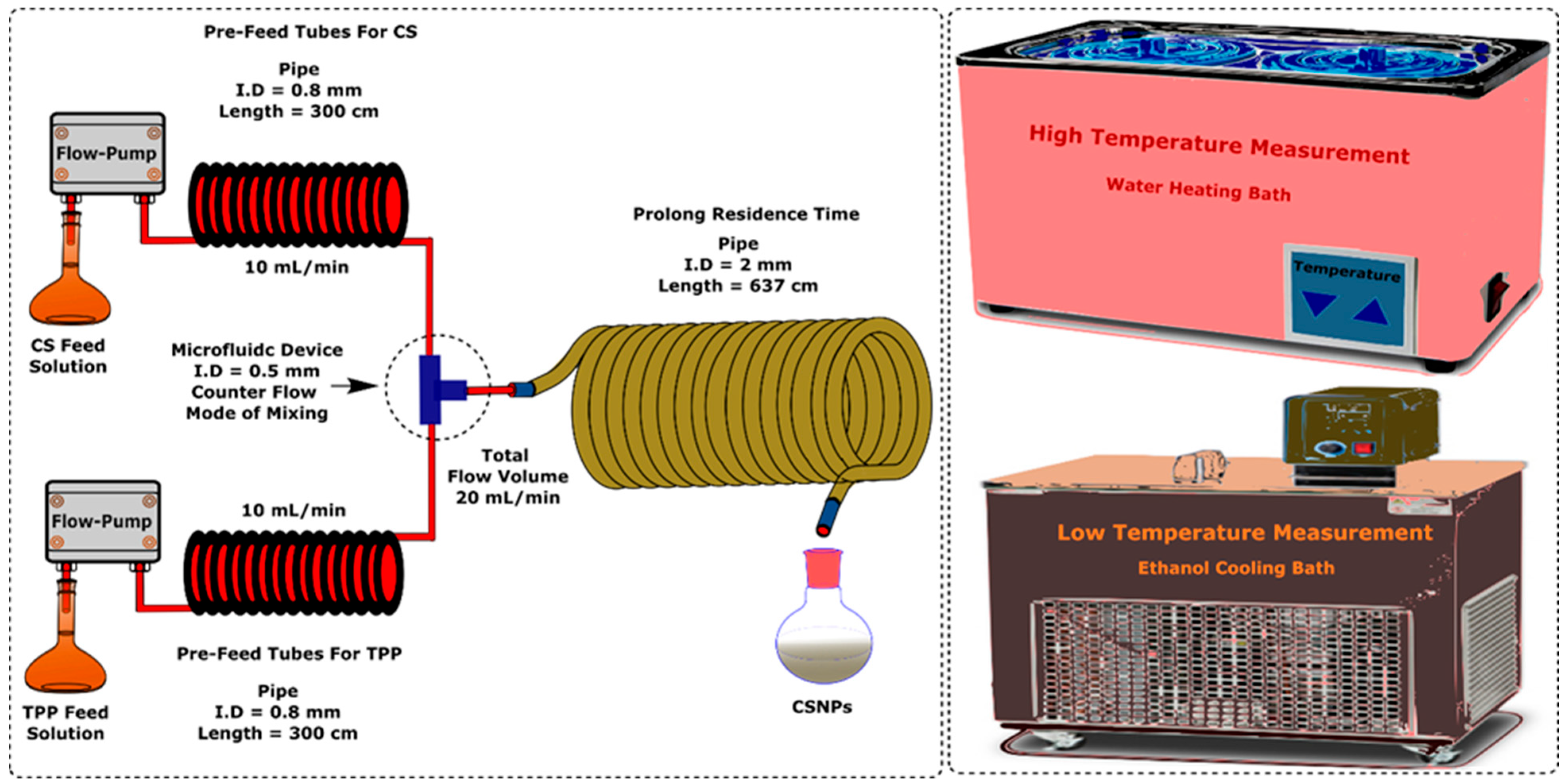
2.3. Equipment and Procedures

The CSNPs were prepared with the ionic gelation method by the electrostatic interaction between the chitosan polymer and sodium tri-polyphosphate. When using a buffer solution of pH = 5 prepared with acetic acid (0.158 wt%) and sodium acetate (0.443 wt%) to dissolve chitosan and TPP separately, the amino groups in the CS can be pronated to obtain positively charged CS, and the TPP can be dissociated into negative phosphoric ions (P3O10−5). Mixing the two solutions will start up the gelation process due to the static interaction under a pH controlled at 5.
Figure 2 shows a scheme of a continuous synthesis platform for CSNPs. It includes a T-junction with a 0.5 mm internal diameter and a delay tube with a 2 mm diameter and 637 cm length. This configuration was chosen to promote rapid nucleation from the initial mixing and ensure stable flow without considerable back-mixing throughout the downstream. To control the temperature, the feed bottles, 300 cm pre-cooling/heating tubes, the T-junction, and the delay tube were immersed in a thermostat. More information about the platform and temperature control evaluation can be found in Figures S1–S5. Two constant flux pumps (Xingda, Beijing, China) were used to deliver the CS and TPP solutions with a flow rate of 10 mL/min separately. The platform worked under ambient pressure. The temperature and feed concentrations of CS and TPP were adjusted on demand. At least three experiments were repeated to ensure the reliability of the experimental results.

3. Results and Discussion

3.1. Tailoring the Fabrication of CSNPs: Insights from Temperature Variation

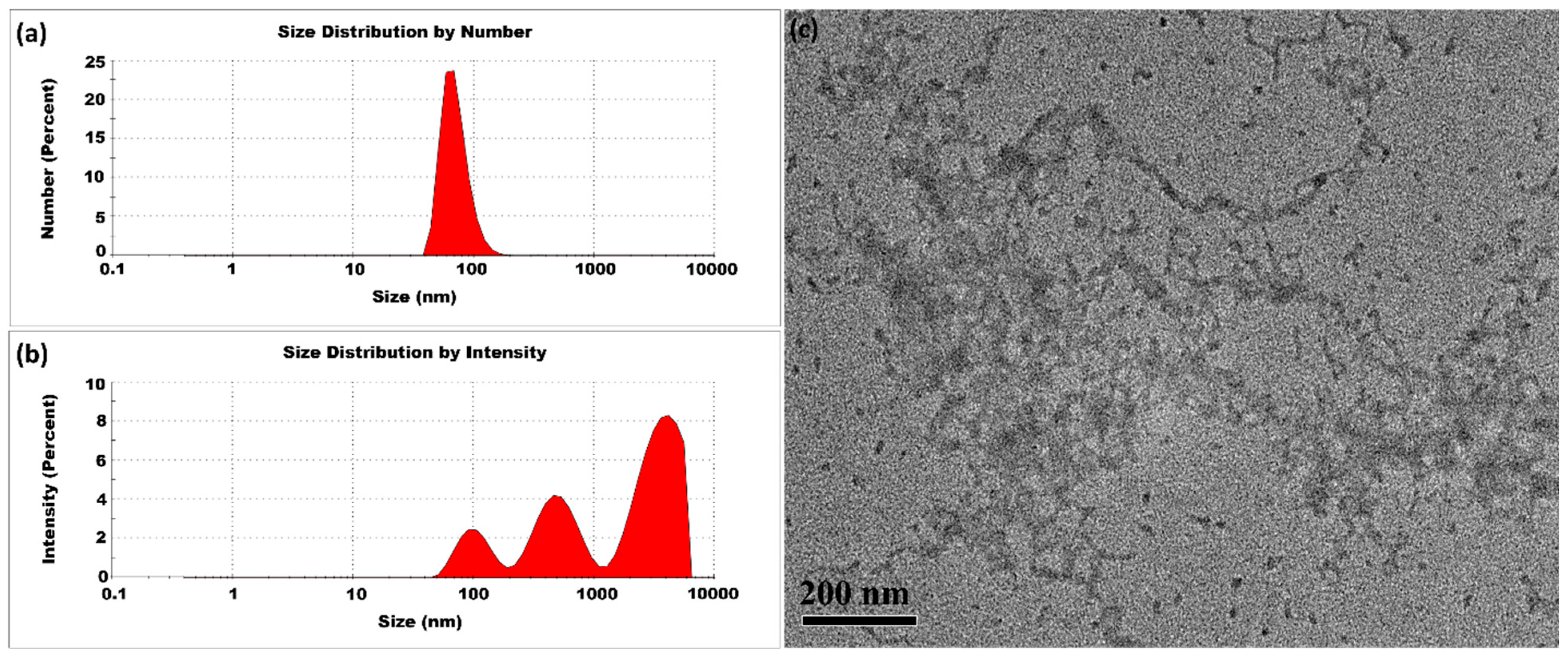
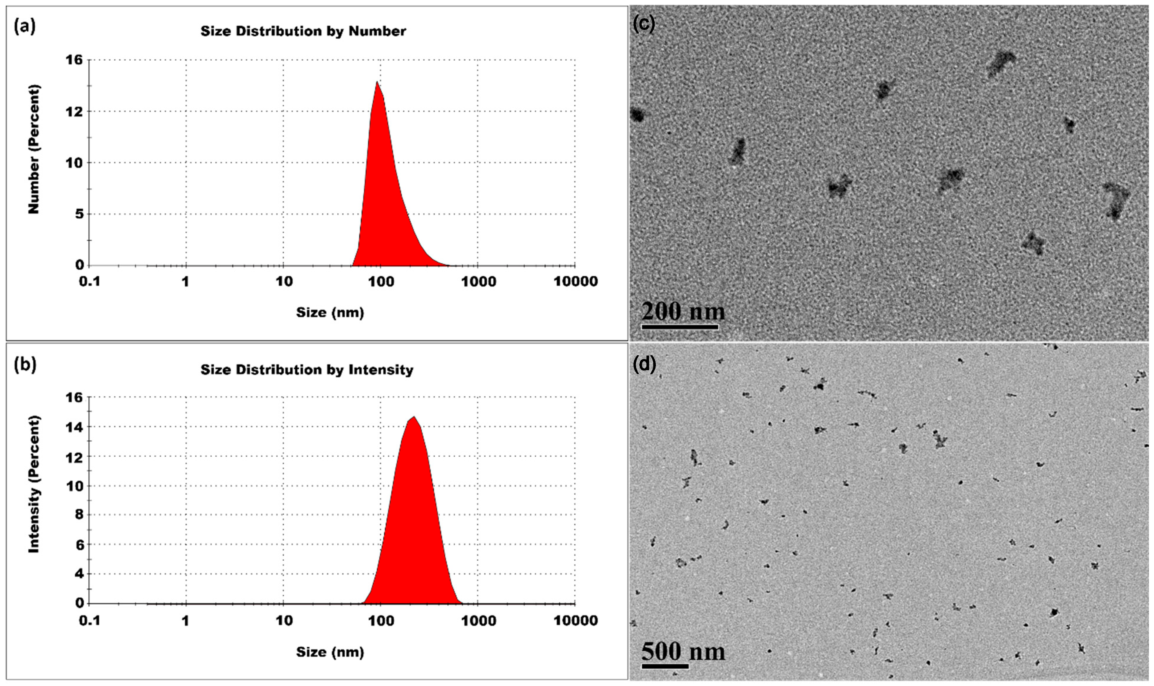
3.1.1. Room Temperature

The experiment was first conducted at room temperature. The synthesis of CSNPs was performed with a CS-to-TPP weight ratio of 5:1, corresponding to a CS concentration of 2 mg/mL and a TPP concentration of 0.4 mg/mL. Figure 3 presents typical DLS and TEM measurements. The size distribution by intensity percentage indicated the presence of small-sized CSNPs, along with two additional peaks representing larger aggregates. Herein, we used Z-average method to measure the average size and polydispersity index. Because the scattered light intensity is proportional to the square of the particle volume, the Z-average size is significantly skewed toward larger particles. The CSNPs prepared at room temperature had an average size of 368 ± 10 nm and a high PDI value of 0.956 ± 0.024. Post-treatment of CSNPs also revealed the presence of aggregates with variable sizes, as seen in Figure S7. These findings implied that to stabilize the small nuclei and prevent aggregation, a rapid surface curing process might be essential for limiting their aggregation. The presence of small-sized CSNPs alongside aggregates indicated that some nuclei achieved complete curing, while others remained uncured. This uncured fraction contributes to a diverse range of sizes. In addition, a TPP concentration of 0.4 mg/mL was identified to be enough for the complete conversion of the CS feed of 2 mg/mL, as evidenced by the carbon equilibrium between the fed CS and the CSNPs separated out from the product suspension. Herein, the ratio of CS to TPP was not higher than 5:1 to guarantee the sufficient conversion of CS.

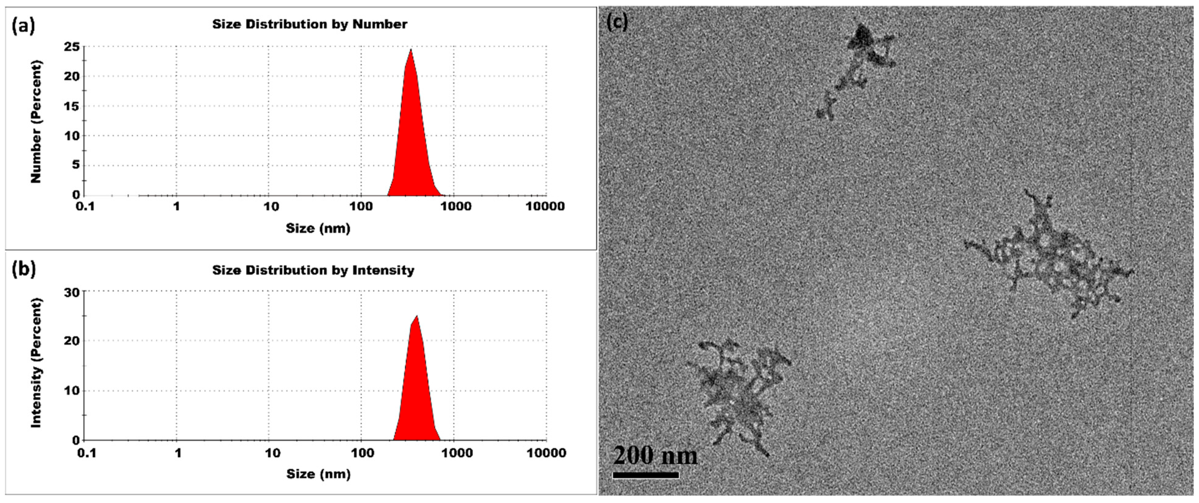
3.1.2. Lower Temperatures

The CSNPs synthesized at 10 °C had an average particle size of 509 ± 17 nm and a PDI of 1.00 ± 0.00, indicating substantial variability in particle size. The intensity profile shown in Figure 4 possessed three distinct peaks, suggesting the presence of multiple size populations. The moderate reduction in thermal energy slowed the molecular motion of both CS and TPP, which in turn reduced the frequency of ionic interactions. This slower interaction rate extended the curing phase and allowed larger aggregates to form. The higher PDI value come from a slower and more gradual crosslinking dependent on the moderated kinetic energy at 10 °C.

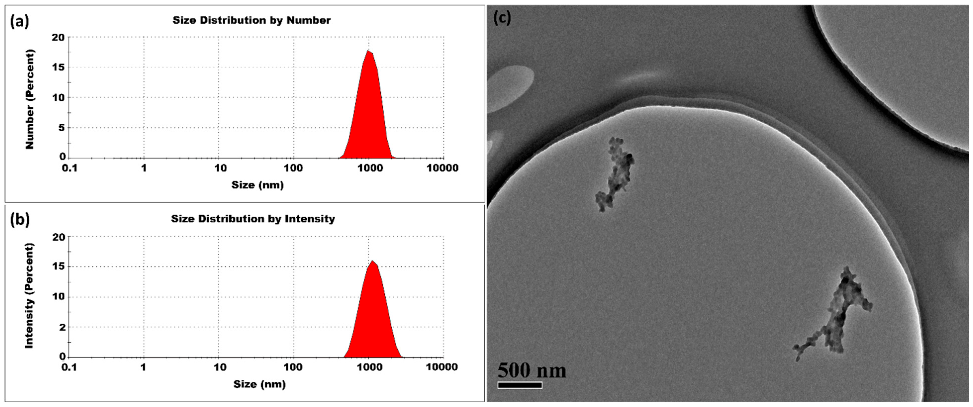
3.1.3. High Temperatures

The CSNPs synthesized at 40 °C had an average size of 429 ± 2 nm and a PDI of 0.359 ± 0.007. As shown in Figure 5, the size distribution analysis by number percentage indicated the presence of large-sized CSNPs, and the intensity percentage distribution also revealed a single peak, confirming the predominance of large-sized CSNPs in the sample. At 40 °C, the mobility of the initially formed small nuclei was significantly enhanced, leading to more frequent and effective collisions. This increased kinetic activity promotes the uniform aggregation of these small nuclei into larger CSNPs. These findings suggest that at 40 °C, the synthesis process may not provide enough thermal energy to fully overcome activation barriers, preventing complete curing of the small nuclei, but it does enable considerable curing. A temperature of 40 °C is sufficient to facilitate the formation of well-structured, large-sized CSNPs. This controlled aggregation effectively regulates the variance of the size, resulting in the formation of CSNPs with improved uniformity.
Figure 6 shows results for CSNPs synthesized at 50 °C. The DLS and TEM analysis shown in Figure 6 indicates a notable change in the average particle size, with measurements of 370 ± 2 nm and PDI of 0.285 ± 0.001. At 50 °C, the curing process becomes more efficient than at 40 °C. This moderate improvement in curing efficiency at 50 °C reduces the tendency for multiple small nuclei to aggregate further.
The CSNPs synthesized at 80 °C showed an average particle size of 310 ± 2 nm, and a PDI value of 0.429 ± 0.008. The size distribution analysis by number percentage of DLS and TEM indicates the presence of small particles, as shown in Figure 7. At 80 °C, the rapid stabilization of small nuclei effectively curtails aggregations, leading to the decrease in nanoparticles size. The PDI of CSNPs significantly decreased from 0.956 under room temperature and 1.00 at 10 °C to 0.429 or lower at elevated temperature, highlighting the critical role of temperature in the curing process. The elevated temperature shortens the curing duration and reduces aggregation, resulting in the fabrication of small-sized CSNPs with a narrow size distribution.
Figure 8 illustrates the dependence of average size and size distribution on temperature. At lower temperatures, the reduced Brownian motion, high activation barriers, slower reaction rates, and slow, inefficient, and uneven surface curing of small nuclei leads to uncontrollable aggregation, resulting in the formation of polydispersed CSNPs, as observed at 5 °C, 10 °C, 20 °C, and 30 °C. At 40 °C or higher temperatures, the increased Brownian motion promoted more frequent collisions between small nuclei, the activation barrier was well overcome to achieve a faster reaction rate and rapid cross-linking, and the small aggregates could remain largely independent, resulting in the stabilization of small-sized CSNPs with utmost uniformity. The abnormal increase in size from 30 °C to 40 °C might reflect the complex temperature effects on the aggregation (such as colloid probability and interface property).
In summary, the increasing temperature might simultaneously promote two factors: (1) particle surface curing and (2) particle collisions. The first factor, particle surface curing, acts to hinder agglomeration. When the effect of surface curing dominates, it can reduce agglomeration, which is a fundamental to produce CSNPs with utmost uniformity.

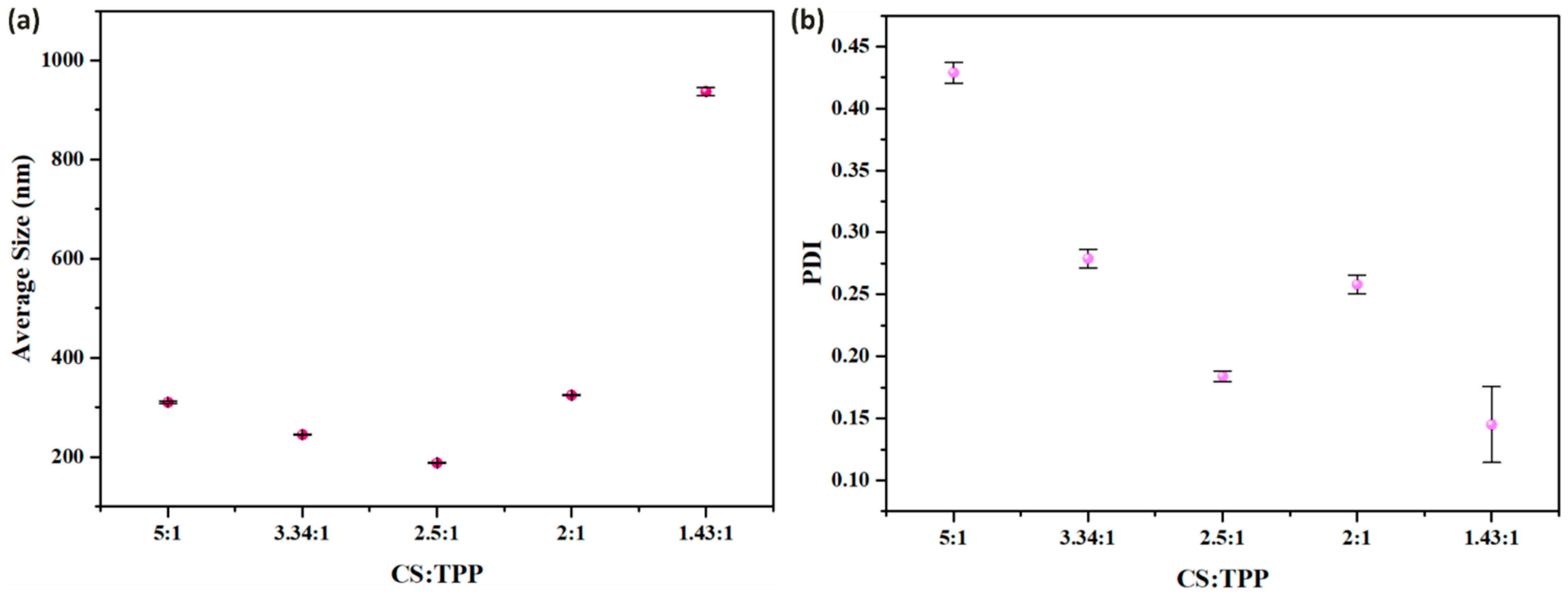
3.2. Impact of TPP Concentration on the Synthesis of CSNPs

The CSNPs were synthesized using a CS to TPP ratio of 3.34:1 at 80 °C, i.e., the TPP and CS concentrations were set at 0.6 mg/mL and 2 mg/mL, respectively. The products displayed an average particle size of 245 ± 1 nm and a PDI of 0.279 ± 0.007 (Figure 9). Compared to CSNPs synthesized using a 5:1 ratio, these results highlight a substantial reduction in particle average size and a decrease in the PDI. Since TPP functions as an ionic crosslinking agent to bind the amino groups on the CS molecules and form a gel-like matrix, the density of crosslinking sites will increase with the increasing of TPP concentration, thereby accelerating the curing process. This accelerated crosslinking facilitates the rapid formation of a more compact and rigid surface. Consequently, the aggregation of CSNPs will be more effectively regulated, preventing uncontrolled aggregation and size growth.
Next, the CSNPs were synthesized by reacting CS with TPP at a 2.5:1 ratio and 80 °C. The concentrations of CS and TPP were 2 mg/mL and 0.6 mg/mL, respectively. As depicted in Figure 10, a decrease in the CS:TPP ratio from 3.34:1 to 2.5:1 caused a significant reduction in the average particle size, which decreased from 245 ± 1 nm to 188 ± 0.6 nm. Moreover, the PDI also improved, dropping from 0.279 ± 0.007 to 0.184 ± 0.004. The elevated TPP concentration significantly accelerates the crosslinking process, promoting the rapid formation of small nanoparticle nuclei within a very short period. This fast-paced crosslinking ensures that the nascent nuclei are stabilized almost immediately, preventing premature interactions between initially nucleated small nuclei. This early-stage stabilization is crucial for maintaining consistent particle size and preventing undesirable particle aggregation.
When employing an even lower CS to TPP ratio, 2:1, moderate-sized CSNPs were synthesized instead of smaller ones as imagine. In detail, a CS concentration of 2 mg/mL and a TPP concentration of 1 mg/mL were used, resulting in CSNPs with an average size of 325 ± 0.7 nm and a PDI of 0.258 ± 0.007, as shown in Figure 11. We supposed that the excessive TPP content facilitated inter-crosslinking between the CSNPs, causing the nanoparticles to fuse together rather than remaining separate, leading to an increase in overall particle size.
When further decreasing the CS:TPP ratio to 1.43:1 (the concentrations of CS and TPP were 2 mg/mL and 1.4 mg/mL, respectively), we obtained even larger particles. The average size was 937 ± 8 nm and the PDI was 0.145 ± 0.03, respectively, as shown in Figure 12. Fortunately, the elevated temperature of 80 °C still played a crucial role in regulating the inter-crosslinking process, ultimately facilitating the production of relatively large-sized CSNPs with utmost uniformity.
Figure 13 demonstrates the influential role of TPP in governing the aggregation behavior of CSNPs. A CS:TPP ratio of 2.5:1 represents a critical threshold in CSNP: at this specific ratio, TPP efficiently facilitates crosslinking and stabilizes nascent nuclei within a short timeframe, yielding a highly uniform size distribution. As the ratio increases beyond this point, slower curing enhance the likelihood of continued interaction between nascent nuclei, even after they exit the delay tube, leading to uncontrolled aggregation and broad size dispersity. Conversely, decreasing the ratio below 2.5:1 intensifies interparticle crosslinking, promoting the formation of larger secondary aggregates.
The final suspensions of CSNPs prepared at 80 °C with CS:TPP ratios of 3.34:1, 2.5:1, and 2:1 exhibited stable colloidal behavior without turbidity or precipitation, indicating successful fabrication of CSNPs free of large aggregates or clusters. In contrast, at room temperature, the 3.34:1 ratio suspension showed turbidity and the 2:1 ratio suspension developed precipitation. These results highlight that elevated temperatures more effectively prevent uncontrollable aggregation, offering a superior approach to improve CSNP size uniformity and directly optimize target product yield.
Overall, this study demonstrates that high-temperature synthesis of CSNPs promotes more controlled nanoparticle growth compared to lower temperatures. CSNP aggregation is profoundly influenced by synthesis parameters, including microfluidic technology, temperature, and TPP concentration. Microfluidic systems enable rapid mixing and precise nucleation, generating small primary nuclei. At lower temperatures, slow and uneven crosslinking leads to insufficient stabilization, significantly increasing the likelihood of uncontrolled aggregation between nascent nuclei. Conversely, higher temperatures enhance molecular mobility, facilitating efficient crosslinking and shortening the surface-curing process of small nuclei. Optimizing surface curing under controlled collision rates allows precise regulation of CSNP aggregation and improves size uniformity. The synergistic effect of temperature and TPP is critical for enhancing CSNP stability and uniformity, ensuring the production of high-quality nanoparticles.
The influence of temperature on the acceleration of surface curing can be effectively correlated with the work of Shimiao Zhang et al. [68], who established a clear relationship between temperature and polymer surface curing. Their findings demonstrate that temperature plays a critical role in the curing process. At lower temperatures, the available thermal energy is insufficient to drive complete cross-linking, resulting in partial curing. In contrast, higher temperatures facilitate more extensive cross-linking, leading to a polymer network that approaches maximum density. This enhanced curing is achieved by gradually ramping the temperature to drive the reaction to completion and maximize cross-linking density [68].
Several theoretical models support our findings regarding the effect of temperature on the crosslinking process. According to the principles of Brownian motion, temperature directly influences the mobility of molecules; higher temperatures increase molecular movement, which can enhance crosslinking density. Fick’s First Law of Diffusion also indicates that the diffusion coefficient increases with temperature, promoting faster molecular transport and improving the crosslinking process. Additionally, the Arrhenius model demonstrates that elevated temperatures accelerate reaction rates by increasing the kinetic energy of molecules. In this context of CSNPs, higher temperatures enhance the reactivity of TPP, enabling it to more effectively optimize the crosslinking density and surface curing of CSNPs [69,70,71].

4. Conclusions

This study validates the effective synthesis of CSNPs via a microfluidic approach, where rapid mixing drives quick nucleation and formation of small nuclei. However, an initial high PDI of 0.956 ± 0.0239 at room temperature indicated substantial aggregation, with lower temperatures (10 °C and 5 °C) further increasing the polydispersity. High temperatures are pivotal for controlling CSNP growth by optimizing curing through regulated nuclear mobility, essential for fabricating highly uniform particles. At 40 °C and 50 °C, controlled interactions between small nuclei governed nanoparticle growth, yielding uniformly sized CSNPs with diameters of 429 ± 2 nm and 370 ± 2 nm, respectively. Raising the temperature to 80 °C drastically accelerated small-nucleus curing, resulting in smaller CSNPs. The CS:TPP ratio also critically influenced surface curing and particle size at 80 °C: ratios of 5:1, 3.34:1, and 2.5:1 produced CSNPs with mean sizes of 310 ± 2 nm, 245 ± 1 nm, and 188 ± 0.6 nm, respectively. Continuous decreases in both average size and PDI values reflected improved uniformity and reduced aggregation. The TPP-facilitated enhanced surface-curing process stabilized CSNPs early and minimized agglomeration, enabling successful synthesis of small CSNPs (188 ± 0.6 nm) at 80 °C with a 2.5:1 ratio. Additionally, precise size regulation at 80 °C yielded moderate and large CSNPs (325 ± 0.7 nm and 937 ± 8 nm, respectively). Collectively, temperature and TPP concentration are critical factors in modulating nanoparticle curing to control CSNP agglomeration. These parameters work in tandem to precisely regulate nucleus–nucleus interactions and particle–particle crosslinking, governing CSNP growth and size with exceptional consistency. This optimization enhances the translational potential of CSNPs for diverse applications.

Supplementary Materials

The following supporting information can be downloaded at: https://www.mdpi.com/article/10.3390/mi16060642/s1.

Author Contributions

M.A.: data curation, formal analysis, investigation, writing—original draft. Y.L.: conceptualization, funding acquisition, methodology, project administration, resources, supervision, writing—review and editing. All authors have read and agreed to the published version of the manuscript.

Funding

This work was supported by the National Natural Science Foundation of China (grant no.: 21978152).

Data Availability Statement

The original contributions presented in this study are included in the article/Supplementary Material. Further inquiries can be directed to the corresponding author.

Conflicts of Interest

The authors declare that they have no known competing financial interests or personal relationships that could have appeared to influence the work reported in this paper.

References

  1. Saravanakumar, K.; Mariadoss, A.V.A.; Sathiyaseelan, A.; Venkatachalam, K.; Hu, X.; Wang, M.-H. pH-sensitive release of fungal metabolites from chitosan nanoparticles for effective cytotoxicity in prostate cancer (PC3) cells. Process Biochem. 2021, 102, 165–172. [Google Scholar] [CrossRef]
  2. Garg, U.; Chauhan, S.; Nagaich, U.; Jain, N. Current advances in chitosan nanoparticles based drug delivery and targeting. Adv. Pharm. Bull. 2019, 9, 195–204. [Google Scholar] [CrossRef] [PubMed]
  3. Rostami, E. Progresses in targeted drug delivery systems using chitosan nanoparticles in cancer therapy: A mini-review. J. Drug Deliv. Sci. Technol. 2020, 58, 101813. [Google Scholar] [CrossRef]
  4. Pan, C.; Qian, J.; Zhao, C.; Yang, H.; Zhao, X.; Guo, H. Study on the relationship between crosslinking degree and properties of TPP crosslinked chitosan nanoparticles. Carbohydr. Polym. 2020, 241, 116349. [Google Scholar] [CrossRef]
  5. Ryu, J.H.; Yoon, H.Y.; Sun, I.C.; Kwon, I.C.; Kim, K. Tumor-Targeting Glycol Chitosan Nanoparticles for Cancer Heterogeneity. Adv. Mater. 2020, 32, e2002197. [Google Scholar] [CrossRef]
  6. Ahmad, M.Z.; Rizwanullah, M.; Ahmad, J.; Alasmary, M.Y.; Akhter, M.H.; Abdel-Wahab, B.A.; Warsi, M.H.; Haque, A. Progress in nanomedicine-based drug delivery in designing of chitosan nanoparticles for cancer therapy. Int. J. Polym. Mater. Polym. Biomater. 2021, 71, 602–623. [Google Scholar] [CrossRef]
  7. Fernández-Marín, R.; Morales, A.; Erdocia, X.; Iturrondobeitia, M.; Labidi, J.; Lizundia, E. Chitosan–chitin nanocrystal films from lobster and spider crab: Properties and environmental sustainability. ACS Sustain. Chem. Eng. 2024, 12, 10363–10375. [Google Scholar] [CrossRef]
  8. Hassan, U.A.; Hussein, M.Z.; Alitheen, N.B.; Yahya Ariff, S.A.; Masarudin, M.J. In vitro cellular localization and efficient accumulation of fluorescently tagged biomaterials from monodispersed chitosan nanoparticles for elucidation of controlled release pathways for drug delivery systems. Int. J. Nanomed. 2018, 13, 5075–5095. [Google Scholar] [CrossRef]
  9. Alamassi, M.N.; Chia, S.L.; Abdullah, C.A.C.; Masarudin, M.J. Increased efficacy of biologics following inhibition of autophagy in A549 lung cancer cells in bimodal treatment of doxorubicin and SAR405-loaded chitosan nanoparticles. OpenNano 2023, 11, 100142. [Google Scholar] [CrossRef]
  10. Deepa, G.; Sivakumar, K.; Sajeevan, T. Molecular simulation and in vitro evaluation of chitosan nanoparticles as drug delivery systems for the controlled release of anticancer drug cytarabine against solid tumours. 3 Biotech 2018, 8, 493. [Google Scholar] [CrossRef]
  11. Bugnicourt, L.; Ladavière, C. Interests of chitosan nanoparticles ionically cross-linked with tripolyphosphate for biomedical applications. Prog. Polym. Sci. 2016, 60, 1–17. [Google Scholar] [CrossRef]
  12. Branca, C.; D’Angelo, G.; Crupi, C.; Khouzami, K.; Rifici, S.; Ruello, G.; Wanderlingh, U. Role of the OH and NH vibrational groups in polysaccharide-nanocomposite interactions: A FTIR-ATR study on chitosan and chitosan/clay films. Polymer 2016, 99, 614–622. [Google Scholar] [CrossRef]
  13. Ali, M.E.A.; Aboelfadl, M.M.S.; Selim, A.M.; Khalil, H.F.; Elkady, G.M. Chitosan nanoparticles extracted from shrimp shells, application for removal of Fe (II) and Mn (II) from aqueous phases. Sep. Sci. Technol. 2018, 53, 2870–2881. [Google Scholar] [CrossRef]
  14. Lipatova, I.M.; Makarova, L.I.; Yusova, A.A. Adsorption removal of anionic dyes from aqueous solutions by chitosan nanoparticles deposited on the fibrous carrier. Chemosphere 2018, 212, 1155–1162. [Google Scholar] [CrossRef]
  15. Punjabi, K.; Adhikary, R.R.; Patnaik, A.; Bendale, P.; Saxena, S.; Banerjee, R. Lectin-Functionalized Chitosan Nanoparticle-Based Biosensor for Point-of-Care Detection of Bacterial Infections. Bioconjug. Chem. 2022, 33, 1552–1563. [Google Scholar] [CrossRef]
  16. Choudhary, R.C.; Joshi, A.; Kumari, S.; Kumaraswamy, R.; Saharan, V. Preparation of Cu-chitosan nanoparticle and its effect on growth and enzyme activity during seed germination in maize. J. Pharmacogn. Phytochem. 2017, 6, 669–673. [Google Scholar]
  17. Saharan, V.; Kumaraswamy, R.; Choudhary, R.C.; Kumari, S.; Pal, A.; Raliya, R.; Biswas, P. Cu-chitosan nanoparticle mediated sustainable approach to enhance seedling growth in maize by mobilizing reserved food. J. Agric. Food Chem. 2016, 64, 6148–6155. [Google Scholar] [CrossRef]
  18. Kashyap, P.L.; Xiang, X.; Heiden, P. Chitosan nanoparticle based delivery systems for sustainable agriculture. Int. J. Biol. Macromol. 2015, 77, 36–51. [Google Scholar] [CrossRef]
  19. Liu, Y.; Chen, J.; Li, H.; Wang, Y. Nanocomplexes film composed of gallic acid loaded ovalbumin/chitosan nanoparticles and pectin with excellent antibacterial activity: Preparation, characterization and application in coating preservation of salmon fillets. Int. J. Biol. Macromol. 2024, 259, 128934. [Google Scholar] [CrossRef]
  20. Xu, W.; McClements, D.J.; Zhang, Z.; Zhang, R.; Qiu, C.; Zhao, J.; Jin, Z.; Chen, L. Effect of tannic acid modification on antioxidant activity, antibacterial activity, environmental stability and release characteristics of quercetin loaded zein-carboxymethyl chitosan nanoparticles. Int. J. Biol. Macromol. 2024, 280, 135853. [Google Scholar] [CrossRef]
  21. Ta, Q.; Ting, J.; Harwood, S.; Browning, N.; Simm, A.; Ross, K.; Olier, I.; Al-Kassas, R. Chitosan nanoparticles for enhancing drugs and cosmetic components penetration through the skin. Eur. J. Pharm. Sci. 2021, 160, 105765. [Google Scholar] [CrossRef] [PubMed]
  22. Saraiva, S.M.; Crespo, A.M.; Vaz, F.; Filipe, M.; Santos, D.; Jacinto, T.A.; Paiva-Santos, A.C.; Rodrigues, M.; Ribeiro, M.P.; Coutinho, P.; et al. Development and characterization of thermal water gel comprising Helichrysum italicum essential oil-loaded chitosan nanoparticles for skin care. Cosmetics 2023, 10, 8. [Google Scholar] [CrossRef]
  23. Ntohogian, S.; Gavriliadou, V.; Christodoulou, E.; Nanaki, S.; Lykidou, S.; Naidis, P.; Mischopoulou, L.; Barmpalexis, P.; Nikolaidis, N.; Bikiaris, D.N. Chitosan nanoparticles with encapsulated natural and UF-purified annatto and saffron for the preparation of UV protective cosmetic emulsions. Molecules 2018, 23, 2107. [Google Scholar] [CrossRef]
  24. Anusha, J.; Raj, C.J.; Cho, B.-B.; Fleming, A.T.; Yu, K.-H.; Kim, B.C. Amperometric glucose biosensor based on glucose oxidase immobilized over chitosan nanoparticles from gladius of Uroteuthis duvauceli. Sens. Actuators B Chem. 2015, 215, 536–543. [Google Scholar] [CrossRef]
  25. Gigli, V.; Tortolini, C.; Capecchi, E.; Angeloni, A.; Lenzi, A.; Antiochia, R. Novel amperometric biosensor based on tyrosinase/chitosan nanoparticles for sensitive and interference-free detection of total catecholamine. Biosensors 2022, 12, 519. [Google Scholar] [CrossRef]
  26. Sapre, N.; Gumathannavar, R.; Shirolkar, M.; Dalai, S.; Kanojiya, P.; Saroj, S.; Kulkarni, A. Optically tuneable chitosan nanoparticles for biomedical imaging application. Sens. Technol. 2024, 2, 2428590. [Google Scholar] [CrossRef]
  27. Nesalin, J.A.J.; Smith, A.A. Preparation and evaluation of stavudine loaded chitosan nanoparticles. J. Pharm. Res. 2013, 6, 268–274. [Google Scholar]
  28. Sawaengsak, C.; Mori, Y.; Yamanishi, K.; Mitrevej, A.; Sinchaipanid, N. Chitosan nanoparticle encapsulated hemagglutinin-split influenza virus mucosal vaccine. AAPS PharmSciTech 2014, 15, 317–325. [Google Scholar] [CrossRef]
  29. Tabynov, K.; Solomadin, M.; Turebekov, N.; Babayeva, M.; Fomin, G.; Yadagiri, G.; Renu, S.; Yerubayev, T.; Petrovsky, N.; Renukaradhya, G.J.; et al. An intranasal vaccine comprising SARS-CoV-2 spike receptor-binding domain protein entrapped in mannose-conjugated chitosan nanoparticle provides protection in hamsters. Sci. Rep. 2023, 13, 12115. [Google Scholar]
  30. Khan, M.A.; Zafaryab, M.; Mehdi, S.H.; Ahmad, I.; Rizvi, M.M. Characterization and anti-proliferative activity of curcumin loaded chitosan nanoparticles in cervical cancer. Int. J. Biol. Macromol. 2016, 93, 242–253. [Google Scholar] [CrossRef]
  31. Nam, T.; Park, S.; Lee, S.-Y.; Park, K.; Choi, K.; Song, I.C.; Han, M.H.; Leary, J.J.; Yuk, S.A.; Kwon, I.C.; et al. Tumor targeting chitosan nanoparticles for dual-modality optical/MR cancer imaging. Bioconjug. Chem. 2010, 21, 578–582. [Google Scholar] [CrossRef] [PubMed]
  32. Ma, Z.; Lim, T.M.; Lim, L.-Y. Pharmacological activity of peroral chitosan–insulin nanoparticles in diabetic rats. Int. J. Pharm. 2005, 293, 271–280. [Google Scholar] [CrossRef] [PubMed]
  33. Jabbari, F.; Farani, M.R.; Abednejad, A.; Akbari, B.; Mostafavi, E.; Zare, I. Chitosan nanoparticles in tissue engineering and regenerative medicine. In Fundamentals and Biomedical Applications of Chitosan Nanoparticles; Elsevier: Amsterdam, The Netherlands, 2025; pp. 497–526. [Google Scholar]
  34. Biranje, S.S.; Madiwale, P.V.; Patankar, K.C.; Chhabra, R.; Dandekar-Jain, P.; Adivarekar, R.V. Hemostasis and anti-necrotic activity of wound-healing dressing containing chitosan nanoparticles. Int. J. Biol. Macromol. 2019, 121, 936–946. [Google Scholar] [CrossRef]
  35. Loo, H.L.; Goh, B.H.; Lee, L.-H.; Chuah, L.H. Application of chitosan-based nanoparticles in skin wound healing. Asian J. Pharm. Sci. 2022, 17, 299–332. [Google Scholar] [CrossRef]
  36. Shirolkar, M.M.; Athavale, R.; Ravindran, S.; Rale, V.; Kulkarni, A.; Deokar, R. Antibiotics functionalization intervened morphological, chemical and electronic modifications in chitosan nanoparticles. Nano-Struct. Nano-Objects 2021, 25, 100657. [Google Scholar] [CrossRef]
  37. Naskar, S.; Kuotsu, K.; Sharma, S. Chitosan-based nanoparticles as drug delivery systems: A review on two decades of research. J. Drug Target. 2019, 27, 379–393. [Google Scholar] [CrossRef]
  38. Nokhodi, F.; Nekoei, M.; Goodarzi, M.T. Hyaluronic acid-coated chitosan nanoparticles as targeted-carrier of tamoxifen against MCF7 and TMX-resistant MCF7 cells. J. Mater. Sci. Mater. Med. 2022, 33, 24. [Google Scholar] [CrossRef]
  39. Song, H.; Su, C.; Cui, W.; Zhu, B.; Liu, L.; Chen, Z.; Zhao, L. Folic acid-chitosan conjugated nanoparticles for improving tumor-targeted drug delivery. BioMed Res. Int. 2013, 2013, 723158. [Google Scholar] [CrossRef]
  40. Wang, T.; Hou, J.; Su, C.; Zhao, L.; Shi, Y. Hyaluronic acid-coated chitosan nanoparticles induce ROS-mediated tumor cell apoptosis and enhance antitumor efficiency by targeted drug delivery via CD44. J. Nanobiotechnol. 2017, 15, 7. [Google Scholar] [CrossRef]
  41. Zhang, W.; Xu, W.; Lan, Y.; He, X.; Liu, K.; Liang, Y. Antitumor effect of hyaluronic-acid-modified chitosan nanoparticles loaded with siRNA for targeted therapy for non-small cell lung cancer. Int. J. Nanomed. 2019, 14, 5287–5301. [Google Scholar] [CrossRef]
  42. Hejjaji, E.M.A.; Smith, A.M.; Morris, G.A. Evaluation of the mucoadhesive properties of chitosan nanoparticles prepared using different chitosan to tripolyphosphate (CS:TPP) ratios. Int. J. Biol. Macromol. 2018, 120, 1610–1617. [Google Scholar] [CrossRef] [PubMed]
  43. Holtze, C.; Boehling, R. Batch or flow chemistry?—A current industrial opinion on process selection. Curr. Opin. Chem. Eng. 2022, 36, 100798. [Google Scholar] [CrossRef]
  44. Zhang, H.; Yang, J.; Sun, R.; Han, S.; Yang, Z.; Teng, L. Microfluidics for nano-drug delivery systems: From fundamentals to industrialization. Acta Pharm. Sin. B 2023, 13, 3277–3299. [Google Scholar] [CrossRef]
  45. Yao, F.; Zhu, P.; Chen, J.; Li, S.; Sun, B.; Li, Y.; Zou, M.; Qi, X.; Liang, P.; Chen, Q. Synthesis of nanoparticles via microfluidic devices and integrated applications. Microchim. Acta 2023, 190, 256. [Google Scholar] [CrossRef]
  46. Alarfaj, A.A. Antibacterial effect of chitosan nanoparticles against food spoilage bacteria. J. Pure Appl. Microbiol. 2019, 13, 1273–1278. [Google Scholar] [CrossRef]
  47. Ahmed, M.E.; Mohamed, M.I.; Ahmed, H.Y.; Elaasser, M.M.; Kandile, N.G. Fabrication and characterization of unique sustain modified chitosan nanoparticles for biomedical applications. Sci. Rep. 2024, 14, 13869. [Google Scholar] [CrossRef]
  48. Saharan, V.; Mehrotra, A.; Khatik, R.; Rawal, P.; Sharma, S.; Pal, A. Synthesis of chitosan based nanoparticles and their in vitro evaluation against phytopathogenic fungi. Int. J. Biol. Macromol. 2013, 62, 677–683. [Google Scholar] [CrossRef]
  49. Jha, A.; Ghormade, V.; Kolge, H.; Paknikar, K.M. Dual effect of chitosan-based nanoparticles on the inhibition of β-amyloid peptide aggregation and disintegration of the preformed fibrils. J. Mater. Chem. B 2019, 7, 3362–3373. [Google Scholar] [CrossRef]
  50. Antoniou, J.; Liu, F.; Majeed, H.; Qi, J.; Yokoyama, W.; Zhong, F. Physicochemical and morphological properties of size-controlled chitosan–tripolyphosphate nanoparticles. Colloids Surf. A Physicochem. Eng. Asp. 2015, 465, 137–146. [Google Scholar] [CrossRef]
  51. Masarudin, M.J.; Cutts, S.M.; Evison, B.J.; Phillips, D.R.; Pigram, P.J. Factors determining the stability, size distribution, and cellular accumulation of small, monodisperse chitosan nanoparticles as candidate vectors for anticancer drug delivery: Application to the passive encapsulation of [14C]-doxorubicin. Nanotechnol. Sci. Appl. 2015, 8, 67–80. [Google Scholar] [CrossRef]
  52. Hussain, Z.; Sahudin, S. Preparation, characterisation and colloidal stability of chitosan-tripolyphosphate nanoparticles: Optimisation of formulation and process parameters. Int. J. Pharm. Pharm. Sci. 2016, 8, 297–308. [Google Scholar]
  53. Douglas, K.L.; Tabrizian, M. Effect of experimental parameters on the formation of alginate–chitosan nanoparticles and evaluation of their potential application as DNA carrier. J. Biomater. Sci. Polym. Ed. 2005, 16, 43–56. [Google Scholar] [CrossRef] [PubMed]
  54. Van Bavel, N.; Issler, T.; Pang, L.; Anikovskiy, M.; Prenner, E.J. A simple method for synthesis of chitosan nanoparticles with ionic gelation and homogenization. Molecules 2023, 28, 4328. [Google Scholar] [CrossRef]
  55. Zhao, J.; Wu, J. Preparation and characterization of the fluorescent chitosan nanoparticle probe. Chin. J. Anal. Chem. 2006, 34, 1555–1559. [Google Scholar] [CrossRef]
  56. Körpe, D.A.; Malekghasemi, S.; Aydın, U.; Duman, M. Fabrication of monodispersive nanoscale alginate–chitosan core–shell particulate systems for controlled release studies. J. Nanopart. Res. 2014, 16, 2754. [Google Scholar] [CrossRef]
  57. Majedi, F.S.; Hasani-Sadrabadi, M.M.; Emami, S.H.; Taghipoor, M.; Dashtimoghadam, E.; Bertsch, A.; Moaddel, H.; Renaud, P. Microfluidic synthesis of chitosan-based nanoparticles for fuel cell applications. Chem. Commun. 2012, 48, 7744–7746. [Google Scholar] [CrossRef]
  58. Greco, A.; Gabold, B.; Chen, S.; Wang, X.; Xu, Z.; Hartschuh, A.; Chiesa, E.; Genta, I.; Ried, C.L.; Merdan, T. Microfluidic mixing as platform technology for production of chitosan nanoparticles loaded with different macromolecules. Eur. J. Pharm. Biopharm. 2023, 188, 170–181. [Google Scholar] [CrossRef]
  59. Siavashy, S.; Soltani, M.; Ghorbani-Bidkorbeh, F.; Fallah, N.; Farnam, G.; Mortazavi, S.A.; Shirazi, F.H.; Tehrani, M.H.H.; Hamedi, M.H. Microfluidic platform for synthesis and optimization of chitosan-coated magnetic nanoparticles in cisplatin delivery. Carbohydr. Polym. 2021, 265, 118027. [Google Scholar] [CrossRef]
  60. Nathanael, K.; Cheng, S.; Kovalchuk, N.M.; Arcucci, R.; Simmons, M.J. Optimization of microfluidic synthesis of silver nanoparticles: A generic approach using machine learning. Chem. Eng. Res. Des. 2023, 193, 65–74. [Google Scholar] [CrossRef]
  61. Karnik, R.; Gu, F.; Basto, P.; Cannizzaro, C.; Dean, L.; Kyei-Manu, W.; Langer, R.; Farokhzad, O.C. Microfluidic platform for controlled synthesis of polymeric nanoparticles. Nano Lett. 2008, 8, 2906–2912. [Google Scholar] [CrossRef]
  62. Chiesa, E.; Bellotti, M.; Caimi, A.; Conti, B.; Dorati, R.; Conti, M.; Genta, I.; Auricchio, F. Development and optimization of microfluidic assisted manufacturing process to produce PLGA nanoparticles. Int. J. Pharm. 2022, 629, 122368. [Google Scholar] [CrossRef] [PubMed]
  63. Liu, D.; Cito, S.; Zhang, Y.; Wang, C.-F.; Sikanen, T.M.; Santos, H.A. A versatile and robust microfluidic platform toward high throughput synthesis of homogeneous nanoparticles with tunable properties. Adv. Mater. 2015, 27, 2298–2304. [Google Scholar] [CrossRef] [PubMed]
  64. Hung, L.-H.; Lee, A.P. Microfluidic devices for the synthesis of nanoparticles and biomaterials. J. Med. Biol. Eng. 2007, 27, 1–6. [Google Scholar]
  65. Matsunaga, T.; Lee, H.-J.; Nishino, K. An approach for accurate simulation of liquid mixing in a T-shaped micromixer. Lab Chip 2013, 13, 1515–1521. [Google Scholar] [CrossRef]
  66. Makgwane, P.R.; Ray, S.S. Synthesis of nanomaterials by continuous-flow microfluidics: A review. J. Nanosci. Nanotechnol. 2014, 14, 1338–1363. [Google Scholar] [CrossRef]
  67. Schmidt, S.; Nguyen, A.T.; Vu, H.Q.; Tran, N.N.; Sareela, M.; Fisk, I.; Hessel, V. Microfluidic Spontaneous Emulsification for Generation of O/W Nanoemulsions—Opportunity for In-Space Manufacturing. Adv. Healthc. Mater. 2023, 12, 2203363. [Google Scholar] [CrossRef]
  68. Zhang, S.; So, L.L.C.; Faucher, S.; Xi, L. Polymer coating over solid particles with in situ thermal curing. Ind. Eng. Chem. Res. 2016, 55, 5574–5584. [Google Scholar] [CrossRef]
  69. Lamkin-Kennard, K.A.; Popovic, M.B. Molecular and cellular level—Applications in biotechnology and medicine addressing molecular and cellular level. In Biomechatronics; Elsevier: Amsterdam, The Netherlands, 2019; pp. 201–233. [Google Scholar]
  70. An, F.; Jia, H.; Feng, Y. Effect of stress, concentration and temperature on gas diffusion coefficient of coal measured through a direct method and its model application. Fuel 2022, 312, 122991. [Google Scholar] [CrossRef]
  71. Laidler, K.J. The development of the Arrhenius equation. J. Chem. Educ. 1984, 61, 494. [Google Scholar] [CrossRef]
Figure 1. The size distributions of CSNPs for different potential applications reported in the literature.
Micromachines 16 00642 g001
Figure 2. Schematic diagram of a continuous synthesis system for the fabrication of CSNPs.
Micromachines 16 00642 g002
Figure 3. CSNPs synthesized at room temperature with a CS:TPP ratio of 5:1. The final suspension was analyzed directly. (a) Size distribution by number percentage; (b) size distribution by intensity percentage. (c) TEM diagram.
Micromachines 16 00642 g003
Figure 4. CSNPs synthesized at 10 °C with a CS:TPP ratio of 5:1. The final suspension was analyzed directly. (a) Size distribution by number percentage; (b) size distribution by intensity percentage. (c) TEM diagram.
Micromachines 16 00642 g004
Figure 5. CSNPs synthesized at 40 °C with a CS:TPP ratio of 5:1. The final suspension was analyzed directly. (a) Size distribution by number percentage; (b) size distribution by intensity percentage. (c) TEM diagram.
Micromachines 16 00642 g005
Figure 6. CSNPs synthesized at 50 °C with a CS:TPP ratio of 5:1. The final suspension was analyzed directly. (a) Size distribution by number percentage; (b) size distribution by intensity percentage. (c) TEM diagram.
Micromachines 16 00642 g006
Figure 7. CSNPs synthesized at 80 °C with a CS:TPP ratio of 5:1. The final suspension was analyzed directly. (a) Size distribution by number percentage; (b) size distribution by intensity percentage. (c) TEM diagram.
Micromachines 16 00642 g007
Figure 8. Impact of temperature on CSNPs: (a) Size, (b) PDI. The concentrations of CS and TPP are 2 mg/mL and 0.4 mg/mL, respectively. The final suspension was analyzed directly.
Micromachines 16 00642 g008
Figure 9. CSNPs synthesized at 80 °C with a CS:TPP ratio of 3.34:1. The final suspension was analyzed directly. (a) Size distribution by number percentage; (b) size distribution by intensity percentage. (c) TEM diagram.
Micromachines 16 00642 g009
Figure 10. CSNPs synthesized at 80 °C with a CS:TPP ratio of 2.5:1. The final suspension was analyzed directly. (a) Size distribution by number percentage; (b) size distribution by intensity percentage; (c,d) TEM diagram.
Micromachines 16 00642 g010
Figure 11. CSNPs synthesized at 80 °C with a CS:TPP ratio of 2:1. The final suspension was analyzed directly. (a) Size distribution by number percentage; (b) size distribution by intensity percentage. (c) TEM diagram.
Micromachines 16 00642 g011
Figure 12. CSNPs synthesized at 80 °C with a CS:TPP ratio of 1.43:1. The final suspension was analyzed directly. (a) Size distribution by number percentage; (b) size distribution by intensity percentage. (c) TEM diagram.
Micromachines 16 00642 g012
Figure 13. Impact of CS:TPP ratio on CSNPs: (a) particle size, (b) PDI. The concentration of CS is 2 mg/mL, and the temperature is 80 °C. The final suspension was analyzed directly.
Micromachines 16 00642 g013
Disclaimer/Publisher’s Note: The statements, opinions and data contained in all publications are solely those of the individual author(s) and contributor(s) and not of MDPI and/or the editor(s). MDPI and/or the editor(s) disclaim responsibility for any injury to people or property resulting from any ideas, methods, instructions or products referred to in the content.

