Review

TiO2 Nanotube-Enabled Glucose Biosensing: Transformative Insights from 2009 to 2024

by
Joydip Sengupta
1 and
Chaudhery Mustansar Hussain
2,*
1
Department of Electronic Science, Jogesh Chandra Chaudhuri College, Kolkata 700033, India
2
Department of Chemistry and Environmental Science, New Jersey Institute of Technology, Newark, NJ 07102, USA
*
Author to whom correspondence should be addressed.
Micromachines 2025, 16(11), 1235; https://doi.org/10.3390/mi16111235
Submission received: 20 September 2025 / Revised: 15 October 2025 / Accepted: 20 October 2025 / Published: 30 October 2025

Abstract

The global rise in diabetes has intensified the demand for advanced glucose monitoring technologies that provide continuous, accurate, and real-time detection. Traditional sensing approaches often face challenges related to sensitivity, long-term stability, and suitability for wearable or implantable systems. In this context, titanium dioxide (TiO2) nanotube arrays (NTAs) have emerged as a versatile platform owing to their well-defined nanostructure, tunable surface properties, and semiconductor nature, which collectively enable enhanced performance across different sensing modes. These include enzymatic systems, non-enzymatic configurations, and photoelectrochemical (PEC) sensors. While each sensing strategy offers considerable potential, certain inherent limitations continue to be explored. Ongoing research is gradually uncovering various pathways to enhance performance and reliability through the introduction of novel materials and system designs. Looking forward, the broader integration of TiO2-based sensing platforms with evolving technological frameworks may contribute to the advancement of more adaptive and user-friendly glucose monitoring solutions.

1. Introduction

The global prevalence of diabetes mellitus has reached epidemic proportions, affecting over 537 million adults worldwide and representing one of the most significant public health challenges of the 21st century [1]. This figure is projected to rise to more than 780 million by 2045 [2], underscoring the alarming and sustained increase in diabetes cases year after year. Such an exponential rise not only reflects changes in lifestyle and dietary habits but also highlights the urgent need for early diagnosis and continuous glucose surveillance to effectively manage this chronic disease. This chronic metabolic disorder, characterized by persistent hyperglycemia due to defects in insulin production or action, necessitates continuous glucose monitoring to prevent severe complications including cardiovascular disease [3], nephropathy [4], retinopathy [5], and neuropathy [6]. The critical importance of accurate, reliable, and real-time glucose monitoring has driven intensive research into advanced biosensing technologies that can overcome the limitations of conventional glucose detection methods.
Conventional glucose detection techniques, such as finger-prick blood tests and enzymatic colorimetric assays, although accurate, are invasive, painful, and not suitable for continuous monitoring. As a result, there has been a paradigm shift toward the development of miniaturized, wearable, and portable electrochemical biosensors capable of providing continuous and real-time glucose monitoring [7]. These next-generation devices—integrated into smartwatches [8], contact lenses [9], skin patches [10], and textile-based sensors [11]—enable seamless and non-invasive glucose measurement from biofluids such as sweat, tears, saliva, and interstitial fluid. Such continuous screening technologies have revolutionized diabetes management by enabling trend analysis, early hypoglycemia detection, and personalized feedback through smartphone connectivity and AI-assisted analytics. The advancement of flexible and stretchable electronic materials, combined with nanostructured electrodes, has further enhanced the performance, comfort, and reliability of these wearable systems [12]. Figure 1 shows the year-wise distribution of journal articles on glucose biosensors, based on Scopus (Elsevier) data.
The emergence of nanotechnology has revolutionized the field of biosensing by providing novel materials with unique properties that can address the limitations of conventional detection methods. Among the diverse array of nanomaterials investigated for glucose sensing applications, TiO2 nanotubes have garnered exceptional attention due to their remarkable combination of structural, chemical, and electronic properties. These one-dimensional nanostructures exhibit a highly ordered, vertically aligned architecture [13] that provides unprecedented surface area for biomolecule immobilization and enhanced electron transfer kinetics.
The unique properties of TiO2 nanotubes that make them particularly attractive for glucose sensing applications include their exceptional biocompatibility [14], chemical stability [15], and tunable surface chemistry [16]. The large surface-to-volume ratio of these nanostructures enables high-density enzyme loading, while their ordered architecture facilitates efficient mass transport and electron transfer processes.
In the context of electrochemical biosensors, TiO2-based nanostructures offer several compelling advantages, such as superior charge-transfer efficiency, photocatalytic activity, and compatibility with various transduction platforms [17]. Their inherent stability and low toxicity make them ideal candidates for integration into wearable and implantable biosensors [18]. Moreover, the tunable band gap and surface functionality of TiO2 enable improved enzyme immobilization and enhanced signal transduction, bridging the existing research gap between traditional rigid biosensors and flexible, real-time monitoring systems [19].
Additionally, the semiconducting properties of TiO2 can be exploited for PEC applications, opening new avenues for light-enhanced glucose detection with improved sensitivity and selectivity. The combination of photoelectrochemical activity and electrochemical sensing not only enhances the signal-to-noise ratio but also reduces interference from coexisting biomolecules—an essential aspect for accurate continuous monitoring. The versatility of TiO2 nanotubes has enabled the development of three distinct yet complementary approaches to glucose sensing: enzymatic, non-enzymatic, and PEC detection strategies. The integration of these diverse detection modalities on the TiO2 nanotube platform represents a paradigm shift in glucose sensing technology, offering the potential for multifunctional sensors that can adapt to different operating conditions and requirements.
This review explores the advancements in TiO2 nanotube-based glucose sensors across enzymatic, non-enzymatic, and PEC modalities, highlighting their structural advantages, fabrication methods, and sensing performance. Beyond their relevance to diabetes management, these platforms hold promise for broader applications in personalized healthcare and wearable diagnostics. Given the rapid increase in global diabetes incidence and the growing emphasis on continuous, non-invasive, and real-time glucose monitoring, TiO2 nanotube-based sensors present a powerful solution for next-generation wearable and portable biosensing devices. Their ability to combine flexibility, durability, and high analytical performance positions them as a cornerstone material for future electrochemical biosensor innovations and real-time healthcare monitoring systems.

2. Fundamentals of TiO2 NTAs

2.1. Synthesis

The synthesis of TiO2 NTs plays a pivotal role in determining their morphological, structural, and surface properties, all of which are critical for optimizing biosensing performance. While electrochemical anodization remains the dominant fabrication technique due to its scalability and the formation of uniform, vertically aligned arrays, alternative methods such as hydrothermal, solvothermal, template-assisted, and electrospinning techniques offer tailored advantages for specific sensor configurations and integration strategies.
Electrochemical anodization [20] involves the oxidation of titanium substrates in fluoride-rich electrolytes under controlled electrochemical conditions, where parameters like electrolyte composition, pH, applied potential, and processing time significantly influence the dimensions, wall thickness, and crystalline nature of the resulting nanotubes [21]. Despite offering high reproducibility and compatibility with device-level fabrication, the anodized TiO2 typically requires subsequent thermal annealing to transition from its as-formed amorphous state to the anatase or rutile crystalline phases that are more suitable for biosensing due to enhanced photoactivity and surface reactivity [22].
Hydrothermal and solvothermal methods provide an alternative bottom-up strategy that enables direct synthesis of crystalline nanotubular structures under moderate temperature and pressure in a sealed autoclave [23]. These processes involve the transformation of titanium precursors in alkaline environments, facilitating the formation of titanate intermediates that can be proton-exchanged and converted to anatase TiO2 through acid washing [24]. The choice of solvent system not only affects the crystalline outcome but also modulates surface functionalities, thereby enhancing molecular recognition and specificity in biosensing applications. Moreover, the relatively low processing temperatures of these methods allow better integration with flexible or heat-sensitive substrates [24].
Template-assisted synthesis utilizes pre-structured nanoporous scaffolds, such as anodic aluminum oxide or polymeric membranes, to guide the deposition of TiO2 within predefined geometries [25]. After deposition, the sacrificial template is chemically removed, yielding free-standing nanotube arrays with precise control over diameter, alignment, and aspect ratio—features that are particularly advantageous for applications requiring directional transport, spatially resolved detection, or photonic coupling.
Electrospinning offers a solution-based route to fabricate hybrid or composite one-dimensional TiO2 nanostructures by generating precursor-polymer nanofibers, which are then thermally treated to remove organic components [26]. Depending on the precursor composition and calcination protocol, the resulting structures can range from hollow nanotubes to solid nanofibers. This method facilitates the incorporation of functional dopants or secondary phases during synthesis, enabling the design of multifunctional sensing platforms with enhanced electrochemical or optical response [27].
The synthesis method governs the morphology and surface characteristics of TiO2 nanotubes, directly impacting biosensor performance. Electrochemical anodization remains the most common technique, producing uniform arrays after thermal annealing to enhance crystallinity. Hydrothermal and solvothermal methods enable direct formation of crystalline nanotubes with tunable surface properties suitable for flexible substrates. Template-assisted synthesis provides precise control over geometry and alignment, while electrospinning offers a simple route to hybrid nanostructures with integrated dopants. Together, these approaches allow tailored fabrication of TiO2 nanostructures optimized for high-performance biosensing applications.

2.2. Properties

The electronic properties of TiO2 NTs are central to their function in electrochemical and PEC glucose sensing. As n-type semiconductors with wide band gaps (~3.2 eV for anatase and ~3.0 eV for rutile) [28], TiO2 NTs support photoexcitation-induced charge generation, with a valence band derived from O 2p orbitals and a conduction band composed of Ti 3d orbitals [29]. Although their intrinsic electrical conductivity is limited, it can be significantly improved by introducing oxygen vacancies [30], doping with heteroatoms [31], or coupling with conductive nanomaterials [32]. The highly ordered, one-dimensional architecture of NTs facilitates directional and rapid electron transport, reducing charge recombination and improving signal transduction. Upon UV illumination, TiO2 NTs generate electron–hole pairs that drive surface redox reactions [33], forming the basis of PEC glucose sensing. Their optical response is governed by crystal phase and morphology, with anatase showing strong UV absorption and nanotube geometry enhancing light harvesting through confinement effects [34]. Surface states, particularly oxygen vacancies, can boost visible light absorption and conductivity but may also introduce recombination centers, necessitating defect optimization [35]. Moreover, the interface between TiO2 NTs and biomolecular or nanomaterial components plays a critical role in determining sensor performance by influencing band alignment and interfacial charge transfer kinetics.

2.3. Surface Chemistry and Functionalization

The surface chemistry of TiO2 NTs plays a pivotal role in determining their performance in glucose sensing applications [36], as it governs the interactions with enzymes, metal nanoparticles, and other functional moieties necessary for signal transduction. The surface is typically rich in hydroxyl groups [37] and oxygen vacancies [38], which serve as reactive sites for functionalization. Surface hydroxyl groups can be exploited for silanization reactions, allowing covalent attachment of organic linkers and subsequent immobilization of enzymes or other biomolecules. The density and distribution of these groups can be tuned through annealing [39] or chemical treatments [40], enabling surface tailoring for specific sensing applications. Oxygen vacancies, on the other hand, function as intrinsic electron donors that improve charge transport and provide catalytically active sites, especially beneficial for non-enzymatic and PEC sensing [41]. However, an excess of vacancies may lead to instability or undesired recombination effects, thus requiring controlled introduction during synthesis.
There are various functionalization strategies, such as adsorption [42], electrodeposition [43], covalent bonding [44], carbodiimide chemistry [45], etc., for the enhancement of TiO2 NT properties. The choice of strategy depends on the sensor configuration. For enzymatic glucose sensors, stable and oriented enzyme immobilization is essential to retain catalytic activity. In non-enzymatic sensors, the attachment of electrocatalytic nanoparticles such as Pt or Cu must preserve surface accessibility and conductivity. For PEC sensors, surface functionalization must not hinder light absorption or charge separation, requiring strategies that enhance photocatalytic activity without compromising optical or electronic properties. Hence, rational surface engineering of TiO2 NTs is central to achieving sensitive, stable, and application-specific glucose detection.

3. TiO2-Based Sensors: Types and Working Principles

TiO2 nanotube-based sensors offer a multifunctional platform for glucose detection, owing to their high surface area, excellent charge transport, chemical stability, and tunable surface chemistry. Based on the detection mechanism, these sensors are categorized as enzymatic, non-enzymatic, and PEC.
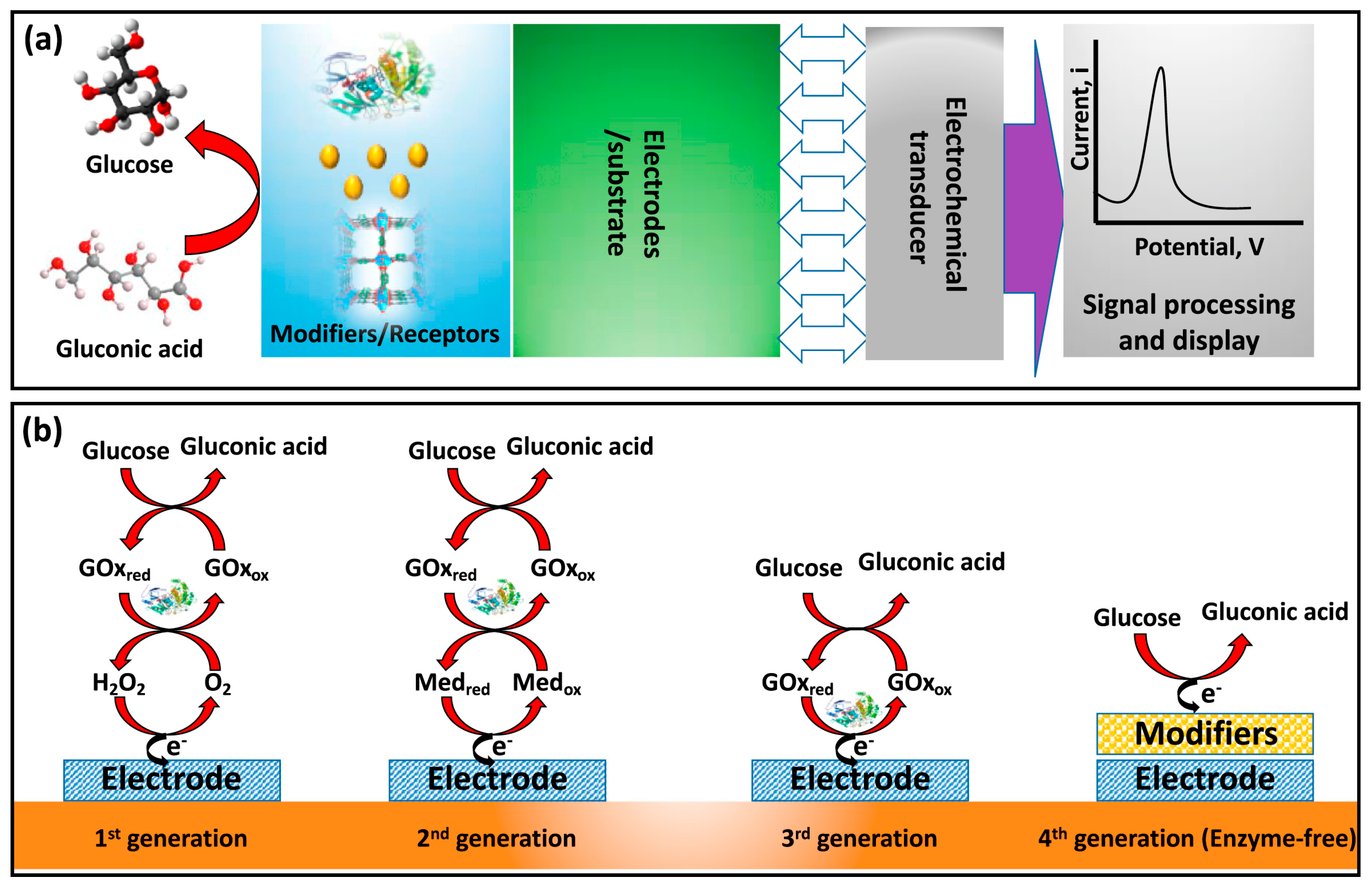
In enzymatic sensors (Figure 2), TiO2 nanotubes act as a scaffold for immobilizing GOx, which catalyzes glucose oxidation, generating H2O2 [46]. The electrochemical oxidation of H2O2 produces a measurable current proportional to glucose concentration. TiO2 enhances electron transfer and enzyme loading. Further improvements are achieved through surface modification with mediators like AuNPs, Prussian Blue (PB), or carbon nanomaterials, enhancing sensitivity and reducing interferences [47]. Despite high selectivity, enzymatic systems are limited by enzyme instability.
Non-enzymatic sensors (Figure 2) utilize electrocatalytic metals (e.g., Pt, Ni, Cu) anchored onto TiO2 nanotubes to directly oxidize glucose, eliminating the need for biological elements [49]. This configuration offers improved stability, longer shelf life, and broad linear response ranges. TiO2 ensures uniform nanoparticle dispersion and supports synergistic charge transfer. Bimetallic and transition metal-decorated TiO2 sensors exhibit excellent anti-interference capabilities and ultra-low detection limits in complex biofluids [50].
In PEC glucose sensors (Figure 3), the semiconducting properties of TiO2 enable photoexcitation under UV or visible light, generating electron–hole pairs that drive the oxidation of glucose, producing a measurable photocurrent proportional to its concentration [51]. Performance enhancements have been achieved through the incorporation of plasmonic metals (e.g., Au, Pt), co-catalysts (e.g., BiOBr, CuO), and heterostructures (e.g., CdS, CdxZn1−xS), which extend light absorption into the visible region and improve charge separation. These modifications contribute to improved sensor sensitivity, lower detection thresholds, faster response times, and strong selectivity, even in complex biological environments like blood, sweat, and serum.
Collectively, these sensor modalities highlight the versatility of TiO2 nanotubes, offering tailored solutions for selective, stable, and ultrasensitive glucose sensing in diverse environments.

4. Application of TiO2-Based Sensors in Glucose Biosensing

TiO2, particularly in NTA configurations, has emerged as a highly promising platform for glucose biosensing due to its unique physicochemical properties such as high surface area, biocompatibility, photoactivity, and ease of functionalization. This section categorizes and discusses various TiO2-based glucose biosensors according to their operational mechanisms—enzymatic, non-enzymatic, PEC, and other novel formats.

4.1. TiO2-Based Enzymatic Glucose Biosensors

4.1.1. TiO2/GOx-Based Biosensors

Recent progress in TiO2 NTA-based enzymatic glucose biosensors reflects a growing emphasis on fine-tuning the nanostructure–enzyme interface to enhance sensitivity, selectivity, and operational robustness. Wang et al. [53] addressed the challenge of preserving the intrinsic properties of TiO2 NTAs by developing a biosensor in which GOx was immobilized via a carefully optimized cross-linking method that deliberately excluded hybrid material integration. The vertically aligned TiO2 NTAs, anodically grown on titanium substrates, ensured strong adhesion, uniform morphology, and high surface area for enzyme loading. The resulting biosensor exhibited a broad dynamic range, ultra-low detection limit, and strong resistance to electrochemical interference from ascorbic acid and sucrose, which are commonly encountered in biological matrices. Importantly, the sensor demonstrated excellent reproducibility and retained functional stability during long-term storage, underscoring the value of structural preservation in enzyme–nanotube coupling.
Building on this, Hu et al. [54] tackled the conductivity bottleneck and enzyme immobilization inefficiencies by introducing a defect-engineering strategy through thermal annealing in an inert argon atmosphere. This process generated oxygen vacancies within the TiO2 lattice, enhancing both surface reactivity and charge carrier density, which in turn improved enzyme anchoring and electron transfer kinetics. GOx-functionalized NTAs derived from this process displayed significantly enhanced electrochemical responses, with high sensitivity, a lowered detection threshold, and excellent selectivity toward glucose in the presence of common interferents. The authors emphasized that their method is not only effective but also scalable and economically viable, making it suitable for large-scale biosensor fabrication. Unlike Wang et al.’s [53] structurally conservative approach, Hu et al. [54] introduced deliberate lattice-level modifications to tailor the physicochemical landscape of TiO2, reflecting a shift from passive immobilization to active structural optimization.
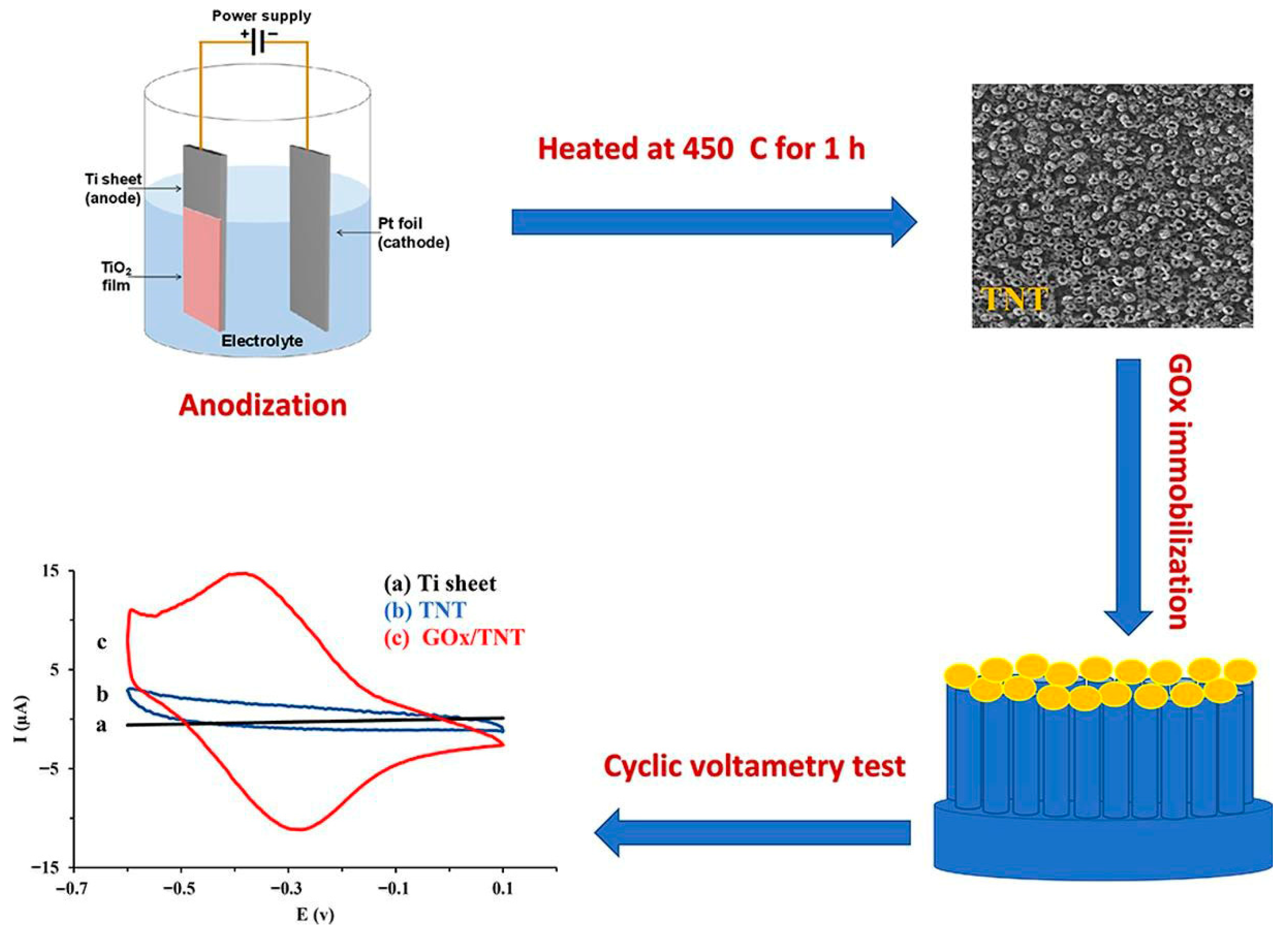
Akhbari Varkani et al. [55] framed their work within a broader context of integrating redox-active biocatalysts with engineered nanostructures for advanced biosensing applications (Figure 4). They reaffirmed that enzyme immobilization is not only a stability-enhancing step but also a functional determinant of electrocatalytic performance. Employing electrochemically anodized TiO2 NTAs, they immobilized GOx to construct a biosensing platform that demonstrated high sensitivity, low detection limits, and an extended linear range for glucose detection. Their results confirmed that TiO2 NTAs serve as excellent supports due to their high surface area, biocompatibility, and ability to retain enzyme activity over prolonged use.
Together, these studies highlight the evolving design philosophy in TiO2-based biosensor research: from minimalistic structural preservation to defect-driven performance enhancement and finally to system-level integration of nanomaterials with bioactive components.

4.1.2. TiO2/Noble Metal/GOx-Based Hybrid Biosensors

Recent developments in TiO2 NTA-based enzymatic glucose biosensors reflect a systematic evolution in design strategies that leverage noble metals, redox mediators, and conductive nanomaterials to enhance electrochemical response, enzyme immobilization efficiency, and transduction performance.
Benvenuto et al. [56] developed a high-performance biosensor by sequentially modifying TiO2 NTAs with a Au layer via argon plasma treatment, followed by electrodeposition of PB, a known artificial peroxidase. Subsequently, GOx was co-immobilized with chitosan on the PB/Au-modified surface, which provided a redox-active and biocompatible matrix facilitating effective enzyme anchoring and electron mediation. The resulting biosensor exhibited high electrocatalytic activity, a significantly reduced detection limit, and excellent operational stability under repetitive use, with negligible signal drift over extended periods. Notably, the integration of PB facilitated direct electron transfer pathways between GOx and the electrode surface, while the Au layer improved conductivity, collectively enhancing the sensor’s responsiveness and durability under physiological conditions.
Extending the nanocomposite strategy, Feng et al. [57] reported the fabrication of a GOx-based biosensor through the immobilization of GOx onto TiO2 NTAs modified with photoreduced Ag nanoparticles (AgNPs). The AgNPs, formed by immersing the TiO2 arrays in silver nitrate solution under controlled photoreduction conditions, exhibited uniform distribution and tunable size and density depending on the immersion duration. This metallic decoration significantly enhanced the catalytic surface and electron transfer kinetics, leading to improved amperometric sensitivity and a linear detection range compatible with physiological glucose concentrations. The study demonstrated that precise control over AgNP distribution plays a pivotal role in optimizing sensor performance without compromising the structural integrity of the TiO2 substrate.
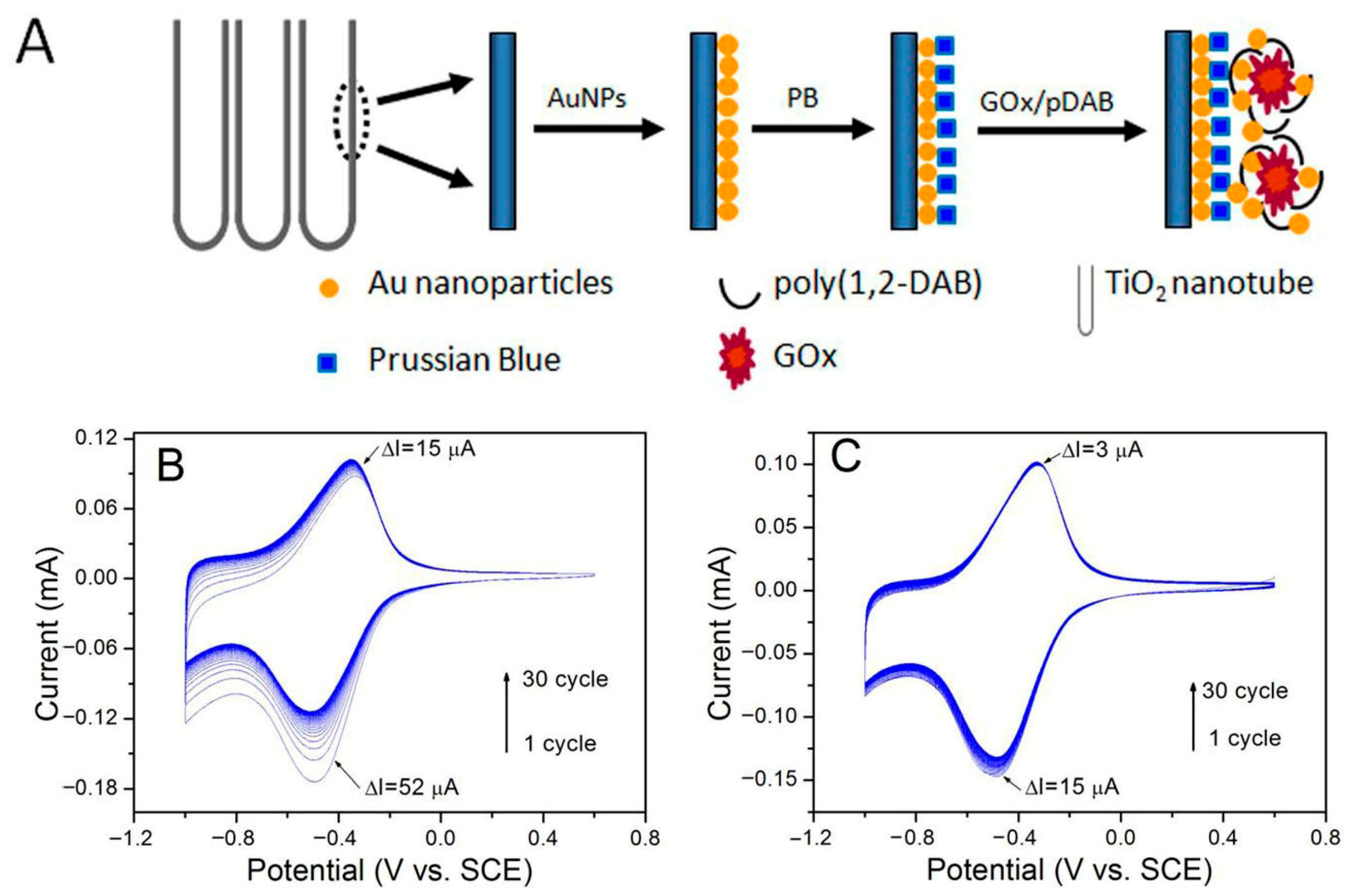
Building on the role of photoactivity in TiO2, Gao et al. [58] employed a photocatalytic deposition technique to embed PB nanocrystals within the inner walls of the TiO2 nanotubes, utilizing UV illumination to achieve spatially controlled and stable incorporation (Figure 5). Following this, a polymer-assisted electrodeposition process was used to immobilize GOx in conjunction with AuNPs, creating a nanobiocomposite interface with enhanced electrocatalytic properties. The fabricated bioelectrode demonstrated a rapid and stable amperometric response to glucose, with a broad linear detection range and high reproducibility. The synergy between the peroxidase-mimicking PB, the conductive AuNPs, and the high-surface-area TiO2 NTAs enabled efficient electron transfer and preserved enzyme activity, making the system highly suitable for continuous monitoring applications. Furthermore, the spatially resolved functionalization method illustrated a scalable pathway for tailoring the internal architecture of NTAs to host multiple bioactive and redox-active components.
In a distinct approach, Pang et al. [59] engineered a hybrid TiO2/CNT electrode by incorporating vapor-grown carbon nanotubes into the internal channels of the TiO2 NTAs to address inherent limitations in charge transport and active surface area. This CNT integration dramatically improved electrical conductivity and enzyme loading capacity due to the interconnected nanotubular structure and high aspect ratio of CNTs. Pt NPs were subsequently deposited onto the TiO2/CNT composite, forming a uniform catalytic layer optimized for H2O2 oxidation, a key intermediate in GOx-mediated glucose sensing. GOx immobilization on this multifunctional composite yielded a biosensor characterized by a fast electrochemical response, high sensitivity across physiologically relevant glucose concentrations, and an exceptionally low detection limit. The hybrid configuration demonstrated superior electron mobility and catalytic efficiency compared to conventional TiO2-based electrodes, confirming the critical role of CNTs and Pt NPs in advancing sensor performance.
Together, these studies exemplify the progression in biosensor engineering from single-metal nanoparticle decoration to complex multi-component nanocomposite architectures. The integration of noble metals (Ag, Au, Pt), redox mediators (PB), and conductive frameworks (CNTs) into TiO2 NTAs has been shown to address the multifaceted challenges of electron transfer, catalytic efficiency, and enzyme stability.

4.2. TiO2-Based Non-Enzymatic Glucose Biosensors

4.2.1. TiO2/Noble Metal-Based Biosensors

Recent developments in non-enzymatic glucose detection have emphasized the integration of noble metals with TiO2 nanostructures to enhance electrocatalytic activity, electron transport efficiency, and sensor reusability. TiO2-based hybrid platforms modified with noble metals have therefore attracted considerable attention due to their synergistic physicochemical and electrochemical properties.
Song et al. [60] systematically demonstrated the fabrication and electrochemical application of Pt-modified TiO2 NTAs as a reusable sensor for glucose detection. In this architecture, Pt nanoparticles functioned dually as catalytic agents for glucose oxidation and as conductive facilitators, enhancing electron mobility along the TiO2 framework to support stable amperometric operation. Importantly, the intrinsic photocatalytic functionality of TiO2 was preserved, allowing for regeneration of Pt catalytic activity via photodegradation of surface-bound interfering species. This configuration enabled selective, reusable glucose sensing with minimal interference from common electroactive compounds.
In a subsequent study, Wang et al. [61] introduced a bimetallic modification approach by depositing hollow Ag and Pt nanostructures onto annealed TiO2 nanotube substrates using a solution-phase reduction and galvanic replacement technique. The resulting Ag&Pt-TiO2 composites exhibited well-defined hollow morphologies with uniform nanoparticle distribution, as verified by electron microscopy and elemental mapping. Electrochemical analysis revealed enhanced redox activity and rapid electron transfer kinetics, and demonstrated high sensitivity, wide linear range, and low detection threshold for glucose. This dual-metal TiO2-based sensor offered a simplified synthesis route with superior analytical performance.

4.2.2. TiO2/Transition Metal-Based Biosensors

TiO2/Nickel-Based Biosensors
Transition metal-functionalized TiO2 nanostructures have garnered increasing interest in non-enzymatic glucose sensing due to their cost-effectiveness, abundance, and catalytic redox activity. Among these, nickel-based modifications of TiO2 nanotubes have shown considerable promise.
Yu et al. [62] designed a non-enzymatic glucose sensor using TiO2 NTAs decorated with nickel nanoparticles, synthesized via anodization followed by pulsed electrodeposition. Structural characterizations confirmed the uniform incorporation of spherical Ni nanoparticles within the nanotube matrix. Electrochemical assessments demonstrated substantial catalytic activity for glucose oxidation, attributed to the enlarged electroactive surface area and enhanced charge transfer enabled by the nanotubular architecture. The sensor exhibited high sensitivity, a broad linear detection range, and a low detection threshold. Importantly, the straightforward and cost-effective synthesis protocol underscored its feasibility for scalable manufacturing.
Expanding upon the catalytic functionality, Huo et al. [63] introduced a light-renewable TiO2-based sensor composed of Ni/NiTiO3-modified TiO2 NTAs fabricated via hydrothermal treatment in nickel acetate, followed by annealing in a reducing atmosphere. This electrode architecture combined the redox-active Ni/NiTiO3 phases with the inherent photocatalytic properties of TiO2, yielding a sensor capable of self-regeneration under light irradiation (Figure 6). The vertically aligned nanotubes and uniformly distributed active phases facilitated rapid electron transfer and superior electrocatalytic performance at low applied potential. Notably, the photocatalytic restoration of electrode sensitivity after fouling events represented a substantial advancement toward achieving long-term stability in non-enzymatic glucose biosensing.
Kang et al. [64] pursued an alternative strategy by incorporating nickel and diamond-like carbon (DLC) into TiO2 NTAs through anodization, electrodeposition, and magnetron sputtering. Electrochemical evaluation revealed markedly improved glucose oxidation performance compared to bare and Ni-coated TiO2 electrodes. The enhanced activity was linked to the synergistic interaction between the redox-active Ni species and the conductive DLC layer, which collectively enhanced charge mobility and surface reactivity. The resulting sensor demonstrated high sensitivity and low detection limits across a broad linear range.
TiO2/Copper-Based Biosensors
The integration of Cu-based nanostructures with TiO2 NTAs has been widely explored to enhance the electrochemical performance of non-enzymatic glucose biosensors. Luo et al. [65] developed a CuO-functionalized TiO2 nanotube electrode using a two-step process involving electrodeposition of Cu nanoparticles followed by thermal oxidation. The fabricated electrode demonstrated significant electrocatalytic efficiency in alkaline media, exhibiting strong linearity and high sensitivity in glucose oxidation. Their design strategy effectively minimized common interferences and ensured electrode stability over prolonged periods. They concluded that the structural synergy between CuO and TiO2 nanotubes was key to the observed selectivity and operational robustness.
In another study, Stanley et al. [66] modified vertically aligned TiO2 nanotubes with CuO mesoclusters through electrochemical methods. Their surface morphology analysis revealed well-distributed CuO aggregates that enhanced the catalytic surface area. The electrode facilitated glucose oxidation at a relatively higher potential and achieved remarkable sensitivity over a wide concentration range. The system displayed negligible cross-reactivity with interfering biomolecules and sugars, ensuring reliable performance in complex biological samples. The authors further validated its clinical potential through successful testing with real blood serum, aligning well with commercial glucose sensing tools.
Bhanu et al. [67] introduced a UV-assisted photoreduction route for depositing Cu nanostructures on anodically synthesized TiO2 nanotubes. Their approach ensured uniform surface coverage and optimal electronic properties, confirmed by comprehensive structural and optical analyses. The Cu-modified electrodes demonstrated enhanced sensitivity and a broad detection range, with significant improvement in signal selectivity. Moreover, the hybrid structures exhibited superior photocurrent response, indicating dual functionality in both sensing and solar-driven electrochemical applications. They further emphasized that the incorporation of copper not only improved glucose detection but also opened pathways for multifunctional device development.
TiO2/Transition Metals Hybrid-Based Biosensors
TiO2 nanotube-based hybrid structures with transition metals have emerged as effective platforms for non-enzymatic glucose sensing due to their unique electrochemical characteristics and tunable surface functionalities.
Li et al. [68] synthesized Ni-Cu nanoparticles on TiO2 NTAs using a potential step method to improve non-enzymatic glucose detection. Surface morphology and composition analysis confirmed uniform alloy distribution with enhanced electrocatalytic properties. Compared to individual nickel or copper modifications, the bimetallic Ni–Cu system showed superior performance in glucose oxidation under alkaline conditions. Interference studies indicated minimal signal distortion from biologically relevant molecules. The sensor also exhibited strong operational stability and repeatability, highlighting its application in complex biological systems.
A sensor was fabricated by Suneesh et al. [69] via electrodepositing Co-Cu alloy nanoparticles onto vertically aligned TiO2 NTAs. The electrodeposition parameters were optimized to tailor the alloy composition for maximum glucose oxidation activity. The resulting nanostructured electrode demonstrated dual linear detection ranges with outstanding sensitivity and low detection thresholds. Selectivity tests revealed robust resistance against common electroactive species and sugars. The system was successfully applied for serum glucose estimation, showcasing clinical applicability.
Chen et al. [70] employed polydiallyldimethylammonium chloride (PDDA) to stabilize functionalized TiO2 nanotubes, enabling uniform self-assembly of Pd nanoparticles on the electrode surface. The resulting sensor demonstrated enhanced electrocatalytic oxidation of glucose at low potential, maintaining high resistance to interference from common biological species. Surface treatment with acid and base further amplified the glucose oxidation current, attributed to increased surface defects and oxide species. These modifications significantly improved the sensor’s performance within physiologically relevant glucose concentrations.
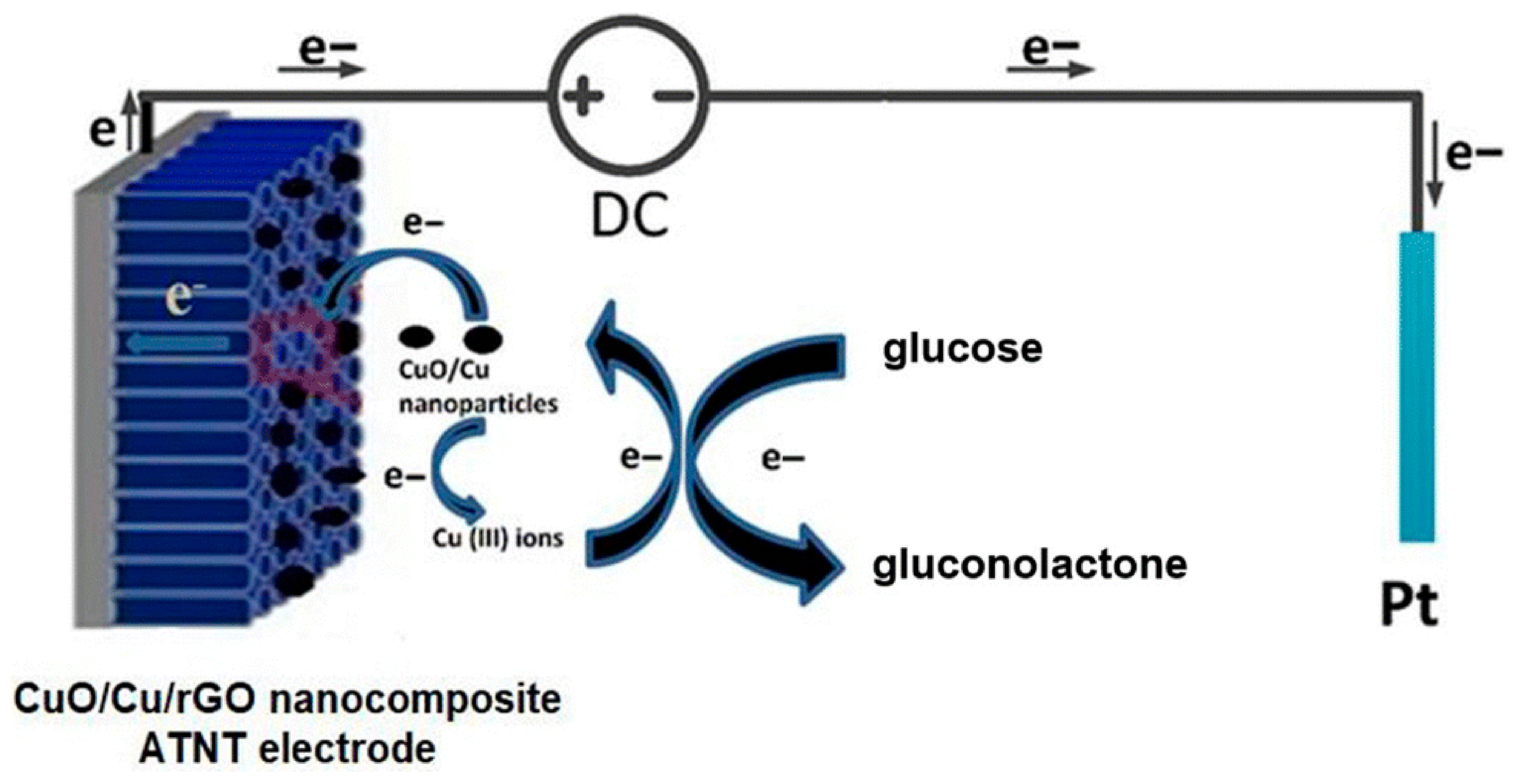
In another study, Chahrour et al. [71] introduced a hybrid nanocomposite comprising CuO/Cu and reduced graphene oxide on anodically formed TiO2 NTAs (Figure 7). The stepwise electrodeposition and reduction processes facilitated uniform nanoparticle dispersion and enhanced conductivity through graphene integration. Structural and elemental characterization verified effective composite formation with improved electrochemical surface properties. The biosensor delivered a high electrocatalytic response to glucose with excellent selectivity in neutral media. Its reproducibility and long-term stability emphasize its promise for biomedical diagnostics and pharmaceutical screening.
Kumar & Sinha [72] developed a tungsten oxide-decorated TiO2 NTA through electrochemical methods to enhance glucose detection without enzymes. The integration of WO3 nanostructures provided a high electroactive surface area and improved stability under electrochemical conditions. Detailed structural analyses confirmed uniform deposition and strong crystalline features of the hybrid material. Electrochemical testing revealed excellent sensitivity and response characteristics, indicating efficient electron transport and charge transfer kinetics. The sensor exhibited consistent behavior in real sample analysis, validating its potential for practical glucose monitoring.
Non-enzymatic glucose biosensors based on TiO2 nanostructures have advanced through integration with noble and transition metals to enhance catalytic activity, conductivity, and stability. Noble metal hybrids (Pt, Ag, Au) improved electron transfer and reusability, while transition metal systems (Ni, Cu, Co, Pd, W) offered cost-effective, redox-active alternatives with strong sensitivity and durability. Bimetallic and hybrid designs further boosted selectivity and operational stability, underscoring a clear shift from enzyme-reliant to catalyst-driven TiO2 platforms for reliable and scalable glucose detection.

4.3. TiO2-Based Photoelectrochemical Glucose Biosensors

The development of TiO2-based PEC glucose biosensors has attracted increasing attention due to their high sensitivity, stability, and potential for non-invasive diagnostics.

4.3.1. TiO2/Noble Metal-Based Biosensors

Noble metal-functionalized TiO2-based PEC biosensors have gained significant attention due to their enhanced charge separation, plasmonic light absorption, and catalytic properties. The incorporation of metals such as Au and Pt onto TiO2 nanotube architectures has enabled the development of highly sensitive and selective glucose sensing platforms with superior PEC performance.
Liu et al. [73] reported the fabrication of a PEC glucose sensor utilizing TiO2 nanotubes functionalized with Au nanoparticles, capitalizing on the superior charge carrier separation efficiency intrinsic to one-dimensional architectures and the pronounced surface plasmon resonance (SPR) properties of Au. The sensor was responsive to visible red-light irradiation, enabling efficient PEC activity. Under optimized operational parameters, the Au/TiO2NTs-based sensor demonstrated high sensitivity and an exceptionally low detection threshold for glucose quantification. Furthermore, it maintained robust selectivity, exhibited substantial resistance to interference from coexisting analytes, and preserved functional stability over extended periods. Owing to these advantageous attributes, the system was identified as a promising candidate for integration into non-invasive PEC biosensing platforms.
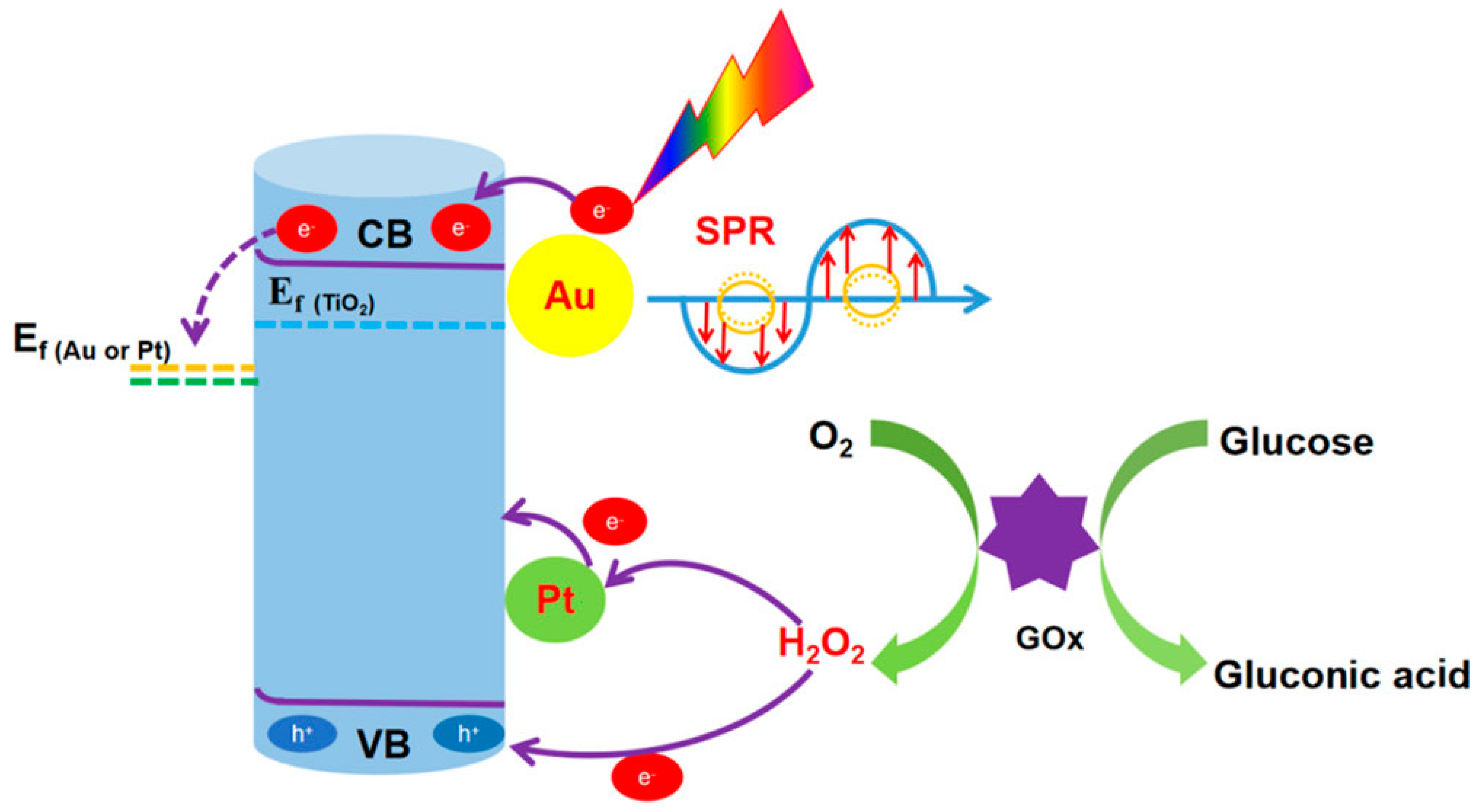
Yang et al. [74] developed a high-performance PEC glucose biosensor by modifying TiO2 NTAs with Au and Pt nanoparticles (TiO2NTs/Au/Pt/GOx). The TiO2 nanotubes enhanced charge transport by aligning light absorption with carrier diffusion (Figure 8). Au nanoparticles improved light harvesting via plasmonic effects and promoted charge separation through Schottky junctions, while Pt nanoparticles catalyzed hydrogen peroxide reduction from the GOx-mediated glucose reaction, enhancing electron transfer. This dual modification significantly outperformed single-metal systems, demonstrating that Pt plays a critical role in boosting sensitivity and detection efficiency in enzymatic PEC glucose sensing.

4.3.2. TiO2/Metal Oxide-Based Biosensors

The integration of TiO2 with metal oxides has emerged as a strategic approach to enhance PEC glucose sensing by leveraging synergistic heterojunction effects and catalytic functionalities. These composite systems enable improved charge separation, broadened light absorption, and enzyme-free or self-powered detection capabilities, broadening the scope of non-invasive biosensing applications.
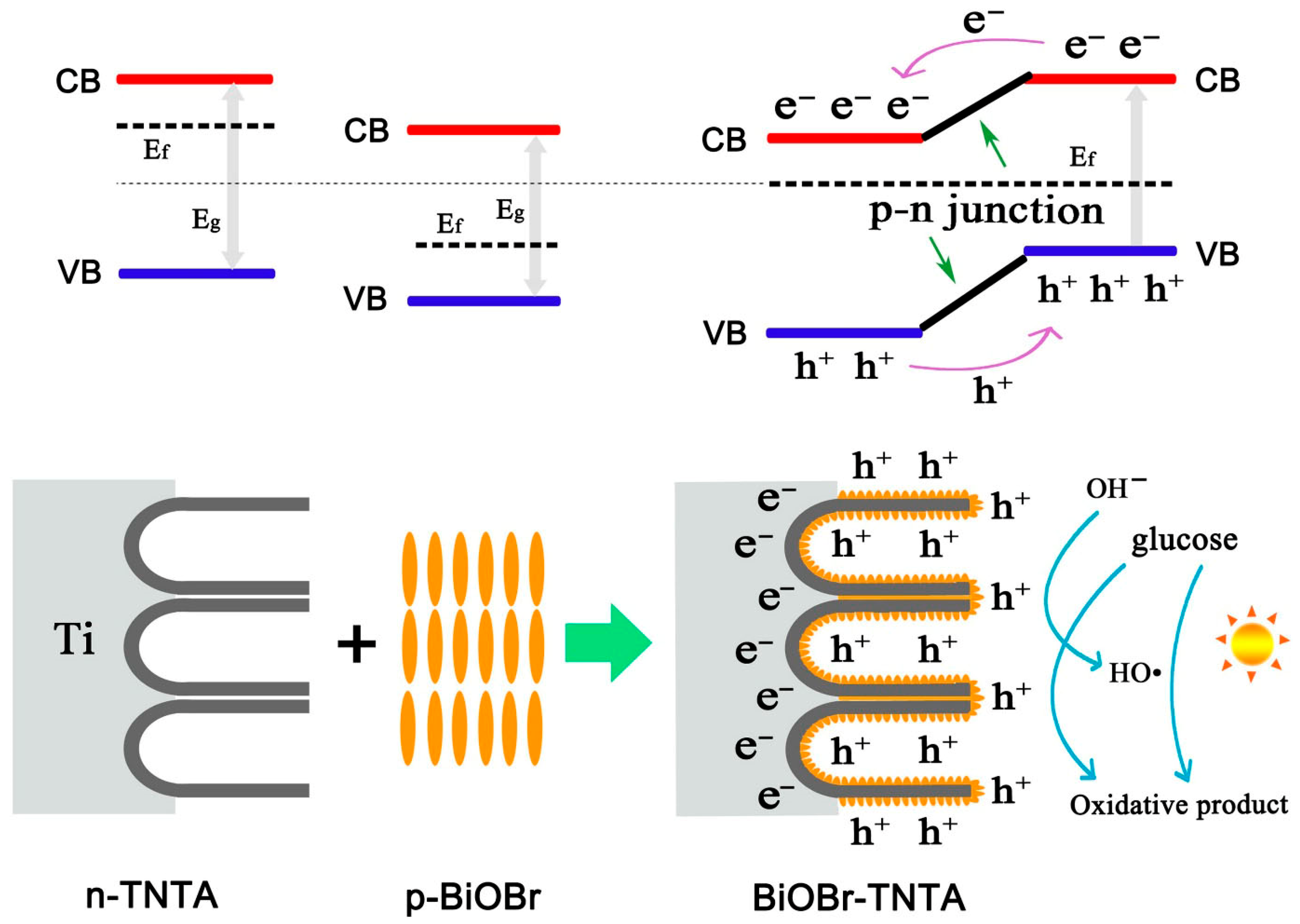
Wu et al. [75] engineered a p-n heterojunction PEC electrode by depositing BiOBr nanostructures onto TiO2 NTAs (Figure 9). The heterojunction structure facilitated effective photogenerated charge separation due to favorable energy band alignment between BiOBr and TiO2. The electrode preparation parameters, including the Bi precursor concentration and nanotube morphology, critically influenced the sensor’s electrochemical properties. Under optimized conditions, the device displayed enhanced response and reproducibility for glucose detection in alkaline media. This study expanded the material design strategy for PEC biosensors beyond traditional semiconductors.
Ke et al. [76] critically addressed the limitations posed by conventional invasive glucose sensors, which often resulted in adverse physical and psychological effects during fingertip blood sampling, constrained applicability of enzyme-dependent electrodes, and operational complexities associated with externally powered integrated systems. In response, they proposed a PEC platform capable of enzyme-free, self-powered glucose detection via sweat, utilizing a CuO nanoparticle-modified TiO2 hierarchical nanotube (CuO\@TiO2 HNT) structure. The CuO\@TiO2 HNT-based PEC sensor demonstrated reliable glucose detection under bias-free conditions and maintained functional integrity across a wide thermal operating range. Analytical performance for sweat glucose quantification before and after food intake showed strong agreement with results obtained from standard commercial glucometers. The superior sensing capability was primarily attributed to the enhanced photon absorption and high surface area provided by the hierarchical TiO2 nanotube framework, in conjunction with the CuO nanoparticles’ ability to accelerate interfacial charge transfer and promote glucose oxidation kinetics. This study introduced a promising self-sustained, non-invasive strategy for sweat-based glucose monitoring, offering a viable route toward the development of portable and enzyme-independent biosensing technologies.
Yang et al. [77] emphasized that achieving elevated sensitivity and minimal detection thresholds represented essential benchmarks in the advancement of biosensing technologies. They synthesized a PEC glucose sensor by employing three-dimensional hydrogen titanate nanotubes grown on titanium substrates as precursors, subsequently forming a CuO–TiO2 heterojunction nanotube array (CuO–TiO2NTs/Ti) photoelectrode. The integrated architecture, comprising a conductive titanium foil, a mesoporous nanotubular morphology, and a p-n CuO–TiO2 interface, markedly enhanced charge carrier dissociation efficiency and promoted rapid interfacial electron transport, resulting in amplified photoelectronic responses under visible light exposure. The sensor exhibited high analytical sensitivity, low detection thresholds, and notable selectivity in glucose recognition. Density functional theory calculations confirmed the role of the heterojunction in improving photoinduced electron–hole separation and overall PEC performance. Furthermore, the incorporation of chemometric methodologies significantly optimized signal interpretation, thereby advancing the precision of sensitivity measurements and reinforcing the practical viability of the PEC sensor in bioanalytical applications.

4.3.3. TiO2/Semiconductor-Based Biosensors

The formation of heterojunctions between TiO2 and narrow band gap semiconductors has emerged as an effective approach to enhance PEC glucose sensing by promoting light absorption and charge carrier separation. Such TiO2-based semiconductor composites offer enzyme-free detection capabilities with improved sensitivity, selectivity, and practical applicability in clinical diagnostics.
Liu et al. [78] reported that a composite structure integrating g-C3N4 and TiO2 nanosheets was strategically engineered to construct a photoelectrochemical enzyme biosensor platform. The coupling of these semiconductors was designed to overcome the limited visible-light activity of TiO2 and to suppress the recombination of photogenerated charge carriers within g-C3N4, thereby improving overall photoelectrochemical efficiency. Employing glucose oxidase as the biorecognition element, the hybrid system exhibited pronounced visible-light-driven catalytic behavior toward enzymatic glucose oxidation, surpassing the performance of biosensors fabricated from the individual constituents. The enhanced activity was attributed to the synergistic heterojunction effect that facilitated efficient charge transfer, as well as to the high surface area and biocompatible characteristics of the TiO2 nanosheets, which collectively promoted superior enzyme immobilization and signal transduction within the photoelectrochemical sensing interface.
Esmaeili et al. [79] designed an enzyme-free PEC glucose sensor by decorating TiO2 NTAs with CdxZn1−nS nanofilms. This heterojunction composite structure significantly improved light absorption and electron transport, resulting in amplified photocurrent signals. The modified electrode showed excellent analytical performance with respect to detection limit and sensitivity, attributed to the discrete morphology of the sulfide films. Characterization confirmed the successful integration of the composite and its structural stability. The sensor further demonstrated robust application potential in detecting glucose in real plasma samples, underscoring its clinical relevance and economic viability.
Li et al. [80] demonstrated that heterojunctions composed of dissimilar semiconducting materials exhibited superior photon-harvesting capabilities and enhanced photoelectrical responses relative to single-component systems. They engineered CdSe/TiO2 nanotube (CdSe/TiO2NT) heterojunctions via a hydrothermal synthesis route, utilizing structurally stable TiO2 nanotubes as the foundational scaffold. The resultant nanostructures were extensively characterized through X-ray diffraction, transmission and scanning electron microscopy, photoluminescence spectroscopy, UV–visible absorption spectroscopy, and electrochemical impedance spectroscopy. Under visible light excitation, the CdSe/TiO2NT heterojunctions displayed enhanced optical absorption and a significant reduction in charge carrier recombination compared to pristine TiO2. Theoretical simulations substantiated the effective spatial separation of photogenerated electrons and holes within the heterostructure. When optimized, the PEC system demonstrated a linear photocurrent response across a defined glucose concentration range and maintained a low detection threshold. Additionally, the sensor showed high selectivity and operational stability. The underlying PEC sensing mechanism was elucidated by integrating empirical observations with theoretical analyses, reinforcing the potential of CdSe/TiO2-based heterostructures in advanced biosensing platforms.

4.3.4. TiO2/Graphene-Based Biosensors

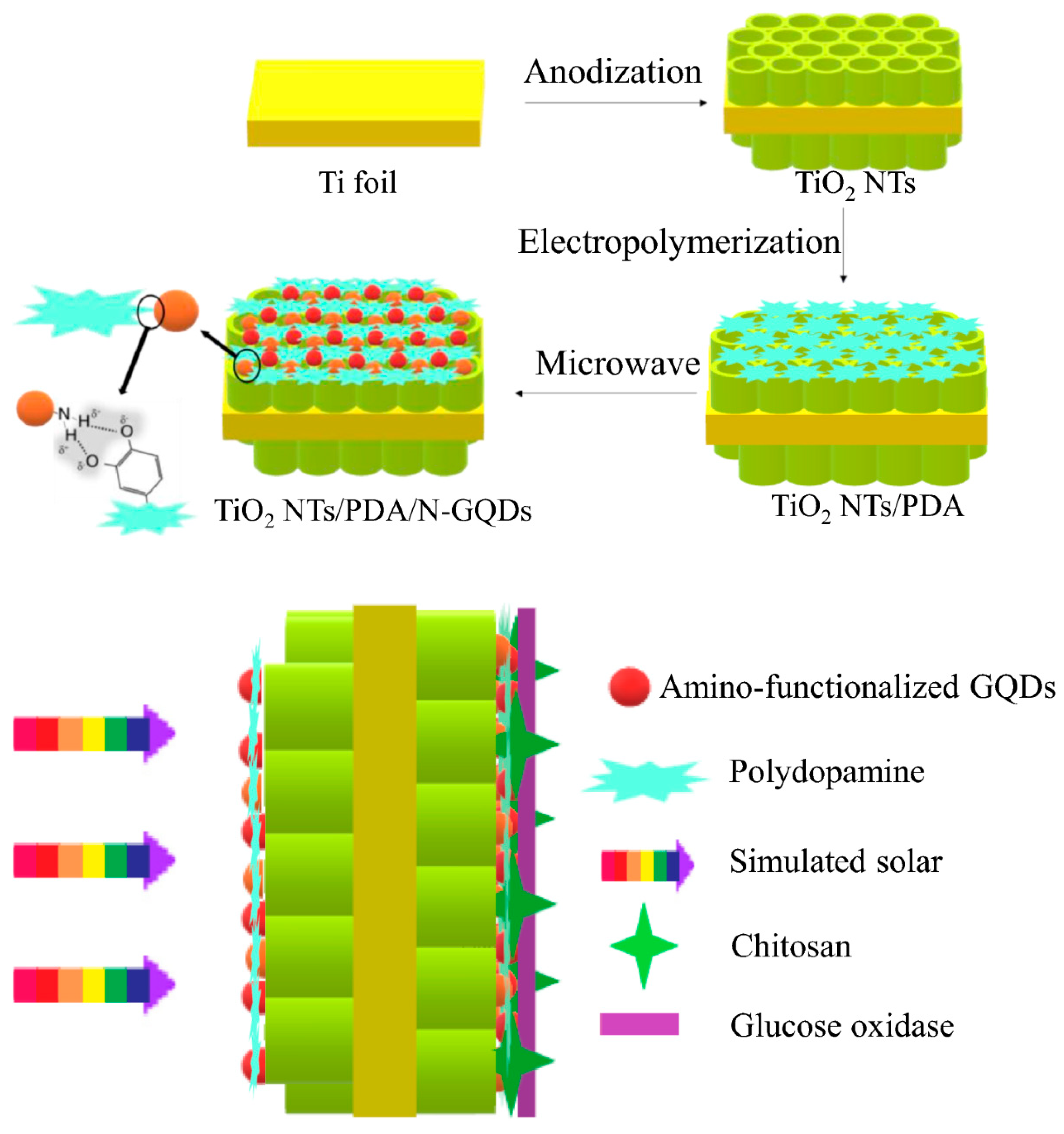
The integration of graphene-based nanomaterials with TiO2 has shown great promise in enhancing PEC biosensing performance through improved electron transfer and charge separation. Such hybrid architectures enable high photocurrent responses and reliable glucose detection, highlighting their potential for advanced biosensor development. Yang et al. [81] fabricated a PEC biosensor by functionalizing TiO2 nanotubes with polydopamine and amino-functionalized graphene quantum dots, integrated with GOx (Figure 10). The electropolymerized polydopamine provided an efficient electron-transfer interface, while the microwave-assisted incorporation of quantum dots improved charge carrier separation. The system demonstrated notable enhancement in photocurrent generation and exhibited superior analytical performance in terms of selectivity and operational stability. This dual-electron-acceptor design indicated the potential of synergistic material integration in advancing PEC biosensor platforms.
TiO2-based PEC glucose biosensors have progressed through integration with noble metals, metal oxides, semiconductors, and graphene to improve charge separation, light absorption, and catalytic efficiency. Noble metals enhanced plasmonic response and electron transfer, while metal oxides and semiconductors enabled enzyme-free, self-powered detection with high sensitivity and stability. Graphene hybrids further boosted conductivity and photocurrent. Overall, these developments reflect a move toward light-driven, wearable PEC sensors for practical glucose monitoring.

4.4. TiO2-Based Other Novel Glucose Biosensors

Beyond traditional electrochemical and PEC designs, novel TiO2-based biosensors have employed organic electrochemical transistors (OECTs) and optical detection strategies for glucose monitoring.
Liao et al. [82] developed an organic electrochemical transistor (OECT) sensor incorporating TiO2 NTAs as the gate electrode, replacing traditionally used Pt-based materials. The TiO2 NTAs offered comparable electrocatalytic performance while being cost-effective and biocompatible. When modified with Pt nanoparticles and GOx, the device demonstrated a strong linear correlation between glucose concentration and output signal, along with enhanced selectivity against common interferents through Nafion functionalization. Furthermore, the sensor maintained stable signal reproducibility, indicating the robustness of TiO2 NTAs as an alternative gate material in OECT configurations. This approach suggested significant potential for expanding OECT-based biosensing technologies using TiO2 nanostructures.
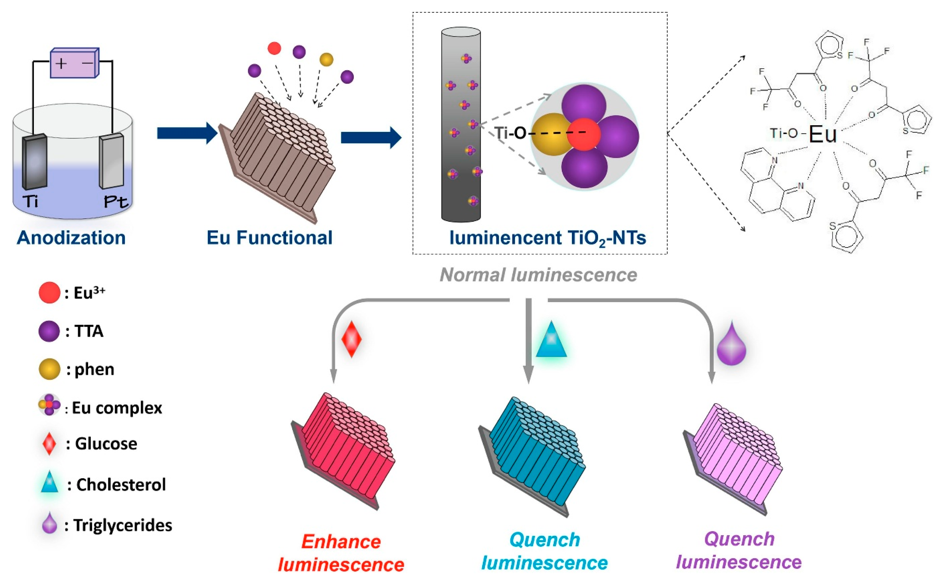
Building upon the multifunctionality of TiO2 NTAs, Su et al. [83] introduced a luminescent sensor platform by integrating europium complexes onto TiO2-nanotubes through a one-step anodization and in-situ modification technique (Figure 11). The europium-functionalized nanotubes exhibited distinct fluorescence behavior upon interaction with glucose, cholesterol, and triglycerides, which was attributed to an energy transfer mechanism. A linear response in fluorescence intensity relative to the analyte concentration allowed quantitative detection with high sensitivity. This photoluminescent sensor was further validated through practical applications in food matrices, exhibiting strong agreement with certified analytical methods. The study highlighted the potential of rare-earth-doped TiO2 nanostructures for constructing simple, selective, and multifunctional biosensing platforms, expanding the utility of TiO2-based systems in real-world scenarios.
The prior discussion of TiO2 NT-based biosensing platforms for glucose detection is summarized in Table 1.

5. Challenges and Future Perspectives

Despite significant advancements in TiO2-based glucose sensor technologies, several critical challenges hinder their broader clinical translation, long-term operation, and scalability. Enzymatic sensors, while achieving prolonged stability and fast response times, face limitations due to the inherent instability of GOx, which is prone to denaturation under varying pH, temperature, and storage conditions. Although studies have demonstrated operational stability exceeding 30 days and response times under one second, issues such as enzyme leaching and biofouling in complex biofluids like serum persist, restricting their use in continuous monitoring applications. Non-enzymatic sensors, particularly those incorporating transition metals or bimetallic nanostructures, offer superior electrocatalytic activity, wide linear ranges, and ultra-low detection limits. However, their reliance on strongly alkaline media raises compatibility concerns with physiological fluids and wearable applications. Additionally, despite improved selectivity through nanostructuring, interference from electroactive species in high-protein environments remains problematic. Challenges such as nanoparticle agglomeration, structural instability, and limited shelf life further impede their commercial viability. PEC sensors, exploiting TiO2’s semiconductor properties under light illumination, present a promising alternative with advantages like low-power operation, high sensitivity, and background current suppression. Systems incorporating materials such as CdxZn1−xS and BiOBr have achieved remarkably low detection limits, yet challenges persist in optimizing light-harvesting efficiency, particularly for visible-light-driven PEC sensors. Ensuring stable performance under fluctuating ambient lighting and standardizing optical setups remain unresolved issues, complicating their integration into compact, real-world devices. Another critical gap lies in real-world validation, as many studies focus on performance in controlled buffer solutions rather than complex biological matrices like serum or plasma. The lack of extensive clinical trials, long-term monitoring studies, and standardized fabrication methods further delays their transition from lab-scale prototypes to commercial products. Table 2 highlights the features, advantages, and limitations of enzymatic, non-enzymatic, and PEC TiO2-based glucose sensors.
Future research must prioritize hybrid sensor architectures that combine the selectivity of enzymatic recognition with the robustness of non-enzymatic and PEC systems, potentially enabling dual-signal amplification for enhanced reliability. The development of flexible, wearable TiO2-based sensors is essential for continuous glucose monitoring, requiring innovations in biocompatible materials and stretchable substrates to improve user comfort and signal stability. Integrating advanced data processing tools such as machine learning [84] and artificial intelligence [85] could further refine dynamic calibration, drift correction, and predictive analytics, enabling personalized glucose monitoring tailored to individual metabolic patterns. Scalable and cost-effective fabrication techniques, including roll-to-roll printing [86] and 3D nanostructuring [87], are needed to replace lab-intensive synthesis methods and facilitate mass production. Additionally, sustainability considerations, such as eco-friendly synthesis routes, reduced use of rare metals, and recyclable materials, must be incorporated to align with green chemistry principles. Finally, rigorous clinical validation, long-term in vivo studies, and adherence to regulatory standards are imperative to bridge the gap between research and practical implementation, ensuring these sensors meet the demands of point-of-care diagnostics and global healthcare markets.

6. Conclusions

In conclusion, TiO2-based glucose sensors have demonstrated remarkable potential across enzymatic, non-enzymatic, PEC, and emerging hybrid platforms. Their tunable surface chemistry, excellent biocompatibility, and compatibility with a wide range of nanostructures enable sensitive, selective, and stable glucose detection in both physiological and complex real-world matrices. The reviewed literature highlights significant advancements in detection range, response time, and anti-interference capabilities, achieved through strategic material modifications such as metal nanoparticle doping, heterojunction formation, and surface functionalization. Despite these achievements, key challenges remain in long-term stability, reproducibility, and scalability. Recent developments in wearable biosensors based on TiO2 nanostructures have further expanded their applicability toward continuous, non-invasive glucose monitoring. Such devices offer distinct advantages, including real-time data acquisition, user convenience, and integration with wireless or smartphone-based platforms for personalized healthcare. Future progress will hinge on the integration of smart data analytics, sustainable material strategies, and wearable device architectures. In this context, designing flexible, skin-compatible, and energy-efficient TiO2-based wearable systems represents a promising direction for next-generation biosensing technologies. Collectively, these innovations hold the promise of transforming TiO2-based glucose sensors from laboratory prototypes into clinically relevant, real-time monitoring tools capable of addressing the growing demands of personalized and point-of-care diagnostics.

Author Contributions

Conceptualization, C.M.H.; methodology, J.S.; writing—original draft preparation, J.S.; writing—review and editing, C.M.H. All authors have read and agreed to the published version of the manuscript.

Funding

This research received no external funding.

Data Availability Statement

No new data were created or analyzed in this study.

Conflicts of Interest

The authors declare no conflicts of interest.

References

  1. Hossain, M.J.; Al-Mamun, M.; Islam, M.R. Diabetes Mellitus, the Fastest Growing Global Public Health Concern: Early Detection Should Be Focused. Health Sci. Rep. 2024, 7, e2004. [Google Scholar] [CrossRef]
  2. Sun, H.; Saeedi, P.; Karuranga, S.; Pinkepank, M.; Ogurtsova, K.; Duncan, B.B.; Stein, C.; Basit, A.; Chan, J.C.N.; Mbanya, J.C.; et al. IDF Diabetes Atlas: Global, Regional and Country-Level Diabetes Prevalence Estimates for 2021 and Projections for 2045. Diabetes Res. Clin. Pract. 2021, 183, 109119. [Google Scholar] [CrossRef]
  3. Eckel, R.H.; Bornfeldt, K.E.; Goldberg, I.J. Cardiovascular Disease in Diabetes, beyond Glucose. Cell Metab. 2021, 33, 1519–1545. [Google Scholar] [CrossRef]
  4. Samsu, N. Diabetic Nephropathy: Challenges in Pathogenesis, Diagnosis, and Treatment. BioMed Res. Int. 2021, 2021, 1497449. [Google Scholar] [CrossRef]
  5. Tan, T.-E.; Wong, T.Y. Diabetic Retinopathy: Looking Forward to 2030. Front. Endocrinol. 2023, 13, 1077669. [Google Scholar] [CrossRef]
  6. Eid, S.A.; Rumora, A.E.; Beirowski, B.; Bennett, D.L.; Hur, J.; Savelieff, M.G.; Feldman, E.L. New Perspectives in Diabetic Neuropathy. Neuron 2023, 111, 2623–2641. [Google Scholar] [CrossRef] [PubMed]
  7. Jamshidnejad-Tosaramandani, T.; Kashanian, S.; Omidfar, K.; Schiöth, H.B. The Role of Nanomaterials in the Wearable Electrochemical Glucose Biosensors for Diabetes Management. Biosensors 2025, 15, 451. [Google Scholar] [CrossRef] [PubMed]
  8. Pikulin, S.; Yehezkel, I.; Moskovitch, R. Enhanced Blood Glucose Levels Prediction with a Smartwatch. PLoS ONE 2024, 19, e0307136. [Google Scholar] [CrossRef]
  9. Bamgboje, D.; Christoulakis, I.; Smanis, I.; Chavan, G.; Shah, R.; Malekzadeh, M.; Violaris, I.; Giannakeas, N.; Tsipouras, M.; Kalafatakis, K.; et al. Continuous Non-Invasive Glucose Monitoring via Contact Lenses: Current Approaches and Future Perspectives. Biosensors 2021, 11, 189. [Google Scholar] [CrossRef] [PubMed]
  10. Yoon, S.; Yoon, H.; Zahed, M.A.; Park, C.; Kim, D.; Park, J.Y. Multifunctional Hybrid Skin Patch for Wearable Smart Healthcare Applications. Biosens. Bioelectron. 2022, 196, 113685. [Google Scholar] [CrossRef]
  11. Khosravi, S.; Soltanian, S.; Servati, A.; Khademhosseini, A.; Zhu, Y.; Servati, P. Screen-Printed Textile-Based Electrochemical Biosensor for Noninvasive Monitoring of Glucose in Sweat. Biosensors 2023, 13, 684. [Google Scholar] [CrossRef] [PubMed]
  12. Hou, W.; Liao, Q.; Xie, S.; Song, Y.; Qin, L. Prospects and Challenges of Flexible Stretchable Electrodes for Electronics. Coatings 2022, 12, 558. [Google Scholar] [CrossRef]
  13. Zhang, J.; Li, J.; Wen, X.; Liu, L.; Wei, Q.; Zhang, P.; Zhang, J.-Y.; Zhao, D.; Lan, K. Facile Synthesis of Mesoporous TiO2 Architectures with Tunable Configurations and Nanometer Precision. Nat. Protoc. 2025, 1–23. [Google Scholar] [CrossRef]
  14. Tong, S.; Sun, X.; Wu, A.; Guo, S.; Zhang, H. Improved Biocompatibility of TiO2 Nanotubes via Co-Precipitation Loading with Hydroxyapatite and Gentamicin. Coatings 2021, 11, 1191. [Google Scholar] [CrossRef]
  15. Zakir, O.; Ait-Karra, A.; Idouhli, R.; Khadiri, M.; Dikici, B.; Aityoub, A.; Abouelfida, A.; Outzourhit, A. A Review on TiO2 Nanotubes: Synthesis Strategies, Modifications, and Applications. J. Solid State Electrochem. 2023, 27, 2289–2307. [Google Scholar] [CrossRef]
  16. Patra, S.; Das, P.; Chatterjee, S. Tuning the Optoelectronic and Surface Properties of TiO2 Nanotubes. Mater. Chem. Phys. 2023, 308, 128308. [Google Scholar] [CrossRef]
  17. Hua, L.; Yin, Z.; Cao, S. Recent Advances in Synthesis and Applications of Carbon-Doped TiO2 Nanomaterials. Catalysts 2020, 10, 1431. [Google Scholar] [CrossRef]
  18. Sanattalab, E.; Kanarya, D.; Ebrahimi, A.; Didarian, R.; Doğan Güzel, F.; Yıldırım Tirgil, N. Cutting-Edge Applications of Titanium Dioxide in Biosensors. Electroanalysis 2025, 37, e70049. [Google Scholar] [CrossRef]
  19. Bertel, L.; Miranda, D.A.; García-Martín, J.M. Nanostructured Titanium Dioxide Surfaces for Electrochemical Biosensing. Sensors 2021, 21, 6167. [Google Scholar] [CrossRef]
  20. Isik, E.; Tasyurek, L.B.; Isik, I.; Kilinc, N. Synthesis and Analysis of TiO2 Nanotubes by Electrochemical Anodization and Machine Learning Method for Hydrogen Sensors. Microelectron. Eng. 2022, 262, 111834. [Google Scholar] [CrossRef]
  21. Puga, M.L.; Venturini, J.; ten Caten, C.S.; Bergmann, C.P. Influencing Parameters in the Electrochemical Anodization of TiO2 Nanotubes: Systematic Review and Meta-Analysis. Ceram. Int. 2022, 48, 19513–19526. [Google Scholar] [CrossRef]
  22. Benčina, M.; Iglič, A.; Mozetič, M.; Junkar, I. Crystallized TiO2 Nanosurfaces in Biomedical Applications. Nanomaterials 2020, 10, 1121. [Google Scholar] [CrossRef]
  23. Saker, R.; Shammout, H.; Regdon, G.; Sovány, T. An Overview of Hydrothermally Synthesized Titanate Nanotubes: The Factors Affecting Preparation and Their Promising Pharmaceutical Applications. Pharmaceutics 2024, 16, 635. [Google Scholar] [CrossRef]
  24. Alkanad, K.; Hezam, A.; Al-Zaqri, N.; Bajiri, M.A.; Alnaggar, G.; Drmosh, Q.A.; Almukhlifi, H.A.; Neratur Krishnappagowda, L. One-Step Hydrothermal Synthesis of Anatase TiO2 Nanotubes for Efficient Photocatalytic CO2 Reduction. ACS Omega 2022, 7, 38686–38699. [Google Scholar] [CrossRef]
  25. Cossuet, T.; Rapenne, L.; Renou, G.; Appert, E.; Consonni, V. Template-Assisted Growth of Open-Ended TiO2 Nanotubes with Hexagonal Shape Using Atomic Layer Deposition. Cryst. Growth Des. 2021, 21, 125–132. [Google Scholar] [CrossRef]
  26. Barakat, N.A.M.; Hamza, A.M.; Al-Deyab, S.S.; Qurashi, A.; Kim, H.Y. Titanium-Based Polymeric Electrospun Nanofiber Mats as a Novel Organic Semiconductor. Mater. Sci. Eng. B 2012, 177, 34–42. [Google Scholar] [CrossRef]
  27. Ahmad, A.; Khan, M.A.; Nazir, A.; Arshad, S.N.; Qadir, M.B.; Khaliq, Z.; Khan, Z.S.; Satti, A.N.; Mushtaq, B.; Shahzad, A. Triaxial Electrospun Mixed-Phased TiO2 Nanofiber-in-Nanotube Structure with Enhanced Photocatalytic Activity. Microporous Mesoporous Mater. 2021, 320, 111104. [Google Scholar] [CrossRef]
  28. Etafa Tasisa, Y.; Kumar Sarma, T.; Krishnaraj, R.; Sarma, S. Band Gap Engineering of Titanium Dioxide (TiO2) Nanoparticles Prepared via Green Route and Its Visible Light Driven for Environmental Remediation. Results Chem. 2024, 11, 101850. [Google Scholar] [CrossRef]
  29. Alrashedi, W.; Kochkar, H.; Berhault, G.; Younas, M.; Ben Ali, A.; Alomair, N.A.; Hamdi, R.; Abubshait, S.A.; Alagha, O.; Gondal, M.F.; et al. Enhancement of the Photocatalytic Response of Cu-Doped TiO2 Nanotubes Induced by the Addition of Strontium. J. Photochem. Photobiol. A Chem. 2022, 428, 113858. [Google Scholar] [CrossRef]
  30. Li, J.; Wang, D.; Guan, R.; Zhang, Y.; Zhao, Z.; Zhai, H.; Sun, Z. Vacancy-Enabled Mesoporous TiO2 Modulated by Nickel Doping with Enhanced Photocatalytic Nitrogen Fixation Performance. ACS Sustain. Chem. Eng. 2020, 8, 18258–18265. [Google Scholar] [CrossRef]
  31. Siliavka, E.S.; Rudakova, A.V.; Bakiev, T.V.; Murashkina, A.A.; Murzin, P.D.; Kataeva, G.V.; Emeline, A.V.; Bahnemann, D.W. Effect of the Heterovalent Sc3+ and Nb5+ Doping on Photoelectrochemical Behavior of Anatase TiO2. Catalysts 2024, 14, 76. [Google Scholar] [CrossRef]
  32. Sultana, S.; Darowska, I.; Pisarek, M.; Sulka, G.D.; Syrek, K. Designing TiO2 Nanotubular Arrays with Au-CoOx Core–Shell Nanoparticles for Enhanced Photoelectrochemical Methanol and Lignin Oxidation. ACS Appl. Mater. Interfaces 2024, 16, 49262–49274. [Google Scholar] [CrossRef]
  33. Liang, X.; Yu, S.; Meng, B.; Wang, X.; Yang, C.; Shi, C.; Ding, J. Advanced TiO2-Based Photoelectrocatalysis: Material Modifications, Charge Dynamics, and Environmental–Energy Applications. Catalysts 2025, 15, 542. [Google Scholar] [CrossRef]
  34. Meng, M.; Zhou, H.; Yang, J.; Wang, L.; Yuan, H.; Hao, Y.; Gan, Z. Exploiting the Bragg Mirror Effect of TiO2 Nanotube Photonic Crystals for Promoting Photoelectrochemical Water Splitting. Nanomaterials 2024, 14, 1695. [Google Scholar] [CrossRef]
  35. Zhang, Y.; Yan, J. Recent Advances in the Synthesis of Defective TiO2 Nanofibers and Their Applications in Energy and Catalysis. Chem. Eng. J. 2023, 472, 144831. [Google Scholar] [CrossRef]
  36. Lia, F.; Farrugia, C.; Buccheri, M.A.; Rappazzo, G.; Zammit, E.; Rizzo, A.; Grech, M.; Refalo, P.; Abela, S. Effect of the Surface Morphology of TiO2 Nanotubes on Photocatalytic Efficacy Using Electron-Transfer-Based Assays and Antimicrobial Tests. Appl. Sci. 2020, 10, 5243. [Google Scholar] [CrossRef]
  37. Cheng, Q.; Wang, A.; Song, Z.; Bao, J.; Xue, J.; Wei, Y.; Li, S.; Lv, L.; Ding, J.; Cai, M.; et al. Enhancement and Stabilization of Isolated Hydroxyl Groups via the Construction of Coordinatively Unsaturated Sites on Surface and Subsurface of Hydrogenated TiO2 Nanotube Arrays for Photocatalytic Complete Mineralization of Toluene. J. Environ. Chem. Eng. 2021, 9, 105080. [Google Scholar] [CrossRef]
  38. Cui, H.; Yao, C.; Cang, Y.; Liu, W.; Zhang, Z.; Miao, Y.; Xin, Y. Oxygen Vacancy-Regulated TiO2 Nanotube Photoelectrochemical Sensor for Highly Sensitive and Selective Detection of Tetracycline Hydrochloride. Sens. Actuators B Chem. 2022, 359, 131564. [Google Scholar] [CrossRef]
  39. Sebek, M.; Peppel, T.; Lund, H.; Medic, I.; Springer, A.; Mazierski, P.; Zaleska-Medynska, A.; Strunk, J.; Steinfeldt, N. Thermal Annealing of Ordered TiO2 Nanotube Arrays with Water Vapor-Assisted Crystallization under a Continuous Gas Flow for Superior Photocatalytic Performance. Chem. Eng. J. 2021, 425, 130619. [Google Scholar] [CrossRef]
  40. Sabri, B.A.; Satgunam, M.; Manap, A.; Gnanasagaran, C.L.; Ramachandran, K. Physical and Chemical Investigation of TiO2 Nanotubes Treated with Isopropyl Triisostearoyl Titanate (KR-TTS). J. Nanomater. 2023, 2023, 7961857. [Google Scholar] [CrossRef]
  41. Rezaei, M.; Nezamzadeh-Ejhieh, A.; Massah, A.R. A Comprehensive Review on the Boosted Effects of Anion Vacancy in the Heterogeneous Photocatalytic Degradation, Part II: Focus on Oxygen Vacancy. ACS Omega 2024, 9, 6093–6127. [Google Scholar] [CrossRef]
  42. Shen, X.; Hu, J.; Lu, X.; Liu, A.; Lu, Z.; Xie, J.; Cao, Y. Interesting Molecular Adsorption Strategy Induced Surface Charge Redistribution of Commercial TiO2: Boosting 9 Times Photocatalytic Hydrogen Production Performance. Appl. Surf. Sci. 2022, 596, 153622. [Google Scholar] [CrossRef]
  43. Pandey, B.; Rani, S.; Roy, S.C. A Scalable Approach for Functionalization of TiO2 Nanotube Arrays with G-C3N4 for Enhanced Photo-Electrochemical Performance. J. Alloys Compd. 2020, 846, 155881. [Google Scholar] [CrossRef]
  44. Tian, Z.; Wang, S.; Wu, Y.; Yan, F.; Qin, S.; Yang, J.; Li, J.; Cui, Z. Fabrication of polymer@TiO2 NPs Hybrid Membrane Based on Covalent Bonding and Coordination and Its Mechanism of Enhancing Photocatalytic Performance. J. Alloys Compd. 2022, 910, 164887. [Google Scholar] [CrossRef]
  45. Khan, H.; Shah, M.U.H. Modification Strategies of TiO2 Based Photocatalysts for Enhanced Visible Light Activity and Energy Storage Ability: A Review. J. Environ. Chem. Eng. 2023, 11, 111532. [Google Scholar] [CrossRef]
  46. Liu, W.; Yao, C.; Cui, H.; Cang, Y.; Zhang, Z.; Miao, Y.; Xin, Y. A Nano-Enzymatic Photoelectrochemical L-Cysteine Biosensor Based on Bi2MoO6 Modified Honeycomb TiO2 Nanotube Arrays Composite. Microchem. J. 2022, 175, 107200. [Google Scholar] [CrossRef]
  47. Liang, L.; Yin, J.; Bao, J.; Cong, L.; Huang, W.; Lin, H.; Shi, Z. Preparation of Au Nanoparticles Modified TiO2 Nanotube Array Sensor and Its Application as Chemical Oxygen Demand Sensor. Chin. Chem. Lett. 2019, 30, 167–170. [Google Scholar] [CrossRef]
  48. Adeel, M.; Rahman, M.M.; Caligiuri, I.; Canzonieri, V.; Rizzolio, F.; Daniele, S. Recent Advances of Electrochemical and Optical Enzyme-Free Glucose Sensors Operating at Physiological Conditions. Biosens. Bioelectron. 2020, 165, 112331. [Google Scholar] [CrossRef] [PubMed]
  49. Kumar, B.; Sinha, S.K. Nanostructured Cu2O Deposited on TiO2 Nanotube Arrays for Ultra-Sensitive Non-Enzymatic Cholesterol Electrochemical Biosensor. Mater. Today Commun. 2023, 37, 107171. [Google Scholar] [CrossRef]
  50. Wang, K.; Amani Hamedani, H. Glucose Oxidation Performance of Zinc Nano-Hexagons Decorated on TiO2 Nanotube Arrays. Nanomanufacturing 2024, 4, 187–201. [Google Scholar] [CrossRef]
  51. Alaridhee, Z.A.I.; Alqaraguly, M.B.; Formanova, S.; Kuryazov, R.; Mahdi, M.S.; Taher, W.M.; Alwan, M.; Jabir, M.S.; Zankanah, F.H.; Majdi, H.; et al. Recent Advances in Microfluidic-Based Photoelectrochemical (PEC) Sensing Platforms for Biomedical Applications. Microchim. Acta 2025, 192, 297. [Google Scholar] [CrossRef]
  52. Yuan, R.; Yan, B.; Lai, C.; Wang, X.; Cao, Y.; Tu, J.; Li, Y.; Wu, Q. Carbon Dot-Modified Branched TiO2 Photoelectrochemical Glucose Sensors with Visible Light Response. ACS Omega 2023, 8, 22099–22107. [Google Scholar] [CrossRef]
  53. Wang, W.; Xie, Y.; Wang, Y.; Du, H.; Xia, C.; Tian, F. Glucose Biosensor Based on Glucose Oxidase Immobilized on Unhybridized Titanium Dioxide Nanotube Arrays. Microchim. Acta 2014, 181, 381–387. [Google Scholar] [CrossRef]
  54. Hu, Z.; Rong, J.; Zhan, Z.; Yu, X. Glucose Biosensor Based on Glucose Oxidase Immobilized on Multi-Vacancy TiO2 Nanotube Arrays. Int. J. Electrochem. Sci. 2019, 14, 9661–9670. [Google Scholar] [CrossRef]
  55. Akhbari Varkani, R.; Rafiee-Pour, H.-A.; Noormohammadi, M. One Step Immobilization of Glucose Oxidase on TiO2 Nanotubes towards Glucose Biosensing. Microchem. J. 2021, 170, 106712. [Google Scholar] [CrossRef]
  56. Benvenuto, P.; Kafi, A.K.M.; Chen, A. High Performance Glucose Biosensor Based on the Immobilization of Glucose Oxidase onto Modified Titania Nanotube Arrays. J. Electroanal. Chem. 2009, 627, 76–81. [Google Scholar] [CrossRef]
  57. Feng, C.; Xu, G.; Liu, H.; Lv, J.; Zheng, Z.; Wu, Y. Glucose Biosensors Based on Ag Nanoparticles Modified TiO2 Nanotube Arrays. J. Solid State Electrochem. 2014, 18, 163–171. [Google Scholar] [CrossRef]
  58. Gao, Z.-D.; Qu, Y.; Li, T.; Shrestha, N.K.; Song, Y.-Y. Development of Amperometric Glucose Biosensor Based on Prussian Blue Functionlized TiO2 Nanotube Arrays. Sci. Rep. 2014, 4, 6891. [Google Scholar] [CrossRef] [PubMed]
  59. Pang, X.; He, D.; Luo, S.; Cai, Q. An Amperometric Glucose Biosensor Fabricated with Pt Nanoparticle-Decorated Carbon Nanotubes/TiO2 Nanotube Arrays Composite. Sens. Actuators B Chem. 2009, 137, 134–138. [Google Scholar] [CrossRef]
  60. Song, Y.-Y.; Gao, Z.; Lee, K.; Schmuki, P. A Self-Cleaning Nonenzymatic Glucose Detection System Based on Titania Nanotube Arrays Modified with Platinum Nanoparticles. Electrochem. Commun. 2011, 13, 1217–1220. [Google Scholar] [CrossRef]
  61. Wang, X.; Xia, X.; Zhang, X.; Meng, W.; Yuan, C.; Guo, M. Nonenzymatic Glucose Sensor Based on Ag&Pt Hollow Nanoparticles Supported on TiO2 Nanotubes. Mater. Sci. Eng. C 2017, 80, 174–179. [Google Scholar] [CrossRef]
  62. Yu, S.; Peng, X.; Cao, G.; Zhou, M.; Qiao, L.; Yao, J.; He, H. Ni Nanoparticles Decorated Titania Nanotube Arrays as Efficient Nonenzymatic Glucose Sensor. Electrochim. Acta 2012, 76, 512–517. [Google Scholar] [CrossRef]
  63. Huo, K.; Li, Y.; Chen, R.; Gao, B.; Peng, C.; Zhang, W.; Hu, L.; Zhang, X.; Chu, P.K. Recyclable Non-Enzymatic Glucose Sensor Based on Ni/NiTiO3/TiO2 Nanotube Arrays. ChemPlusChem 2015, 80, 576–582. [Google Scholar] [CrossRef] [PubMed]
  64. Kang, Y.; Ren, X.; Li, Y.; Yu, Z. Ni-Coated Diamond-like Carbon-Modified TiO2 Nanotube Composite Electrode for Electrocatalytic Glucose Oxidation. Molecules 2022, 27, 5815. [Google Scholar] [CrossRef] [PubMed]
  65. Luo, S.; Su, F.; Liu, C.; Li, J.; Liu, R.; Xiao, Y.; Li, Y.; Liu, X.; Cai, Q. A New Method for Fabricating a CuO/TiO2 Nanotube Arrays Electrode and Its Application as a Sensitive Nonenzymatic Glucose Sensor. Talanta 2011, 86, 157–163. [Google Scholar] [CrossRef]
  66. Stanley, J.; Sree, R.J.; Ramachandran, T.; Babu, T.G.S.; Nair, B.G. Vertically Aligned TiO2 Nanotube Arrays Decorated with CuO Mesoclusters for the Nonenzymatic Sensing of Glucose. J. Nanosci. Nanotechnol. 2017, 17, 2732–2739. [Google Scholar] [CrossRef] [PubMed]
  67. Gousiya, B.G.; Garlapally, R.; Rao, B. A Study on Cu Decorated Anodic TiO2 Nanotubes for Non-Enzymatic Glucose Sensing and Photoelectrochemical Water Splitting Application. J. Electrochem. Soc. 2024, 171, 107517. [Google Scholar] [CrossRef]
  68. Li, X.; Yao, J.; Liu, F.; He, H.; Zhou, M.; Mao, N.; Xiao, P.; Zhang, Y. Nickel/Copper Nanoparticles Modified TiO2 Nanotubes for Non-Enzymatic Glucose Biosensors. Sens. Actuators B Chem. 2013, 181, 501–508. [Google Scholar] [CrossRef]
  69. Suneesh, P.V.; Sara Vargis, V.; Ramachandran, T.; Nair, B.G.; Satheesh Babu, T.G. Co–Cu Alloy Nanoparticles Decorated TiO2 Nanotube Arrays for Highly Sensitive and Selective Nonenzymatic Sensing of Glucose. Sens. Actuators B Chem. 2015, 215, 337–344. [Google Scholar] [CrossRef]
  70. Chen, X.; Li, G.; Zhang, G.; Hou, K.; Pan, H.; Du, M. Self-Assembly of Palladium Nanoparticles on Functional TiO2 Nanotubes for a Nonenzymatic Glucose Sensor. Mater. Sci. Eng. C 2016, 62, 323–328. [Google Scholar] [CrossRef]
  71. Chahrour, K.M.; Ooi, P.C.; Nazeer, A.A.; Al-Hajji, L.A.; Jubu, P.R.; Dee, C.F.; Ahmadipour, M.; Hamzah, A.A. CuO/Cu/rGO Nanocomposite Anodic Titania Nanotubes for Boosted Non-Enzymatic Glucose Biosensors. New J. Chem. 2023, 47, 7890–7902. [Google Scholar] [CrossRef]
  72. Kumar, B.; Sinha, S.K. Growth of WO3 Nanostructure Deposited on TiO2 Nanotube Arrays for Electrochemical Enzyme-Free Glucose Sensor. J. Food Compos. Anal. 2024, 135, 106666. [Google Scholar] [CrossRef]
  73. Liu, W.; Duan, W.; Jia, L.; Wang, S.; Guo, Y.; Zhang, G.; Zhu, B.; Huang, W.; Zhang, S. Surface Plasmon-Enhanced Photoelectrochemical Sensor Based on Au Modified TiO2 Nanotubes. Nanomaterials 2022, 12, 2058. [Google Scholar] [CrossRef]
  74. Yang, Z.; Xu, W.; Yan, B.; Wu, B.; Ma, J.; Wang, X.; Qiao, B.; Tu, J.; Pei, H.; Chen, D.; et al. Gold and Platinum Nanoparticle-Functionalized TiO2 Nanotubes for Photoelectrochemical Glucose Sensing. ACS Omega 2022, 7, 2474–2483. [Google Scholar] [CrossRef]
  75. Wu, Z.; Zhao, J.; Yin, Z.; Wang, X.; Li, Z.; Wang, X. Highly Sensitive Photoelectrochemical Detection of Glucose Based on BiOBr/TiO2 Nanotube Array p-n Heterojunction Nanocomposites. Sens. Actuators B Chem. 2020, 312, 127978. [Google Scholar] [CrossRef]
  76. Ke, S.; Qin, L.; Zhang, R.; Zhu, W.; Lu, W.; Ma, L.; Wu, S.; Li, X. Towards Self-Driven and Enzyme-Free Sweat Glucose Photoelectrochemical Sensing via Decorating CuO Nanoparticles on TiO2 Hierarchical Nanotubes. Surf. Interfaces 2023, 40, 103102. [Google Scholar] [CrossRef]
  77. Yang, R.-F.; Zhang, S.-S.; Shi, D.-J.; Dong, J.-X.; Li, Y.-L.; Li, J.-X.; Guo, C.; Yue, Z.; Li, G.; Huang, W.-P.; et al. Glucose Detection via Photoelectrochemical Sensitivity of 3D CuO-TiO2 Heterojunction Nanotubes/Ti Combined with Chemometric Tools. Microchem. J. 2024, 199, 110017. [Google Scholar] [CrossRef]
  78. Liu, P.; Huo, X.; Tang, Y.; Xu, J.; Liu, X.; Wong, D.K.Y. A TiO2 Nanosheet-g-C3N4 Composite Photoelectrochemical Enzyme Biosensor Excitable by Visible Irradiation. Anal. Chim. Acta 2017, 984, 86–95. [Google Scholar] [CrossRef] [PubMed]
  79. Esmaeili, F.O.; Tasviri, M.; Mohaghegh, N. A CdxZn1−xS/TiO2 Nanotube Array Electrode for a Highly Sensitive and Selective Nonenzymatic Photoelectrochemical Glucose Sensor. New J. Chem. 2022, 46, 9880–9888. [Google Scholar] [CrossRef]
  80. Li, Y.-L.; Tian, J.; Shi, D.-J.; Dong, J.-X.; Yue, Z.; Li, G.; Huang, W.-P.; Zhang, S.-M.; Zhu, B.-L. CdSe/TiO2NTs Heterojunction-Based Nonenzymatic Photoelectrochemical Sensor for Glucose Detection. Langmuir 2023, 39, 14935–14944. [Google Scholar] [CrossRef] [PubMed]
  81. Yang, W.; Xu, W.; Zhang, N.; Lai, X.; Peng, J.; Cao, Y.; Tu, J. TiO2 Nanotubes Modified with Polydopamine and Graphene Quantum Dots as a Photochemical Biosensor for the Ultrasensitive Detection of Glucose. J. Mater. Sci. 2020, 55, 6105–6117. [Google Scholar] [CrossRef]
  82. Liao, J.; Lin, S.; Yang, Y.; Liu, K.; Du, W. Highly Selective and Sensitive Glucose Sensors Based on Organic Electrochemical Transistors Using TiO2 Nanotube Arrays-Based Gate Electrodes. Sens. Actuators B Chem. 2015, 208, 457–463. [Google Scholar] [CrossRef]
  83. Su, B.; Yang, W.; Wang, Y.; Huang, L.; Popat, K.C.; Kipper, M.J.; Belfiore, L.A.; Tang, J. Europium-Functionalized Luminescent Titania Nanotube Arrays: Utilizing Interactions with Glucose, Cholesterol and Triglycerides for Rapid Detection Application. Mater. Sci. Eng. C 2020, 114, 111054. [Google Scholar] [CrossRef] [PubMed]
  84. Zhou, Z.; Tian, D.; Yang, Y.; Cui, H.; Li, Y.; Ren, S.; Han, T.; Gao, Z. Machine Learning Assisted Biosensing Technology: An Emerging Powerful Tool for Improving the Intelligence of Food Safety Detection. Curr. Res. Food Sci. 2024, 8, 100679. [Google Scholar] [CrossRef] [PubMed]
  85. Nashruddin, S.N.A.B.M.; Salleh, F.H.M.; Yunus, R.M.; Zaman, H.B. Artificial Intelligence−powered Electrochemical Sensor: Recent Advances, Challenges, and Prospects. Heliyon 2024, 10, e37964. [Google Scholar] [CrossRef] [PubMed]
  86. Kim, H.-S.; Suh, J. 4-Roll-to-Roll Printing. In Smart and Connected Wearable Electronics; Yeo, W.-H., Kim, Y.-S., Eds.; Woodhead Publishing Series in Electronic and Optical Materials; Woodhead Publishing: Cambridge, UK, 2024; pp. 123–152. ISBN 978-0-323-99147-6. [Google Scholar]
  87. Jaggessar, A.; Senevirathne, S.W.M.A.I.; Velic, A.; Yarlagadda, P.K.D.V. Antibacterial Activity of 3D versus 2D TiO2 Nanostructured Surfaces to Investigate Curvature and Orientation Effects. Curr. Opin. Biomed. Eng. 2022, 23, 100404. [Google Scholar] [CrossRef]
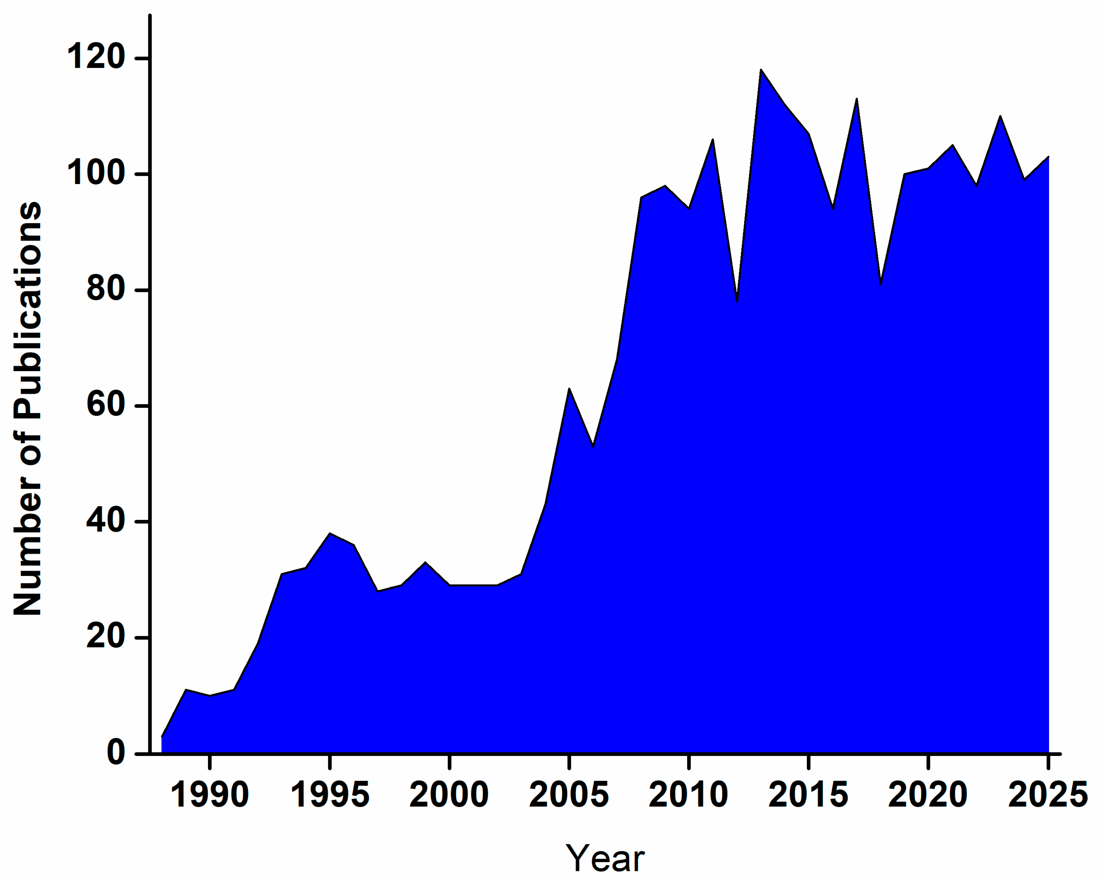
Figure 1. Year-wise distribution of journal articles on glucose biosensors based on data from Scopus, Elsevier.
Micromachines 16 01235 g001
Figure 2. (a) Schematic overview and (b) operational mechanism illustrating various generations of electrochemical glucose sensors. (Reproduced with permission from [48]).
Micromachines 16 01235 g002
Figure 3. PEC detection mechanism of glucose by CDs/B-TiO2/Gox. (Reproduced with permission from [52]).
Micromachines 16 01235 g003
Figure 4. Schematic illustration of the one-step immobilization process of GOx onto TiO2 NTAs, enabling the development of a sensitive and efficient glucose biosensor. (Reproduced with permission from [55]).
Micromachines 16 01235 g004
Figure 5. (A) Schematic representation of the fabrication procedure for the “bienzyme” electrode based on TiO2 NTAs. (B) Cyclic voltammograms of the PB/AuNP/TiNT electrode and (C) the pDAB-PB/AuNP/TiNT nanocomposite electrode, measured in air-saturated 0.01 M phosphate-buffered saline (PBS, pH 6.0) at a scan rate of 50 mV s−1. (Reproduced with permission from [58]).
Micromachines 16 01235 g005
Figure 6. Schematic illustration of the fabrication procedure for the Ni/NiTiO3/TiO2 electrode and the photoinduced regeneration mechanism of its surface. (Reproduced with permission from [63]).
Micromachines 16 01235 g006
Figure 7. Electrochemical mechanism of glucose sensing by the CuO/Cu/rGO nanocomposite ATNT electrode at an applied potential of 0.6 V. (Reproduced with permission from [71]).
Micromachines 16 01235 g007
Figure 8. Photoelectrochemical sensing mechanism of the TiO2NTs/Au/Pt/GOx biosensor for glucose detection. (Reproduced with permission from [74]).
Micromachines 16 01235 g008
Figure 9. Illustrative representation of the photoelectrochemical glucose oxidation mechanism at the BiOBr-TNTA electrode. (Reproduced with permission from [75]).
Micromachines 16 01235 g009
Figure 10. The fabrication process of the TiO2 NTs/PDA/N-GQD dual-electron-acceptor biosensing platform. (Reproduced with permission from [81]).
Micromachines 16 01235 g010
Figure 11. Schematic representation depicting the fabrication of Eu(III) complex-functionalized TiO2 NTAs and their fluorescence-based sensing mechanism for glucose, cholesterol, and triglycerides. (Reproduced with permission from [83]).
Micromachines 16 01235 g011
Table 1. Key parameters of TiO2 NT-based biosensing platforms for glucose detection.
Biosensor ConfigurationBiosensor TypeTargetMedium UsedPrimary Detection MethodLinear RangeSensitivityDetection LimitResponse TimeStabilityAnti-Interference PerformanceReference
TiO2/GOxElectrochemical (Enzymatic)GlucosePBSAmperometry0.05 to 0.65 mM199.6 μA mM−1cm−23.8 μMNA86% (after 6 days)Minimal response to ascorbic acid, sucrose, L-cysteine, L-histidine, and L-glycineWang et al. [53]
TiO2/GOxElectrochemical (Enzymatic)GlucosePBSAmperometry0.05 to 3.2 mM8.5 μAmM−1cm−23.2 μM<10 s93.5% (after 40 days)Minimal response to ascorbic acid, uric acid, and dopamine, KCLHu et al. [54]
TiO2/GOxElectrochemical (Enzymatic)GlucosePBSVoltammetry0.03 to 1.0 mM56.60 µA mM−1 cm−28.5 µM10 s90% (after 20 days)Minimal response to ascorbic acid, acetaminophen, and uric acidAkhbari Varkani et al. [55]
Ti/TiO2/Au/PB/GOxElectrochemical (Enzymatic)GlucosePBSAmperometry0.015 to 4.00 mM36 μA mM−15 μM<10 s90% (after 21 days)Minimal response to ascorbic acid, uric acid, acetaminophenBenvenuto et al. [56]
GOx/Ag/TiO2Electrochemical (Enzymatic)GlucosePBSAmperometry0.1 to 4 mM0.39 μA mM−1 cm−20.1 mM40 sNAMinimal response to H2O2Feng et al. [57]
GOx/Au/pDAB)-PB/AuNP/TiO2Electrochemical (Enzymatic)GlucosePBS, SerumAmperometry0.01 to 0.70 mM248 mA M−1 cm−23.2 μM<1 s>90% (after 30 days)Minimal response to ascorbic acid, uric acid, p-acetamidophenolGao et al. [58]
TiO2/CNT/Pt/GOxElectrochemical (Enzymatic)GlucosePBSAmperometry0.006 to 1.5 mM0.24 μA mM−1 cm−25.7 μM<3 s82% (after 30 days)Not specifiedPang et al. [59]
Pt/TiO2Electrochemical (Non-enzymatic)GlucoseH2SO4AmperometryNANANANASelf-cleaning via UV exposure Minimal response to ascorbic acid, uric acid, and p-acetamidophenol Song et al. [60]
Ag/Pt-TiO2Electrochemical (Non-enzymatic)GlucosePBSVoltammetry30 to 180 mM3.99 μA·cm−2·mM−122.6 μMNANAMinimal response to Cl ionWang et al. [61]
Ni-NPs/TiO2Electrochemical (Non-enzymatic)GlucoseNaOH solutionAmperometry0.004 to 4.8 mM700.2 μA mM−1 cm−22 μM<5 s80.3% (after 20 days)Minimal response to ascorbic acid, uric acidYu et al. [62]
Ni/NiTiO3/TiO2Electrochemical (Non-enzymatic)GlucoseSerumAmperometry0.005 to 0.5 mM456.4 μA mM−1 cm−20.7 μM<5 sNAMinimal response to ascorbic acid, uric acidHuo et al. [63]
Ni-DLC/TiO2Electrochemical (Non-enzymatic)GlucoseNaOHAmperometry0.99 to 22.97 mM 1063.78 μA·mM−1·cm−2 0.53 μM<5 s82.6% (after 30 days)Minimal response to dopamine, ascorbic acid, uric acid, and galactoseKang et al. [64]
CuO/TiO2Electrochemical (Non-enzymatic)GlucoseSerumAmperometryUp to 2.0 mM79.79 μA·mM−1·cm−2 1 μM<4 s>90% (after 30 days)Minimal response to Cl ion, ascorbic acid, uric acid, lactose, sucrose, fructose, dopamineLuo et al. [65]
CuO/TiO2Electrochemical (Non-enzymatic)GlucoseSerumAmperometry0.625 to 6.25 m mol L−1; 6.87 to 12.5 m mol L−11836 μA mmol−1 L cm−2 (low range); 1416 μA mmol−1 L cm−2 (high range)3.4 μ mol L−1≤2 s>96% (after 30 days)Minimal response to ascorbic acid, dopamine, galactose, uric acid, lactoseStanley et al. [66]
Cu/TiO2Electrochemical (Non-enzymatic)GlucoseNaOHAmperometry0.5 to 7 mM522 μA mM−1 cm−2NA0.1 sNAMinimal response to ascorbic acid, NaCl, lactose, sucrose, D-fructose Bhanu et al. [67]
Ni-Cu/TiO2Electrochemical (Non-enzymatic)GlucoseNaOHAmperometry10 μM to 3.2 mM1590.9 μA mM−1 cm−25 μM<5 s98% (after 49 days)Minimal response to uric acid, ascorbic acidLi et al. [68]
Co/Cu/TiO2Electrochemical (Non-enzymatic)GlucoseSerum, NaOHAmperometryUp to 12 mM4651.0 μA mM−1 cm−2 up to 5 mM and 2581.70 μA mM−1 cm−2 from 5 mM to 12 mM.0.6 μMNA92% (after 90 days)Minimal response to fructose, maltose, galactose, lactose, ascorbic acid, uric acid, acetamidophenol, creatinine, urea, chlorideSuneesh et al. [69]
Pd NPs/PDDA/TiO2Electrochemical (Non-enzymatic)GlucoseSerumAmperometry4 × 10−7 to 8 × 10⁻⁴ MNA8 × 10−8 M NAStable (after 14 days)Minimal response to chloride ions, ascorbic acid, uric acid, ureaChen et al. [70]
CuO/Cu/rGO/TiO2Electrochemical (Non-enzymatic)GlucosePBSAmperometry0.5 to 16 mM371.6 μA mM−1 cm−222.8 μM~5 s92% (after 5 days)Minimal response to uric acid, ascorbic acid, lactose, sucrose, fructoseChahrour et al. [71]
WO3/TiO2Electrochemical (Non-enzymatic)GlucoseOrange juiceAmperometry1.0 to 6.5 mM1228.12 μA mM−1 cm−20.19 mM2 s97.1% (after 25 days)Minimal response to uric acid, ascorbic acid, NaClKumar & Sinha [72]
Au/TiO2PhotoelectrochemicalGlucoseNaOHVisible red light1 to 90 μM170.37 μA·mM−1·cm−21.3 μMNA96% after 25 daysMinimal response to sucrose, lactose, ascorbic acid, saccharose, fructoseLiu et al. [73]
TiO2/Au/Pt/GOxPhotoelectrochemicalGlucosePBSAmperometry (visible light)0 to 4 mM81.93 μA mM–1 cm–21.39 μMNANAMinimal response to NaCl, sucrose, ascorbic acid, uric acid, galactose, fructoseYang et al. [74]
BiOBr/TiO2PhotoelectrochemicalGlucoseSerum, NaOH)Amperometry (visible light)5 × 10² to 3 × 10⁷ nMNA10 nMNA>95% (after 28 days)Minimal response to ascorbic acid, uric acid, urea, dopamineWu et al. [75]
CuO/TiO2PhotoelectrochemicalGlucoseHuman sweatVisible light1 to 200 μM (sweat)/0.5 to 10 mM (blood)138.5 μA·mM−1·cm−2 0.7 μM<1 sNAMinimal response to NaCl, KCl, dopamine, uric acid, lactic acid, ascorbic acidKe et al. [76]
3D CuO/TiO2/TiPhotoelectrochemicalGlucoseSerumVisible light70 to 900 μM155 μA·mM−1·cm−220 μMNANAMinimal response to fructose, lactose, sucrose, dopamine, ascorbic acid, uric acid, and L-cysteineYang et al. [77]
GOx/g-C3N4-TiO2/ITOPhotoelectrochemicalGlucoseSerumVisible light0.05 to 16 mM16.7 µA mM−1 cm−20.01 mMNA90.5% (after 14 days)Minimal response to ascorbic acid, uric acid, dopamine, fructose, lactose, and sucrose.Liu et al. [78]
CdxZn1-xS/TiO2PhotoelectrochemicalGlucosePlasma, NaNO3Amperometry (UV light)0.014 to 214 mM1331.7 μA mM−1 cm−20.225 μMNA82% (after 35 days)Minimal response to ascorbic acid, uric acid, dopamine, urea, lysine, tyrosine, histidineEsmaeili et al. [79]
CdSe/TiO2PhotoelectrochemicalGlucoseSerumVisible light10 to 90 μMNA3.1 μMNANAMinimal response to ascorbic acid, uric acid, urea, fructose, xyloseLi et al. [80]
TiO2/PDA/N-GQDs/GOxPhotoelectrochemicalGlucosePBSAmperometry (visible light)Up to 11 mM13.6 μA mM−1 cm−20.015 mM <1 s86.95% (after 30 days)Minimal response to NaCl, sucrose, ascorbic acid, uric acid, dopamineYang et al. [81]
Nafion/GOx/Pt-NPs/TiO2Organic Electrochemical TransistorGlucosePBS, SerumAmperometry100 nM to 5 mM0.09 NCR/1μM100 nMNA90% (after 10 days)Minimal response to ascorbic acid, uric acidLiao et al. [82]
Eu(III) complex/TiO2Fluorescence (Optical)GlucoseOrange juiceFluorescence intensity change0 to 15 mmol/LNA1.02 mmol/LNANAMinimal response to urea, fructose, sucrose, galactoseSu et al. [83]
Table 2. Comparison of TiO2-based glucose sensor types.
FeatureEnzymatic SensorsNon-Enzymatic SensorsPhotoelectrochemical (PEC) Sensors
Sensing PrincipleUses an enzyme, such as glucose oxidase (GOx), for specific biorecognition of the target analyte. The biological event is then converted into a measurable signal by a transducer, often electrochemical.Uses an electrode material (e.g., noble metal, metal oxide, or carbon nanomaterial) to directly catalyze the oxidation or reduction in the target analyte.Combines light excitation and an electrical signal readout. A photoactive material converts an optical signal into a measurable electrical current or voltage.
StrengthsHigh selectivity and sensitivity: Enzymes provide excellent specificity for their target molecules, resulting in high accuracy.
Rapid response: Enzymatic reactions can provide a fast response time.
High stability and long-term lifespan: Not dependent on biological components, so they are more robust against environmental factors like temperature and pH.
Lower cost: Enzymes are often expensive to produce and immobilize, so non-enzymatic sensors have lower manufacturing costs.
Simpler fabrication: Avoids the complex and delicate process of immobilizing enzymes.
High sensitivity: Separating the optical excitation and electrical detection minimizes background noise, leading to very high sensitivity and low detection limits.
High signal-to-noise ratio: The separation of the input light signal and output electrical signal allows for reduced noise and drift.
Low background signal: Inherently low background current enables sensitive detection.
Miniaturization: Relies on simple light sources and electrodes, allowing for smaller, more portable devices.
WeaknessesLow stability: Enzymes can denature due to changes in temperature, pH, or exposure to organic solvents, which reduces sensor lifespan.
High cost: Enzymes are costly to source, purify, and immobilize.
Complex immobilization: Ensuring the enzyme remains active and securely attached to the electrode is a complicated process.
Oxygen dependence: Early generations of sensors were limited by the availability of oxygen, though this has been addressed in later generations.
Lower selectivity: Catalytic materials are not as specific as enzymes, leading to potential interference from other electroactive species in the sample.
High working potential: Some non-enzymatic sensors require a high potential, which can increase interference.
Surface fouling: The electrode surface can become blocked or “poisoned” by intermediate oxidation products, which degrades performance over time.
Sensitivity can be lower: It remains a challenge to achieve sensitivity levels comparable to enzymatic sensors.
Limited material choice: Performance is highly dependent on the photoactive material used, which can have intrinsic limitations like a narrow light absorption range.
Complex systems: The use of nanomaterials and heterojunctions can increase complexity, which may affect scalability.
Potential for toxicity: Some photoactive materials, like early quantum dots, have toxicity concerns, though research is shifting to more biocompatible options.
Developmental stage: PEC technology is still an emerging field, and widespread commercialization is not yet realized for many applications.
Disclaimer/Publisher’s Note: The statements, opinions and data contained in all publications are solely those of the individual author(s) and contributor(s) and not of MDPI and/or the editor(s). MDPI and/or the editor(s) disclaim responsibility for any injury to people or property resulting from any ideas, methods, instructions or products referred to in the content.