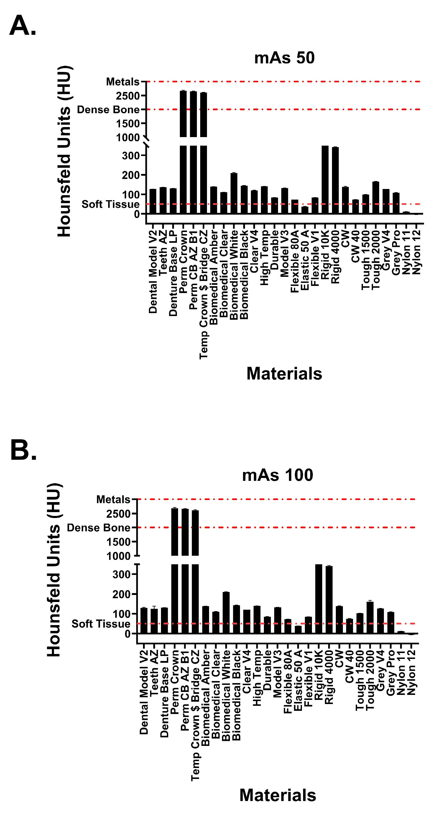
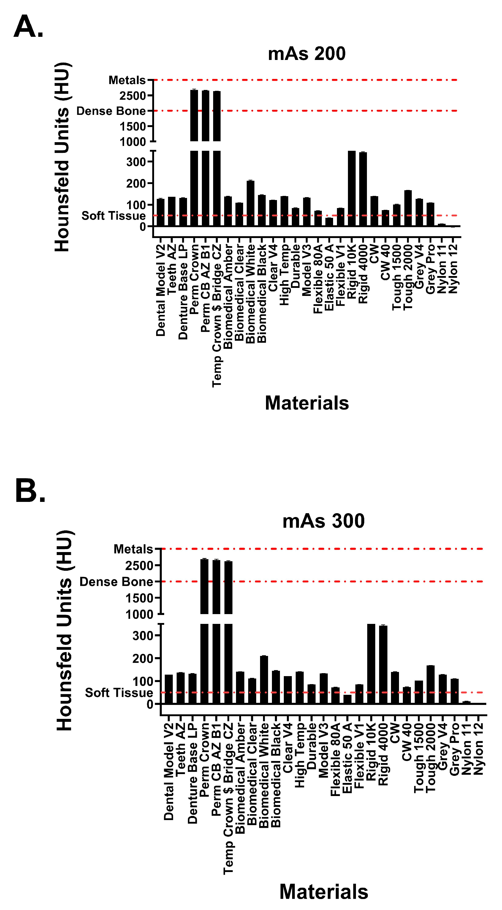
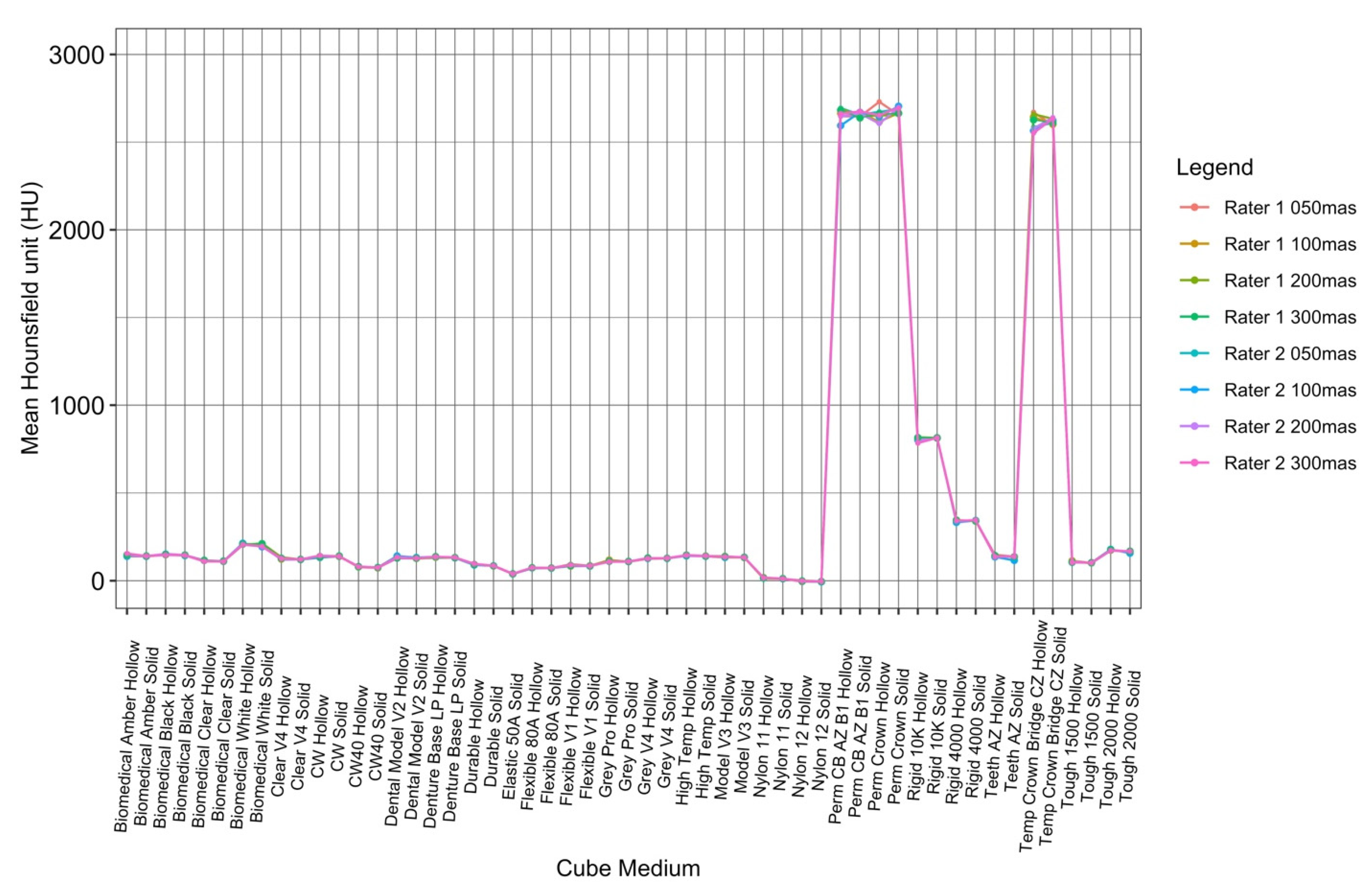
Computed Tomography Attenuation of Three-Dimensional (3D) Printing Materials—Depository to Aid in Constructing 3D-Printed Phantoms

 , , ,
, , , Abstract
Share and Cite
Kalidindi, Y.; Ganapathy, A.K.; Nayak, Y.; Elumalai, A.; Chen, D.Z.; Bishop, G.; Sanchez, A.; Albers, B.; Shetty, A.S.; Ballard, D.H. Computed Tomography Attenuation of Three-Dimensional (3D) Printing Materials—Depository to Aid in Constructing 3D-Printed Phantoms. Micromachines 2023, 14, 1928. https://doi.org/10.3390/mi14101928
Kalidindi Y, Ganapathy AK, Nayak Y, Elumalai A, Chen DZ, Bishop G, Sanchez A, Albers B, Shetty AS, Ballard DH. Computed Tomography Attenuation of Three-Dimensional (3D) Printing Materials—Depository to Aid in Constructing 3D-Printed Phantoms. Micromachines. 2023; 14(10):1928. https://doi.org/10.3390/mi14101928
Chicago/Turabian StyleKalidindi, Yuktesh, Aravinda Krishna Ganapathy, Yash Nayak, Anusha Elumalai, David Z. Chen, Grace Bishop, Adrian Sanchez, Brian Albers, Anup S. Shetty, and David H. Ballard. 2023. "Computed Tomography Attenuation of Three-Dimensional (3D) Printing Materials—Depository to Aid in Constructing 3D-Printed Phantoms" Micromachines 14, no. 10: 1928. https://doi.org/10.3390/mi14101928
APA StyleKalidindi, Y., Ganapathy, A. K., Nayak, Y., Elumalai, A., Chen, D. Z., Bishop, G., Sanchez, A., Albers, B., Shetty, A. S., & Ballard, D. H. (2023). Computed Tomography Attenuation of Three-Dimensional (3D) Printing Materials—Depository to Aid in Constructing 3D-Printed Phantoms. Micromachines, 14(10), 1928. https://doi.org/10.3390/mi14101928