Review

Recent Advances in Microfluidic Devices for Contamination Detection and Quality Inspection of Milk

1
Division of Nephrology, Kaohsiung Chang Gung Memorial Hospital, College of Medicine, Chang Gung University, Kaohsiung 833, Taiwan
2
Department of Emergency Medicine, Kaohsiung Chang Gung Memorial Hospital, College of Medicine, Chang Gung University, Kaohsiung 833, Taiwan
3
Department of Engineering Science, National Cheng Kung University, Tainan 701, Taiwan
*
Author to whom correspondence should be addressed.
These authors contributed equally to this paper.
Micromachines 2021, 12(5), 558; https://doi.org/10.3390/mi12050558
Submission received: 16 April 2021 / Revised: 5 May 2021 / Accepted: 11 May 2021 / Published: 14 May 2021
(This article belongs to the Special Issue Microfluidic-Based Approaches for Detection in Water and Food Samples)

Abstract

:
Milk is a necessity for human life. However, it is susceptible to contamination and adulteration. Microfluidic analysis devices have attracted significant attention for the high-throughput quality inspection and contaminant analysis of milk samples in recent years. This review describes the major proposals presented in the literature for the pretreatment, contaminant detection, and quality inspection of milk samples using microfluidic lab-on-a-chip and lab-on-paper platforms in the past five years. The review focuses on the sample separation, sample extraction, and sample preconcentration/amplification steps of the pretreatment process and the determination of aflatoxins, antibiotics, drugs, melamine, and foodborne pathogens in the detection process. Recent proposals for the general quality inspection of milk samples, including the viscosity and presence of adulteration, are also discussed. The review concludes with a brief perspective on the challenges facing the future development of microfluidic devices for the analysis of milk samples in the coming years.

1. Introduction

Milk is one of the main sources of nutrients necessary for human growth and maintenance of health. As such, it is the most important dietary product for all age groups. However, milk is also a major transmission source for contaminants, such as pathogens, antibiotics, heavy metals, pesticides, and drugs, which can cause various adverse physiological changes and have a serious impact on human health, particularly in infants and young children [1]. Consequently, effective methods for detecting the presence of contaminants, or adulterants, are essential. The quality and safety of milk are generally tested at the downstream processing plant using high-precision instruments and systems. However, while these systems are highly reliable, they tend to be expensive and bulky, such as automatic milk composition analyzer, automatic real-time microbial counting analyzer, etc. Moreover, they require the involvement of professional technical staff. Consequently, the development of microfluidic analysis devices capable of performing the testing process on-site at the point of production has attracted significant interest in recent years.
Microfluidic technology has advanced rapidly over the past decade and has resulted in the development of many lab-on-a-chip [2,3,4,5,6,7,8] and lab-on-paper [9,10,11,12,13,14,15,16] platforms for total analysis systems in fields such as medicine and healthcare, environmental monitoring, cell biology, food safety inspection, and so on. Both platforms offer the advantages of cost-effectiveness, a simple operation, high reliability, high sensitivity, low sample/reagent consumption, and a near-instant on-site detection capability. However, in terms of the transport theory and driving mechanisms used by the two systems, they are very different. In particular, the substrates in lab-on-a-chip devices are generally fabricated of glass, PMMA (poly(methyl methacrylate)), and PDMS (polydimethylsiloxane), and are patterned using photolithography, etching, or laser ablation techniques. Moreover, the working fluid is transported by externally produced pressure or electrical forces [17,18,19,20]. By contrast, in lab-on-paper devices, the substrates are fabricated of paper, and hydrophilic microchannels are defined by printing, wax dipping, cutting, photolithography, or spraying techniques. Furthermore, the sample and buffer solutions flow naturally within the microchannels under the effects of capillary action alone, without the need for any external driving source [21,22,23,24].
The most important purpose of developing microfluidic lab-on-a-chip or lab-on-paper devices is to provide end users with rapid, accurate, easy-to-operate, and cost-effective on-site analysis tools. However, the selection and integration of appropriate detection systems must also be carefully considered. In practice, the choice of detection system depends largely on the limit of detection (LOD) requirements of the target application. For example, the detection of heavy metal residues in milk requires high-sensitivity detection with a LOD on the order of ppb. Such applications generally require the use of high-resolution detection systems such as fluorescence, electrochemistry (EC), electrophoresis (EP), surface-enhanced Raman spectroscopy (SERS), and so on [25,26,27]. However, many alternative quantitative detection systems have also been proposed in recent years, including colorimetry, spectrometry, chemiluminescence (CL), photoelectrochemistry (PEC), and electrochemiluminescence (ECL) [25,28,29]. In general, all of these methods provide a reliable, simple, miniaturized, easily integrated, and cost-effective solution for analyte detection and determination in many different application areas.
This review describes developments in the microfluidic platform technology field for the analysis of milk samples in the past five years. The review commences by describing the methods proposed for sample pretreatment in microfluidic devices, including the separation, sample extraction, and amplification steps. An in-depth review of the latest advances in the use of microfluidic platforms for the detection of aflatoxins, antibiotics, drugs, melamine, and foodborne pathogens in milk samples is then presented. Recent proposals for milk quality inspection and adulterant detection are then briefly described. The review concludes with a brief perspective on the challenges facing the microfluidic platform development field in the dairy business over the coming years, together with the associated opportunities for future research.

2. Sample Pretreatment of Microfluidic Devices

The difficulty of sample pretreatment/preparation in food testing is much higher than that for human biological samples. For example, whole blood analysis and diagnosis tests usually require only centrifugation to separate the blood cells and plasma for detection [30]. By contrast, when detecting acidic preservatives in food samples, for example, it is necessary to homogenize the sample first and then extract the test substance by steam distillation before detection can be performed [31,32]. As a result, sample processing in traditional food detection methods usually involves a high cost, a long process, and the use of multiple equipment. This section therefore reviews the pretreatment methods proposed in the literature for milk sample analysis using compact and low-cost microfluidic devices. Broadly speaking, pretreatment methods on microfluidic platforms fall into three main categories, namely separation, extraction, and concentration/amplification.

2.1. Sample Separation Microfluidic Devices

In food testing, the sample usually has a complex composition consisting of fats, proteins, and many other components. As a result, it is generally necessary to separate the sample prior to the detection process in order to avoid possible interference with the detection of the target analyte. Singh et al. [33] presented a hand-held microfluidic device incorporating a gold-coated glass slide for the separation of microbes from model solutions and complex matrices. The feasibility of the proposed device was demonstrated by capturing the microbial cells in a yoghurt sample. The results showed that the device captured up to 99.3% of the cells within 60 min. Many other researchers have presented microfluidic platforms for the separation of milk samples in recent years, including antibiotics and toxins [34], quinolone drugs [35], and foodborne bacteria [36]. For instance, Jung et al. [36] developed a magnetophoresis-based microfluidic device for the rapid separation and concentration of foodborne bacteria consisting of a polyethylene tube wrapped around a magnet (see Figure 1a). It was shown that an efficient sample separation performance could be achieved even under high flow rates simply by increasing the length of the tube. For example, when applied to the analysis of milk and homogenized cabbage samples, the device showed a separation efficiency of more than 92% at a flow rate of 40 mL/h.

2.2. Sample Extraction Microfluidic Devices

In developing microfluidic devices for the analysis of food samples, one of the key requirements is that of developing miniaturized extraction techniques compatible with lab-on-a-chip and lab-on-paper platforms. The literature contains many proposals for the extraction of various substances in milk samples, including antibiotics [37] and foodborne pathogens [38,39,40,41]. For example, Park et al. [39] presented an integrated rotary microfluidic system for performing the extraction and amplification of foodborne pathogens. As shown in Figure 1b, the system was fabricated using PC (polycarbonate) and PMMA substrates and was designed to perform three functions, namely solid-phase DNA extraction, loop-mediated isothermal amplification (LAMP), and lateral flow strip paper detection. In the proposed device, the extraction step was performed using a solid phase DNA extraction method based on the use of silica bead as a solid matrix. The experimental results showed that the proposed platform was capable of detecting Salmonella (S.) Typhimurium and Vibrio (V.) parahaemolyticus bacteria in contaminated milk samples with a LOD of 50 CFU (colony-forming unit) within 80 min.

2.3. Sample Amplification Microfluidic Devices

In food analysis samples, the target analyte is typically present in only very small quantities. Consequently, some form of sample amplification system must be employed in order to improve the LOD performance. Several microfluidic platforms with integrated amplification systems have been proposed for the detection and analysis of milk based on polymerase chain reaction (PCR) [42,43,44], LAMP [45,46], hybridization chain reaction (HCR) [47], and catalyzed hairpin assembly (CHA) [48]. For example, Li et al. [47] presented an HCR amplification microfluidic aptasensor for the detection of E. coli O157:H7 in milk samples. As shown in Figure 1c, an HCR initiator and aptamer were deposited on the substrate, and the stronger binding ability of the pathogen with the aptamer was then exploited to dissociate the aptamer from the initiator and trigger a series of hybridization events to achieve sample amplification. It was shown that the aptasensor had good specificity for the detection of E. coli O157:H7 and amplified the signal by around 100 times compared to that of the original (non-amplified) sample.

3. Microfluidic Platforms for Milk Sample Analysis

Generally speaking, fresh milk consists of 87% water and 13% total solids (including 4% fat, 3.4% protein, 4.8% lactose, and 0.8% minerals). While the exact quantity of the main components may vary depending on the individual animal, lactation stage, breed, age, and health status, the overall composition does not change very much. As described in Section 1, milk provides many of the essential nutrients required to support human development and growth. However, it is prone to contamination and adulteration, and hence strict quality control and inspection measures are required to safeguard human health. This function is traditionally performed using large-scale, professional equipment in a laboratory environment. However, to reduce costs and enable a more immediate detection result, it is desirable to analyze the milk quality at the point of production using simple, low-cost methods. As a result, the development of microfluidic platforms for the detection and analysis of milk samples has attracted significant interest in recent years [49,50,51]. This section of the review describes some of the most significant lab-on-a-chip and lab-on-paper proposals reported in the literature over the past five years for the detection of some of the most common contaminants in milk, namely aflatoxins, antibiotics, drugs, melamine, and foodborne pathogens. The section concludes by describing several other recent microfluidics-based proposals for general milk quality inspection and adulterant detection.

3.1. Aflatoxin Analysis

Aflatoxins are toxic carcinogens and mutagens produced by certain fungi or molds. They can pose serious health risks to humans, including developmental delay, liver damage, and liver cancer [1,52]. Consequently, reliable methods for their detection in foods and beverages are urgently required. While there are many different types of aflatoxins naturally occurring in nature, two of the most common in contaminated milk samples include aflatoxin B1 (AFB1; reference values less than 0.5 μg/L) and aflatoxin M1 (AFM1; reference values less than 0.5 μg/L) (European commission regulation (EC) no. 1881, 2006) [1]. The recent literature thus contains many microfluidic lab-on-a-chip and lab-on-paper devices for the detection of AFB1 in milk [53,54,55] and AFM1 [56,57,58,59] in milk samples. For example, Sheini [54] developed a microfluidic lab-on-paper colorimetric aggregation assay sensor array for the detection of five mycotoxins classified into three categories, namely aflatoxins, ochratoxins A (OTA), and zearalenone. As shown in Figure 2a, multiple hydrophilic zones were patterned on the paper substrate using an ink printer and the zones were then coated with gold and silver nanoparticles (AuNPs and AgNPs) and different reducing agents in order to form a sensing array. The color change induced by the presence of mycotoxins was then analyzed in order to determine the corresponding toxin concentration. The experimental results showed that the platform achieved detection limits of 2.7, 7.3, 2.1, 3.3, and 7.0 ng/mL for AFB1, AFG1, AFM1, OTA, and zearalenone, respectively. Furthermore, the detection results obtained for AFM1 in milk samples were shown to be consistent with those obtained using a conventional high-performance liquid chromatography (HPLC) detection method [54]. Soares et al. [55] presented a multiplexed microfluidic lab-on-a-chip fluorescence immunoassay sensor for the rapid detection of AFB1, OTA, and deoxynivalenol (DON) in food samples. The device consisted of a PDMS substrate patterned with a sample injection microchannel (20 μm height) and four microbead-filled microchannels (100 μm height), and an integrated array of thin-film photodiodes. In the detection process, the microbeads served as the binding medium between the antibody and the target analyte and the analyte concentration was determined using a fluorescence signal acquisition method. When applied to the detection of AFB1, OTA, and DON, the corresponding LODs were found to be 1, 3, and 10 ng/mL, respectively.
Aissa et al. [56] developed a microfluidic lab-on-paper aptasensor device for the detection of AFM1 in milk samples based on electrochemical capacitance spectroscopy (ECS). In the proposed device, carbon screen-printed working electrodes were prepared on the substrate surface and the electrodes were modified with silicon nanocomposite to enhance the capacitance signal. Finally, an anti-AFM1 aptamer was attached to the working electrode to provide a sensing function. The device was shown to achieve a linear ECS response over the AFM1 concentration range of 10 to 500 fM and a LOD of 4.53 fM. He et al. [34] presented a microfluidic lab-on-a-chip aptasensor based on magnetic DNA nanostructure probes for kanamycin (KANA), AFM1, and 17β-estradiol (E2) detection in milk samples. The detection process was implemented using Au-Fe3O4 magnetic complex as a probe, and the target captured by the aptamer on the probe was separated magnetically and transported to a microfluidic electrophoresis chip (MEC) for further analysis by a laser-induced fluorescence (LIF) detection system. It was shown that the device achieved LODs of 0.32, 0.95, and 6.8 pg/mL for KANA, AFM1, and E2 respectively, with a response time of less than three minutes. Table 1 briefly reviews several other microfluidic devices proposed in recent studies for the detection of aflatoxin and other contaminants in milk samples.

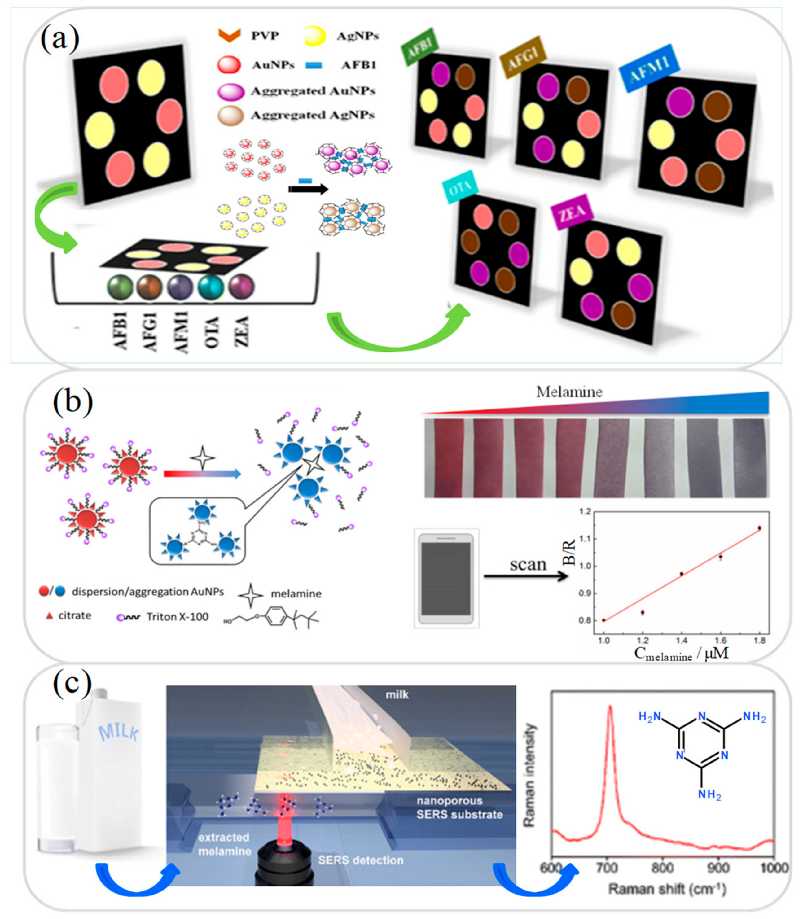
3.2. Melamine Analysis

Melamine is an organic compound with a high nitrogen content and is often added illegally to food in order to increase its apparent protein content. However, melamine is toxic and can cause many serious health issues, including reproduction damage, bladder infection, kidney stones, acute kidney failure, bladder cancer, and infant death [60]. The detection of melamine in milk samples (non-detectable) requires an extremely high resolution, and hence recent microfluidic devices for melamine analysis generally use high-sensitivity detection techniques such as electrochemistry (EC) [61,62], colorimetric AuNP sensing [63,64], surface-enhanced Raman scattering (SERS) [65,66], and immunosensor [67,68]. For example, Li et al. [62] developed a microfluidic EC DNA-based sensor platform for continuous, real-time melamine detection in whole milk samples. The EC electrode of the biosensor was modified with nucleobase thymine, which subsequently reacted with the melamine in the milk sample to form a DNA triplex. The resulting conformational change produced a corresponding shift in the EC current, from which the melamine concentration was then inversely derived. The results obtained using spiked milk samples showed that the DNA-based device achieved a melamine LOD performance of 2.5 ppm. Gao et al. [63] presented a microfluidic colorimetric lab-on-paper sensor for the detection of melamine in milk samples based on Triton X-100-modified AuNPs. As shown in Figure 2b, Triton X-100 was added to citrate-capped AuNPs to produce a dispersible and stable Triton X-100-AuNP complex. In the presence of melamine, a ligand exchange occurred, which resulted in the release of some of the Triton X-100 from the AuNPs and the subsequent formation of AuNP agglomerates. The resulting color change was then used to determine the corresponding melamine concentration. The results showed that the device achieved a LOD of just 5.1 nM.
Krafft et al. [66] developed a microfluidic lab-on-a-chip device combined with a SERS detector for the detection of melamine in whole milk samples. As shown in Figure 2c, the device consisted of a nanoporous membrane designed to both enrich the sample through a selective electrokinetic filtering process and facilitate SERS through the use of a AgNP layer on its under surface. The experimental results showed that the device achieved a detection limit of 1 ppm. However, it was speculated that the detection performance could be further improved by on-target sample drying using an additional gas flow. Chen et al. [69] presented a centrifugal microfluidic lab-on-a-chip immunoassay device for the detection of six contaminants’ residues in milk samples, namely chloramphenicol (CAP), tetracycline (TC), enrofloxacin (ENR), cephalexin (CEX), sulfonamides (SAs), and melamine. In the immunoassay process, the mixture in the sample chamber was centrifuged into the reaction chamber (containing antibody solution) and two affinity and binding operations were then performed to complete the immunoassay reaction. It was shown that the device allowed the determination of CAP, TC, ENR, CEX, SAs, and melamine with LODs of 0.92, 1.01, 1.83, 1.14, 1.96, and 7.80 μg/kg, respectively. Furthermore, the total reaction time was less than 17 min. Table 1 briefly reviews several other microfluidic devices proposed in recent studies for the detection of melamine and other contaminants in milk samples.

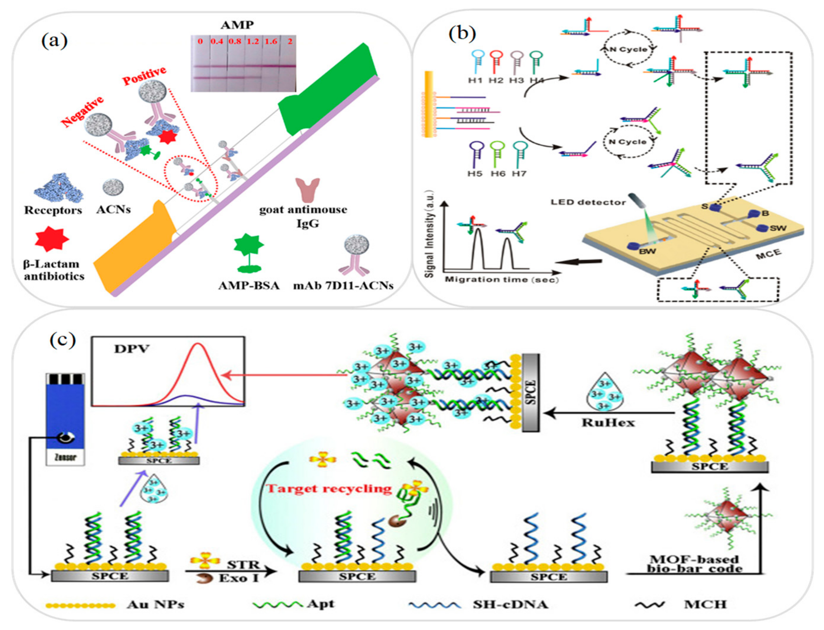
3.3. Antibiotic and Drug Analysis

In large-scale dairy production, the cows are often injected with drugs and antibiotics in order to increase the milk yield. However, residues from these drugs and antibiotics may have a harmful effect on human health. A similar problem can arise in the use of certain animal feeds and environmental medications [70]. Consequently, the literature contains many microfluidic devices for the detection of antibiotics and drugs, such as ampicillin [71], β-lactamase [72,73], 17β-estradiol [74], bacitracin zinc [75], chloramphenicol [76,77], clarithromycin [78], clenbuterol [79], ciprofloxacin [80], enrofloxacin [81], monensin [82], norfloxacin [83], streptomycin [84], and tylosin and tilmicosin [85].
Zhang et al. [73] developed a microfluidic lab-on-paper lateral flow immunoassay device for the detection of β-lactams in milk samples. As shown in Figure 3a, the immunoassay platform used amorphous carbon nanoparticles (ACNs) to label anti-receptor monoclonal antibodies (mAb) to form a complex with the unlabeled β-lactam receptors. The results obtained for the detection of 22 β-lactams showed that the measurement sensitivity achieved using ACNs was around 2 times better than that using the same immunoreagents but with colloidal gold as indirect labeling receptors. Furthermore, the sensitivity was around 10 times better than that obtained without using any labeling at all. Zhang et al. [76] presented a microfluidic MCE aptasensor for the detection of chloramphenicol and kanamycin in milk and fish samples. As shown in Figure 3b, the device used a stir bar coated with AuNPs to facilitate DNA multi-arm junctions recycling (MAJR) for signal transduction and amplification purposes. In the detection process, the target antibiotics reacted specifically with the aptamer probes on the stir bar to produce different single-stranded DNA primers. The primers then triggered the MAJR process to connect the three-arm and four-arm DNA strands of the different targets. Finally, the multi-arm DNA was separated by MCE for quantitative detection. It was shown that the device achieved LODs of 0.52 and 0.41 pg/mL for chloramphenicol and kanamycin, respectively.
Zeng et al. [78] developed a microfluidic lab-on-paper immunochromatographic assay device for the rapid detection of four macrolides in milk samples, namely clarithromycin, erythromycin, roxithromycin, and azithromycin. In performing the detection process, antigen and goat anti-mouse IgG antibody were first coated on an immune paper strip as the test (T) line and control (C) line, respectively. Monoclonal antibody was then labeled with colloidal gold in the detection solution, and sample solution was added to prompt the antibiotics to react with the monoclonal antibody. Finally, the mixed solution consisting of the target antibodies and the monoclonal antibodies was dropped onto the previously prepared immune paper strip. The antibodies and colloidal gold-labeled monoclonal antibodies then underwent a competitive interaction with the immunochromatographic band to produce a color change from which the antibiotic concentration was determined. The microfluidic device was shown to be capable of detecting clarithromycin, erythromycin, roxithromycin, and azithromycin in milk samples with LODs of 0.095, 0.085, 0.055, and 0.175 ng/mL, respectively. Meng et al. [84] presented a microfluidic EC aptasensor for the detection of streptomycin in milk samples. As shown in Figure 3c, AuNPs were first coated on the working electrode, and monolayer thiolated cDNA/aptamer duplex (dsDNA) and 6-mercapto-1-hexanol (MCH) were then fixed on the AuNPs to modify the working electrode (Au/SPCE). Finally, a metal-organic framework (MOF) bio-bar code and Exo I enzyme were used to assist the target cycle for dual-signal amplification and achieve a high-sensitivity streptomycin detection performance. The results showed that the aptasensor provided a good linear response over the streptomycin concentration range of 0.005~150 ng/mL and achieved a LOD of 2.6 pg/mL. Table 2 briefly reviews several other microfluidic devices proposed in recent studies for the detection of antibiotics and drugs in milk samples.

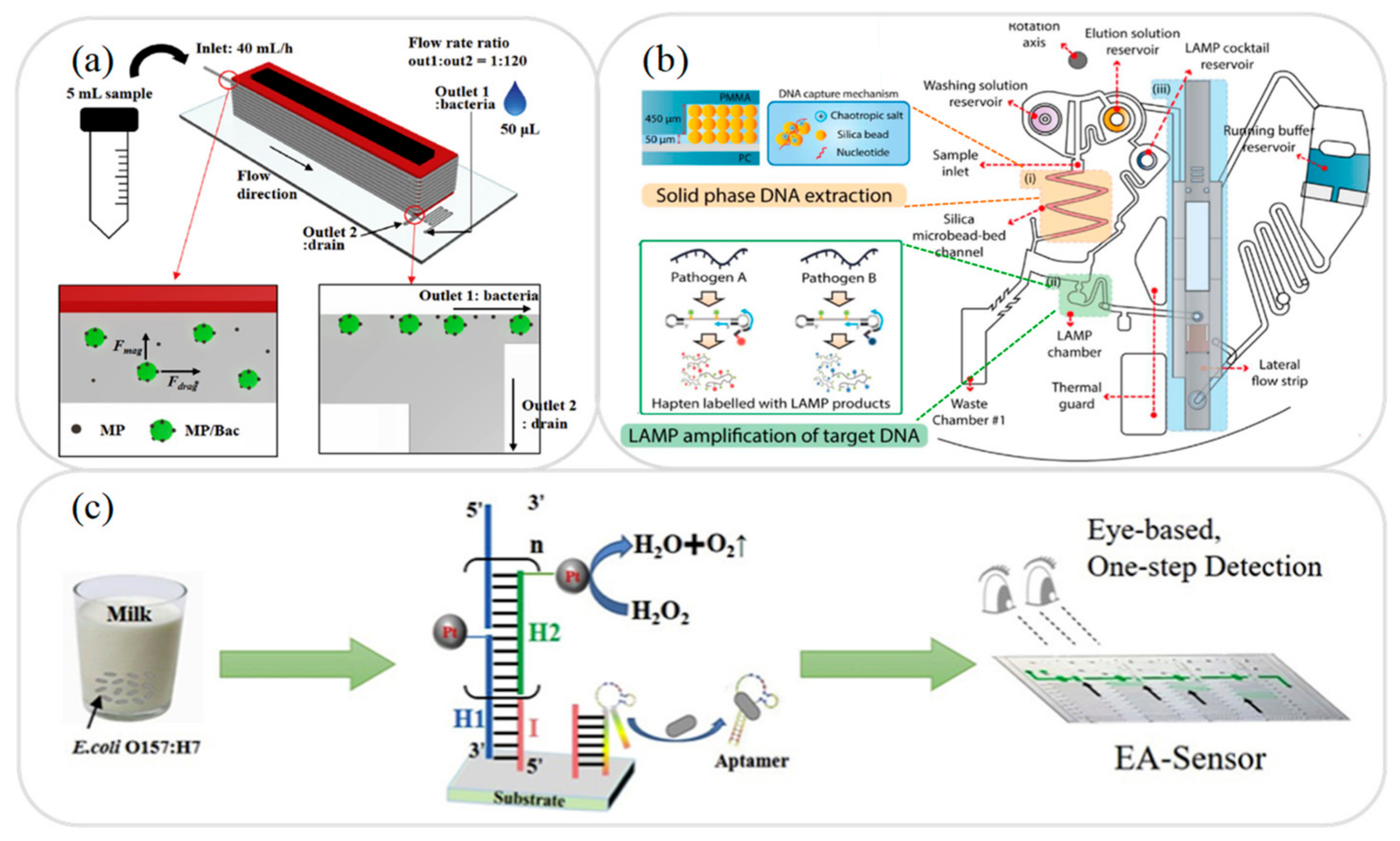
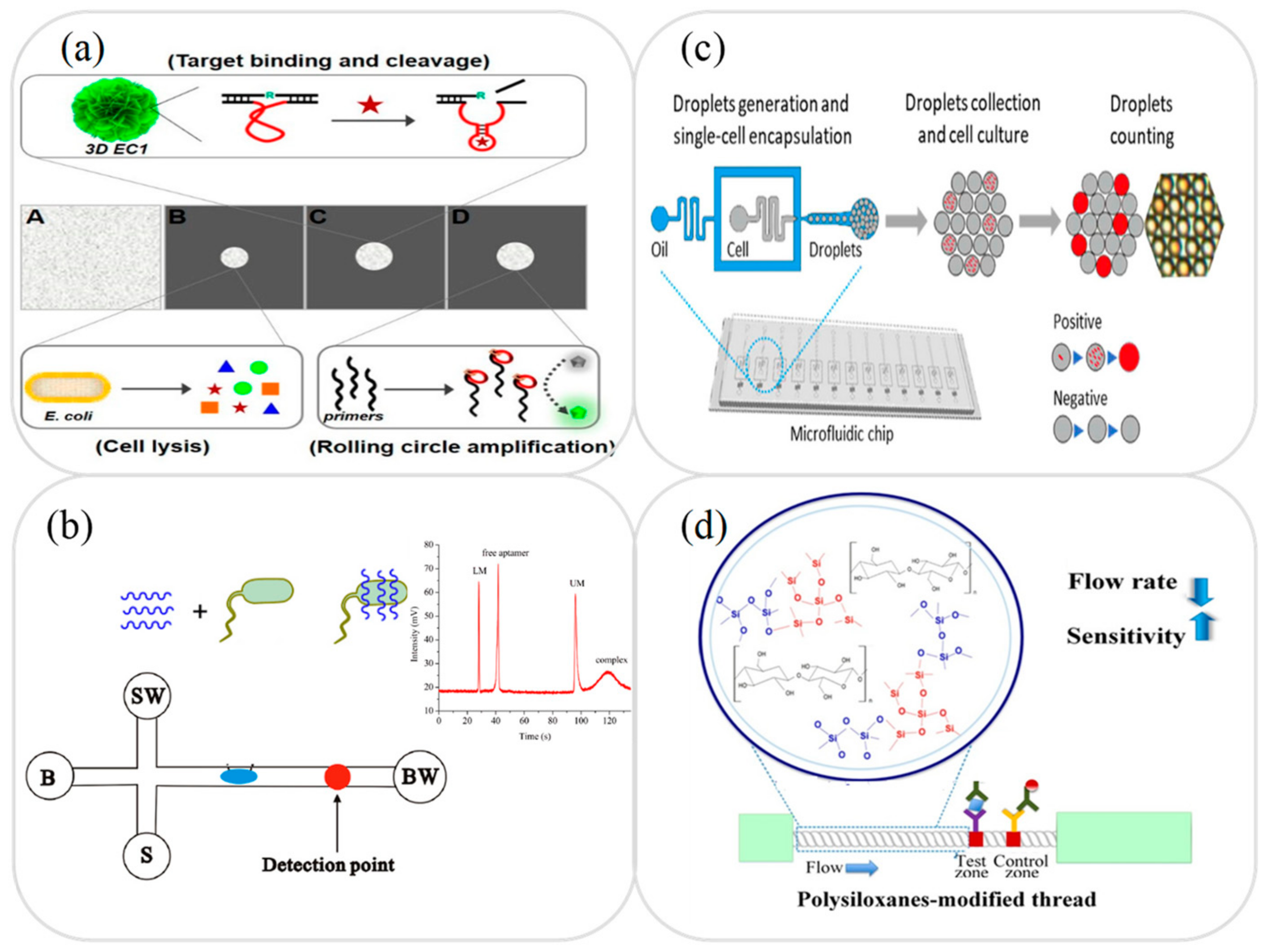
3.4. Foodborne Pathogens Analysis

The contamination of milk by pathogens is one of the main causes of human health problems and gastrointestinal infectious diseases. Pathogenic bacteria grow and reproduce rapidly within milk and metabolize to produce all manner of toxins. Although the pathogens in milk are killed in the downstream treatment process, the toxins remain active and can cause serious health issues if consumed. Among the many pathogenic bacteria, Staphylococcus (S.) aureus, Streptococcus, Escherichia (E.) coli, Listeria, Salmonella, Vibrio (V.) vulnificus, and Bacillus anthracis are among the most common [86]. Many of these bacteria are pathogens of bovine mastitis, one of the most widespread and economically costly diseases in the dairy industry. Traditional detection methods for foodborne bacteria involve sample culturing using an appropriate medium. However, while these methods have a high sensitivity, a low cost, and good accuracy, they are time-consuming, laborious, and must be performed in a laboratory setting. To address these problems, various alternative techniques for pathogen detection in milk have been proposed in recent years, including PCR, LAMP, enzyme-linked immunosorbent assay (ELISA), and microarray-based systems [87,88,89]. However, these methods still require the use of laboratory equipment and skilled technical personnel. Thus, the problem of developing microfluidic platforms for the on-site detection of common foodborne pathogens in milk samples has attracted great interest in the recent literature. Typically, these platforms have been designed for the detection of E. coli [86,90,91,92,93], S. aureus [94,95,96], Listeria [97,98,99], and Salmonella [100,101,102,103,104].
E. coli is a Gram-negative bacterium which lives mainly in the human intestine. In mild cases, it can cause symptoms such as diarrhea, gastroenteritis, inflammation, and malnutrition. However, in severe cases, it can cause more serious problems such as sepsis and hemolytic uremic syndrome [105]. Various microfluidic platforms have been developed for the detection of E. coli in milk samples, including lab-on-a-chip devices combined with immunoassay [78], aptamer and fluorescence detection [76], rolling circle amplification (RCA), and fluorescence detection [91], and lab-on-paper devices based on RCA and colorimetric detection [105], colorimetric detection [86,106], and immunoassay [90,92,93]. Sun et al. [105] developed a folding microfluidic lab-on-paper RCA device with DNAzyme-containing DNA superstructures for E. coli detection in juice and milk samples. As shown in Figure 4a, the device first extracted the protein molecules from the cells in the sample and then used a DNA superstructure to immobilize the RNA and produce a target-induced cleavage of the DNAzymes. Finally, isothermal RCA was performed to replicate the E. coli pathogens. The experimental results showed that the proposed device allowed the detection of E. coli K12 with a LOD of 103 CFU/mL within 35 min. Zhang et al. [107] presented a microfluidic MCE device based on a specific aptamer binding strategy for detecting the concentration of E. coli in drinking water and milk samples. In the proposed device, the target pathogen initially combined with SYBR gold-labeled aptamer to form a complex. The free aptamer was then separated from this complex by capillary electrophoresis and detected using a LIF technique (see Figure 4b). The LOD of the microfluidic device was shown to be 3.7 × 10² CFU/mL.
S. aureus is a Gram-positive bacterium associated with various gastrointestinal infections and dermatitis. The literature contains various proposals for determining the S. aureus concentration in milk samples based on techniques such as optical cell counting, colorimetric detection, EC biosensing, and immunoassays [94,95,96]. For example, Eissa and Zourob [95] developed a microfluidic dual EC/colorimetric platform for the detection of S. aureus in milk and water samples based on a gold electrode sensor coated with a specific peptide sequence coupled with magnetic nanoparticles. In the as-prepared condition, the electrode surface had a black appearance. However, the subsequent reaction between the peptide sequence and the S. aureus protease solution prompted the release of some of the magnetic nanoparticles, causing a partial uncovering of the gold electrode surface. The S. aureus concentration was then determined either qualitatively based on a simple visual inspection of the electrode color by the naked eye, or more precisely by means of square wave voltammetry using an iron/ferricyanide redox pair as the EC detection signal. The LOD of the latter approach was determined to be 3 CFU/mL with a response time of 1 min.
Listeria, also known as Listeria (L.) monocytogenes, is a pathogen of listeriosis and is one of the deadliest foodborne pathogens. Listeria usually uses food as a vector of infection and can cause many serious diseases, including meningitis, sepsis, and miscarriage [99]. Various microfluidic platforms have been proposed for detecting Listeria in milk samples using such techniques as chemiluminescence, colorimetry, immunoassays, EC biosensors, and PCR amplification combined with LIF [97,98,99]. For example, Silva et al. [98] developed a microfluidic EC immunosensor device for the detection of Listeria p60 proteins. In the proposed device, a sandwich immune pair consisting of antibodies for the target protein and Listeria spp. p60 proteins was immobilized on the surface of the electrode and blocked with BSA. The reaction process was then facilitated using an additional secondary antibody and alkaline phosphatase (ALP) with 3-indoxyl phosphate/silver ions. Finally, the concentration of p60 proteins was determined via the voltametric stripping of the enzymatically deposited silver. The LOD of the microfluidic device was found to be 1.5 ng/mL under optimal conditions with a response time of less than 3 h.
Salmonella is a foodborne pathogen with symptoms of dehydration, fever, diarrhea, and stomach cramps within 6–72 h of consumption [108]. Many microfluidic devices have been proposed for the detection of Salmonella in milk samples. Generally speaking, these devices use colorimetric, immunoassay (microchip-based, thread-based, rotary paper-based), EC biosensing, fluorescence, and PCR amplification combined with LIF [100,101,102,103,104]. For example, An et al. [108] presented a microfluidic lab-on-a-chip platform in which a microfluidic droplet device was used to encapsulate single Salmonella cells in a growth medium and the encapsulated droplets were then collected. Finally, the Salmonella concentration was determined by counting the positive sample droplets using a fluorescence detection technique (see Figure 4c). The experimental results obtained using spiked milk samples showed that the device achieved a LOD of 50 CFU/mL. Choi et al. [109] developed a microfluidic thread-based immunoassay device for the detection of Salmonella enterica serotype Enteritidis in food samples. As shown in Figure 4d, the hydrophilicity of the cotton thread was enhanced by using polysiloxane as a pore modifier and antibody-gold nanoparticles (dAb-AuNPs) and anti-mouse IgG were then coated on thread-based strips as the test zone and control zone, respectively. The interaction between the target antigen and the antigen-dAb-AuNPs produced complexes which subsequently bound to the captured antibodies and generated a detection signal with an intensity proportional to the Salmonella concentration. The results showed that the intensity of the signal obtained using the proposed device was around 10 times higher than that produced by an equivalent device without polysiloxane modification. Moreover, the LOD was of the order of 100 CFU/mL. Table 2 briefly reviews several other microfluidic devices proposed in recent studies for the detection of foodborne pathogens in milk samples.

3.5. Other Analysis and Applications

In addition to the analytes described above, microfluidic devices have also been proposed for the testing of various other contaminants or adulterants in milk, including alkaline phosphatase (ALP) [110], preservative H2O2 [111], and heavy metals [69,112]. For example, Mahato and Chandra [110] developed a microfluidic lab-on-paper immunosensor device for the detection of ALP, in which the immunosensor probe was covalently immobilized on the surface of the functionalized paper device by ALP antibody and the interaction between the probe and the ALP produced a blue-green precipitate immune complex as an analysis signal (see Figure 5a). The ALP concentration was then determined either qualitatively by the naked eye or quantitatively via colorimetric detection. The linear range of the microfluidic device for ALP detection was shown to be 10–1000 U/mL with a LOD of 0.87 U/mL. Khoshbin et al. [112] presented a microfluidic lab-on-paper array aptasensor device for the simultaneous detection of Hg2+ and Ag+ in human serum, water and milk samples. As shown in Figure 5b, the device was implanted with fluorescent aptamer and ion detection was performed by measuring the fluorescence quenching intensity induced by the Förster Resonance Energy Transfer (FRET) effect in the presence of the target analyte. The results showed that the platform achieved LODs of 1.33 and 1.01 pM for Hg2+ and Ag+, respectively.
Milk quality inspection generally involves checking the milk fat, milk protein (lactoferrin), lactose, total solids, somatic cell count, viscosity, and specific DNA sequences. Various microfluidic devices have been proposed for evaluating the lactoferrin [113], lactose [114], somatic cell count [115], adulterants [116,117] viscosity [118], and specific DNA sequences [119] of milk samples. For example, Wang et al. [113] developed a lab-on-paper immunoassay device for the detection of bovine lactoferrin in which a microfluidic immunoassay strip was coated with lactoferrin antigen as the test line and anti-mouse IgG as the control line (see Figure 5c). In the detection process, the correlated antigen and colloidal gold-labeled anti-lactoferrin monoclonal antibody served as competitors in the presence of hybridoma cells secreting IgG in an ELISA assay. The linear detection range for lactoferrin in milk samples was found to be 9.76–625 ng/mL with a LOD of 0.01 ng/mL. Fan et al. [116] developed a milk carton with an integrated microfluidic lab-on-paper colorimetric device for rapid milk quality testing. As shown in Figure 5d, the microfluidic device was printed on the outer surface of the milk carton and was coated with a detection reagent in the reaction zone. In the detection process, a drop of milk was dropped onto the sample zone, and the milk flowed to the reaction zone under the effects of capillary action and prompted a colorimetric reaction. It was shown that the microfluidic device enabled the simultaneous detection of urea, protein, and nitrite with concentrations as low as 0.1, 16, and 0.1 mg/mL, respectively. Table 2 briefly reviews several other microfluidic devices proposed in recent studies for the detection of preservatives and other common adulterants in milk samples.

4. Conclusions

Compared with traditional laboratory systems, microfluidic platforms increase the processing speed, reduce the cost, and improve the sensitivity, accuracy, and repeatability of detection in real samples. Furthermore, with their small size and straightforward operation, they can be easily deployed on-site without the need for skilled technical operators. As a result, they have significant potential for many sensing and detection applications in the medicine, environmental monitoring, and food processing and safety monitoring fields. This review has described the major advances in microfluidic lab-on-a-chip and lab-on-paper devices for the pretreatment, contaminant analysis, and quality inspection of milk samples in the past five years.
In general, the review has confirmed that microfluidic devices have significant potential for on-site application in the dairy industry. Many significant devices and platforms have been proposed. The review has shown that microfluidic devices integrated with amplification components such as PCR [39], LAMP [45], HCR [47], CHA [48], and RCA [91] provide an effective means of performing the timely detection of foodborne pathogens, such as S. aureus, Streptococcus, E. coli, Listeria, Salmonella, V. vulnificus, and Bacillus anthracis in milk samples. Integrated rotating microfluidic systems with multiple functions such as DNA extraction and LAMP amplification have also been successfully applied for foodborne pathogen detection [39]. Likewise, a stir bar coated with AuNPs and combined with a microchip device has been shown to be effective in restoring DNA multi-arm link recovery for signal transduction and amplification purposes in the rapid detection of antibiotics [64]. In addition, some microfluidic pre-concentration/amplification techniques have been applied to biological samples such as isotachophoresis (ITP) [120,121,122,123,124,125,126], ion concentration polarization (ICP) [127,128,129,130,131,132], electrokinetic stacking (EKS) [133,134,135,136,137], field amplification stacking (FAS) [138,139,140,141], and multiplex electroanalytical techniques [142,143]. Although it has not been applied to milk analysis, it is also an important reference for future research. The literature contains many other microfluidic lab-on-paper devices for the detection of foodborne pathogens based on immuno-magnetic separation [100,144], droplet encapsulation [108,145,146], and so on. Microfluidic platforms have also been proposed for many other common contaminants and adulterants in milk samples. The results suggest that many of these devices have significant promise for further development and commercialization in the dairy industry.
However, while microfluidic devices have advanced rapidly in recent years and have found growing use in a wide range of on-site detection applications nowadays, there are still several major challenges to be overcome. One of the most pressing challenges is that of the pretreatment of food samples. For example, in performing milk sample detection, it is generally necessary to perform pretreatment steps such as extraction, separation, distillation, amplification, or pre-concentration prior to the detection process itself in order to obtain a purified or enhanced-detection target matrix. In most current microfluidic devices, the pretreatment process is performed using some form of external equipment prior to testing. However, this not only increases the cost and complexity of the detection process, but also increases the detection time and raises the need for additional processing equipment. Thus, the potential of microfluidic devices for on-site timely detection is greatly impaired. Therefore, the need for a pretreatment technology that can be directly integrated with a microfluidic platform or the development of a microfluidic-based pretreatment device is still an important issue. Furthermore, many of the microfluidic devices proposed in the recent literature rely on the use of high-sensitivity detection equipment such as mass spectrometers, electrochemical instruments, fluorescence spectrometers, and Raman analyzers. These microfluidic platforms, combined with high-resolution analytical instruments, can reach specifications in terms of quantification, detection limits, and error ranges. However, these high-resolution analytical instruments still have the disadvantages of being expensive, large in size, requiring professional operation, and not easy to detect on-site. In addition, there are still some disadvantages, such as the difficulty of multistep integration of fluid flow manipulation and the easy clogging of microchannels, and these devices have not yet passed routine use tests [147]. Replacing these instruments with low-cost alternatives, preferably directly integrable with the microfluidic platforms themselves, is also a challenging but highly important concern. Finally, while microfluidic devices have been developed for many common contaminants and adulterants in milk samples, many other analytes have yet to be considered, including calcium, ammonia, chloride, e-fructosyl-lisine, free fatty acids, L-lactic acid, milk urea nitrogen, peroxidase, and preservatives.
In conclusion, this article has reviewed the major developments in the microfluidics field for the analysis of milk and dairy products in the past five years. It is hoped that the analysis and perspectives provided in this review may spur the further development of microfluidic devices in the dairy industry and other food safety testing and monitoring fields and contribute to human health and general wellbeing as a result.

Author Contributions

Conceptualization, L.-M.F.; data curation, H.-Y.N., W.-C.L., C.-T.K., and L.-C.L.; formal analysis, C.-T.L. and L.-M.F.; writing—original draft, H.-Y.N., W.-C.L., C.-T.K., and L.-C.L.; supervision, C.-T.L. and L.-M.F.; writing—review and editing, C.-T.L. and L.-M.F. All authors have read and agreed to the published version of the manuscript.

Funding

This research was funded by the Ministry of Science and Technology in Taiwan, grant number MOST 106-2314-B-006-085-MY3, MOST 109-2622-E-006-009-CC, and MOST 109-2221-E-006-043-MY3.

Institutional Review Board Statement

Not applicable.

Informed Consent Statement

Not applicable.

Data Availability Statement

The data presented in this study are available upon request from the corresponding author.

Conflicts of Interest

The authors declare no conflict of interest.

References

  1. Wochner, K.F.; Becker-Algeri, T.A.; Colla, E.; Badiale-Furlong, E.; Drunkler, D.A. The action of probiotic microorganisms on chemical contaminants in milk. Crit. Rev. Microbiol. 2018, 44, 112–123. [Google Scholar] [CrossRef] [PubMed]
  2. Hsu, S.Y.; Liu, C.C.; Yang, C.E.; Fu, L.M. Multifunctional microchip-based distillation apparatus I-Steam distillation for formaldehyde detection. Anal. Chim. Acta 2019, 1062, 94–101. [Google Scholar] [CrossRef] [PubMed]
  3. Kung, C.T.; Gao, H.; Lee, C.Y.; Wang, Y.N.; Dong, W.; Ko, C.H.; Wang, G.; Fu, L.M. Microfluidic synthesis control technology and its application in drug delivery, bioimaging, biosensing, environmental analysis and cell analysis. Chem. Eng. J. 2020, 399, 125748. [Google Scholar] [CrossRef]
  4. Wu, Y.T.; Yang, C.E.; Ko, C.H.; Wang, Y.N.; Liu, C.C.; Fu, L.M. Microfluidic detection platform with integrated micro-spectrometer system. Chem. Eng. J. 2020, 393, 124700. [Google Scholar] [CrossRef]
  5. Khan, H.; Choi, J.H.; Ullah, A.; Kim, Y.H.; Kim, G.M. Continuous determination of glucose using a membraneless, microfluidic enzymatic biofuel cell. Micromachines 2020, 11, 1129. [Google Scholar]
  6. Zhang, Y.; Yang, N.; Xie, L.; Shu, F.; Shi, Q.; Shaheen, N. A New 3D cultured liver chip and real-time monitoringsystem based on microfluidic technology. Micromachines 2020, 11, 1118. [Google Scholar]
  7. Loessberg-Zahl, J.; Beumer, J.; van den Berg, A.; Eijkel, J.C.T.; van der Meer, A.D. Patterning biological gels for 3D cell culture inside microfluidic devices by local surface modification through Laminar Flow Patterning. Micromachines 2020, 11, 1112. [Google Scholar]
  8. Li, W.; Sun, X.; Ji, B.; Yang, X.; Zhou, B.; Lu, Z.; Gao, X. PLGA nanofiber/PDMS microporous composite membrane-sandwiched microchip for drug testing. Micromachines 2020, 11, 1054. [Google Scholar] [CrossRef]
  9. Fu, L.M.; Liu, C.C.; Yang, C.E.; Wang, Y.N.; Ko, C.H. A PET/paper chip platform for high resolution sulphur dioxide detection in foods. Food Chem. 2019, 286, 316–321. [Google Scholar] [CrossRef]
  10. Kung, C.T.; Hou, C.Y.; Wang, Y.N.; Fu, L.M. Microfluidic paper-based analytical devices for environmental analysis of soil, air, ecology and river water. Sens. Actuators B Chem. 2019, 301, 126855. [Google Scholar] [CrossRef]
  11. Soum, V.; Kim, Y.; Park, S.; Chuong, M.; Ryu, S.R.; Lee, S.H.; Tanev, G.; Madsen, J.; Kwon, O.-S.; Shin, K. Affordable fabrication of conductive electrodes and dielectric films for a paper-based digital microfluidic chip. Micromachines 2019, 10, 109. [Google Scholar] [CrossRef] [Green Version]
  12. Raj, N.; Breedveld, V.; Hess, D.W. Flow control in fully enclosed microfluidics paper based analytical devices using plasma processes. Sens. Actuators B Chem. 2020, 320, 128606. [Google Scholar] [CrossRef]
  13. Kalish, B.; Tan, M.K.; Tsutsui, H. Modifying wicking speeds in paper-based microfluidic devices by laser-etching. Micromachines 2020, 11, 773. [Google Scholar] [CrossRef]
  14. Amin, R.; Ghaderinezhad, F.; Bridge, C.; Temirel, M.; Jones, S.; Toloueinia, P.; Tasoglu, S. Pushing the limits of spatial assay resolution for paper-based microfluidics using low-cost and high-throughput pen plotter approach. Micromachines 2020, 11, 611. [Google Scholar] [CrossRef]
  15. Ko, C.H.; Liu, C.C.; Chen, K.H.; Sheu, F.; Fu, L.M.; Chen, S.J. Microfluidic colorimetric analysis system for sodium benzoate detection in foods. Food Chem. 2021, 345, 128773. [Google Scholar] [CrossRef] [PubMed]
  16. Tseng, C.C.; Ko, C.H.; Lu, S.Y.; Yang, C.E.; Fu, L.M.; Li, C.Y. Rapid electrochemical-biosensor microchip platform for determination of microalbuminuria in CKD patients. Anal. Chim. Acta 2021, 1146, 70–76. [Google Scholar] [CrossRef] [PubMed]
  17. Lee, C.Y.; Fu, L.M. Recent advances and applications of micromixers. Sens. Actuators B Chem. 2018, 259, 677–702. [Google Scholar] [CrossRef]
  18. Li, Y.; Li, D.; Zhao, P.; Nandakumar, K.; Wang, L.; Song, Y. Microfluidics-based systems in diagnosis of Alzheimer’s disease and biomimetic modeling. Micromachines 2020, 11, 787. [Google Scholar] [CrossRef] [PubMed]
  19. Cheng, J.; Liu, Y.; Zhao, Y.; Zhang, L.; Zhang, L.; Mao, H.; Huang, C. Nanotechnology-assisted isolation and analysis of circulating tumor cells on microfluidic devices. Micromachines 2020, 11, 774. [Google Scholar] [CrossRef]
  20. Wu, S.; Wang, X.; Li, Z.; Zhang, S.; Xing, F. Recent advances in the fabrication and application of graphene microfluidic aensors. Micromachines 2020, 11, 1059. [Google Scholar] [CrossRef] [PubMed]
  21. Soum, V.; Park, S.; Brilian, A.I.; Kwon, O.-S.; Shin, K. Programmable paper-based microfluidic devices for biomarker detections. Micromachines 2019, 10, 516. [Google Scholar] [CrossRef] [PubMed] [Green Version]
  22. Schaumburg, F.; Kler, P.A.; Berli, C.L.A. Comprehensive model of electromigrative transport in microfluidic paper based analytical devices. Electrophoresis 2020, 41, 598–606. [Google Scholar] [CrossRef] [PubMed]
  23. Kim, T.H.; Hahn, Y.K.; Kim, M.S. Recent advances of fluid manipulation technologies in microfluidic paper-based analytical devices (μPADs) toward multi-step assays. Micromachines 2020, 11, 269. [Google Scholar] [CrossRef] [Green Version]
  24. Tseng, C.C.; Kung, C.T.; Chen, R.F.; Tsai, M.H.; Chao, H.R.; Wang, Y.N.; Fu, L.M. Recent advances in microfluidic paper-based assay devices for diagnosis of human diseases using saliva, tears and sweat samples. Sens. Actuators B Chem. 2021, 342, 130078. [Google Scholar] [CrossRef]
  25. Fu, L.M.; Wang, Y.N. Detection methods and applications of microfluidic paper-based analytical devices. TrAC Trends Anal. Chem. 2018, 107, 196–211. [Google Scholar] [CrossRef]
  26. Guo, S.; Duan, X.; Xie, M.; Aw, K.C.; Xue, Q. Composites, fabrication and application of polyvinylidene fluoride for flexible electromechanical devices: A review. Micromachines 2020, 11, 1076. [Google Scholar] [CrossRef]
  27. Zhang, X.; Xu, X.; Wang, J.; Wang, C.; Yan, Y.; Wu, A.; Ren, Y. Public-health-driven microfluidic technologies: From separation to detection. Micromachines 2021, 12, 391. [Google Scholar]
  28. Zhou, W.; Le, J.; Chen, Y.; Cai, Y.; Hong, Z.; Chai, Y. Recent advances in microfluidic devices for bacteria and fungus research. TrAC Trends Anal. Chem. 2019, 112, 175–195. [Google Scholar] [CrossRef]
  29. Khan, N.I.; Song, E. Lab-on-a-chip systems for aptamer-based biosensing. Micromachines 2020, 11, 220. [Google Scholar] [CrossRef] [Green Version]
  30. Tseng, C.C.; Yang, R.J.; Ju, W.J.; Fu, L.M. Microfluidic paper-based platform for whole blood creatinine detection. Chem. Eng. J. 2018, 348, 117–124. [Google Scholar] [CrossRef]
  31. Dayao, L.A.N.; Liu, C.C.; Hsu, S.Y.; Tayo, L.L.; Ju, W.J.; Fu, L.M. Multifunctional microchip-based distillation apparatus II-Aerated distillation for sulfur dioxide detection. Anal. Chim. Acta 2019, 1071, 44–52. [Google Scholar] [CrossRef]
  32. Hou, C.Y.; Fu, L.M.; Ju, W.J.; Wu, P.Y. Microfluidic colorimetric system for nitrite detection in foods. Chem. Eng. J. 2021, 398, 125573. [Google Scholar] [CrossRef]
  33. Singh, R.; Brockgreitens, J.; Saiapina, O.; Wu, Y.; Abbas, A. Microbial separation from a complex matrix by a hand-held microfluidic device. Chem. Commun. 2017, 53, 10788–10791. [Google Scholar] [CrossRef] [PubMed]
  34. He, L.; Shen, Z.; Wang, J.; Zeng, J.; Wang, W.; Wu, H.; Wang, Q.; Gan, N. Simultaneously responsive microfluidic chip aptasensor for determination of kanamycin, aflatoxin M1, and 17β-estradiol based on magnetic tripartite DNA assembly nanostructure probes. Microchim. Acta 2020, 187, 176. [Google Scholar] [CrossRef]
  35. Zhao, Y.; Tang, M.; Liu, F.; Li, H.; Wang, H.; Xu, D. Highly integrated microfluidic chip coupled to mass spectrometry for online analysis of residual quinolones in milk. Anal. Chem. 2019, 91, 13418–13426. [Google Scholar] [CrossRef] [PubMed]
  36. Jung, T.; Jung, Y.; Ahn, J.; Yang, S. Continuous, rapid concentration of foodborne bacteria (Staphylococcus aureus, Salmonella typhimurium, and Listeria monocytogenes) using magnetophoresis-based microfluidic device. Food Control 2020, 114, 107229. [Google Scholar] [CrossRef]
  37. He, L.; Shen, Z.; Cao, Y.; Li, T.; Wu, D.; Dong, Y.; Gan, N. A microfluidic chip based ratiometric aptasensor for antibiotic detection in foods using stir bar assisted sorptive extraction and rolling circle amplification. Analyst 2019, 144, 2755–2764. [Google Scholar] [CrossRef]
  38. Choi, Y.; Kim, Y.T.; Lee, S.J.; Lee, E.; Lee, K.G.; Im, S.G. Direct solvent-free modification of the inner wall of the microchip for rapid DNA extraction with enhanced capturing efficiency. Macromol. Res. 2020, 28, 249–256. [Google Scholar] [CrossRef]
  39. Park, B.H.; Oh, S.J.; Jung, J.H.; Choi, G.; Seo, J.H.; Lee, E.Y.; Seo, T.S. An integrated rotary microfluidic system with DNA extraction, loop-mediated isothermal amplification, and lateral flow strip based detection for point-of-care pathogen diagnostics. Biosensors Bioelectron. 2017, 91, 334–340. [Google Scholar] [CrossRef]
  40. Tang, R.; Yang, H.; Gong, Y.; You, M.; Liu, Z.; Choi, J.R.; Wen, T.; Qu, Z.; Mei, Q.; Xu, F. A fully disposable and integrated paper-based device for nucleic acid extraction, amplification and detection. Lab Chip 2017, 17, 1270–1279. [Google Scholar] [CrossRef]
  41. Yin, J.; Zou, Z.; Hu, Z.; Zhang, S.; Zhang, F.; Wang, B.; Lv, S.; Mu, Y. A “sample-in-multiplex-digital-answer-out” chip for fast detection of pathogens. Lab Chip 2020, 20, 979–986. [Google Scholar] [CrossRef]
  42. Zhang, H.; Huang, F.; Cai, G.; Li, Y.; Lin, J. Rapid and sensitive detection of Escherichia coli O157: H7 using coaxial channel-based DNA extraction and microfluidic PCR. J. Dairy Sci. 2018, 101, 9736–9746. [Google Scholar] [CrossRef]
  43. Zhou, L.; Gan, N.; Hu, F.; Li, T.; Cao, Y.; Wu, D. Microchip electrophoresis array-based aptasensor for multiplex antibiotic detection using functionalized magnetic beads and polymerase chain reaction amplification. Sens. Actuators B Chem. 2018, 263, 568–574. [Google Scholar] [CrossRef]
  44. Zhu, L.; Zhang, Y.; He, P.; Zhang, Y.; Wang, Q. A multiplex PCR amplification strategy coupled with microchip electrophoresis for simultaneous and sensitive detection of three foodborne bacteria. J. Chromatogr. B 2018, 1093, 141–146. [Google Scholar] [CrossRef] [PubMed]
  45. Lee, J.W.; Nguyen, V.D.; Seo, T.S. Paper-based molecular diagnostics for the amplification and detection of pathogenic bacteria from human whole blood and milk without a sample preparation step. BioChip J. 2019, 13, 243–250. [Google Scholar] [CrossRef]
  46. Tsougeni, K.; Kastania, A.; Kaprou, G.; Eck, M.; Jobst, G.; Petrou, P.; Kakabakos, S.; Mastellos, D.; Gogolides, E.; Tserepi, A. A modular integrated lab-on-a-chip platform for fast and highly efficient sample preparation for foodborne pathogen screening. Sens. Actuators B Chem. 2019, 288, 171–179. [Google Scholar] [CrossRef]
  47. Li, T.; Ou, G.; Chen, X.; Li, Z.; Hu, R.; Li, Y.; Yang, Y.; Liu, M. Naked-eye based point-of-care detection of E. coli O157: H7 by a signal-amplified microfluidic aptasensor. Anal. Chim. Acta 2020, 1130, 20–28. [Google Scholar] [CrossRef]
  48. Luo, F.; Li, Z.; Dai, G.; Lu, Y.; He, P.; Wang, Q. Ultrasensitive biosensing pathogenic bacteria by combining aptamer-induced catalysed hairpin assembly circle amplification with microchip electrophoresis. Sens. Actuators B Chem. 2020, 306, 127577. [Google Scholar] [CrossRef]
  49. Feng, Y.; Lee, Y. Microfluidic assembly of food-grade delivery systems: Toward functional delivery structure design. Trends Food Sci. Technol. 2019, 86, 465–478. [Google Scholar] [CrossRef]
  50. Khoshbin, Z.; Housaindokht, M.R.; Izadyar, M.; Verdian, A.; Bozorgmehr, M.R. A simple paper-based aptasensor for ultrasensitive detection of lead (II) ion. Anal. Chim. Acta 2019, 1071, 70–77. [Google Scholar] [CrossRef]
  51. Nguyen, Q.H.; Kim, M.I. Nanomaterial-mediated paper-based biosensors for colorimetric pathogen detection. TrAC Trends Anal. Chem. 2020, 132, 116038. [Google Scholar] [CrossRef] [PubMed]
  52. Fang, B.; Xu, S.; Huang, Y.; Su, F.; Huang, Z.; Fang, H.; Peng, J.; Xiong, Y.; Lai, W. Gold nanorods etching-based plasmonic immunoassay for qualitative and quantitative detection of aflatoxin M1 in milk. Food Chem. 2020, 329, 127160. [Google Scholar] [CrossRef] [PubMed]
  53. Kasoju, A.; Shrikrishna, N.S.; Shahdeo, D.; Khan, A.A.; Alanazi, A.M.; Gandhi, S. Microfluidic paper device for rapid detection of aflatoxin B1 using an aptamer based colorimetric assay. RSC Adv. 2020, 10, 11843–11850. [Google Scholar] [CrossRef]
  54. Sheini, A. Colorimetric aggregation assay based on array of gold and silver nanoparticles for simultaneous analysis of aflatoxins, ochratoxin and zearalenone by using chemometric analysis and paper based analytical devices. Microchim. Acta 2020, 187, 167. [Google Scholar] [CrossRef] [PubMed]
  55. Soares, R.R.; Santos, D.R.; Pinto, I.F.; Azevedo, A.M.; Aires-Barros, M.R.; Chu, V.; Conde, J.P. Multiplexed microfluidic fluorescence immunoassay with photodiode array signal acquisition for sub-minute and point-of-need detection of mycotoxins. Lab Chip 2018, 18, 1569–1580. [Google Scholar] [CrossRef] [PubMed]
  56. Aissa, S.B.; Mars, A.; Catanante, G.; Marty, J.-L.; Raouafi, N. Design of a redox-active surface for ultrasensitive redox capacitive aptasensing of aflatoxin M1 in milk. Talanta 2019, 195, 525–532. [Google Scholar] [CrossRef] [PubMed]
  57. Han, M.; Gong, L.; Wang, J.; Zhang, X.; Jin, Y.; Zhao, R.; Yang, C.; He, L.; Feng, X.; Chen, Y. An octuplex lateral flow immunoassay for rapid detection of antibiotic residues, aflatoxin M1 and melamine in milk. Sens. Actuators B Chem. 2019, 292, 94–104. [Google Scholar] [CrossRef]
  58. Kasoju, A.; Shahdeo, D.; Khan, A.A.; Shrikrishna, N.S.; Mahari, S.; Alanazi, A.M.; Bhat, M.A.; Giri, J.; Gandhi, S. Fabrication of microfluidic device for Aflatoxin M1 detection in milk samples with specific aptamers. Sci. Rep. 2020, 10, 4627. [Google Scholar] [CrossRef] [Green Version]
  59. Tsounidi, D.; Koukouvinos, G.; Petrou, P.; Misiakos, K.; Zisis, G.; Goustouridis, D.; Raptis, I.; Kakabakos, S.E. Rapid and sensitive label-free determination of aflatoxin M1 levels in milk through a White Light Reflectance Spectroscopy immunosensor. Sens. Actuators B Chem. 2019, 282, 104–111. [Google Scholar] [CrossRef]
  60. Mu, W.Y.; Huang, P.Z.; Chen, Q.Y.; Wang, W. Determination of melamine and melamine–Cu (II) complexes in milk using a DNA-Ag hydrocolloid as the sensor. Food Chem. 2020, 311, 125889. [Google Scholar] [CrossRef]
  61. Ezhilan, M.; Gumpu, M.B.; Ramachandra, B.L.; Nesakumar, N.; Babu, K.J.; Krishnan, U.M.; Rayappan, J.B.B. Design and development of electrochemical biosensor for the simultaneous detection of melamine and urea in adulterated milk samples. Sens. Actuators B Chem. 2017, 238, 1283–1292. [Google Scholar] [CrossRef]
  62. Li, H.; Somerson, J.; Xia, F.; Plaxco, K.W. Electrochemical DNA-based sensors for molecular quality control: Continuous, real-time melamine detection in flowing whole milk. Anal. Chem. 2018, 90, 10641–10645. [Google Scholar] [CrossRef] [PubMed] [Green Version]
  63. Gao, N.; Huang, P.; Wu, F. Colorimetric detection of melamine in milk based on Triton X-100 modified gold nanoparticles and its paper-based application. Spectrochim. Acta Part A Mol. Biomol. Spectrosc. 2018, 192, 174–180. [Google Scholar] [CrossRef]
  64. Xie, L.; Zi, X.; Zeng, H.; Sun, J.; Xu, L.; Chen, S. Low-cost fabrication of a paper-based microfluidic using a folded pattern paper. Anal. Chim. Acta 2019, 1053, 131–138. [Google Scholar] [CrossRef] [PubMed]
  65. Zhang, C.; You, T.; Yang, N.; Gao, Y.; Jiang, L.; Yin, P. Hydrophobic paper-based SERS platform for direct-droplet quantitative determination of melamine. Food Chem. 2019, 287, 363–368. [Google Scholar] [CrossRef]
  66. Krafft, B.; Panneerselvam, R.; Geissler, D.; Belder, D. A microfluidic device enabling surface-enhanced Raman spectroscopy at chip-integrated multifunctional nanoporous membranes. Anal. Bioanal. Chem. 2020, 412, 267–277. [Google Scholar] [CrossRef] [PubMed]
  67. Chen, X.; Hong, F.; Zhang, W.; Wu, D.; Li, T.; Hu, F.; Gan, N.; Lin, J.; Wang, Q. Microchip electrophoresis based multiplexed assay for silver and mercury ions simultaneous detection in complex samples using a stirring bar modified with encoded hairpin probes for specific extraction. J. Chromatogr. A 2019, 1589, 173–181. [Google Scholar] [CrossRef]
  68. Li, G.; Xu, L.; Wu, W.; Wang, D.; Jiang, J.; Chen, X.; Zhang, W.; Poapolathep, S.; Poapolathep, A.; Zhang, Z. On-Site ultrasensitive detection paper for multiclass chemical contaminants via universal bridge-antibody labeling: Mycotoxin and illegal additives in milk as an example. Anal. Chem. 2018, 91, 1968–1973. [Google Scholar] [CrossRef] [PubMed]
  69. Chen, Y.; Zhu, Y.; Shen, M.; Lu, Y.; Cheng, J.; Xu, Y. Rapid and automated detection of six contaminants in milk using a centrifugal microfluidic platform with two rotation axes. Anal. Chem. 2019, 91, 7958–7964. [Google Scholar] [CrossRef]
  70. Raza, N.; Kim, K.H. Quantification techniques for important environmental contaminants in milk and dairy products. TrAC Trends Anal. Chem. 2018, 98, 79–94. [Google Scholar] [CrossRef]
  71. Rosati, G.; Ravarotto, M.; Scaramuzza, M.; De Toni, A.; Paccagnella, A. Silver nanoparticles inkjet-printed flexible biosensor for rapid label-free antibiotic detection in milk. Sens. Actuators B Chem. 2019, 280, 280–289. [Google Scholar] [CrossRef]
  72. Hassan, S.u.; Zhang, X. Design and fabrication of optical flow cell for multiplex detection of β-lactamase in microchannels. Micromachines 2020, 11, 385. [Google Scholar] [CrossRef] [Green Version]
  73. Zhang, X.; Zhao, F.; Sun, Y.; Mi, T.; Wang, L.; Li, Q.; Li, J.; Ma, W.; Liu, W.; Zuo, J. Development of a highly sensitive lateral flow immunoassay based on receptor-antibody-amorphous carbon nanoparticles to detect 22 β-lactams in milk. Sens. Actuators B Chem. 2020, 321, 128458. [Google Scholar] [CrossRef]
  74. Xiao, L.; Zhang, Z.; Wu, C.; Han, L.; Zhang, H. Molecularly imprinted polymer grafted paper-based method for the detection of 17β-estradiol. Food Chem. 2017, 221, 82–86. [Google Scholar] [CrossRef] [PubMed]
  75. Na, G.; Hu, X.; Yang, J.; Sun, Y.; Kwee, S.; Tang, L.; Xing, G.; Xing, Y.; Zhang, G. Colloidal gold-based immunochromatographic strip assay for the rapid detection of bacitracin zinc in milk. Food Chem. 2020, 327, 126879. [Google Scholar] [CrossRef] [PubMed]
  76. Zhang, K.; Gan, N.; Shen, Z.; Cao, J.; Hu, F.; Li, T. Microchip electrophoresis based aptasensor for multiplexed detection of antibiotics in foods via a stir-bar assisted multi-arm junctions recycling for signal amplification. Biosens. Bioelectron. 2019, 130, 139–146. [Google Scholar] [CrossRef]
  77. Zhao, M.; Li, X.; Zhang, Y.; Wang, Y.; Wang, B.; Zheng, L.; Zhang, D.; Zhuang, S. Rapid quantitative detection of chloramphenicol in milk by microfluidic immunoassay. Food Chem. 2021, 339, 127857. [Google Scholar] [CrossRef]
  78. Zeng, L.; Liu, L.; Kuang, H.; Cui, G.; Xu, C. A paper-based colorimetric assay for rapid detection of four macrolides in milk. Mater. Chem. Front. 2019, 3, 2175–2183. [Google Scholar] [CrossRef]
  79. Ma, L.; Nilghaz, A.; Choi, J.R.; Liu, X.; Lu, X. Rapid detection of clenbuterol in milk using microfluidic paper-based ELISA. Food Chem. 2018, 246, 437–441. [Google Scholar] [CrossRef]
  80. Bosma, R.; Devasagayam, J.; Eswar, R.; Albuquerque, I.d.F.; Collier, C.M. Voltage control for microchip capillary electrophoresis analyses. Electrophoresis 2020, 41, 1961–1968. [Google Scholar] [CrossRef]
  81. Yang, M.; Zhang, W.; Zheng, W.; Cao, F.; Jiang, X. Inkjet-printed barcodes for a rapid and multiplexed paper-based assay compatible with mobile devices. Lab Chip 2017, 17, 3874–3882. [Google Scholar] [CrossRef]
  82. Hu, M.; Hu, X.; Zhang, Y.; Teng, M.; Deng, R.; Xing, G.; Tao, J.; Xu, G.; Chen, J.; Zhang, Y. Label-free electrochemical immunosensor based on AuNPs/Zn/Ni-ZIF-8-800@ graphene composites for sensitive detection of monensin in milk. Sens. Actuators B Chem. 2019, 288, 571–578. [Google Scholar] [CrossRef]
  83. Zong, L.; Jiao, Y.; Guo, X.; Zhu, C.; Gao, L.; Han, Y.; Li, L.; Zhang, C.; Liu, Z.; Liu, J. based fluorescent immunoassay for highly sensitive and selective detection of norfloxacin in milk at picogram level. Talanta 2019, 195, 333–338. [Google Scholar] [CrossRef]
  84. Meng, X.; Gu, H.; Yi, H.; He, Y.; Chen, Y.; Sun, W. Sensitive detection of streptomycin in milk using a hybrid signal enhancement strategy of MOF-based bio-bar code and target recycling. Anal. Chim. Acta 2020, 1125, 1–7. [Google Scholar] [CrossRef]
  85. Li, X.; Wu, X.; Wang, J.; Hua, Q.; Wu, J.; Shen, X.; Sun, Y.; Lei, H. Three lateral flow immunochromatographic assays based on different nanoparticle probes for on-site detection of tylosin and tilmicosin in milk and pork. Sens. Actuators B Chem. 2019, 301, 127059. [Google Scholar] [CrossRef]
  86. Kim, H.J.; Kwon, C.; Lee, B.S.; Noh, H. One-step sensing of foodborne pathogenic bacteria using a 3D paper-based device. Analyst 2019, 144, 2248–2255. [Google Scholar] [CrossRef] [PubMed]
  87. Gorgannezhad, L.; Stratton, H.; Nguyen, N.-T. Microfluidic-based nucleic acid amplification systems in microbiology. Micromachines 2019, 10, 408. [Google Scholar] [CrossRef] [Green Version]
  88. Zhang, H.; Xu, Y.; Fohlerova, Z.; Chang, H.; Iliescu, C.; Neuzil, P. LAMP-on-a-chip: Revising microfluidic platforms for loop-mediated DNA amplification. Trends Anal. Chem. 2019, 113, 44–53. [Google Scholar] [CrossRef] [PubMed]
  89. Yin, J.; Suo, Y.; Zou, Z.; Sun, J.; Zhang, S.; Wang, B.; Xu, Y.; Darland, D.; Zhao, J.; Mu, Y. Integrated microfluidic systems with sample preparation and nucleic acid amplification. Lab Chip 2019, 19, 2769–2785. [Google Scholar] [CrossRef]
  90. Ilhan, H.; Guven, B.; Dogan, U.; Torul, H.; Evran, S.; Çetin, D.; Suludere, Z.; Saglam, N.; Boyaci, İ.H.; Tamer, U. The coupling of immunomagnetic enrichment of bacteria with paper-based platform. Talanta 2019, 201, 245–252. [Google Scholar] [CrossRef]
  91. Jiang, Y.; Qiu, Z.; Le, T.; Zou, S.; Cao, X. Developing a dual-RCA microfluidic platform for sensitive E. coli O157: H7 whole-cell detections. Anal. Chim. Acta 2020, 1127, 79–88. [Google Scholar] [CrossRef] [PubMed]
  92. Liu, C.; Fang, S.; Tian, Y.; Ma, J.; Wang, Z.; Xu, D.; Li, Y.; Hou, D.; Liu, Q. Rapid detection of Escherichia coli O157: H7 in milk, bread, and jelly by lac dye coloration-based bidirectional lateral flow immunoassay strip. J. Food Saf. 2021, 41, e12862. [Google Scholar] [CrossRef]
  93. Luo, K.; Ryu, J.; Seol, I.-H.; Jeong, K.-B.; You, S.-M.; Kim, Y.-R. Based Radial Chromatographic Immunoassay for the Detection of Pathogenic Bacteria in Milk. ACS Appl. Mater. Interfaces 2019, 11, 46472–46478. [Google Scholar] [CrossRef] [PubMed]
  94. Asif, M.; Awan, F.R.; Khan, Q.M.; Ngamsom, B.; Pamme, N. based analytical devices for colorimetric detection of S. aureus and E. coli and their antibiotic resistant strains in milk. Analyst 2020, 145, 7320–7329. [Google Scholar] [CrossRef]
  95. Eissa, S.; Zourob, M. A dual electrochemical/colorimetric magnetic nanoparticle/peptide-based platform for the detection of Staphylococcus aureus. Analyst 2020, 145, 4606–4614. [Google Scholar] [CrossRef] [PubMed]
  96. Soares, A.C.; Soares, J.C.; Rodrigues, V.C.; Oliveira, O.N.; Mattoso, L.H.C. Controlled molecular architectures in microfluidic immunosensors for detecting Staphylococcus aureus. Analyst 2020, 145, 6014–6023. [Google Scholar] [CrossRef] [PubMed]
  97. Shang, Q.; Su, Y.; Liang, Y.; Lai, W.; Jiang, J.; Wu, H.; Zhang, C. Ultrasensitive cloth-based microfluidic chemiluminescence detection of Listeria monocytogenes hlyA gene by hemin/G-quadruplex DNAzyme and hybridization chain reaction signal amplification. Anal. Bioanal. Chem. 2020, 412, 3787–3797. [Google Scholar] [CrossRef]
  98. Silva, N.F.; Neves, M.M.; Magalhães, J.M.; Freire, C.; Delerue-Matos, C. Electrochemical immunosensor towards invasion-associated protein p60: An alternative strategy for Listeria monocytogenes screening in food. Talanta 2020, 216, 120976. [Google Scholar] [CrossRef]
  99. Zhang, Y.; Hu, X.; Wang, Q. Sensitive and specific detection of E. coli, Listeria monocytogenes, and Salmonella enterica serovar Typhimurium in milk by microchip electrophoresis combined with multiplex PCR amplification. Microchem. J. 2020, 157, 104876. [Google Scholar] [CrossRef]
  100. Carrell, C.S.; Wydallis, R.M.; Bontha, M.; Boehle, K.E.; Beveridge, J.R.; Geiss, B.J.; Henry, C.S. Rotary manifold for automating a paper-based Salmonella immunoassay. RSC Adv. 2019, 9, 29078–29086. [Google Scholar] [CrossRef] [Green Version]
  101. De Oliveira, T.R.; Martucci, D.H.; Faria, R.C. Simple disposable microfluidic device for Salmonella typhimurium detection by magneto-immunoassay. Sens. Actuators B Chem. 2018, 255, 684–691. [Google Scholar] [CrossRef]
  102. Luo, F.; Li, Z.; Dai, G.; Lu, Y.; He, P.; Wang, Q. Simultaneous detection of different bacteria by microchip electrophoresis combined with universal primer-duplex polymerase chain reaction. J. Chromatogr. A 2020, 1615, 460734. [Google Scholar] [CrossRef] [PubMed]
  103. Murasova, P.; Kovarova, A.; Kasparova, J.; Brozkova, I.; Hamiot, A.; Pekarkova, J.; Dupuy, B.; Drbohlavova, J.; Bilkova, Z.; Korecka, L. Direct culture-free electrochemical detection of Salmonella cells in milk based on quantum dots-modified nanostructured dendrons. J. Electroanal. Chem. 2020, 863, 114051. [Google Scholar] [CrossRef]
  104. Srbova, J.; Krulisova, P.; Holubova, L.; Pereiro, I.; Bendali, A.; Hamiot, A.; Podzemna, V.; Macak, J.; Dupuy, B.; Descroix, S. Advanced immunocapture of milk-borne Salmonella by microfluidic magnetically stabilized fluidized bed. Electrophoresis 2018, 39, 526–533. [Google Scholar] [CrossRef]
  105. Sun, Y.; Chang, Y.; Zhang, Q.; Liu, M. An origami paper-based device printed with DNAzyme-containing DNA superstructures for escherichia coli detection. Micromachines 2019, 10, 531. [Google Scholar] [CrossRef] [Green Version]
  106. Kim, H.J.; Kwon, C.; Noh, H. Based diagnostic system facilitating escherichia coli assessments by duplex coloration. ACS Sens. 2019, 4, 2435–2441. [Google Scholar] [CrossRef]
  107. Zhang, Y.; Zhu, L.; He, P.; Zi, F.; Hu, X.; Wang, Q. Sensitive assay of Escherichia coli in food samples by microchip capillary electrophoresis based on specific aptamer binding strategy. Talanta 2019, 197, 284–290. [Google Scholar] [CrossRef] [PubMed]
  108. An, X.; Zuo, P.; Ye, B.-C. A single cell droplet microfluidic system for quantitative determination of food-borne pathogens. Talanta 2020, 209, 120571. [Google Scholar] [CrossRef]
  109. Choi, J.R.; Nilghaz, A.; Chen, L.; Chou, K.C.; Lu, X. Modification of thread-based microfluidic device with polysiloxanes for the development of a sensitive and selective immunoassay. Sens. Actuators B Chem. 2018, 260, 1043–1051. [Google Scholar] [CrossRef]
  110. Mahato, K.; Chandra, P. based miniaturized immunosensor for naked eye ALP detection based on digital image colorimetry integrated with smartphone. Biosensors Bioelectron. 2019, 128, 9–16. [Google Scholar] [CrossRef]
  111. Zhang, W.; Niu, X.; Li, X.; He, Y.; Song, H.; Peng, Y.; Pan, J.; Qiu, F.; Zhao, H.; Lan, M. A smartphone-integrated ready-to-use paper-based sensor with mesoporous carbon-dispersed Pd nanoparticles as a highly active peroxidase mimic for H2O2 detection. Sens. Actuators B Chem. 2018, 265, 412–420. [Google Scholar] [CrossRef]
  112. Khoshbin, Z.; Housaindokht, M.R.; Verdian, A. A low-cost paper-based aptasensor for simultaneous trace-level monitoring of mercury (II) and silver (I) ions. Anal. Biochem. 2020, 597, 113689. [Google Scholar] [CrossRef] [PubMed]
  113. Wang, R.; Wang, J.; Liu, H.; Gao, Y.; Zhao, Q.; Ling, S.; Wang, S. Sensitive immunoassays based on specific monoclonal IgG for determination of bovine lactoferrin in cow milk samples. Food Chem. 2021, 338, 127820. [Google Scholar] [CrossRef]
  114. Tůma, P.; Sommerová, B.; Daněček, V. On-line coupling of capillary electrophoresis with microdialysis for determining saccharides in dairy products and honey. Food Chem. 2020, 316, 126362. [Google Scholar] [CrossRef] [PubMed]
  115. Zeng, W.; Fu, H. Quantitative measurements of the somatic cell count of fat-free milk based on droplet microfluidics. J. Mater. Chem. C 2020, 8, 13770–13776. [Google Scholar] [CrossRef]
  116. Fan, Y.; Wang, H.; Liu, S.; Zhang, B.; Zhang, Y. Milk carton with integrated paper-based microfluidics for milk quality rapid test. J. Food Saf. 2018, 38, e12548. [Google Scholar] [CrossRef]
  117. Younas, M.; Maryam, A.; Khan, M.; Nawaz, A.A.; Jaffery, S.H.I.; Anwar, M.N.; Ali, L. Parametric analysis of wax printing technique for fabricating microfluidic paper-based analytic devices (µPAD) for milk adulteration analysis. Microfluid. Nanofluid. 2019, 23, 38. [Google Scholar] [CrossRef]
  118. Puneeth, S.; Goel, S. Novel 3D printed microfluidic paper-based analytical device with integrated screen-printed electrodes for automated viscosity measurements. IEEE Trans. Electron. Devices 2019, 66, 3196–3201. [Google Scholar] [CrossRef]
  119. Bougadi, E.T.; Kalogianni, D.P. based DNA biosensor for food authenticity testing. Food Chem. 2020, 322, 126758. [Google Scholar] [CrossRef] [PubMed]
  120. Han, C.M.; Catoe, D.; Munro, S.A.; Khnouf, R.; Snyder, M.P.; Santiago, J.G.; Salit, M.L.; Cenik, C. Simultaneous RNA purification and size selection using on-chip isotachophoresis with an ionic spacer. Lab Chip 2019, 19, 2741–2749. [Google Scholar] [CrossRef] [Green Version]
  121. Guo, S.; Schlecht, W.; Li, L.; Dong, W.J. Paper-based cascade cationic isotachophoresis: Multiplex detection of cardiac markers. Talanta 2019, 205, 120112. [Google Scholar] [CrossRef] [PubMed]
  122. Ward, C.L.; Linz, T.H. Characterizing the impact of thermal gels on isotachophoresis in microfluidic devices. Electrophoresis 2020, 41, 691–696. [Google Scholar] [CrossRef]
  123. Schaumburg, F.; Kler, P.A.; Carrell, C.S.; Berli, C.L.A.; Henry, C.S. USB powered microfluidic paper-based analytical device. Electrophoresis 2020, 41, 562–569. [Google Scholar] [CrossRef] [PubMed]
  124. Somaweera, H.; Estlack, Z.; Devadhasan, J.P.; Kim, J.; Kim, J. Characterization and optimization of isotachophoresis parameters for pacific blue succinimidyl ester dye on a PDMS microfluidic chip. Micromachines 2020, 11, 951. [Google Scholar]
  125. Guo, S.; Xu, J.; Estell, A.P.; Ivory, C.F.; Du, D.; Lin, Y.; Dong, W.J. Paper-based ITP technology: An application to specific cancer-derived exosome detection and analysis. Biosens. Bioelectron. 2020, 164, 112292. [Google Scholar] [CrossRef] [PubMed]
  126. Sullivan, B.P.; Bender, A.T.; Ngyuen, D.N.; Zhang, J.Y.; Posner, J.D. Nucleic acid sample preparation from whole blood in a paper microfluidic device using isotachophoresis. J. Chromatogr. B 2021, 1163, 122494. [Google Scholar] [CrossRef]
  127. Li, J.; Chen, D.; Ye, J.; Zhang, L.; Zhou, T.; Zhou, Y. Direct numerical simulation of seawater desalination based on ion concentration polarization. Micromachines 2019, 10, 562. [Google Scholar]
  128. Papadimitriou, V.A.; Segerink, L.I.; Eijkel, J.C.T. Continuous focusing, fractionation and extraction of anionic analytes in a microfluidic chip. Lab Chip 2019, 19, 3238–3248. [Google Scholar] [CrossRef] [Green Version]
  129. Kim, H.; Lee, K.H.; Han, S.I.; Lee, D.; Chung, S.; Lee, D.; Lee, J.H. Origami-paper-based device for microvesicle/exosome preconcentration and isolation. Lab Chip 2019, 19, 3917–3921. [Google Scholar] [CrossRef]
  130. Subramanian, V.; Lee, S.; Jena, S.; Jana, S.K.; Ray, D.; Kim, S.J.; Thalappil, P. Enhancing the sensitivity of point-of-use electrochemical microfluidic sensors by ion concentration polarization—A case study on arsenic. Sens. Actuators B Chem. 2020, 304, 127340. [Google Scholar] [CrossRef]
  131. Perera, A.T.K.; Pudasaini, S.; Ahmed, S.S.U.; Phan, D.T.; Liu, Y.; Yang, C. Rapid pre-concentration of Escherichia coli in a microfluidic paper-based device using ion concentration polarization. Electrophoresis 2020, 41, 867–874. [Google Scholar] [CrossRef]
  132. Lee, J.; Yoo, Y.K.; Lee, D.; Kim, C.; Kim, K.H.; Lee, S.; Kwak, S.; Kang, J.Y.; Kim, H.; Yoon, D.S.; et al. Origami paper-based sample preconcentration using sequentially driven ion concentration polarization. Lab Chip 2021, 21, 867–874. [Google Scholar] [CrossRef] [PubMed]
  133. Niu, J.; Hu, X.; Ouyang, W.; Chen, Y.; Liu, S.; Han, J.; Liu, L. Femtomolar detection of lipopolysaccharide in injectables and serum samples using aptamer-coupled reduced graphene oxide in a continuous injection-electrostacking biochip. Anal. Chem. 2019, 91, 2360–2367. [Google Scholar] [CrossRef] [PubMed]
  134. Liu, L.; Xie, M.R.; Chen, Y.Z.; Wu, Z.Y. Simultaneous electrokinetic stacking and separation of anionic and cationic species on a paper fluidic channel. Lab Chip 2019, 19, 845–850. [Google Scholar] [CrossRef] [PubMed]
  135. Ouyang, W.; Han, J. One-step nucleic acid purification and noise-resistant polymerase chain reaction by electrokinetic concentration for ultralow abundance nucleic acid detection. Angew. Chem. Int. Ed. 2020, 59, 10981–10988. [Google Scholar] [CrossRef]
  136. Xi, L.; De, R.; Dutta, D. Electrokinetic stacking of particle zones in confined channels enabling their UV absorbance detection on microchips. Anal. Chim. Acta 2020, 1135, 83–90. [Google Scholar] [CrossRef]
  137. Zhai, H.M.; Zhou, T.; Fang, F.; Wu, Z.Y. Colorimetric speciation of Cr on paper-based analytical devices based on field amplified stacking. Talanta 2020, 210, 120635. [Google Scholar] [CrossRef]
  138. Cai, Y.; Niu, J.C.; Du, X.L.; Fang, F.; Wu, Z.Y. Novel field amplification for sensitive colorimetric detection of microalbuminuria on a paper-based analytical device. Anal. Chim. Acta 2019, 1080, 146–152. [Google Scholar] [CrossRef]
  139. Zhang, X.X.; Liu, J.J.; Cai, Y.; Zhao, S.; Wu, Z.Y. A field amplification enhanced paper-based analytical device with a robust chemiluminescence detection module. Analyst 2019, 144, 498–503. [Google Scholar] [CrossRef]
  140. Zhou, T.; Liu, J.J.; Xu, Y.; Wu, Z.Y. Fast and sensitive screening detection of tetracyclines with a paper-based analytical device. Microchem. J. 2019, 145, 703–707. [Google Scholar] [CrossRef]
  141. Cai, Y.; Niu, J.C.; Liu, Y.Q.; Du, X.L.; Wu, Z.Y. Online sample clean-up and enrichment of proteins from salty media with dynamic double gradients on a paper fluidic channel. Anal. Chim. Acta 2020, 1100, 149–155. [Google Scholar] [CrossRef]
  142. Costa-Rama, E.; Nouws, H.P.A.; Delerue-Matos, C.; Blanco-Lopez, M.C.; Fernandez-Abedul, M.T. Preconcentration and sensitive determination of the anti-inflammatory drug diclofenac on a paper-based electroanalytical platform. Anal. Chim. Acta 2019, 1074, 89–97. [Google Scholar] [CrossRef] [PubMed]
  143. Lee, W.C.; Ng, H.Y.; Hou, C.Y.; Lee, C.T.; Fu, L.M. Recent advances in lab-on-paper diagnostic devices using blood samples. Lab Chip 2021, 21, 1433–1453. [Google Scholar] [CrossRef] [PubMed]
  144. Kim, Y.; Abafogi, A.T.; Tran, B.M.; Kim, J.; Lee, J.; Chen, Z.; Bae, P.K.; Park, K.; Shin, Y.-B.; van Noort, D.; et al. Integrated microfluidic preconcentration and nucleic amplification system for detection of influenza a virus H1N1 in saliva. Micromachines 2020, 11, 203. [Google Scholar] [CrossRef] [Green Version]
  145. Kalsi, S.; Valiadi, M.; Turner, C.; Sutton, M.; Morgan, H. Sample pre-concentration on a digital microfluidic platform for rapid AMR detection in urine. Lab Chip 2019, 19, 168–177. [Google Scholar] [CrossRef] [Green Version]
  146. Kopp, M.R.G.; Linsenmeier, M.; Hettich, B.; Prantl, S.; Stavrakis, S.; Leroux, J.C.; Arosio, P. Microfluidic shrinking droplet concentrator for analyte detection and phase separation of protein solutions. Anal. Chem. 2020, 92, 5803–5812. [Google Scholar] [CrossRef] [PubMed]
  147. Lim, H.; Jafry, A.T.; Lee, J. Fabrication, flow control, and applications of microfluidic paper-based analytical devices. Molecules 2019, 24, 2869. [Google Scholar] [CrossRef] [PubMed] [Green Version]
Figure 1. (a) Schematic illustration showing working principle of magnetophoresis-based microfluidic device for sample separation and concentration during pretreatment process. Reprinted with permission from ref. [36]. Copyright 2020 Elsevier. (b) Schematic illustration showing working principle of extraction and amplification of DNA on microfluidic device. Reprinted with permission from ref. [39]. Copyright 2017 Elsevier. (c) Schematic illustration showing HCR amplification of bacteria on microfluidic device. Reprinted with permission from ref. [47]. Copyright 2020 Elsevier.
Micromachines 12 00558 g001
Figure 2. (a) Microfluidic colorimetric array device based on AuNPs and AgNPs for detection of aflatoxins, ochratoxins, and zearalenone. Reprinted with permission from ref. [54]. Copyright 2020 Springer. (b) Schematic illustration showing working principle of microfluidic colorimetric sensor based on modified AuNPs for detection of melamine. Reprinted with permission from ref. [63]. Copyright 2018 Elsevier. (c) Schematic illustration showing microfluidic device combined with SERS detector for determination of melamine. Reprinted with permission from ref. [66]. Copyright 2020 Springer.
Micromachines 12 00558 g002
Figure 3. (a) Microfluidic lateral flow immunoassay device for detection of 22 β-lactams. Reprinted with permission from ref. [73]. Copyright 2020 Elsevier. (b) Schematic illustration showing working principle of MAJR amplification biosensor and microfluidic MCE aptasensor for detection of chloramphenicol and kanamycin. Reprinted with permission from ref. [76]. Copyright 2019 Elsevier. (c) Schematic illustration showing working principle of microfluidic EC aptasensor for detection of streptomycin. Reprinted with permission from ref. [84]. Copyright 2020 Elsevier.
Micromachines 12 00558 g003
Figure 4. (a) Folded microfluidic RCA amplification device for E. coli detection. Reprinted with permission from ref. [105]. Copyright 2019 MPDI. (b) Schematic illustration showing operating principle of microfluidic MCE device based on specific aptamer binding strategy for E. coli detection. Reprinted with permission from ref. [107]. Copyright 2019 Elsevier. (c) Microfluidic droplet device for quantitative determination of Salmonella. Reprinted with permission from ref. [108]. Copyright 2020 Elsevier. (d) Microfluidic thread-based immunoassay device for detection of Salmonella. Reprinted with permission from ref. [109]. Copyright 2018 Elsevier.
Micromachines 12 00558 g004
Figure 5. (a) Schematic illustration showing working principle of microfluidic immunosensor device for dual naked eye/colorimetric detection of ALP concentration. Reprinted with permission from ref. [110]. Copyright 2019 Elsevier. (b) Schematic illustration showing working principle of microfluidic aptasensor device with FRET process for detection of Hg2+ and Ag+ ions. Reprinted with permission from ref. [112]. Copyright 2020 Elsevier. (c) Schematic illustration showing working principle of microfluidic immunoassay device for detection of lactoferrin. Reprinted with permission from ref. [113]. Copyright 2021 Elsevier. (d) Schematic illustration and photograph showing milk carton with integrated microfluidic colorimetric device for rapid milk quality testing. Reprinted with permission from ref. [116]. Copyright 2018 Wiley.
Micromachines 12 00558 g005
Table 1. Summary of the microfluidic devices used for milk contamination analysis.
Device Type, Materials, and StructuresFabrication MethodsDetection MethodsTargetLODRef.
Lab-on-a-chip, 3-D PlasticInjection MoldingCMAflatoxin M10.11 ng/mL[52]
Lab-on-paper, 2-D
Filter paper
Soft Lithography CMAflatoxin B110 nM[53]
Lab-on-paper, 2-D Nitrocellulose MB SpottingCMAflatoxin M1 Melamine Β-Lactams0.016 ng/mL
2.5 ng/mL
0.13 ng/mL
[57]
Lab-on-paper, 2-D
Filter paper
Soft Lithography CMAflatoxin M13 pM @Water
10 nM @milk
[58]
Lab-on-a-chip, 3-D
Si/SiO2 chip
DepositionWLRSAflatoxin M16 pg/mL[59]
Electrode, 3-D Pt/ZnO/AChE CoatingECMelamine Urea3 pM, 1 pM[61]
Lab-on-paper, 3-D
Filter paper/PDMS
Cutting and CoatingCMMelamine0.1 ppm[64]
Lab-on-paper, 2-D
Filter paper
Dip-coatingSERSMelamine1 ppm[65]
Lab-on-a-Chip, 3-D gold/Quartz Soft Lithography FluAg+ Hg2+0.038 nM, 0.054 nM[67]
Lab-on-paper, 2-D Nitrocellulose MB SpottingFluAflatoxin M1 Melamine0.009 ng/mL
0.024 ng/mL
[68]
CM: Colorimetric; EC: Electrochemical; Flu: Fluorescence; MB: Membrane; SERS: Surface-enhanced Raman scattering; WLRS: White light reflectance spectroscopy.
Table 2. Summary of the microfluidic devices used for milk contamination analysis.
Device Type, Materials, and StructuresFabrication MethodsDetection MethodsTargetLODRef.
Lab-on-paper, 2-D
Paper/AgNPs
PrintingECAntibiotic10 μg/mL[71]
Lab-on-a-chip, 3-D
PMMA
Cutting and Adhesive CMΒ-Lactamase0.05 mg/mL[72]
Lab-on-paper, 2-D
Millipore MB
Dip-coatingCM17 Β-Estradiol0.25 μg/L[74]
Lab-on-paper, 2-D
Nitrocellulose MB
SpottingCMBacitracin Zinc0.82 ng/mL[75]
Lab-on-a-Chip, 3-D
Gold/Quartz
Soft Lithography and ModifyFluCP Kanamycin0.52 pg/mL
0.41 pg/mL
[76]
Lab-on-paper, 3-D
CG paper
Waxing and Coating CMClenbuterol0.2 ppb[79]
Electrode, 3-D
AuNPs
CoatingECMonensin0.11 ng/mL[82]
Lab-on-paper, 2-D
Filter paper
Waxing and spottingFluNorfloxacin10 pg/mL[83]
Lab-on-paper, 2-D
Nitrocellulose MB
SpottingFluTylosin 2 ng/mL[85]
Lab-on-a-chip, 3-D
PDMS
Soft Lithography FluE. Coli80 cells/m[91]
Lab-on-a-Chip, 3-D
Quartz
Soft Lithography FluS. typhimurium
P. aeruginosa
15 CFU/mL
5 CFU/mL
[93]
Lab-on-paper, 3-D chitosan/chondroitin sulfate Deposited layer-by-layer ECS. aureus2.8 CFU /mL[96]
Lab-on-a-Chip, 3-D
cloth-based
Cutting and screen-printingCLL. monocytogenes1.1 fM.[97]
Lab-on-a-Chip, 3-D
Quartz
Soft Lithography FluE. coli
L. monocytogenes
S. typhimurium
2.1 ng/μL
1.8 ng/μL
2.4 ng/μL
[99]
Lab-on-a-Chip, 3-D
Ag/AgCl polystyrene
Screen printing and CuttingECS. typhimurium7.7 cells/mL[101]
Lab-on-a-Chip, 3-D
Electrodes
Deposition modifiedECSalmonella Cells4 CFU/mL[103]
Lab-on-paper, 3-D
Filter paper/PL
Waxing CME. coli10 CFU/mL[106]
Lab-on-a-chip, 3-D
PLA/PDMS
3D-printing and Soft Lithography CME. coli O157:H750 CFU/mL[107]
Lab-on-paper, 2-D
Filter paper
Cutting and Dip-coatingCMH2O21 μM[111]
Lab-on-a-Chip, 3-D
Quartz
FusedCE analysis Lactose2.2 mg/L[114]
CG: Chromatography; CL: Chemiluminescence; CM: Colorimetric; CP: Chloramphenicol; EC: Electrochemical; Flu: Fluorescence; MB: Membrane; SERS: Surface-enhanced Raman scattering; WLRS: White light reflectance spectroscopy.
Publisher’s Note: MDPI stays neutral with regard to jurisdictional claims in published maps and institutional affiliations.

