
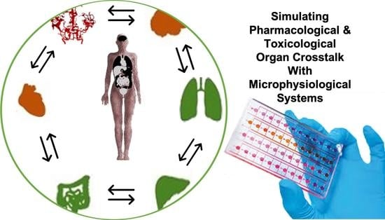
A Decade of Organs-on-a-Chip Emulating Human Physiology at the Microscale: A Critical Status Report on Progress in Toxicology and Pharmacology

 ,
,
Abstract
1. Introduction
2. Lung-on-a-Chip Systems: Biomechanically Improved Disease Models
3. Skin-on-a-Chip Systems: Combined Epidermis Full Skin Thickness Models
4. Liver-on-a-Chip: Valid Models for Pharmaceutical Screening
5. Kidney-on-a-Chip: Evaluating Nephrotoxicity and Drug Efficacy
6. Heart-on-a-Chip: Identifying Beneficial and Adverse Effects of Drugs in Health and Disease
7. Vasculature-on-a-Chip: Vascular Barrier Transport and Anti-Vasculogenic Drug Screening
8. Blood-Brain-Barrier-on-a-Chip: Barrier Function, Transport Properties, and Neuroinflammatory Model
9. Central Nervous System-on-a-Chip: From Neurodevelopment to Drug Testing and Brain Disease
10. Multi-Organ on-Chip Models: Combined Tissue Models to Screen Drug-Drug Interactions, Pharmacokinetics, and Pharmacodynamics
11. Conclusions

| Device Specifications | Assays | Additional Information | |||||
|---|---|---|---|---|---|---|---|
| Device Material | 2D/3D | Application | Cell Type(s) | Assays | Special Feature | Outcome | Ref. |
| PDMS and PET porous membranes | Monolayer | Skin epidermis, inflamatory, edem | Human keratinocytes HaCaT, fibroblasts, Human umbilical vein endothelial cells (HUVECs) | Response to TNF-a (mRNA expression levels ELISA), Tight junction staining, leakage assay | Multi barrier competent Chip (3) | TNF-α-induced inflammation treatment with drug DEX | [74] |
| Polycarbonate cover-plates, PDMS-glass chip | Biopsy | Oral and ermal ubstance absorption | Prepuce samples | Stratified stratum corneum at the air–liquid interface, mRNA | 4-organ-models for 28 days | Absorption, distribution, metabolism, and excretion | [203] |
| Hydrogel-based (collagen) | 3D hydrogel Co-culture | Epidermis and dermis | Normal human dermal fibroblasts (NHDFs), normal human epidermal keratinocytes (NHEKs), Human umbilical vein endothelial cells (HUVECs) | Drug uptake on skin—concentration measured in the perfusion | Perfusion from within | Percutaneous absorption of caffeine and ISDN | [69] |
| PDMS and PC membrane | 3D | Air-Skin-Interface | Fibroblasts and keratinocytes | Transdermal transport of FAM-tagged oligonucleotides | Air-Skin-Interfaces | Anti-proliferative role of doxorubicin | [73] |
| PDMS, membrane | 3D | Differentiation | Fibroblasts and keratinocytes | H&E, Masson trichrome (MT), and Sirius red staining | Gravity flow | Skin differentiation on chip | [72] |
| PC, PMMA, PTFE filter membranes | 3D | Skin permeation | N/TERT-1 keratinocyte, human primary foreskin-derived dermal fibroblasts | Diffusion of caffeine, testosterone, salicylic acid | Micro device vs. Franz diffusion cells | As good as Franz diffusion cells | [71] |
| PDMS, PMMA, PS, PET membrane | 2D | Immune-competent | Human keratinocytes (HaCaT) human leukemic monocyte lymphoma cell line (U937) | TEER, lipopolysaccharides impact, UV, nickle sulfate, cobalt sulfate, glycerol, DNCB | Immune response, TEER on chip | ALI not possible | [75] |
| Commercial Biochip | 2D/3D | Air-Skin-Interface | Murine fibroblasts (L929) and EpiDerm™ RhE. | TEER, impact of sodium dodecyl sulphate | Air-Skin-Interface, TEER on chip | Detection of tissue break down | [67] |
| PMMA | 3D in fibrin dermal matrix | epidermal morphogenesis | Human primary foreskin-derived dermal fibroblasts | TEER, Raman, 2PP microscopy | orthokeratinized full-thickness SE within a microfluidic platform | Industrial close working chip design | [68] |
| Device Specifics | Assays | Additional Information | |||||
|---|---|---|---|---|---|---|---|
| Device Material | 2D/3D | Application | Cell Type(s) | Special Feature | Sensing Principle/Assay | Outcome | Ref. |
| PDMS | Monolayer, co-culture | Alveolar-capillary interface | human alveolar epithelial cells and microvascular endothelial cells | Stretchable membrane | TEER, albumin transport, neutrophil activation through e. coli, ROS, nanoparticle | First biomimetic device for breathing lung | [28] |
| PDMS | Monolayer | Pulmonary edema | Human pulmonary microvascular endothelial cells (Lonza), Alveolar epithelial cells NCI-H441 | Stretchable membrane | interleukin-2 (IL-2) (for leakage of ALI = edema) vs. angiopoietin-1 and drug GSK219387 | GSK219387 closes leakage from edema | [51] |
| PDMS, PE Membrane | Monolayer, co-culture | Disease COPD | Primary hAECs, human lung microvascular endothelial cells | ALI | interleukin-13 (IL-13) reconstituted the goblet cell hyperplasia, cytokine hypersecretion, and decreased ciliary function of asthmatics | Cytokines IL-8, M-CSF, IP-10, and RANTES healthy vs. COPD | [53] |
| PDMS, PET membrane | Monolayer, co-culture | Impact of smoking | Primary human small airway epithelial cell, microvascular endothelium | Cigarette smoke as ALI. Also e-cigarette. | upregulation of anti-oxidant heme oxygenase 1 (HMOX1) gene expression with smoke exposure phosphorylation of the antioxidant regulator Nrf2 ciliary beat frequency interleukin 8 (IL-8) secretion of COPD cells influenced by smoke | Smoking upregulates expression of oxidation-reduction | [45] |
| PDMS, PET Membrane, gold electrodes on PC | monolayer | TEER and barriers (also gut with CaCo2) | primary human airway epithelial cells (hAECs) | TEER of ALI | Application of chelating agent EGTA | EGTA leads to destruction of barrier | [60] |
| PDMS | 3D organoids | Organ toxicity | Hepatic stellate cells (HSCs) (+), primary human hepatocytes (+), Kupffer cells (+), induced pluripotent stem cell-derived cardiomyocytes (iPSC CMs) (*), human primary cardiac fibroblasts (+), lung microvasculature endothelial cells (+), airway stromal mesenchymal cells (+), bronchial epithelial cells (+) | Cardiac beat rate measurement by real-time imaging and computational analysis, antibody-binding by impedance measurement, and barrier function by TEER measurement | acetaminophen, N-acetyl-L-cysteine on liver, epinephrine and propranolol on heart, Bleomycin | Interlinked responds of combined organoids | [209] |
| PDMS, PDMS membrane | Lung monolayer, whole blood HUVECS channel wall | Blood-air interface pulmonary thrombosis | HUVECs, human lung microvascular endothelial cells (HMVEC-L), blood | Blood for perfusion of vessel | Inflammatory cytokine-induced pulmonary thrombosis | LPS endotoxin indirectly stimulates intravascular thrombosis | [62] |
| PDMS, PDMS membrane | Monolayer | Blood-air interface | Bronchial epithelial 16HBE14o−cells, Primary human pulmonary alveolar epithelial cells (pHPAEC) | Mechanical strain of alveolar barrier during breathing | IL-8 concentration, viability | Stretching influencing the transport of small molecules | [56] |
| PDMS, flexible circuit board | Monolayer | Blood-air interface | Human type II alveolar epithelial-like A549 cells (–) | Mechanical strain of alveolar barrier during breathing | Barrier movement and membrane permeabilization sensing by real-time measurement of resistivity changes in three impedimetric coplanar electrodes | Monitoring in real-time the integrity of an epithelial barrier located at a distance of 1 mm | [57] |
| PMMA | Monolayer | Epithelial smooth muscle interface | Human airway epithelial cells (Calu-3), human bronchial smooth muscle cells (hBSMCs) | ALI | Device characterization, tight junctions | Stable hydrogel-based ALI | [65] |
| PDMS | Spheroids | photodynamic therapy (PDT) | A549 and non-malignant MRC-5 | 3D spheroids, 384 wellplate based | Verification of spheroid viability, ALA accumulation, ROS generation | High PDT effectiveness on lung spheroids required additional time after treatment. | [66] |
| PDMS, PET membrane, glass | Monolayer | Various barrier models, gut, and BBB | A549 | TEER on chip | TEER, ZO1, b-cantenin, leakage assay | One device for different barrier models | [212] |
| PDMS, silicon | Monolayer | Lung inflammation | Beas-2B (ATCCsnumber: CRL-9609t) is a human bronchialepithelial cell line infected with an adenovirus (12-SV40 hybridvirus, Ad12SV40 peripheral blood mononuclearcells (PBMCs) | Fibrocyte migration | Cell migration, ELISA | CP, CXCL12, and CXCR4 responsible for fibrocyte extravasation in lung inflammation | [63] |
| PDMS, PLGA for membrane | Monolayer | Tumor invasion | A549, HFL1, and HUVECs | Electro spinning of PLGA membrane | Device characterization, f-actin, DAPI, Calcein-AM, CD31 expression of HUVECs | Simulate in vitro the tumor microenvironment alveolar biochemical factors | [64] |
| PDMS, glass, PET membrane | Monolayer | Lung | A549 | TEER (Evom) | Surface tension influenced by surfactant secretion | Chip higher TEER | [58] |
| Paraffin, glass, PET, and PC membrane | Monolayer | Lung | Calu-3 | TEER (Evom) | 14C-sucrose permeability | Permeability higher through | [59] |
| PDMS, glass | Monolayer | Lung | A549 | Phosphor lipids, lamellar bodies, F-actin | Formation of laminar bodies | Laminar body formation depends on shear | [61] |
| Device Specifics | Assays | Additional Information | ||||||
|---|---|---|---|---|---|---|---|---|
| Device Material | 2D/3D | Culture Conditions | Cell Type(s) | Application | Staining | Assays | Outcome | Ref. |
| Polysterene (Nortis) | 2D | Flow conditions, Collagen I | Primary rat and human hepatocytes | Hepatotoxicity | Calcein-AM, Ethidium-homodimer1, HNF4α | Lactate dehydrogenase (LDH) release, alanine aminotransferase (ALT), albumin CYP1A1/2, and CYP3A4 activities, visualization o canalicular structures | Exhibited higher viability and improved hepatic functions | [96] |
| PDMS | Bio-printed 3D spheroids | Spheroid culture encapsulated within photocrosslinkable Gelatin methacryloyl (GelMA) hydrogel | human HepG2/C3Aspheroids | Drug toxicity assessment | cytokeratin 18, MRP2 bile canalicular protein, and tight junction protein ZO-1 | Albumin, alpha-1 antitrypsin, transferrin, and ceruloplasmin | Treatment with acetaminophen induced a toxic response in the hepatic construct that was similar to published studies on animals | [213] |
| PDMS | 3D liver spheroids | Flow conditions, low-attachment spheroid microplates | HepaRG cells, 3D pancreatic islet microtissues | Pancreatic islet–liver crosstalk based on insulin and glucose regulation | Insulin, glucacon, cytokeratin 8/18, vimentin, albumin, CYP3A4 | Albumin, glucose, insulin | Demonstrated a functional feedback loop between the liver and the insulin-secreting islet micro-tissues | [214] |
| 3D Perfusion IbiTreat, (Ibidi) | 2D and 3D | hyaluronan and poly(ethylene glycol) (HA-PEG), alginate and agarose hydrogels for 3D, PLL or HEP Coat coated for 2D | HepG2 and hiPS-HEP | Comparison of agarose, alginate, and HA-PEG hydrogels | - | Rheology, dextran diffusion | In HA-PEG hydrogels, 3D hiPS-HEPs showed an increased viability and higher albumin production compared to cultures in the other hydrogels | [95] |
| PDMS, PMMA, PC membrane | 3D | Flow, conditions, Matrigel | HepG2, LX-2, EAhy926, U937 | Investigation of pathophysiological process of individual non-parenchymal cells in alcohol-induced ALD | ZO-1, CD14 and F-actin, VE-Cadherin, and eNOS for EAhy926, VEGF, and α-SMA for LX-2 | Albumin, urea, ROS | Alcohol damages the tight junction and reduces the release of NO of EAhy926 cells | [99] |
| PDMS | 2D | static and flow, fibronectin and collagen I | Primary human hepatocytes, LX-2, EA.hy926, U937 | Recapitulation of a liver sinusoid-on-a-chip | CD31 | Albumin, urea, CYP3A4 | Higher albumin synthesis (synthetic), urea excretion (detoxification) was observed under flow compared to static cultures. | [100] |
| PDMS | 2D | static and flow conditions, co-Culture of primary rat hepatocytes and endothelial cells | Primary rat hepatocytes, primary rat adrenal medullary endothelial cells (RAMECs), bovine aortic endothelial cells (BAECs) | Analysis of viral replication for the hepatotropic hepatitis B virus (HBV) | - | Urea, analysis of secreted HBV, RT-PCR | A dual-channel configuration under flow condition seems to be the best long-term liver model and more closely mimics the structure and microenvironment of the liver sinusoid. | [102] |
| PDMS | 2D/3D | Flow conditions, monolayer, collagen sandwich, 3D spheroids, bioreactor | Primary rat hepatocytes | Assessment of repeated dosing chronic hepatotoxicity | - | Oxygen concentration, shear stress, viability, urea, albumin, CYP1A2, CYP2B1/2, CYP3A2 | Chronic drug response to repeated dosing of Diclofenac and acetaminophen evaluated in PIC were more sensitive than the static culture control. | [215] |
| PMMA | 2D/3D | Flow conditions, dECM bioink | HepaRG, HUVEC | Vascular/biliary fluidic channels for creating vascular and biliary systems | MRP2, CK31, Cholyllysyl-fluorescein | Albumin, urea, CYP expression | Drug treatment in the chip was highly influential and that liver functions were disrupted during the culture | [216] |
| PDMS | 3D | Liver microsome into 3-D hydrogel pillars | Rat liver microsomes | Analyzing the reaction kinetics inside the microfluidic chip by liver enzymes | - | P450 reactions, enzyme kinetic parameters (Km, Vm, Diffusivity) | Reaction rates were significantly lower than the solution phase mathematical model that showed a good correlation with the results | [93] |
| Glass (Micronit) | 2D | Flow conditions, collagen I | Primary human hepatocytes, THP-1, LX-2, HMVEC, primary human PMNs, LSECs | ADME/TOX studies | F-actin, VE-cadherin, α-SMA | Bile efflux, permeability, ROS | Partial immunologic functions within the liver sinusoid, including the activation of LSECs, promoting the binding of polymorphonuclear leukocytes (PMNs) followed by transmigration into the hepatic chamber | [101] |
| PDMS | 3D | Flow conditions, 3D spheroid culture | Primary rat hepatocytes, hepatic stellate cells | Investigation of hepatocyte-hepatic stellate cell interactions | Albumin, P450 reductase | Albumin, urea | Enzymatic activity of spheroids co-cultured for 8 days was greater than that of mono-cultured spheroids | [97] |
| PMMA, PDMS, PC membrane | 3D | flow conditions, 3D spheroid culture | human HepG2/C3A | development of bio-artificial livers, disease modeling, and drug toxicity screening | ZO-1, MRP-2, CPR, NADPH-cytochrome P450 reductase | Albumin, urea, expression of CYP enzymes, phase II enzymes, hepatic nuclear receptors, hepatic transporters, bile canaliculi transporters | timesaving, efficient, and safe in situ perfusion culture of hepatic spheroids in 3D culture | [217] |
| Device Specifics | Assays | Additional Information | - | |||||
|---|---|---|---|---|---|---|---|---|
| Device Material | 2D/3D | Culture Conditions | Cell Type | Application | Staining | Assay | Outcome | Ref. |
| PDMS | 2D, human proximal tubule | Flow conditions, 2D on collagen type I | Human primary proximal tubular epithelial cells (PTECs) | Renal drug clearance and drug-induced nephrotoxicity | CD13, E-cadherin, aquaporin 1, prominin 2, uromodulin, KIM-1, Na+/K+ ATPase, γ-glutamyl transpeptidase (GGT) | γ-Glutamyl transpeptidase (GGT) activity, ATP assay, ammoniagenesis, glucose reabsorption 25-(OH)2, vitamin D3 metabolism | Morphological and functional phenotypes of proximal tubule epithelium in vivo | [110] |
| PDMS | 2D human renal vascular–tubular system | Flow conditions, 2D on collagen I | /human fetal kidney tissue, endothelial cells (HUVECs and HKMECs), fetal kidney pericytes | Reabsorption of albumin and glucose | F-actin, VE-cadherin, E-cadherin, tubulin | Histology, dextran perfusion, blood perfusion, albumin -and inulin perfusion, glucose perfusion | Double-layer human renal vascular–tubular unit (hRVTU) enabled by a thin collagen membrane that replicates the kidney exchange interface | [112] |
| PDMS | Glomerular vascular system | Flow conditions, ECM with fibrinogen, 2 wt% gelatin, 2.5 mM CaCl2, and 0.2 wt% transglutaminase | hPSC-derived kidney pretubular aggregates | nephrotoxicity, tubular and glomerular disease, and kidney regeneration | DNA, MCAM, PECAM1, PODXL, PDGFRβ, TUBA4A, ATP1A1 (Na/K ATPase subunit-α1), collagen IV and LTL | Flow profile analysis, flow cytometry, qRT-PCR | Vascularized kidney organoids cultured under flow had more mature podocyte and tubular compartments with enhanced cellular polarity and adult gene expression ompared with that in static controls | [113] |
| Polysterene (Mimetas) | 3D, proximal tubule microvillus | Flow conditions, ECM, Collagen I | Cortical kidney tissue, urine-derived kidney cells | Personalized medicine | ZO-1, Ezrin, CDH-1, tubulin, | Barrier integrity assayP-gp transporter assay, trans-epithelial transport assay | Tubuloid-derived cells can form leak-tight, polarized kidney tubules, enabling personalized transporter studies in tubuloids | [218] |
| Acryl chambers | 2D, glomerular filter barrier | Flow conditions, collagen I on porous membrane 0.02 µm pore size | Immortalized podocytes | transmembrane pressure in glomerular filter membrane, with potential implications for drug development | Actin | Dextran filtration, qPCR | Dysfunction of renal filtration is correlated with the reduction of synaptopodin expression and disorganized actin cytoskeleton | [219] |
| Polysterene (Mimetas) | 2D and 3D | Flow conditions, 3D ECM Collagen I | Immortalized proximal tubule epithelial cells (CiPTEC-OAT1) | Screenings assay for renal drug-transporter interactions | ZO-1, tubulin, pericentrin | Functionality of P-gp and MRP2/4 | Increased accumulation of glutathione-methylfluorescein (GS-MF) was observed upon inhibition with a combination of PSC833, MK571, and KO143 | [220] |
| PDMS | 2D | Matrigel-coated, flow and static conditions | ureteric bud (UB) cells isolated from primary mouse embryonic kidneys cells | Fluid shear stress studies | Dolichosbiflorus agglutinin | qRT-PCR | Link between the fluid shear stress from the initiation of urine flow and the development and function of the kidney | [221] |
| PDMS | Kidney glomerular capillary wall | Flow conditions, Matrigel coated | PS cells (PGP1, IMR-90-1, and IISH3i-CB6) and ES cell (H9) lines | Podocyte differentiation | podocin | Whole-transcriptome analysis | Podocyte differentiation protocol for iPS cells, and the development of a glomerulus chip | [222] |
| COP | 2D | Flow conditions PET membrane, coated with collagen type I | Renal epithelial cells (raTAL and NRK-52E) | Transepithelial reabsorption of NaCl | - | TEER | RaTAL cells might sense ion concentrations on either side and adjust tight junction permeability accordingly. | [223] |
| Device Specifics | Assays | Additional Information | |||||
|---|---|---|---|---|---|---|---|
| Organ(s) | Device Material | Application | Cell Type | 2D/3D | Sensing Principle | Outcome | Ref. |
| Heart and liver | PMMA, PDMS, titanium, platinum (electrodes) | Cardiotoxicity (primarily from hepatic cytochrome P450 (CYP) metabolism) | Human iPSC derived cardiomyocytes and human primary hepatocytes | 2D monolayers | Multi-electrode array for electrical activity sensing and cantilever array for sensing of cardiac mechanical function | Culture period of 28 days with stable cellular function/viability. Cardiotoxicity of two known drugs due to hepatic metabolism could be predicted. | [224] |
| Heart | PDMS, titanium, palladium, gold (electrodes) | Cardiac biomarker secretion | Human embryonic stem cell-derived cardiomyocytes | 3D cell-laden hydrogel structures | Creatine kinase (CK)-MB sensing by impedance measurements using an aptamer functionalized micro-electrode | Aptamers specific to cardiac damage markers could detect dose-dependent usage of drugs. Beating rate and cell viability confirmed these measurements. | [225] |
| Blood vessel, heart, liver | PDMS, polystyrene | Cancer metastasis | HUVECs, HepG2, and hPSC line BJ1D | 3D cell-laden hydrogel structures | Cardiac beat frequency sensing by fluorescence microscopy and computational analysis of micro-cantilevers. | Simulation of cancer invasion and metastasis from tumor to downstream liver model. | [226] |
| Heart | PDMS, titanium, platinum (electrodes) | Barrier function and electrical activity of endothelialized myocardium | HUVECs and human iPSC derived cardiomyocytes | Barrier model | Barrier integrity and electrical activity sensing by TEER-multielectrode array measurements | Measurement of biological processes and drug effects by TEER and MEA, allowing analysis of barrier function, changes in cell layer conformation, and electrical activity. | [208] |
| Heart and liver | PDMS, gold, silver (electrodes) | Organ toxicity | Human iPSC derived cardiomyocytes and HepG2/C3A hepatocellular carcinoma cells | 3D organoids | pH sensing by light absorption of phenol red, oxygen sensing by fluorescence measurements of quenching effects of oxygen sensitive ruthenium dye and immunosensing by functionable electrodes | Platform for automated measurement of pH, oxygen, temperature, and biomarkers and microscope imaging over a period of 5 days of a dual organ-on-a-chip system with and without drug inducement. | [207] |
| Heart | PDMS, carbon (electrodes) | Beating behavior, cardiotoxicity | Cardiomyocytes isolated from neonatal Sprague Dawley rat pups | 3D cell-laden hydrogel structures | Generated force sensing by fluorescence measurement of deflection of cantilevers | Generated cardiac microtissues showed that mechanical response (dynamic response, spontaneous stress) to compounds (isoproterenol and digoxin) can be used for drug testing | [227] |
| Heart | PDMS | Beating behavior, cardiotoxicity | Human iPSC derived cardiomyocytes | Small (100 μm height) 3D cell-laden hydrogel structures | Beat rate, conduction velocity, and field potential duration were measured by field potential analysis using commercially available MEAs | The micro-molded gelatin system with incorporated MEAs is able to measure electrophysiological changes in the cardiac tissue. Beat rate responded accordingly to drug (isoproterenol) application. | [228] |
| Heart, liver, and lung | PDMS, PMMA, gold (electrodes) | Organ toxicity | Hepatic stellate cells, primary human hepatocytes, Kupffer cells, iPSC-derived cardiomyocytes, human primary cardiac fibroblasts, lung microvasculature endothelial cells, airway stromal mesenchymal cells, and bronchial epithelial cells | 3D organoids | Cardiac beat rate measurement by real-time imaging and computational analysis, antibody-binding by impedance measurement and barrier function by TEER measurement | The impact of multi-organ crosstalk is displayed by the effects of bleomycin on the cardiac tissue with and without a lung tissue present. | [209] |
| Heart, liver, skeletal muscle, and neuronal network | PDMS, PMMA, Titanium, platinum (electrodes) | Organ toxicity | Human hepatocellular carcinoma HepG2/C3A, human iPSC derived cardiomyocytes, human skeletal myofibers, human motorneurons differentiated from human spinal cord stem cell line (hSCSC) and human iPSC dervied cortical-like neurons | 2D monolayers | Cardiomyocyte contraction (force) sensing by cantilever deflection (laser beam reflection) 10, 11 and electrical activity of cardiomyocytes or motoneurons by a multielectrode array 11 | The system could detect influences of organ-cross talk in drug testing over a period of 14 days. 10 The 4-organ-system revealed stable conditions for all organs over a period of 28 days, making it a good fit for drug testing to investigate repeat dose toxicity. 11 | [229] |
| Heart | Petri dish as housing for MEA, titanium (electrodes) | Beating behavior | Human embryoid stem cell line CCTL14 and human iPSCs | 3D organoid | Cardiomyocyte beating force sensing by multi-electrode array and atomic force microscopy measurements | The system was able to determine the relation between cardiac beating behavior and correlated force in case of Duchenne Muscular Dystrophy. | [208] |
| Embryoid body (cardiac cells) | Glass, PEDOT:PSS (electrodes) | Autonomous beat rate of embryoid bodies | Mouse embryonic stem cells (mESC) derived cardiomyocytes | 3D embryoid body | Cardiac beat rate sensing by voltage and displacement current measurement by large area electrodes | The system could measure the beating rate of whole embryoid bodies due to a combination of large, high capacity electrodes and usage of the displacement current measurement technique. | [230] |
| Heart | Glass, PDMS, poly(N-isopropylacrylamide)(PIPAAm), platinum (electrodes) | Muscle contraction | Cardiac ventricular myocytes harvested from two-day old neonatal Sprague-Dawley rats | 2D monolayer | Cardiac tissue stress/contraction sensing by measurement of deformation of cells on elastic, thin film | Measurement of spontaneous contraction of cardiac tissues due to the impact of epinephrine. | [27] |
| Heart | PDMS, copper (force probe) | Muscle contraction/strength | Myocytes isolated from 0–2-day-old neonatal Sprague-Dawley rats, cardiac fibroblasts, and a transfected human embryonic kidney (HEK) 293T | 3D cell-laden hydrogel fibers | Force sensing of the muscle strips by usage of a copper force probe, which applied force/deformation prior to the illumination. The deformation prior to and after the illumination was compared | The system was able to individually activate and pace the muscle fibers. With the addition of multiple fibers, the force can be graded. This offers a non-invasive way (compared to electrodes) for muscle stimulation. | [25] |
| Heart | Glass, PDMS, poly(N-isopropylacrylamide)(PIPAAm), aluminum, platinum (electrodes) | Muscle contraction | Cardiac myocytes extracted from ventricles of 2-day-old neonatal Sprague-Dawley rats | 2D monolayer | Stress sensing by visual observation of cantilever deformation due to cardiac contraction. | Diastolic and systolic stress of cardiac tissues could be assessed and electrically stimulated with and without drug exposure. | [129] |
| Heart | PDMS, polystyrene beads (cyclic strain measurement), | Cardiac differentiation under cyclic strain | Neonatal rat cardiac cells and human iPSC-derived cardiomyocytes | 3D cell-laden hydrogel structures | Cellular property sensing by immunofluorescence (live/dead, histochemistry) and scanning/transmission electron microscopy | The device showed that by applying cyclic strain, the cell-cell and cell-matrix interactions are better, and also micro-tissues displayed early synchronous beating. | [126] |
| Heart | Dextran, elastollan, silver flakes, PDMS | Long-term non-invasive measurement of contractile strength of cardiac tissues | Neonatal rat ventricular myocytes (NRVMs) and human iPSC-derived cardiomyocytes | Multiple 2D monolayers | Contractile stress and beat rate sensing by relative resistance changes of embedded sensors | 3D multi-material printing allowed the creation of a device for non-invasive measurement of contractile stress development over a period of 4 weeks. | [130] |
| Heart | PDMS, glass, poly(3,4-ethylenedioxythiophene) carbon nanotube (PEDOT-CNT) (electrodes); UV crosslinkable methacrylated gelatin (GelMA) | On-chip culture of human 3D myocardium using cells from a single patient | Human iPSC-derived myocardial cells and endothelial cells | 3D cell-laden hydrogel structures | Cell population alignment sensing by microscopy and beating frequency sensing by measurement of extracellular membrane potential | Usage of two cell types derived from the same hiPSCs this device mimics the myocardium of an individual human. Integration of hydrogel allowed 3D structuring and analysis was performed by integrated micro-electrode arrays and standard cell culture analysis. | [121] |
| Heart | PDMS, matrigel, fibrinogen, thrombin | Contractile activity | Neonatal rat heart cells | 3D cell-laden hydrogel structures | Contractile force and beat rate measurement by video-optical measurement of pillar deflection | The device was able to measure the impact of drug and toxic substances on the beat rate of heart constructs, allowing a fast and simple screening method. | [127] |
| Heart | Polydopamine (PDA), polycaprolactone (PCL) (nanofibers), gelatin (cantilever), TiO2, and Ag (nanoparticels) | Contractile activity | Neonatal rat ventricular myocytes (NRVMs) isolated from 2-day-old Sprague Dawley rats | 2D monolayer | Beat rate sensing by optical detection of cantilever deflection | By using 3D fiber scaffolds, the device supported the formation of anisotropic and contractile cardiac tissues. Non-invasive measurement of nanoparticle impact enables good drug screening capabilities. | [128] |
| Heart | Quartz (substrate), gold (electrodes) | Beating behavior | Neonatal rat cardiomyocytes | 2D monolayer | Beat rate and amplitude sensing by impedance detection | The device can measure the mechanical contraction status of the cardiac tissue, which can be linked to the pumping of blood and plays a vital role in drug screening for heart medication. | [123] |
| Heart | PMMA, alginate, gelatin methacryloyl (GelMA), photoinitiator Irgacure 2959 (bioink) | Contractile activity | HUVECs, GFP-labeled HUVECs, and neonatal cardiomyocytes isolated from two-day-old Sprague-Dawley rats | 3D bioprinted endothelial structure with 2D cardiac monolayer on top | Beat rate and amplitude sensing by optical microscopy and computational analysis | The endothelial cells in the bio-printed fibers of the scaffold would migrate toward the edge of the fibers and form a layer of confluent endothelium, mimicking a blood vessel. In combination with the myocardium and a perfusion bioreactor, this was then used for drug screening. | [132] |
| Heart | PEG-silane, PDMS, silicon (cantilever) | Electrical and contractile activity | Human cardiomyocytes derived from the hESC-10-0061 stem cell line | 2D monolayer | Electrical activity sensing by using an MEA and contractile stress sensing using cantilevers deflection measured by a photodiode laser and a photodetector. | The system is able to detect electrical and contractile activity of cardiac tissues and both sensing areas can be upscaled. | [131] |
| Heart | PDMS, glass | Cell proliferation/viability increase in 3D vs 2D | Human primary cardiomyocytes | 3D cell-laden hydrogel structures, 2D monolayer | Cell proliferation and viability sensing by fluorescence microscopy | The device showed that the proliferation rate was higher in 3D and the negative effects of isoproterenol was lower in 3D. | [122] |
| Device Specifics | Assays | Additional Information | |||||||
|---|---|---|---|---|---|---|---|---|---|
| Method | Device Material | Hydrogel | Concentration | Cell Source | Cell Type(s) | Assays | Application | Outcome | Ref |
| Microchannel patterning | PDMS | Collagen 1 | 0.1 mg/mL (coating) | Primary | HUVEC | Live/dead staining Immunofluorescence staining Blood perfusion | Thrombosis | Thrombosis-on-chip model created by PDMS molding of 3D-printed healthy and stenotic vessel | [139] |
| PDMS | PDL/fibronectin | 1 mg/mL (coating) | Primary | VSMC HUVEC | Immunofluorescence staining | Platform for blood vessel-related diseases | Successful cultivation of SMC and HUVEC on micro-wrinkled PDMS | [140] | |
| PDMS | Fibronectin | 250 µg/mL | Primary | HUVEC | Immunofluorescence staining | Identify combinatorial effect of fluid flow and substrate rigidity on endothelial cells | Pronounced cell response to flow-induced shear stress and underlying substrate mechanics | [141] | |
| Hydrogels | Alginate Gelatin Gel-MA | 1–5% 1–9% 1–9% | Primary | HUVEC | Diffusional permeability | Enhancing physiological relevance via microfabrication of hydrogels | Hydrogel-based microfluidic device as a vessel-on-chip model | [143] | |
| Gelatin | Gelatin | 9% | Primary | HUVEC | Immunofluorescence staining Diffusional permeability | Circular microfluidic channels | Fabrication of branched vascular networks in a hydrogel-based microfluidic device | [144] | |
| PDMS | Collagen 1 | 2.5 mg/mL | Primary | HUVEC SMC Neutrophil | Diffusional permeability Leucocyte transmigration Blood perfusion | Atherosclerosis | Formation of pathophysiological architectures for real-time study of cardiovascular disease | [146] | |
| PDMS Polycarbonate | Fibrin | Scaffold | Primary | HUVEC lrVEC hMSC | Endothelial sprouting Blood perfusion Leukocyte adhesion and migration | Vascularization of tissue engineered scaffold | Engineering of vasculature in novel polycarbonate scaffold with subsequent surgical anastomosis | [147] | |
| Sacrificial molding | PDMS | Collagen 1 | 3 mg/mL | Primary | HUVEC hBMSC/hLF/ hASMCs/ hKPC | Diffusional permeability Immunofluorescence staining Western blot RT-qPCR | Barrier function variation with inflammatory exposure | Identification of key proteins in regulating barrier function in inflammation | [153] |
| PDMS | Collagen 1 | 2.5 mg/mL | Primary | hMVEC | Diffusional permeability Immunofluorescence staining RT-qPCR | Influence of hemodynamic shear stress on endothelial barrier function | Identification of novel mechanosensory complex | [231] | |
| PDMS | Gel-MA | 4–16% | Primary cell line | HUVEC rSMC 3T3 fibroblast | Live/Dead staining Immunofluorescence staining Diffusional permeability Mechanical compression | Vascularization of tissue engineered structures | Engineering of all three layers of a mature blood vessel | [232] | |
| PDMS | Collagen 1 | 3 mg/mL | Primary | HUVEC hASMC | Metabolic activity Immunofluorescence staining | Defining physiological vessel functionality or pathologic remodeling | Formation of stable arteriole and artery-like structures | [151] | |
| PDMS | Collagen 1 | 6.5 mg/mL | Primary | hMVEC | LifeACT actin remodeling Diffusional permeability Immunofluorescence staining Live/Dead staining | Model for low-intensity anti-vascular ultrasound therapy | Efficacy of low-intensity ultrasound treatment of tumors dependent on fluid flow | [149] | |
| PDMS | Collagen 1 | 3 mg/mL–6 mg/mL | Primary | hLEC/HUVEC nhMF/CAF | Live/Dead staining Cytokine quantification RT-qPCR Diffusional permeability Immunofluorescence staining | Lymphatic vessel model for understanding physiologic and pathology | Organotypic lymphatic function dependent on cell-to-cell signaling | [154] | |
| Self-assembly | PDMS | Collagen 1 | 3 mg/mL | Primary | HUVEC | Sprouting analysis Immunofluorescence staining Calpain and MMP inhibition | Platform to investigate hemodynamic and biochemical factors in angiogenesis | Angiogenesis dependent on IF and VEGF concentration | [159] |
| PDMS | Fibrin | 2.5 mg/mL fibrinogen 1 U/mL Thrombin | Primary | HUVEC hLF | Sprouting analysis Immunofluorescence staining | Flow-mediated endothelial dynamics and phenotype changes | Interstitial flow regulates angiogenic sprouting and endothelial cell phenotype | [158] | |
| PDMS | Fibrin | 2.5 mg/mL fibrinogen 1 U/mL thrombin | Primary | HUVEC hASC | Network quantification | Direct and indirect perfusion of cell-seeded hydrogels | Direct cell-to-cell contact as well as reciprocal signaling molecules play a vital role in vasculogenesis | [8] | |
| PDMS | Fibrin | 2.5 mg/mL fibrinogen 1 U/mL thrombin | Primary | hMVEC-dLyAd NHLF | Sprouting analysis Immunofluorescence staining Cell viability | Lymphangiogenesis in tumor formation | Interstitial flow augments lymphatic endothelial sprouting in synergy with lymphangiogenic factors | [160] | |
| PDMS | Fibrin | 2.5 mg/mL fibrinogen 1 U/mL thrombin | Primary | HUVEC NHLF | Diffusional permeability Glycocalyx staining Colocalization analysis Transcytosis mechanism | Drug transport across the vascular endothelium and into the target tissue | On-chip model recapitulates physiologic paracellular and transcellular permeability | [161] | |
| PDMS | Fibrin | 5 mg/mL fibrinogen 1 U/mL thrombin | Primary | HUVEC NHLF | Immunofluorescence staining Cell viability Network quantification Diffusional permeability Colocalization analysis | Vascular remodeling in Idiopathic pulmonary fibrosis | Anti-vasculogenic drug acts on endothelial network formation and endothelial–perivascular interactions | [162] | |
| PDMS | Fibrin | 2.5 mg/mL fibrinogen 0.5 U/mL thrombin | Primary | HUVEC NHLF hPC-PL | Blood vessel contraction Oxidative stress Immunofluorescence staining Diffusional permeability | Nanoparticle toxicity on endothelial barrier | Nanoparticle exposure leads to microvessel contraction and nanoparticles are transported via caveolae-mediated transcytosis | [163] | |
| PDMS | Fibrin | 1 U/mL thrombin | Primary | HUVEC NHLF | Immunofluorescence staining Diffusional permeability | Pathogenesis of inhaled atmospheric nanoparticles | Atmospheric nanoparticle inhalation leads to a loss of tight junctions and increased vascular permeability | [164] | |
| Device Specifics | Assays | Additional Information | |||||||||
|---|---|---|---|---|---|---|---|---|---|---|---|
| Method | Device Material | Culture Condition | Cell Type | Immuno-Staining | Fluorescent Tracker [kDa] | TEER Values | Other Features | Shear Stress [Pa] | External Stimuli | Outcome | Ref. |
| Sandwich Design | PDMS 3D | 17 days ECM: laminin | Primary hBMVEC (human) iPSC astrocytes pericytes | ZO-1 + Phalloidin staining | Dextran: 10, 70 | 35,000 Ω | Ascorbate transport | 2 × 10⁻³ | Cold shock Glutamate | Pericyte conditioned medium enhances BBB integrity | [171] |
| Sandwich Design | PDMS 2D | 7 days ECM: Col IV, fibronectin, Col I | b.End3 (mouse) C8D1A (mouse) | Claudin-5 | Dextran: 70 | - | - | 0.5 | - | Optically transparent PTFE membrane might be a suitable alternative | [169] |
| Sandwich Design | PDMS 2D | 6 days ECM: Col I/IV | hCMEC/D3 or primary rat endothelial cells primary pericytes (rat) primary astrocytes (rat) | ZO-1 β-catenin | Dextran: 4.4 Albumin: 67 Sodium fluorescin | 175 Ω cm2 | Direct contact between endothelial cells and pericytes | 1.5 × 10⁻² | - | Primary cells exhibited better BBB properties than hCMEC/D3 cells. | [212] |
| Sandwich Design | PMMA 2D | 7 days ECM: - | bEnd.3 (mouse) | Claudin-5 | - | 1150 Ω cm2 | Internalization of nanoparticles | 1.5 × 10⁻² | - | GH625 peptide modification on nanoparticle facilitates the transport across the BBB | [170] |
| Sandwich Design | Objet VeroClear photo-polymer - Parylene-C coating 2D | 10 days ECM: Col I Fibronectin | iPSC derived BMEC (human) primary astrocytes (rat) | ZO-1 Claudin-5 | Dextran: 4, 20, 70 | 2000 Ω cm2 | Caffeine Cimetidine Doxorubicin | 1.4–25 × 10−3 | - | First time the application of iPSCs-derived BMEC in co-culture with astrocyte (enhanced BBB integrity) Doxorubicin disrupts BBB integrity after 24-h treatment | [233] |
| Sandwich Design | PDMS and polycarbonate 2D | 4 days ECM: Fibronectin vs. Matrigel | Primary BMVEC (mouse) Astrocytes (mouse) | ZO-1 GFAP | Dextran: 3, 7, 10 | 3500 Ω | - | 0.1–3 | Histamine disruption | Multi-channel model with an integrated electrical impedance sensor array Matrigel provides better barrier integrity than fibronectin | [234] |
| Sandwich Design | PDMS 2D | 6 days ECM: Fibronectin and Col IV | bEnd.3 (mouse) | Claudin-5 | Dextran: 4, 20, 500 | 172 Ω cm2 | - | 0.1–0.6 | - | Angiopep-2 peptide modification facilitates nanoparticle transport across the BBB | [235] |
| Parallel design | PDMS 3D | 5 days ECM: fibronectin | RBE4 (rat) astrocytes (rat) | ZO-1 GFAP | Dextran: 40 | 250 Ω cm2 | - | 3.8 × 10⁻⁴ | - | Co-culture with astrocytes increases barrier integrity Cell/cell interactions are observed Endothelial cells are sensitive to astrocyte conditioned medium | [174] |
| Parallel design | PDMS 3D | 7 days ECM: Col I | HUVEC/hCMEC/D3 (human) primary astrocytes (rat) primary neurons (rat) | ZO-1 VE-cadherin GFAP DCX | Dextran: 10, 70 | - | Glutamate transport Calcium imaging | - | - | hCMEC/D3 shows significantly higher barrier integrity compared with HUVEC Neurons in triple co-cultures grow progressively in 3D Col I hydrogels | [176] |
| Parallel design | PDMS 3D | 4 days ECM: Fibronectin | HUVEC (human) astrocytes (rat) Met-1 | - | Dextran: 3, 70 | - | Efflux activity (Rhodamine 123) | 1.9 × 10⁻⁴ | Brain tumor model disrupts BBB integrity but retains P-gp activity | [175] | |
| Parallel design | OrganoPlate Mimetas Polystyrene 3D | 5–9 days ECM: Col I | TY 10 (human) hBPCT (human) hAst (human) Immortalized cell lines | VE-cadherin Claudin-5 PECAM-1 | Dextran: 20 | - | Antibody transcytosis | ~0.12 | - | Applicable to HTS | [177] |
| Tubular design | PDMS 3D | 3 days ECM: Col I | RBE4 (rat) Human neutrophils | ZO-1 VE-cadherin | Dextran: 40 | - | Transmigration of neutrophils | - | TNF-α/OGD | TNF- α elevates the release of several cytokines OGD induced the ischemia model ROCK inhibition restores BBB integrity | [179] |
| Tubular design | PDMS 3D | 3 days ECM: Col I/IV | hBMVEC (human) astrocytes (human) or pericytes (human) | ZO-1 VE-cadherin GFAP SMA | Dextran: 3 | 40–50 Ω cm2 | - | - | TNF-α | Co-culture with pericyte or astrocyte enhances BBB integrity Distinct differences in G-CSF and IL-6 secretion level between Transwell and chip cultures | [180] |
| Tubular Design | PDMS 3D | 3 days ECM: Col I | Primary BMEC (rat) primary astrocyte (rat) | ZO-1 VE-cadherin | Sodium fluorescein | 1298 Ω cm2 | 1.Expression level of P-gp and Glut-1 2.Modeling of extravasation in brain metastasis 3. Drug screening | 1 × 10⁻² | - | Co-culture enhances BBB integrity U87 were unable to cross the BBB Only temozolomide is able to pass through BBB and kill U87 Applicable to HTS | [236] |
| Tubular Design | PDMS 3D | 4 days ECM: Col I Matrigel HA | hCMEC/D3 (human) astrocytes (human) | ZO-1 GFAP | Dextran: 4 | 1000–1200 Ω cm2 | TEM | 0.07 | TNF-α | Shear stress significantly improves BBB integrity Pulsatile flow drives retrograde transport along the basement membrane | [237] |
| Tubular Design | IP-DiLL -SU8 3D | 3 days ECM: - | b.End.3 (mouse) U-87 (human) | ZO-1 | Dextran: 10 | 71 Ω cm2 | - | - | - | Two-photon lithography fabricated 3D tubular structures with 1:1 scale of capillaries | [238] |
| Vasculogenesis Design | PDMS 3D | 3 days ECM: Fibrinogen | iPSC-EC (human) brain pericytes (human) brain astrocytes (human) | CD31 ZO-1 GFAP | Dextran: 20, 70 | - | - | - | - | The best combination of cells for co-culture is identified | [182] |
| Device Specifics | Assays | Additional Information | ||||||
|---|---|---|---|---|---|---|---|---|
| Application | 2D/3D | Device Material | Cell Type | Assays | Flow | External Stimuli | Outcome | Ref. |
| Neuronal development-3D Organoid formation on chip | 3D ECM: Matrigel | PDMS | iPSCs | Immunocytochemistry PCR TUNEL assay | 25 µL h−1 | - | Organoids display feature specific identities such as neuronal differentiation, brain regionalization, and cortical spatial organization Improved cortical development compared to static culture conditions | [189] |
| Neuronal development-Prenatal nicotine exposure | 3D ECM: Matrigel | PDMS | iPSCs | Immunocytochemistry PCR TUNEL assay Neurite outgrowth assay (TUJ1) | 25 µL h−1 | Nicotine | Nicotine exposure led to premature neuronal differentiation, disrupted brain regional organization, abnormal cortical development, neuronal outgrowth, and increased cell apoptosis. | [190] |
| Neuronal development-Effect of endothelial cells on neuronal development | 2D | PDMS | iPSC derived spinal motor neurons iPSC derived BMECs | Immunocytochemistry Transcriptomics Live Calcium Transient Imaging Analysis | - | - | Vascular-neural interaction and specific gene activation enhances neuronal function and enables in vivo-like signatures | [186] |
| Neuronal development-High throughput generation of human brain organoids | 3D ECM: Matrigel | PDMS | - | Immunocytochemistry PCR | - | - | Controlled formation of human brain organoids on chip | [192] |
| Neuronal development-Neuronal differentiation and chemotaxis | 3D | PDMS | hNT-2 hNPCs Immortalized hBMECs | Immunocytochemistry Chemotaxis assay | - | CXCL12 SLIT2-N | hNPCs respond to shallow gradients of CXCL12 only in the presence of a micro-environment mimicking the brain parenchyma milieu hNPCs are more polarized in the presence of a neuronal-glial cell population, which leads to more directed movement | [239] |
| Neuronal development-Physics of brain folding | 3D ECM: Matrigel | PDMS | hESC | Immunocytochemistry RNA sequencing AFM | - | Blebbistatin | On-chip organoid approach successfully mimics the early developing cortex Lissencephalic organoids display reduced convolutions, modified scaling, and a reduced elastic modulus | [191] |
| Neuronal networks - Multi-regional brain on chip | 2D | PDMS | Hippocampal neurons (rat) Amygdala neurons (rat) Prefrontal cortex neurons (rat) | Immunocytochemistry Proteomics MS Oxygen Measurement MEA | Phencyclidine hydro chloride | Distinct differences in metabolism in different brain regions in vitro Changed electrical activity profile upon connecting individual regions PCP treatment alters electrical activity in a region-dependent manner | [184] | |
| Neuronal networks - 3D printed nervous system | 3D | Silicone Polycap-rolactone | Hippocampal neurons (rat) SCG neurons (rat) Schwann cells (rat) PK-15 cells | Immunocytochemistry Viral assay | - | Pseudorabies virus | Interconnectivity in-between individual cellular components Schwann cells and hippocampal neurons are refractory to axon-to-cell infection of pseudorabies virus | [240] |
| Neuronal networks - 3D neural circuit | 3D ECM: Matrigel | PDMS | Cortical neurons (rat) | Immunocytochemistry Calcium imaging | - | - | Axons formed in vivo like neural bundles with fasciculation and defasciculation. Aligned neural circuits with visualizable synapses. | [185] |
| Neuronal networks - Neuronal network for active compound testing | 2D | PDMS | Hippocampal neurons (rat) | Immunocytochemistry Viability assay Calcium imaging | 0.4–10 µL min−1 | Glutamate | Integration of microfluidic perfusion with Ca2+ imaging techniques | [187] |
| Neuro-degenerative diseases - Alzheimer’s disease | 3D neurospheroid | PDMS | Primary cortical neurons (rat) | Immunocytochemistry CCK-8 SEM | Interstitial fluid flow | Amyloid β | Interstitial fluid flow positively affects the maturation of neurospheroids Amyloid β treatment induced neural network induction | [198] |
| Neuro-degenerative diseases-ALS | 3D ECM: Col I | PDMS | iPSCs derived motor neurons iPSCs derived skeletal muscle cells iPSCs derived endothelial cells | Immunocytochemistry PCR Ca2+ oscillation imaging SNP genotyping Whole exome sequencing Western blot | - | Rapamycin Bosutinib | Muscle contraction could be induced by MN activity once NMJ is formed ALS motor unit displayed higher levels of apoptosis and reduced muscle contraction force Combinatorial treatments improved neuronal survival and an increased muscle contraction force | [199] |
| Brain cancer chip for HTS | 3D | PEGDa | U87 Primary glioblastoma tumors | Immunocytochemistry Diffusion test Viability | - | Pitavastatin Irinotecan Temozo-lomide Bevacizumab | HTS platform for spheroid formation and drug screening Combinatorial treatment with TMZ and BEV were more effective on glioblastoma spheroids | [195,241] |
| Cancer migration study | - | PDMS | Hippocampal neurons (rat) Cortical neurons (rat) DRG neurons (rat) PC-3 (human) Panc-1 (human) MCF-1 (human) | Immunocytochemistry | - | β -blockers muscarinic antagonists Neuron injury (6-hydroxy-dopamine) | Neurites guide the directional movement of cancer cells. Cancer cells with high levels of perineural invasion display greater migration along neurites. Neuron injury reduces migration of cancer cells. Muscarinic antagonists reduced migration. | [236] |
| Effects of GDNF on NMJ | 2D | PDMS | Skeletal myocytes (murine) Neurons (murine) | Immunocytochemistry Calcium live imaging | - | GDNF Oxidative stress BTX | Spatially distinct effects of GDNF on MN. | [242] |
| Organophosphate toxicity | 3D | Organoplate - PS | bEnd.3 (murine) C8-D1A (murine) BV-2 (murine) | Immunocytochemistry Acetylcholinesterase assay | 1.5 μL h−1 | DMMP DEMP DECP DCP | OPs penetrate the blood brain barrier (BBB) and rapidly inhibit AChE activity. In vitro toxicity was correlated with available in vivo data. | [193] |
| Combinatorial systems - Co-culture of liver and neurospheres | 3D | PDMS | HepaRG (human) Hepatic stellate cells (human) hNT-2 | Immunocytochemistry qPCR LDH activity Metabolic activity TUNEL assay | NA | 2,5-hexanedione | Successful co-culture of neuro-spheres and liver spheroids for 14 days. Co-culture was more susceptible to 2,5-hexanedione treatment compared to monocultures. | [194] |
Author Contributions
Funding
Data Availability Statement
Acknowledgments
Conflicts of Interest
References
- Whitesides, G.M. The origins and the future of microfluidics. Nature 2006, 442, 368–373. [Google Scholar] [CrossRef]
- Cann, O. These are the top 10 emerging technologies of 2016. World Econ. Forum. 2016. Available online: https://www.weforum.org/agenda/2016/06/top-10-emerging-technologies-2016/ (accessed on 31 March 2021).
- Ertl, P.; Sticker, D.; Charwat, V.; Kasper, C.; Lepperdinger, G. Lab-on-a-chip technologies for stem cell analysis. Trends Biotechnol. 2014, 32, 245–253. [Google Scholar] [CrossRef] [PubMed]
- Benam, K.H.; Novak, R.; Nawroth, J.; Hirano-Kobayashi, M.; Ferrante, T.C.; Choe, Y.; Prantil-Baun, R.; Weaver, J.C.; Bahinski, A.; Parker, K.K.; et al. Matched-Comparative Modeling of Normal and Diseased Human Airway Responses Using a Microengineered Breathing Lung Chip. Cell Syst. 2016, 3, 456–466.e4. [Google Scholar] [CrossRef]
- Ergir, E.; Bachmann, B.; Redl, H.; Forte, G.; Ertl, P. Small Force, Big Impact: Next Generation Organ-on-a-Chip Systems Incorporating Biomechanical Cues. Front. Physiol. 2018, 9, 1417. [Google Scholar] [CrossRef]
- Zirath, H.; Rothbauer, M.; Spitz, S.; Bachmann, B.; Jordan, C.; Müller, B.; Ehgartner, J.; Priglinger, E.; Mühleder, S.; Redl, H.; et al. Every Breath You Take: Non-invasive Real-Time Oxygen Biosensing in Two- and Three-Dimensional Microfluidic Cell Models. Front. Physiol. 2018, 9, 815. [Google Scholar] [CrossRef]
- Eilenberger, C.; Rothbauer, M.; Ehmoser, E.-K.; Ertl, P.; Küpcü, S. Effect of Spheroidal Age on Sorafenib Diffusivity and Toxicity in a 3D HepG2 Spheroid Model. Sci. Rep. 2019, 9, 4863. [Google Scholar] [CrossRef] [PubMed]
- Bachmann, B.; Spitz, S.; Rothbauer, M.; Jordan, C.; Purtscher, M.; Zirath, H.; Schuller, P.; Eilenberger, C.; Ali, S.F.; Mühleder, S.; et al. Engineering of three-dimensional pre-vascular networks within fibrin hydrogel constructs by microfluidic control over reciprocal cell signaling. Biomicrofluidics 2018, 12. [Google Scholar] [CrossRef]
- Sticker, D.; Rothbauer, M.; Ehgartner, J.; Steininger, C.; Liske, O.; Liska, R.; Neuhaus, W.; Mayr, T.; Haraldsson, T.; Kutter, J.P.; et al. Oxygen Management at the Microscale: A Functional Biochip Material with Long-Lasting and Tunable Oxygen Scavenging Properties for Cell Culture Applications. ACS Appl. Mater. Interfaces 2019, 11, 9730–9739. [Google Scholar] [CrossRef] [PubMed]
- Li, A.P.; Bode, C.; Sakai, Y. A novel in vitro system, the integrated discrete multiple organ cell culture (IdMOC) system, for the evaluation of human drug toxicity: Comparative cytotoxicity of tamoxifen towards normal human cells from five major organs and MCF-7 adenocarcinoma breast cancer cells. Chem. Biol. Interact. 2004, 150, 129–136. [Google Scholar] [CrossRef] [PubMed]
- Sin, A.; Chin, K.C.; Jamil, M.F.; Kostov, Y.; Rao, G.; Shuler, M.L. The Design and Fabrication of Three-Chamber Microscale Cell Culture Analog Devices with Integrated Dissolved Oxygen Sensors. Biotechnol. Prog. 2004, 20, 338–345. [Google Scholar] [CrossRef] [PubMed]
- Hosokawa, K.; Maeda, R. Pneumatically-actuated three-way microvalve fabricated with polydimethylsiloxane using the membrane transfer technique. J. Micromech. Microeng. 2000, 10, 415–420. [Google Scholar] [CrossRef]
- Kim, J.; Kang, M.; Jensen, E.C.; Mathies, R.A. Lifting gate polydimethylsiloxane microvalves and pumps for microfluidic control. Anal. Chem. 2012, 84, 2067–2071. [Google Scholar] [CrossRef] [PubMed]
- Unger, M.A.; Chou, H.P.; Thorsen, T.; Scherer, A.; Quake, S.R. Monolithic microfabricated valves and pumps by multilayer soft lithography. Science 2000, 288, 113–116. [Google Scholar] [CrossRef]
- Rothbauer, M.; Zirath, H.; Ertl, P. Recent advances in microfluidic technologies for cell-to-cell interaction studies. Lab Chip 2018, 18, 249–270. [Google Scholar] [CrossRef] [PubMed]
- Rothbauer, M.; Rosser, J.M.; Zirath, H.; Ertl, P. Tomorrow today: Organ-on-a-chip advances towards clinically relevant pharmaceutical and medical in vitro models. Curr. Opin. Biotechnol. 2019, 55, 81–86. [Google Scholar] [CrossRef]
- Shafiee, A.; Ghadiri, E.; Kassis, J.; Pourhabibi Zarandi, N.; Atala, A. Biosensing Technologies for Medical Applications, Manufacturing, and Regenerative Medicine. Curr. Stem Cell Rep. 2018, 4, 105–115. [Google Scholar] [CrossRef]
- Kratz, S.R.A.; Höll, G.; Schuller, P.; Ertl, P.; Rothbauer, M. Latest trends in biosensing for microphysiological organs-on-a-chip and body-on-a-chip systems. Biosensors 2019, 9, 110. [Google Scholar] [CrossRef]
- Cho, S.; Yoon, J.Y. Organ-on-a-chip for assessing environmental toxicants. Curr. Opin. Biotechnol. 2017, 45, 34–42. [Google Scholar] [CrossRef]
- Günther, A.; Yasotharan, S.; Vagaon, A.; Lochovsky, C.; Pinto, S.; Yang, J.; Lau, C.; Voigtlaender-Bolz, J.; Bolz, S.S. A microfluidic platform for probing small artery structure and function. Lab Chip 2010, 10, 2341–2349. [Google Scholar] [CrossRef]
- Blundell, C.; Tess, E.R.; Schanzer, A.S.; Coutifaris, C.; Su, E.J.; Parry, S.; Huh, D. A microphysiological model of the human placental barrier. Lab Chip 2016, 16, 3065–3073. [Google Scholar] [CrossRef]
- Mandt, D.; Gruber, P.; Markovic, M.; Tromayer, M.; Rothbauer, M.; Adam Kratz, S.R.; Ali, S.F.; van Hoorick, J.; Holnthoner, W.; Mühleder, S.; et al. Fabrication of biomimetic placental barrier structures within a microfluidic device utilizing two-photon polymerization. Int. J. Biopr. 2018, 4. [Google Scholar] [CrossRef]
- Griep, L.M.; Wolbers, F.; de Wagenaar, B.; ter Braak, P.M.; Weksler, B.B.; Romero, I.A.; Couraud, P.O.; Vermes, I.; van der Meer, A.D.; van den Berg, A. BBB on CHIP: Microfluidic platform to mechanically and biochemically modulate blood-brain barrier function. Biomed. Microdevices 2013, 15, 145–150. [Google Scholar] [CrossRef]
- Uzel, S.G.; Pavesi, A.; Kamm, R.D. Microfabrication and microfluidics for muscle tissue models. Prog. Biophys. Mol. Biol. 2014, 115, 279–293. [Google Scholar] [CrossRef]
- Chan, V.; Neal, D.M.; Uzel, S.G.; Kim, H.; Bashir, R.; Asada, H.H. Fabrication and characterization of optogenetic, multi-strip cardiac muscles. Lab Chip 2015, 15, 2258–2268. [Google Scholar] [CrossRef] [PubMed]
- Grosberg, A.; Nesmith, A.P.; Goss, J.A.; Brigham, M.D.; McCain, M.L.; Parker, K.K. Muscle on a chip: In vitro contractility assays for smooth and striated muscle. J. Pharmacol. Toxicol. Methods 2012, 65, 126–135. [Google Scholar] [CrossRef]
- Grosberg, A.; Alford, P.W.; McCain, M.L.; Parker, K.K. Ensembles of engineered cardiac tissues for physiological and pharmacological study: Heart on a chip. Lab Chip 2011, 11, 4165–4173. [Google Scholar] [CrossRef] [PubMed]
- Huh, D.; Matthews, B.D.; Mammoto, A.; Montoya-Zavala, M.; Yuan Hsin, H.; Ingber, D.E. Reconstituting organ-level lung functions on a chip. Science 2010, 328, 1662–1668. [Google Scholar] [CrossRef] [PubMed]
- Park, S.H.; Sim, W.Y.; Min, B.H.; Yang, S.S.; Khademhosseini, A.; Kaplan, D.L. Chip-Based Comparison of the Osteogenesis of Human Bone Marrow- and Adipose Tissue-Derived Mesenchymal Stem Cells under Mechanical Stimulation. PLoS ONE 2012, 7. [Google Scholar] [CrossRef] [PubMed]
- Benam, K.H.; Dauth, S.; Hassell, B.; Herland, A.; Jain, A.; Jang, K.J.; Karalis, K.; Kim, H.J.; MacQueen, L.; Mahmoodian, R.; et al. Engineered in vitro disease models. Annu. Rev. Pathol. Mech. Dis. 2015, 10, 195–262. [Google Scholar] [CrossRef]
- Zheng, F.; Fu, F.; Cheng, Y.; Wang, C.; Zhao, Y.; Gu, Z. Organ-on-a-Chip Systems: Microengineering to Biomimic Living Systems. Small 2016, 12, 2253–2282. [Google Scholar] [CrossRef] [PubMed]
- Mittal, R.; Woo, F.W.; Castro, C.S.; Cohen, M.A.; Karanxha, J.; Mittal, J.; Chhibber, T.; Jhaveri, V.M. Organ-on-chip models: Implications in drug discovery and clinical applications. J. Cell. Physiol. 2019, 234, 8352–8380. [Google Scholar] [CrossRef]
- Polini, A.; Prodanov, L.; Bhise, N.S.; Manoharan, V.; Dokmeci, M.R.; Khademhosseini, A. Organs-on-a-chip: A new tool for drug discovery. Expert Opin. Drug Discov. 2014, 9, 335–352. [Google Scholar] [CrossRef] [PubMed]
- Allwardt, V.; Ainscough, A.J.; Viswanathan, P.; Sherrod, S.D.; McLean, J.A.; Haddrick, M.; Pensabene, V. Translational Roadmap for the Organs-on-a-Chip Industry toward Broad Adoption. Bioengineering 2020, 7, 112. [Google Scholar] [CrossRef] [PubMed]
- Rodrigues, R.O.; Sousa, P.C.; Gaspar, J.; Bañobre-López, M.; Lima, R.; Minas, G. Organ-on-a-Chip: A Preclinical Microfluidic Platform for the Progress of Nanomedicine. Small 2020, 16. [Google Scholar] [CrossRef]
- Zhang, Y.S.; Zhang, Y.N.; Zhang, W. Cancer-on-a-chip systems at the frontier of nanomedicine. Drug Discov. Today 2017, 22, 1392–1399. [Google Scholar] [CrossRef] [PubMed]
- Zhang, R.X.; Wong, H.L.; Xue, H.Y.; Eoh, J.Y.; Wu, X.Y. Nanomedicine of synergistic drug combinations for cancer therapy—Strategies and perspectives. J. Control. Release 2016, 240, 489–503. [Google Scholar] [CrossRef]
- Di Wu, M.S.; Xue, H.Y.; Wong, H.L. Nanomedicine applications in the treatment of breast cancer: Current state of the art. Int. J. Nanomed. 2017, 12, 5879–5892. [Google Scholar] [CrossRef]
- Patwa, A.; Shah, A. Anatomy and physiology of respiratory system relevant to anaesthesia. Indian J. Anaesth. 2015, 59, 533. [Google Scholar] [CrossRef] [PubMed]
- Leslie, K.O.; Wick, M.R. Lung Anatomy. Practical Pulmonary Pathology: A Diagnostic Approach; Elsevier: Amsterdam, The Netherlands, 2018; pp. 1–14.e2. ISBN 9780323442848. [Google Scholar]
- Cowan, M.L. Diseases of the Respiratory System. In Reptile Medicine and Surgery in Clinical Practice; John Wiley & Sons Ltd.: Hoboken, NJ, USA, 2017. [Google Scholar]
- Matute-Bello, G.; Frevert, C.W.; Martin, T.R. Animal models of acute lung injury. Am. J. Physiol. Lung Cell. Mol. Physiol. 2008, 295, L379–L399. [Google Scholar] [CrossRef] [PubMed]
- Moore, B.B.; Lawson, W.E.; Oury, T.D.; Sisson, T.H.; Raghavendran, K.; Hogaboam, C.M. Animal models of fibrotic lung disease. Am. J. Respir. Cell Mol. Biol. 2013, 49, 167–179. [Google Scholar] [CrossRef] [PubMed]
- Sanderson, M.J. Exploring lung physiology in health and disease with lung slices. Pulm. Pharmacol. Ther. 2011, 24, 452–465. [Google Scholar] [CrossRef]
- Dvornikov, D.; Zimmermann, N.; Khan, M.; Halavatyi, A.; Hessel, E.; Pöckel, D.; Beinke, S.; Pepperkok, R. An ex vivo model to study response of human COPD and non-COPD small airways to infections and therapeutic interventions. ERJ Open Res. 2019, 5, PP226. [Google Scholar] [CrossRef]
- Coraux, C.; Nawrocki-Raby, B.; Hinnrasky, J.; Kileztky, C.; Gaillard, D.; Dani, C.; Puchelle, E. Embryonic stem cells generate airway epithelial tissue. Am. J. Respir. Cell Mol. Biol. 2005, 32, 87–92. [Google Scholar] [CrossRef]
- Anthony, P. The Liver. Biology and Pathobiology, 5th ed.; Raven Press: Ely, MN, USA, 1988. [Google Scholar]
- Hannan, N.R.; Sampaziotis, F.; Segeritz, C.P.; Hanley, N.A.; Vallier, L. Generation of Distal Airway Epithelium from Multipotent Human Foregut Stem Cells. Stem Cells Dev. 2015, 24, 1680–1690. [Google Scholar] [CrossRef]
- Rock, J.R.; Randell, S.H.; Hogan, B.L. Airway basal stem cells: A perspective on their roles in epithelial homeostasis and remodeling. Dis. Models Mech. 2010, 3, 545–556. [Google Scholar] [CrossRef]
- Miller, A.J.; Dye, B.R.; Ferrer-Torres, D.; Hill, D.R.; Overeem, A.W.; Shea, L.D.; Spence, J.R. Generation of lung organoids from human pluripotent stem cells in vitro. Nat. Protoc. 2019, 14, 518–540. [Google Scholar] [CrossRef]
- Huh, D.; Leslie, D.C.; Matthews, B.D.; Fraser, J.P.; Jurek, S.; Hamilton, G.A.; Thorneloe, K.S.; McAlexander, M.A.; Ingber, D.E. A human disease model of drug toxicity-induced pulmonary edema in a lung-on-a-chip microdevice. Sci. Transl. Med. 2012, 4. [Google Scholar] [CrossRef]
- Thorneloe, K.S.; Cheung, M.; Bao, W.; Alsaid, H.; Lenhard, S.; Jian, M.-Y.; Costell, M.; Maniscalco-Hauk, K.; Krawiec, J.A.; Olzinski, A.; et al. An orally active TRPV4 channel blocker prevents and resolves pulmonary edema induced by heart failure. Sci. Transl. Med. 2012, 4, 159ra148. [Google Scholar] [CrossRef]
- Benam, K.; Villenave, R.; Lucchesi, C. Small airway-on-a-chip enables analysis of human lung inflammation and drug responses in vitro. Nat. Methods 2016, 13, 151–157. [Google Scholar] [CrossRef]
- Nawroth, J.C.; Lucchesi, C.; Cheng, D.; Shukla, A.; Ngyuen, J.; Shroff, T.; Varone, A.; Karalis, K.; Lee, H.-H.; Alves, S.; et al. A Microengineered Airway Lung Chip Models Key Features of Viral-induced Exacerbation of Asthma. Am. J. Respir. Cell Mol. Biol. 2020, 63, 591–600. [Google Scholar] [CrossRef]
- Athari, S.S. Targeting cell signaling in allergic asthma. Signal Transduct. Target. Ther. 2019, 4, 45. [Google Scholar] [CrossRef]
- Stucki, A.O.; Stucki, J.D.; Hall, S.R.; Felder, M.; Mermoud, Y.; Schmid, R.A.; Geiser, T.; Guenat, O.T. A lung-on-a-chip array with an integrated bio-inspired respiration mechanism. Lab Chip 2015, 15, 1302–1310. [Google Scholar] [CrossRef]
- Mermoud, Y.; Felder, M.; Stucki, J.D.; Stucki, A.O.; Guenat, O.T. Microimpedance tomography system to monitor cell activity and membrane movements in a breathing lung-on-chip. Sens. Actuators B Chem. 2018, 255, 3647–3653. [Google Scholar] [CrossRef]
- Nalayanda, D.D.; Puleo, C.; Fulton, W.B.; Sharpe, L.M.; Wang, T.H.; Abdullah, F. An open-access microfluidic model for lung-specific functional studies at an air-liquid interface. Biomed. Microdevices 2009, 11, 1081–1089. [Google Scholar] [CrossRef]
- Bol, L.; Galas, J.C.; Hillaireau, H.; Le Potier, I.; Nicolas, V.; Haghiri-Gosnet, A.M.; Fattal, E.; Taverna, M. A microdevice for parallelized pulmonary permeability studies. Biomed. Microdevices 2014, 16, 277–285. [Google Scholar] [CrossRef]
- Henry, O.Y.; Villenave, R.; Cronce, M.J.; Leineweber, W.D.; Benz, M.A.; Ingber, D.E. Organs-on-chips with integrated electrodes for trans-epithelial electrical resistance (TEER) measurements of human epithelial barrier function. Lab Chip 2017, 17, 2264–2271. [Google Scholar] [CrossRef]
- Mahto, S.K.; Tenenbaum-Katan, J.; Greenblum, A.; Rothen-Rutishauser, B.; Sznitman, J. Microfluidic shear stress-regulated surfactant secretion in alveolar epithelial type II cells in vitro. Am. J. Physiol. Lung Cell. Mol. Physiol. 2014, 306, 672–683. [Google Scholar] [CrossRef]
- Jain, A.; Barrile, R.; van der Meer, A.D.; Mammoto, A.; Mammoto, T.; de Ceunynck, K.; Aisiku, O.; Otieno, M.A.; Louden, C.S.; Hamilton, G.A.; et al. Primary Human Lung Alveolus-on-a-chip Model of Intravascular Thrombosis for Assessment of Therapeutics. Clin. Pharmacol. Ther. 2018, 103, 332–340. [Google Scholar] [CrossRef]
- Punde, T.H.; Wu, W.H.; Lien, P.C.; Chang, Y.L.; Kuo, P.H.; Chang, M.D.T.; Lee, K.Y.; Da Huang, C.; Kuo, H.P.; Chan, Y.F.; et al. A biologically inspired lung-on-a-chip device for the study of protein-induced lung inflammation. Integr. Biol. 2015, 7, 162–169. [Google Scholar] [CrossRef]
- Yang, X.; Li, K.; Zhang, X.; Liu, C.; Guo, B.; Wen, W.; Gao, X. Nanofiber membrane supported lung-on-a-chip microdevice for anti-cancer drug testing. Lab Chip 2018, 18, 486–495. [Google Scholar] [CrossRef]
- Humayun, M.; Chow, C.W.; Young, E.W. Microfluidic lung airway-on-a-chip with arrayable suspended gels for studying epithelial and smooth muscle cell interactions. Lab Chip 2018, 18, 1298–1309. [Google Scholar] [CrossRef] [PubMed]
- Zuchowska, A.; Jastrzebska, E.; Chudy, M.; Dybko, A.; Brzozka, Z. 3D lung spheroid cultures for evaluation of photodynamic therapy (PDT) procedures in microfluidic Lab-on-a-Chip system. Anal. Chim. Acta 2017, 990, 110–120. [Google Scholar] [CrossRef]
- Alexander, F.A.; Eggert, S.; Wiest, J. Skin-on-a-chip: Transepithelial electrical resistance and extracellular acidification measurements through an automated air-liquid interface. Genes 2018, 9, 114. [Google Scholar] [CrossRef]
- Sriram, G.; Alberti, M.; Dancik, Y.; Wu, B.; Wu, R.; Feng, Z.; Ramasamy, S.; Bigliardi, P.L.; Bigliardi-Qi, M.; Wang, Z. Full-thickness human skin-on-chip with enhanced epidermal morphogenesis and barrier function. Mater. Today 2018, 21, 326–340. [Google Scholar] [CrossRef]
- Mori, N.; Morimoto, Y.; Takeuchi, S. Skin integrated with perfusable vascular channels on a chip. Biomaterials 2017, 116, 48–56. [Google Scholar] [CrossRef]
- Lee, S.; Jin, S.P.; Kim, Y.K.; Sung, G.Y.; Chung, J.H.; Sung, J.H. Construction of 3D multicellular microfluidic chip for an in vitro skin model. Biomed. Microdevices 2017, 19, 22. [Google Scholar] [CrossRef]
- Alberti, M.; Dancik, Y.; Sriram, G.; Wu, B.; Teo, Y.L.; Feng, Z.; Bigliardi-Qi, M.; Wu, R.G.; Wang, Z.P.; Bigliardi, P.L. Multi-chamber microfluidic platform for high-precision skin permeation testing. Lab Chip 2017, 17, 1625–1634. [Google Scholar] [CrossRef] [PubMed]
- Song, H.J.; Lim, H.Y.; Chun, W.; Choi, K.C.; Sung, J.H.; Sung, G.Y. Fabrication of a pumpless, microfluidic skin chip from different collagen sources. J. Ind. Eng. Chem. 2017, 56, 375–381. [Google Scholar] [CrossRef]
- Abaci, H.E.; Gledhill, K.; Guo, Z.; Christiano, A.M.; Shuler, M.L. Pumpless microfluidic platform for drug testing on human skin equivalents. Lab Chip 2015, 15, 882–888. [Google Scholar] [CrossRef]
- Wufuer, M.; Lee, G.H.; Hur, W.; Jeon, B.; Kim, B.J.; Choi, T.H.; Lee, S.H. Skin-on-a-chip model simulating inflammation, edema and drug-based treatment. Sci. Rep. 2016, 6. [Google Scholar] [CrossRef]
- Ramadan, Q.; Ting, F.C.W. In vitro micro-physiological immune-competent model of the human skin. Lab Chip 2016, 16, 1899–1908. [Google Scholar] [CrossRef]
- Yuki, T.; Hachiya, A.; Kusaka, A.; Sriwiriyanont, P.; Visscher, M.O.; Morita, K.; Muto, M.; Miyachi, Y.; Sugiyama, Y.; Inoue, S. Characterization of tight junctions and their disruption by UVB in human epidermis and cultured keratinocytes. J. Investig. Dermatol. 2011, 131, 744–752. [Google Scholar] [CrossRef] [PubMed]
- Jackson, E.; Shoemaker, R.; Larian, N.; Cassis, L. Adipose tissue as a site of toxin accumulation. Compr. Physiol. 2017, 7, 1085–1135. [Google Scholar] [CrossRef]
- Loskill, P.; Sezhian, T.; Tharp, K.M.; Lee-Montiel, F.T.; Jeeawoody, S.; Reese, W.M.; Zushin, P.J.H.; Stahl, A.; Healy, K.E. WAT-on-a-chip: A physiologically relevant microfluidic system incorporating white adipose tissue. Lab Chip 2017, 17, 1645–1654. [Google Scholar] [CrossRef]
- Vidal, S.E.; Tamamoto, K.A.; Nguyen, H.; Abbott, R.D.; Cairns, D.M.; Kaplan, D.L. 3D biomaterial matrix to support long term, full thickness, immuno-competent human skin equivalents with nervous system components. Biomaterials 2019, 198, 194–203. [Google Scholar] [CrossRef]
- Lee, J.; Rabbani, C.C.; Gao, H.; Steinhart, M.R.; Woodruff, B.M.; Pflum, Z.E.; Kim, A.; Heller, S.; Liu, Y.; Shipchandler, T.Z.; et al. Hair-bearing human skin generated entirely from pluripotent stem cells. Nature 2020, 582, 399–404. [Google Scholar] [CrossRef] [PubMed]
- Kuehlmann, B.; Bonham, C.A.; Zucal, I.; Prantl, L.; Gurtner, G.C. Mechanotransduction in Wound Healing and Fibrosis. J. Clin. Med. 2020, 9, 1423. [Google Scholar] [CrossRef] [PubMed]
- Peters, A.S.; Brunner, G.; Krieg, T.; Eckes, B. Cyclic mechanical strain induces TGFβ1-signalling in dermal fibroblasts embedded in a 3D collagen lattice. Arch. Dermatol. Res. 2015, 307, 191–197. [Google Scholar] [CrossRef]
- Du, Y.; Li, N.; Long, M. Liver sinusoid on a chip. Methods Cell Biol. 2018, 146, 105–134. [Google Scholar] [CrossRef]
- Almazroo, O.A.; Miah, M.K.; Venkataramanan, R. Drug Metabolism in the Liver. Clin. Liver Dis. 2017, 21, 1–20. [Google Scholar] [CrossRef]
- Michalopoulos, G.K. Liver regeneration. J. Cell. Physiol. 2007, 213, 286–300. [Google Scholar] [CrossRef]
- Taub, R. Liver regeneration: From myth to mechanism. Nat. Rev. Mol. Cell Biol. 2004, 5, 836–847. [Google Scholar] [CrossRef]
- Cole, B.K.; Feaver, R.E.; Wamhoff, B.R.; Dash, A. Non-alcoholic fatty liver disease (NAFLD) models in drug discovery. Expert Opin. Drug Discov. 2018, 13, 193–205. [Google Scholar] [CrossRef] [PubMed]
- Palma, E.; Doornebal, E.J.; Chokshi, S. Precision-cut liver slices: A versatile tool to advance liver research. Hepatol. Int. 2019, 13, 51–57. [Google Scholar] [CrossRef] [PubMed]
- Lau, J.K.C.; Zhang, X.; Yu, J. Animal models of non-alcoholic fatty liver disease: Current perspectives and recent advances. J. Pathol. 2017, 241, 36–44. [Google Scholar] [CrossRef] [PubMed]
- Anstee, Q.M.; Goldin, R.D. Mouse models in non-alcoholic fatty liver disease and steatohepatitis research. Int. J. Exp. Pathol. 2006, 87, 1–16. [Google Scholar] [CrossRef]
- Novik, E.I.; Dwyer, J.; Morelli, J.K.; Parekh, A.; Cho, C.; Pludwinski, E.; Shrirao, A.; Freedman, R.M.; MacDonald, J.S.; Jayyosi, Z. Long-enduring primary hepatocyte-based co-cultures improve prediction of hepatotoxicity. Toxicol. Appl. Pharmacol. 2017, 336, 20–30. [Google Scholar] [CrossRef]
- Vaja, R.; Ghuman, N. Drugs and the liver. Anaesth. Intensiv. Care Med. 2018, 19, 30–34. [Google Scholar] [CrossRef]
- Lee, J.; Kim, S.H.; Kim, Y.C.; Choi, I.; Sung, J.H. Fabrication and characterization of microfluidic liver-on-a-chip using microsomal enzymes. Enzym. Microb. Technol. 2013, 53, 159–164. [Google Scholar] [CrossRef] [PubMed]
- Lee, K.H.; Lee, J.; Lee, S.H. 3D liver models on a microplatform: Well-defined culture, engineering of liver tissue and liver-on-a-chip. Lab Chip 2015, 15, 3822–3837. [Google Scholar] [CrossRef]
- Christoffersson, J.; Aronsson, C.; Jury, M.; Selegård, R.; Aili, D.; Mandenius, C.F. Fabrication of modular hyaluronan-PEG hydrogels to support 3D cultures of hepatocytes in a perfused liver-on-a-chip device. Biofabrication 2019, 11. [Google Scholar] [CrossRef]
- Chang, S.Y.; Voellinger, J.L.; van Ness, K.P.; Chapron, B.; Shaffer, R.M.; Neumann, T.; White, C.C.; Kavanagh, T.J.; Kelly, E.J.; Eaton, D.L. Characterization of rat or human hepatocytes cultured in microphysiological systems (MPS) to identify hepatotoxicity. Toxicol. Vitr. 2017, 40, 170–183. [Google Scholar] [CrossRef] [PubMed]
- Lee, S.A.; Da No, Y.; Kang, E.; Ju, J.; Kim, D.S.; Lee, S.H. Spheroid-based three-dimensional liver-on-a-chip to investigate hepatocyte-hepatic stellate cell interactions and flow effects. Lab Chip 2013, 13, 3529–3537. [Google Scholar] [CrossRef] [PubMed]
- Kamei, K.i.; Yoshioka, M.; Terada, S.; Tokunaga, Y.; Chen, Y. Three-dimensional cultured liver-on-a-Chip with mature hepatocyte-like cells derived from human pluripotent stem cells. Biomed. Microdevices 2019, 21, 73. [Google Scholar] [CrossRef] [PubMed]
- Deng, J.; Chen, Z.; Zhang, X.; Luo, Y.; Wu, Z.; Lu, Y.; Liu, T.; Zhao, W.; Lin, B. A liver-chip-based alcoholic liver disease model featuring multi-non-parenchymal cells. Biomed. Microdevices 2019, 21, 57. [Google Scholar] [CrossRef] [PubMed]
- Prodanov, L.; Jindal, R.; Bale, S.S.; Hegde, M.; Mccarty, W.J.; Golberg, I.; Bhushan, A.; Yarmush, M.L.; Usta, O.B. Long-term maintenance of a microfluidic 3D human liver sinusoid. Biotechnol. Bioeng. 2016, 113, 241–246. [Google Scholar] [CrossRef]
- Li, X.; George, S.M.; Vernetti, L.; Gough, A.H.; Taylor, D.L. A glass-based, continuously zonated and vascularized human liver acinus microphysiological system (vLAMPS) designed for experimental modeling of diseases and ADME/TOX. Lab Chip 2018, 18, 2614–2631. [Google Scholar] [CrossRef]
- Kang, Y.B.A.; Sodunke, T.R.; Lamontagne, J.; Cirillo, J.; Rajiv, C.; Bouchard, M.J.; Noh, M. Liver sinusoid on a chip: Long-term layered co-culture of primary rat hepatocytes and endothelial cells in microfluidic platforms. Biotechnol. Bioeng. 2015, 112, 2571–2582. [Google Scholar] [CrossRef]
- Ortega-Ribera, M.; Fernández-Iglesias, A.; Illa, X.; Moya, A.; Molina, V.; Maeso-Díaz, R.; Fondevila, C.; Peralta, C.; Bosch, J.; Villa, R.; et al. Resemblance of the human liver sinusoid in a fluidic device with biomedical and pharmaceutical applications. Biotechnol Bioeng. 2018, 115, 2585–2594. [Google Scholar] [CrossRef]
- Liu, J.; Li, R.; Xue, R.; Li, T.; Leng, L.; Wang, Y.; Wang, J.; Ma, J.; Yan, J.; Yan, F.; et al. Liver Extracellular Matrices Bioactivated Hepatic Spheroids as a Model System for Drug Hepatotoxicity Evaluations. Adv. Biosys. 2018, 2, 1800110. [Google Scholar] [CrossRef]
- Albrecht, W.; Kappenberg, F.; Brecklinghaus, T.; Stoeber, R.; Marchan, R.; Zhang, M.; Ebbert, K.; Kirschner, H.; Grinberg, M.; Leist, M.; et al. Prediction of human drug-induced liver injury (DILI) in relation to oral doses and blood concentrations. Arch. Toxicol. 2019, 93, 1609–1637. [Google Scholar] [CrossRef] [PubMed]
- Jang, K.-J.; Otieno, M.A.; Ronxhi, J.; Lim, H.-K.; Ewart, L.; Kodella, K.R.; Petropolis, D.B.; Kulkarni, G.; Rubins, J.E.; Conegliano, D.; et al. Reproducing human and cross-species drug toxicities using a Liver-Chip. Sci. Transl. Med. 2019, 11. [Google Scholar] [CrossRef] [PubMed]
- Uchino, S.; Kellum, J.A.; Bellomo, R.; Doig, G.S.; Morimatsu, H.; Morgera, S.; Schetz, M.; Tan, I.; Bouman, C.; Macedo, E.; et al. Acute renal failure in critically ill patients: A multinational, multicenter study. J. Am. Med Assoc. 2005, 294, 813–818. [Google Scholar] [CrossRef] [PubMed]
- Nieskens, T.T.; Sjögren, A.K. Emerging In Vitro Systems to Screen and Predict Drug-Induced Kidney Toxicity. Semin. Nephrol. 2019, 39, 215–226. [Google Scholar] [CrossRef]
- Lee, J.; Kim, S. Kidney-on-a-Chip: A New Technology for Predicting Drug Efficacy, Interactions, and Drug-induced Nephrotoxicity. Curr. Drug Metab. 2018, 19, 577–583. [Google Scholar] [CrossRef] [PubMed]
- Weber, E.J.; Chapron, A.; Chapron, B.D.; Voellinger, J.L.; Lidberg, K.A.; Yeung, C.K.; Wang, Z.; Yamaura, Y.; Hailey, D.W.; Neumann, T.; et al. Development of a microphysiological model of human kidney proximal tubule function. Kidney Int. 2016, 90, 627–637. [Google Scholar] [CrossRef] [PubMed]
- Wilmer, M.J.; Ng, C.P.; Lanz, H.L.; Vulto, P.; Suter-Dick, L.; Masereeuw, R. Kidney-on-a-Chip Technology for Drug-Induced Nephrotoxicity Screening. Trends Biotechnol. 2016, 34, 156–170. [Google Scholar] [CrossRef] [PubMed]
- Rayner, S.G.; Phong, K.T.; Xue, J.; Lih, D.; Shankland, S.J.; Kelly, E.J.; Himmelfarb, J.; Zheng, Y. Reconstructing the Human Renal Vascular–Tubular Unit In Vitro. Adv. Healthc. Mater. 2018, 7, e1801120. [Google Scholar] [CrossRef]
- Homan, K.A.; Gupta, N.; Kroll, K.T.; Kolesky, D.B.; Skylar-Scott, M.; Miyoshi, T.; Mau, D.; Valerius, M.T.; Ferrante, T.; Bonventre, J.V.; et al. Flow-enhanced vascularization and maturation of kidney organoids in vitro. Nat. Methods 2019, 16, 255–262. [Google Scholar] [CrossRef]
- Kim, S.; LesherPerez, S.C.; Kim, B.C.C.; Yamanishi, C.; Labuz, J.M.; Leung, B.; Takayama, S. Pharmacokinetic profile that reduces nephrotoxicity of gentamicin in a perfused kidney-on-a-chip. Biofabrication 2016, 8, 15021. [Google Scholar] [CrossRef]
- Vormann, M.K.; Gijzen, L.; Hutter, S.; Boot, L.; Nicolas, A.; van den Heuvel, A.; Vriend, J.; Ng, C.P.; Nieskens, T.T.; van Duinen, V.; et al. Nephrotoxicity and Kidney Transport Assessment on 3D Perfused Proximal Tubules. AAPS J. 2018, 20, 90. [Google Scholar] [CrossRef]
- Li, Z.; Jiang, L.; Tao, T.; Su, W.; Guo, Y.; Yu, H.; Qin, J. Assessment of cadmium-induced nephrotoxicity using a kidney-on-a-chip device. Toxicol. Res. 2017, 6, 372–380. [Google Scholar] [CrossRef] [PubMed]
- Huang, H.C.; Chang, Y.J.; Chen, W.C.; Harn, H.I.; Tang, M.J.; Wu, C.C. Enhancement of renal epithelial cell functions through microfluidic-based coculture with adipose-derived stem cells. Tissue Eng. Part A 2013, 19, 2024–2034. [Google Scholar] [CrossRef] [PubMed]
- Savoji, H.; Mohammadi, M.H.; Rafatian, N.; Toroghi, M.K.; Wang, E.Y.; Zhao, Y.; Korolj, A.; Ahadian, S.; Radisic, M. Cardiovascular disease models: A game changing paradigm in drug discovery and screening. Biomaterials 2019, 198, 3–26. [Google Scholar] [CrossRef] [PubMed]
- de Hert, M.; Detraux, J.; van Winkel, R.; Yu, W.; Correll, C.U. Metabolic and cardiovascular adverse effects associated with antipsychotic drugs. Nat. Rev. Endocrinol. 2012, 8, 114–126. [Google Scholar] [CrossRef] [PubMed]
- Brown, S.A.; Sandhu, N.; Herrmann, J. Systems biology approaches to adverse drug effects: The example of cardio-oncology. Nat. Rev. Clin. Oncol. 2015, 12, 718–731. [Google Scholar] [CrossRef] [PubMed]
- Ellis, B.W.; Acun, A.; Isik Can, U.; Zorlutuna, P. Human IPSC-derived myocardium-on-chip with capillary-like flow for personalized medicine. Biomicrofluidics 2017, 11, 24105. [Google Scholar] [CrossRef] [PubMed]
- Tomecka, E.; Zukowski, K.; Jastrzebska, E.; Chudy, M.; Brzozka, Z. Microsystem with micropillar array for three- (gel-embaded) and two-dimensional cardiac cell culture. Sens. Actuators B Chem. 2018, 254, 973–983. [Google Scholar] [CrossRef]
- Zhang, X.; Wang, T.; Wang, P.; Hu, N. High-throughput assessment of drug cardiac safety using a high-speed impedance detection technology-based heart-on-a-chip. Micromachines 2016, 7, 122. [Google Scholar] [CrossRef]
- Tandon, N.; Cannizzaro, C.; Chao, P.H.G.; Maidhof, R.; Marsano, A.; Au, H.T.H.; Radisic, M.; Vunjak-Novakovic, G. Electrical stimulation systems for cardiac tissue engineering. Nat. Protoc. 2009, 4, 155–173. [Google Scholar] [CrossRef]
- Stoppel, W.L.; Kaplan, D.L.; Black, L.D. Electrical and mechanical stimulation of cardiac cells and tissue constructs. Adv. Drug Deliv. Rev. 2016, 96, 135–155. [Google Scholar] [CrossRef] [PubMed]
- Marsano, A.; Conficconi, C.; Lemme, M.; Occhetta, P.; Gaudiello, E.; Votta, E.; Cerino, G.; Redaelli, A.; Rasponi, M. Beating heart on a chip: A novel microfluidic platform to generate functional 3D cardiac microtissues. Lab Chip 2016, 16, 599–610. [Google Scholar] [CrossRef]
- Hansen, A.; Eder, A.; Bönstrup, M.; Flato, M.; Mewe, M.; Schaaf, S.; Aksehirlioglu, B.; Schwoerer, A.; Uebeler, J.; Eschenhagen, T. Erratum: Development of a drug screening platform based on engineered heart tissue. Circ. Res. 2011, 109, 35. [Google Scholar] [CrossRef]
- Ahn, S.; Ardoña, H.A.M.; Lind, J.U.; Eweje, F.; Kim, S.L.; Gonzalez, G.M.; Liu, Q.; Zimmerman, J.F.; Pyrgiotakis, G.; Zhang, Z.; et al. Mussel-inspired 3D fiber scaffolds for heart-on-a-chip toxicity studies of engineered nanomaterials. Anal. Bioanal. Chem. 2018, 410, 6141–6154. [Google Scholar] [CrossRef]
- Agarwal, A.; Goss, J.A.; Cho, A.; McCain, M.L.; Parker, K.K. Microfluidic heart on a chip for higher throughput pharmacological studies. Lab Chip 2013, 13, 3599–3608. [Google Scholar] [CrossRef] [PubMed]
- Lind, J.U.; Busbee, T.A.; Valentine, A.D.; Pasqualini, F.S.; Yuan, H.; Yadid, M.; Park, S.J.; Kotikian, A.; Nesmith, A.P.; Campbell, P.H.; et al. Instrumented cardiac microphysiological devices via multimaterial three-dimensional printing. Nat. Mater. 2017, 16, 303–308. [Google Scholar] [CrossRef]
- Stancescu, M.; Molnar, P.; McAleer, C.W.; McLamb, W.; Long, C.J.; Oleaga, C.; Prot, J.M.; Hickman, J.J. A phenotypic invitro model for the main determinants of human whole heart function. Biomaterials 2015, 60, 20–30. [Google Scholar] [CrossRef]
- Zhang, Y.S.; Arneri, A.; Bersini, S.; Shin, S.R.; Zhu, K.; Goli-Malekabadi, Z.; Aleman, J.; Colosi, C.; Busignani, F.; Dell’Erba, V.; et al. Bioprinting 3D microfibrous scaffolds for engineering endothelialized myocardium and heart-on-a-chip. Biomaterials 2016, 110, 45–59. [Google Scholar] [CrossRef] [PubMed]
- Swartz, M.A. The physiology of the lymphatic system. Adv. Drug Deliv. Rev. 2001, 50, 3–20. [Google Scholar] [CrossRef]
- Cochrane, A.; Albers, H.J.; Passier, R.; Mummery, C.L.; van den Berg, A.; Orlova, V.V.; van der Meer, A.D. Advanced in vitro models of vascular biology: Human induced pluripotent stem cells and organ-on-chip technology. Adv. Drug Deliv. Rev. 2019, 140, 68–77. [Google Scholar] [CrossRef]
- Haase, K.; Kamm, R.D. Advances in on-chip vascularization. Regen. Med. 2017, 12, 285–302. [Google Scholar] [CrossRef]
- Chen, M.B.; Whisler, J.A.; Fröse, J.; Yu, C.; Shin, Y.; Kamm, R.D. On-chip human microvasculature assay for visualization and quantification of tumor cell extravasation dynamics. Nat. Protoc. 2017, 12, 865–880. [Google Scholar] [CrossRef] [PubMed]
- Pisano, M.; Triacca, V.; Barbee, K.A.; Swartz, M.A. An in vitro model of the tumor-lymphatic microenvironment with simultaneous transendothelial and luminal flows reveals mechanisms of flow enhanced invasion. Integr. Biol. 2015, 7, 525–533. [Google Scholar] [CrossRef] [PubMed]
- Kim, S.; Kim, W.; Lim, S.; Jeon, J.S. Vasculature-on-a-chip for in vitro disease models. Bioengineering 2017, 4, 8. [Google Scholar] [CrossRef] [PubMed]
- Costa, P.F.; Albers, H.J.; Linssen, J.E.; Middelkamp, H.H.; van der Hout, L.; Passier, R.; van den Berg, A.; Malda, J.; van der Meer, A.D. Mimicking arterial thrombosis in a 3D-printed microfluidic: In vitro vascular model based on computed tomography angiography data. Lab Chip 2017, 17, 2785–2792. [Google Scholar] [CrossRef]
- Choi, J.S.; Seo, T.S. Orthogonal co-cultivation of smooth muscle cell and endothelial cell layers to construct in vivo-like vasculature. Biomicrofluidics 2019, 13. [Google Scholar] [CrossRef] [PubMed]
- Rathod, M.L.; Ahn, J.; Jeon, N.L.; Lee, J. Hybrid polymer microfluidic platform to mimic varying vascular compliance and topology. Lab Chip 2017, 17, 2508–2516. [Google Scholar] [CrossRef] [PubMed]
- Fenech, M.; Girod, V.; Claveria, V.; Meance, S.; Abkarian, M.; Charlot, B. Microfluidic blood vasculature replicas using backside lithography. Lab Chip 2019, 19, 2096–2106. [Google Scholar] [CrossRef] [PubMed]
- Nie, J.; Gao, Q.; Wang, Y.; Zeng, J.; Zhao, H.; Sun, Y.; Shen, J.; Ramezani, H.; Fu, Z.; Liu, Z.; et al. Vessel-on-a-chip with Hydrogel-based Microfluidics. Small 2018, 14. [Google Scholar] [CrossRef]
- He, J.; Chen, R.; Lu, Y.; Zhan, L.; Liu, Y.; Li, D.; Jin, Z. Fabrication of circular microfluidic network in enzymatically-crosslinked gelatin hydrogel. Mater. Sci. Eng. C 2016, 59, 53–60. [Google Scholar] [CrossRef]
- Cueni, L.N.; Detmar, M. New insights into the molecular control of the lymphatic vascular system and its role in disease. J. Invest. Dermatol. 2006, 126, 2167–2177. [Google Scholar] [CrossRef] [PubMed]
- Menon, N.V.; Tay, H.M.; Wee, S.N.; Li, K.H.H.; Hou, H.W. Micro-engineered perfusable 3D vasculatures for cardiovascular diseases. Lab Chip 2017, 17, 2960–2968. [Google Scholar] [CrossRef] [PubMed]
- Zhang, B.; Montgomery, M.; Chamberlain, M.D.; Ogawa, S.; Korolj, A.; Pahnke, A.; Wells, L.A.; Masse, S.; Kim, J.; Reis, L.; et al. Biodegradable scaffold with built-in vasculature for organ-on-a-chip engineering and direct surgical anastomosis. Nat. Mater. 2016, 15, 669–678. [Google Scholar] [CrossRef] [PubMed]
- Chrobak, K.M.; Potter, D.R.; Tien, J. Formation of perfused, functional microvascular tubes in vitro. Microvasc. Res. 2006, 71, 185–196. [Google Scholar] [CrossRef]
- DeOre, B.J.; Galie, P.A.; Sehgal, C.M. Fluid flow rate dictates the efficacy of low-intensity anti-vascular ultrasound therapy in a microfluidic model. Microcirculation 2019, 26. [Google Scholar] [CrossRef]
- Kinoshita, K.; Iwase, M.; Yamada, M.; Yajima, Y.; Seki, M. Fabrication of multilayered vascular tissues using microfluidic agarose hydrogel platforms. Biotechnol. J. 2016, 11, 1415–1423. [Google Scholar] [CrossRef]
- Tan, A.; Fujisawa, K.; Yukawa, Y.; Matsunaga, Y.T. Bottom-up fabrication of artery-mimicking tubular co-cultures in collagen-based microchannel scaffolds. Biomater. Sci. 2016, 4, 1503–1514. [Google Scholar] [CrossRef]
- Polacheck, W.J.; Kutys, M.L.; Tefft, J.B.; Chen, C.S. Microfabricated blood vessels for modeling the vascular transport barrier. Nat. Protoc. 2019, 14, 1425–1454. [Google Scholar] [CrossRef]
- Alimperti, S.; Mirabella, T.; Bajaj, V.; Polacheck, W.; Pirone, D.M.; Duffield, J.; Eyckmans, J.; Assoian, R.K.; Chen, C.S. Three-dimensional biomimetic vascular model reveals a RhoA, Rac1, and N-cadherin balance in mural cell–endothelial cell-regulated barrier function. Proc. Natl. Acad. Sci. USA 2017, 114, 8758–8763. [Google Scholar] [CrossRef]
- Gong, M.M.; Lugo-Cintron, K.M.; White, B.R.; Kerr, S.C.; Harari, P.M.; Beebe, D.J. Human organotypic lymphatic vessel model elucidates microenvironment-dependent signaling and barrier function. Biomaterials 2019, 214. [Google Scholar] [CrossRef]
- Bischel, L.L.; Lee, S.-H.; Beebe, D.J. A practical method for patterning lumens through ECM hydrogels via viscous finger patterning. J. Lab. Autom. 2012, 17, 96–103. [Google Scholar] [CrossRef]
- de Graaf, M.N.S.; Cochrane, A.; van den Hil, F.E.; Buijsman, W.; van der Meer, A.D.; van den Berg, A.; Mummery, C.L.; Orlova, V.V. Scalable microphysiological system to model three-dimensional blood vessels. APL Bioeng. 2019, 3, 26105. [Google Scholar] [CrossRef]
- Whisler, J.A.; Chen, M.B.; Kamm, R.D. Control of perfusable microvascular network morphology using a multiculture microfluidic system. Tissue Eng. Part C Methods 2014, 20, 543–552. [Google Scholar] [CrossRef] [PubMed]
- Kim, S.; Chung, M.; Ahn, J.; Lee, S.; Jeon, N.L. Interstitial flow regulates the angiogenic response and phenotype of endothelial cells in a 3D culture model. Lab Chip 2016, 16, 4189–4199. [Google Scholar] [CrossRef] [PubMed]
- Abe, Y.; Watanabe, M.; Chung, S.; Kamm, R.D.; Tanishita, K.; Sudo, R. Balance of interstitial flow magnitude and vascular endothelial growth factor concentration modulates three-dimensional microvascular network formation. APL Bioeng. 2019, 3. [Google Scholar] [CrossRef]
- Kim, S.; Chung, M.; Jeon, N.L. Three-dimensional biomimetic model to reconstitute sprouting lymphangiogenesis in vitro. Biomaterials 2016, 78, 115–128. [Google Scholar] [CrossRef] [PubMed]
- Offeddu, G.S.; Haase, K.; Gillrie, M.R.; Li, R.; Morozova, O.; Hickman, D.; Knutson, C.G.; Kamm, R.D. An on-chip model of protein paracellular and transcellular permeability in the microcirculation. Biomaterials 2019, 212, 115–125. [Google Scholar] [CrossRef] [PubMed]
- Zeinali, S.; Bichsel, C.A.; Hobi, N.; Funke, M.; Marti, T.M.; Schmid, R.A.; Guenat, O.T.; Geiser, T. Human microvasculature-on-a chip: Anti-neovasculogenic effect of nintedanib in vitro. Angiogenesis 2018, 21, 861–871. [Google Scholar] [CrossRef]
- Ahn, J.; Cho, C.S.; Cho, S.W.; Kang, J.H.; Kim, S.Y.; Min, D.H.; Song, J.M.; Park, T.E.; Jeon, N.L. Investigation on vascular cytotoxicity and extravascular transport of cationic polymer nanoparticles using perfusable 3D microvessel model. Acta Biomater. 2018, 76, 154–163. [Google Scholar] [CrossRef] [PubMed]
- Li, Y.; Wu, Y.; Liu, Y.; Deng, Q.H.; Mak, M.; Yang, X. Atmospheric nanoparticles affect vascular function using a 3D human vascularized organotypic chip. Nanoscale 2019, 11, 15537–15549. [Google Scholar] [CrossRef]
- Phan, D.T.T.; Wang, X.; Craver, B.M.; Sobrino, A.; Zhao, D.; Chen, J.C.; Lee, L.Y.N.; George, S.C.; Lee, A.P.; Hughes, C.C.W. A vascularized and perfused organ-on-a-chip platform for large-scale drug screening applications. Lab Chip 2017, 17, 511–520. [Google Scholar] [CrossRef] [PubMed]
- Sobrino, A.; Phan, D.T.T.; Datta, R.; Wang, X.; Hachey, S.J.; Romero-López, M.; Gratton, E.; Lee, A.P.; George, S.C.; Hughes, C.C.W. 3D microtumors in vitro supported by perfused vascular networks. Sci. Rep. 2016, 6, 31589. [Google Scholar] [CrossRef] [PubMed]
- Abbott, N.J.; Patabendige, A.A.; Dolman, D.E.; Yusof, S.R.; Begley, D.J. Structure and function of the blood-brain barrier. Neurobiol. Dis. 2010, 37, 13–25. [Google Scholar] [CrossRef] [PubMed]
- Pardridge, W.M. The blood-brain barrier: Bottleneck in brain drug development. NeuroRx 2005, 2, 3–14. [Google Scholar] [CrossRef] [PubMed]
- Sellgren, K.L.; Hawkins, B.T.; Grego, S. An optically transparent membrane supports shear stress studies in a three-dimensional microfluidic neurovascular unit model. Biomicrofluidics 2015, 9. [Google Scholar] [CrossRef] [PubMed]
- Falanga, A.P.; Melone, P.; Cagliani, R.; Borbone, N.; D’Errico, S.; Piccialli, G.; Netti, P.A.; Guarnieri, D. Design, synthesis and characterization of novel co-polymers decorated with peptides for the selective nanoparticle transport across the cerebral endothelium. Molecules 2018, 23, 1655. [Google Scholar] [CrossRef] [PubMed]
- Brown, J.A.; Pensabene, V.; Markov, D.A.; Allwardt, V.; Diana Neely, M.; Shi, M.; Britt, C.M.; Hoilett, O.S.; Yang, Q.; Brewer, B.M.; et al. Recreating blood-brain barrier physiology and structure on chip: A novel neurovascular microfluidic bioreactor. Biomicrofluidics 2015, 9, 54124. [Google Scholar] [CrossRef]
- Maoz, B.M.; Herland, A.; FitzGerald, E.A.; Grevesse, T.; Vidoudez, C.; Pacheco, A.R.; Sheehy, S.P.; Park, T.E.; Dauth, S.; Mannix, R.; et al. A linked organ-on-chip model of the human neurovascular unit reveals the metabolic coupling of endothelial and neuronal cells. Nat. Biotechnol. 2018, 36, 865–877. [Google Scholar] [CrossRef]
- Park, T.E.; Mustafaoglu, N.; Herland, A.; Hasselkus, R.; Mannix, R.; FitzGerald, E.A.; Prantil-Baun, R.; Watters, A.; Henry, O.; Benz, M.; et al. Hypoxia-enhanced Blood-Brain Barrier Chip recapitulates human barrier function and shuttling of drugs and antibodies. Nat. Commun. 2019, 10. [Google Scholar] [CrossRef]
- Deosarkar, S.P.; Prabhakarpandian, B.; Wang, B.; Sheffield, J.B.; Krynska, B.; Kiani, M.F. A novel dynamic neonatal blood-brain barrier on a chip. PLoS ONE 2015, 10, e0142725. [Google Scholar] [CrossRef]
- Terrell-Hall, T.B.; Ammer, A.G.; Griffith, J.I.; Lockman, P.R. Permeability across a novel microfluidic blood-tumor barrier model. Fluids Barriers CNS 2017, 14, 3. [Google Scholar] [CrossRef]
- Adriani, G.; Ma, D.; Pavesi, A.; Kamm, R.D.; Goh, E.L. A 3D neurovascular microfluidic model consisting of neurons, astrocytes and cerebral endothelial cells as a blood-brain barrier. Lab Chip 2017, 17, 448–459. [Google Scholar] [CrossRef]
- Wevers, N.R.; Kasi, D.G.; Gray, T.; Wilschut, K.J.; Smith, B.; Vught, R.; Shimizu, F.; Sano, Y.; Kanda, T.; Marsh, G.; et al. A perfused human blood-brain barrier on-a-chip for high-throughput assessment of barrier function and antibody transport. Fluids Barriers CNS 2018, 15, 23. [Google Scholar] [CrossRef]
- Kim, J.A.; Kim, H.N.; Im, S.K.; Chung, S.; Kang, J.Y.; Choi, N. Collagen-based brain microvasculature model in vitro using three-dimensional printed template. Biomicrofluidics 2015, 9. [Google Scholar] [CrossRef] [PubMed]
- Cho, H.; Seo, J.H.; Wong, K.H.; Terasaki, Y.; Park, J.; Bong, K.; Arai, K.; Lo, E.H.; Irimia, D. Three-dimensional blood-brain barrier model for in vitro studies of neurovascular pathology. Sci. Rep. 2015, 5. [Google Scholar] [CrossRef] [PubMed]
- Herland, A.; van der Meer, A.D.; FitzGerald, E.A.; Park, T.E.; Sleeboom, J.J.; Ingber, D.E. Distinct contributions of astrocytes and pericytes to neuroinflammation identified in a 3D human blood-brain barrier on a chip. PLoS ONE 2016, 11, e0150360. [Google Scholar] [CrossRef] [PubMed]
- Bang, S.; Lee, S.R.; Ko, J.; Son, K.; Tahk, D.; Ahn, J.; Im, C.; Jeon, N.L. A Low Permeability Microfluidic Blood-Brain Barrier Platform with Direct Contact between Perfusable Vascular Network and Astrocytes. Sci. Rep. 2017, 7, 1–10. [Google Scholar] [CrossRef] [PubMed]
- Campisi, M.; Shin, Y.; Osaki, T.; Hajal, C.; Chiono, V.; Kamm, R.D. 3D self-organized microvascular model of the human blood-brain barrier with endothelial cells, pericytes and astrocytes. Biomaterials 2018, 180, 117–129. [Google Scholar] [CrossRef] [PubMed]
- Mastorakos, P.; McGavern, D. The anatomy and immunology of vasculature in the central nervous system. Sci. Immunol. 2019, 4. [Google Scholar] [CrossRef] [PubMed]
- Dauth, S.; Maoz, B.M.; Sheehy, S.P.; Hemphill, M.A.; Murty, T.; Macedonia, M.K.; Greer, A.M.; Budnik, B.; Parker, K.K. Neurons derived from different brain regions are inherently different in vitro: A novel multiregional brain-on-a-chip. J. Neurophysiol. 2017, 117, 1320–1341. [Google Scholar] [CrossRef] [PubMed]
- Bang, S.; Na, S.; Jang, J.M.; Kim, J.; Jeon, N.L. Engineering-Aligned 3D Neural Circuit in Microfluidic Device. Adv. Healthc. Mater. 2016, 5, 159–166. [Google Scholar] [CrossRef]
- Sances, S.; Ho, R.; Vatine, G.; West, D.; Laperle, A.; Meyer, A.; Godoy, M.; Kay, P.S.; Mandefro, B.; Hatata, S.; et al. Human iPSC-Derived Endothelial Cells and Microengineered Organ-Chip Enhance Neuronal Development. Stem Cell Rep. 2018, 10, 1222–1236. [Google Scholar] [CrossRef]
- MacKerron, C.; Robertson, G.; Zagnoni, M.; Bushell, T.J. A Microfluidic Platform for the Characterisation of CNS Active Compounds. Sci. Rep. 2017, 7. [Google Scholar] [CrossRef]
- Takahashi, K.; Yamanaka, S. Induction of Pluripotent Stem Cells from Mouse Embryonic and Adult Fibroblast Cultures by Defined Factors. Cell 2006, 126, 663–676. [Google Scholar] [CrossRef]
- Wang, Y.; Wang, L.; Guo, Y.; Zhu, Y.; Qin, J. Engineering stem cell-derived 3D brain organoids in a perfusable organ-on-a-chip system. RSC Adv. 2018, 8, 1677–1685. [Google Scholar] [CrossRef]
- Wang, Y.; Wang, L.; Zhu, Y.; Qin, J. Human brain organoid-on-a-chip to model prenatal nicotine exposure. Lab Chip 2018, 18, 851–860. [Google Scholar] [CrossRef]
- Karzbrun, E.; Kshirsagar, A.; Cohen, S.R.; Hanna, J.H.; Reiner, O. Human brain organoids on a chip reveal the physics of folding. Nat. Phys. 2018, 14, 515–522. [Google Scholar] [CrossRef]
- Zhu, Y.; Wang, L.; Yu, H.; Yin, F.; Wang, Y.; Liu, H.; Jiang, L.; Qin, J. In situ generation of human brain organoids on a micropillar array. Lab Chip 2017, 17, 2941–2950. [Google Scholar] [CrossRef] [PubMed]
- Koo, Y.; Hawkins, B.T.; Yun, Y. Three-dimensional (3D) tetra-culture brain on chip platform for organophosphate toxicity screening. Sci. Rep. 2018, 8. [Google Scholar] [CrossRef] [PubMed]
- Materne, E.M.; Ramme, A.P.; Terrasso, A.P.; Serra, M.; Alves, P.M.; Brito, C.; Sakharov, D.A.; Tonevitsky, A.G.; Lauster, R.; Marx, U. A multi-organ chip co-culture of neurospheres and liver equivalents for long-term substance testing. J. Biotechnol. 2015, 205, 36–46. [Google Scholar] [CrossRef] [PubMed]
- Lei, Y.; Li, J.; Wang, N.; Yang, X.; Hamada, Y.; Li, Q.; Zheng, W.; Jiang, X. An on-chip model for investigating the interaction between neurons and cancer cells. Integr. Biol. 2016, 8, 359–367. [Google Scholar] [CrossRef]
- Fan, Y.; Nguyen, D.T.; Akay, Y.; Xu, F.; Akay, M. Engineering a Brain Cancer Chip for High-throughput Drug Screening. Sci. Rep. 2016, 6, 25062. [Google Scholar] [CrossRef] [PubMed]
- Osaki, T.; Shin, Y.; Sivathanu, V.; Campisi, M.; Kamm, R.D. In Vitro Microfluidic Models for Neurodegenerative Disorders. Adv. Healthc. Mater. 2018, 7, 1700489. [Google Scholar] [CrossRef] [PubMed]
- Park, J.; Lee, B.K.; Jeong, G.S.; Hyun, J.K.; Lee, C.J.; Lee, S.H. Three-dimensional brain-on-a-chip with an interstitial level of flow and its application as an in vitro model of Alzheimer’s disease. Lab Chip 2015, 15, 141–150. [Google Scholar] [CrossRef] [PubMed]
- Osaki, T.; Uzel, S.G.; Kamm, R.D. Microphysiological 3D model of amyotrophic lateral sclerosis (ALS) from human iPS-derived muscle cells and optogenetic motor neurons. Sci. Adv. 2018, 4, eaat5847. [Google Scholar] [CrossRef]
- Wilke, R.A.; Lin, D.W.; Roden, D.M.; Watkins, P.B.; Flockhart, D.; Zineh, I.; Giacomini, K.M.; Krauss, R.M. Identifying genetic risk factors for serious adverse drug reactions: Current progress and challenges. Nat. Rev. Drug Discov. 2007, 6, 904–916. [Google Scholar] [CrossRef]
- Shinha, K.; Nihei, W.; Ono, T.; Nakazato, R.; Kimura, H. A pharmacokinetic-pharmacodynamic model based on multi-organ-on-a-chip for drug-drug interaction studies. Biomicrofluidics 2020, 14, 44108. [Google Scholar] [CrossRef]
- Satoh, T.; Sugiura, S.; Shin, K.; Onuki-Nagasaki, R.; Ishida, S.; Kikuchi, K.; Kakiki, M.; Kanamori, T. A multi-throughput multi-organ-on-a-chip system on a plate formatted pneumatic pressure-driven medium circulation platform. Lab Chip 2018, 18, 115–125. [Google Scholar] [CrossRef]
- Maschmeyer, I.; Lorenz, A.K.; Schimek, K.; Hasenberg, T.; Ramme, A.P.; Hübner, J.; Lindner, M.; Drewell, C.; Bauer, S.; Thomas, A.; et al. A four-organ-chip for interconnected long-term co-culture of human intestine, liver, skin and kidney equivalents. Lab Chip 2015, 15, 2688–2699. [Google Scholar] [CrossRef] [PubMed]
- Zhao, Y.; Rafatian, N.; Wang, E.Y.; Wu, Q.; Lai, B.F.; Lu, R.X.; Savoji, H.; Radisic, M. Towards chamber specific heart-on-a-chip for drug testing applications. Adv. Drug Deliv. Rev. 2020, 165-166, 60–76. [Google Scholar] [CrossRef]
- Oleaga, C.; Riu, A.; Rothemund, S.; Lavado, A.; McAleer, C.W.; Long, C.J.; Persaud, K.; Narasimhan, N.S.; Tran, M.; Roles, J.; et al. Investigation of the effect of hepatic metabolism on off-target cardiotoxicity in a multi-organ human-on-a-chip system. Biomaterials 2018, 182, 176–190. [Google Scholar] [CrossRef]
- Lee, J.; Mehrotra, S.; Zare-Eelanjegh, E.; Rodrigues, R.O.; Akbarinejad, A.; Ge, D.; Amato, L.; Kiaee, K.; Fang, Y.C.; Rosenkranz, A.; et al. A Heart-Breast Cancer-on-a-Chip Platform for Disease Modeling and Monitoring of Cardiotoxicity Induced by Cancer Chemotherapy. Small 2020, 2004258. [Google Scholar] [CrossRef] [PubMed]
- Zhang, Y.S.; Aleman, J.; Shin, S.R.; Kilic, T.; Kim, D.; Shaegh, S.A.M.; Massa, S.; Riahi, R.; Chae, S.; Hu, N.; et al. Multisensor-integrated organs-on-chips platform for automated and continual in situ monitoring of organoid behaviors. Proc. Natl. Acad. Sci. USA 2017, 114, E2293–E2302. [Google Scholar] [CrossRef] [PubMed]
- Maoz, B.M.; Herland, A.; Henry, O.Y.; Leineweber, W.D.; Yadid, M.; Doyle, J.; Mannix, R.; Kujala, V.J.; FitzGerald, E.A.; Parker, K.K.; et al. Organs-on-Chips with combined multi-electrode array and transepithelial electrical resistance measurement capabilities. Lab Chip 2017, 17, 2294–2302. [Google Scholar] [CrossRef] [PubMed]
- Skardal, A.; Murphy, S.V.; Devarasetty, M.; Mead, I.; Kang, H.W.; Seol, Y.J.; Zhang, Y.S.; Shin, S.R.; Zhao, L.; Aleman, J.; et al. Multi-tissue interactions in an integrated three-tissue organ-on-a-chip platform. Sci. Rep. 2017, 7, 8837. [Google Scholar] [CrossRef]
- Novak, R.; Ingram, M.; Marquez, S.; Das, D.; Delahanty, A.; Herland, A.; Maoz, B.M.; Jeanty, S.S.F.; Somayaji, M.R.; Burt, M.; et al. Robotic fluidic coupling and interrogation of multiple vascularized organ chips. Nat. Biomed. Eng. 2020, 4, 407–420. [Google Scholar] [CrossRef]
- Petrosyan, A.; Cravedi, P.; Villani, V.; Angeletti, A.; Manrique, J.; Renieri, A.; de Filippo, R.E.; Perin, L.; Da Sacco, S. A glomerulus-on-a-chip to recapitulate the human glomerular filtration barrier. Nat. Commun. 2019, 10. [Google Scholar] [CrossRef]
- Walter, F.R.; Valkai, S.; Kincses, A.; Petneházi, A.; Czeller, T.; Veszelka, S.; Ormos, P.; Deli, M.A.; Dér, A. A versatile lab-on-a-chip tool for modeling biological barriers. Sens. Actuators B Chem. 2016, 222, 1209–1219. [Google Scholar] [CrossRef]
- Bhise, N.S.; Manoharan, V.; Massa, S.; Tamayol, A.; Ghaderi, M.; Miscuglio, M.; Lang, Q.; Zhang, Y.S.; Shin, S.R.; Calzone, G.; et al. A liver-on-a-chip platform with bioprinted hepatic spheroids. Biofabrication 2016, 8. [Google Scholar] [CrossRef]
- Bauer, S.; Wennberg Huldt, C.; Kanebratt, K.P.; Durieux, I.; Gunne, D.; Andersson, S.; Ewart, L.; Haynes, W.G.; Maschmeyer, I.; Winter, A.; et al. Functional coupling of human pancreatic islets and liver spheroids on-a-chip: Towards a novel human ex vivo type 2 diabetes model. Sci. Rep. 2017, 7, 14620. [Google Scholar] [CrossRef]
- Yu, F.; Deng, R.; Hao Tong, W.; Huan, L.; Chan Way, N.; Islambadhan, A.; Iliescu, C.; Yu, H. A perfusion incubator liver chip for 3D cell culture with application on chronic hepatotoxicity testing. Sci. Rep. 2017, 7, 14528. [Google Scholar] [CrossRef]
- Lee, H.; Chae, S.; Kim, J.Y.; Han, W.; Kim, J.; Choi, Y.; Cho, D.W. Cell-printed 3D liver-on-a-chip possessing a liver microenvironment and biliary system. Biofabrication 2019, 11, 25001. [Google Scholar] [CrossRef]
- Ma, L.D.; Wang, Y.T.; Wang, J.R.; Wu, J.L.; Meng, X.S.; Hu, P.; Mu, X.; Liang, Q.L.; Luo, G.A. Design and fabrication of a liver-on-a-chip platform for convenient, highly efficient, and safe: In situ perfusion culture of 3D hepatic spheroids. Lab Chip 2018, 18, 2547–2562. [Google Scholar] [CrossRef] [PubMed]
- Schutgens, F.; Rookmaaker, M.B.; Margaritis, T.; Rios, A.; Ammerlaan, C.; Jansen, J.; Gijzen, L.; Vormann, M.; Vonk, A.; Viveen, M.; et al. Tubuloids derived from human adult kidney and urine for personalized disease modeling. Nat. Biotechnol. 2019, 37, 303–313. [Google Scholar] [CrossRef]
- Chen, T.H.; Chen, J.S.; Ko, Y.C.; Chen, J.W.; Chu, H.Y.; Lu, C.S.; Chu, C.W.; Hsu, H.H.; Tseng, F.G. A microfluidic platform for investigating transmembrane pressure-induced glomerular leakage. Micromachines 2018, 9, 228. [Google Scholar] [CrossRef]
- Vriend, J.; Nieskens, T.T.; Vormann, M.K.; van den Berge, B.T.; van den Heuvel, A.; Russel, F.G.; Suter-Dick, L.; Lanz, H.L.; Vulto, P.; Masereeuw, R.; et al. Screening of Drug-Transporter Interactions in a 3D Microfluidic Renal Proximal Tubule on a Chip. AAPS J. 2018, 20, 87. [Google Scholar] [CrossRef] [PubMed]
- Kimura, H.; Nishikawa, M.; Yanagawa, N.; Nakamura, H.; Miyamoto, S.; Hamon, M.; Hauser, P.; Zhao, L.; Jo, O.D.; Komeya, M.; et al. Effect of fluid shear stress on in vitro cultured ureteric bud cells. Biomicrofluidics 2018, 12. [Google Scholar] [CrossRef] [PubMed]
- Musah, S.; Dimitrakakis, N.; Camacho, D.M.; Church, G.M.; Ingber, D.E. Directed differentiation of human induced pluripotent stem cells into mature kidney podocytes and establishment of a Glomerulus Chip. Nat. Protoc. 2018, 13, 1662–1685. [Google Scholar] [CrossRef]
- Yeste, J.; Martinez-Gimeno, L.; Illa, X.; Laborda, P.; Guimera, A.; Sanchez-Marin, J.P.; Villa, R.; Gimenez, I. A perfusion chamber for monitoring transepithelial NaCl transport in an in vitro model of the renal tubule. Biotechnol. Bioeng. 2018, 115, 1604–1613. [Google Scholar] [CrossRef]
- Oleaga, C.; Lavado, A.; Riu, A.; Rothemund, S.; Carmona-Moran, C.A.; Persaud, K.; Yurko, A.; Lear, J.; Narasimhan, N.S.; Long, C.J.; et al. Long-Term Electrical and Mechanical Function Monitoring of a Human-on-a-Chip System. Adv. Funct. Mater. 2019, 29. [Google Scholar] [CrossRef]
- Shin, S.R.; Zhang, Y.S.; Kim, D.J.; Manbohi, A.; Avci, H.; Silvestri, A.; Aleman, J.; Hu, N.; Kilic, T.; Keung, W.; et al. Aptamer-Based Microfluidic Electrochemical Biosensor for Monitoring Cell-Secreted Trace Cardiac Biomarkers. Anal. Chem. 2016, 88, 10019–10027. [Google Scholar] [CrossRef] [PubMed]
- Lai, B.F.L.; Huyer, L.D.; Lu, R.X.Z.; Drecun, S.; Radisic, M.; Zhang, B. InVADE: Integrated Vasculature for Assessing Dynamic Events. Adv. Funct. Mater. 2017, 27, 1703524. [Google Scholar] [CrossRef]
- Boudou, T.; Legant, W.R.; Mu, A.; Borochin, M.A.; Thavandiran, N.; Radisic, M.; Zandstra, P.W.; Epstein, J.A.; Margulies, K.B.; Chen, C.S. A microfabricated platform to measure and manipulate the mechanics of engineered cardiac microtissues. In Proceedings of the ASME 2012 Summer Bioengineering Conference, Fajardo, PR, USA, 20–23 July 2012; Volume 18, pp. 243–244. [Google Scholar] [CrossRef]
- Kujala, V.J.; Pasqualini, F.S.; Goss, J.A.; Nawroth, J.C.; Parker, K.K. Laminar ventricular myocardium on a microelectrode array-based chip. J. Mater. Chem. B 2016, 4, 3534–3543. [Google Scholar] [CrossRef] [PubMed]
- Oleaga, C.; Bernabini, C.; Smith, A.S.; Srinivasan, B.; Jackson, M.; McLamb, W.; Platt, V.; Bridges, R.; Cai, Y.; Santhanam, N.; et al. Multi-Organ toxicity demonstration in a functional human in vitro system composed of four organs. Sci. Rep. 2016, 6, 20030. [Google Scholar] [CrossRef] [PubMed]
- Inácio, P.M. Bioelectrical signal detection using conducting polymer electrodes and the displacement current method. IEEE Sens. J. 2017, 17, 3961–3966. [Google Scholar] [CrossRef]
- Polacheck, W.J.; Kutys, M.L.; Yang, J.; Eyckmans, J.; Wu, Y.; Vasavada, H.; Hirschi, K.K.; Chen, C.S. A non-canonical Notch complex regulates adherens junctions and vascular barrier function. Nature 2017, 552, 258–262. [Google Scholar] [CrossRef]
- Hasan, A.; Paul, A.; Memic, A.; Khademhosseini, A. A multilayered microfluidic blood vessel-like structure. Biomed. Microdevices 2015, 17. [Google Scholar] [CrossRef]
- Wang, Y.I.; Abaci, H.E.; Shuler, M.L. Microfluidic blood–brain barrier model provides in vivo-like barrier properties for drug permeability screening. Biotechnol. Bioeng. 2017, 114, 184–194. [Google Scholar] [CrossRef]
- Jeong, S.; Kim, S.; Buonocore, J.; Park, J.; Welsh, C.J.; Li, J.; Han, A. A three-dimensional arrayed microfluidic blood-brain barrier model with integrated electrical sensor array. IEEE Trans. Biomed. Eng. 2018, 65, 431–439. [Google Scholar] [CrossRef]
- Papademetriou, I.; Vedula, E.; Charest, J.; Porter, T. Effect of flow on targeting and penetration of angiopep-decorated nanoparticles in a microfluidic model blood-brain barrier. PLoS ONE 2018, 13, e0205158. [Google Scholar] [CrossRef]
- Xu, H.; Li, Z.; Yu, Y.; Sizdahkhani, S.; Ho, W.S.; Yin, F.; Wang, L.; Zhu, G.; Zhang, M.; Jiang, L. A dynamic in vivo-like organotypic blood-brain barrier model to probe metastatic brain tumors. Sci. Rep. 2016, 6, 1–12. [Google Scholar] [CrossRef]
- Partyka, P.P.; Godsey, G.A.; Galie, J.R.; Kosciuk, M.C.; Acharya, N.K.; Nagele, R.G.; Galie, P.A. Mechanical stress regulates transport in a compliant 3D model of the blood-brain barrier. Biomaterials 2017, 115, 30–39. [Google Scholar] [CrossRef]
- Marino, A.; Tricinci, O.; Battaglini, M.; Filippeschi, C.; Mattoli, V.; Sinibaldi, E.; Ciofani, G. A 3D Real-Scale, Biomimetic, and Biohybrid Model of the Blood-Brain Barrier Fabricated through Two-Photon Lithography. Small 2018, 14, 1702959. [Google Scholar] [CrossRef]
- Kilic, O.; Pamies, D.; Lavell, E.; Schiapparelli, P.; Feng, Y.; Hartung, T.; Bal-Price, A.; Hogberg, H.T.; Quinones-Hinojosa, A.; Guerrero-Cazares, H. Brain-on-a-chip model enables analysis of human neuronal differentiation and chemotaxis. Lab. Chip 2016, 16, 4152–4162. [Google Scholar] [CrossRef] [PubMed]
- Johnson, B.N.; Lancaster, K.Z.; Hogue, I.B.; Meng, F.; Kong, Y.L.; Enquist, L.W.; McAlpine, M.C. Correction: 3D printed nervous system on a chip. Lab. Chip 2016, 16, 1946. [Google Scholar] [CrossRef] [PubMed]
- Akay, M.; Hite, J.; Avci, N.G.; Fan, Y.; Akay, Y.; Lu, G.; Zhu, J.J. Drug Screening of Human GBM Spheroids in Brain Cancer Chip. Sci. Rep. 2018, 8, 1–9. [Google Scholar] [CrossRef] [PubMed]
- Zahavi, E.E.; Ionescu, A.; Gluska, S.; Gradus, T.; Ben-Yaakov, K.; Perlson, E. A compartmentalized microfluidic neuromuscular co-culture system reveals spatial aspects of GDNF functions. J. Cell. Sci. 2015, 15, 1241–1252. [Google Scholar] [CrossRef]


Publisher’s Note: MDPI stays neutral with regard to jurisdictional claims in published maps and institutional affiliations. |
© 2021 by the authors. Licensee MDPI, Basel, Switzerland. This article is an open access article distributed under the terms and conditions of the Creative Commons Attribution (CC BY) license (https://creativecommons.org/licenses/by/4.0/).
Share and Cite
Rothbauer, M.; Bachmann, B.E.M.; Eilenberger, C.; Kratz, S.R.A.; Spitz, S.; Höll, G.; Ertl, P. A Decade of Organs-on-a-Chip Emulating Human Physiology at the Microscale: A Critical Status Report on Progress in Toxicology and Pharmacology. Micromachines 2021, 12, 470. https://doi.org/10.3390/mi12050470
Rothbauer M, Bachmann BEM, Eilenberger C, Kratz SRA, Spitz S, Höll G, Ertl P. A Decade of Organs-on-a-Chip Emulating Human Physiology at the Microscale: A Critical Status Report on Progress in Toxicology and Pharmacology. Micromachines. 2021; 12(5):470. https://doi.org/10.3390/mi12050470
Chicago/Turabian StyleRothbauer, Mario, Barbara E.M. Bachmann, Christoph Eilenberger, Sebastian R.A. Kratz, Sarah Spitz, Gregor Höll, and Peter Ertl. 2021. "A Decade of Organs-on-a-Chip Emulating Human Physiology at the Microscale: A Critical Status Report on Progress in Toxicology and Pharmacology" Micromachines 12, no. 5: 470. https://doi.org/10.3390/mi12050470
APA StyleRothbauer, M., Bachmann, B. E. M., Eilenberger, C., Kratz, S. R. A., Spitz, S., Höll, G., & Ertl, P. (2021). A Decade of Organs-on-a-Chip Emulating Human Physiology at the Microscale: A Critical Status Report on Progress in Toxicology and Pharmacology. Micromachines, 12(5), 470. https://doi.org/10.3390/mi12050470