
The Mutation Glu151Asp in the B-Component of the Bacillus cereus Non-Hemolytic Enterotoxin (Nhe) Leads to a Diverging Reactivity in Antibody-Based Detection Systems

Abstract
:
1. Introduction
2. Results
2.1. B. Cereus Strains with Uncommon Reactivity Pattern

| Strain Number | Origin | Type | AA or Mutation at Position 151 |
|---|---|---|---|
| MHI 241 | stew with vegetables | high toxicity | Glu |
| MHI 3178 | infant food | low toxicity | Glu |
| MHI 1440 | unknown | 151mutant | Glu › Asp |
| MHI 1541 | unknown | 151mutant | Glu › Asp |
| MHI 1668 | infant food | 151mutant | Glu › Asp |
| MHI 2970 | milk powder | 151mutant | Glu › Asp |
| MHI 3173 | human faeces | 151mutant | Glu › Asp |
| MHI 3225 | coffee cream | 151mutant | Glu › Asp |
| MHI 1430 | unknown | intermediate performance in EIA and WB | Glu |
| MHI 1444 | unknown | intermediate performance in EIA and WB | Glu |
| MMI 1758 | unknown | intermediate performance in EIA and WB | Glu |
| Strain | Cytotoxicity titer (Vero cells) | Cytotoxicity titer (CaCo-2 cells) | Sandwich EIA titer | Indirect EIA 1E11 titer | Indirect EIA 2B11 titer | Duopath® |
|---|---|---|---|---|---|---|
| MHI 241 | 1381 (±8.0) | 358 (±46.2) | 27072 (±2841.9) | 2669 (±128.7) | 118 (±0.7) | + |
| MHI 3178 | 16 (±4.2) | 4 (±0.3) | 192 (±7.8) | 113 (±9.2) | 27 (±2.8) | + |
| MHI 1440 | 218 (±20.5) | 202 (±39.2) | 5.4 (±0.2) | 583 (±33.9) | 2.6 (±1.3) | + |
| MHI 1541 | 533 (±46.0) | 198 (±22.4) | 19 (±1.4) | 775 (±76.4) | 6.7 (±1.1) | + |
| MHI 1668 | 628 (±36.8) | 157 (±26.6) | 47.5 (±0.7) | 2330 (±753.1) | 47.5 (±0.7) | + |
| MHI 2970 | 755 (±4.2) | 345 (±80.2) | 41.5 (±2.1) | 1204 (±125.2) | 11 (±1.3) | + |
| MHI 3173 | 1210 (±253.1) | 271 (±46.3) | 38.5 (±2.7) | 1682 (±14.1) | 5.7 (±2.0) | + |
| MHI 3225 | 1274 (±247.5) | 200 (±10.2) | 36.5 (±0.7) | 1692 (±14.1) | 6.9 (±1.3) | + |
| MHI 1430 | 679 (±89.0) | 280 (±15.4) | 467 (±26.9) | 2383 (±501.3) | 51 (±9.9) | + |
| MHI 1444 | 740 (±162.3) | 508 (±60.9) | 445 (±85.6) | 2591 (±413.0) | 47 (±2.8) | + |
| MHI 1758 | 1053 (±215.0) | 699 (±109.0) | 961 (±56.6) | 2755 (±166.2) | 52 (±4.2) | * |
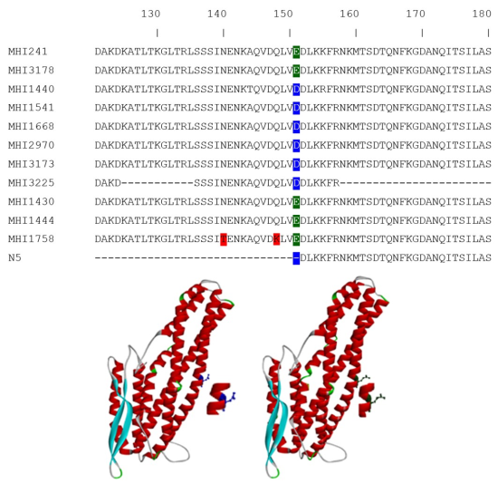
2.2. Sequencing of the NheB Gene Unravels a Point Mutation at Position 151
2.3. Performance of the Mutant Strains in a Lateral Flow Device
2.4. Western Blot Reactivity of the Mutants

3. Discussion and Conclusions
4. Experimental Section
4.1. Bacterial Strains and Culture
4.2. DNA Preparation and Sequencing of NheB
4.3. Indirect and Sandwich EIA
4.4. Duopath® Cereus Enterotoxins Test
4.5. Western Blot
4.6. Cytotoxicity Assay
4.7. Statistics
Supplementary Materials
Acknowledgments
Author Contributions
Conflicts of Interest
References
- Teufel, P.; Bräuning, J.; Hartung, M.; Kleer, J.; Schütt-Abraham, I. Mikrobiologische Aspekte der Ernährung. In Ernährungsbericht 2004; DGE Medien Service: Bonn, Germany, 2006. [Google Scholar]
- Anonymous. The european union summary report on trends and sources of zoonoses, zoonotic agents and food-bourne outbreaks. EFSA J. 2013, 11, 3129. [Google Scholar]
- Dietrich, R.; Moravek, M.; Burk, C.; Granum, P.E.; Martlbauer, E. Production and characterization of antibodies against each of the three subunits of the Bacillus cereus nonhemolytic enterotoxin complex. Appl. Environ. Microbiol. 2005, 71, 8214–8220. [Google Scholar] [CrossRef] [PubMed]
- Jessberger, N.; Dietrich, R.; Bock, S.; Didier, A.; Martlbauer, E. Bacillus cereus enterotoxins act as major virulence factors and exhibit distinct cytotoxicity to different human cell lines. Toxicon 2013, 77, 49–57. [Google Scholar] [CrossRef] [PubMed]
- Kyei-Poku, G.; Gauthier, D.; Pang, A.; van Frankenhuyzen, K. Detection of Bacillus cereus virulence factors in commercial products of bacillus thuringiensis and expression of diarrheal enterotoxins in a target insect. Can. J. Microbiol. 2007, 53, 1283–1290. [Google Scholar] [CrossRef] [PubMed]
- Ehling-Schulz, M.; Messelhausser, U. Bacillus “next generation” diagnostics: Moving from detection toward subtyping and risk-related strain profiling. Front. Microbiol. 2013, 4, 32. [Google Scholar] [CrossRef] [PubMed]
- Lund, T.; Granum, P.E. Characterisation of a non-haemolytic enterotoxin complex from Bacillus cereus isolated after a foodborne outbreak. FEMS Microbiol. Lett. 1996, 141, 151–156. [Google Scholar] [CrossRef] [PubMed]
- Wehrle, E.; Moravek, M.; Dietrich, R.; Burk, C.; Didier, A.; Martlbauer, E. Comparison of multiplex PCR, enzyme immunoassay and cell culture methods for the detection of enterotoxinogenic Bacillus cereus. J. Microbiol. Methods 2009, 78, 265–270. [Google Scholar] [CrossRef] [PubMed]
- Beecher, D.J.; Macmillan, J.D. Characterization of the components of hemolysin BL from Bacillus cereus. Infect. Immun. 1991, 59, 1778–1784. [Google Scholar] [PubMed]
- Hansen, B.M.; Hendriksen, N.B. Detection of enterotoxic Bacillus cereus and Bacillus thuringiensis strains by PCR analysis. Appl. Environ. Microbiol. 2001, 67, 185–189. [Google Scholar] [CrossRef] [PubMed]
- Guinebretiere, M.H.; Broussolle, V.; Nguyen-The, C. Enterotoxigenic profiles of food-poisoning and food-borne Bacillus cereus strains. J. Clin. Microbiol. 2002, 40, 3053–3056. [Google Scholar] [CrossRef] [PubMed]
- Wehrle, E.; Didier, A.; Moravek, M.; Dietrich, R.; Martlbauer, E. Detection of Bacillus cereus with enteropathogenic potential by multiplex real-time PCRbased on SYBR green. Mol. Cell Probes 2010, 24, 124–130. [Google Scholar] [CrossRef] [PubMed]
- Guinebretiere, M.H.; Velge, P.; Couvert, O.; Carlin, F.; Debuyser, M.L.; Nguyen, C. The Ability of Bacillus cereus group strains to cause food poisoning varies according to phylogenetic affiliation (groups I to VII) rather than species affiliation. J. Clin. Microbiol. 2010, 48, 3388–3391. [Google Scholar] [CrossRef] [PubMed]
- Moravek, M.; Dietrich, R.; Buerk, C.; Broussolle, V.; Guinebretiere, M.H.; Granum, P.E.; Nguyen-the, C.; Martlbauer, E. Determination of the toxic potential of Bacillus cereus isolates by quantitative enterotoxin analyses. FEMS Microbiol. Lett. 2006, 257, 293–298. [Google Scholar] [CrossRef] [PubMed]
- Spira, W.M.; Goepfert, J.M. Bacillus cereus-induced fluid accumulation in rabbit ileal loops. Appl. Microbiol. 1972, 24, 341–348. [Google Scholar] [PubMed]
- Glatz, B.A.; Spira, W.M.; Goepfert, J.M. Alteration of vascular permeability in rabbits by culture filtrates of Bacillus cereus and related species. Infect. Immun. 1974, 10, 299–303. [Google Scholar] [PubMed]
- Jessberger, N.; Krey, V.M.; Rademacher, C.; Bohm, M.E.; Mohr, A.K.; Ehling-Schulz, M.; Scherer, S.; Martlbauer, E. From genome to toxicity: A combinatory approach highlights the complexity of enterotoxin production in Bacillus cereus. Front. Microbiol. 2015, 6, 560. [Google Scholar] [PubMed]
- Sievers, F.; Wilm, A.; Dineen, D.; Gibson, T.J.; Karplus, K.; Li, W.; Lopez, R.; McWilliam, H.; Remmert, M.; Soding, J.; et al. Fast, scalable generation of high-quality protein multiple sequence alignments using clustal omega. Mol. Syst. Biol. 2011, 7, 539. [Google Scholar] [PubMed]
- Ganash, M.; Phung, D.; Sedelnikova, S.E.; Lindback, T.; Granum, P.E.; Artymiuk, P.J. Structure of the nhea component of the nhe toxin from Bacillus cereus: Implications for function. PLoS ONE 2013, 8, e74748. [Google Scholar] [CrossRef] [PubMed]
- Williams, L.D.; Burdock, G.A.; Jiménez, G.; Castillo, M. Literature review on the safety of Toyocerin, a non-toxigenic and non-pathogenic Bacillus cereus var. toyoi preparation. Regul. Toxicol. Pharmacol. 2009, 55, 236–246. [Google Scholar] [CrossRef] [PubMed]
- Ceuppens, S.; Boon, N.; Uyttendaele, M. Diversity of Bacillus cereus group strains is reflected in their broad range of pathogenicity and diverse ecological lifestyles. FEMS Microbiol. Ecol. 2013, 84, 433–450. [Google Scholar] [CrossRef] [PubMed]
- Stenfors Arnesen, L.P.; Fagerlund, A.; Granum, P.E. From soil to gut: Bacillus cereus and its food poisoning toxins. FEMS Microbiol. Rev. 2008, 32, 579–606. [Google Scholar] [CrossRef] [PubMed]
- Cadot, C.; Tran, S.L.; Vignaud, M.L.; De Buyser, M.L.; Kolsto, A.B.; Brisabois, A.; Nguyen-The, C.; Lereclus, D.; Guinebretiere, M.H.; Ramarao, N. InhA1, NprA, and HlyII as candidates for markers to differentiate pathogenic from nonpathogenic Bacillus cereus strains. J. Clin. Microbiol. 2010, 48, 1358–1365. [Google Scholar] [CrossRef] [PubMed]
- Doll, V.M.; Ehling-Schulz, M.; Vogelmann, R. Concerted action of sphingomyelinase and non-hemolytic enterotoxin in pathogenic Bacillus cereus. PLoS ONE 2013, 8, e61404. [Google Scholar] [CrossRef] [PubMed]
- Ceuppens, S.; Rajkovic, A.; Hamelink, S.; van de Wiele, T.; Boon, N.; Uyttendaele, M. Enterotoxin production by Bacillus cereus under gastrointestinal conditions and their immunological detection by commercially available kits. Foodborne Pathog. Dis. 2012, 9, 1130–1136. [Google Scholar] [CrossRef] [PubMed]
- Didier, A.; Dietrich, R.; Gruber, S.; Bock, S.; Moravek, M.; Nakamura, T.; Lindback, T.; Granum, P.E.; Martlbauer, E. Monoclonal antibodies neutralize Bacillus cereus Nhe enterotoxin by inhibiting ordered binding of its three exoprotein components. Infect. Immun. 2012, 80, 832–838. [Google Scholar] [CrossRef] [PubMed]
- Makins, C.; Pickering, A.V.; Mariani, C.; Wolthers, K.R. Mutagenesis of a conserved glutamate reveals the contribution of electrostatic energy to adenosylcobalamin Co–C bond homolysis in ornithine 4,5-aminomutase and methylmalonyl-CoA mutase. Biochemistry 2013, 52, 878–888. [Google Scholar] [CrossRef] [PubMed]
- Nice, T.J.; Strong, D.W.; McCune, B.T.; Pohl, C.S.; Virgin, H.W. A single-amino-acid change in murine norovirus NS1/2 is sufficient for colonic tropism and persistence. J. Virol. 2013, 87, 327–334. [Google Scholar] [CrossRef] [PubMed]
- Parent, A.; Caux-Thang, C.; Signor, L.; Clemancey, M.; Sethu, R.; Blondin, G.; Maldivi, P.; Duarte, V.; Latour, J.M. Single glutamate to aspartate mutation makes ferric uptake regulator (Fur) as sensitive to H2O2 as peroxide resistance regulator (PerR). Angew. Chem. Int. Ed. 2013, 52, 10339–10343. [Google Scholar] [CrossRef] [PubMed]
- Böhm, M.L.; Huptas, C.; Krey, M.L.; Scherer, S. Massive horizontal gene transfer, strictly vertical inheritance and ancient duplications differentially shape the evolution of Bacillus cereus enterotoxin operons hbl, cytK and nhe. BMC Evol. Biol. 2015, in press. [Google Scholar]
© 2015 by the authors; licensee MDPI, Basel, Switzerland. This article is an open access article distributed under the terms and conditions of the Creative Commons Attribution license (http://creativecommons.org/licenses/by/4.0/).
Share and Cite
Didier, A.; Jeßberger, N.; Krey, V.; Dietrich, R.; Scherer, S.; Märtlbauer, E. The Mutation Glu151Asp in the B-Component of the Bacillus cereus Non-Hemolytic Enterotoxin (Nhe) Leads to a Diverging Reactivity in Antibody-Based Detection Systems. Toxins 2015, 7, 4655-4667. https://doi.org/10.3390/toxins7114655
Didier A, Jeßberger N, Krey V, Dietrich R, Scherer S, Märtlbauer E. The Mutation Glu151Asp in the B-Component of the Bacillus cereus Non-Hemolytic Enterotoxin (Nhe) Leads to a Diverging Reactivity in Antibody-Based Detection Systems. Toxins. 2015; 7(11):4655-4667. https://doi.org/10.3390/toxins7114655
Chicago/Turabian StyleDidier, Andrea, Nadja Jeßberger, Victoria Krey, Richard Dietrich, Siegfried Scherer, and Erwin Märtlbauer. 2015. "The Mutation Glu151Asp in the B-Component of the Bacillus cereus Non-Hemolytic Enterotoxin (Nhe) Leads to a Diverging Reactivity in Antibody-Based Detection Systems" Toxins 7, no. 11: 4655-4667. https://doi.org/10.3390/toxins7114655
APA StyleDidier, A., Jeßberger, N., Krey, V., Dietrich, R., Scherer, S., & Märtlbauer, E. (2015). The Mutation Glu151Asp in the B-Component of the Bacillus cereus Non-Hemolytic Enterotoxin (Nhe) Leads to a Diverging Reactivity in Antibody-Based Detection Systems. Toxins, 7(11), 4655-4667. https://doi.org/10.3390/toxins7114655