Twenty-Five Years of Human Urinary Biomonitoring of Mycotoxins: Analytical Advances and Applications in Risk Assessment

 ,
,  ,
,  , , , and
Abstract
1. Introduction
2. Mycotoxin Biomonitoring
3. Analytical Methods for Mycotoxin Analysis in Human Urine

| Mycotoxins (Number: Name) | Extraction Details | Detection Method | LOD (Range) ng/mL | LOQ (Range) ng/mL | Year of Publication | Ref. |
|---|---|---|---|---|---|---|
| 10: OTA, 4-OH-OTA, OTB, AFB1, AFM1, AFL, AFB2, AFM2, AFG1, AFG2 | LLE/SPE | HPLC-FLD | NA | NA | 2001 | [99] |
| 1: OTA | IAC | HPLC | 0.005 | NA | 2001 | [100] |
| 1: OTA | IAC | HPLC-FLD | NA | 0.01 | 2001 | [101] |
| 1: DON | IAC | HPLC-ESI-MS | NA | 4 | 2003 | [102] |
| 1: OTA | LLE/SPE | HPLC | 0.3 | 0.9 | 2003 | [103] |
| 1: OTA | IAC | HPLC | 0.004 | 0.006 | 2005 | [104] |
| 1: OTA | IAC | HPLC-FLD | NA | 0.02 | 2006 | [105] |
| 1: OTA | SPME | LC-FLD | 0.01 | 0.05 | 2007 | [106] |
| 1: DON | IAC | HPLC-MS | NA | 0.6 | 2007 | [107] |
| 1: OTA | SPME | LC-MS/MS | 0.3 | 0.7 | 2008 | [108] |
| 1: OTA | IAC | HPLC-FLD | NA | 0.007 | 2008 | [109] |
| 6: ZEA, α-ZEL, β-ZEL, α-ZAL, β-ZAL, ZAN | SPE | HPLC-MWCNTs | 1.3–1.4 | 4.2–4.8 | 2008 | [110] |
| 1: FB1 | SPE | HPLC-ESI-MS | 0.02 | NA | 2008 | [111] |
| 1: OTA | IAC | HPLC-FLD | NA | 0.008 | 2009 | [112] |
| 2: OTA, OTα | LLE | LC-FLD | 0.02 | 0.05 | 2010 | [113] |
| 1: OTA | IAC | HPLC-FLD | NA | 0.008 | 2010 | [114] |
| 2: FB1, FB2 | IAC | LC-MS/MS | 5 | 10 | 2010 | [115] |
| 5: OTA, OTα, AFM1, FB1, FB2 | IAC | LC-MS/MS | 0.001–0.045 | 0.004–0.135 | 2010 | [116] |
| 1: OTA | IAC | HPLC-FLD | 0.006 | 0.018 | 2010 | [117] |
| 1: DON | IAC | HPLC-MS | 0.5 | NA | 2010 | [118] |
| 2: OTA, OTα | LLE | HPLC | 0.023–0.034 | 0.076–0.112 | 2011 | [119] |
| 11: DON, T-2, HT-2, ZEA, OTA, AFB1, AFB2, AFG1, AFG2, FB1, FB2 | IAC | LC-MS/MS | 0.4–10 | 1.2–35 | 2011 | [120] |
| 1: DON-3-GlcA | LLE (dilute and shoot) | LC-MS/MS | 3 | 10 | 2011 | [121] |
| 7: AFM1, OTA, DON, DOM-1, α-ZEL, β-ZEL, FB1 | IAC-SPE | LC-MS/MS | 0.01–2.2 | 0.02–4.4 | 2011 | [122] |
| 1: OTA | IAC | HPLC-FLD | 0.0024 | 0.008 | 2012 | [123] |
| 15: DON, DON-3-GlcA, DON-15-GlcA, DOM-1, NIV, T-2, HT-2, ZEA, ZEA-14-GlcA, α-ZEL, β-ZEL, FB1, FB2, OTA, AFM1 | SPE | LC-ESI-MS/MS | 0.05–10 | 0.17–67 | 2012 | [124] |
| 18: AFB1, AFM1, AFB1-N7-Gua, OTA, OTα, 4-OH-OTA, DON, DOM-1, DON-3-GlcA, ZEA, α-ZEL, β-ZEL, ZEA-14-GlcA, T-2, HT-2, CIT, FB1, HFB1 | LLE-SPE | UHPLC-MS/MS | 0.01–3.65 | 0.02–7.3 | 2012 | [125] |
| 2: CIT, DH-CIT | IAC-SPE | LC–MS/MS | 0.02–0.05 | 0.05–0.1 | 2013 | [126] |
| 15: FB1, FB2, AFM1, OTA, DON, DON-3-GlcA, DON-15-GlcA, DOM-1, NIV, T-2, HT-2, ZEA, ZEA-14-GlcA, α-ZEL, β-ZEL | LLE (dilute and shoot) | LC–MS/MS | 0.002–0.45 | 0.007–1.51 | 2013 | [127] |
| 14: AFM1, FB1, FB2, OTA, DON, DON-3-GlcA, DOM-1, NIV, T-2, HT-2, ZEA, ZEA-14-GlcA, α-ZEL, β-ZEL | LLE (dilute and shoot) | LC–MS/MS | 0.05–12 | 0.15–40 | 2014 | [98] |
| 15: DOM-1, DON, 3-AcDON, FUS-X, DAS, NIV, NEO, HT-2, T-2, ZAN, α-ZAL, β-ZAL, ZEA, α-ZEL, β-ZEL | d-SPE | GC-QqQ-MS/MS | 0.12–4 | 0.25–8 | 2014 | [93] |
| 23: DON, DON-3-GlcA, T-2, HT-2, HT-2-4-GlcA, FB1, AFB1, AFB2, AFG1, AFG2, AFM1, ZEA, ZAN, α-ZEL, β-ZEL, ZEA-14-GlcA, ZAN-14-GlcA, α-ZEL-14-GlcA, β-ZEL-14-GlcA, OTA, OTα, ENB, DH-CIT | dilute and shoot | LC-MS/MS | 0.0005–0.3125 | 0.0013–0.3125 | 2014 | [128] |
| 2: CIT, DH-CIT | IAC | LC–MS/MS | 0.02–0.05 | 0.05–0.1 | 2015 | [129] |
| 23: DON, DON-3-GlcA, T-2, HT-2, HT-2-4-GlcA, FB1, AFB1, AFB2, AFG1, AFG2, AFM1, ZEA, ZAN, α-ZEL, β-ZEL, ZEA-14-GlcA, ZAN-14-GlcA, α-ZEL-14-GlcA, β-ZEL-14-GlcA, OTA, OTα, ENB, DH-CIT | dilute and shoot | LC-MS/MS | 0.000125–0.45 | 0.0005–0.9 | 2015 | [130] |
| 32: AFB1, AFB2, AFG1, AFG2, OTA, AFM1, DAS, Fus-X, 3-AcDON, 15-AcDON, β-ZEL, α-ZEL, CIT, DH-CIT, OTα, DOM-1, FB1, FB2, FB3, DON, ZEA, T-2, HT-2, DON-3-GlcA, DOM-GlcA, ZEA-14-GlcA, β-ZEL-7-GlcA, β-ZEL-14-GlcA, α-ZEL-7-GlcA, α-ZEL-14-GlcA, 15-AcDON-3-GlcA, 3-AcDON-15-GlcA | Filter-shot/ IAC (only for OTA, CIT, AFM1) | LC-MS/MS | 0.001–0.2 | 0.003–0.5 | 2015 | [131] |
| 9: CIT, DH-CIT, DON, DOM-1, OTA, OTα, ZEA, α-ZEL, β-ZEL | IAC/LLE (only for OTA) | HPLC/LC–MS/MS | 0.002–0.150 | 0.005–0.300 | 2016 | [132] |
| 2: AFM1, AFB1-N7-Gua | IAC | HPLC-FLD | 0.000075–0.003 | 0.00025–0.01 | 2016 | [97] |
| 2: ZEA, α-ZEL | SLE | UPLC-MS/MS | NA | NA | 2016 | [90] |
| 3: OTA, OTA-8-GlcA, open lactone OTA-8-GlcA | LLE | LC–MS/MS | 0.03–0.5 | 0.05–1 | 2017 | [133] |
| 2: DON, DOM-1 | IAC | LC-MS/MS | 0.12–0.25 | NA | 2017 | [134] |
| 2: OTA, OTα | LLE | HPLC-FLD | 0.01 | 0.02 | 2017 | [135] |
| 12: NIV, DON, DOM-1, OTA, AFM1, CIT, DH-CIT, FB1, ZEA, α-ZEL, β-ZEL, AOH | IAC-SPE | UHPLC-MS/MS | 0.0003–0.3 | 0.001–0.5 | 2018 | [136] |
| 6: ZEA, ZAN, α-ZEL, β-ZEL, α-ZAL, β-ZAL | LLE-SPE | UHPLC-MS/MS | 0.02–0.06 | 0.05–0.2 | 2018 | [137] |
| 3: DON, DON-GlcA, DOM-1 | IAC | LC-QTOF-MS | NA | 0.25–0.5 | 2018 | [138] |
| 26: AFB1, AFB2, AFG1, AFG2, AFB1-lysine, AFM1, AFM2, OTA, OTα, FB1, T-2, HT-2, DON, 3-AcDON, 15-AcDON, DON-3-GlcA, DON-15-GlcA, FUS-X, ZEA, ZAN, α-ZEL, β-ZEL, α-ZAL, β-ZAL, ZEA-14-GlcA, ZAN-14-GlcA | LLE | UHPLC-MS/MS | 0.03–0.5 | 0.1–1 | 2019 | [139] |
| 3: OTA, CIT, DH-CIT | IAC | HPLC-FLD | 0.001–0.05 | 0.002–0.1 | 2019 | [140] |
| 37: NIV, DON, DOM-1, 3-AcDON, 15-AcDON, DON-3G, CIT, OTA, OTα, T-2, HT-2, T-2 triol, T-2 tetraol, FB1, FB2, FB3, HFB1, ZEA, ZAN, α-ZEL, β-ZEL, α-ZAL, β-ZAL, roquefortine C, DAS, NEO, FUS-X, STG, PAT, AOH, AME, DON-3-GlcA, DON-15-GlcA, α-ZEL-GlcA, β-ZEL-GlcA, ZEA-14-sulfate, ZEA-14-GlcA | QuEChERS-based procedure | LC–MS/MS | 0.01–2.6 | 0.02–4.9 | 2019 | [91] |
| 2: DON, DOM-1 | IAC | LC-MS | 0.25 | 0.5 | 2019 | [141] |
| 3: AFM1, DON, DOM-1 | IAC | HPLC-FLD (AFM1) LC–MS/MS (DON, DOM-1) | 0.0017–0.16 | 0.005–0.3 | 2020 | [142] |
| 6: ZEA, ZAN, α-ZEL, β-ZEL, α-ZAL, β-ZAL | IAC | LC–MS/MS | 0.02–0.06 | NA | 2020 | [143] |
| 2: PAT, CIT | QuEChERS-based procedure | LC–MS/MS | 0.14–1.67 | 0.2–2.65 | 2020 | [144] |
| 2: DON, AFM1 | IAC | LC–MS/MS | 0.002–0.25 | 0.005–0.5 | 2020 | [145] |
| 10: DON, 3-AcDON, 15-AcDON, FUS-X, NIV, NEO, ZEA, ZAN, T-2, HT-2 | DLLME | GC–MS/MS | 0.12–4 | 0.25–8 | 2021 | [146] |
| 11: AFB1, AFM1, OTA, OTα, DON, ZEA, α-ZEL, β-ZEL, FB1, HT-2, T-2 | IAC, SPE (only for DON) | LC-HRMS | NA | 0.01–0.5 | 2021 | [147] |
| 12: FB1, FB2, DON, DOM-1, ZEA, α-ZEL, β-ZEL, OTA, AFM1, T-2, HT-2, NIV | IAC + HLB column | LC–MS/MS | 0.0003–6.25 | 0.001–20.8 | 2021 | [148] |
| 14: AFB1, AFB2, AFG2, AFM1, CIT, CPA, FB1, OTA, OTB, STG, T-2, α-ZEL, β-ZEL, roquefortine | LLE | HPLC–ESI–HRAMS | NA | 0.2–0.5 | 2021 | [149] |
| 4: DON, DOM-1, DON-3-GlcA, DON-15-GlcA | IAC | LC-HRMS | 0.005–0.009 | 0.015–0.27 | 2021 | [150] |
| 1: OTA | IAC | HPLC-FLD | 0.007 | 0.01 | 2021 | [151] |
| 9: AFB1, AFM1, OTα, OTA, DON, DOM-1, ZEA, α-ZEL, β-ZEL | SPE | UHPLC-MS/MS | 0.08–6.6 | 0.1–20.1 | 2021 | [152] |
| 11: AFM1, ALT, AME, AOH, CIT, DH-CIT, FB1, OTA, ZEA, α-ZEL, β-ZEL | SPE | UHPLC-MS/MS | 0.0036–0.27 | 0.012–0.91 | 2021 | [153] |
| 14: CIT, DH-CIT, DON, FB1, T-2, HT-2, OTA, 2′R-OTA, OTα, TeA + allo-TeA, ZEA, ZAN, α-ZEL, β-ZEL | dried urine spots/ dilute and shoot | HPLC–ESI-MS/MS | 0.004–1.4 | 0.013–4.5 | 2021 | [95] |
| 6: AFB2, AFG2, OTA, OTB, ZEA, α-ZEL | QuEChERS-based procedure | LC-ESI-QTOF-MS | 1.5–5 | 3–10 | 2022 | [87] |
| 1: DON | IAC | HPLC-MS/MS | NA | 0.3 | 2023 | [154] |
| 1: DON | NA | HPLC-MS/LC-HRMS | 0.04–0.2 | 0.05–0.5 | 2024 | [155] |
| 23: AFB1, AFB2, AFG1, AFG2, AFM1, DON, DOM-1, 15-AcDON, 3-AcDON, ZEA, α-ZEL, β-ZEL, ZAN, OTα, OTA, OTB, BEA, ENA, ENA1, ENB, ENB1, T-2, HT-2 | SALLE | LC-ESI-QTOF-MS | NA | 0.1–5 | 2024 | [88] |
| 22: 15-AcDON, 3-AcDON, AFB1, AFB2, AFG1, AFG2, AFM1, α-ZEL, β-ZEL, DOM-1, DON, ENA, ENA1, ENB, ENB1, HT-2, T-2, OTα, OTA, OTB, ZEA, ZAN | SALLE | LC-ESI-QTOF-MS | 0.01–0.750 | 0.03–2.5 | 2024 | [89] |
| 1: AFM1 | filtration and dilution | ELISA | 0.988 | NA | 2025 | [156] |
| 10: AFM1, OTA, FB1, FB2, ZEA, α-ZEL, β-ZEL, DON, T-2, HT-2 | IAC | UPLC-MS/MS | 0.01–0.46 | 0.03–1.32 | 2025 | [157] |
| 3: T-2, HT-2, T-2 triol | IAC | UPLC-MS/MS | 0.013–0.10 | 0.05–0.30 | 2025 | [158] |
| 12: AFB1, AFB2, AFG1, AFG2, AFM1, ZEA, ZAN, OTA, AME, CIT, STG, AOH | dilute and shoot | UHPLC-MS/MS | NA | 0.005–0.5 | 2025 | [96] |
| 36: AFM1, STG, FB1, FB2, CIT, DH-CIT, OTA, 2’R-OTA, OTα, 10-OH-OTA, OTB-NAC, ZEA, ZEA-14-GlcA, α-ZEL, β-ZEL, α-ZEL-14-GlcA, β-ZEL-14-GlcA, ZAN-14-GlcA, T-2, HT-2, HT-2-3-GlcA, HT-2-4-GlcA, DON, DON-3-GlcA, ALT, AME, AOH, TeA + allo-TeA, CPA, PENA, PAX, BEA, ENA, ENA1, ENB, ENB1 | online SPE | LC-MS/MS | 0.001–0.833 | 0.002–5.0 | 2025 | [94] |
| 10: AFM1, FB1, OTA, DON, CIT, NIV, ZEA, DHC, α-ZEL, β-ZEL | SPE | UHPLC-ESI-MS/MS | 0.0005–0.05 | 0.01–0.5 | 2025 | [159] |
| 10: AME, AOH, ZEA, ZAN, α-ZAL, β-ZAL, α-ZEL, β-ZEL, ZEA-14-GlcA, ZEA-14-sulfate | LLE | UHPLC-MS/MS | 0.0029–0.22 | 0.010–0.74 | 2025 | [160] |
4. Occurrence of Mycotoxins in Human Urine—Step-by-Step Review
4.1. Aflatoxins (AFs)
4.2. Ochratoxin A (OTA)
4.3. Zearalenone (ZEA)
4.4. Trichothecenes
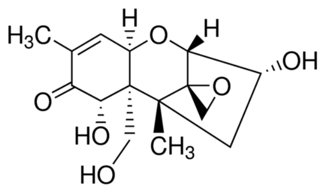
4.4.1. Deoxynivalenol (DON)
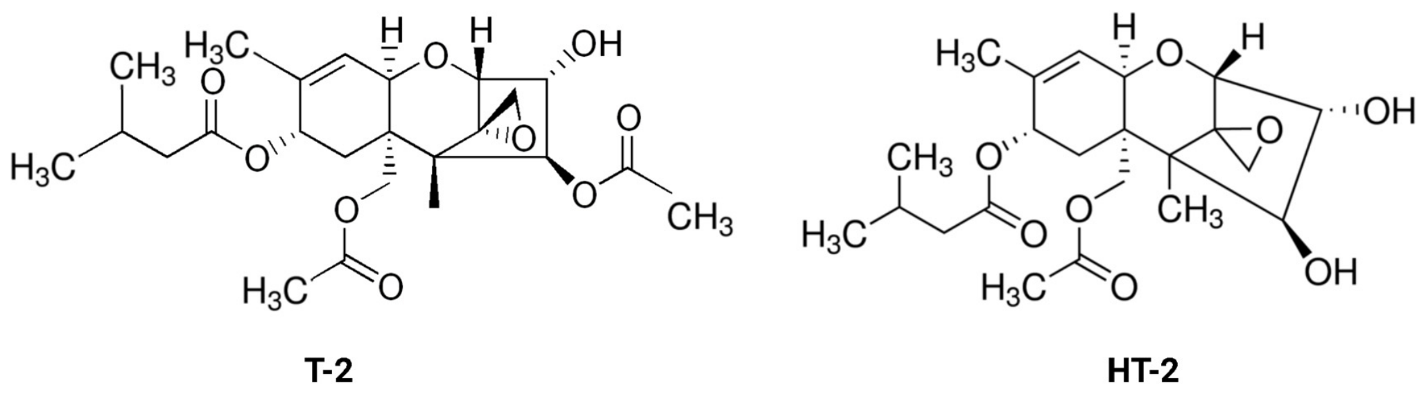
4.4.2. T-2 Toxin (T-2) and HT-2 Toxin (HT-2)
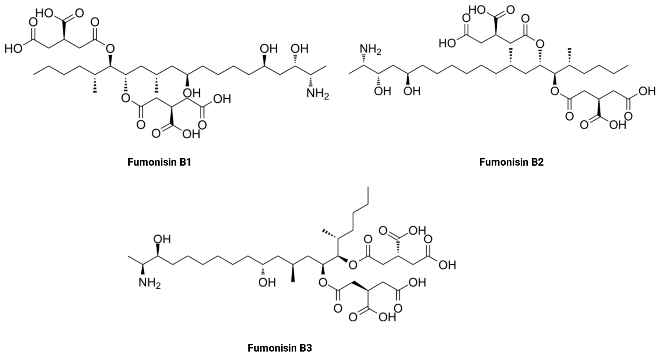
4.5. Fumonisins (FUMOs)
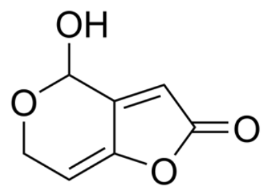
4.6. Patulin (PAT)
4.7. Citrinin (CIT)
4.8. Emerging Mycotoxins
5. Mycotoxin Exposure Assessment Using Human Urine Analysis
- risk assessment (a thorough examination of the prevalence of known or possible harmful health effects from dietary risks in humans or animals),
- risk management (a process of scaling policy alternatives considering risk assessment and strategies of surveillance and regulatory responses),
- food consumption surveys,
- food monitoring studies,
- total diet studies,
- duplicate diet studies [80].
6. Conclusions
7. Materials and Methods
Author Contributions
Funding
Institutional Review Board Statement
Informed Consent Statement
Data Availability Statement
Acknowledgments
Conflicts of Interest
Abbreviations
| 10-OH-OTA | 10-Hydroxy-ochratoxin A |
| 15-AcDON | 15-Acetyl-deoxynivalenol |
| 15-AcDON-3-GlcA | 15-Acetyl-deoxynivalenol-3-glucuronide |
| 2′R-OTA | 2′R-Ochratoxin A |
| 3-AcDON | 3-Acetyl-deoxynivalenol |
| 3-AcDON-15-GlcA | 3-Acetyl-deoxynivalenol-15-glucuronide |
| 4-OH-OTA | 4-Hydroxy-ochratoxin A |
| ADI | Acceptable daily intake |
| ADME | Absorption, distribution, metabolism, and excretion |
| AFB1 | Aflatoxin B1 |
| AFB1-N7-Gua | Aflatoxin B1-N7-guanine |
| AFB2 | Aflatoxin B2 |
| AFG1 | Aflatoxin G1 |
| AFG2 | Aflatoxin G2 |
| AFL | Aflatoxicol |
| AFM1 | Aflatoxin M1 |
| AFM2 | Aflatoxin M2 |
| AFs | Aflatoxins |
| ALARA | As low as reasonably achievable |
| ALT | Altenuene |
| AME | Alternariol monomethyl ether |
| AOH | Alternariol |
| BEA | Beauvericin |
| BMDL10 | Benchmark Dose Lower Confidence Limit 10% |
| bw | Body weight |
| CAC | Codex Alimentarius Commission |
| CIT | Citrinin |
| CPA | Cyclopiazonic acid |
| DAS | Diacetoxyscirpenol |
| DH-CIT | Dihydrocitrinone |
| DLLME | Dispersive liquid–liquid microextraction |
| d-SPE | Dispersive solid-phase extraction |
| DOM-1 | Deepoxy-deoxynivalenol |
| DOM-GlcA | Deepoxy-deoxynivalenol-glucuronides |
| DON | Deoxynivalenol |
| DON-15-GlcA | Deoxynivalenol-15-O-β-glucuronide |
| DON-3G | Deoxynivalenol-3-glucoside |
| DON-3-GlcA | Deoxynivalenol-3-O-β-glucuronide |
| DON-GlcA | Deoxynivalenol-glucuronides |
| EDI | Estimated daily intake |
| EFSA | European Food Safety Authority |
| ELISA | Enzyme-linked immunosorbent assay |
| ENA | Enniatin A |
| ENA1 | Enniatin A1 |
| ENB | Enniatin B |
| ENB1 | Enniatin B1 |
| ENs | Enniatins |
| FAO | Food and Agriculture Organization |
| FB1 | Fumonisin B1 |
| FB2 | Fumonisin B2 |
| FB3 | Fumonisin B3 |
| FDA | Food and Drug Administration |
| FLD | Fluorescence detection |
| FUMOs | Fumonisins |
| FUS-X | Fusarenone-X |
| GC | Gas chromatography |
| GC–MS/MS | Gas chromatography–tandem mass spectrometry |
| GC–QqQ–MS/MS | Gas chromatography–triple quadrupole–tandem mass spectrometry |
| HBM | Human biomonitoring |
| HBM4EU | European Human Biomonitoring Initiative |
| HFB1 | Hydrolyzed fumonisin B1 |
| HLB | Hydrophilic–lipophilic balanced |
| HPLC | High-performance liquid chromatography |
| HPLC-ESI-MS | High-performance liquid chromatography–electrospray ionization–single quadrupole mass spectrometry |
| HPLC-FLD | High-performance liquid chromatography–fluorescence detection |
| HPLC-MS | High-performance liquid chromatography–mass spectrometry |
| HPLC-MWCNTs | High-performance liquid chromatography–multi-wall carbon nanotubes detection |
| HQ | Hazard Quotient |
| HRMS | High-resolution mass spectrometry |
| HT-2 | HT-2 toxin |
| HT-2-3-GlcA | HT-2 toxin-3-glucuronide |
| HT-2-4-GlcA | HT-2 toxin-4-glucuronide |
| IAC | Immunoaffinity column |
| IARC | International Agency for Research on Cancer |
| JECFA | Joint FAO/WHO Expert Committee on Food Additives |
| LB | Lower bound |
| LC | Liquid chromatography |
| LC-ESI-QTOF-MS | Liquid chromatography coupled to quadrupole time-of-flight mass spectrometry with electrospray ionization |
| LC-FLD | Liquid chromatography–fluorescence detection |
| LC-HRMS | Liquid chromatography–high-resolution mass spectrometry |
| LC-MS/MS | Liquid chromatography–tandem mass spectrometry |
| LLE | Liquid–liquid extraction |
| LOD | Limit of detection |
| LOQ | Limit of quantification |
| ML | Maximum level |
| MOE | Margin of exposure |
| MON | Moniliformin |
| MOS | Margin of safety |
| MPL | Maximum permitted level |
| MS | Mass spectrometry |
| MS/MS | Tandem mass spectrometry |
| MWCNTs | Multi-wall carbon nanotubes |
| NEO | Neosolaniol |
| NIV | Nivalenol |
| NOAEL | No-observed-adverse-effect level |
| NRC | National Research Council |
| OTA | Ochratoxin A |
| OTA-8-GlcA | Ochratoxin A-8-β-glucuronide |
| OTB | Ochratoxin B |
| OTC | Ochratoxin C |
| OTB-NAC | Ochratoxin-N-acetyl-L-cysteine |
| OTα | Ochratoxin α |
| OTβ | Ochratoxin β |
| PAT | Patulin |
| PAX | Paxilline |
| PDI | Probable daily intake |
| PENA | Penitrem A |
| PMTDI | Provisional maximum tolerable daily intake |
| QuEChERS | Quick, Easy, Cheap, Effective, Rugged, and Safe |
| RASFF | Rapid Alert System for Food and Feed |
| rpm | Revolutions per minute |
| SALLE | Salting-out-assisted liquid–liquid extraction |
| SLE | Supported liquid extraction |
| SPE | Solid-phase extraction |
| SPME | Solid-phase microextraction |
| STG | Sterigmatocystin |
| T-2 | T-2 toxin |
| TeA | Tenuazonic acid |
| TDI | Tolerable daily intake |
| UB | Upper bound |
| UHPLC | Ultra-high-performance liquid chromatography |
| UHPLC-MS/MS | Ultra-high-performance liquid chromatography–tandem mass spectrometry |
| USA | United States of America |
| WHO | World Health Organization |
| ZAN | Zearalanone |
| ZAN-14-GlcA | Zearalanone-14-glucuronide |
| ZEA | Zearalenone |
| ZEA-14-GlcA | Zearalenone-14-O-β-glucuronide |
| ZEA-14-sulfate | Zearalenone-14-sulfate |
| α-ZAL | α-Zearalanol |
| α-ZEL | α-Zearalenol |
| α-ZEL-GlcA | α-Zearalenol-glucuronides |
| β-ZAL | β-Zearalanol |
| β-ZEL | β-Zearalenol |
| β-ZEL-GlcA | β-Zearalenol-glucuronides |
References
- Coppock, R.W.; Dziwenka, M. Mycotoxins. In Biomarkers in Toxicology; Gupta, R., Ed.; Elsevier Academic Press: Medford, MA, USA, 2014; pp. 549–562. [Google Scholar]
- Khan, R. Mycotoxins in Food: Occurrence, Health Implications, and Control Strategies-A Comprehensive Review. Toxicon 2024, 248, 108038. [Google Scholar] [CrossRef] [Scilit] [PubMed]
- Pitt, J.I. Chapter 30-Mycotoxins. In Foodborne Infections and Intoxications, 4th ed.; Morris, J.G., Potter, M.E., Eds.; Food Science and Technology; Academic Press: San Diego, CA, USA, 2013; pp. 409–418. [Google Scholar]
- Stein, R.A.; Bulboacӑ, A.E. Chapter 21-Mycotoxins. In Foodborne Diseases, 3rd ed.; Dodd, C.E.R., Aldsworth, T., Stein, R.A., Cliver, D.O., Riemann, H.P., Eds.; Academic Press: Cambridge, MA, USA, 2017; pp. 407–446. [Google Scholar]
- Kotowicz, N.K.; Frąc, M.; Lipiec, J. The Importance of Fusarium Fungi in Wheat Cultivation–Pathogenicity and Mycotoxins Production: A Review. J. Anim. Plant Sci. 2014, 21, 3326–3343. [Google Scholar]
- Nesic, K.; Ivanovic, S.; Nesic, V. Fusarial Toxins: Secondary Metabolites of Fusarium Fungi. Rev. Environ. Contam. Toxicol. 2014, 228, 101–120. [Google Scholar] [CrossRef] [Scilit] [PubMed]
- Zahra, N.; Jamil, N.; Ahmad, S.R.; Saeed, M.K.; Kalim, I.; Sheikh, A. A Review of Mycotoxin Types, Occurrence, Toxicity, Detection Methods and Control. Biol. Sci.-PJSIR 2019, 62, 206–218. [Google Scholar] [CrossRef] [Scilit]
- Alshannaq, A.; Yu, J.H. Occurrence, Toxicity, and Analysis of Major Mycotoxins in Food. Int. J. Environ. Res. Public Health 2017, 14, 632. [Google Scholar] [CrossRef] [Scilit] [PubMed]
- Santos Pereira, C.; C Cunha, S.; Fernandes, J.O. Prevalent Mycotoxins in Animal Feed: Occurrence and Analytical Methods. Toxins 2019, 11, 290. [Google Scholar] [CrossRef] [Scilit] [PubMed]
- Popescu, R.G.; Rădulescu, A.L.; Georgescu, S.E.; Dinischiotu, A. Aflatoxins in Feed: Types, Metabolism, Health Consequences in Swine and Mitigation Strategies. Toxins 2022, 14, 853. [Google Scholar] [CrossRef] [Scilit] [PubMed]
- Stoev, S.D. Food Security, Underestimated Hazard of Joint Mycotoxin Exposure and Management of the Risk of Mycotoxin Contamination. Food Control 2024, 159, 110235. [Google Scholar] [CrossRef] [Scilit]
- Alina Romina, M. Implications of Mycotoxins in Food Safety. In Mycotoxins and Food Safety-Recent Advances; Alina Romina, M., Ed.; IntechOpen: London, UK, 2022. [Google Scholar]
- Nagda, A.; Meena, M. Alternaria Mycotoxins in Food and Feed: Occurrence, Biosynthesis, Toxicity, Analytical Methods, Control and Detoxification Strategies. Food Control 2024, 158, 110211. [Google Scholar] [CrossRef] [Scilit]
- Kolawole, O.; Siri-Anusornsak, W.; Petchkongkaew, A.; Elliott, C. A Systematic Review of Global Occurrence of Emerging Mycotoxins in Crops and Animal Feeds, and Their Toxicity in Livestock. Emerg. Contam. 2024, 10, 100305. [Google Scholar] [CrossRef] [Scilit]
- European Commission. Food Safety. The EU Agri-Food Fraud Network. Reports and publications. The 2023 Annual Report Alert and Cooperation Network. Available online: https://food.ec.europa.eu/food-safety/eu-agri-food-fraud-network/reports-and-publications_en#alert-and-cooperation-network-acn (accessed on 28 January 2025).
- CAST. Mycotoxins: Risks in Plant, Animal, and Human Systems; CAST: Ames, IA, USA, 2003. [Google Scholar]
- Mitchell, N.J.; Bowers, E.; Hurburgh, C.; Wu, F. Potential Economic Losses to the US Corn Industry from Aflatoxin Contamination. Food Addit. Contam. Part A 2016, 33, 540–550. [Google Scholar] [CrossRef] [Scilit] [PubMed]
- Pizzolato Montanha, F.; Anater, A.; Burchard, J.F.; Luciano, F.B.; Meca, G.; Manyes, L.; Pimpão, C.T. Mycotoxins in Dry-Cured Meats: A Review. Food Chem. Toxicol. 2018, 111, 494–502. [Google Scholar] [CrossRef] [Scilit] [PubMed]
- Smith, M.; Madec, S.; Coton, E.; Hymery, N. Natural Co-Occurrence of Mycotoxins in Foods and Feeds and Their in Vitro Combined Toxicological Effects. Toxins 2016, 8, 94. [Google Scholar] [CrossRef] [Scilit] [PubMed]
- Eskola, M.; Kos, G.; Elliott, C.T.; Hajšlová, J.; Mayar, S.; Krska, R. Worldwide Contamination of Food-Crops with Mycotoxins: Validity of the Widely Cited ‘FAO Estimate’ of 25%. Crit. Rev. Food Sci. Nutr. 2020, 60, 2773–2789. [Google Scholar] [CrossRef] [Scilit] [PubMed]
- Garvey, M.; Rowan, N.J. Pathogenic Drug Resistant Fungi: A Review of Mitigation Strategies. Int. J. Mol. Sci. 2023, 24, 1584. [Google Scholar] [CrossRef] [Scilit] [PubMed]
- El-Sayed, R.A.; Jebur, A.B.; Kang, W.; El-Demerdash, F.M. An Overview on the Major Mycotoxins in Food Products: Characteristics, Toxicity, and Analysis. J. Future Foods 2022, 2, 91–102. [Google Scholar] [CrossRef] [Scilit]
- Khan, R.; Anwar, F.; Ghazali, F.M. A Comprehensive Review of Mycotoxins: Toxicology, Detection, and Effective Mitigation Approaches. Heliyon 2024, 10, e28361. [Google Scholar] [CrossRef] [Scilit] [PubMed]
- Suo, Z.; Niu, X.; Wei, M.; Jin, H.; He, B. Latest Strategies for Rapid and Point of Care Detection of Mycotoxins in Food: A Review. Anal. Chim. Acta 2023, 1246, 340888. [Google Scholar] [CrossRef] [Scilit] [PubMed]
- Munkvold, G.P.; Arias, S.; Taschl, I.; Gruber-Dorninger, C. Chapter 9-Mycotoxins in Corn: Occurrence, Impacts, and Management. In Corn: Chemistry and Technology; Serna-Saldivar, S., Ed.; Elsevier Inc.: Oxford, UK; AACC International: St. Paul, MN, USA, 2019; pp. 235–287. [Google Scholar]
- Eslamizad, S.; Yazdanpanah, H.; Hadian, Z.; Tsitsimpikou, C.; Goumenou, M.; Shojaee AliAbadi, M.H.; Kamalabadi, M.; Tsatsakis, A. Exposure to Multiple Mycotoxins in Domestic and Imported Rice Commercially Traded in Tehran and Possible Risk to Public Health. Toxicol. Rep. 2021, 8, 1856–1864. [Google Scholar] [CrossRef] [Scilit] [PubMed]
- Dweba, C.C.; Figlan, S.; Shimelis, H.A.; Motaung, T.E.; Sydenham, S.; Mwadzingeni, L.; Tsilo, T.J. Fusarium Head Blight of Wheat: Pathogenesis and Control Strategies. Crop Prot. 2017, 91, 114–122. [Google Scholar] [CrossRef] [Scilit]
- Vogelgsang, S.; Musa, T.; Bänziger, I.; Kägi, A.; Bucheli, T.D.; Wettstein, F.E.; Pasquali, M.; Forrer, H.R. Fusarium Mycotoxins in Swiss Wheat: A Survey of Growers’ Samples between 2007 and 2014 Shows Strong Year and Minor Geographic Effects. Toxins 2017, 9, 246. [Google Scholar] [CrossRef] [Scilit] [PubMed]
- Beccari, G.; Caproni, L.; Tini, F.; Uhlig, S.; Covarelli, L. Presence of Fusarium Species and Other Toxigenic Fungi in Malting Barley and Multi-Mycotoxin Analysis by Liquid Chromatography-High-Resolution Mass Spectrometry. J. Agric. Food Chem. 2016, 64, 4390–4399. [Google Scholar] [CrossRef] [Scilit] [PubMed]
- Stanciu, O.; Juan, C.; Miere, D.; Berrada, H.; Loghin, F.; Mañes, J. First Study on Trichothecene and Zearalenone Exposure of the Romanian Population through Wheat-Based Products Consumption. Food Chem. Toxicol. 2018, 121, 336–342. [Google Scholar] [CrossRef] [Scilit] [PubMed]
- Schabo, D.C.; Freire, L.; Sant’Ana, A.S.; Schaffner, D.W.; Magnani, M. Mycotoxins in Artisanal Beers: An Overview of Relevant Aspects of the Raw Material, Manufacturing Steps and Regulatory Issues Involved. Food Res. Int. 2021, 141, 110114. [Google Scholar] [CrossRef] [Scilit] [PubMed]
- Kunz, B.M.; Wanko, F.; Kemmlein, S.; Bahlmann, A.; Rohn, S.; Maul, R. Development of a Rapid Multi-Mycotoxin LC-MS/MS Stable Isotope Dilution Analysis for Grain Legumes and Its Application on 66 Market Samples. Food Control 2020, 109, 106949. [Google Scholar] [CrossRef] [Scilit]
- Carbonell-Rozas, L.; Albasi, V.; Camardo Leggieri, M.; Dall’Asta, C.; Battilani, P. Apple Mycotoxins: From Orchard to Processed Apple Puree. Fungal Biol. 2024, 128, 2422–2430. [Google Scholar] [CrossRef] [Scilit] [PubMed]
- Łozowicka, B.; Kaczyński, P.; Iwaniuk, P.; Rutkowska, E.; Socha, K.; Orywal, K.; Farhan, J.A.; Perkowski, M. Nutritional Compounds and Risk Assessment of Mycotoxins in Ecological and Conventional Nuts. Food Chem. 2024, 458, 140222. [Google Scholar] [CrossRef] [Scilit] [PubMed]
- Belasli, A.; Herrera, M.; Ariño, A.; Djenane, D. Occurrence and Exposure Assessment of Major Mycotoxins in Foodstuffs from Algeria. Toxins 2023, 15, 449. [Google Scholar] [CrossRef] [Scilit] [PubMed]
- Thanushree, M.P.; Sailendri, D.; Yoha, K.S.; Moses, J.A.; Anandharamakrishnan, C. Mycotoxin Contamination in Food: An Exposition on Spices. Trends Food Sci. Technol. 2019, 93, 69–80. [Google Scholar] [CrossRef] [Scilit]
- Casas-Junco, P.P.; Solís-Pacheco, J.R.; Ragazzo-Sánchez, J.A.; Aguilar-Uscanga, B.R.; Bautista-Rosales, P.U.; Calderón-Santoyo, M. Cold Plasma Treatment as an Alternative for Ochratoxin A Detoxification and Inhibition of Mycotoxigenic Fungi in Roasted Coffee. Toxins 2019, 11, 337. [Google Scholar] [CrossRef] [Scilit] [PubMed]
- Kochman, J.; Jakubczyk, K.; Janda, K. Mycotoxins in Red Wine: Occurrence and Risk Assessment. Food Control 2021, 129, 108229. [Google Scholar] [CrossRef] [Scilit]
- Welke, J.E. Fungal and Mycotoxin Problems in Grape Juice and Wine Industries. Curr. Opin. Food Sci. 2019, 29, 7–13. [Google Scholar] [CrossRef] [Scilit]
- Haque, M.A.; Wang, Y.; Shen, Z.; Li, X.; Saleemi, M.K.; He, C. Mycotoxin Contamination and Control Strategy in Human, Domestic Animal and Poultry: A Review. Microb. Pathog. 2020, 142, 104095. [Google Scholar] [CrossRef] [Scilit] [PubMed]
- Corassin, C.H.; de Oliveira, C.A.F. Mycotoxins in the Dairy Industry. Dairy 2023, 4, 392–394. [Google Scholar] [CrossRef] [Scilit]
- Wang, L.; Zhang, Q.; Yan, Z.; Tan, Y.; Zhu, R.; Yu, D.; Yang, H.; Wu, A. Occurrence and Quantitative Risk Assessment of Twelve Mycotoxins in Eggs and Chicken Tissues in China. Toxins 2018, 10, 477. [Google Scholar] [CrossRef] [Scilit] [PubMed]
- Smaoui, S.; D’Amore, T.; Agriopoulou, S.; Mousavi Khaneghah, A. Mycotoxins in Seafood: Occurrence, Recent Development of Analytical Techniques and Future Challenges. Separations 2023, 10, 217. [Google Scholar] [CrossRef] [Scilit]
- Binder, E.M.; Tan, L.M.; Chin, L.J.; Handl, J.; Richard, J. Worldwide Occurrence of Mycotoxins in Commodities, Feeds and Feed Ingredients. Anim. Feed Sci. Technol. 2007, 137, 265–282. [Google Scholar] [CrossRef] [Scilit]
- Buszewska-Forajta, M. Mycotoxins, Invisible Danger of Feedstuff with Toxic Effect on Animals. Toxicon 2020, 182, 34–53. [Google Scholar] [CrossRef] [Scilit] [PubMed]
- Freire, L.; Sant’Ana, A.S. Modified Mycotoxins: An Updated Review on Their Formation, Detection, Occurrence, and Toxic Effects. Food Chem. Toxicol. 2018, 111, 189–205. [Google Scholar] [CrossRef] [Scilit] [PubMed]
- da Rocha, M.E.B.; da Chagas Oliveira Freire, F.; Feitosa Maia, F.E.; Florindo Guedes, M.I.; Rondina, D. Mycotoxins and Their Effects on Human and Animal Health. Food Control 2014, 36, 159–165. [Google Scholar] [CrossRef] [Scilit]
- Galvez-Llompart, M.; Zanni, R.; Manyes, L.; Meca, G. Elucidating the Mechanism of Action of Mycotoxins through Machine Learning-Driven QSAR Models: Focus on Lipid Peroxidation. Food Chem. Toxicol. 2023, 182, 114120. [Google Scholar] [CrossRef] [Scilit] [PubMed]
- Kim, Y.; Kang, S.; Ajani, O.S.; Mallipeddi, R.; Ha, Y. Predicting Early Mycotoxin Contamination in Stored Wheat Using Machine Learning. J. Stored Prod. Res. 2024, 106, 102294. [Google Scholar] [CrossRef] [Scilit]
- Inglis, A.; Parnell, A.C.; Subramani, N.; Doohan, F.M. Machine Learning Applied to the Detection of Mycotoxin in Food: A Systematic Review. Toxins 2024, 16, 268. [Google Scholar] [CrossRef] [Scilit] [PubMed]
- Moretti, A.; Logrieco, A.F.; Susca, A. Mycotoxins: An Underhand Food Problem. Methods Mol. Biol. 2017, 1542, 3–12. [Google Scholar] [CrossRef] [Scilit] [PubMed]
- Habschied, K.; Kanižai Šarić, G.; Krstanović, V.; Mastanjević, K. Mycotoxins—Biomonitoring and Human Exposure. Toxins 2021, 13, 113. [Google Scholar] [CrossRef] [Scilit] [PubMed]
- European Commission. Communication from the Commission to the European Parliament, the Council, the European Economic and Social Committee and the Committee of the Regions. A Farm to Fork Strategy for a Fair, Healthy and Environmentally-Friendly Food System; European Commission: Brussels, Belgium, 2020. [Google Scholar]
- European Commission. Communication from the Commission to the European Parliament, the European Council, the Council, the European Economic and Social Committee and the Committee of the Regions. The European Green Deal; European Commission: Brussels, Belgium, 2019. [Google Scholar]
- Milani, J.M. Ecological Conditions Affecting Mycotoxin Production in Cereals: A Review. Vet. Med. 2013, 58, 405–411. [Google Scholar] [CrossRef] [Scilit]
- Müller, M.E.H.; Brenning, A.; Verch, G.; Koszinski, S.; Sommer, M. Multifactorial Spatial Analysis of Mycotoxin Contamination of Winter Wheat at the Field and Landscape Scale. Agric. Ecosyst. Environ. 2010, 139, 245–254. [Google Scholar] [CrossRef] [Scilit]
- Champeil, A.; Fourbet, J.F.; Doré, T.; Rossignol, L. Influence of Cropping System on Fusarium Head Blight and Mycotoxin Levels in Winter Wheat. Crop Prot. 2004, 23, 531–537. [Google Scholar] [CrossRef] [Scilit]
- Kos, J.; Anić, M.; Radić, B.; Zadravec, M.; Janić Hajnal, E.; Pleadin, J. Climate Change—A Global Threat Resulting in Increasing Mycotoxin Occurrence. Foods 2023, 12, 2704. [Google Scholar] [CrossRef] [Scilit] [PubMed]
- Casu, A.; Camardo Leggieri, M.; Toscano, P.; Battilani, P. Changing Climate, Shifting Mycotoxins: A Comprehensive Review of Climate Change Impact on Mycotoxin Contamination. Compr. Rev. Food Sci. Food Saf. 2024, 23, e13323. [Google Scholar] [CrossRef] [Scilit] [PubMed]
- De Battistis, F.; Civitelli, C.; Prota, V.; Caloni, F.; Mantovani, A.; Vincentini, O. Enniatins and Beauvericin as Emerging Mycotoxins in the Context of Climate Change in Europe. Toxins 2026, 18, 209. [Google Scholar] [CrossRef] [Scilit] [PubMed]
- Wang, J.; Wu, Y.; Cai, D.; Li, L.; Wang, S.; Zhang, Y.; Han, X.; Wang, S.; Pan, L.; Ye, J. Occurrence, Distribution Characteristics, Risk Assessment, and Climatic Drivers of Type B Trichothecenes and Their Transformation Products in Major Wheat-Producing Areas of China. Toxins 2026, 18, 150. [Google Scholar] [CrossRef] [Scilit] [PubMed]
- Neme, K.; Mohammed, A. Mycotoxin Occurrence in Grains and the Role of Postharvest Management as a Mitigation Strategies. A Review. Food Control 2017, 78, 412–425. [Google Scholar] [CrossRef] [Scilit]
- Yadavalli, R.; Valluru, P.; Raj, R.; Reddy, C.N.; Mishra, B. Biological Detoxification of Mycotoxins: Emphasizing the Role of Algae. Algal Res. 2023, 71, 103039. [Google Scholar] [CrossRef] [Scilit]
- Gamlath, C.J.; Wu, F. AI and Biotechnology to Combat Aflatoxins: Future Directions for Modern Technologies in Reducing Aflatoxin Risk. Toxins 2025, 17, 524. [Google Scholar] [CrossRef] [Scilit] [PubMed]
- FDA. Guidance for Industry: Action Levels for Poisonous or Deleterious Substances in Human Food and Animal Feed; FDA: Silver Spring, MD, USA, 2000.
- FDA. CPG Sec 510.150 Apple Juice, Apple Juice Concentrates, and Apple Juice Products-Adulteration with Patulin; FDA: Silver Spring, MD, USA, 2005.
- FDA. Guidance for Industry and FDA: Advisory Levels for Deoxynivalenol (DON) in Finished Wheat Products for Human Consumption and Grains and Grain By-Products Used for Animal Feed; FDA: Silver Spring, MD, USA, 2010.
- FDA. Guidance for Industry: Fumonisin Levels in Human Foods and Animal Feeds; FDA: Silver Spring, MD, USA, 2001.
- European Commission. Commission Regulation (EC) No 1881/2006 of 19 December 2006 Setting Maximum Levels for Certain Contaminants in Foodstuffs. Off. J. Eur. Union 2006, L364, 5–24. [Google Scholar]
- European Commission. Commission Regulation (EU) 2023/915 of 25 April 2023 on Maximum Levels for Certain Contaminants in Food and Repealing Regulation (EC) No 1881/2006. Off. J. Eur. Union 2023, L119, 103–157. [Google Scholar]
- European Commission. Commission Regulation (EU) 2024/1038 of 9 April 2024 Amending Regulation (EU) 2023/915 as Regards Maximum Levels of T-2 and HT-2 Toxins in Food. Off. J. Eur. Union 2024, L1038. [Google Scholar]
- Awuchi, C.G.; Ondari, E.N.; Nwozo, S.; Odongo, G.A.; Eseoghene, I.J.; Twinomuhwezi, H.; Ogbonna, C.U.; Upadhyay, A.K.; Adeleye, A.O.; Okpala, C.O.R. Mycotoxins’ Toxicological Mechanisms Involving Humans, Livestock and Their Associated Health Concerns: A Review. Toxins 2022, 14, 167. [Google Scholar] [CrossRef] [Scilit] [PubMed]
- Marcelloni, A.M.; Pigini, D.; Chiominto, A.; Gioffrè, A.; Paba, E. Exposure to Airborne Mycotoxins: The Riskiest Working Environments and Tasks. Ann. Work Expo. Health 2024, 68, 19–35. [Google Scholar] [CrossRef] [Scilit] [PubMed]
- Lee, H.J.; Ryu, D. Advances in Mycotoxin Research: Public Health Perspectives. J. Food Sci. 2015, 80, T2970–T2983. [Google Scholar] [CrossRef] [Scilit] [PubMed]
- Arce-López, B.; Lizarraga, E.; Vettorazzi, A.; González-Peñas, E. Human Biomonitoring of Mycotoxins in Blood, Plasma and Serum in Recent Years: A Review. Toxins 2020, 12, 147. [Google Scholar] [CrossRef] [Scilit] [PubMed]
- Arce-López, B.; Lizarraga, E.; López de Mesa, R.; González-Peñas, E. Assessment of Exposure to Mycotoxins in Spanish Children through the Analysis of Their Levels in Plasma Samples. Toxins 2021, 13, 150. [Google Scholar] [CrossRef] [Scilit] [PubMed]
- Owolabi, I.O.; Siwarak, K.; Greer, B.; Rajkovic, A.; Dall’asta, C.; Karoonuthaisiri, N.; Uawisetwathana, U.; Elliott, C.T.; Petchkongkaew, A. Applications of Mycotoxin Biomarkers in Human Biomonitoring for Exposome-Health Studies: Past, Present, and Future. Expo. Health 2024, 16, 837–859. [Google Scholar] [CrossRef] [Scilit]
- Marín, S.; Cano-Sancho, G.; Sanchis, V.; Ramos, A.J. The Role of Mycotoxins in the Human Exposome: Application of Mycotoxin Biomarkers in Exposome-Health Studies. Food Chem. Toxicol. 2018, 121, 504–518. [Google Scholar] [CrossRef] [Scilit] [PubMed]
- Berthiller, F.; Brera, C.; Iha, M.H.; Krska, R.; Lattanzio, V.M.T.; MacDonald, S.; Malone, R.J.; Maragos, C.; Solfrizzo, M.; Stranska-Zachariasova, M.; et al. Developments in Mycotoxin Analysis: An Update for 2015–2016. World Mycotoxin J. 2017, 10, 5–29. [Google Scholar] [CrossRef] [Scilit]
- de Nijs, M.; Mengelers, M.J.B.; Boon, P.E.; Heyndrickx, E.; Hoogenboom, L.A.P.; Lopez, P.; Mol, H.G.J. Strategies for Estimating Human Exposure to Mycotoxins via Food. World Mycotoxin J. 2016, 9, 831–845. [Google Scholar] [CrossRef] [Scilit]
- Hernandez-Vargas, H.; Castelino, J.; Silver, M.J.; Dominguez-Salas, P.; Cros, M.-P.; Durand, G.; Calvez-Kelm, F.L.; Prentice, A.M.; Wild, C.P.; Moore, S.E.; et al. Exposure to Aflatoxin B in Utero Is Associated with DNA Methylation in White Blood Cells of Infants in The Gambia. Int. J. Epidemiol. 2015, 44, 1238–1248. [Google Scholar] [CrossRef] [Scilit] [PubMed]
- Turner, P.C.; Flannery, B.; Isitt, C.; Ali, M.; Pestka, J. The Role of Biomarkers in Evaluating Human Health Concerns from Fungal Contaminants in Food. Nutr. Res. Rev. 2012, 25, 162–179. [Google Scholar] [CrossRef] [Scilit] [PubMed]
- Al-Jaal, B.A.; Jaganjac, M.; Barcaru, A.; Horvatovich, P.; Latiff, A. Aflatoxin, Fumonisin, Ochratoxin, Zearalenone and Deoxynivalenol Biomarkers in Human Biological Fluids: A Systematic Literature Review, 2001–2018. Food Chem. Toxicol. 2019, 129, 211–228. [Google Scholar] [CrossRef] [Scilit] [PubMed]
- McCormick, S.P.; Kato, T.; Maragos, C.M.; Busman, M.; Lattanzio, V.M.T.; Galaverna, G.; Dall-Asta, C.; Crich, D.; Price, N.P.J.; Kurtzman, C.P. Anomericity of T-2 Toxin-Glucoside: Masked Mycotoxin in Cereal Crops. J. Agric. Food Chem. 2015, 63, 731–738. [Google Scholar] [CrossRef] [Scilit] [PubMed]
- Berthiller, F.; Crews, C.; Dall’Asta, C.; Saeger, S.D.; Haesaert, G.; Karlovsky, P.; Oswald, I.P.; Seefelder, W.; Speijers, G.; Stroka, J. Masked Mycotoxins: A Review. Mol. Nutr. Food Res. 2013, 57, 165–186. [Google Scholar] [CrossRef] [Scilit] [PubMed]
- Escrivá, L.; Font, G.; Manyes, L.; Berrada, H. Studies on the Presence of Mycotoxins in Biological Samples: An Overview. Toxins 2017, 9, 251. [Google Scholar] [CrossRef] [Scilit] [PubMed]
- Pallarés, N.; Carballo, D.; Ferrer, E.; Rodríguez-Carrasco, Y.; Berrada, H. High-Throughput Determination of Major Mycotoxins with Human Health Concerns in Urine by LC-Q TOF MS and Its Application to an Exposure Study. Toxins 2022, 14, 42. [Google Scholar] [CrossRef] [Scilit] [PubMed]
- Marín-Sáez, J.; Hernández-Mesa, M.; Gallardo-Ramos, J.A.; Gámiz-Gracia, L.; García-Campaña, A.M. Assessing Human Exposure to Pesticides and Mycotoxins: Optimization and Validation of a Method for Multianalyte Determination in Urine Samples. Anal. Bioanal. Chem. 2024, 416, 1935–1949. [Google Scholar] [CrossRef] [Scilit] [PubMed]
- Gallardo-Ramos, J.A.; Marín-Sáez, J.; Sanchis, V.; Gámiz-Gracia, L.; García-Campaña, A.M.; Hernández-Mesa, M.; Cano-Sancho, G. Simultaneous Detection of Mycotoxins and Pesticides in Human Urine Samples: A 24-h Diet Intervention Study Comparing Conventional and Organic Diets in Spain. Food Chem. Toxicol. 2024, 188, 114650. [Google Scholar] [CrossRef] [Scilit] [PubMed]
- Fleck, S.C.; Churchwell, M.I.; Doerge, D.R.; Teeguarden, J.G. Urine and Serum Biomonitoring of Exposure to Environmental Estrogens II: Soy Isoflavones and Zearalenone in Pregnant Women. Food Chem. Toxicol. 2016, 95, 19–27. [Google Scholar] [CrossRef] [Scilit] [PubMed]
- Martins, C.; Vidal, A.; De Boevre, M.; De Saeger, S.; Nunes, C.; Torres, D.; Goios, A.; Lopes, C.; Assunção, R.; Alvito, P. Exposure Assessment of Portuguese Population to Multiple Mycotoxins: The Human Biomonitoring Approach. Int. J. Hyg. Environ. Health 2019, 222, 913–925. [Google Scholar] [CrossRef] [Scilit] [PubMed]
- Rodríguez-Carrasco, Y.; Moltó, J.C.; Mañes, J.; Berrada, H. Exposure Assessment Approach through Mycotoxin/Creatinine Ratio Evaluation in Urine by GC-MS/MS. Food Chem. Toxicol. 2014, 72, 69–75. [Google Scholar] [CrossRef] [Scilit] [PubMed]
- Rodríguez-Carrasco, Y.; Moltó, J.C.; Mañes, J.; Berrada, H. Development of a GC–MS/MS Strategy to Determine 15 Mycotoxins and Metabolites in Human Urine. Talanta 2014, 128, 125–131. [Google Scholar] [CrossRef] [Scilit] [PubMed]
- Kuhn, M.; Kyei, N.N.A.; Cramer, B.; Humpf, H.U. Online Solid Phase Extraction-LC-MS/MS with Two Step Peak Focusing for Sensitive Multi-Analyte Analysis of Mycotoxins in Urine. Microchem. J. 2025, 219, 115821. [Google Scholar] [CrossRef] [Scilit]
- Schmidt, J.; Lindemann, V.; Olsen, M.; Cramer, B.; Humpf, H.-U. Dried Urine Spots as Sampling Technique for Multi-Mycotoxin Analysis in Human Urine. Mycotoxin Res. 2021, 37, 129–140. [Google Scholar] [CrossRef] [Scilit] [PubMed]
- Peris-Camarasa, B.; Pardo, O.; Dualde, P.; Coscollà, C. Multi-Mycotoxin Determination in Human Urine by UHPLC-MS/MS: An Environmentally Friendly and High-Throughput Approach. J. Chromatogr. A 2025, 1760, 466317. [Google Scholar] [CrossRef] [Scilit] [PubMed]
- Jager, A.V.; Tonin, F.G.; Baptista, G.Z.; Souto, P.C.M.C.; Oliveira, C.A.F. Assessment of Aflatoxin Exposure Using Serum and Urinary Biomarkers in São Paulo, Brazil: A Pilot Study. Int. J. Hyg. Environ. Health 2016, 219, 294–300. [Google Scholar] [CrossRef] [Scilit] [PubMed]
- Ezekiel, C.N.; Warth, B.; Ogara, I.M.; Abia, W.A.; Ezekiel, V.C.; Atehnkeng, J.; Sulyok, M.; Turner, P.C.; Tayo, G.O.; Krska, R.; et al. Mycotoxin Exposure in Rural Residents in Northern Nigeria: A Pilot Study Using Multi-Urinary Biomarkers. Environ. Int. 2014, 66, 138–145. [Google Scholar] [CrossRef] [Scilit] [PubMed]
- Jonsyn-Ellis, F.E. Seasonal Variation in Exposure Frequency and Concentration Levels of Aflatoxins and Ochratoxins in Urine Samples of Boys and Girls. Mycopathologia 2001, 152, 35–40. [Google Scholar] [CrossRef] [Scilit] [PubMed]
- Pascale, M.; Visconti, A. Rapid Method for the Determination of Ochratoxin A in Urine by Immunoaffinity Column Clean-up and High-Performance Liquid Chromatography. Mycopathologia 2001, 152, 91–95. [Google Scholar] [CrossRef] [Scilit] [PubMed]
- Gilbert, J.; Brereton, P.; MacDonald, S. Assessment of Dietary Exposure to Ochratoxin A in the UK Using a Duplicate Diet Approach and Analysis of Urine and Plasma Samples. Food Addit. Contam. 2001, 18, 1088–1093. [Google Scholar] [CrossRef] [Scilit] [PubMed]
- Meky, F.A.; Turner, P.C.; Ashcroft, A.E.; Miller, J.D.; Qiao, Y.-L.; Roth, M.J.; Wild, C.P. Development of a Urinary Biomarker of Human Exposure to Deoxynivalenol. Food Chem. Toxicol. 2003, 41, 265–273. [Google Scholar] [CrossRef] [Scilit] [PubMed]
- Domijan, A.-M.; Peraica, M.; Miletić-Medved, M.; Lucić, A.; Fuchs, R. Two Different Clean-up Procedures for Liquid Chromatographic Determination of Ochratoxin A in Urine. J. Chromatogr. B 2003, 798, 317–321. [Google Scholar] [CrossRef] [Scilit] [PubMed]
- Fazekas, B.; Tar, A.; Kovács, M. Ochratoxin A Content of Urine Samples of Healthy Humans in Hungary. Acta Vet. Hung. 2005, 53, 35–44. [Google Scholar] [CrossRef] [Scilit] [PubMed]
- Pena, A.; Seifrtová, M.; Lino, C.; Silveira, I.; Solich, P. Estimation of Ochratoxin A in Portuguese Population: New Data on the Occurrence in Human Urine by High Performance Liquid Chromatography with Fluorescence Detection. Food Chem. Toxicol. 2006, 44, 1449–1454. [Google Scholar] [CrossRef] [Scilit] [PubMed]
- Vatinno, R.; Aresta, A.; Zambonin, C.G.; Palmisano, F. Determination of Ochratoxin A in Human Urine by Solid-Phase Microextraction Coupled with Liquid Chromatography-Fluorescence Detection. J. Pharm. Biomed. Anal. 2007, 44, 1014–1018. [Google Scholar] [CrossRef] [Scilit] [PubMed]
- Turner, P.C.; Rothwell, J.A.; White, K.L.M.; Gong, Y.; Cade, J.E.; Wild, C.P. Urinary Deoxynivalenol Is Correlated with Cereal Intake in Individuals from the United Kingdom. Environ. Health Perspect. 2008, 116, 21–25. [Google Scholar] [CrossRef] [Scilit] [PubMed]
- Vatinno, R.; Vuckovic, D.; Zambonin, C.G.; Pawliszyn, J. Automated High-Throughput Method Using Solid-Phase Microextraction–Liquid Chromatography–Tandem Mass Spectrometry for the Determination of Ochratoxin A in Human Urine. J. Chromatogr. A 2008, 1201, 215–221. [Google Scholar] [CrossRef] [Scilit] [PubMed]
- Manique, R.; Pena, A.; Lino, C.M.; Moltó, J.C.; Mañes, J. Ochratoxin A in the Morning and Afternoon Portions of Urine from Coimbra and Valencian Populations. Toxicon 2008, 51, 1281–1287. [Google Scholar] [CrossRef] [Scilit] [PubMed]
- de Andrés, F.; Zougagh, M.; Castañeda, G.; Ríos, A. Determination of Zearalenone and Its Metabolites in Urine Samples by Liquid Chromatography with Electrochemical Detection Using a Carbon Nanotube-Modified Electrode. J. Chromatogr. A 2008, 1212, 54–60. [Google Scholar] [CrossRef] [Scilit] [PubMed]
- Gong, Y.Y.; Torres-Sanchez, L.; Lopez-Carrillo, L.; Peng, J.H.; Sutcliffe, A.E.; White, K.L.; Humpf, H.-U.; Turner, P.C.; Wild, C.P. Association between Tortilla Consumption and Human Urinary Fumonisin B1 Levels in a Mexican Population. Cancer Epidemiol. Biomark. Prev. 2008, 17, 688–694. [Google Scholar] [CrossRef] [Scilit] [PubMed]
- Duarte, S.C.; Bento, J.M.V.; Pena, A.; Lino, C.M. Ochratoxin A Exposure Assessment of the Inhabitants of Lisbon during Winter 2007/2008 through Bread and Urine Analysis. Food Addit. Contam. Part A 2009, 26, 1411–1420. [Google Scholar] [CrossRef] [Scilit] [PubMed]
- Muñoz, K.; Blaszkewicz, M.; Degen, G.H. Simultaneous Analysis of Ochratoxin A and Its Major Metabolite Ochratoxin Alpha in Plasma and Urine for an Advanced Biomonitoring of the Mycotoxin. J. Chromatogr. B 2010, 878, 2623–2629. [Google Scholar] [CrossRef] [Scilit] [PubMed]
- Duarte, S.; Bento, J.; Pena, A.; Lino, C.M.; Delerue-Matos, C.; Oliva-Teles, T.; Morais, S.; Correia, M.; Oliveira, M.B.P.P.; Alves, M.R.; et al. Monitoring of Ochratoxin A Exposure of the Portuguese Population through a Nationwide Urine Survey—Winter 2007. Sci. Total Environ. 2010, 408, 1195–1198. [Google Scholar] [CrossRef] [Scilit] [PubMed]
- Silva, L.J.G.; Pena, A.; Lino, C.M.; Fernández, M.F.; Mañes, J. Fumonisins Determination in Urine by LC-MS-MS. Anal. Bioanal. Chem. 2010, 396, 809–816. [Google Scholar] [CrossRef] [Scilit] [PubMed]
- Ahn, J.; Kim, D.; Kim, H.; Jahng, K.-Y. Quantitative Determination of Mycotoxins in Urine by LC-MS/MS. Food Addit. Contam. Part A 2010, 27, 1674–1682. [Google Scholar] [CrossRef] [Scilit] [PubMed]
- Akdemir, C.; Ulker, O.C.; Basaran, A.; Ozkaya, S.; Karakaya, A. Estimation of Ochratoxin A in Some Turkish Populations: An Analysis in Urine as a Simple, Sensitive and Reliable Biomarker. Food Chem. Toxicol. 2010, 48, 877–882. [Google Scholar] [CrossRef] [Scilit] [PubMed]
- Turner, P.C.; White, K.L.M.; Burley, V.J.; Hopton, R.P.; Rajendram, A.; Fisher, J.; Cade, J.E.; Wild, C.P. A Comparison of Deoxynivalenol Intake and Urinary Deoxynivalenol in UK Adults. Biomarkers 2010, 15, 553–562. [Google Scholar] [CrossRef] [Scilit] [PubMed]
- Coronel, M.B.; Marin, S.; Tarragó, M.; Cano-Sancho, G.; Ramos, A.J.; Sanchis, V. Ochratoxin A and Its Metabolite Ochratoxin Alpha in Urine and Assessment of the Exposure of Inhabitants of Lleida, Spain. Food Chem. Toxicol. 2011, 49, 1436–1442. [Google Scholar] [CrossRef] [Scilit] [PubMed]
- Rubert, J.; Soriano, J.M.; Mañes, J.; Soler, C. Rapid Mycotoxin Analysis in Human Urine: A Pilot Study. Food Chem. Toxicol. 2011, 49, 2299–2304. [Google Scholar] [CrossRef] [Scilit] [PubMed]
- Warth, B.; Sulyok, M.; Berthiller, F.; Schuhmacher, R.; Fruhmann, P.; Hametner, C.; Adam, G.; Fröhlich, J.; Krska, R. Direct Quantification of Deoxynivalenol Glucuronide in Human Urine as Biomarker of Exposure to the Fusarium Mycotoxin Deoxynivalenol. Anal. Bioanal. Chem. 2011, 401, 195–200. [Google Scholar] [CrossRef] [Scilit] [PubMed]
- Solfrizzo, M.; Gambacorta, L.; Lattanzio, V.M.T.; Powers, S.; Visconti, A. Simultaneous LC–MS/MS Determination of Aflatoxin M1, Ochratoxin A, Deoxynivalenol, de-Epoxydeoxynivalenol, α and β-Zearalenols and Fumonisin B1 in Urine as a Multi-Biomarker Method to Assess Exposure to Mycotoxins. Anal. Bioanal. Chem. 2011, 401, 2831–2841. [Google Scholar] [CrossRef] [Scilit] [PubMed]
- Duarte, S.C.; Alves, M.R.; Pena, A.; Lino, C.M. Determinants of Ochratoxin A Exposure—A One Year Follow-up Study of Urine Levels. Int. J. Hyg. Environ. Health 2012, 215, 360–367. [Google Scholar] [CrossRef] [Scilit] [PubMed]
- Warth, B.; Sulyok, M.; Fruhmann, P.; Mikula, H.; Berthiller, F.; Schuhmacher, R.; Hametner, C.; Abia, W.A.; Adam, G.; Fröhlich, J.; et al. Development and Validation of a Rapid Multi-biomarker Liquid Chromatography/Tandem Mass Spectrometry Method to Assess Human Exposure to Mycotoxins. Rapid Commun. Mass Spectrom. 2012, 26, 1533–1540. [Google Scholar] [CrossRef] [Scilit] [PubMed]
- Njumbe Ediage, E.; Diana Di Mavungu, J.; Song, S.; Wu, A.; Van Peteghem, C.; De Saeger, S. A Direct Assessment of Mycotoxin Biomarkers in Human Urine Samples by Liquid Chromatography Tandem Mass Spectrometry. Anal. Chim. Acta 2012, 741, 58–69. [Google Scholar] [CrossRef] [Scilit] [PubMed]
- Blaszkewicz, M.; Muñoz, K.; Degen, G.H. Methods for Analysis of Citrinin in Human Blood and Urine. Arch. Toxicol. 2013, 87, 1087–1094. [Google Scholar] [CrossRef] [Scilit] [PubMed]
- Shephard, G.S.; Burger, H.-M.; Gambacorta, L.; Gong, Y.Y.; Krska, R.; Rheeder, J.P.; Solfrizzo, M.; Srey, C.; Sulyok, M.; Visconti, A.; et al. Multiple Mycotoxin Exposure Determined by Urinary Biomarkers in Rural Subsistence Farmers in the Former Transkei, South Africa. Food Chem. Toxicol. 2013, 62, 217–225. [Google Scholar] [CrossRef] [Scilit] [PubMed]
- Gerding, J.; Cramer, B.; Humpf, H. Determination of Mycotoxin Exposure in Germany Using an LC-MS/MS Multibiomarker Approach. Mol. Nutr. Food Res. 2014, 58, 2358–2368. [Google Scholar] [CrossRef] [Scilit] [PubMed]
- Ali, N.; Blaszkewicz, M.; Degen, G.H. Occurrence of the Mycotoxin Citrinin and Its Metabolite Dihydrocitrinone in Urines of German Adults. Arch. Toxicol. 2015, 89, 573–578. [Google Scholar] [CrossRef] [Scilit] [PubMed]
- Gerding, J.; Ali, N.; Schwartzbord, J.; Cramer, B.; Brown, D.L.; Degen, G.H.; Humpf, H.-U. A Comparative Study of the Human Urinary Mycotoxin Excretion Patterns in Bangladesh, Germany, and Haiti Using a Rapid and Sensitive LC-MS/MS Approach. Mycotoxin Res. 2015, 31, 127–136. [Google Scholar] [CrossRef] [Scilit] [PubMed]
- Huybrechts, B.; Martins, J.C.; Debongnie, P.; Uhlig, S.; Callebaut, A. Fast and Sensitive LC–MS/MS Method Measuring Human Mycotoxin Exposure Using Biomarkers in Urine. Arch. Toxicol. 2015, 89, 1993–2005. [Google Scholar] [CrossRef] [Scilit] [PubMed]
- Föllmann, W.; Ali, N.; Blaszkewicz, M.; Degen, G.H. Biomonitoring of Mycotoxins in Urine: Pilot Study in Mill Workers. J. Toxicol. Environ. Health A 2016, 79, 1015–1025. [Google Scholar] [CrossRef] [Scilit] [PubMed]
- Muñoz, K.; Cramer, B.; Dopstadt, J.; Humpf, H.-U.; Degen, G.H. Evidence of Ochratoxin A Conjugates in Urine Samples from Infants and Adults. Mycotoxin Res. 2017, 33, 39–47. [Google Scholar] [CrossRef] [Scilit] [PubMed]
- Wells, L.; Hardie, L.; Williams, C.; White, K.; Liu, Y.; De Santis, B.; Debegnach, F.; Moretti, G.; Greetham, S.; Brera, C.; et al. Deoxynivalenol Biomarkers in the Urine of UK Vegetarians. Toxins 2017, 9, 196. [Google Scholar] [CrossRef] [Scilit] [PubMed]
- Ali, N.; Muñoz, K.; Degen, G.H. Ochratoxin A and Its Metabolites in Urines of German Adults—An Assessment of Variables in Biomarker Analysis. Toxicol. Lett. 2017, 275, 19–26. [Google Scholar] [CrossRef] [Scilit] [PubMed]
- Šarkanj, B.; Ezekiel, C.N.; Turner, P.C.; Abia, W.A.; Rychlik, M.; Krska, R.; Sulyok, M.; Warth, B. Ultra-Sensitive, Stable Isotope Assisted Quantification of Multiple Urinary Mycotoxin Exposure Biomarkers. Anal. Chim. Acta 2018, 1019, 84–92. [Google Scholar] [CrossRef] [Scilit] [PubMed]
- Li, C.; Deng, C.; Zhou, S.; Zhao, Y.; Wang, D.; Wang, X.; Gong, Y.Y.; Wu, Y. High-Throughput and Sensitive Determination of Urinary Zearalenone and Metabolites by UPLC-MS/MS and Its Application to a Human Exposure Study. Anal. Bioanal. Chem. 2018, 410, 5301–5312. [Google Scholar] [CrossRef] [Scilit] [PubMed]
- Papageorgiou, M.; Wells, L.; Williams, C.; White, K.; De Santis, B.; Liu, Y.; Debegnach, F.; Miano, B.; Moretti, G.; Greetham, S.; et al. Assessment of Urinary Deoxynivalenol Biomarkers in UK Children and Adolescents. Toxins 2018, 10, 50. [Google Scholar] [CrossRef] [Scilit] [PubMed]
- Fan, K.; Xu, J.; Jiang, K.; Liu, X.; Meng, J.; Di Mavungu, J.D.; Guo, W.; Zhang, Z.; Jing, J.; Li, H.; et al. Determination of Multiple Mycotoxins in Paired Plasma and Urine Samples to Assess Human Exposure in Nanjing, China. Environ. Pollut. 2019, 248, 865–873. [Google Scholar] [CrossRef] [Scilit] [PubMed]
- Malir, F.; Louda, M.; Ostry, V.; Toman, J.; Ali, N.; Grosse, Y.; Malirova, E.; Pacovsky, J.; Pickova, D.; Brodak, M.; et al. Analyses of Biomarkers of Exposure to Nephrotoxic Mycotoxins in a Cohort of Patients with Renal Tumours. Mycotoxin Res. 2019, 35, 391–403. [Google Scholar] [CrossRef] [Scilit] [PubMed]
- De Santis, B.; Debegnach, F.; Miano, B.; Moretti, G.; Sonego, E.; Chiaretti, A.; Buonsenso, D.; Brera, C. Determination of Deoxynivalenol Biomarkers in Italian Urine Samples. Toxins 2019, 11, 441. [Google Scholar] [CrossRef] [Scilit] [PubMed]
- Ali, N.; Manirujjaman, M.; Rana, S.; Degen, G.H. Determination of Aflatoxin M1 and Deoxynivalenol Biomarkers in Infants and Children Urines from Bangladesh. Arch. Toxicol. 2020, 94, 3775–3786. [Google Scholar] [CrossRef] [Scilit] [PubMed]
- Zhang, S.; Zhou, S.; Gong, Y.Y.; Zhao, Y.; Wu, Y. Human Dietary and Internal Exposure to Zearalenone Based on a 24-Hour Duplicate Diet and Following Morning Urine Study. Environ. Int. 2020, 142, 105852. [Google Scholar] [CrossRef] [Scilit] [PubMed]
- Ouhibi, S.; Vidal, A.; Martins, C.; Gali, R.; Hedhili, A.; De Saeger, S.; De Boevre, M. LC-MS/MS Methodology for Simultaneous Determination of Patulin and Citrinin in Urine and Plasma Applied to a Pilot Study in Colorectal Cancer Patients. Food Chem. Toxicol. 2020, 136, 110994. [Google Scholar] [CrossRef] [Scilit] [PubMed]
- Xia, L.; Routledge, M.N.; Rasheed, H.; Ismail, A.; Dong, Y.; Jiang, T.; Gong, Y.Y. Biomonitoring of Aflatoxin B1 and Deoxynivalenol in a Rural Pakistan Population Using Ultra-Sensitive LC-MS/MS Method. Toxins 2020, 12, 591. [Google Scholar] [CrossRef] [Scilit] [PubMed]
- Niknejad, F.; Escrivá, L.; Adel Rad, K.B.; Khoshnia, M.; Barba, F.J.; Berrada, H. Biomonitoring of Multiple Mycotoxins in Urine by GC–MS/MS: A Pilot Study on Patients with Esophageal Cancer in Golestan Province, Northeastern Iran. Toxins 2021, 13, 243. [Google Scholar] [CrossRef] [Scilit] [PubMed]
- Ndaw, S.; Jargot, D.; Antoine, G.; Denis, F.; Melin, S.; Robert, A. Investigating Multi-Mycotoxin Exposure in Occupational Settings: A Biomonitoring and Airborne Measurement Approach. Toxins 2021, 13, 54. [Google Scholar] [CrossRef] [Scilit] [PubMed]
- Szabó-Fodor, J.; Szeitzné-Szabó, M.; Bóta, B.; Schieszl, T.; Angeli, C.; Gambacorta, L.; Solfrizzo, M.; Szabó, A.; Kovács, M. Assessment of Human Mycotoxin Exposure in Hungary by Urinary Biomarker Determination and the Uncertainties of the Exposure Calculation: A Case Study. Foods 2021, 11, 15. [Google Scholar] [CrossRef] [Scilit] [PubMed]
- Al-Jaal, B.; Latiff, A.; Salama, S.; Hussain, H.M.; Al-Thani, N.A.; Al-Naimi, N.; Al-Qasmi, N.; Horvatovich, P.; Jaganjac, M. Analysis of Multiple Mycotoxins in the Qatari Population and Their Relation to Markers of Oxidative Stress. Toxins 2021, 13, 267. [Google Scholar] [CrossRef] [Scilit] [PubMed]
- Eriksen, G.S.; Knutsen, H.K.; Sandvik, M.; Brantsæter, A.-L. Urinary Deoxynivalenol as a Biomarker of Exposure in Different Age, Life Stage and Dietary Practice Population Groups. Environ. Int. 2021, 157, 106804. [Google Scholar] [CrossRef] [Scilit] [PubMed]
- Al Ayoubi, M.; Salman, M.; Gambacorta, L.; El Darra, N.; Solfrizzo, M. Assessment of Dietary Exposure to Ochratoxin A in Lebanese Students and Its Urinary Biomarker Analysis. Toxins 2021, 13, 795. [Google Scholar] [CrossRef] [Scilit] [PubMed]
- Foerster, C.; Ríos-Gajardo, G.; Gómez, P.; Muñoz, K.; Cortés, S.; Maldonado, C.; Ferreccio, C. Assessment of Mycotoxin Exposure in a Rural County of Chile by Urinary Biomarker Determination. Toxins 2021, 13, 439. [Google Scholar] [CrossRef] [Scilit] [PubMed]
- Schmidt, J.; Cramer, B.; Turner, P.C.; Stoltzfus, R.J.; Humphrey, J.H.; Smith, L.E.; Humpf, H.-U. Determination of Urinary Mycotoxin Biomarkers Using a Sensitive Online Solid Phase Extraction-UHPLC-MS/MS Method. Toxins 2021, 13, 418. [Google Scholar] [CrossRef] [Scilit] [PubMed]
- Schmied, A.; Marske, L.; Berger, M.; Kujath, P.; Weber, T.; Kolossa-Gehring, M. Human Biomonitoring of Deoxynivalenol (DON)-Assessment of the Exposure of Young German Adults from 1996–2021. Int. J. Hyg. Environ. Health 2023, 252, 114198. [Google Scholar] [CrossRef] [Scilit] [PubMed]
- Namorado, S.; Martins, C.; Ogura, J.; Assunção, R.; Vasco, E.; Appenzeller, B.; I Halldorsson, T.; Janasik, B.; Kolossa-Gehring, M.; Van Nieuwenhuyse, A.; et al. Exposure Assessment of the European Adult Population to Deoxynivalenol–Results from the HBM4EU Aligned Studies. Food Res. Int. 2024, 198, 115281. [Google Scholar] [CrossRef] [Scilit] [PubMed]
- Elabed, S.; Khaled, R.; Farhat, N.; Madkour, M.; Mohammad Zadeh, S.A.; Shousha, T.; Taneera, J.; Semerjian, L.; Abass, K. Assessing Aflatoxin Exposure in the United Arab Emirates (UAE): Biomonitoring AFM1 Levels in Urine Samples and Their Association with Dietary Habits. Environ. Toxicol. Pharmacol. 2025, 114, 104644. [Google Scholar] [CrossRef] [Scilit] [PubMed]
- Ali, S.; Franco, B.B.; Rezende, V.T.; Ullah, S.; Wahab, N.; Dionisio Freire, L.G.; Rosim, R.E.; Tonin, F.G.; Corassin, C.H.; Del Ciampo, L.A.; et al. Children’s Exposure to Mycotoxins and Risk Characterization Using Urinary Biomarkers in São Paulo, Brazil. Environ. Pollut. 2025, 385, 127160. [Google Scholar] [CrossRef] [Scilit] [PubMed]
- McKeon, H.P.; Hoogenveen, R.; Sopel, M.M.; Schepens, M.A.A.; Mengelers, M.J.B.; van den Brand, A.D.; de Heer, J.A.; Brantsæter, A.L.; Kalyva, M.; Husøy, T. Exploring the Relationship between Daily Intake and Urinary Excretion of the Mycotoxins T-2 and HT-2 Toxin in Humans. Food Chem. Toxicol. 2025, 201, 115491. [Google Scholar] [CrossRef] [Scilit] [PubMed]
- Ezekiel, C.N.; Ayeni, K.I.; Sarkanj, B.; Sulyok, M.; Akinyemi, M.O.; Ogara, I.M.; Turner, P.C.; Warth, B.; Krska, R. Urinary Biomarker-Based Seasonal Mycotoxin Exposure Assessment in Rural Resident Populations of North-Central Nigeria. Environ. Int. 2025, 203, 109713. [Google Scholar] [CrossRef] [Scilit] [PubMed]
- Feuerstein, M.L.; Hossain, M.Z.; Kyei, N.N.A.; Gabrysch, S.; Warth, B. Next-Generation Biomonitoring in a Cohort of Pregnant Women from Rural Bangladesh. Environ. Int. 2026, 208, 110029. [Google Scholar] [CrossRef] [Scilit] [PubMed]
- Galaverna, G.; Dall’Asta, C. Sampling Techniques for the Determination of Mycotoxins in Food Matrices. In Comprehensive Sampling and Sample Preparation; Pawliszyn, J., Ed.; Elsevier Academic Press: Medford, MA, USA, 2012; pp. 381–403. [Google Scholar]
- Kumar, P.; Mahato, D.K.; Kamle, M.; Mohanta, T.K.; Kang, S.G. Aflatoxins: A Global Concern for Food Safety, Human Health and Their Management. Front. Microbiol. 2017, 7, 2170. [Google Scholar] [CrossRef] [Scilit] [PubMed]
- IARC. ARC Monographs on the Evaluation of Carcinogenic Risks to Humans. Preamble. 2006; IARC: Lyon, France, 2006. [Google Scholar]
- IARC. Some Naturally Occurring Substances: Food Items and Constituents, Heterocyclic Aromatic Amines and Mycotoxins. In IARC (International Agency for Research on Cancer) Monographs on the Evaluation of Carcinogenic Risk of Chemicals to Humans; IARC: Lyon, France, 1993; Volume 56, pp. 489–521. [Google Scholar]
- Turner, P.C.; Snyder, J.A. Development and Limitations of Exposure Biomarkers to Dietary Contaminants Mycotoxins. Toxins 2021, 13, 314. [Google Scholar] [CrossRef] [Scilit] [PubMed]
- El Khoury, A.; Atoui, A. Ochratoxin A: General Overview and Actual Molecular Status. Toxins 2010, 2, 461–493. [Google Scholar] [CrossRef] [Scilit] [PubMed]
- Scudamore, K.A. Prevention of Ochratoxin A in Commodities and Likely Effects of Processing Fractionation and Animal Feeds. Food Addit. Contam. 2005, 22, 17–25. [Google Scholar] [CrossRef] [Scilit] [PubMed]
- Hope, J.H.; Hope, B.E. A Review of the Diagnosis and Treatment of Ochratoxin A Inhalational Exposure Associated with Human Illness and Kidney Disease Including Focal Segmental Glomerulosclerosis. J. Environ. Public Health 2012, 2012, 835059. [Google Scholar] [CrossRef] [Scilit] [PubMed]
- Ostry, V.; Malir, F.; Toman, J.; Grosse, Y. Mycotoxins as Human Carcinogens—The IARC Monographs Classification. Mycotoxin Res. 2017, 33, 65–73. [Google Scholar] [CrossRef] [Scilit] [PubMed]
- Krausová, M.; Ayeni, K.I.; Gu, Y.; Borutzki, Y.; O’Bryan, J.; Perley, L.; Silasi, M.; Wisgrill, L.; Johnson, C.H.; Warth, B. Longitudinal Biomonitoring of Mycotoxin Exposure during Pregnancy in the Yale Pregnancy Outcome Prediction Study. Environ. Int. 2024, 194, 109081. [Google Scholar] [CrossRef] [Scilit] [PubMed]
- Haschek, W.M.; Voss, K.A. Mycotoxins. In Haschek and Rousseaux’s Handbook of Toxicologic Pathology; Elsevier: Amsterdam, The Netherlands, 2013; pp. 1187–1258. [Google Scholar]
- Zinedine, A.; Soriano, J.M.; Moltó, J.C.; Mañes, J. Review on the Toxicity, Occurrence, Metabolism, Detoxification, Regulations and Intake of Zearalenone: An Oestrogenic Mycotoxin. Food Chem. Toxicol. 2007, 45, 1–18. [Google Scholar] [CrossRef] [Scilit] [PubMed]
- EFSA. Scientific Opinion on the Risks for Public Health Related to the Presence of Zearalenone in Food. EFSA J. 2011, 9, 2197. [Google Scholar] [CrossRef] [Scilit]
- Tuanny Franco, L.; Mousavi Khaneghah, A.; In Lee, S.H.; Fernandes Oliveira, C.A. Biomonitoring of Mycotoxin Exposure Using Urinary Biomarker Approaches: A Review. Toxin Rev. 2021, 40, 383–403. [Google Scholar] [CrossRef] [Scilit]
- Sudakin, D.L. Trichothecenes in the Environment: Relevance to Human Health. Toxicol. Lett. 2003, 143, 97–107. [Google Scholar] [CrossRef] [Scilit] [PubMed]
- McCormick, S.P.; Stanley, A.M.; Stover, N.A.; Alexander, N.J. Trichothecenes: From Simple to Complex Mycotoxins. Toxins 2011, 3, 802–814. [Google Scholar] [CrossRef] [Scilit] [PubMed]
- Zhang, Y.; Ouyang, B.; Zhang, W.; Guang, C.; Xu, W.; Mu, W. Deoxynivalenol: Occurrence, Toxicity, and Degradation. Food Control 2024, 155, 110027. [Google Scholar] [CrossRef] [Scilit]
- Creppy, E.E. Update of Survey, Regulation and Toxic Effects of Mycotoxins in Europe. Toxicol. Lett. 2002, 127, 19–28. [Google Scholar] [CrossRef] [Scilit] [PubMed]
- Nathanail, A.V.; Varga, E.; Meng-Reiterer, J.; Bueschl, C.; Michlmayr, H.; Malachova, A.; Fruhmann, P.; Jestoi, M.; Peltonen, K.; Adam, G.; et al. Metabolism of the Fusarium Mycotoxins T-2 Toxin and HT-2 Toxin in Wheat. J. Agric. Food Chem. 2015, 63, 7862–7872. [Google Scholar] [CrossRef] [Scilit] [PubMed]
- Warensjö Lemming, E.; Montano Montes, A.; Schmidt, J.; Cramer, B.; Humpf, H.-U.; Moraeus, L.; Olsen, M. Mycotoxins in blood and urine of Swedish adolescents—Possible associations to food intake and other background characteristics. Mycotoxin Res. 2020, 36, 193–206. [Google Scholar] [CrossRef] [Scilit] [PubMed]
- Welsch, T.; Humpf, H.-U. HT-2 Toxin 4-Glucuronide as New T-2 Toxin Metabolite: Enzymatic Synthesis, Analysis, and Species Specific Formation of T-2 and HT-2 Toxin Glucuronides by Rat, Mouse, Pig, and Human Liver Microsomes. J. Agric. Food Chem. 2012, 60, 10170–10178. [Google Scholar] [CrossRef] [Scilit] [PubMed]
- Narváez, A.; Izzo, L.; Pallarés, N.; Castaldo, L.; Rodríguez-Carrasco, Y.; Ritieni, A. Human Biomonitoring of T-2 Toxin, T-2 Toxin-3-Glucoside and Their Metabolites in Urine through High-Resolution Mass Spectrometry. Toxins 2021, 13, 869. [Google Scholar] [CrossRef] [Scilit] [PubMed]
- Yu, S.; Jia, B.; Lin, H.; Zhang, S.; Yu, D.; Liu, N.; Wu, A. Effects of Fumonisin B and Hydrolyzed Fumonisin B on Growth and Intestinal Microbiota in Broilers. Toxins 2022, 14, 163. [Google Scholar] [CrossRef] [Scilit] [PubMed]
- Li, K.; Yu, S.; Yu, D.; Lin, H.; Liu, N.; Wu, A. Biodegradation of Fumonisins by the Consecutive Action of a Fusion Enzyme. Toxins 2022, 14, 266. [Google Scholar] [CrossRef] [Scilit] [PubMed]
- Bacha, S.A.S.; Li, Y.; Nie, J.; Xu, G.; Han, L.; Farooq, S. Comprehensive Review on Patulin and Alternaria Toxins in Fruit and Derived Products. Front. Plant Sci. 2023, 14, 1139757. [Google Scholar] [CrossRef] [Scilit] [PubMed]
- Zargar, S.; Wani, T.A. Food Toxicity of Mycotoxin Citrinin and Molecular Mechanisms of Its Potential Toxicity Effects through the Implicated Targets Predicted by Computer-Aided Multidimensional Data Analysis. Life 2023, 13, 880. [Google Scholar] [CrossRef] [Scilit] [PubMed]
- Degen, G.H.; Ali, N.; Gundert-Remy, U. Preliminary data on citrinin kinetics in humans and their use to estimate citrinin ex-posure based on biomarkers. Toxicol. Lett. 2018, 282, 43–48. [Google Scholar] [CrossRef] [Scilit] [PubMed]
- Zingales, V.; Fernández-Franzón, M.; Ruiz, M.-J. Sterigmatocystin: Occurrence, Toxicity and Molecular Mechanisms of Action—A Review. Food Chem. Toxicol. 2020, 146, 111802. [Google Scholar] [CrossRef] [Scilit] [PubMed]
- Křížová, L.; Dadáková, K.; Dvořáčková, M.; Kašparovský, T. Feedborne Mycotoxins Beauvericin and Enniatins and Livestock Animals. Toxins 2021, 13, 32. [Google Scholar] [CrossRef] [Scilit] [PubMed]
- Stanciu, O.; Juan, C.; Miere, D.; Loghin, F.; Mañes, J. Presence of Enniatins and Beauvericin in Romanian Wheat Samples: From Raw Material to Products for Direct Human Consumption. Toxins 2017, 9, 189. [Google Scholar] [CrossRef] [Scilit] [PubMed]
- Saleh, I.; Zeidan, R.; Abu-Dieyeh, M. The Characteristics, Occurrence, and Toxicological Effects of Alternariol: A Mycotoxin. Arch. Toxicol. 2024, 98, 1659–1683. [Google Scholar] [CrossRef] [Scilit] [PubMed]
- FAO. Worldwide Regulations for Mycotoxins in Food and Feed in 2003; FAO Food and Nutrition Paper 81; FAO: Rome, Italy, 2004. [Google Scholar]
- NRC. Science and Decisions: Advancing Risk Assessment/Committee on Improving Risk Analysis Approaches Used; The National Academies Press: Washington, DC, USA, 2009. [Google Scholar]
- FAO. Working Principles for Risk Analysis for Food Safety for Application by Governments; FAO: Rome, Italy, 2007. [Google Scholar]
- De Boevre, M.; Jacxsens, L.; Lachat, C.; Eeckhout, M.; Di Mavungu, J.D.; Audenaert, K.; Maene, P.; Haesaert, G.; Kolsteren, P.; De Meulenaer, B.; et al. Human Exposure to Mycotoxins and Their Masked Forms through Cereal-Based Foods in Belgium. Toxicol. Lett. 2013, 218, 281–292. [Google Scholar] [CrossRef] [Scilit] [PubMed]
- World Health Organization. IPCS Chapter 6: Dietary Exposure Assessment of Chemicals in Food. In Principles and Methods for the Risk Assessment of Chemicals in Food; International Programme On Chemical Safety; World Health Organization: Geneve, Switzerland, 2009; pp. 6.1–6.98. [Google Scholar]
- Liu, C.; Xu, W.; Ni, L.; Chen, H.; Hu, X.; Lin, H. Development of a Sensitive Simultaneous Analytical Method for 26 Targeted Mycotoxins in Coix Seed and Monte Carlo Simulation-Based Exposure Risk Assessment for Local Population. Food Chem. 2024, 435, 137563. [Google Scholar] [CrossRef] [Scilit] [PubMed]
- Scientific Committee on Food. Opinion of the Scientific Committee on Food on Fusarium Toxins. Part 6: Group Evaluation of T-2 Toxin, HT-2 Toxin, Nivalenol and Deoxynivalenol. 2002. Available online: https://food.ec.europa.eu/system/files/2020-12/sci-com_scf_out123_en.pdf (accessed on 13 February 2026).
- Scientific Committee on Food. Opinion of the Committee on Food on Fusarium Toxins. Part 2: Zearalenone (ZEA). 2000. Available online: https://ec.europa.eu/food/fs/sc/scf/out65_en.pdf (accessed on 13 February 2026).
- Joint FAO/WHO Expert Committee in Food Additives (JECFA). Summary Report of the Seventy-Second Meeting of JECFA. 2010. Available online: https://www.who.int/publications/i/item/JECFA-72-SC (accessed on 13 February 2026).
- EFSA. Scientific Opinion on the Risks for Animal and Public Health Related to the Presence of T-2 and HT-2 Toxin in Food and Feed. EFSA J. 2011, 9, 2481. [Google Scholar] [CrossRef] [Scilit]
- EFSA CONTAM Panel Scientific Opinion on the Risk to Human and Animal Health Related to the Presence of 4,15-Diacetoxyscirpenol in Food and Feed. EFSA J. 2018, 16, 5367.
- EFSA CONTAM Panel MINUTE STATEMENT ON PATULIN Expressed by the Scientific Committee on Food during the Plenary Meeting on 8 March 2000. Available online: https://food.ec.europa.eu/document/download/d7e3af18-ae73-4a91-8591-d1aca78bbcfa_en?filename=cs_contaminants_catalogue_patulin_out55_en.pdf (accessed on 13 February 2026).
- JECFA; World Health Organization; Joint FAO/WHO Expert Committee on Food Additives. Evaluation of Certain Contaminants in Food: Eighty-Third Report of the Joint FAO/WHO Expert Committee on Food Additives, 83rd ed.; WHO Technical Report Series; World Health Organization: Geneva, Switzerland, 2017. [Google Scholar]
- EFSA Panel on Contaminants in the Food Chain (CONTAM). Appropriateness to Set a Group Health-based Guidance Value for Fumonisins and Their Modified Forms. EFSA J. 2018, 16, 5172. [CrossRef] [Scilit] [PubMed]
- Codex Alimentarius Commission (CAC). Codex General Standard for Contaminants and Toxins in Food and Feed, (CODEX STAN 193-1995). General Standard for Contaminants and Toxins in Food and Feed; Codex Alimentarius Commission: Rome, Italy, 1995. [Google Scholar]
- EFSA. Panel on Contaminants in the Food Chain Scientific Opinion—Risk Assessment of Aflatoxins in Food. EFSA J. 2020, 18, 6040. [Google Scholar]
- EFSA. Panel on Contaminants in the Food Chain Scientific Opinion on the Risk Assessment of Ochratoxin A in Food. EFSA J. 2020, 18, 6113. [Google Scholar]
- van den Brand, A.D.; Bokkers, B.G.H.; te Biesebeek, J.D.; Mengelers, M.J.B. Combined Exposure to Multiple Mycotoxins: An Example of Using a Tiered Approach in a Mixture Risk Assessment. Toxins 2022, 14, 303. [Google Scholar] [CrossRef] [Scilit] [PubMed]
- Cuadros-Rodríguez, L.; Bagur-González, M.G.; Sánchez-Viñas, M.; González-Casado, A.; Gómez-Sáez, A.M. Principles of Analytical Calibration/Quantification for the Separation Sciences. J. Chromatogr. A 2007, 1158, 33–46. [Google Scholar] [CrossRef] [Scilit] [PubMed]
- Dragsted, L.O.; Gao, Q.; Scalbert, A.; Vergères, G.; Kolehmainen, M.; Manach, C.; Brennan, L.; Afman, L.A.; Wishart, D.S.; Andres Lacueva, C.; et al. Validation of Biomarkers of Food Intake—Critical Assessment of Candidate Biomarkers. Genes Nutr. 2018, 13, 14. [Google Scholar] [CrossRef] [Scilit] [PubMed]
- EFSA. Management of Left-Censored Data in Dietary Exposure Assessment of Chemical Substances. EFSA J. 2010, 8, 1557. [Google Scholar] [CrossRef] [Scilit]
- Hazinski, M.F. Normal Urine Output in Children. In Nursing Care of the Critically Ill Child; Hazinski, M.F., Ed.; Elsevier: St. Louis, MO, USA, 2012. [Google Scholar]
- EFSA Scientific Committee. Guidance on Selected Default Values to Be Used by the EFSA Scientific Committee, Scientific Panels and Units in the Absence of Actual Measured Data. EFSA J. 2012, 10, 2579. [Google Scholar] [CrossRef] [Scilit] [PubMed]
- Riley, R.T.; Torres, O.; Showker, J.L.; Zitomer, N.C.; Matute, J.; Voss, K.A.; Gelineau-van Waes, J.; Maddox, J.R.; Gregory, S.G.; Ashley-Koch, A.E. Kinetics of Urinary Fumonisin B1 Excretion in Humans Consuming Maize-Based Diets. Mol. Nutr. Food Res. 2012, 56, 1445–1455. [Google Scholar] [CrossRef] [Scilit] [PubMed]


| Country | Sample Size (Particularities) | Biomarker * | LOQ (ng/mL) | No. Positive Samples | % Positive Samples | Range (ng/mL) | Ref. |
|---|---|---|---|---|---|---|---|
| Sierra Leone | 134 (boys, dry season) | AFB1 | NA | 47 | 35 | 0.6–188 | [99] |
| AFM1 | NA | 56 | 41 | 0.5–374 | |||
| AFL | NA | 83 | 62 | 0.04–14.2 | |||
| AFB2 | NA | 40 | 30 | 0.01–15.5 | |||
| AFM2 | NA | 71 | 53 | 4.5–130 | |||
| AFG1 | NA | 51 | 38 | 2.9–169 | |||
| AFG2 | NA | 3 | 2 | 0.1–1.5 | |||
| 110 (girls, dry season) | AFB1 | NA | 53 | 49 | 0.04–319 | ||
| AFM1 | NA | 48 | 44 | 2.3–34 | |||
| AFL | NA | 68 | 62 | 0.05–8.9 | |||
| AFB2 | NA | 18 | 17 | 0.2–152 | |||
| AFM2 | NA | 48 | 44 | 4.5–94 | |||
| AFG1 | NA | 42 | 39 | 0.4–138 | |||
| AFG2 | NA | 0 | 0 | nd | |||
| 97 (boys, rainy season) | AFB1 | NA | 32 | 33 | 1.2–115 | ||
| AFM1 | NA | 42 | 43 | 0.1–35 | |||
| AFL | NA | 52 | 54 | 0.02–7.2 | |||
| AFB2 | NA | 9 | 9 | 0.2–48 | |||
| AFM2 | NA | 62 | 64 | 1.3–41.3 | |||
| AFG1 | NA | 27 | 28 | 0.8–57.4 | |||
| AFG2 | NA | 2 | 2 | 0.2–0.7 | |||
| 93 (girls, rainy season) | AFB1 | NA | 38 | 41 | 0.08–127 | ||
| AFM1 | NA | 55 | 59 | 0.3–124 | |||
| AFL | NA | 35 | 38 | 0.1–9.0 | |||
| AFB2 | NA | 19 | 20 | 0.1–12 | |||
| AFM2 | NA | 66 | 71 | 5.1–86 | |||
| AFG1 | NA | 18 | 19 | 1.0–150 | |||
| AFG2 | NA | 3 | 3 | 1.1–2.0 | |||
| Korea | 12 | AFM1 | 0.01 | 1 | 8.3 | 0.09 | [116] |
| Spain | 27 (healthy volunteers) | AFB1 | 1.5 | 0 | 0 | nd | [120] |
| AFB2 | 1.2 | 0 | 0 | nd | |||
| AFG1 | 1.2 | 0 | 0 | nd | |||
| AFG2 | 2 | 1 | 4 | uq | |||
| Italy | 10 (5 female, 5 male) | AFM1 | 0.12 | 0 | 0 | nd | [122] |
| Belgium | 40 | AFB1 | 1.66 | 0 | 0 | nd | [125] |
| AFM1 | 0.02 | 0 | 0 | nd | |||
| AFB1-N7-Gua | 1.7 | 0 | 0 | nd | |||
| Nigeria | 120 (all ages) | AFM1 | 0.15 | 17 | 14.2 | max. 1.5 | [98] |
| Brazil | 113 | AFM1 | 0.00025 | 74 | 65 | NA | [97] |
| AFB1-N7-Gua | 0.01 | 0 | 0 | nd | |||
| Nigeria | 120 (children, adolescent and adults) | AFM1 | 0.001 | 87 | 72.5 | 0.001–0.62 | [136] |
| China | 260 (adults) | AFB1 | 0.2 | 0 | 0 | nd | [139] |
| AFB2 | 0.1 | 0 | 0 | nd | |||
| AFG1 | 0.1 | 0 | 0 | nd | |||
| AFG2 | 0.2 | 0 | 0 | nd | |||
| AFB1-lysine | NA | 0 | 0 | nd | |||
| AFM1 | 0.1 | 27 | 10.4 | 0.125–0.464 | |||
| AFM2 | 0.2 | 0 | 0 | nd | |||
| Bangladesh | 154 (children and infants) | AFM1 | 0.005 | 67 | 43.5 | 1.7–75.3 | [142] |
| Pakistan | 264 | AFM1 | 0.005 | 182 | 69 | max. 0.393 | [145] |
| France | 9 (occupational exposure) | AFB1 | 0.01 | 5 | 55.5 | max. 0.239 | [147] |
| AFM1 | 0.05 | 4 | 44.4 | max. 0.46 | |||
| Hungary | 41 (healthy people) | AFM1 | 0.063 | 0 | 0 | nd | [148] |
| 19 (coeliac patients) | AFM1 | 0.063 | 0 | 0 | nd | ||
| Qatar | 559 | AFB1 | 0.2 | 2 | 0.4 | uq | [149] |
| AFB2 | 0.2 | 13 | 2.3 | uq | |||
| AFG2 | 0.2 | 9 | 1.6 | 0.19–0.34 | |||
| AFM1 | 0.2 | 3 | 0.5 | uq | |||
| Chile | 172 | AFB1 | 0.1 | 14 | 8 | max. 0.3 | [152] |
| AFM1 | 1.1 | 2 | 1 | max. 2.2 | |||
| Zimbabwe | 50 | AFM1 | 0.025 | 9 | 18 | max. 0.87 | [153] |
| Spain | 56 (healthy people) | AFB2 | 5 | 18 | 32 | max. 60.98 | [87] |
| AFG2 | 5 | 23 | 41 | max. 69.42 | |||
| United Arab Emirates | 144 | AFM1 | NA | 100 | 69 | NA | [156] |
| (healthy people) | |||||||
| Brazil | 26 (preschoolers) | AFM1 | 0.03 | 4 | 15 | NA | [157] |
| 29 (schoolers) | AFM1 | 0.03 | 5 | 17 | NA | ||
| 49 (adolescents) | AFM1 | 0.03 | 3 | 6 | NA | ||
| Spain | 25 | AFB1 | 0.005 | 25 | 100 | 0.015–0.498 | [96] |
| AFB2 | 0.005 | 25 | 100 | 0.009–0.299 | |||
| AFG1 | 0.005 | 6 | 24 | max. 0.244 | |||
| AFG2 | 0.01 | 25 | 100 | 0.369–5.276 | |||
| AFM1 | 0.01 | 25 | 100 | 0.025–3.415 | |||
| Bangladesh | 50 (pregnant women) | AFM1 | 0.06 | 0 | 0 | nd | [94] |
| Nigeria | 286 (adults—rural areas) | AFM1 | 0.01 | 115 | 40.2 | 0.10–3.14 | [159] |
| Country | Sample size (Particularities) | Biomarker * | LOQ (ng/mL) | No. Positive Samples | % Positive Samples | Range (ng/mL) | Ref. |
|---|---|---|---|---|---|---|---|
| Sierra Leone | 134 (boys, dry season) | OTA | NA | 29 | 21 | 0.07–59 | [99] |
| 4-OH-OTA | NA | 50 | 37 | 0.1–29 | |||
| OTB | NA | 64 | 47 | 0.4–218 | |||
| 110 (girls, dry season) | OTA | NA | 34 | 31 | 0.08–148 | ||
| 4-OH-OTA | NA | 41 | 38 | 0.1–14.7 | |||
| OTB | NA | 51 | 47 | 0.6–124 | |||
| 97 (boys, rainy season) | OTA | NA | 26 | 27 | 0.6–72.2 | ||
| 4-OH-OTA | NA | 28 | 29 | 0.2–37 | |||
| OTB | NA | 31 | 32 | 0.05–45 | |||
| 93 (girls, rainy season) | OTA | NA | 21 | 23 | 0.7–4.9 | ||
| 4-OH-OTA | NA | 48 | 52 | 0.2–33 | |||
| OTB | NA | 41 | 44 | 0.06–81 | |||
| Italy | 38 (healthy volunteers) | OTA | NA | 22 | 58 | 0.012–0.046 | [100] |
| 3 (patients with KIN) | OTA | NA | 3 | 100 | max. 0.140 | ||
| UK | 50 | OTA | 0.01 | 46 | 92 | 0.01–0.058 | [101] |
| Croatia | 35 (healthy volunteers) | OTA | 0.9 | 33 | 94 | 0.99–5.22 | [103] |
| Hungary | 88 | OTA | 0.006 | 54 | 61 | 0.006–0.065 | [104] |
| Portugal | 60 | OTA | 0.02 | 42 | 70 | 0.02–0.105 | [105] |
| Portugal | 30 (morning sampling) | OTA | 0.007 | 13 | 43.3 | 0.011–0.208 | [109] |
| 30 (afternoon sampling) | OTA | 0.007 | 14 | 46.7 | 0.008–0.110 | ||
| Spain | 31 (morning sampling) | OTA | 0.007 | 25 | 80.6 | 0.007–0.124 | [109] |
| 31 (afternoon sampling) | OTA | 0.007 | 26 | 83.9 | 0.008–0.089 | ||
| Portugal | 43 (healthy volunteers) | OTA | 0.008 | 27 | 62.8 | max. 0.071 | [112] |
| Germany | 13 (healthy volunteers) | OTA | 0.05 | 13 | 100 | 0.20–0.29 | [113] |
| OTα | 0.05 | 13 | 100 | 0.65–1.64 | |||
| Portugal | 155 (winter season) | OTA | 0.008 | 122 | 78.7 | max. 0.069 | [114] |
| Korea | 12 | OTA | 0.004 | 12 | 100 | 0.012–0.093 | [116] |
| OTα | 0.135 | 0 | 0 | nd | |||
| Turkey | 233 (healthy individuals) | OTA | 0.018 | 193 | 83 | NA | [117] |
| Spain | 72 | OTA | 0.112 | 9 | 12.5 | 0.057–0.562 | [119] |
| OTα | 0.076 | 43 | 60.6 | 0.056–2.894 | |||
| Spain | 27 (healthy volunteers) | OTA | 1.5 | 3 | 11.1 | uq | [120] |
| Italy | 10 (5 female, 5 male) | OTA | 0.02 | 9 | 90 | max 0.25 | [122] |
| Portugal | 50 (female, winter) | OTA | 0.008 | 43 | 86 | max. 0.062 | [123] |
| 45 (male, winter) | OTA | 0.008 | 40 | 88.9 | max. 0.071 | ||
| 50 (female, summer) | OTA | 0.008 | 42 | 84 | max. 0.040 | ||
| 45 (male, summer) | OTA | 0.008 | 35 | 77.8 | max. 0.039 | ||
| Belgium | 40 | OTA | 0.06 | 4 | 10 | 0.1–0.61 | [125] |
| OTα | 0.06 | 3 | 7.5 | 5.1–15 | |||
| 4-OH-OTA | 0.24 | 2 | 2.5 | uq | |||
| Nigeria | 120 (children, adolescents, adults) | OTA | 0.15 | 34 | 28.3 | max. 0.06 | [98] |
| Germany | 17 (mill workers) | OTA | 0.02 | 17 | 100 | 0.016–0.228 | [132] |
| OTα | 0.02 | 7 | 41 | max. 0.31 | |||
| Germany | 10 (infants) | OTA | 0.05 | 8 | 80 | max. 0.22 | [133] |
| 11 (adults) | OTA | 0.05 | 9 | 81.8 | max. 0.319 | ||
| Turkey | 28 (infants) | OTA | 0.05 | 14 | 50 | max. 1.36 | [133] |
| Germany | 50 | OTA | 0.02 | 50 | 100 | 0.02–1.82 | [135] |
| OTα | 0.02 | 39 | 78 | max. 14.25 | |||
| Nigeria | 120 | OTA | 0.001 | 94 | 78.3 | 0.003–0.31 | [136] |
| China | 260 (adults) | OTA | 0.1 | 3 | 1.2 | 0.153–0.557 | [139] |
| OTα | 0.2 | 0 | 0 | nd | |||
| Czech Republic | 50 (patients with KC) | OTA | 0.002 | 31 | 62 | 0.001–0.0278 | [140] |
| Portugal | 94 (24 h urine) | OTA | 0.02 | 16 | 17 | max. 1.23 | [91] |
| OTα | 0.02 | 0 | 0 | nd | |||
| France | 9 (working exposure) | OTA | 0.01 | 9 | 100 | 0.02–0.042 | [147] |
| OTα | 0.25 | 0 | 0 | nd | |||
| Hungary | 41 (healthy people) | OTA | 0.001 | 41 | 100 | 0.071–1.024 | [148] |
| 19 (coeliac patients) | OTA | 0.001 | 19 | 100 | 0.052–0.243 | ||
| Qatar | 559 | OTA | 1.5 | 0 | 0 | nd | [149] |
| OTB | 5 | 3 | 0.5 | uq | |||
| Lebanon | 43 | OTA | 0.01 | 43 | 100 | 0.007–0.058 | [151] |
| Chile | 172 | OTA | 2.1 | 1 | 0.6 | uq | [152] |
| OTα | NA | 0 | 0 | nd | |||
| Zimbabwe | 50 | OTA | 0.012 | 19 | 38 | max. 0.11 | [153] |
| Sweden | 91 (adolescents) | OTA | 0.013 | 74 | 81.3 | max. 0.434 | [95] |
| OTα | 0.14 | 0 | 0 | nd | |||
| 2′R-OTA | 0.016 | 0 | 0 | nd | |||
| Spain | 56 (healthy people) | OTA | 10 | 0 | 0 | nd | [87] |
| OTB | 10 | 5 | 9 | max. 38.8 | |||
| United States of America | 200 (pregnant women) | OTA | 0.04 | 4 | 2 | 0.02–0.08 | [170] |
| Brazil | 26 (preschoolers) | OTA | 0.06 | 10 | 38 | NA | [157] |
| 29 (schoolers) | OTA | 0.06 | 7 | 24 | NA | ||
| 49 (adolescents) | OTA | 0.06 | 9 | 18 | NA | ||
| Spain | 25 | OTA | 0.05 | 12 | 48 | max. 0.258 | [96] |
| Bangladesh | 50 (pregnant women) | OTA | 0.008 | 49 | 98 | max. 2.37 | [94] |
| 2’R-OTA | 0.008 | 0 | 0 | nd | |||
| OTα | 0.12 | 4 | 8 | uq | |||
| 10-OH-OTA | 0.02 | 0 | 0 | nd | |||
| OTB-NAC | 0.04 | 0 | 0 | nd | |||
| Nigeria | 286 (adults—rural areas) | OTA | 0.001 | 245 | 85.7 | 0.02–2.73 | [159] |
| Country | Sample Size (Particularities) | Biomarker * | LOQ (ng/mL) | No. Positive Samples | % Positive Samples | Range (ng/mL) | Ref. |
|---|---|---|---|---|---|---|---|
| Spain | 27 (healthy volunteers) | ZEA | 10 | 0 | 0 | nd | [120] |
| Italy | 10 (5 female, 5 male) | α-ZEL | 1.6 | 0 | 0 | nd | [122] |
| β-ZEL | 4.4 | 0 | 0 | nd | |||
| Belgium | 40 | ZEA | 2.48 | 4 | 10 | max. 12.6 | [125] |
| α-ZEL | 1.22 | 0 | 0 | nd | |||
| β-ZEL | 2.2 | 4 | 10 | 4–24.8 | |||
| ZEA-14-GlcA | 7.3 | 0 | 0 | nd | |||
| Nigeria | 120 (children, adolescents, adults) | ZEA | 0.6 | 1 | 0.8 | uq | [98] |
| ZEA-14-GlcA | 1 | 8 | 6.7 | max. 44.5 | |||
| α-ZEL | 1 | 0 | 0 | nd | |||
| β-ZEL | 1 | 0 | 0 | nd | |||
| Germany | 17 (mill workers) | ZEA | 0.005 | 17 | 100 | max. 0.1 | [132] |
| α-ZEL | 0.025 | 4 | 23.5 | max. 0.04 | |||
| β-ZEL | 0.025 | 3 | 17.6 | max. 0.037 | |||
| United States of America | 30 (pregnant subjects) | ZEA | NA | NA | NA | max. 0.31 | [90] |
| α-ZEL | NA | NA | NA | max. 0.25 | |||
| Nigeria | 120 | ZEA | 0.003 | 98 | 81.7 | 0.03–19.99 | [136] |
| α-ZEL | 0.01 | 5 | 4.2 | 0.52–2.52 | |||
| β-ZEL | 0.003 | 7 | 5.8 | 0.06–2.74 | |||
| China | 301 (healthy volunteers aged 0–84 years) | ZEA | 0.02 | 185 | 61.4 | max. 3.7 | [137] |
| ZAN | 0.03 | 2 | 0.67 | max. 0.52 | |||
| α-ZEL | 0.04 | 3 | 1 | max. 2.6 | |||
| β-ZEL | 0.06 | 19 | 6.3 | max. | |||
| α-ZAL | 0.04 | 0 | 0 | nd | |||
| β-ZAL | 0.02 | 0 | 0 | nd | |||
| China | 260 (adults) | ZEA | 0.1 | 18 | 6.9 | 0.056 -0.311 | [139] |
| ZAN | 0.1 | 20 | 7.7 | 0.106–1.82 | |||
| α-ZEL | 0.5 | 0 | 0 | nd | |||
| β-ZEL | 0.5 | 0 | 0 | nd | |||
| α-ZAL | 0.2 | 0 | 0 | nd | |||
| β-ZAL | 0.5 | 0 | 0 | nd | |||
| ZEA-14-GlcA | 0.2 | 0 | 0 | nd | |||
| ZAN-14-GlcA | 0.1 | 0 | 0 | nd | |||
| Portugal | 94 (24 h urine) | ZEA | 0.8 | 24 | 26 | max. 3.98 | [91] |
| ZAN | 0.32 | 0 | 0 | nd | |||
| α-ZEL | 1.4 | 0 | 0 | nd | |||
| β-ZEL | 2.1 | 0 | 0 | nd | |||
| α-ZAL | 3.8 | 0 | 0 | nd | |||
| β-ZAL | 4.2 | 0 | 0 | nd | |||
| α-ZEL-GlcA | 3 | 0 | 0 | nd | |||
| β-ZEL-GlcA | 3.5 | 0 | 0 | nd | |||
| ZEA-14-sulfate | 4.9 | 0 | 0 | nd | |||
| ZEA-14-GlcA | 1.3 | 15 | 16 | max. 25.7 | |||
| China | 199 (children, adolescents, adults, elders) | ZEA | NA | 175 | 87.9 | 0.01–3.77 | [143] |
| ZAN | NA | 0 | 0 | nd | |||
| α-ZEL | NA | 51 | 25.6 | 0.02–2.66 | |||
| β-ZEL | NA | 48 | 24.1 | 0.03–2.84 | |||
| α-ZAL | NA | 0 | 0 | nd | |||
| β-ZAL | NA | 0 | 0 | nd | |||
| Iran | 10 (healthy volunteers) | ZEA | 6 | 0 | 0 | nd | [146] |
| ZAN | 8 | 0 | 0 | nd | |||
| 17 (patients with EC) | ZEA | 6 | 0 | 0 | nd | ||
| ZAN | 8 | 0 | 0 | nd | |||
| France | 9 (working exposure) | ZEA | 0.025 | 6 | 66.7 | max. 0.364 | [147] |
| α-ZEL | 1 | 2 | 11.1 | max. 1.2 | |||
| β-ZEL | 0.5 | 0 | 0 | nd | |||
| Hungary | 41 (healthy people) | ZEA | 0.003 | 40 | 97.6 | 0.012–0.174 | [148] |
| α-ZEL | 0.007 | 34 | 82.9 | 0.004–0.063 | |||
| β-ZEL | 0.025 | 8 | 19.5 | 0.013–0.033 | |||
| 19 (coeliac patients) | ZEA | 0.003 | 19 | 100 | 0.011–0.121 | ||
| α-ZEL | 0.007 | 11 | 57.9 | 0.004–0.071 | |||
| β-ZEL | 0.025 | 2 | 10.5 | max. 0.013 | |||
| Qatar | 559 | α-ZEL | 5 | 22 | 3.9 | 5.08–7.01 | [149] |
| β-ZEL | 5 | 42 | 7.5 | 5.11–19.58 | |||
| Chile | 172 | ZEA | 1.7 | 1 | 0.6 | uq | [152] |
| α-ZEL | 3.7 | 14 | 8 | max. 16.5 | |||
| β-ZEL | 2.3 | 8 | 4.5 | max. 31.3 | |||
| Zimbabwe | 50 | ZEA | 0.035 | 5 | 10 | max. 0.4 | [153] |
| α-ZEL | 0.15 | 1 | 2 | uq | |||
| β-ZEL | 0.15 | 0 | 0 | nd | |||
| Sweden | 91 (adolescents) | ZEA | 0.57 | 0 | 0 | nd | [95] |
| ZAN | 0.31 | 0 | 0 | nd | |||
| α-ZEL | 0.58 | 0 | 0 | nd | |||
| β-ZEL | 0.85 | 0 | 0 | nd | |||
| Spain | 56 (healthy people) | ZEA | 5 | 0 | 0 | nd | [87] |
| α-ZEL | 5 | 0 | 0 | nd | |||
| United States of America | 200 (pregnant women) | ZEA | 0.1 | 126 | 63 | 0.07–2.5 | [170] |
| α-ZEL | 0.2 | 44 | 22 | 0.1–0.9 | |||
| β-ZEL | 0.1 | 3 | 1.5 | 0.07–0.1 | |||
| Brazil | 26 (preschoolers) | ZEA | 0.05 | 15 | 57 | NA | [157] |
| α-ZEL | 0.15 | 10 | 38 | NA | |||
| β-ZEL | 0.15 | 7 | 26 | NA | |||
| 29 (schoolers) | ZEA | 0.05 | 19 | 66 | NA | ||
| α-ZEL | 0.15 | 8 | 28 | NA | |||
| β-ZEL | 0.15 | 4 | 14 | NA | |||
| 49 (adolescents) | ZEA | 0.05 | 26 | 53 | NA | ||
| α-ZEL | 0.15 | 22 | 45 | NA | |||
| β-ZEL | 0.15 | 13 | 27 | NA | |||
| Spain | 25 | ZEA | 0.01 | 16 | 64 | max. 0.094 | [96] |
| ZAN | 0.01 | 8 | 32 | max. 0.099 | |||
| Bangladesh | 50 (pregnant women) | ZEA | 0.027 | 1 | 2 | uq | [94] |
| ZEA-14-GlcA | 0.67 | 0 | 0 | nd | |||
| α-ZEL | 0.107 | 0 | 0 | nd | |||
| β-ZEL | 0.117 | 0 | 0 | nd | |||
| α-ZEL-14-GlcA | 1.33 | 0 | 0 | nd | |||
| β-ZEL-14-GlcA | 2.67 | 0 | 0 | nd | |||
| ZAN-14-GlcA | 2.67 | 1 | 2 | max. 9.43 | |||
| Nigeria | 286 (adults—rural areas) | ZEA | 0.05 | 166 | 58 | 27.2–398 | [159] |
| α-ZEL | 0.01 | 7 | 2.5 | 1.29–38.2 | |||
| β-ZEL | 0.01 | 4 | 1.4 | 0.96–1.87 | |||
| Bangladesh | 446 (pregnant women) | ZEA | 0.015 | 5 | 1.1 | max. 0.031 | [160] |
| ZAN | 0.039 | 0 | 0 | nd | |||
| α-ZAL | 0.029 | 0 | 0 | nd | |||
| β-ZAL | 0.035 | 0 | 0 | nd | |||
| α-ZEL | 0.013 | 0 | 0 | nd | |||
| β-ZEL | 0.035 | 0 | 0 | nd | |||
| ZEA-14-GlcA | 0.74 | 0 | 0 | nd | |||
| ZEA-14-sulfate | 0.011 | 0 | 0 | nd |
| Country | Sample Size (Particularities) | Biomarker * | LOQ (ng/mL) | No. Positive Samples | % Positive Samples | Range (ng/mL) | Ref. |
|---|---|---|---|---|---|---|---|
| China | 11 (high-risk area) | DON | 4 | 11 | 100 | 14–94 | [102] |
| 4 (low-risk area) | DON | 4 | 4 | 100 | 4–18 | ||
| UK | 300 | DON | 0.6 | 296 | 98.7 | NA | [107] |
| UK | 210 (normal diet) | DON | NA | 198 | 94.2 | max. 78.2 | [118] |
| 98 (partial cereal-based diet) | DON | NA | 94 | 95.9 | max. 34 | ||
| 40 (cereal-based diet) | DON | NA | 17 | 42.5 | max. 3.2 | ||
| Spain | 27 (healthy volunteers) | DON | 35 | 9 | 33.3 | uq | [120] |
| Austria | 4 (cereal-restricted diet) | DON-3-GlcA | 10 | 0 | 0 | nd | [121] |
| 4 (normal diet) | DON-3-GlcA | 10 | 2 | 50 | 31–32 | ||
| Italy | 10 (5 female, 5 male) | DON | 1.6 | 7 | 70 | 1.1–14.2 | [122] |
| DOM-1 | 1.6 | 0 | 0 | nd | |||
| Belgium | 40 | DON | 5.7 | 5 | 12.5 | 5.9–68.3 | [125] |
| DOM-1 | 1.3 | 0 | 0 | nd | |||
| DON-3-GlcA | 4.5 | 0 | 0 | nd | |||
| Nigeria | 120 (children, adolescents, adults) | DON | 4 | 1 | 0.8 | uq | [98] |
| DOM-1 | 30 | 0 | 0 | nd | |||
| DON-3-GlcA | 6 | 6 | 5 | max. 8 | |||
| Germany | 17 (mill workers) | DON | 0.3 | 17 | 100 | 0.85–13.8 | [132] |
| DOM-1 | 0.2 | 10 | 58.8 | max. 0.22 | |||
| UK | 64 (vegetarians) | DON | NA | 50 | 78.1 | 0.9–58.8 | [134] |
| DOM-1 | NA | 0 | 0 | nd | |||
| 62 (normal diet) | DON | NA | 62 | 100 | max. 60.5 | ||
| DOM-1 | NA | 0 | 0 | nd | |||
| Nigeria | 120 | DON | 0.15 | 23 | 19.2 | 0.08–6.22 | [136] |
| UK | 40 (children) | DON | 0.25 | NA | NA | 1.2–140.9 | [138] |
| DOM-1 | 0.5 | 0 | 0 | nd | |||
| 39 (adolescents) | DON | 0.25 | NA | NA | max. 104.3 | ||
| DOM-1 | 0.5 | 0 | 0 | nd | |||
| China | 260 (adults) | DON | 1 | 26 | 10 | 1.39–14.7 | [139] |
| 3-AcDON | 1 | 0 | 0 | nd | |||
| 15-AcDON | 1 | 0 | 0 | nd | |||
| DON-3-GlcA | 1 | 41 | 15.8 | 0.583–5.84 | |||
| DON-15-GlcA | NA | 114 | 43.8 | 0.828–37.7 | |||
| Portugal | 94 (24 h urine) | DON | 1 | 59 | 63 | max. 36.31 | [91] |
| DOM-1 | 0.5 | 37 | 39 | max. 5.13 | |||
| 3-AcDON | 0.8 | 0 | 0 | nd | |||
| 15-AcDON | 0.8 | 0 | 0 | nd | |||
| DON-3G | 1.1 | 7 | 7 | max. 2.09 | |||
| DON-3-GlcA | 1 | 35 | 37 | max. 34.67 | |||
| DON-15-GlcA | 0.9 | 48 | 51 | max. 204.17 | |||
| Italy | 406 | DON | 0.5 | 151 | 37.2 | mean 7.80 | [141] |
| DOM-1 | 0.51 | 6 | 1.5 | NA | |||
| Bangladesh | 120 (children and infants) | DON | 0.3 | 40 | 33.3 | 0.3–8.6 | [142] |
| DOM-1 | 0.2 | 0 | 0 | nd | |||
| Pakistan | 264 | DON | 0.5 | 54 | 20 | max. 1.247 | [145] |
| Iran | 10 (healthy volunteers) | DON | 0.25 | 1 | 10 | 8.42 | [146] |
| 3-AcDON | 0.5 | 0 | 0 | nd | |||
| 15-AcDON | 0.5 | 0 | 0 | nd | |||
| 17 (patients with EC) | DON | 0.25 | 0 | 0 | nd | ||
| 3-AcDON | 0.5 | 0 | 0 | nd | |||
| 15-AcDON | 0.5 | 0 | 0 | nd | |||
| France | 9 (working exposure) | DON | 0.05 | 9 | 100 | 3.9–18.8 | [147] |
| Hungary | 41 (healthy people) | DON | 0.42 | 18 | 43.9 | 2.01–18.947 | [148] |
| DOM-1 | 0.45 | 0 | 0 | nd | |||
| 19 (coeliac patients) | DON | 0.42 | 9 | 47.3 | 3.876–9.484 | ||
| DOM-1 | 0.45 | 0 | 0 | nd | |||
| Norway | 257 (normal diet and vegetarians) | DON | 0.015 | 256 | 99.6 | NA | [150] |
| DOM-1 | 0.27 | 31 | 12 | NA | |||
| Chile | 172 | DON | 20.1 | 124 | 73 | max. 61.1 | [152] |
| DOM-1 | NA | 0 | 0 | nd | |||
| Sweden | 91 (adolescents) | DON | 3.03 | 69 | 75.8 | max. 136 | [95] |
| Germany | 360 (24-h urine samples) | DON | 0.3 | 357 | 99 | 0.3–99.1 | [154] |
| France | 300 (first sample morning) | DON | 0.05 | 280 | 93.3 | max. 108.1 | [155] |
| Germany | 120 (24 h) | DON | 0.3 | 118 | 98.3 | max. 24.9 | [155] |
| Iceland | 171 (spot sample) | DON | 0.14 | 160 | 93.6 | max. 33.45 | [155] |
| Luxembourg | 191 (spot sample) | DON | 0.5 | 183 | 95.8 | max. 124 | [155] |
| Poland | 193 (spot sample) | DON | 0.5 | 192 | 99.5 | max. 133.2 | [155] |
| Portugal | 295 (first sample morning) | DON | 0.05 | 288 | 97.6 | max. 77.87 | [155] |
| USA | 200 (pregnant women) | DON | 1.9 | 198 | 99 | 0.95–436 | [170] |
| DOM-1 | 0.94 | 5 | 2.5 | 0.5–2.6 | |||
| Brazil | 26 (preschoolers) | DON | 1.32 | 4 | 15 | NA | [157] |
| 29 (schoolers) | DON | 1.32 | 6 | 21 | NA | ||
| 49 (adolescents) | DON | 1.32 | 5 | 10 | NA | ||
| Bangladesh | 50 (pregnant women) | DON | 0.67 | 2 | 4 | max. 1.05 | [94] |
| DON-3-GlcA | 1.3 | 6 | 12 | max. 11 | |||
| Nigeria | 286 (adults—rural areas) | DON | 0.5 | 15 | 5.2 | 4.13–47.2 | [159] |
| Country | Sample Size (Particularities) | Biomarker * | LOQ (ng/mL) | No. Positive Samples | % Positive Samples | Range (ng/mL) | Ref. |
|---|---|---|---|---|---|---|---|
| Spain | 27 (healthy volunteers) | T-2 | 6 | 0 | 0 | nd | [120] |
| HT-2 | 10 | 0 | 0 | nd | |||
| Belgium | 40 | T-2 | 0.1 | 0 | 0 | nd | [125] |
| HT-2 | 0.84 | 0 | 0 | nd | |||
| Nigeria | 120 (children, adolescents, adults) | T-2 | 1 | 0 | 0 | nd | [98] |
| HT-2 | 40 | 0 | 0 | nd | |||
| China | 260 (adults) | T-2 | 0.1 | 6 | 2.3 | 0.248–3.61 | [139] |
| HT-2 | 0.5 | 0 | 0 | nd | |||
| Portugal | 94 (24 h urine) | T-2 | 0.2 | 0 | 0 | nd | [91] |
| HT-2 | 0.4 | 0 | 0 | nd | |||
| Iran | 10 (healthy volunteers) | T-2 | 1 | 0 | 0 | nd | [146] |
| HT-2 | 2 | 1 | 10 | 23.97 | |||
| 17 (patients with EC) | T-2 | 1 | 1 | 5.8 | 50.09 | ||
| HT-2 | 2 | 3 | 17.6 | 18.91–50.38 | |||
| France | 9 (working exposure) | T-2 | 0.5 | 0 | 0 | nd | [147] |
| HT-2 | 1 | 0 | 0 | nd | |||
| Hungary | 41 (healthy people) | T-2 | 0.83 | 0 | 0 | nd | [148] |
| HT-2 | 20.8 | 0 | 0 | nd | |||
| 19 (coeliac patients) | T-2 | 0.83 | 0 | 0 | nd | ||
| HT-2 | 20.8 | 0 | 0 | nd | |||
| Qatar | 559 | T-2 | 1.5 | 3 | 0.5 | uq | [149] |
| Sweden | 91 (adolescents) | T-2 | 0.33 | 0 | 0 | nd | [95] |
| HT-2 | 4.5 | 0 | 0 | nd | |||
| Brazil | 26 (preschoolers) 29 (schoolers) 49 (adolescents) | T-2 | 0.04 | 3 | 11 | NA | [157] |
| HT-2 | 0.08 | 1 | 4 | uq | |||
| T-2 | 0.04 | 2 | 7 | NA | |||
| HT-2 | 0.08 | 0 | 0 | nd | |||
| T-2 | 0.04 | 2 | 4 | NA | |||
| HT-2 | 0.08 | 1 | 2 | uq | |||
| Norway | 117 (adults) | HT-2 | 0.05 | 99 | 85 | 0.078–0.476 | [158] |
| Bangladesh | 50 (pregnant women) | T-2 | 0.08 | 5 | 10 | uq | [94] |
| HT-2 | 5 | 0 | 0 | nd | |||
| HT-2-3-GlcA | 0.02 | 0 | 0 | nd | |||
| HT-2-4-GlcA | 0.016 | 0 | 0 | nd |
| Country | Sample Size (Particularities) | Biomarker * | LOQ (ng/mL) | No. Positive Samples | % Positive Samples | Range (ng/mL) | Ref. | |
|---|---|---|---|---|---|---|---|---|
| Mexic | 75 (only women) | FB1 | NA | 56 | 75 | 0.02–9.312 | [111] | |
| Portugal | 68 (urban & rural areas) | FB1 | 10 | 0 | 0 | nd | [115] | |
| FB2 | 10 | 0 | 0 | nd | ||||
| Korea | 12 | FB1 | 0.022 | 0 | 0 | nd | [116] | |
| FB2 | 0.01 | 0 | 0 | nd | ||||
| Spain | 27 (healthy volunteers) | FB1 | 15 | 0 | 0 | nd | [120] | |
| FB2 | 15 | 0 | 0 | nd | ||||
| Belgium | 40 | FB1 | 0.1 | 0 | 0 | nd | [125] | |
| HFB1 | 1.02 | 0 | 0 | nd | ||||
| Nigeria | 120 (children, adolescents, adults) | FB1 | 2 | 16 | 13.3 | max. 12.8 | [98] | |
| FB2 | 0.7 | 2 | 1.7 | max. 1 | ||||
| Nigeria | 120 | FB1 | 0.01 | 85 | 70.8 | 0.08–14.88 | [136] | |
| China | 260 (adults) | FB1 | 0.5 | 8 | 3.1 | 0.230–1.33 | [139] | |
| Portugal | 94 (24 h urine) | FB1 | 0.42 | 2 | 2 | max. 0.48 | [91] | |
| FB2 | 0.36 | 0 | 0 | nd | ||||
| FB3 | 0.37 | 0 | 0 | nd | ||||
| HFB1 | 0.42 | 0 | 0 | nd | ||||
| France | 9 (working exposure) | FB1 | 0.25 | 0 | 0 | nd | [147] | |
| Hungary | 41 (healthy people) | FB1 | 0.017 | 41 | 100 | 0.141–1.525 | [148] | |
| FB2 | 0.016 | 41 | 100 | 0.008–0.875 | ||||
| 19 (coeliac patients) | FB1 | 0.017 | 19 | 100 | 0.146–0.356 | |||
| FB2 | 0.016 | 19 | 100 | 0.016–0.226 | ||||
| Qatar | 559 | FB1 | 0.2 | 1 | 0.2 | uq | [149] | |
| Zimbabwe | 50 | FB1 | 0.014 | 50 | 100 | 0.032–4.6 | [153] | |
| Sweden | 91 (adolescents) | FB1 | 0.53 | 0 | 0 | nd | [95] | |
| Brazil | 26 (preschoolers) | FB1 | 0.05 | 9 | 34 | NA | [157] | |
| FB2 | 0.06 | 4 | 15 | NA | ||||
| 29 (schoolers) | FB1 | 0.05 | 14 | 48 | NA | |||
| FB2 | 0.06 | 2 | 7 | NA | ||||
| 49 (adolescents) | FB1 | 0.05 | 14 | 29 | NA | |||
| FB2 | 0.06 | 7 | 14 | NA | ||||
| Bangladesh | 50 (pregnant women) | FB1 | 0.005 | 0 | 0 | nd | [94] | |
| FB2 | 0.01 | 0 | 0 | nd | ||||
| Nigeria | 286 (adults—rural areas) | FB1 | 0.01 | 204 | 71.3 | 0.78–566 | [159] | |
| Country | Sample Size (Particularities) | Biomarker * | LOQ (ng/mL) | No. Positive Samples | % Positive Samples | Range (ng/mL) | Ref. |
|---|---|---|---|---|---|---|---|
| Portugal | 94 (24 h urine) | PAT | 1 | 0 | 0 | nd | [91] |
| Tunisia | 50 (healthy volunteers) | PAT | 2.65 | 0 | 0 | nd | [144] |
| 50 (CRC patients) | PAT | 2.65 | 0 | 0 | nd |
| Country | Sample Size (Particularities) | Biomarker * | LOQ (ng/mL) | No. Positive Samples | % Positive Samples | Range (ng/mL) | Ref. |
|---|---|---|---|---|---|---|---|
| Belgium | 40 | CIT | 5.76 | 1 | 2.5 | 6.8 | [125] |
| Turkey | 10 (adults and infants) | CIT | 0.05 | 8 | 80 | 0.16–0.79 | [126] |
| DH-CIT | 0.1 | 5 | 50 | 0.15–1.12 | |||
| Germany | 50 (healthy adults) | CIT | 0.05 | 41 | 82 | max 0.08 | [129] |
| DH-CIT | 0.1 | 42 | 84 | max. 0.51 | |||
| Germany | 17 (mill workers) | CIT | 0.05 | 17 | 100 | max. 0.076 | [132] |
| DH-CIT | 0.1 | 17 | 100 | max. 0.506 | |||
| Nigeria | 120 | CIT | 0.01 | 79 | 65.8 | 0.015–241.46 | [136] |
| DH-CIT | 0.01 | 69 | 57.5 | 0.05–16.89 | |||
| Czech Republic | 50 (patients with KC) | CIT | 0.05 | 45 | 90 | max. 0.087 | [140] |
| DH-CIT | 0.1 | 50 | 100 | max. 0.160 | |||
| Portugal | 94 (24 h urine) | CIT | 1 | 1 | 1 | 1.2 | [91] |
| Tunisia | 50 (healthy volunteers) | CIT | 0.2 | 36 | 72 | max. 5.72 | [144] |
| 50 (CRC patients) | CIT | 0.2 | 38 | 76 | max. 0.96 | ||
| Qatar | 559 | CIT | 1.5 | 6 | 1.1 | uq | [149] |
| Zimbabwe | 50 | CIT | 0.32 | 7 | 14 | max. 2.3 | [153] |
| DH-CIT | 0.23 | 9 | 18 | max. 1.3 | |||
| Sweeden | 91 (adolescents) | CIT | 0.2 | 0 | 0 | nd | [95] |
| DH-CIT | 0.06 | 50 | 54.9 | max. 0.97 | |||
| USA | 200 (pregnant women) | DH-CIT | 0.6 | 4 | 2 | 0.82–66 | [170] |
| Spain | 25 | CIT | 0.5 | 0 | 0 | nd | [96] |
| Bangladesh | 50 (pregnant women) | CIT DH-CIT | 0.125 0.016 | 20 39 | 40 78 | max. 4.62 max. 6.11 | [94] |
| Nigeria | 286 (adults—rural areas) | CIT DH-CIT | 0.05 0.05 | 107 172 | 37.4 60.1 | 5.89–425 0.53–190 | [159] |
| Country | Sample Size (Particularities) | Biomarker * | LOQ (ng/mL) | No. Positive Samples | % Positive Samples | Range (ng/mL) | Ref. |
|---|---|---|---|---|---|---|---|
| Nigeria | 120 (children, adolescents, adults) | NIV | 4 | 0 | 0 | nd | [98] |
| Nigeria | 120 | NIV | 0.1 | 40 | 33.3 | 0.24–3.02 | [136] |
| China | 260 (adults) | FUS-X | 1 | 0 | 0 | nd | [139] |
| Portugal | 94 (24 h urine) | NIV | 0.4 | 0 | 0 | nd | [91] |
| DAS | 1.5 | 0 | 0 | nd | |||
| NEO | 1.2 | 0 | 0 | nd | |||
| FUS-X | 0.8 | 0 | 0 | nd | |||
| STG | 0.05 | 0 | 0 | nd | |||
| Iran | 10 (healthy volunteers) | FUS-X | 4 | 0 | 0 | nd | [146] |
| NIV | 1 | 0 | 0 | nd | |||
| NEO | 0.5 | 4 | 40 | 10.5–22.5 | |||
| 17 (patients with EC) | FUS-X | 4 | 0 | 0 | nd | ||
| NIV | 1 | 0 | 0 | nd | |||
| NEO | 0.5 | 1 | 5.8 | 12.9 | |||
| Hungary | 41 (healthy people) | NIV | 0.06 | 1 | 2.4 | 0.475 | [148] |
| 19 (coeliac patients) | NIV | 0.06 | 0 | 0 | nd | ||
| Qatar | 559 | STG | 0.2 | 5 | 0.9 | uq | [149] |
| Spain | 25 | AME | 0.5 | 0 | 0 | nd | [96] |
| AOH | 0.5 | 17 | 68 | max. 8.683 | |||
| STG | 0.005 | 48 | max. 0.03 | ||||
| Bangladesh | 50 (pregnant women) | STG | 0.002 | 38 | 76 | max. 0.033 | [94] |
| ALT | 2 | 0 | 0 | nd | |||
| AME | 0.004 | 7 | 14 | 0.009 | |||
| AOH | 0.132 | 1 | 2 | uq | |||
| TeA + allo-TeA | 0.192 | 43 | 86 | max. 7.69 | |||
| CPA | 0.008 | 0 | 0 | nd | |||
| PENA | 0.027 | 0 | 0 | nd | |||
| PAX | 0.17 | 0 | 0 | nd | |||
| BEA | 0.067 | 0 | 0 | nd | |||
| ENA | 0.012 | 0 | 0 | nd | |||
| ENA1 | 0.012 | 4 | 8 | max. 0.034 | |||
| ENB | 0.002 | 2 | 4 | max. 0.005 | |||
| ENB1 | 0.01 | 0 | 0 | nd | |||
| Nigeria | 286 (adults—rural areas) | NIV | 0.1 | 39 | 13.6 | 1.24–13.8 | [159] |
| Bangladesh | 446 (pregnant women) | AME | 0.01 | 153 | 34 | max. 0.54 | [160] |
| AOH | 0.06 | 40 | 8.9 | max. 0.47 |
Disclaimer/Publisher’s Note: The statements, opinions and data contained in all publications are solely those of the individual author(s) and contributor(s) and not of MDPI and/or the editor(s). MDPI and/or the editor(s) disclaim responsibility for any injury to people or property resulting from any ideas, methods, instructions or products referred to in the content. |
© 2026 by the authors. Licensee MDPI, Basel, Switzerland. This article is an open access article distributed under the terms and conditions of the Creative Commons Attribution (CC BY) license.
Share and Cite
Mîrza, O.; Pașca, D.; Manyes, L.; Cozma-Petruț, A.; Banc, R.; Hegheș, S.C.; Filip, L.; Kiss, B. Twenty-Five Years of Human Urinary Biomonitoring of Mycotoxins: Analytical Advances and Applications in Risk Assessment. Toxins 2026, 18, 390. https://doi.org/10.3390/toxins18090390
Mîrza O, Pașca D, Manyes L, Cozma-Petruț A, Banc R, Hegheș SC, Filip L, Kiss B. Twenty-Five Years of Human Urinary Biomonitoring of Mycotoxins: Analytical Advances and Applications in Risk Assessment. Toxins. 2026; 18(9):390. https://doi.org/10.3390/toxins18090390
Chicago/Turabian StyleMîrza, Oana, Denisia Pașca, Lara Manyes, Anamaria Cozma-Petruț, Roxana Banc, Simona Codruța Hegheș, Lorena Filip, and Béla Kiss. 2026. "Twenty-Five Years of Human Urinary Biomonitoring of Mycotoxins: Analytical Advances and Applications in Risk Assessment" Toxins 18, no. 9: 390. https://doi.org/10.3390/toxins18090390
APA StyleMîrza, O., Pașca, D., Manyes, L., Cozma-Petruț, A., Banc, R., Hegheș, S. C., Filip, L., & Kiss, B. (2026). Twenty-Five Years of Human Urinary Biomonitoring of Mycotoxins: Analytical Advances and Applications in Risk Assessment. Toxins, 18(9), 390. https://doi.org/10.3390/toxins18090390