Biological Detoxification of Mycotoxins by Lactic Acid Bacteria: Safeguarding Food from Fungal Contaminants

 , , , ,
  , and
, and 
Abstract
1. Introduction
2. Major Mycotoxins in Food: Occurrence, Chemistry, and Health Impacts
3. Lactic Acid Bacteria: Classification, Ecology, and Safety Status
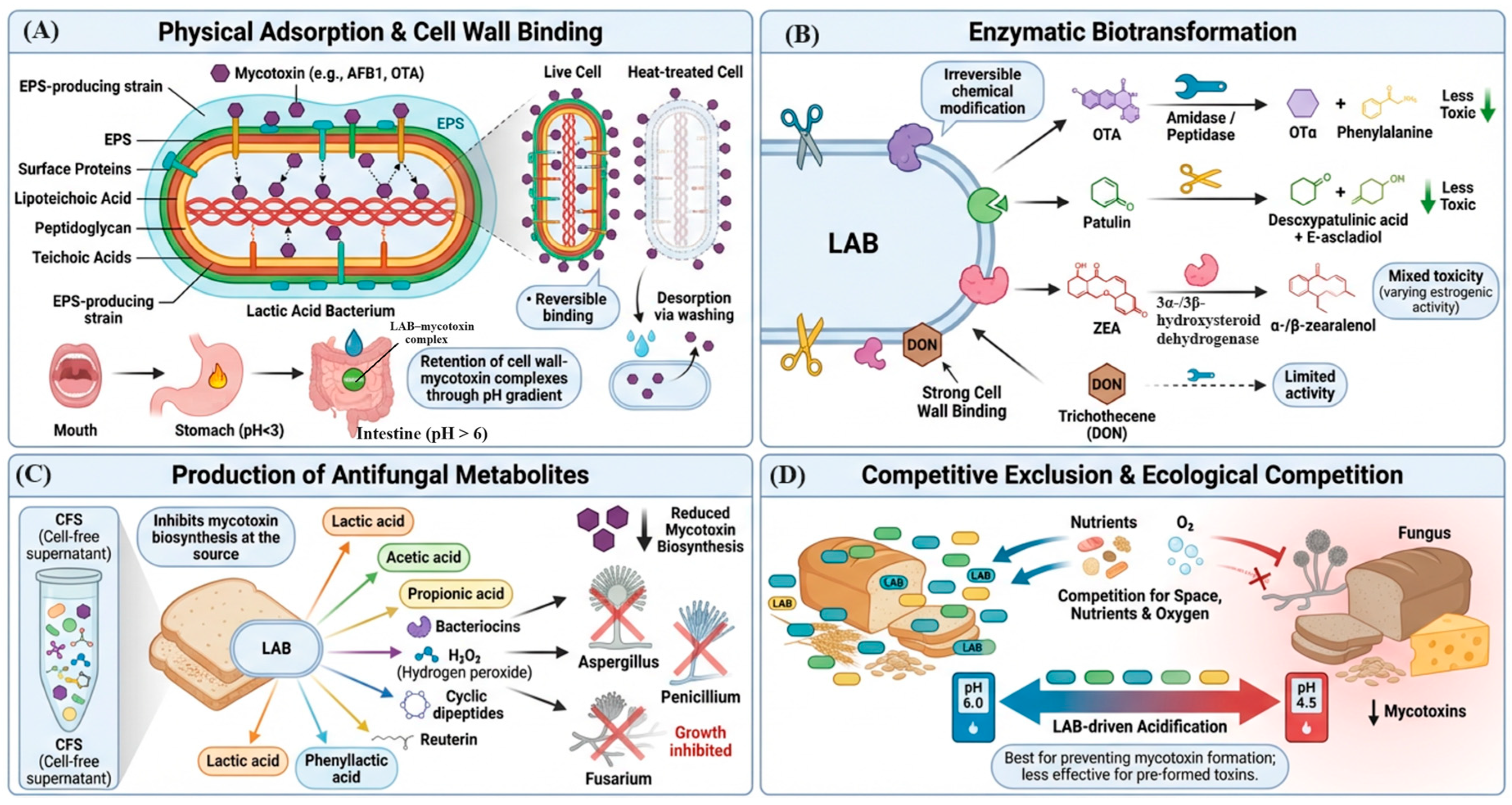
4. Mechanisms of Mycotoxin Detoxification by Lactic Acid Bacteria
4.1. Physical Adsorption and Cell Wall Binding
4.2. Enzymatic Biotransformation
4.3. Production of Antifungal Metabolites
4.4. Competitive Exclusion and Ecological Competition
5. LAB-Mediated Detoxification of Specific Mycotoxins: Evidence and Efficacy
5.1. Aflatoxins
5.2. Ochratoxin A
5.3. Deoxynivalenol (DON)
5.4. Zearalenone (ZEA)
5.5. Fumonisins
5.6. Patulin (PAT)
5.7. T-2 Toxin and HT-2 Toxin
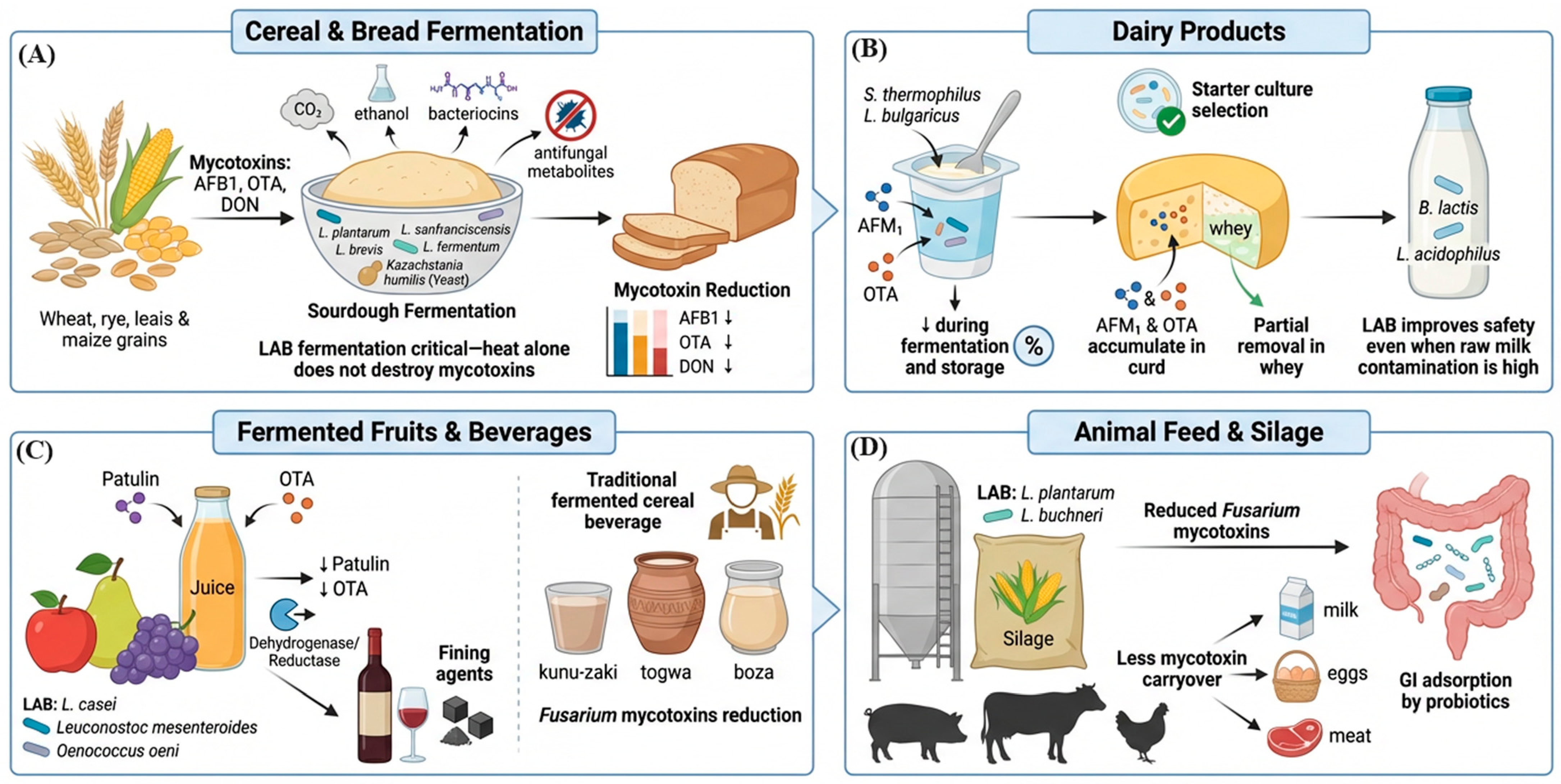
6. Application of LAB Detoxification in Food Systems
6.1. Cereal and Bread Fermentation
6.2. Dairy Products
6.3. Fermented Fruits and Beverages
6.4. Animal Feed and Silage
7. Toxicity Profiles of Major Mycotoxin Degradation Products
8. Factors Governing LAB Mycotoxin Detoxification Efficiency
8.1. Strain-Specific and Species-Level Variation
8.2. Mycotoxin Concentration and Structural Class
8.3. Environmental and Process Conditions
8.4. Viable vs. Non-Viable (Heat-Killed) Cells
8.5. Fermentation Duration and Inoculum Density
9. In Vivo Evidence and Safety of Degradation Products
10. Regulatory Considerations for LAB-Based Detoxification
11. Challenges, Emerging Strategies, and Future Perspectives
11.1. Current Limitations
11.2. Encapsulation and Strain Engineering
11.3. Multi-Target and Combination Approaches
11.4. Climate Change and Emerging Mycotoxins
12. Conclusions
Author Contributions
Funding
Institutional Review Board Statement
Informed Consent Statement
Data Availability Statement
Acknowledgments
Conflicts of Interest
Abbreviations
| LAB | Lactic Acid Bacteria |
| AFB1 | Aflatoxin B1 |
| AFM1 | Aflatoxin M1 |
| OTA | Ochratoxin A |
| DON | Deoxynivalenol |
| ZEA | Zearalenone |
| FB1/FB2 | Fumonisin B1/B2 |
| PAT | Patulin |
| GRAS | Generally Recognized as Safe |
| QPS | Qualified Presumption of Safety |
| EFSA | European Food Safety Authority |
| FDA | Food and Drug Administration |
| EPS | Exopolysaccharides |
| PLA | Phenyllactic acid |
| HACCP | Hazard Analysis and Critical Control Points |
| MAPKinase | mitogen-activated protein kinase |
References
- Forgacs, J.; Carll, W.T. Mycotoxicoses. Adv. Vet. Sci. 1962, 7, 273–382. [Google Scholar]
- Edite Bezerra da Rocha, M.; Freire, F.d.C.O.; Erlan Feitosa Maia, F.; Izabel Florindo Guedes, M.; Rondina, D. Mycotoxins and their effects on human and animal health. Food Control 2014, 36, 159–165. [Google Scholar] [CrossRef]
- Eskola, M.; Kos, G.; Elliott, C.T.; Hajšlová, J.; Mayar, S.; Krska, R. Worldwide contamination of food-crops with mycotoxins: Validity of the widely cited ‘FAO estimate’ of 25%. Crit. Rev. Food Sci. Nutr. 2020, 60, 2773–2789. [Google Scholar] [CrossRef]
- Goda, A.A.; Shi, J.; Xu, J.; Liu, X.; Zhou, Y.; Xiao, L.; Abdel-Galil, M.; Salem, S.H.; Ayad, E.G.; Deabes, M.; et al. Global health and economic impacts of mycotoxins: A comprehensive review. Environ. Sci. Eur. 2025, 37, 122. [Google Scholar] [CrossRef]
- Janik, E.; Niemcewicz, M.; Ceremuga, M.; Stela, M.; Saluk-Bijak, J.; Siadkowski, A.; Bijak, M. Molecular aspects of mycotoxins—A serious problem for human health. Int. J. Mol. Sci. 2020, 21, 8187. [Google Scholar] [CrossRef]
- Mamo, F.T.; Abate, B.A.; Tesfaye, K.; Nie, C.; Wang, G.; Liu, Y. Mycotoxins in Ethiopia: A review on prevalence, economic and health impacts. Toxins 2020, 12, 648. [Google Scholar] [CrossRef]
- McCullough, A.K.; Lloyd, R.S. Mechanisms underlying aflatoxin-associated mutagenesis—Implications in carcinogenesis. DNA Repair 2019, 77, 76–86. [Google Scholar] [CrossRef]
- Wenndt, A.; Mutua, F.; Grace, D.; Thomas, L.F.; Lambertini, E. Quantitative assessment of aflatoxin exposure and hepatocellular carcinoma (HCC) risk associated with consumption of select Nigerian staple foods. Front. Sustain. Food Syst. 2023, 7, 1128540. [Google Scholar] [CrossRef]
- Chen, T.; Liu, J.; Li, Y.; Wei, S. Burden of disease associated with dietary exposure to aflatoxins in China in 2020. Nutrients 2022, 14, 1027. [Google Scholar] [CrossRef]
- Qin, M.; Lin, L.; Wang, L.; Zhang, Y.; Zhang, L.; Song, Y.; Chen, J. Disease burden estimation of hepatocellular carcinoma attributable to dietary aflatoxin exposure in Sichuan Province, China. Nutrients 2024, 16, 4381. [Google Scholar] [CrossRef]
- Zhang, W.; He, H.; Zang, M.; Wu, Q.; Zhao, H.; Lu, L.-L.; Ma, P.; Zheng, H.; Wang, N.; Zhang, Y.; et al. Genetic features of aflatoxin-associated hepatocellular carcinoma. Gastroenterology 2017, 153, 249–262. [Google Scholar] [CrossRef]
- Pitt, J.I.; Miller, J.D. A concise history of mycotoxin research. J. Agric. Food Chem. 2017, 65, 7021–7033. [Google Scholar] [CrossRef]
- Magan, N.; Medina, A.; Aldred, D. Possible climate-change effects on mycotoxin contamination of food crops pre- and postharvest. Plant Pathol. 2011, 60, 150–163. [Google Scholar] [CrossRef]
- Zhang, J.; Tang, X.; Cai, Y.; Zhou, W.-W. Mycotoxin contamination status of cereals in China and potential microbial decontamination methods. Metabolites 2023, 13, 551. [Google Scholar] [CrossRef]
- Battilani, P.; Toscano, P.; Van der Fels-Klerx, H.J.; Moretti, A.; Camardo Leggieri, M.; Brera, C.; Rortais, A.; Goumperis, T.; Robinson, T. Aflatoxin B1 contamination in maize in Europe increases due to climate change. Sci. Rep. 2016, 6, 24328. [Google Scholar] [CrossRef]
- Streit, E.; Naehrer, K.; Rodrigues, I.; Schatzmayr, G. Mycotoxin occurrence in feed and feed raw materials worldwide: Long-term analysis with special focus on Europe and Asia. J. Sci. Food Agric. 2013, 93, 2892–2899. [Google Scholar] [CrossRef]
- EFSA Panel on Contaminants in the Food Chain (CONTAM); Knutsen, H.K.; Alexander, J.; Barregård, L.; Bignami, M.; Brüschweiler, B.; Ceccatelli, S.; Cottrill, B.; DiNovi, M.; Grasl-Kraupp, B.; et al. Risks to human and animal health related to the presence of deoxynivalenol and its acetylated and modified forms in food and feed. EFSA J. 2017, 15, e04718. [Google Scholar] [CrossRef] [PubMed]
- Bullerman, L.B.; Bianchini, A. Stability of mycotoxins during food processing. Int. J. Food Microbiol. 2007, 119, 140–146. [Google Scholar] [CrossRef]
- Jalili, M. A review on aflatoxins reduction in food. Iran. J. Health Saf. Environ. 2016, 3, 445–459. [Google Scholar]
- Wacoo, A.P.; Wendiro, D.; Vuzi, P.C.; Hawumba, J.F. Methods for detection of aflatoxins in agricultural food crops. J. Appl. Chem. 2014, 2014, 706291. [Google Scholar] [CrossRef]
- Dalie, D.K.D.; Deschamps, A.M.; Richard-Forget, F. Lactic acid bacteria—Potential for control of mould growth and mycotoxins: A review. Food Control 2010, 21, 370–380. [Google Scholar] [CrossRef]
- Shetty, P.H.; Jespersen, L. Saccharomyces cerevisiae and lactic acid bacteria as potential mycotoxin decontaminating agents. Trends Food Sci. Technol. 2006, 17, 48–55. [Google Scholar] [CrossRef]
- Nasrollahzadeh, A.; Mokhtari, S.; Khomeiri, M.; Saris, P.E.J. Antifungal biopreservation of food using lactic acid bacteria. Curr. Opin. Food Sci. 2022, 47, 100892. [Google Scholar] [CrossRef]
- Escriva, L.; Calpe, J.; Lafuente, C.; Moreno, A.; Musto, L.; Meca, G.; Luz, C. Aflatoxin B1 and ochratoxin A reduction by Lactobacillus spp. during bread making. J. Sci. Food Agric. 2023, 103, 7095–7103. [Google Scholar] [CrossRef]
- Crowley, S.; Mahony, J.; van Sinderen, D. Current perspectives on antifungal lactic acid bacteria as natural bio-preservatives. Trends Food Sci. Technol. 2013, 33, 93–109. [Google Scholar] [CrossRef]
- Tian, M.; Zhang, G.; Ding, S.; Jiang, Y.; Jiang, B.; Ren, D.; Chen, P. Lactobacillus plantarum T3 as an adsorbent of aflatoxin B1 effectively mitigates the toxic effects on mice. Food Biosci. 2022, 49, 101984. [Google Scholar] [CrossRef]
- Banicod, R.J.S.; Tabassum, N.; Javaid, A.; Kim, Y.-M.; Khan, F. Lactic acid bacteria-derived secondary metabolites: Emerging natural alternatives for food preservation. Probiotics Antimicrob. Proteins 2026, 18, 3113–3150. [Google Scholar] [CrossRef] [PubMed]
- Jard, G.; Liboz, T.; Mathieu, F.; Guyonvarc’h, A.; Lebrihi, A. Review of mycotoxin reduction in food and feed: From prevention in the field to detoxification by adsorption or transformation. Food Addit. Contam. Part A 2011, 28, 1590–1609. [Google Scholar] [CrossRef] [PubMed]
- Karlovsky, P.; Suman, M.; Berthiller, F.; De Meester, J.; Eisenbrand, G.; Perrin, I.; Dussort, P. Impact of food processing and detoxification treatments on mycotoxin contamination. Mycotoxin Res. 2016, 32, 179–205. [Google Scholar] [CrossRef] [PubMed]
- Kensler, T.W.; Roebuck, B.D.; Wogan, G.N.; Groopman, J.D. Aflatoxin: A 50-year odyssey of mechanistic and translational toxicology. Toxicol. Sci. 2011, 120, S28–S48. [Google Scholar] [CrossRef]
- Liu, Y.; Wu, F. Global burden of aflatoxin-induced hepatocellular carcinoma: A risk assessment. Environ. Health Perspect. 2010, 118, 818–824. [Google Scholar] [CrossRef]
- European Commission. Commission Regulation (EC) No 1881/2006 of 19 December 2006 setting maximum levels for certain contaminants in foodstuffs. Off. J. Eur. Union 2006, L 364, 5–24. [Google Scholar]
- Gonçalves, B.L.; Muaz, K.; Coppa, C.F.S.C.; Rosim, R.E.; Kamimura, E.S.; Oliveira, C.A.F.; Corassin, C.H. Aflatoxin M1 absorption by non-viable cells of lactic acid bacteria and Saccharomyces cerevisiae strains in Frescal cheese. Food Res. Int. 2020, 136, 109604. [Google Scholar] [CrossRef] [PubMed]
- Codex Alimentarius Commission. General Standard for Contaminants and Toxins in Food and Feed (CXS 193-1995); Codex Alimentarius Commission: Rome, Italy, 2019. [Google Scholar]
- Pfohl-Leszkowicz, A.; Manderville, R.A. Ochratoxin A: An overview on toxicity and carcinogenicity in animals and humans. Mol. Nutr. Food Res. 2007, 51, 61–99. [Google Scholar] [CrossRef]
- Niderkorn, V.; Morgavi, D.P.; Aboab, B.; Lemaire, M.; Boudra, H. Cell wall component and mycotoxin moieties involved in the binding of fumonisin B1 and B2 by lactic acid bacteria. J. Appl. Microbiol. 2009, 107, 977–984. [Google Scholar] [CrossRef]
- EFSA Panel on Contaminants in the Food Chain (CONTAM); Schrenk, D.; Bignami, M.; Bodin, L.; Chipman, J.K.; Del Mazo, J.; Grasl-Kraupp, B.; Hogstrand, C.; Hoogenboom, L.; Leblanc, J.-C.; et al. Risks for animal health related to the presence of ochratoxin A (OTA) in feed. EFSA J. 2023, 21, e08375. [Google Scholar] [CrossRef]
- Pestka, J.J. Deoxynivalenol: Mechanisms of action, human exposure, and toxicological relevance. Arch. Toxicol. 2010, 84, 663–679. [Google Scholar] [CrossRef]
- Maresca, M. From the gut to the brain: Journey and pathophysiological effects of the food-associated trichothecene mycotoxin deoxynivalenol. Toxins 2013, 5, 784–820. [Google Scholar] [CrossRef]
- Niderkorn, V.; Boudra, H.; Morgavi, D.P. Binding of Fusarium mycotoxins by fermentative bacteria in vitro. J. Appl. Microbiol. 2006, 101, 849–856. [Google Scholar] [CrossRef] [PubMed]
- Franco, T.S.; Garcia, S.; Hirooka, E.Y.; Ono, Y.S.; dos Santos, J.S. Lactic acid bacteria in the inhibition of Fusarium graminearum and deoxynivalenol detoxification. J. Appl. Microbiol. 2011, 111, 739–748. [Google Scholar] [CrossRef] [PubMed]
- Codex Alimentarius Commission. Code of Practice for the Prevention and Reduction of Mycotoxin Contamination in Cereals (CXC 51-2003); Codex Alimentarius Commission: Rome, Italy, 2003. [Google Scholar]
- Rogowska, A.; Pomastowski, P.; Sagandykova, G.; Buszewski, B. Zearalenone and its metabolites: Effect on human health, metabolism and neutralisation methods. Toxicon 2019, 162, 46–56. [Google Scholar] [CrossRef]
- Kleinova, M.; Zollner, P.; Kahlbacher, H.; Hochsteiner, W.; Lindner, W. Metabolic profiles of the mycoestrogen zearalenone and of the growth promoter zeranol in urine, liver, and muscle of heifers. J. Agric. Food Chem. 2002, 50, 4769–4776. [Google Scholar] [CrossRef] [PubMed]
- Merrill, A.H., Jr.; Sullards, M.C.; Wang, E.; Voss, K.A.; Riley, R.T. Sphingolipid metabolism: Roles in signal transduction and disruption by fumonisins. Environ. Health Perspect. 2001, 109, 283–289. [Google Scholar] [CrossRef]
- Alberts, J.F.; van Zyl, W.H.; Gelderblom, W.C.A. Biologically based methods for control of fumonisin-producing Fusarium species and reduction of the mycotoxin in food and feed. Front. Microbiol. 2016, 7, 548. [Google Scholar] [CrossRef]
- Puel, O.; Galtier, P.; Oswald, I.P. Biosynthesis and toxicological effects of patulin. Toxins 2010, 2, 613–631. [Google Scholar] [CrossRef]
- Moake, M.M.; Padilla-Zakour, O.I.; Worobo, R.W. Comprehensive review of patulin control methods in foods. Compr. Rev. Food Sci. Food Saf. 2005, 4, 8–21. [Google Scholar] [CrossRef]
- Ioi, J.D.; Zhou, T.; Tsao, R.; Marcone, M.F. Mitigation of patulin in fresh and processed apple products. Toxins 2017, 9, 157. [Google Scholar] [CrossRef]
- Ferrigo, D.; Raiola, A.; Causin, R. Fusarium toxins in cereals: Occurrence, legislation, factors promoting the appearance and their management. Molecules 2016, 21, 627. [Google Scholar] [CrossRef]
- Marin, S.; Ramos, A.J.; Cano-Sancho, G.; Sanchis, V. Mycotoxins: Occurrence, toxicology, and exposure assessment. Food Chem. Toxicol. 2013, 60, 218–237. [Google Scholar] [CrossRef] [PubMed]
- Rose, M.; Guérin, T. Scientific opinion on the risks for animal and public health related to the presence of T-2 and HT-2 toxin in food and feed. EFSA J. 2011, 9, 2481. [Google Scholar] [CrossRef]
- Zain, M.E. Impact of mycotoxins on humans and animals. J. Saudi Chem. Soc. 2011, 15, 129–144. [Google Scholar] [CrossRef]
- Fraeyman, S.; Croubels, S.; Devreese, M.; Antonissen, G. Emerging Fusarium and Alternaria mycotoxins: Occurrence, toxicity and toxicokinetics. Toxins 2017, 9, 228. [Google Scholar] [CrossRef]
- Mischler, S.; Andre, A.; Chetschik, I.; Miescher Schwenninger, S. Potential for the bio-detoxification of the mycotoxins enniatin B and deoxynivalenol by lactic acid bacteria and Bacillus spp. Microorganisms 2024, 12, 1892. [Google Scholar] [CrossRef]
- Behr, A.-C.; Fæste, C.K.; Azqueta, A.; Tavares, A.M.; Spyropoulou, A.; Solhaug, A.; Olsen, A.-K.; Vettorazzi, A.; Mertens, B.; Zegura, B.; et al. Hazard characterization of the mycotoxins enniatins and beauvericin to identify data gaps and improve risk assessment for human health. Arch. Toxicol. 2025, 99, 1791–1841. [Google Scholar] [CrossRef]
- Martínez, J.; Hernández-Rodríguez, M.; Méndez-Albores, A.; Téllez-Isaías, G.; Mera Jiménez, E.; Nicolás-Vázquez, M.I.; Miranda Ruvalcaba, R. Computational studies of aflatoxin B1 (AFB1): A review. Toxins 2023, 15, 135. [Google Scholar] [CrossRef]
- Marchese, S.; Polo, A.; Ariano, A.; Velotto, S.; Costantini, S.; Severino, L. Aflatoxin B1 and M1: Biological properties and their involvement in cancer development. Toxins 2018, 10, 214. [Google Scholar] [CrossRef]
- Nazhand, A.; Durazzo, A.; Lucarini, M.; Souto, E.B.; Santini, A. Characteristics, occurrence, detection and detoxification of aflatoxins in foods and feeds. Foods 2020, 9, 644. [Google Scholar] [CrossRef] [PubMed]
- Syraji, Y.; Jeyaramraja, P.R.; Mada, T.; Gobikanila, K. Comprehensive review of aflatoxin contamination, its occurrence, effects, management, and future perspectives. Discov. Food 2025, 5, 377. [Google Scholar] [CrossRef]
- Foroud, N.A.; Baines, D.; Gagkaeva, T.Y.; Thakor, N.; Badea, A.; Steiner, B.; Bürstmayr, M.; Bürstmayr, H. Trichothecenes in cereal grains—An update. Toxins 2019, 11, 634. [Google Scholar] [CrossRef]
- Janik, E.; Niemcewicz, M.; Podogrocki, M.; Ceremuga, M.; Stela, M.; Bijak, M. T-2 toxin—The most toxic trichothecene mycotoxin: Metabolism, toxicity, and decontamination strategies. Molecules 2021, 26, 6868. [Google Scholar] [CrossRef] [PubMed]
- Makarova, K.S.; Koonin, E.V. Evolutionary genomics of lactic acid bacteria. J. Bacteriol. 2007, 189, 1199–1208. [Google Scholar] [CrossRef]
- Zheng, J.; Wittouck, S.; Salvetti, E.; Franz, C.; Harris, H.M.B.; Mattarelli, P.; O’Toole, P.W.; Pot, B.; Vandamme, P.; Walter, J.; et al. A taxonomic note on the genus Lactobacillus: Description of 23 novel genera, emended description of the genus Lactobacillus Beijerinck 1901, and union of Lactobacillaceae and Leuconostocaceae. Int. J. Syst. Evol. Microbiol. 2020, 70, 2782–2858. [Google Scholar] [CrossRef]
- Pot, B.; Felis, G.E.; De Bruyne, K.; Tsakalidou, E.; Papadimitriou, K.; Leisner, J.; Vandamme, P. The genus Lactobacillus. In Lactic Acid Bacteria: Biodiversity and Taxonomy; Wiley-Blackwell: Oxford, UK, 2014; pp. 249–353. [Google Scholar]
- Hammes, W.P.; Hertel, C. The genera Lactobacillus and Carnobacterium. In The Prokaryotes; Springer: New York, NY, USA, 2006; pp. 320–403. [Google Scholar]
- Salminen, S.; von Wright, A.; Morelli, L.; Marteau, P.; Brassart, D.; de Vos, W.M.; Fondén, R.; Saxelin, M.; Collins, K.; Mogensen, G.; et al. Demonstration of safety of probiotics—A review. Int. J. Food Microbiol. 1998, 44, 93–106. [Google Scholar] [CrossRef]
- EFSA BIOHAZ Panel; Allende, A.; Alvarez-Ordonez, A.; Bortolaia, V.; Bover-Cid, S.; De Cesare, A.; Dohmen, W.; Guillier, L.; Jacxsens, L.; Nauta, M.; et al. Updated list of QPS-recommended microorganisms for safety risk assessments carried out by EFSA. Zenodo 2026. [Google Scholar] [CrossRef]
- Cotter, P.D.; Ross, R.P.; Hill, C. Bacteriocins—A viable alternative to antibiotics? Nat. Rev. Microbiol. 2013, 11, 95–105. [Google Scholar] [CrossRef]
- Stiles, M.E. Biopreservation by lactic acid bacteria. Antonie Van Leeuwenhoek 1996, 70, 331–345. [Google Scholar] [CrossRef]
- Kim, M.; Khatun, J.; Khan, F.; Kim, Y.-M. Lactic acid bacteria as natural antimicrobials: Biofilm control in food and food industry. Antibiotics 2026, 15, 248. [Google Scholar] [CrossRef] [PubMed]
- Hill, C.; Guarner, F.; Reid, G.; Gibson, G.R.; Merenstein, D.J.; Pot, B.; Morelli, L.; Canani, R.B.; Flint, H.J.; Salminen, S.; et al. The International Scientific Association for Probiotics and Prebiotics consensus statement on the scope and appropriate use of the term probiotic. Nat. Rev. Gastroenterol. Hepatol. 2014, 11, 506–514. [Google Scholar] [CrossRef] [PubMed]
- Jo, D.-M.; Ko, S.-C.; Kim, K.W.; Yang, D.; Kim, J.-Y.; Oh, G.-W.; Choi, G.; Lee, D.-S.; Tabassum, N.; Kim, Y.-M.; et al. Artificial intelligence-driven strategies to enhance the application of lactic acid bacteria as functional probiotics: Health promotion and optimization for industrial applications. Trends Food Sci. Technol. 2025, 165, 105309. [Google Scholar] [CrossRef]
- El-Nezami, H.; Kankaanpaa, P.; Salminen, S.; Ahokas, J. Ability of dairy strains of lactic acid bacteria to bind a common food carcinogen, aflatoxin B1. Food Chem. Toxicol. 1998, 36, 321–326. [Google Scholar] [CrossRef]
- Murtaza, B.; Guo, L.-l.; Wang, L.; Li, X.; Zeb, L.; Jin, B.; Li, J.-b.; Xu, Y. Innovative probiotic fermentation approach for zearalenone detoxification in dried distiller’s grains. Front. Microbiol. 2025, 16, 1533515. [Google Scholar] [CrossRef] [PubMed]
- Murtaza, B.; Jin, B.; Wang, L.; Li, X.; Saleemi, M.; Majeed, S.; Khatoon, A.; Li, G.; Xu, Y. Mitigation of zearalenone in vitro using probiotic strains. LWT 2023, 185, 115265. [Google Scholar] [CrossRef]
- Haskard, C.A.; El-Nezami, H.S.; Kankaanpaa, P.E.; Salminen, S.; Ahokas, J.T. Surface binding of aflatoxin B1 by lactic acid bacteria. Appl. Environ. Microbiol. 2001, 67, 3086–3091. [Google Scholar] [CrossRef]
- Krishnan, S.V.; Nampoothiri, K.M.; Suresh, A.; Linh, N.T.; Balakumaran, P.A.; Pócsi, I.; Pusztahelyi, T. Fusarium biocontrol: Antagonism and mycotoxin elimination by lactic acid bacteria. Front. Microbiol. 2024, 14, 1260166. [Google Scholar] [CrossRef]
- Hernandez-Mendoza, A.; Garcia, H.S.; Steele, J.L. Screening of Lactobacillus casei strains for their ability to bind aflatoxin B1. Food Chem. Toxicol. 2009, 47, 1064–1068. [Google Scholar] [CrossRef]
- Kabak, B.; Var, I. Factors affecting the removal of aflatoxin M1 from food model by Lactobacillus and Bifidobacterium strains. J. Environ. Sci. Health Part B 2008, 43, 617–624. [Google Scholar] [CrossRef]
- Bangar, S.P.; Sharma, N.; Singh, T.; Phimolsiripol, Y.; Brennan, C.S. Mycotoxin degradation via microorganisms: A review of mechanisms and applications. J. Food Sci. 2022, 87, 871–886. [Google Scholar] [CrossRef]
- Peltonen, K.; El-Nezami, H.; Haskard, C.; Ahokas, J.; Salminen, S. Aflatoxin B1 binding by dairy strains of lactic acid bacteria and bifidobacteria. J. Dairy Sci. 2001, 84, 2152–2156. [Google Scholar] [CrossRef]
- Ogunremi, O.R.; Freimuller Leischtfeld, S.; Miescher Schwenninger, S. Potential of lactic acid bacteria and Bacillus spp. in a bio-detoxification strategy for mycotoxin contaminated wheat grains. Appl. Biosci. 2024, 4, 7. [Google Scholar] [CrossRef]
- Chelule, P.K.; Mbongwa, H.P.; Carries, S.; Gqaleni, N. Lactic acid fermentation improves the quality of amahewu, a traditional South African maize-based porridge. Food Chem. 2010, 122, 656–661. [Google Scholar] [CrossRef]
- Schnurer, J.; Magnusson, J. Antifungal lactic acid bacteria as biopreservatives. Trends Food Sci. Technol. 2005, 16, 70–78. [Google Scholar] [CrossRef]
- Gobbetti, M.; De Angelis, M.; Di Cagno, R.; Calasso, M.; Archetti, G.; Rizzello, C.G. Novel insights on the functional/nutritional features of the sourdough fermentation. Int. J. Food Microbiol. 2019, 302, 103–113. [Google Scholar] [CrossRef]
- Govaris, A.; Roussi, V.; Koidis, P.A.; Botsoglou, N.A. Distribution and stability of aflatoxin M1 during production and storage of yoghurt. Food Addit. Contam. 2002, 19, 1043–1050. [Google Scholar] [CrossRef]
- Topcu, A.; Bulat, T.; Wishah, R.; Boyaci, I.H. Detoxification of aflatoxin B1 and patulin by Enterococcus faecium strains. Int. J. Food Microbiol. 2010, 139, 202–205. [Google Scholar] [CrossRef] [PubMed]
- Hernandez-Mendoza, A.; Guzman de Pena, D.; Garcia, H.S. Key role of teichoic acids on aflatoxin B1 binding by probiotic bacteria. J. Appl. Microbiol. 2009, 107, 395–403. [Google Scholar] [CrossRef]
- Gratz, S.W.; Mykkanen, H.; El-Nezami, H.S. Probiotics and gut health: A special focus on liver diseases. World J. Gastroenterol. 2010, 16, 403–410. [Google Scholar] [CrossRef]
- Pietri, A.; Bertuzzi, T.; Pallaroni, L.; Piva, G. Occurrence of ochratoxin A in Italian wines. Food Addit. Contam. 2001, 18, 647–654. [Google Scholar] [CrossRef]
- Kavitake, D.; Singh, S.P.; Kandasamy, S.; Devi, P.B.; Shetty, P.H. Report on aflatoxin-binding activity of galactan exopolysaccharide produced by Weissella confusa KR780676. 3 Biotech 2020, 10, 181. [Google Scholar] [CrossRef]
- Fochesato, A.S.; Cuello, D.; Poloni, V.; Galvagno, M.A.; Dogi, C.A.; Cavaglieri, L.R. Aflatoxin B1 adsorption/desorption dynamics in the presence of Lactobacillus rhamnosus RC007 in a gastrointestinal tract-simulated model. J. Appl. Microbiol. 2019, 126, 223–229. [Google Scholar] [CrossRef]
- Liu, M.; Zhang, X.; Luan, H.; Zhang, Y.; Xu, W.; Feng, W.; Song, P. Bioenzymatic detoxification of mycotoxins. Front. Microbiol. 2024, 15, 1434987. [Google Scholar] [CrossRef] [PubMed]
- Abraham, N.; Chan, E.T.S.; Zhou, T.; Seah, S.Y.K. Microbial detoxification of mycotoxins in food. Front. Microbiol. 2022, 13, 957148. [Google Scholar] [CrossRef]
- Bueno, D.J.; Di Paolo, O.C.; Casale, C.H.; Soprani, M. Aflatoxin B1 and aflatoxin M1 binding capacity of lactic acid bacteria isolated from domestic and industrial products. Adv. Microbiol. 2018, 8, 277–296. [Google Scholar] [CrossRef]
- Li, P.; Su, R.; Yin, R.; Lai, D.; Wang, M.; Liu, Y.; Zhou, L. Detoxification of mycotoxins through biotransformation. Toxins 2020, 12, 121. [Google Scholar] [CrossRef]
- Chen, W.; Li, C.; Zhang, B.; Zhou, Z.; Shen, Y.; Liao, X.; Yang, J.; Wang, Y.; Li, X.; Li, Y.; et al. Advances in biodetoxification of ochratoxin A—A review of the past five decades. Front. Microbiol. 2018, 9, 1386. [Google Scholar] [CrossRef]
- Castoria, R.; Mannina, L.; Duran-Patron, R.; Maffei, F.; Piontelli, E.; De Felice, D.V.; Pinedo-Rivilla, C.; Ritieni, A.; Ferracane, R.; Wright, S.A.I. Conversion of the mycotoxin patulin to the less toxic desoxypatulinic acid by the biocontrol yeast Rhodosporidium kratochvilovae strain LS11. J. Agric. Food Chem. 2011, 59, 11571–11578. [Google Scholar] [CrossRef]
- Zheng, X.; Wei, W.; Rao, S.; Gao, L.; Li, H.; Yang, Z. Degradation of patulin in fruit juice by a lactic acid bacteria strain Lactobacillus casei YZU01. Food Control 2020, 112, 107147. [Google Scholar] [CrossRef]
- Bahati, P.; Zeng, X.; Uzizerimana, F.; Tsoggerel, A.; Awais, M.; Qi, G.; Cai, R.; Yue, T.; Yuan, Y. Adsorption mechanism of patulin from apple juice by inactivated lactic acid bacteria isolated from kefir grains. Toxins 2021, 13, 434. [Google Scholar] [CrossRef] [PubMed]
- Chen, S.W.; Hsu, J.T.; Chou, Y.A.; Wang, H.T. The application of digestive tract lactic acid bacteria with high esterase activity for zearalenone detoxification. J. Sci. Food Agric. 2018, 98, 3870–3879. [Google Scholar] [CrossRef] [PubMed]
- Adunphatcharaphon, S.; Petchkongkaew, A.; Visessanguan, W. In vitro mechanism assessment of zearalenone removal by plant-derived Lactobacillus plantarum BCC 47723. Toxins 2021, 13, 286. [Google Scholar] [CrossRef]
- Ragoubi, C.; Quintieri, L.; Greco, D.; Mehrez, A.; Maatouk, I.; D’Ascanio, V.; Landoulsi, A.; Avantaggiato, G. Mycotoxin removal by Lactobacillus spp. and their application in animal liquid feed. Toxins 2021, 13, 185. [Google Scholar] [CrossRef] [PubMed]
- Gan, M.; Hu, J.; Wan, K.; Liu, X.; Chen, P.; Zeng, R.; Wang, F.-J.; Zhao, Y. Isolation and characterization of Lactobacillus paracasei 85 and Lactobacillus buchneri 93 to absorb and biotransform zearalenone. Toxics 2022, 10, 680. [Google Scholar] [CrossRef]
- Volkl, A.; Vogler, B.; Schollenberger, M.; Karlovsky, P. Microbial detoxification of mycotoxin deoxynivalenol. J. Basic Microbiol. 2004, 44, 147–156. [Google Scholar] [CrossRef] [PubMed]
- Leyva Salas, M.; Mounier, J.; Valence, F.; Coton, M.; Thierry, A.; Coton, E. Antifungal microbial agents for food biopreservation—A review. Microorganisms 2017, 5, 37. [Google Scholar] [CrossRef]
- Lavermicocca, P.; Valerio, F.; Evidente, A.; Lazzaroni, S.; Corsetti, A.; Gobbetti, M. Purification and characterization of novel antifungal compounds from the sourdough Lactobacillus plantarum strain 21B. Appl. Environ. Microbiol. 2000, 66, 4084–4090. [Google Scholar] [CrossRef]
- Russo, P.; Fares, C.; Longo, A.; Spano, G.; Capozzi, V. Lactobacillus plantarum with broad antifungal activity as a protective starter culture for bread production. Foods 2017, 6, 110. [Google Scholar] [CrossRef]
- Quattrini, M.; Bernardi, C.; Stuknytė, M.; Masotti, F.; Passera, A.; Ricci, G.; Vallone, L.; De Noni, I.; Brasca, M.; Fortina, M.G. Functional characterization of Lactobacillus plantarum ITEM 17215: A potential biocontrol agent of fungi with plant growth promoting traits, able to enhance the nutritional value of cereal products. Food Res. Int. 2018, 106, 936–944. [Google Scholar] [CrossRef]
- Guimarães, A.; Santiago, A.P.; Teixeira, J.; Venâncio, A.; Abrunhosa, L. Anti-aflatoxigenic effect of organic acids produced by Lactobacillus plantarum. Int. J. Food Microbiol. 2018, 264, 31–38. [Google Scholar] [CrossRef]
- Zhu, Y.; Xu, Y.; Yang, Q. Antifungal properties and AFB1 detoxification activity of a new strain of Lactobacillus plantarum. J. Hazard. Mater. 2021, 408, 125569. [Google Scholar] [CrossRef] [PubMed]
- Zhao, C.; Penttinen, P.; Zhang, L.; Dong, L.; Zhang, F.; Li, Z.; Zhang, X. Mechanism of inhibiting the growth and aflatoxin B1 biosynthesis of Aspergillus flavus by phenyllactic acid. Toxins 2023, 15, 370. [Google Scholar] [CrossRef]
- Nazareth, T.d.M.; Luz, C.; Torrijos, R.; Quiles, J.M.; Luciano, F.B.; Mañes, J.; Meca, G. Potential application of lactic acid bacteria to reduce aflatoxin B1 and fumonisin B1 occurrence on corn kernels and corn ears. Toxins 2020, 12, 21. [Google Scholar] [CrossRef] [PubMed]
- De Vuyst, L.; Vandamme, E.J. Bacteriocins of Lactic Acid Bacteria: Microbiology, Genetics, and Applications; Blackie Academic and Professional: London, UK, 1994. [Google Scholar]
- Lindgren, S.E.; Dobrogosz, W.J. Antagonistic activities of lactic acid bacteria in food and feed fermentations. FEMS Microbiol. Rev. 1990, 7, 149–163. [Google Scholar] [CrossRef]
- Pradhan, S.; Ananthanarayan, L.; Prasad, K.; Bhatnagar-Mathur, P. Anti-fungal activity of lactic acid bacterial isolates against aflatoxigenic fungi inoculated on peanut kernels. LWT 2021, 143, 111104. [Google Scholar] [CrossRef]
- Strom, K.; Sjogren, J.; Broberg, A.; Schnurer, J. Lactobacillus plantarum MiLAB 393 produces the antifungal cyclic dipeptides cyclo(l-Phe-l-Pro) and cyclo(l-Phe-trans-4-OH-l-Pro) and 3-phenyllactic acid. Appl. Environ. Microbiol. 2002, 68, 4322–4327. [Google Scholar] [CrossRef]
- Vimont, A.; Fernandez, B.; Ahmed, G.; Fortin, H.-P.; Fliss, I. Quantitative antifungal activity of reuterin against food isolates of yeasts and moulds and its potential application in yogurt. Int. J. Food Microbiol. 2019, 289, 182–188. [Google Scholar] [CrossRef] [PubMed]
- Rodrigues, F.J.; Cedran, M.F.; Bicas, J.L.; Sato, H.H. Inhibitory effect of reuterin-producing Limosilactobacillus reuteri and edible alginate-konjac gum film against foodborne pathogens and spoilage microorganisms. Food Biosci. 2023, 52, 102443. [Google Scholar] [CrossRef]
- Magnusson, J.; Schnurer, J. Lactobacillus coryniformis subsp. coryniformis strain Si3 produces a broad-spectrum proteinaceous antifungal compound. Appl. Environ. Microbiol. 2001, 67, 1–5. [Google Scholar] [CrossRef]
- Rizzello, C.G.; Baruzzi, F.; Zannini, E.; Gobbetti, M. Microbiota of wheat flour and representative strains of bacteria and yeasts of sourdough: A review. Front. Microbiol. 2009, 7, 1261. [Google Scholar]
- Guo, Y.; Yuan, Y.; Yue, T. Patulin in apple products and its removal by LAB and yeasts. Food Control 2012, 28, 8–14. [Google Scholar]
- El-Nezami, H.; Polychronaki, N.; Salminen, S.; Mykkanen, H. Binding rather than metabolism may explain the interaction of two food-grade Lactobacillus strains with zearalenone and its derivative alpha-zearalenol. Appl. Environ. Microbiol. 2002, 68, 3545–3549. [Google Scholar] [CrossRef]
- Gratz, S.; Mykkänen, H.; El-Nezami, H. Aflatoxin B1 binding by a mixture of Lactobacillus and Propionibacterium: In vitro versus ex vivo. J. Food Prot. 2005, 68, 2470–2474. [Google Scholar] [CrossRef] [PubMed]
- Gratz, S.; Täubel, M.; Juvonen, R.O.; Viluksela, M.; Turner, P.C.; Mykkänen, H.; El-Nezami, H. Lactobacillus rhamnosus strain GG modulates intestinal absorption, fecal excretion, and toxicity of aflatoxin B1 in rats. Appl. Environ. Microbiol. 2006, 72, 7398–7400. [Google Scholar] [CrossRef]
- Huang, L.; Duan, C.; Zhao, Y.; Gao, L.; Niu, C.; Xu, J.; Li, S. Reduction of aflatoxin B1 toxicity by Lactobacillus plantarum C88: A potential probiotic strain isolated from Chinese traditional fermented food ‘Tofu’. PLoS ONE 2017, 12, e0170109. [Google Scholar] [CrossRef]
- Saghir, S.A.M.; Al Hroob, A.M.; Al-Tarawni, A.H.; Abdulghani, M.A.M.; Tabana, Y.; Aldhalmi, A.K.; Mothana, R.A.; Al-Yousef, H.M. Effect of Lactiplantibacillus plantarum on the growth, hemato-biochemical, inflammation, apoptosis, oxidative stress markers, involved genes and histopathological alterations in growing rabbits challenged with aflatoxin B1. Poult. Sci. 2024, 103, 104002. [Google Scholar] [CrossRef] [PubMed]
- Liu, N.; Ding, K.; Wang, J.Q.; Jia, S.C.; Wang, J.P.; Xu, T.S. Detoxification, metabolism, and glutathione pathway activity of aflatoxin B1 by dietary lactic acid bacteria in broiler chickens. J. Anim. Sci. 2017, 95, 4399–4406. [Google Scholar] [CrossRef] [PubMed]
- Zeng, Y.; Zeng, D.; Zhang, Y.; Ni, X.Q.; Wang, J.; Jian, P.; Zhou, Y.; Li, Y.; Yin, Z.Q.; Pan, K.C.; et al. Lactobacillus plantarum BS22 promotes gut microbial homeostasis in broiler chickens exposed to aflatoxin B1. J. Anim. Physiol. Anim. Nutr. 2018, 102, e449–e459. [Google Scholar] [CrossRef]
- Śliżewska, K.; Cukrowska, B.; Smulikowska, S.; Cielecka-Kuszyk, J. The effect of probiotic supplementation on performance and the histopathological changes in liver and kidneys in broiler chickens fed diets with aflatoxin B1. Toxins 2019, 11, 112. [Google Scholar] [CrossRef]
- Nout, M.J.R.; Aidoo, K.E. Asian fungal fermented food. In The Mycota: A Comprehensive Treatise on Fungi; Springer: Berlin/Heidelberg, Germany, 2002; Volume 10, pp. 23–47. [Google Scholar]
- Piotrowska, M. The Adsorption of Ochratoxin A by Lactobacillus Species. Toxins 2014, 6, 2826–2839. [Google Scholar] [CrossRef]
- La Placa, L.; Tsitsigiannis, D.; Camardo Leggieri, M.; Battilani, P. From grapes to wine: Impact of the vinification process on ochratoxin A contamination. Foods 2023, 12, 260. [Google Scholar] [CrossRef] [PubMed]
- Zjalic, S.; Markov, K.; Loncar, J.; Jakopovic, Z.; Beccaccioli, M.; Reverberi, M. Biocontrol of occurrence of ochratoxin A in wine: A review. Toxins 2024, 16, 277. [Google Scholar] [CrossRef] [PubMed]
- Luo, Y.; Liu, X.; Yuan, L.; Li, J. Complicated interactions between bio-adsorbents and mycotoxins during mycotoxin adsorption: Current research and future prospects. Trends Food Sci. Technol. 2020, 96, 127–134. [Google Scholar] [CrossRef]
- Lucio, O.; Pardo, I.; Heras, J.M.; Krieger-Weber, S.; Ferrer, S. Use of starter cultures of Lactobacillus to induce malolactic fermentation in wine. Aust. J. Grape Wine Res. 2017, 23, 15–21. [Google Scholar] [CrossRef]
- Zheng, X.; Xia, F.; Li, J.; Zheng, L.; Rao, S.; Gao, L.; Yang, Z. Reduction of ochratoxin A from contaminated food by Lactobacillus rhamnosus Bm01. Food Control 2023, 143, 109315. [Google Scholar] [CrossRef]
- El Khoury, R.; Choque, E.; El Khoury, A.; Snini, S.P.; Cairns, R.; Andriantsiferana, C.; Mathieu, F. OTA prevention and detoxification by actinobacterial strains and activated carbon fibers: Preliminary results. Toxins 2018, 10, 137. [Google Scholar] [CrossRef] [PubMed]
- Vekiru, E.; Hametner, C.; Mitterbauer, R.; Rechthaler, J.; Adam, G.; Schatzmayr, G.; Krska, R.; Schuhmacher, R. Cleavage of zearalenone by Gliocladium roseum to a novel non-estrogenic metabolite. Appl. Environ. Microbiol. 2010, 76, 2353–2359. [Google Scholar] [CrossRef]
- Bullerman, L.B.; Bianchini, A.; Diskus, M. Effect of fermentation on mycotoxins. In Mycotoxins: Detection Methods, Management, Public Health and Agricultural Trade; CAB International: Wallingford, UK, 2008; pp. 257–268. [Google Scholar]
- Kolawole, O.; Meneely, J.; Greer, B.; Chevallier, O.; Jones, D.S.; Connolly, L.; Elliott, C. Comparative in vitro assessment of a range of commercial feed additives with multiple mycotoxin binding claims. Toxins 2019, 11, 659. [Google Scholar] [CrossRef]
- Lili, Z.; Junyan, W.; Hongfei, Z.; Baoqing, Z.; Bolin, Z. Detoxification of cancerogenic compounds by lactic acid bacteria strains. Crit. Rev. Food Sci. Nutr. 2018, 58, 2727–2742. [Google Scholar] [CrossRef]
- Li, Z.; Wang, Y.; Liu, Z.; Jin, S.; Pan, K.; Liu, H.; Liu, T.; Li, X.; Zhang, C.; Luo, X.; et al. Biological detoxification of fumonisin by a novel carboxylesterase from Sphingomonadales bacterium and its biochemical characterization. Int. J. Biol. Macromol. 2021, 169, 18–27. [Google Scholar] [CrossRef]
- Alberts, J.; Schatzmayr, G.; Moll, W.-D.; Davids, I.; Rheeder, J.; Burger, H.-M.; Shephard, G.; Gelderblom, W. Detoxification of the fumonisin mycotoxins in maize: An enzymatic approach. Toxins 2019, 11, 523. [Google Scholar] [CrossRef]
- Incze, D.J.; Molnár, Z.; Nagy, G.N.; Leveles, I.; Vértessy, B.G.; Poppe, L.; Bata, Z. Understanding the molecular mechanism of fumonisin esterases by kinetic and structural studies. Food Chem. 2025, 473, 143110. [Google Scholar] [CrossRef]
- Xu, H.; Wang, L.; Sun, J.; Wang, L.; Guo, H.; Ye, Y.; Sun, X. Microbial detoxification of mycotoxins in food and feed. Crit. Rev. Food Sci. Nutr. 2022, 62, 4951–4969. [Google Scholar] [CrossRef] [PubMed]
- Smaoui, S.; Agriopoulou, S.; D’Amore, T.; Tavares, L.; Mousavi Khaneghah, A. The control of Fusarium growth and decontamination of produced mycotoxins by lactic acid bacteria. Crit. Rev. Food Sci. Nutr. 2023, 63, 11125–11152. [Google Scholar] [CrossRef] [PubMed]
- Krishnan, S.V.; Anaswara, P.A.; Nampoothiri, K.M.; Kovács, S.; Adácsi, C.; Szarvas, P.; Király, S.; Pócsi, I.; Pusztahelyi, T. Biocontrol activity of new lactic acid bacteria isolates against Fusaria and Fusarium mycotoxins. Toxins 2025, 17, 68. [Google Scholar] [CrossRef]
- Mateo, E.M.; Tarazona, A.; Aznar, R.; Mateo, F. Exploring the impact of lactic acid bacteria on the biocontrol of toxigenic Fusarium spp. and their main mycotoxins. Int. J. Food Microbiol. 2023, 387, 110054. [Google Scholar] [CrossRef]
- Nasrollahzadeh, A.; Mokhtari, S.; Khomeiri, M.; Saris, P. Mycotoxin detoxification of food by lactic acid bacteria. Int. J. Food Contam. 2022, 9, 1. [Google Scholar] [CrossRef]
- Sadiq, F.A.; Yan, B.; Tian, F.; Zhao, J.; Zhang, H.; Chen, W. Lactic acid bacteria as antifungal and anti-mycotoxigenic agents: A comprehensive review. Compr. Rev. Food Sci. Food Saf. 2019, 18, 1403–1436. [Google Scholar] [CrossRef]
- N, D.; Achar, P.N.; Sreenivasa, M.Y. Current perspectives of biocontrol agents for management of Fusarium verticillioides and its fumonisin in cereals—A review. J. Fungi 2021, 7, 776. [Google Scholar] [CrossRef] [PubMed]
- Tangni, E.K.; Masquelier, J.; Van Hoeck, E. Analysis of patulin in apple products marketed in Belgium: Intra-laboratory validation study and occurrence. Toxins 2023, 15, 368. [Google Scholar] [CrossRef]
- Hussain, S.; Asi, M.R.; Iqbal, M.; Akhtar, M.; Imran, M.; Ariño, A. Surveillance of patulin in apple, grapes, juices and value-added products for sale in Pakistan. Foods 2020, 9, 1744. [Google Scholar] [CrossRef] [PubMed]
- Lien, K.W.; Ling, M.P.; Pan, M.H. Probabilistic risk assessment of patulin in imported apple juice and apple-containing beverages in Taiwan. J. Sci. Food Agric. 2020, 100, 4776–4781. [Google Scholar] [CrossRef] [PubMed]
- Rosa da Silva, C.; Tonial Simões, C.; Kobs Vidal, J.; Reghelin, M.A.; Araújo de Almeida, C.A.; Mallmann, C.A. Development and validation of an extraction method using liquid chromatography-tandem mass spectrometry to determine patulin in apple juice. Food Chem. 2022, 366, 130654. [Google Scholar] [CrossRef]
- Zhang, K.; Zhang, L. Determination of patulin in apple juice and apple-derived products using a robotic sample preparation system and LC-APCI-MS/MS. Toxins 2024, 16, 238. [Google Scholar] [CrossRef]
- Li, X.; Li, H.; Ma, W.; Guo, Z.; Li, X.; Li, X.; Zhang, Q. Determination of patulin in apple juice by single-drop liquid-liquid-liquid microextraction coupled with liquid chromatography-mass spectrometry. Food Chem. 2018, 257, 1–6. [Google Scholar] [CrossRef]
- Wei, C.; Zhang, C.; Gao, Y.; Yu, L.; Zhao, J.; Zhang, H.; Chen, W.; Tian, F. Insights into the metabolic response of Lactiplantibacillus plantarum CCFM1287 upon patulin exposure. Int. J. Mol. Sci. 2022, 23, 11652. [Google Scholar] [CrossRef]
- Zoghi, A.; Khosravi-Darani, K.; Sohrabvandi, S.; Attar, H.; Alavi, S.A. Effect of probiotics on patulin removal from synbiotic apple juice. J. Sci. Food Agric. 2017, 97, 2601–2609. [Google Scholar] [CrossRef] [PubMed]
- Niu, J.; Ma, B.; Shen, J.; Zhu, H.; Lu, Y.; Lu, Z.; Lu, F.; Zhu, P. Enzymatic degradation of mycotoxin patulin by a short-chain dehydrogenase/reductase from Bacillus subtilis and its application in apple juice. Food Microbiol. 2025, 126, 104676. [Google Scholar] [CrossRef]
- Zhang, Z.; Li, J.; Yang, Y.; Gong, Q.; Li, H.; Rao, S.; Zheng, X.; Yang, Z. Degradation of patulin by a yeast strain Kluyveromyces marxianus XZ1 and its mechanism. Food Microbiol. 2025, 129, 104758. [Google Scholar] [CrossRef] [PubMed]
- Xing, M.; Chen, Y.; Li, B.; Tian, S. Characterization of a short-chain dehydrogenase/reductase and its function in patulin biodegradation in apple juice. Food Chem. 2021, 348, 129046. [Google Scholar] [CrossRef]
- Tannous, J.; Snini, S.; Khoury, R.; Canlet, C.; Pinton, P.; Lippi, Y.; Alassane-Kpembi, I.; Gauthier, T.; El Khoury, A.; Atoui, A.; et al. Patulin transformation products and last intermediates in its biosynthetic pathway, E- and Z-ascladiol, are not toxic to human cells. Arch. Toxicol. 2017, 91, 2455–2467. [Google Scholar] [CrossRef] [PubMed]
- EFSA; Arcella, D.; Gergelova, P.; Innocenti, M.L.; Steinkellner, H. Human and animal dietary exposure to T-2 and HT-2 toxin. EFSA J. 2017, 15, e04972. [Google Scholar] [CrossRef]
- Wu, Q.; Qin, Z.; Kuca, K.; You, L.; Zhao, Y.; Liu, A.; Musilek, K.; Chrienova, Z.; Nepovimova, E.; Oleksak, P.; et al. An update on T-2 toxin and its modified forms: Metabolism, immunotoxicity mechanism, and human exposure assessment. Arch. Toxicol. 2020, 94, 3645–3669. [Google Scholar] [CrossRef]
- Kamala, A.; Kimanya, M.; Lachat, C.; Jacxsens, L.; Haesaert, G.; Kolsteren, P.; Ortiz, J.; Tiisekwa, B.; De Meulenaer, B. Risk of exposure to multiple mycotoxins from maize-based complementary foods in Tanzania. J. Agric. Food Chem. 2017, 65, 7106–7114. [Google Scholar] [CrossRef]
- Pourmohammadi, K.; Sayadi, M.; Abedi, E.; Mousavifard, M. Determining the adsorption capacity and stability of aflatoxin B1, Ochratoxin A, and Zearalenone on single and co-culture L. acidophilus and L. rhamnosus surfaces. J. Food Compos. Anal. 2022, 110, 104517. [Google Scholar] [CrossRef]
- Taheur, F.B.; Fedhila, K.; Chaieb, K.; Kouidhi, B.; Bakhrouf, A.; Abrunhosa, L. Adsorption of aflatoxin B1, zearalenone and ochratoxin A by microorganisms isolated from kefir grains. Int. J. Food Microbiol. 2017, 251, 1–7. [Google Scholar] [CrossRef]
- Suo, B.; Chen, X.; Wang, Y. Recent research advances of lactic acid bacteria in sourdough: Origin, diversity, and function. Curr. Opin. Food Sci. 2021, 37, 66–75. [Google Scholar] [CrossRef]
- Zhang, G.; Tu, J.; Sadiq, F.A.; Zhang, W.; Wang, W. Prevalence, genetic diversity, and technological functions of Lactobacillus sanfranciscensis in sourdough: A review. Compr. Rev. Food Sci. Food Saf. 2019, 18, 1209–1226. [Google Scholar] [CrossRef]
- Lafuente, C.; Nazareth, T.d.M.; Dopazo, V.; Meca, G.; Luz, C. Enhancing bread quality and extending shelf life using dried sourdough. LWT 2024, 203, 116379. [Google Scholar] [CrossRef]
- Pakfetrat, S.; Amiri, S.; Radi, M.; Abedi, E.; Torri, L. Reduction of phytic acid, aflatoxins and other mycotoxins in wheat during germination. J. Sci. Food Agric. 2019, 99, 4695–4701. [Google Scholar] [CrossRef] [PubMed]
- Mousavi Khaneghah, A.; Fakhri, Y.; Sant’Ana, A.S. Impact of unit operations during processing of cereal-based products on the levels of deoxynivalenol, total aflatoxin, ochratoxin A, and zearalenone: A systematic review and meta-analysis. Food Chem. 2018, 268, 611–624. [Google Scholar] [CrossRef] [PubMed]
- Wang, J.; Hasanalieva, G.; Wood, L.; Markellou, E.; Iversen, P.O.; Bernhoft, A.; Seal, C.; Baranski, M.; Vigar, V.; Ernst, L.; et al. Effect of wheat species (Triticum aestivum vs T. spelta), farming system (organic vs conventional) and flour type (wholegrain vs white) on composition of wheat flour; results of a retail survey in the UK and Germany—1. Mycotoxin content. Food Chem. 2020, 327, 127011. [Google Scholar] [CrossRef] [PubMed]
- Deligeorgakis, C.; Magro, C.; Skendi, A.; Gebrehiwot, H.H.; Valdramidis, V.; Papageorgiou, M. Fungal and toxin contaminants in cereal grains and flours: Systematic review and meta-analysis. Foods 2023, 12, 4328. [Google Scholar] [CrossRef]
- Escrivá, L.; Agahi, F.; Vila-Donat, P.; Mañes, J.; Meca, G.; Manyes, L. Bioaccessibility study of aflatoxin B1 and ochratoxin A in bread enriched with fermented milk whey and/or pumpkin. Toxins 2022, 14, 6. [Google Scholar] [CrossRef]
- Sevim, S.; Topal, G.G.; Tengilimoglu-Metin, M.M.; Sancak, B.; Kizil, M. Effects of inulin and lactic acid bacteria strains on aflatoxin M1 detoxification in yoghurt. Food Control 2019, 100, 235–239. [Google Scholar] [CrossRef]
- Møller, C.O.D.A.; Freire, L.; Rosim, R.; Margalho, L.P.; Balthazar, C.; Franco, L.T.; Sant’ana, A.d.S.; Corassin, C.H.; Rattray, F.P.; de Oliveira, C.A.F. Effect of lactic acid bacteria strains on the growth and aflatoxin production potential of Aspergillus parasiticus, and their ability to bind aflatoxin B1, ochratoxin A, and zearalenone in vitro. Front. Microbiol. 2021, 12, 655386. [Google Scholar] [CrossRef]
- Salas, M.L.; Thierry, A.; Lemaître, M.; Garric, G.; Harel-Oger, M.; Chatel, M.; Sant’ana, A.d.S.; Corassin, C.H.; Rattray, F.P.; de Oliveira, C.A.F. Antifungal activity of lactic acid bacteria combinations in dairy mimicking models and their potential as bioprotective cultures in pilot scale applications. Front. Microbiol. 2018, 9, 1787. [Google Scholar] [CrossRef]
- Ngolong Ngea, G.L.; Yang, Q.; Tchabo, W.; Castoria, R.; Zhang, X.; Zhang, H. Leuconostoc mesenteroides subsp. mesenteroides LB7 isolated from apple surface inhibits P. expansum in vitro and reduces patulin in fruit juices. Int. J. Food Microbiol. 2021, 339, 109025. [Google Scholar] [CrossRef]
- Li, N.; Cui, R.; Zhang, F.; Meng, X.; Liu, B. Current situation and future challenges of patulin reduction—A review. Food Control 2022, 138, 108996. [Google Scholar] [CrossRef]
- Zhao, M.; Ren, H.; Yan, Z.; Ma, J.; Feng, X.; Liu, D.; Long, F. Reusable thiol-modification Lactobacillus plantarum embedded in cellulose nanocrystals composite aerogel for efficient removal of ochratoxin A in grape juice. Food Chem. X 2024, 22, 101336. [Google Scholar] [CrossRef] [PubMed]
- Nout, M.J.R. Fermented foods and food safety. Food Res. Int. 1994, 27, 291–298. [Google Scholar] [CrossRef]
- Ashbell, G.; Weinberg, Z.G.; Hen, Y.; Filya, I. The effects of temperature on the aerobic stability of wheat and corn silages. J. Ind. Microbiol. Biotechnol. 2002, 28, 261–263. [Google Scholar] [CrossRef]
- Zhang, Y.; Wang, P.; Kong, Q.; Cotty, P.J. Biotransformation of aflatoxin B1 by Lactobacillus helveticus FAM22155 in wheat bran by solid-state fermentation. Food Chem. 2021, 341, 128180. [Google Scholar] [CrossRef] [PubMed]
- Aiko, V.; Edamana, P.; Mehta, A. Decomposition and detoxification of aflatoxin B1 by lactic acid. J. Sci. Food Agric. 2016, 96, 2388–2394. [Google Scholar] [CrossRef]
- Luz, C.; Ferrer, J.; Mañes, J.; Meca, G. Toxicity reduction of ochratoxin A by lactic acid bacteria. Food Chem. Toxicol. 2018, 114, 550–556. [Google Scholar] [CrossRef]
- Wei, C.; Yu, L.; Qiao, N.; Wang, S.; Tian, F.; Zhao, J.; Zhang, H.; Zhai, Q.; Chen, W. The characteristics of patulin detoxification by Lactiplantibacillus plantarum 13M5. Food Chem. Toxicol. 2020, 146, 111787. [Google Scholar] [CrossRef]
- Zheng, X.; Zheng, L.; Xia, F.; Wei, W.; Wang, S.; Rao, S.; Gao, L.; Yang, Z. In vivo evaluation of the toxicity of patulin degradation products produced by Lactobacillus casei YZU01. Biol. Control 2022, 169, 104878. [Google Scholar] [CrossRef]
- Niderkorn, V.; Morgavi, D.P.; Pujos, E.; Tissandier, A.; Boudra, H. Screening of fermentative bacteria for their ability to bind and biotransform deoxynivalenol, zearalenone and fumonisins in an in vitro simulated corn silage model. Food Addit. Contam. 2007, 24, 406–415. [Google Scholar] [CrossRef] [PubMed]
- Fruhauf, S.; Novak, B.; Nagl, V.; Hackl, M.; Hartinger, D.; Rainer, V.; Labudová, S.; Adam, G.; Aleschko, M.; Moll, W.-D.; et al. Biotransformation of the mycotoxin zearalenone to its metabolites hydrolyzed zearalenone (HZEN) and decarboxylated hydrolyzed zearalenone (DHZEN) diminishes its estrogenicity in vitro and in vivo. Toxins 2019, 11, 481. [Google Scholar] [CrossRef] [PubMed]
- Pierron, A.; Sabria, M.; Murate, L.S.; Loiseau, N.; Lippi, Y.; Bracarense, A.; Schatzmayr, G.; He, J.W.; Zhou, T.; Moll, W.-D.; et al. Microbial biotransformation of DON: Molecular basis for reduced toxicity. Sci. Rep. 2016, 6, 29105. [Google Scholar] [CrossRef] [PubMed]
- Bracarense, A.; Pierron, A.; Pinton, P.; Gerez, J.; Schatzmayr, G.; Moll, W.; Ting, Z.; Oswald, I. Reduced toxicity of 3-epi-deoxynivalenol and de-epoxy-deoxynivalenol through deoxynivalenol bacterial biotransformation: In vivo analysis in piglets. Food Chem. Toxicol. 2020, 145, 111241. [Google Scholar] [CrossRef]
- Ccori Martinez, T.; Atanasova-Pénichon, V.; Chéreau, S.; Ferrer, N.; Marchegay, G.; Savoie, J.; Richard-Forget, F. Yeast and bacteria from ensiled high moisture maize grains as potential mitigation agents of fumonisin B1. J. Sci. Food Agric. 2017, 97, 2932–2939. [Google Scholar] [CrossRef]
- Grenier, B.; Bracarense, A.; Schwartz, H.; Trumel, C.; Cossalter, A.; Schatzmayr, G.; Kolf-Clauw, M.; Moll, W.-D.; Oswald, I.P. The low intestinal and hepatic toxicity of hydrolyzed fumonisin B1 correlates with its inability to alter the metabolism of sphingolipids. Biochem. Pharmacol. 2012, 83, 1674–1683. [Google Scholar] [CrossRef]
- Fang, J.; Sheng, L.; Ye, Y.; Ji, J.; Sun, J.; Zhang, Y.; Sun, X. Recent advances in biosynthesis of mycotoxin-degrading enzymes and their applications in food and feed. Crit. Rev. Food Sci. Nutr. 2025, 65, 1465–1481. [Google Scholar] [CrossRef]
- Loi, M.; Fanelli, F.; Liuzzi, V.C.; Logrieco, A.F.; Mulè, G. Mycotoxin biotransformation by native and commercial enzymes: Present and future perspectives. Toxins 2017, 9, 111. [Google Scholar] [CrossRef] [PubMed]
- Peng, Z.; Chen, L.; Zhu, Y.; Huang, Y.; Hu, X.; Wu, Q.; Nussler, A.K.; Liu, L.; Yang, W. Current major degradation methods for aflatoxins: A review. Trends Food Sci. Technol. 2018, 80, 155–166. [Google Scholar] [CrossRef]
- Lemmetty, J.; Lee, Y.; Laitila, T.; Bredehorst, S.; Coda, R.; Katina, K.; Maina, N.H. Sequestration of aflatoxin B1 by lactic acid bacteria: Role of binding and biotransformation. Food Res. Int. 2025, 199, 115351. [Google Scholar] [CrossRef]
- Badji, T.; Durand, N.; Bendali, F.; Piro-Metayer, I.; Zinedine, A.; Ben Salah-Abbès, J.; Abbès, S.; Montet, D.; Riba, A.; Brabet, C. In vitro detoxification of aflatoxin B1 and ochratoxin A by lactic acid bacteria isolated from Algerian fermented foods. Biol. Control 2023, 179, 105181. [Google Scholar] [CrossRef]
- Kabak, B. The fate of mycotoxins during thermal food processing. J. Sci. Food Agric. 2009, 89, 549–554. [Google Scholar] [CrossRef]
- Ji, C.; Fan, Y.; Zhao, L. Review on biological degradation of mycotoxins. Anim. Nutr. 2016, 2, 127–133. [Google Scholar] [CrossRef]
- Muaz, K.; Riaz, M.; Rosim, R.E.; Akhtar, S.; Corassin, C.H.; Gonçalves, B.L.; Oliveira, C.A.F. In vitro ability of nonviable cells of lactic acid bacteria strains in combination with sorbitan monostearate to bind to aflatoxin M1 in skimmed milk. LWT 2021, 147, 111666. [Google Scholar] [CrossRef]
- Abedi, E.; Pourmohammadi, K.; Mousavifard, M.; Sayadi, M. Comparison between surface hydrophobicity of heated and thermosonicated cells to detoxify aflatoxin B1 by co-culture Lactobacillus plantarum and Lactobacillus rhamnosus in sourdough: Modeling studies. LWT 2022, 154, 112616. [Google Scholar] [CrossRef]
- Ma, Z.X.; Amaro, F.X.; Romero, J.J.; Pereira, O.G.; Jeong, K.C.; Adesogan, A.T. The capacity of silage inoculant bacteria to bind aflatoxin B1 in vitro and in artificially contaminated corn silage. J. Dairy Sci. 2017, 100, 7198–7210. [Google Scholar] [CrossRef] [PubMed]
- Salem, R.; El-Habashi, N.; Fadl, S.E.; Sakr, O.A.; Elbialy, Z.I. Effect of probiotic supplement on aflatoxicosis and gene expression in the liver of broiler chicken. Environ. Toxicol. Pharmacol. 2018, 60, 118–127. [Google Scholar] [CrossRef]
- Longobardi, C.; Ferrara, G.; Andretta, E.; Montagnaro, S.; Damiano, S.; Ciarcia, R. Ochratoxin A and kidney oxidative stress: The role of nutraceuticals in veterinary medicine—A review. Toxins 2022, 14, 398. [Google Scholar] [CrossRef]
- Marin, D.E.; Pistol, G.C.; Gras, M.A.; Palade, M.L.; Taranu, I. Comparative effect of ochratoxin A on inflammation and oxidative stress parameters in gut and kidney of piglets. Regul. Toxicol. Pharmacol. 2017, 89, 224–231. [Google Scholar] [CrossRef] [PubMed]
- Marin, D.E.; Braicu, C.; Dumitrescu, G.; Pistol, G.C.; Cojocneanu, R.; Neagoe, I.B.; Taranu, I. MicroRNA profiling in kidney in pigs fed ochratoxin A contaminated diet. Ecotoxicol. Environ. Saf. 2019, 184, 109637. [Google Scholar] [CrossRef] [PubMed]
- Prasad, S.; Streit, B.; Gruber, C.; Gonaus, C. Enzymatic degradation of ochratoxin A in the gastrointestinal tract of piglets. J. Anim. Sci. 2023, 101, skad171. [Google Scholar] [CrossRef] [PubMed]
- Wei, W.; Qian, Y.; Wu, Y.; Chen, Y.; Peng, C.; Luo, M.; Xu, J.; Zhou, Y. Detoxification of ochratoxin A by Lysobacter sp. CW239 and characteristics of a novel degrading gene carboxypeptidase cp4. Environ. Pollut. 2019, 255, 113677. [Google Scholar] [CrossRef]
- Niaz, K.; Shah, S.Z.A.; Khan, F.; Bule, M. Ochratoxin A-induced genotoxic and epigenetic mechanisms lead to Alzheimer disease: Its modulation with strategies. Environ. Sci. Pollut. Res. 2020, 27, 44673–44700. [Google Scholar] [CrossRef]
- Ganesan, A.; Mohan, K.; Rajan, D.K.; Pillay, A.; Palanisami, T.; Sathishkumar, P.; Conterno, L. Distribution, toxicity, interactive effects, and detection of ochratoxin and deoxynivalenol in food: A review. Food Chem. 2021, 356, 129676. [Google Scholar] [CrossRef]
- Banahene, J.C.M.; Ofosu, I.W.; Odai, B.T.; Lutterodt, H.; Agyemang, P.A.; Ellis, W.O. Ochratoxin A in food commodities: A review of occurrence, toxicity, and management strategies. Heliyon 2024, 10, e39313. [Google Scholar] [CrossRef]
- Broom, L.J. Mycotoxins and the intestine. Anim. Nutr. 2015, 1, 262–265. [Google Scholar] [CrossRef]
- Zheng, X.; Li, Y.; Zhang, H.; Apaliya, M.T.; Zhang, X.; Zhao, L.; Jiang, Z.; Yang, Q.; Gu, X. Identification and toxicological analysis of products of patulin degradation by Pichia caribbica. Biol. Control 2018, 123, 127–136. [Google Scholar] [CrossRef]
- Ianiri, G.; Pinedo, C.; Fratianni, A.; Panfili, G.; Castoria, R. Patulin degradation by the biocontrol yeast Sporobolomyces sp. is an inducible process. Toxins 2017, 9, 61. [Google Scholar] [CrossRef] [PubMed]
- Pinedo, C.; Wright, S.A.I.; Collado, I.G.; Goss, R.J.M.; Castoria, R.; Hrelia, P.; Maffei, F.; Durán-Patrón, R. Isotopic labeling studies reveal the patulin detoxification pathway by the biocontrol yeast Rhodotorula kratochvilovae LS11. J. Nat. Prod. 2018, 81, 2692–2699. [Google Scholar] [CrossRef] [PubMed]
- Yang, S.; Zheng, H.; Xu, J.; Zhao, X.; Shu, W.; Li, X.M.; Song, H.; Ma, Y. New biotransformation mode of zearalenone identified in Bacillus subtilis Y816 revealing a novel ZEN conjugate. J. Agric. Food Chem. 2021, 69, 5555–5562. [Google Scholar] [CrossRef]
- Zada, S.; Alam, S.; Ayoubi, S.A.; Shakeela, Q.; Nisa, S.; Niaz, Z.; Khan, I.; Ahmed, W.; Bibi, Y.; Ahmed, S.; et al. Biological transformation of zearalenone by some bacterial isolates associated with ruminant and food samples. Toxins 2021, 13, 712. [Google Scholar] [CrossRef] [PubMed]
- Liu, J.; Applegate, T. Zearalenone (ZEN) in livestock and poultry: Dose, toxicokinetics, toxicity and estrogenicity. Toxins 2020, 12, 377. [Google Scholar] [CrossRef]
- European Union. Regulation (EC) No 1831/2003 of the European Parliament and of the Council of 22 September 2003 on additives for use in animal nutrition. Off. J. Eur. Union 2003, L 268, 29–43. [Google Scholar]
- Nešić, K.; Habschied, K.; Mastanjević, K. Possibilities for the biological control of mycotoxins in food and feed. Toxins 2021, 13, 198. [Google Scholar] [CrossRef]
- Adegoke, T.V.; Yang, B.; Xing, F.; Tian, X.; Wang, G.; Tai, B.; Si, P.; Hussain, S.; Jahan, I. Microbial enzymes involved in the biotransformation of major mycotoxins. J. Agric. Food Chem. 2023, 71, 35–51. [Google Scholar] [CrossRef]
- Kavitake, D.; Kandasamy, S.; Devi, P.B.; Shetty, P.H. Recent developments on encapsulation of lactic acid bacteria as potential starter culture in fermented foods—A review. Food Biosci. 2018, 21, 34–44. [Google Scholar] [CrossRef]
- Kailasapathy, K. Microencapsulation of probiotic bacteria: Technology and potential applications. Curr. Issues Intest. Microbiol. 2002, 3, 39–48. [Google Scholar] [PubMed]
- Adebo, O.A.; Kayitesi, E.; Njobeh, P.B. Reduction of Mycotoxins during Fermentation of Whole Grain Sorghum to Whole Grain Ting (a Southern African Food). Toxins 2019, 11, 180. [Google Scholar] [CrossRef] [PubMed]
- Mateo, F.; Mateo, E.M.; Tarazona, A.; Garcia-Esparza, M.A.; Soria, J.M.; Jimenez, M. New strategies and artificial intelligence methods for the mitigation of toxigenic fungi and mycotoxins in foods. Toxins 2025, 17, 231. [Google Scholar] [CrossRef]
- Aggarwal, A.; Mishra, A.; Tabassum, N.; Kim, Y.-M.; Khan, F. Detection of mycotoxin contamination in foods using artificial intelligence: A review. Foods 2024, 13, 3339. [Google Scholar] [CrossRef]
- Verma, K.; Duhan, P.; Pal, D.; Verma, P.; Bansal, P. Precision fermentation for the next generation of food ingredients: Opportunities and challenges. Future Foods 2025, 12, 100750. [Google Scholar] [CrossRef]
- Chai, K.F.; Ng, K.R.; Samarasiri, M.H.; Chen, W.N. Precision fermentation to advance fungal food fermentations. Curr. Opin. Food Sci. 2022, 47, 100881. [Google Scholar] [CrossRef]
- Mannaa, M.; Han, G.; Seo, Y.; Park, I. Evolution of food fermentation processes and the use of multi-omics in deciphering the roles of the microbiota. Foods 2021, 10, 2861. [Google Scholar] [CrossRef]
- Hilgendorf, K.; Wang, Y.; Miller, M.J.; Jin, Y.-S. Precision fermentation for improving the quality, flavor, safety, and sustainability of foods. Curr. Opin. Biotechnol. 2024, 86, 103084. [Google Scholar] [CrossRef]
- Okoye, C.O.; Ezenwanne, B.C.; Olalowo, O.O.; Ajanwachukwu, O.J.; Chukwudozie, K. Microbial-mycotoxin interactions in food: A review of ecotoxicological implications and omics approaches for understanding detoxification mechanisms. Food Microbiol. 2025, 128, 104955. [Google Scholar] [CrossRef]
- Eshelli, M.; Qader, M.; Jambi, E.; Hursthouse, A.; Rateb, M. Current status and future opportunities of omics tools in mycotoxin research. Toxins 2018, 10, 433. [Google Scholar] [CrossRef]
- Gavahian, M.; Mathad, G.N.; Oliveira, C.A.F.; Khaneghah, A.M. Combinations of emerging technologies with fermentation: Interaction effects for detoxification of mycotoxins? Food Res. Int. 2021, 144, 110104. [Google Scholar] [CrossRef]
- Adebiyi, J.A.; Kayitesi, E.; Adebo, O.A.; Changwa, R.; Njobeh, P.B. Food fermentation and mycotoxin detoxification: An African perspective. Food Control 2019, 106, 106731. [Google Scholar] [CrossRef]
- Van der Fels-Klerx, H.J.; Olesen, J.E.; Madsen, M.S.; Goedhart, P.W. Climate change increases deoxynivalenol contamination of wheat in north-western Europe. Food Addit. Contam. Part A 2012, 29, 1593–1604. [Google Scholar] [CrossRef] [PubMed]



| Mycotoxin | Producer Fungi | Affected Commodities | Primary Health Effects | EU Limit (µg/kg) | Codex/FDA (µg/kg) | References |
|---|---|---|---|---|---|---|
| Aflatoxin B1 (AFB1) | Aspergillus flavus, A. parasiticus | Maize, peanuts, tree nuts, spices, dried figs, cottonseed | Hepatocarcinogen (Group 1, IARC); immunosuppressive; mutagenic; teratogenic; growth retardation | 2 (cereals); 10 (nuts) | 20 total AFs (FDA); 0.5 AFM1 (Codex) | [30,31,32] |
| Aflatoxin M1 (AFM1) | Metabolite of AFB1 in lactating animals fed contaminated feed | Milk, yogurt, cheese, butter, infant formula | Hepatocarcinogen (Group 2B, IARC); immunosuppressive; detected in breast milk | 0.05 (milk) | 0.5 milk (Codex) | [32,33,34] |
| Ochratoxin A (OTA) | Aspergillus ochraceus, A. carbonarius, Penicillium verrucosum | Cereals, wine, grape juice, dried fruits, coffee, cocoa, spices | Nephrotoxic; immunosuppressive; probable carcinogen (Group 2B, IARC); teratogenic; kidney tumors in rodents | 3 (cereals); 10 (dried grapes) | No Codex limit; country-specific | [32,35,36,37] |
| Deoxynivalenol (DON) | Fusarium graminearum, F. culmorum | Wheat, barley, maize, oats; bread, beer, pasta, breakfast cereals | Ribotoxic stress response; immunotoxic; gastrointestinal toxicity; anorexia; growth retardation; intestinal barrier disruption | 750–1250 (cereals); 200 (infant food) | 1000 µg/kg (WHO); 1 mg/kg advisory (FDA) | [38,39,40,41,42] |
| Zearalenone (ZEA) | Fusarium graminearum, F. culmorum, F. equiseti | Maize, wheat, barley, sorghum, processed cereal products | Mycoestrogen; binds ERα and ERβ; reproductive toxicity; hyperestrogenism in swine; endocrine disruption | 100–350 (cereals); 20 (infant food) | No Codex limit; country-specific | [32,43,44] |
| Fumonisins B1/B2 (FB1/FB2) | Fusarium verticillioides, F. proliferatum | Maize, sorghum, wheat; processed maize products, tortillas | Inhibits ceramide synthase; equine leukoencephalomalacia; porcine pulmonary edema; esophageal cancer risk (Group 2B, IARC) | 200–4000 (cereals); 800–1000 (maize flour) | 2 mg/kg maize (FDA); no Codex limit | [45,46] |
| Patulin (PAT) | Penicillium expansum, Aspergillus clavatus, Byssochlamys spp. | Apple juice, apple products, pear juice, fruit-based baby foods | Reacts with thiol groups; gastrointestinal and neurological toxicity; genotoxic; immunosuppressive | 10–50 (fruit juices/products) | 50 µg/kg apple juice (Codex) | [47,48,49] |
| T-2/HT-2 Toxin | Fusarium sporotrichioides, F. langsethiae, F. poae | Oats, wheat, maize, barley, rye; cereal products | Protein and DNA synthesis inhibition; radiomimetic effects; immunosuppression; alimentary toxic aleukia; acute dermotoxicity | 100–200 indicative (EFSA guidance) | No Codex limit established | [40,50,51,52] |
| Citrinin (CIT) | Penicillium citrinum, Monascus purpureus, Aspergillus niveus | Cereals, red yeast rice, dried beans, food supplements | Nephrotoxic; hepatotoxic; genotoxic; often co-occurs with OTA (synergistic nephrotoxicity) | 100 (food supps., EFSA guidance) | No Codex limit | [16,37,53,54] |
| Enniatins/Beauvericin | Fusarium tricinctum, F. avenaceum, Beauveria bassiana | Wheat, maize, barley, cereal-based products | Emerging mycotoxins; ionophore activity; cytotoxic; disrupts membrane ion homeostasis; increasing occurrence in EU cereals | Under evaluation | No established limits | [54,55,56] |
| LAB Species | GRAS/QPS | Food Applications | Mycotoxins Targeted (Removal %) | Primary Mechanism(s) | References |
|---|---|---|---|---|---|
| Lactiplantibacillus plantarum | QPS (EFSA); GRAS (FDA) | Sourdough, fermented vegetables, silage, fermented meats, probiotics | AFB1 (40–90%); OTA (30–60%); DON (20–45%); ZEA (65–90%); PAT (50–90%); FB1 (10–40%); T-2 (20–40%) | Cell wall adsorption (peptidoglycan, teichoic acids); antifungal metabolites (phenyllactic acid (PLA), lactic acid); competitive exclusion | [21,24,26,41,74,75,76] |
| Lacticaseibacillus rhamnosus | QPS (EFSA); GRAS (FDA) | Probiotic supplements, fermented dairy, yogurt, cheese | AFB1 (60–90%); AFM1 (30–71%); OTA (15–60%); DON (20–55%); T-2 (20–50%) | Surface adsorption (EPS; peptidoglycan); GI-tract sequestration; adsorption/desorption kinetics well characterized | [33,40,74,77,78] |
| Lacticaseibacillus casei | QPS; GRAS | Cheese (e.g., Cheddar), fermented milk, probiotic beverages | AFB1 (30–70%); AFM1 (40–71%); OTA (20–55%); DON (15–40%); PAT (30–65%) | Cell wall adsorption; carboxypeptidase-mediated OTA hydrolysis (OTα); thiol adduction of PAT | [24,79,80] |
| Limosilactobacillus reuteri | QPS; GRAS | Probiotic supplements, fermented dairy, sourdough | AFB1 (25–60%); OTA (20–50%); DON (15–40%); ZEA (20–50%) | Cell wall adsorption; reuterin production (antifungal); competitive exclusion in fermentation matrices | [33,36,40,81] |
| Lactobacillus acidophilus | QPS; GRAS | Yogurt, probiotic dairy, dietary supplements, fermented cereal | AFB1 (30–75%); AFM1 (25–65%); OTA (15–55%); ZEA (25–60%); DON (15–40%) | Surface adsorption; EPS-enhanced binding; carboxypeptidase activity (OTA hydrolysis); antifungal organic acid production | [21,24,33,80,82] |
| Lactobacillus fermentum | QPS; GRAS | Fermented cereals (ogi, kunu), sourdough, African fermented foods | AFB1 (25–65%); DON (15–50%); FB1 (10–35%); OTA (15–40%) | Cell wall adsorption; organic acid and hydrogen peroxide production; antifungal inhibition of Fusarium and Aspergillus | [40,41,83,84] |
| Lactococcus lactis | QPS; GRAS | Dairy fermentation (cheese, butter), nisin production, soft cheeses | OTA (20–55%); ZEA (20–50%); DON (15–40%); PAT (30–60%) | Cell wall adsorption; nisin-mediated antifungal activity; protease secretion (OTA amide bond cleavage) | [21,36,69] |
| Leuconostoc mesenteroides | QPS; GRAS | Sauerkraut, kimchi, fermented vegetables, sourdough, fermented beverages | ZEA (15–45%); DON (10–35%); OTA (10–30%) | Cell wall adsorption; competitive exclusion; acidification via heterofermentation (lactic + acetic acid) | [40,81,85,86] |
| Pediococcus acidilactici | QPS; GRAS | Fermented meats (salami), dry sausages, vegetable fermentation, probiotic feeds | AFB1 (20–50%); OTA (15–45%); DON (10–35%); ZEA (15–45%) | Cell wall adsorption; pediocin production (antifungal); EPS-mediated binding in fermented substrates | [21,33,40,81,83] |
| Streptococcus thermophilus | QPS; GRAS | Yogurt starter (with L. delbrueckii subsp. bulgaricus), mozzarella, fermented milks, thermophilic cheese | AFB1 (20–55%); AFM1 (22–50%); OTA (15–40%) | Surface adsorption; EPS production; dairy-matrix-specific binding during coagulation; acidification | [33,82,87] |
| Enterococcus faecium | Not on EFSA QPS list (excluded from QPS evaluation per 2025 EFSA update; individual strain safety assessment required before use) | Selected probiotic strains; ripened cheese, fermented meat; silage | AFB1 (15–45%); OTA (15–45%); DON (10–30%) | Cell wall adsorption (peptidoglycan, polysaccharide); AFB1 binding via cell wall amide groups; limited enzymatic degradation | [40,68,81,88,89] |
| Bifidobacterium spp. | QPS; GRAS | Probiotic dairy (yogurt, kefir), fermented milk, infant formula supplements | AFB1 (25–65%); AFM1 (20–60%); OTA (15–50%); DON (10–35%) | EPS-enhanced surface adsorption; Bifidobacterium-specific cell wall polysaccharide binding; GI-tract sequestration in vivo | [33,67,80,82,90] |
| Oenococcus oeni | QPS (EFSA, wine-specific) | Malolactic fermentation in wine, cider, and fruit wine | OTA (15–40%); PAT (20–50%) | Cell wall adsorption during malolactic fermentation; malic acid-driven substrate changes reduce toxin stability | [28,33,91] |
| Weissella spp. | QPS; GRAS (selected spp.) | Fermented cereals; kimchi; fermented fish; sourdough (W. confusa) | AFB1 (15–40%); ZEA (10–35%); OTA (10–30%) | EPS-mediated adsorption; antifungal metabolite secretion; sourdough acidification reducing toxigenic mold growth | [21,23,83,86,92] |
| Parent Mycotoxin | LAB/Mechanism | Main Degradation Product(s) | Toxicity vs. Parent Toxin | Remarks | References |
|---|---|---|---|---|---|
| AFB1 | L. helveticus FAM22155; extracellular protein fraction; solid-state fermentation; biotransformation | Four lactone-ring-open products (by LC-MS) | Lower toxicity inferred structurally (lactone ring absent) | No formal cytotoxicity data; toxicity reduction inferred from structure only | [187] |
| Lactic acid (LAB metabolite); heating at 80 °C | AFB2 and AFB2a | AFB2a much less cytotoxic than AFB1 (HeLa MTT assay) | Chemical degradation by lactic acid (not LAB enzymatic); requires heating | [188] | |
| OTA | LAB: enzymatic degradation + cell wall adsorption | OTα + l-β-phenylalanine | OTα confirmed by MS; direct OTα vs. OTA comparison not within the same study | OTα has reduced nephrotoxicity and albumin-binding vs. OTA (literature); residual mutagenic potential reported; long-term safety data absent | [189] |
| PAT | Lactiplantibacillus plantarum 13M5; biotransformation (living cells only) | (E)-ascladiol | Reduced cytotoxicity and barrier disruption vs. PAT in Caco-2 cells | Living cells required; heat-killed cells did not degrade PAT; degradation up to 43.81% | [190] |
| Lactobacilli screened in apple juice; biotransformation + thiol-adduct formation | (E)-ascladiol; Z-ascladiol (traces) | E- and Z-ascladiol are devoid of cytotoxicity across human liver, kidney, intestinal, and immune cell lines | Ascladiol did not alter the human transcriptome (microarray); patulin detoxification strategies producing ascladiol are considered safe | [165] | |
| ZEN | L. paracasei 85 + L. buchneri 93; binding + biotransformation | α-ZOL and β-ZOL | Mixed: β-ZOL formed >2× α-ZOL; β-ZOL is less estrogenic than α-ZOL, net estrogenicity reduced | Metabolite ratio critical: α-ZOL may be more estrogenic than ZEN itself; product profiling essential for any ZEN-detoxifying strain | [105] |
| Fermentative bacteria (8 Lactobacilli, 3 Leuconostoc) in simulated silage | α-zearalenol (α-ZOL) | α-ZOL can be more estrogenic than ZEN | No DON or fumonisin biotransformation detected; only adsorption for those; ZEN biotransformation outcome strain-dependent | [192] | |
| ZEN lactonase Zhd101p (benchmark; non-LAB enzyme) | HZEN and DHZEN | 50–10,000× less estrogenic than ZEN in vitro; no uterotrophic effect in piglets | Benchmark evidence: enzyme is non-LAB. HZEN/DHZEN represent the preferred detoxification endpoint for ZEN; lactonase-producing LAB under investigation | [193] | |
| DON | Non-LAB bacterial transformation (benchmark) | DOM-1 and 3-epi-DON | Not cytotoxic; does not impair intestinal barrier function in human epithelial cell model | DOM-1/3-epi-DON forms only 2 H-bonds with ribosomal A-site (vs. 3 for DON); no MAPKinase activation; LAB primarily adsorbs DON rather than enzymatically transforming it | [194] |
| Non-LAB bacterial transformation products; in vivo test (benchmark) | DOM-1 and 3-epi-DON | Not toxic for piglets (7-day, 3 mg/kg dietary exposure) | No intestinal, hepatic, or lymphoid histological lesions; no pro-inflammatory cytokine overexpression; benchmark for acceptable detoxification endpoint | [195] | |
| FB1 | Lactobacillus sp. from silage; biodegradation | HFB1 (as intermediate metabolite) | The toxicity of HFB1 was not assessed in the same LAB study. However, Grenier et al. [197] have reported. | LAB (Lactobacillus sp.) from silage produced HFB1 during FB1 degradation; HFB1 toxicity data require a separate benchmark study | [196] |
| Enzymatic deesterification → HFB1 (benchmark; non-LAB esterase) | HFB1 | No hepatotoxicity; minimal intestinal effects in piglets (2-week in vivo); does not inhibit ceramide synthase | Benchmark evidence: HFB1 only weakly alters the sphinganine/sphingosine ratio vs. FB1, which strongly disrupts sphingolipid metabolism | [197] |
Disclaimer/Publisher’s Note: The statements, opinions and data contained in all publications are solely those of the individual author(s) and contributor(s) and not of MDPI and/or the editor(s). MDPI and/or the editor(s) disclaim responsibility for any injury to people or property resulting from any ideas, methods, instructions or products referred to in the content. |
© 2026 by the authors. Licensee MDPI, Basel, Switzerland. This article is an open access article distributed under the terms and conditions of the Creative Commons Attribution (CC BY) license.
Share and Cite
Tabassum, N.; Kim, M.; Kim, T.-H.; Jo, D.-M.; Jung, W.-K.; Kim, Y.-M.; Khan, F. Biological Detoxification of Mycotoxins by Lactic Acid Bacteria: Safeguarding Food from Fungal Contaminants. Toxins 2026, 18, 236. https://doi.org/10.3390/toxins18050236
Tabassum N, Kim M, Kim T-H, Jo D-M, Jung W-K, Kim Y-M, Khan F. Biological Detoxification of Mycotoxins by Lactic Acid Bacteria: Safeguarding Food from Fungal Contaminants. Toxins. 2026; 18(5):236. https://doi.org/10.3390/toxins18050236
Chicago/Turabian StyleTabassum, Nazia, Minji Kim, Tae-Hee Kim, Du-Min Jo, Won-Kyo Jung, Young-Mog Kim, and Fazlurrahman Khan. 2026. "Biological Detoxification of Mycotoxins by Lactic Acid Bacteria: Safeguarding Food from Fungal Contaminants" Toxins 18, no. 5: 236. https://doi.org/10.3390/toxins18050236
APA StyleTabassum, N., Kim, M., Kim, T.-H., Jo, D.-M., Jung, W.-K., Kim, Y.-M., & Khan, F. (2026). Biological Detoxification of Mycotoxins by Lactic Acid Bacteria: Safeguarding Food from Fungal Contaminants. Toxins, 18(5), 236. https://doi.org/10.3390/toxins18050236