1. Introduction
Snake venoms are widely recognised for their significant effects on blood haemostasis. In particular, viper venoms contain haemotoxic components that influence haemostasis by either promoting or inhibiting clot formation, thus disrupting the normal clotting process [
1,
2]. Blood clot formation is a vital physiological process where platelets and plasma proteins, specifically fibrinogen, form a fibrin mesh to stop bleeding and maintain the integrity of the haemostatic system during injury [
3,
4]. Any disturbance in this balance can result in thrombosis or haemorrhage, leading to various morbidities and mortalities [
3,
4]. For example, several viper venoms contain procoagulant factors, such as prothrombin activators and thrombin-like proteases, that stimulate the coagulation pathways, increasing fibrin production and depleting clotting factors, a condition known as venom-induced consumption coagulopathy (VICC) [
2,
5]. Conversely, some viper venoms contain anticoagulant components, such as fibrinolytic enzymes and inhibitors of platelet function and specific clotting factors, which disrupt normal clot formation, resulting in uncontrolled bleeding [
1,
2,
5]. Although snakebite envenomation can lead to serious clinical complications, venom components have also garnered significant interest as molecular tools for clinical diagnosis and treatment of human coagulation disorders [
6,
7]. Nonetheless, ongoing research into these venom components remains vital for improving clinical management and developing innovative therapeutic strategies for snakebites and other human diseases. To date, research on venom-induced coagulopathy has mainly concentrated on viperid species due to their well-documented haemotoxic effects [
8]. In such envenomations, coagulopathy most commonly manifests as VICC, characterised by rapid activation and depletion of clotting factors, resulting in incoagulable blood and a high risk of bleeding. This pathology arises from venoms rich in enzymatic toxins, including snake venom metalloproteases (SVMPs), snake venom serine proteases (SVSPs), and phospholipases A
2 (PLA
2s) [
9,
10].
In contrast, the venoms of elapid species (e.g., cobras, kraits, and mambas) are primarily characterised by non-enzymatic low-molecular-mass components, such as three-finger toxins (3FTXs) [
9,
10] and PLA
2, which is why they have been mainly studied for their neurotoxic properties, leading to limited understanding of their potential haemotoxic effects on the coagulation system [
8]. Elapid venoms have long been regarded as solely neurotoxic, inducing paralysis with little effect on blood coagulation. However, recent findings challenge this view, suggesting that some elapid snakes contain venom components that can affect haemostatic processes. For instance, an isolated PLA
2 from the venom of
Naja naja has been reported to disaggregate platelets and inhibit collagen- and thrombin-induced platelet aggregation in platelet-rich plasma [
11]. Several African spitting cobras have been shown to possess toxins with significant anticoagulant effects that inhibit essential clotting factors [
8,
12]. Given this context, this study aims to explore the effects of the venom of the Cape cobra (
Naja nivea), an elapid snake, on haemostasis.
N. nivea is a medically significant cobra species, mainly distributed across South Africa, Botswana, and Namibia [
13,
14]. This snake is classified as a Category 1 medically important snake by the World Health Organisation (WHO), due to its frequent involvement in human envenomation incidents and the high fatality rates reported in these regions [
13,
14,
15]. Envenomation by the Cape cobra is predominantly neurotoxic, often resulting in rapid development of flaccid paralysis that necessitates immediate respiratory support and antivenom administration, and may be fatal if left untreated [
13]. This is because almost 75% of its venom proteome possesses 3FTXs, which can target various molecular receptors, including nicotinic and muscarinic acetylcholine receptors and L-type calcium channels [
16,
17]. In the Western Cape, clinicians have observed that Cape cobra bites, while less common than other snakebites, result in disproportionately high morbidity and mortality [
18].
The rationale of this study is to determine whether N. nivea venom contains potential haemotoxic components. The aim is to investigate the in vitro effects of this venom on platelet activation and coagulation cascades. Cape cobra venom exhibits substantial enzymatic and anticoagulant activity, and fractionation has identified a cytotoxin with potent antiplatelet activity. In addition to understanding the pathophysiology of venom, research on such poorly understood venoms offers significant opportunities for bioprospecting, enabling the discovery of therapeutically valuable molecules for human thrombotic conditions and bleeding disorders.
3. Discussion
Envenomations by cobra snakes are a major health concern, as several
Naja species can result in fatal outcomes [
13,
14,
19]. Unlike viper venoms, which are largely recognised for their severe haemotoxicity through metalloproteases, serine proteases, and other prothrombotic toxins that directly activate clotting factors such as prothrombin, Factor X, and V [
5,
20], cobra venoms are predominantly neurotoxic, leading to rapid flaccid paralysis and respiratory failure [
13,
21]. As a result, most research on elapid snake venoms to date has focused on neurotoxicity, while their haemotoxicity has received less attention [
8]. However, some studies indicate that certain cobra venoms can significantly interfere with the coagulation system. For instance, African spitting cobras contain potent anticoagulant components that inhibit key clotting factors, including Factor Xa and thrombin, thereby impairing clot formation [
8]. These cobras have been linked to at least one death caused by severe bleeding following envenomation [
8]. In contrast, non-spitting cobras such as the Cape cobra (
N. nivea) are primarily neurotoxic and are among the most clinically significant African snakes, capable of causing life-threatening paralysis within a few hours of envenomation [
13]. Epidemiological records from South Africa’s Western Cape report that only 14 Cape cobra bite cases were recorded over five years (out of 122 snake envenomations reported), but
N. nivea was responsible for causing the most morbidity and mortality in that area [
18]. Data on the haemotoxic effects of this venom are limited; existing studies mainly involve proteomic analyses reporting low levels of PLA
2 and snake venom metalloproteases (SVMPs), consistent with our findings, but also highlight three-finger toxins (3FTXs), such as cytotoxins (75.6%) and alpha-neurotoxins (around 7.4%), as the dominant venom components which can explain the clinical pattern of profound systemic paralysis with minimal local injury [
13,
14,
22]. Furthermore, specific antivenom treatments for this venom are limited [
13]. South Africa’s standard polyvalent antivenom (SAIMR Polyvalent) includes Cape cobra venom in the immunising mix; however, evidence suggests that even this specific antivenom may be limited in its ability to neutralise Cape cobra venom once severe envenoming has occurred [
13]. In cases of Cape cobra envenomation, apart from a few reports of mild alterations in coagulation parameters and increased fibrin degradation, significant coagulation impairments have not been observed [
23]. Therefore, we assessed the potential haemotoxic effects of Cape cobra venom, as this remains poorly understood.
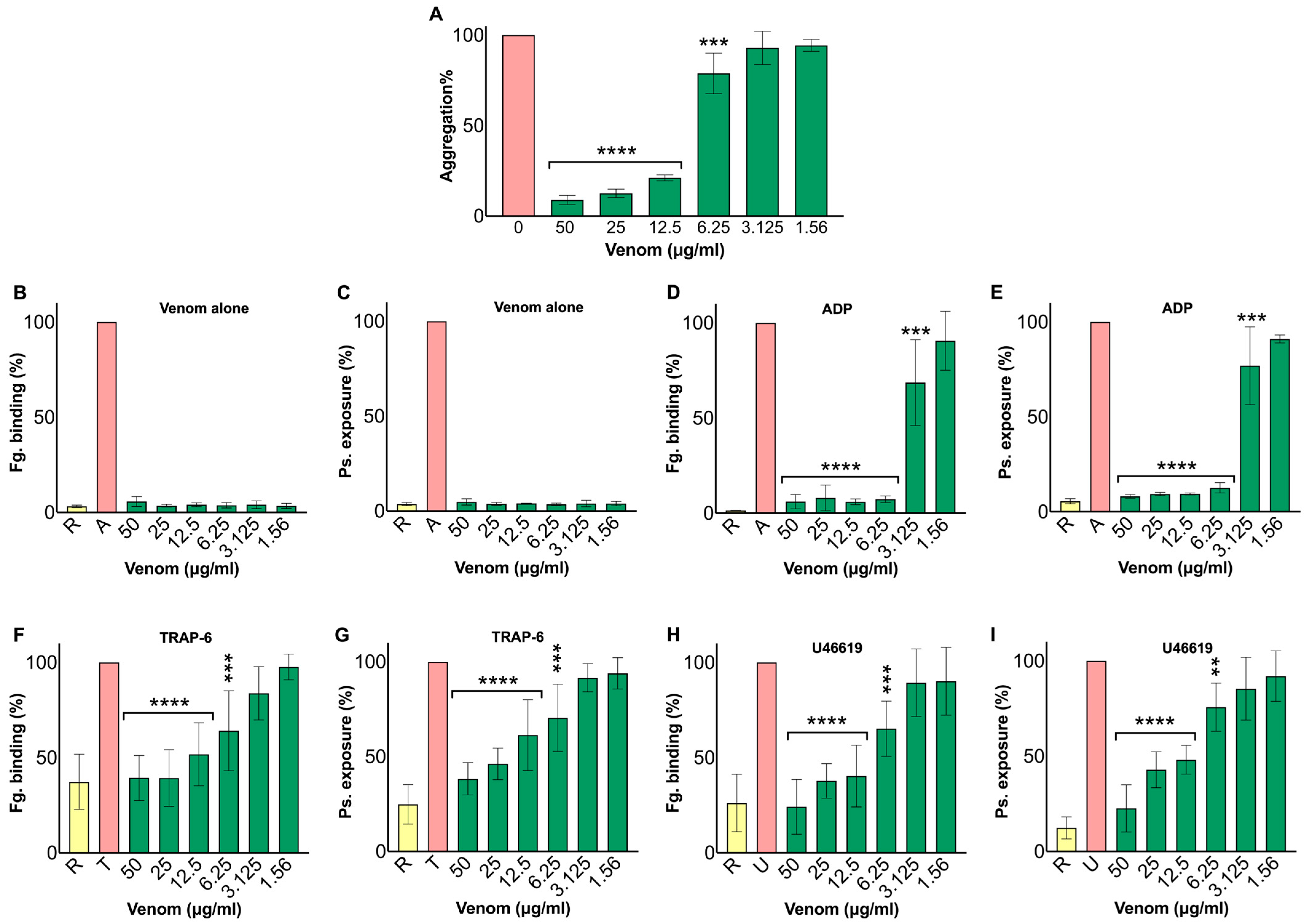
Based on our findings,
N. nivea venom significantly inhibits human platelet aggregation and activation without causing cytotoxic effects on platelets. It markedly diminishes platelet aggregation induced by adenosine diphosphate (ADP) at high concentrations and inhibits platelet activation in a dose-dependent manner. Both markers of platelet activation, fibrinogen binding and P-selectin exposure, were reduced following exposure to different concentrations of venom upon activation with agonists such as ADP, TRAP-6, and U46619. These findings suggest that the antiplatelet activity of
N. nivea venom results from a specific pharmacological mechanism rather than cytotoxic effects on platelets. This aligns with studies on other cobra venoms; for example, a purified 3FTX from
N. kaouthia venom inhibited platelet aggregation following the addition of ADP, thrombin, and arachidonic acid agonists [
24]. Furthermore, PLA
2, a lipid-hydrolysing enzyme that cleaves membrane phospholipids to generate bioactive mediators, is a potent inhibitor of human platelet aggregation in
N. nigricollis venom [
25]. The same effect is observed in other elapid venoms, such as
Notechis scutatus, which also inhibits human platelet aggregation [
26]. Conversely, viper venoms are predominantly procoagulant due to high levels of prothrombin activators and other coagulation enzymes, including metalloproteases (MPs) and serine proteases (SPs) [
27].
Daboia russelii venom exemplifies this, with strong procoagulant components that cause extensive clotting (fibrin formation) via a metalloprotease-derived Factor X activator (RVV-X), which then converts prothrombin to thrombin. Despite this, some viper venoms contain anticoagulant constituents; for instance,
Echis carinatus venom contains echistatin, a small disintegrin that binds to platelet integrin GPIIb/IIIa, thereby inhibiting platelet aggregation [
28]. Overall, the platelet-inhibitory effect of
N. nivea aligns with previously reported findings of other cobra and elapid venoms.
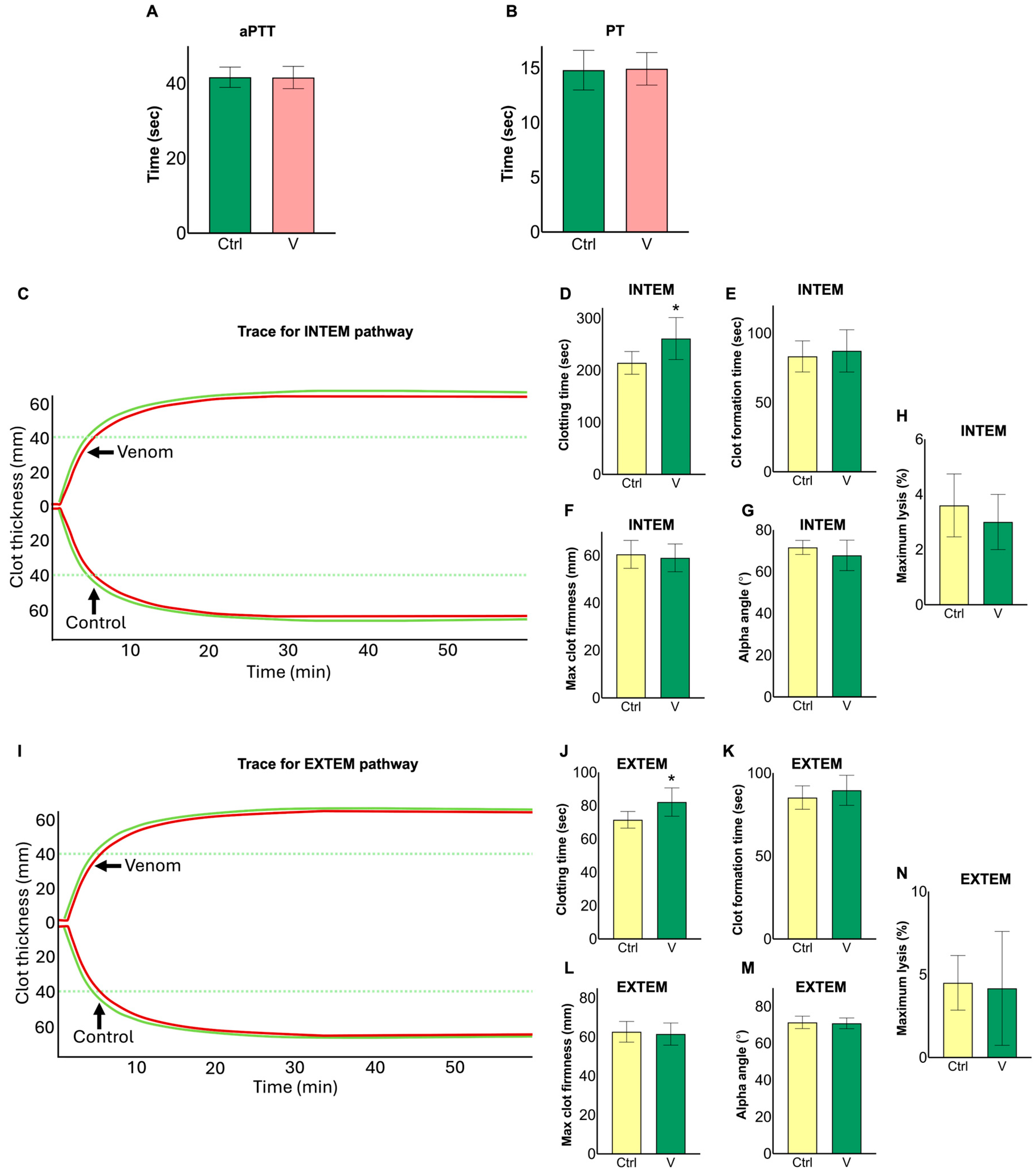
To distinguish platelet-specific effects from direct modulation of the coagulation cascade, plasma and whole blood clotting assays were performed. Results showed that
N. nivea venom did not significantly alter standard plasma clotting times (PT or aPTT), indicating no notable effect on the extrinsic or intrinsic pathways under the tested conditions. Conversely,
N. nivea venom slightly prolonged clotting time in both the intrinsic and extrinsic pathways during ROTEM assays with whole blood, while other thromboelastometry parameters remained unchanged. This pattern suggests that
N. nivea venom may exert a mild anticoagulant effect during the initiation phase of coagulation, without affecting clot formation or strength. African spitting cobra venoms, such as
N. nigricolis,
N. nigricincta,
N. pallida, and
N. mossambica, inhibit Factor Xa via PLA
2 toxins, impairing prothrombinase complex formation and delaying clot initiation [
12]. Conversely, certain elapid venoms act as potent procoagulants. For instance,
Oxyuranus scutellatus venom contains a prothrombin activator that converts prothrombin into thrombin, leading to consumptive coagulopathy [
29].
Pseudonaja textilis venom also causes similar effects through pseutarin C, a prothrombin activator [
30]. In contrast, viper venoms employ a markedly different mechanism to induce coagulotoxic effects. For example, specific viper venoms, such as
Bothrops atrox,
Echis ocellatus, and
D. russelii, have strong procoagulant effects, directly activating clotting factors via SVMP and SVSP enzymes [
31,
32].
Bothrops species possess thrombin-like serine proteases that facilitate fibrinogen clotting, as well as specific enzymes that activate factor X [
4,
33]. Similarly,
E. ocellatus venom uses its SVMP to activate both prothrombin and factor X [
31].
D. russelii venom contains enzymes that activate factor V and X, ultimately leading to consumptive coagulopathy [
32]. For example, 3FTXs from elapid venoms have been characterised as interacting with a variety of cardiovascular targets, including platelet receptors and ion channels, suggesting various mechanisms by which they can influence haemostasis and vascular function, distinct from direct enzymatic activation of coagulation factors used by viper venoms [
34].
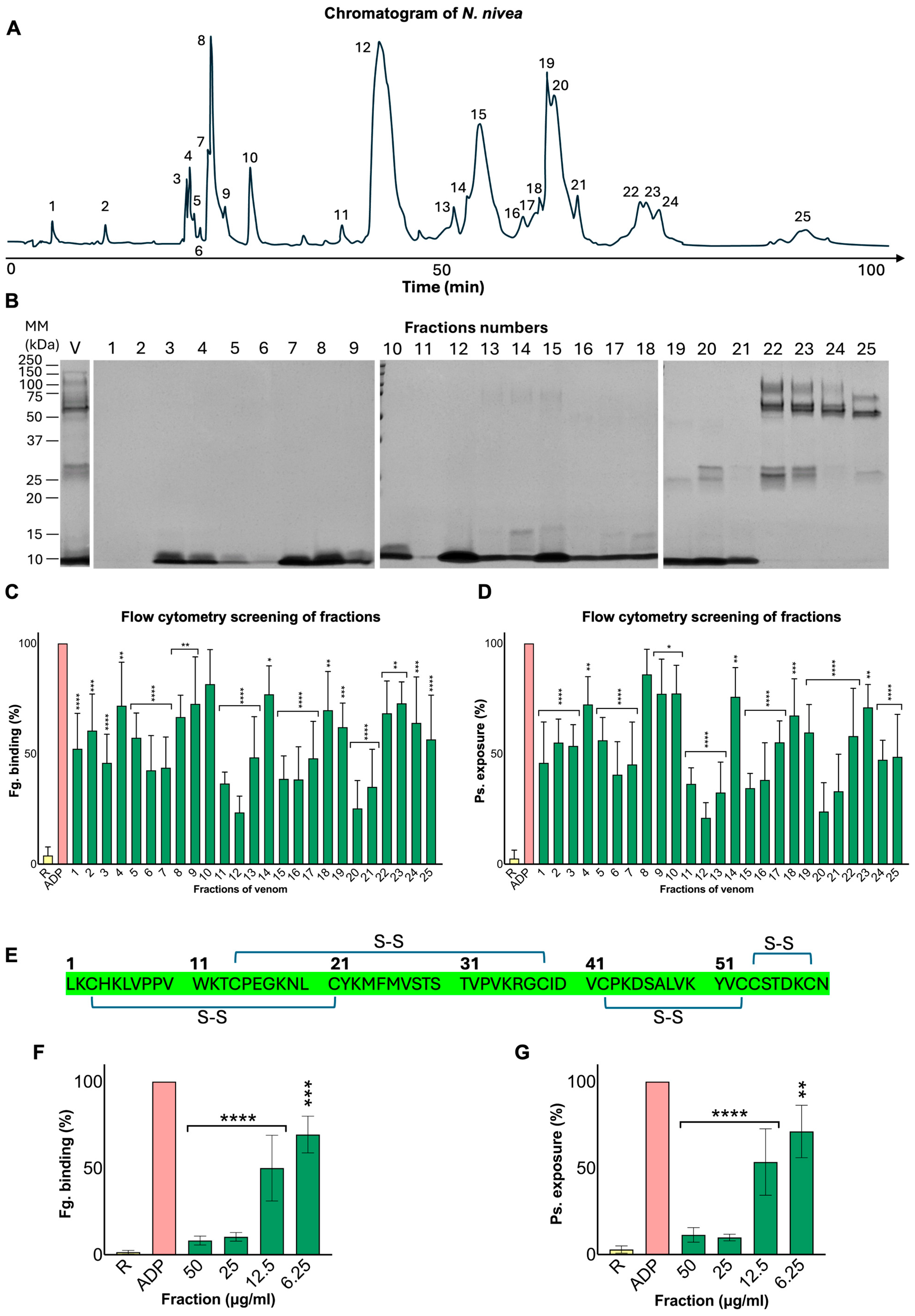
Fractionation of crude
N. nivea venom via RP-HPLC yielded 25 distinct fractions. Among these, one abundant fraction (F12), which showed the greatest inhibitory effect on platelet activation in flow cytometry assays, was identified as a cytotoxin, a 3FTX, through mass spectrometry. 3FTXs are a structurally conserved family of proteins comprising nearly 60 amino acids. These small polypeptides are stabilised by 4–5 disulphide bonds and can be divided into more than 24 subgroups. They are major components of elapid/cobra venoms and are primarily responsible for neurotoxicity or myotoxicity [
35,
36]. They can also impair haemostasis by targeting various stages of blood clotting, although this effect remains inadequately studied [
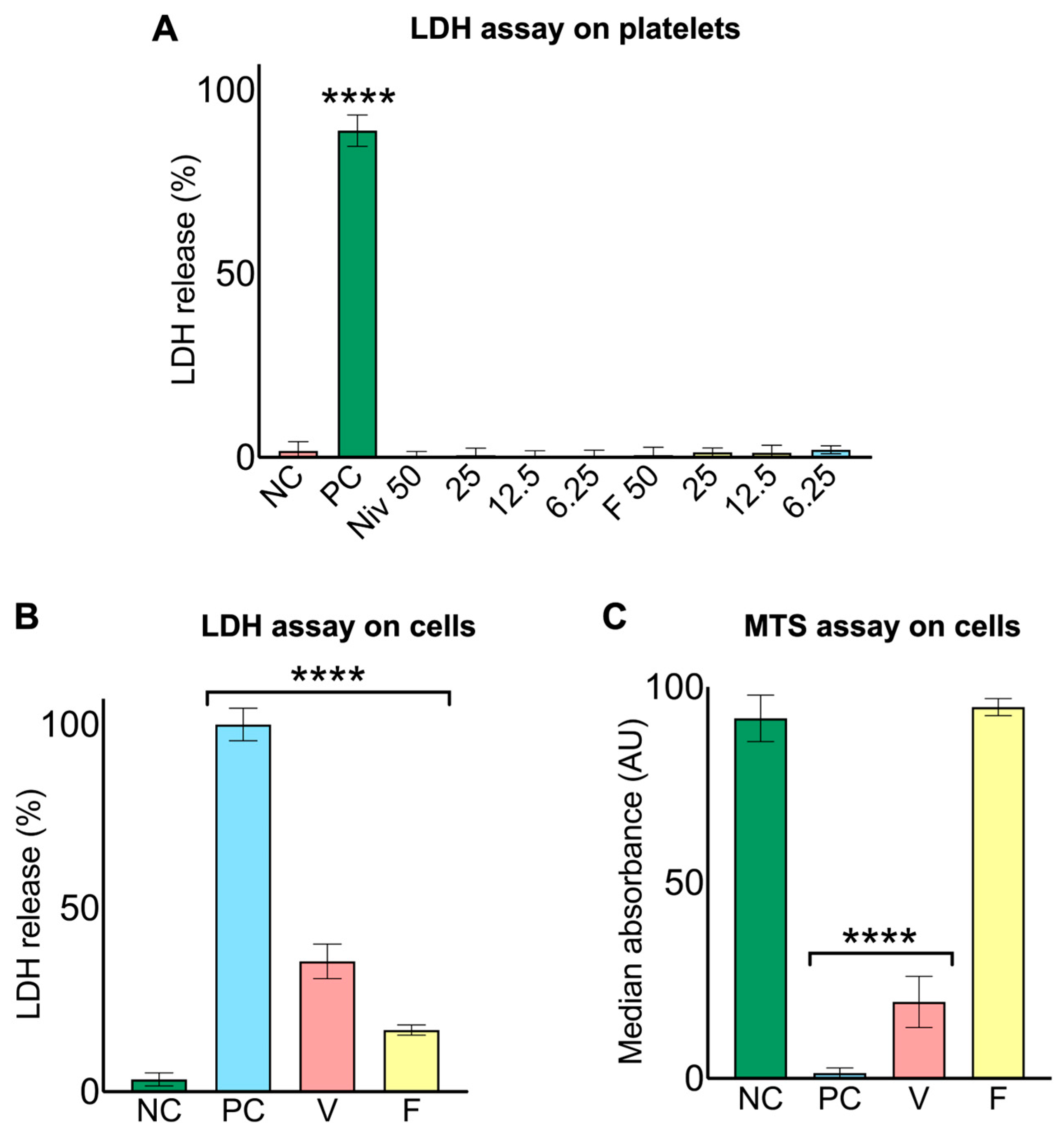
36]. Our findings provide new evidence that, when tested on isolated platelets at various concentrations, F12 significantly inhibited fibrinogen binding and P-selectin exposure in a dose-dependent manner, consistent with the activity of the crude venom. This fraction did not cause platelet lysis and only slightly compromised the cellular integrity of human myoblasts, which released LDH but remained metabolically active and exhibited normal morphology. Previous studies have reported alterations in platelet aggregation or coagulation pathways induced by non-enzymatic venom components [
20,
35]. For example,
N. nigricollis venom contains a cytotoxin that exerts both antiplatelet and anticoagulant effects [
35]. KT-6.9, a 3FTX from
N. kaouthia, has previously demonstrated significant inhibition of ADP-induced aggregation by binding to platelets [
24].
Hemachatus haemachatus venom contains ringhalectin and exactin, 3FTXs, which can block factor X activation by the extrinsic tenase complex, thereby inhibiting fibrin clot formation [
35].
Dendroaspis angusticeps produces thrombostatin, a 3FTX that inhibits fibrinogen binding and platelet aggregation by antagonising glycoprotein alpha-IIb/beta-3 [
35]. Overall, these published findings align with our results and support the hypothesis that this purified 3FTX can influence thrombosis and inhibit platelet activity through a distinct, non-enzymatic mechanism, likely by binding to platelet receptors and coagulation factors. Further research is necessary to elucidate the broader haemotoxic effects and the detailed mechanism of action of this toxin.
This study has some limitations, including its restriction to in vitro and ex vivo assays, and the detailed molecular interactions and in vivo effects of this toxin remain to be elucidated. Further in vivo investigations would be necessary to determine whether the platelet-inhibitory effects observed in vitro translate into measurable alterations in haemostasis during envenoming. Future research should focus on comprehensive mechanistic investigations to identify the precise targets of the purified toxin. It could also investigate the molecular mechanisms underlying the observed inhibition of platelet activation, including potential modulation of integrin αIIbβ3 activation. Future studies could also include assessment of mitochondrial function in isolated platelets to further characterise the cytotoxic effects of the venom on platelets. Employing fluorescence-based approaches with confocal microscopy and flow cytometry, using antivenom or toxin-specific antibodies, could also help determine the precise venom components that directly bind to platelets. In addition, evaluating platelet aggregation with different agonists (e.g., collagen or epinephrine) could help clarify the specific pathways underlying the observed platelet inhibitory activity. Additionally, further characterisation of the proteolytic components of N. nivea venom using more physiologically relevant substrates and techniques, such as gelatin zymography, may help identify the molecular characteristics of the proteases contributing to the observed enzymatic activity. Furthermore, future studies could investigate the venom’s activity against physiological substrates, such as extracellular matrix proteins (e.g., laminin, collagen, and fibronectin), to better understand its potential tissue-related effects. The pharmacological profile of this toxin requires optimisation, including improvements in efficacy, specificity, and safety, to facilitate its development as a novel therapeutic agent.
In conclusion, although cobra envenomation is mainly neurotoxic [
25], our findings highlight this venom’s ability to interfere with platelet activity and the coagulation pathway. This effect is notably less strong than that of vipers. It also emphasises the discovery of a 3FTX in Cape cobra venom that inhibits platelet function. 3FTXs are small, rigid, and highly stable structures, making them suitable for protein engineering, therapeutic uses in bioprospecting, and drug development for conditions such as bleeding disorders and thrombotic complications [
37]. For example, by reducing the cytotoxic effects of 3FTXs while preserving their anti-platelet activity, these toxins could inspire the development of improved antithrombotic agents, similar to how viper venom disintegrins contributed to the development of tirofiban and eptifibatide [
38]. However, many cobra 3FTXs are also known to possess neurotoxic properties [
9]; therefore, their potential therapeutic applications would require careful modification and evaluation of their safety profiles. Hence, even though
N. nivea venom has minimal haemotoxic effects, its components may provide useful insights into mechanisms involved in thrombotic and bleeding disorders. Our findings also suggest that the purified protein may contribute to the venom’s antiplatelet activity without damaging platelet membranes, likely by impairing signalling pathways rather than enzymatically degrading targets. However, this effect appears less potent than that of crude venom, suggesting that other components may act together to produce a stronger haemotoxic response.
4. Materials and Methods
Unless otherwise stated, all chemicals and reagents used in this study were of analytical grade and purchased from Sigma-Aldrich (Gillingham, UK).
4.1. Venom Preparation
Lyophilised N. nivea venom from a pooled stock of multiple specimens was obtained from Latoxan (Valence, France). The lyophilised venom was stored at −80 °C until use. A stock solution was prepared at 2 mg/mL in phosphate-buffered saline (PBS) and stored at −20 °C. On the day of each experiment, the venom stock was freshly diluted in PBS to the required concentrations. During experimental procedures, venom samples were kept on ice to minimise protein degradation. Crotalus atrox venom was obtained in lyophilised form from the Kentucky Reptile Zoo (Bowen, KY, USA) and used as a positive control in selected assays. This venom was prepared under the same conditions as N. nivea venom, and a concentration of 50 μg/mL was used in relevant assays.
4.2. Metalloprotease Assay
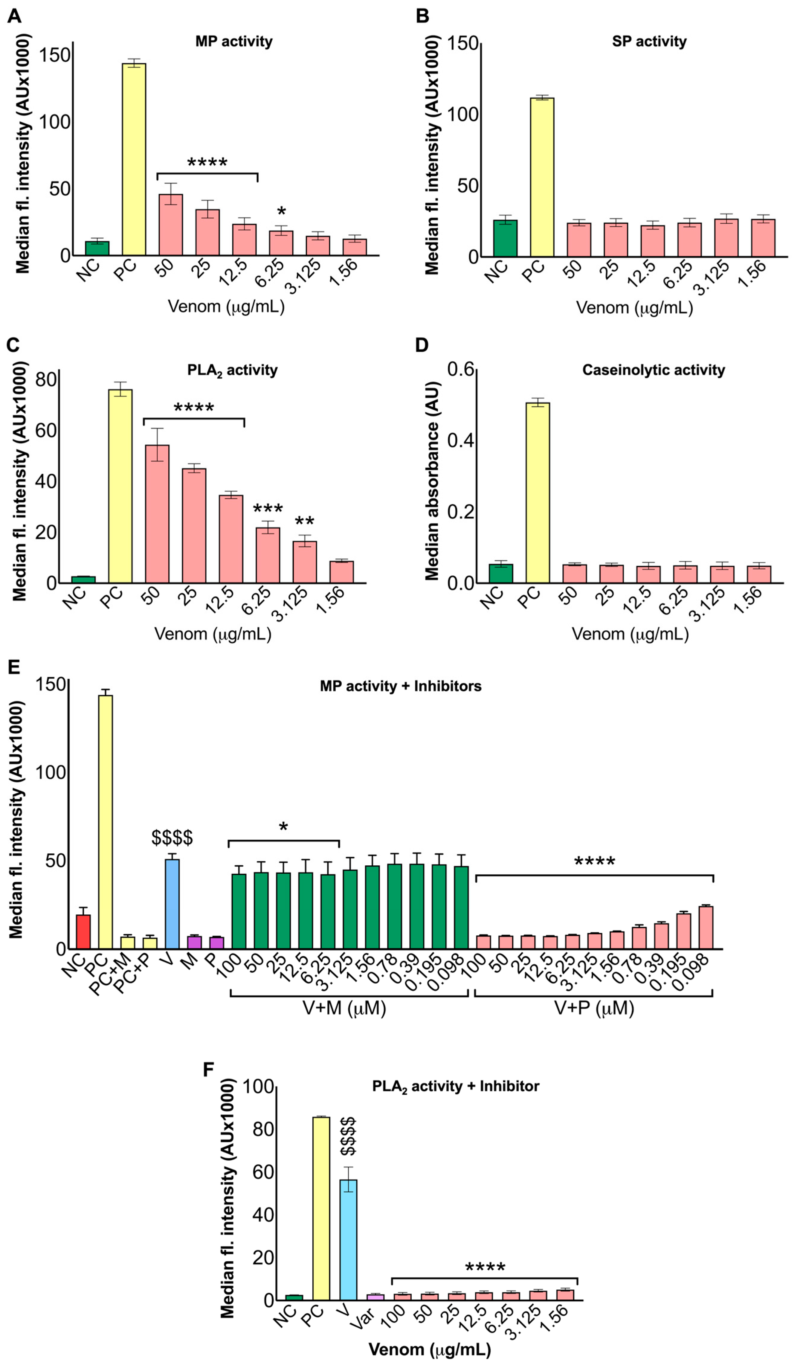
The metalloprotease activity of
N. nivea venom was assessed using DQ-gelatin (1 mg/mL; ThermoFisher Scientific, Abingdon, UK) as a fluorogenic substrate [
39]. Several concentrations of venom (50–1.56 μg/mL) were added to a black 96-well plate. Then, 2 μL (2 μg) of DQ-gelatin was added to each well, and PBS was added to bring the final volume to 100 μL in a 96-well microplate. Then, the fluorescence level was recorded at 10-min intervals for 90 min using a plate reader (FLUOstar OPTIMA, Ortenberg, Germany) with excitation and emission wavelengths of 485 nm and 520 nm, respectively. To assess the inhibitory effects of metalloprotease inhibitors, marimastat (100–0.098 μM) and prinomastat (100–0.098 μM) were added to
N. nivea venom (50 μg/mL) and incubated for 10 min at room temperature in a 96-well plate prior to the addition of DQ-gelatin. Fluorescence was then measured as described above to assess activity. Substrate in PBS and
C. atrox venom (50 μg/mL) were used as the negative and positive controls, respectively, in the same order.
4.3. Serine Protease Assay
Serine protease activity was evaluated using Nα-benzoyl-L-arginine 7-amido-4-methylcoumarin hydrochloride (BAAMC; Sigma Aldrich, Gillingham, UK) as the fluorogenic substrate at a final concentration of 20 μM, reconstituted in Dimethyl sulfoxide (DMSO) [
40]. The substrate was added to different venom concentrations (50–1.56 μg/mL) in a black 96-well plate. Then, fluorescence was measured at 10-min intervals for 90 min, using excitation and emission wavelengths of 355 nm and 460 nm, respectively. A Fluostar Optima (BMG Labtech, Ortenberg, Germany) spectrofluorometer was used for this assay. Substrate in PBS and
C. atrox (50 μg/mL) venom were used as the negative and positive control, respectively.
4.4. Phospholipase A2 Assay
Phospholipase A
2 (PLA
2) activity was evaluated using the Enzcheck
® PLA
2 assay kit (Invitrogen, Paisley, UK), which employed Red/Green BODIPY
® PC-A2 as the substrate [
39]. This experiment was carried out in black 96-well plates by adding 5 μL of venom, at various concentrations (50–1.56 μg/mL), to 45 μL of reaction buffer, followed by 50 μL of substrate mix (prepared according to the manufacturer’s protocol). Fluorescence readings were taken at 10-min intervals for 90 min using a Fluostar Optima (BMG Labtech, Ortenberg, Germany) spectrofluorometer, set at excitation and emission wavelengths of 485 nm and 520 nm, respectively. Bee venom and substrate in PBS served as the positive (PC) and negative (NC) controls, respectively.
4.5. Caseinolytic Assay
Casein was used as a general substrate to assess the venom’s overall proteolytic activity. It is widely used in protease assays due to its stability, low cost, and suitability for detecting broad proteolytic activity. Therefore, this assay was included as a complementary approach to evaluate general protease activity in the venom. The caseinolytic activity was assessed using azocasein (Sigma-Aldrich, Gillingham, UK) as the substrate [
41]. The substrate solution was prepared by dissolving 5 mg of substrate in 1 mL of Tris-HCl buffer (50 mM, pH 8.0). The same buffer was used to dissolve the venom and prepare a final concentration of 1 mg/mL. The samples were prepared by mixing 10 μL of venom (50–1.56 μg/mL) with 90 μL of the substrate solution, followed by incubation for 90 min at 37 °C. After incubation, the reactions were halted by adding 200 μL of 5% (
v/
v) trichloroacetic acid (TCA). The samples were then centrifuged for 5 min at 8000 rpm at room temperature, and 150 μL of the supernatant was transferred into a 96-well microplate, followed by the addition of 150 μL of NaOH (0.5 M). Absorbance of each well was recorded at 440 nm using a Fluostar Optima (BMG Labtech, Ortenberg, Germany) spectrofluorometer.
4.6. Human Blood Collection and Preparation of Plasma, PRP and Washed Platelets
The human blood collection, platelet preparation, and functional assays were performed as previously described [
42]. Blood samples were obtained from healthy human donors in accordance with the protocol approved by the University of Reading Research Ethics Committee (UREC 17/17). After acquiring written informed consent, blood was drawn via venepuncture into vacutainers containing 3.25% (
w/
v) sodium citrate as an anticoagulant. For platelet-rich plasma (PRP), whole blood was centrifuged at 100×
g for 20 min at 20 °C. The upper layer was transferred to a 15 mL Falcon tube without disturbing the remaining layers and then maintained at 30 °C before use. Plasma was prepared by centrifuging whole blood at 5000×
g for 10 min at 20 °C, after which the clear top layer was collected and used in assays.
To isolate platelets, PRP was combined with 3 mL of acid-citrate-dextrose [ACD; 2.5% (w/v) sodium citrate dihydrate, 2.0% (w/v) glucose, and 1.5% (w/v) citric acid] and 10 μL of prostaglandin I2 (PGI2; 125 μg/mL; Sigma-Aldrich, Gillingham, UK) dissolved in Ethanol. The mixture was gently inverted and centrifuged at 1400× g for 10 min at 20 °C. The resulting platelet pellet was resuspended in 1 mL of Tyrode’s-HEPES buffer [134 mmol/L NaCl, 2.9 mmol/L KCl, 0.34 mmol/L Na2HPO4·12H2O, 12 mmol/L NaHCO3, 20 mmol/L HEPES, 1 mmol/L MgCl2, and 5 mM glucose; pH 7.3], followed by the addition of ACD (150 μL). A volume of 25 mL was achieved by adding pre-warmed Tyrode’s-HEPES buffer along with 150 μL ACD. A second centrifugation was performed after adding 3 mL of ACD and prostacyclin (10 ng/mL) at 1400× g for 10 min at 20 °C. The supernatant was discarded, and the final platelet pellet was resuspended in modified Tyrode’s-HEPES buffer to a concentration of 4 × 10⁸ platelets/mL.
4.7. Clotting Assay
The Ceveron T100 fully automated coagulation analyser (Technoclone, Vienna, Austria) was used to measure Prothrombin Time (PT) and activated Partial Thromboplastin Time (aPTT) in accordance with the manufacturer’s instructions and as described previously [
43].
N. nivea venom (50 μg/mL) was added to a fixed volume (40 μL) of human plasma taken from various donors. Standard PT (tissue thromboplastin and 25 mM CaCl
2) and aPTT (silica/sulfatide phospholipids and 25 mM CaCl
2) reagents were added following specific protocols, and samples were analysed using Ceveron T100. PT and aPTT measurements were obtained at wavelengths of 405 nm and 630 nm, respectively.
4.8. ROTEM Analysis
Rotational Thromboelastometry (ROTEM) analysis was carried out using the ROTEM Delta instrument (Werfen, Warrington, UK) to measure the effect of
N. nivea venom (50 μg/mL) on various aspects of the whole blood clotting process [
43]. A venom concentration of 50 μg/mL was selected based on preliminary experiments and concentrations commonly used in venom functional assays to evaluate potential effects on coagulation parameters. The INTEM and EXTEM tests were carried out to evaluate the impact of the venom on the intrinsic and extrinsic (as well as common) pathways of blood coagulation. In each test, the venom (50 μg/mL) was mixed with citrated human whole blood (300 μL), followed by the addition of specific reagents in predetermined volumes as per the manufacturer’s guidelines. Recalcification of the blood samples was carried out using the star-tem reagent (0.2 M CaCl
2 in HEPES buffer, pH 7.4). Subsequently, blood coagulation was activated by intrinsic stimulators (partial thromboplastin phospholipid derived from rabbit brain and ellagic acid activators) or extrinsic stimulators (recombinant tissue factor, heparin and phospholipids).
4.9. Flow Cytometry and Platelet Aggregation Assays
Platelet aggregation was analysed using optical aggregometry (Chrono-Log, Havertown, PA, USA) [
42,
43]. The level of aggregation was recorded for 5 min after adding various concentrations of
N. nivea venom (12.5–1.56 μg/mL) to PRP and incubating at 37 °C. Afterwards, adenosine diphosphate (ADP, 5 μM) was added, the baseline was set to 0, and the aggregation level was recorded for another 5 min. To measure platelet activation by flow cytometry, PRP was incubated with different concentrations of
N. nivea venom (50–1.56 μg/mL) and 35 μL of an antibody master mix. The mastermix contained 2 μL each of P-selectin (PE-Cy™5 labelled mouse anti-human CD62P) and FITC-labelled fibrinogen (polyclonal rabbit anti-human fibrinogen, 2 μg/mL) antibodies and 31 μL of HEPES-buffered solution (containing 2.9 mM KCl, 134 mM NaCl, 0.34 mM Na
2HPO
4·12H
2O, 1 mM MgCl
2, 12 mM NaHCO
3, 20 mM HEPES; pH 7.3). After incubating all samples for 5 min, the agonists ADP (5 μM), thrombin receptor-activating peptide-6 (TRAP-6, 10 μM), or U46619 (4 μM), a thromboxane A
2 (TXA
2) analogue, were added to the relevant samples. Then, samples were incubated for an additional 20 min at 37 °C. Before flow cytometry analysis (using a BD Accuri C6, Wokingham, UK), samples were fixed with 0.2% (
v/
v) formyl saline [0.2% formaldehyde in 0.9% (
w/
v) NaCl]. During analysis, 5000 cells from each sample were recorded to determine median fluorescence intensity. To evaluate the effect of venom treatment, all samples were normalised to the agonist-only controls, which were set at 100%.
4.10. Haemolytic Assay
After removing PRP from centrifuged whole human blood, human erythrocytes were collected from the bottom fraction of the vacutainers, which were dense with red blood cells (RBCs). To wash these erythrocytes, an equal volume of PBS was added to the tube, and the mixture was centrifuged at 2000× g for 2 min; the supernatant was then discarded. Following the washing process, the erythrocytes were resuspended in PBS and treated with N. nivea venom (50 μg/mL). After incubating the samples at different time points (0, 1, 6, and 24 h), they were centrifuged at 2000× g for 2 min, and 50 μL of the supernatant was transferred to a 96-well plate. Finally, absorbance was measured at 540 nm using a Fluostar Optima (BMG Labtech, Ortenberg, Germany) spectrofluorometer. To assess the impact of PLA2 and MP inhibition on venom-induced haemolysis, the venom (50 μg/mL) was incubated with varespladib (100 µM), prinomastat (100 µM), and a combination of both inhibitors (100 µM of each) for the same time intervals prior to analysis. All subsequent steps were performed as described above. In this experiment, PBS and 1% (v/v) Triton X-100 (Sigma-Aldrich, Gillingham, UK) were used as negative (NC) and positive (PC) controls, respectively.
4.11. Fibrinogenolytic Assay
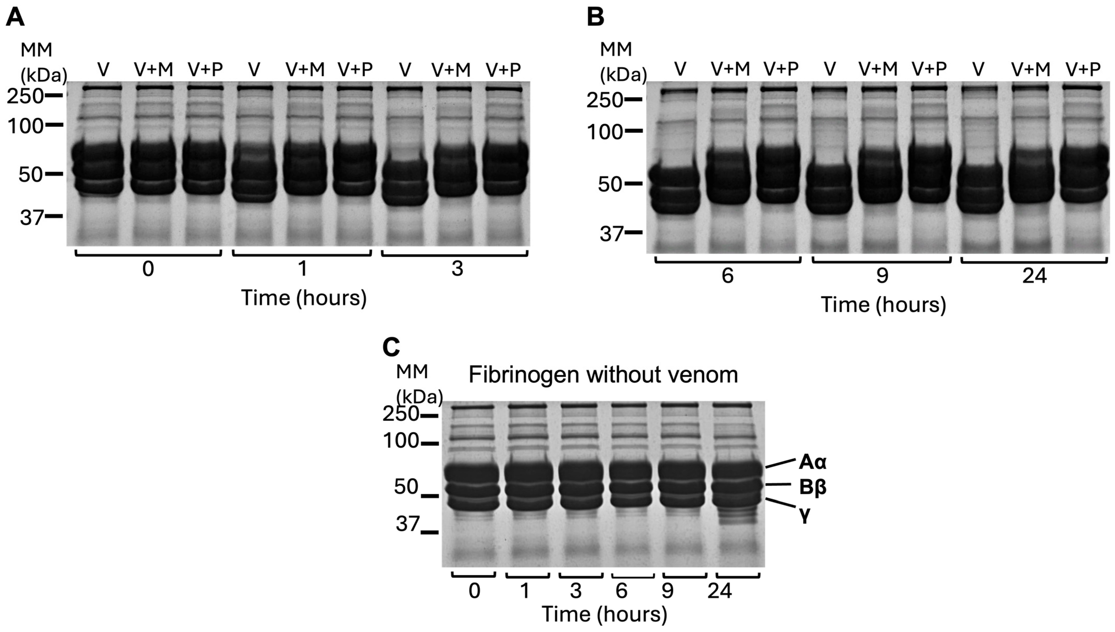
N. nivea venom (100 μg/mL) was added to human fibrinogen (1 mg/mL; Sigma-Aldrich, Gillingham, UK) in PBS at an approximate ratio of 1:19 (venom:fibrinogen), and the samples were incubated at 37 °C for various time intervals (0, 1, 3, 6, 9, 24 h). At each time point, 30 μL of each sample was taken and immediately mixed with 2× reducing sample treatment buffer (2× RSTB) (40% [w/v] SDS, 10% [v/v] β-mercaptoethanol, 20% [v/v] glycerol, and 10% stacking gel buffer in nanopure water, along with a trace of bromophenol blue as a tracking dye), then boiled at 90 °C for 10 min. Afterwards, SDS-PAGE was used to analyse each sample. A venom concentration of 100 μg/mL was used to ensure detectable fibrinogen degradation in the SDS–PAGE analysis. The fibrinogen concentration was selected to ensure clear detection of proteolytic degradation products in the SDS–PAGE analysis.
4.12. Sodium Dodecyl Sulfate-Polyacrylamide Gel Electrophoresis (SDS-PAGE)
To perform gel electrophoresis, SDS-PAGE gels (12%; 10-well) were hand-casted as outlined in the following protocol: resolving gel containing acrylamide–bisacrylamide solution (30% [w/v], 14 mL), SDS (10% [w/v], 350 μL), resolving gel buffer (3 M Tris-HCl, pH 8.8; 4.38 mL), water (14.525 mL), ammonium persulfate (APS, 1.5% [w/v], 1.75 mL) and tetra-methyl-ethylene-diamine (TEMED; 34 μL); the stacking layer is made up of acrylamide–bisacrylamide solution (30% [w/v], 1.35 mL), SDS (10% [w/v], 100 μL), stacking gel buffer (0.5 M Tris-HCl, pH 6.8; 2.5 mL), water (5.75 mL), APS (1.5% [w/v], 500 μL) and TEMED (8 μL). For SDS-PAGE analysis, samples from the fibrinogenolytic assay were prepared by mixing 30 μL of each reaction mixture with an equal volume of 2× RSTB. Samples were heated for 10 min at 90 °C. This was followed by vortexing the samples and loading 30 μL of them onto the gel. Precision Plus Protein™ Dual Colour Standards (5 μL; Bio-Rad, Watford, UK) were used as the molecular mass reference. The run took 2 h at 70 volts using a Mini-PROTEAN Electrophoresis System (Bio-Rad, Watford, UK). Then, Coomassie blue [0.1% (w/v) Coomassie Brilliant Blue in 40% methanol and 10% acetic acid] was used to stain the subsequent gels for 2 h. Then, the gels were de-stained on a rocker for 2 h using a de-staining solution [10% (v/v) methanol and 10% (v/v) acetic acid in double-distilled water].
4.13. Venom Fractionation
Reversed-phase high-performance liquid chromatography (RP-HPLC; Spectra-Physics P200) was employed to separate N. nivea venom using a BDS Hypersil C18 column (250 × 4.6 mm) (Thermo Scientific, Loughborough, UK). An amount of 200 μL of 0.1% [v/v] trifluoroacetic acid (TFA) in water was used to dissolve the lyophilised venom (2 mg). After centrifuging the sample (8000× g for 5 min), 200 μL of the supernatant was manually injected into the instrument. Fractions were collected at a flow rate of 1 mL/min over 100 min, employing a staged gradient of buffer A (0.1% TFA [v/v] in HPLC water) and buffer B (0.1% TFA [v/v] in acetonitrile), with buffer B reaching 60% at almost 70 min into the process. Fractions were detected at 215 nm, and those with an absorbance exceeding 0.05 were collected and stored in the freezer for subsequent analysis. Collected fractions were lyophilised and reconstituted in 100 μL PBS. For SDS-PAGE analysis, 20 μL of each fraction was loaded onto the gel for comparative profiling without prior standardisation of protein concentration. In addition, fractions were screened for biological activity by flow cytometry, in which 5 μL of each fraction was used to assess platelet activation. These analyses were performed as an initial screening step prior to protein quantification, allowing identification of fractions with both biological activity and sufficient abundance for further characterisation.
4.14. Mass Spectrometry Analysis
A prominent protein band from the purified fraction was excised from the Coomassie-stained SDS-PAGE gel and sent to Alta Bioscience (Birmingham, UK) for mass spectrometry analysis. Trypsin digestion was performed using 10 μL of eluted samples (up to 10 μg of protein), to which 40 μL of 100 mM ammonium bicarbonate (pH 8) and 50 µL 10 mM dithiothreitol were added and incubated at 56 °C for 30 min. Samples were then cooled to room temperature and cysteines were alkylated by adding 50 µL of 50 mM iodoacetamide, mixing and incubating at room temperature in the dark for 30 min. Finally, 50 µL of trypsin gold (Promega, Southampton, UK; 6 ng/µL) was added to the samples and incubated at 37 °C overnight. The samples were then desalted using millipore C18 ZipTips (Merck KGaA, Darmstadt, Germany). The tips were prepared by pre-wetting in 100% acetonitrile and rinsed in 2 × 10 µL 0.1% formic acid. Samples were repeat pipetted throughout the volume of the samples ten times. The tip was then washed with 3 × 10 µL 0.1% formic acid to remove excess salts before elution of peptides with 10 µL of 50% acetonitrile/water/0.1% formic acid. Samples were dried down to remove the acetonitrile, and then re-suspended in 0.1% formic acid solution in water.
The peptides were concentrated and separated using a UltiMate® 3000 HPLC (Dionex, Sunnyvale, CA, USA). Samples were trapped on precolumn, Acclaim PepMap 100 C18 HPLC Columns (Thermo Scientific, Loughborough, UK), 3 µm particle size, 2 cm length, 75 µm I.D., (Dionex, Sunnyvale, CA, USA) and separated in Nano Series™ standard solumns 75 µm I.D. × 15 cm, packed with C18 PepMap100, 2 µm, 100 Å (Dionex, Sunnyvale, CA, USA) using a gradient of 3.2% to 44% solvent B (0.1% formic acid in acetonitrile) for 30 min. The column was then washed with 90% mobile phase B before re-equilibrating at 3.2% mobile phase B. Peptides were eluted directly (~300 nL/min) via a Triversa Nanomate nanospray source (Advion Biosciences, Ithaca, NY, USA) into a QExactive HF Orbitrap mass spectrometer (ThermoFisher Scientific, Loughborough, UK). The spray voltage of QE HF was set to 1.7 kV through Triversa NanoMate and heated capillary at 275 °C. The mass spectrometer performed a full MS scan (m/z 360–1600) and subsequent HCD MS/MS scans of the 20 most abundant ions with dynamic exclusion setting 15S. Full-scan mass spectra were recorded at a resolution of 120,000 at m/z 200 and AGC target of 3 × 10⁶. Precursor ions were fragmented in HCD MS/MS with resolution set up at 15,000 and a normalised collision energy of 28. The width of the precursor isolation window was 1.2 m/z and only multiply charged precursor ions were selected for MS/MS.
The MS and MS/MS scans were searched against Uniprot database using Protein Discovery 2.2 software, Sequest HT algorithm (Thermo Fisher, Loughborough, UK). Variable modifications included were deamidation (N and Q), oxidation (M), phosphorylation (S, T and Y) and acetylation (K). The precursor mass tolerance was 10 ppm and the MS/MS mass tolerance was 0.02 Da. Two missed cleavages were allowed and data were filtered with a false discovery rate (FDR) of 0.01. Proteins with at least two high-confidence peptides were accepted as real hits.
4.15. Protein Quantification
Following venom fractionation, the protein concentration of selected venom fractions was determined using a NanoDrop 2000 spectrophotometer (Thermo Fisher Scientific, Waltham, MA, USA). Fractions were dissolved in PBS, with PBS used as the blank. A bovine serum albumin (BSA) standard (2 mg/mL) was serially diluted to generate a standard curve for protein quantification. For each measurement, 2 μL of each sample was loaded onto the NanoDrop pedestal, and measurements were performed in triplicate.
4.16. Lactate Dehydrogenase Cytotoxicity Assay
LDH assays were performed using an LDH cytotoxicity assay kit (Thermo Fisher Scientific, Loughborough, UK), as directed by the manufacturer. Washed platelets were incubated with N. nivea venom (50 μg/mL) for 45 min at 37 °C in a 96-well plate. Subsequently, 25 μL of the supernatant was transferred to a new 96-well plate, followed by the addition of 25 μL of the reaction mixture. Samples were incubated for 30 min at 37 °C in the dark, after which 25 μL of stop solution was added. Absorbance was then recorded at 490 nm and 680 nm using a Fluostar Optima (BMG Labtech, Ortenberg, Germany) spectrofluorometer. To measure the LDH activity of AB1190 cells, cells were seeded in a 96-well tissue culture plate (100 μL of growth media/well) and incubated at 37 °C with 5% CO2. After replacing the media with fresh growth media, N. nivea venom (50 μg/mL) or its purified toxin were added to each well and incubated for 4 h. Afterwards, 25 μL of each sample was transferred to a new 96-well plate, and the remaining steps of the experiment were carried out as described above. Triton X-100 (1%; Sigma, Bilston, UK) was used as the positive control. PBS and the cell culture media served as negative controls for the washed platelets and AB1190 cells cytotoxicity assay, respectively.
4.17. Cell Viability Assay
To investigate the impact of N. nivea venom on the viability of AB1190 myoblasts, an MTS cell viability assay kit (Promega, Southampton, UK) was used. Cells (5000 per well) were seeded in a 96-well plate containing 100 μL growth media and incubated overnight to allow cell adhesion. The growth media were replaced with fresh growth media, after which N. nivea venom (50 μg/mL) and or its purified cytotoxin (50 μg/mL) was added to each well and incubated for 24 h. Following the 24-h incubation, 10 μL MTS reagent was added to each well and incubated further for 4 h at 37 °C with 5% CO2. Absorbance was measured at 490 nm wavelength using Fluostar Optima (BMG Labtech, Ortenberg, Germany) spectrofluorometer. All experiments were performed in triplicate for each condition. Fresh growth media were used for the negative control, and Triton X-100 as a recognised cytotoxic agent (positive control). For analysis, each reading was compared to the negative control, and the percentage cell viability was calculated.
4.18. Statistical Analysis
Statistical analyses were performed using GraphPad Prism (version 7.0; GraphPad Software Inc., San Diego, CA, USA). Differences between multiple groups were analysed using ordinary one-way analysis of variance (ANOVA) followed by Fisher’s least significant difference (LSD) post hoc test. For comparisons between two groups (venom-treated samples and control samples), unpaired Student’s t-tests were performed. Data are presented as mean ± standard deviation (SD), and differences were considered statistically significant at p < 0.05.



 ,
,