Case Report

Azemiops feae (Fea’s Viper) Envenoming: A Case Report and Review of the Literature

1
Adaptive Biotoxicology Lab, School of the Environment, University of Queensland, St. Lucia, QLD 4072, Australia
2
Independent Researcher, Yubei District, Chongqing 404100, China
3
Shizhu Tujia Autonomous County Traditional Chinese Medicine Hospital, Shizhu Tujia Autonomous County, Chongqing 409100, China
*
Authors to whom correspondence should be addressed.
Toxins 2026, 18(5), 201; https://doi.org/10.3390/toxins18050201
Submission received: 18 March 2026 / Revised: 23 April 2026 / Accepted: 24 April 2026 / Published: 26 April 2026
(This article belongs to the Section Animal Venoms)

Abstract

Azemiops feae (Fea’s viper) is a phylogenetically distinctive Asian viper with poorly defined medical significance, and human envenomations remain rarely reported in the English-language literature. We describe a new case of A. feae envenoming from Chongqing, China, and present a scoping review of published clinical case reports and case series to better characterize its epidemiology, clinical manifestations, and management. A 53-year-old male developed marked local pain and swelling following a bite to the hand, accompanied by transient neurotoxic symptoms, as well as mild hypofibrinogenemia. Treatment with a single vial of Gloydius brevicaudus monovalent antivenom was followed by clinical improvement and full recovery. Review of the literature identified nine previously published studies from China and one captive case from Europe. Envenoming typically occurred during agricultural activities, most commonly affected the lower extremities, and was characterized by prominent local effects with occasional mild neurotoxic features and inconsistent, generally mild coagulation abnormalities. Antivenom use was highly variable, involving multiple heterologous monovalent antivenoms, and outcomes were uniformly favourable regardless of antivenom administration. Collectively, available evidence indicates that A. feae envenoming is usually self-limited, with predominantly local effects and infrequent, mild systemic involvement. However, the absence of species-specific antivenom and the heterogeneity of current treatment practices highlight the need for systematic venom characterization and functional antivenom efficacy studies to inform evidence-based clinical management.
Key Contribution: This paper reports a case of medical significant envenoming by the rare Asian viperid Azemiops feae and provides the most comprehensive literature on the envenomation by this species to date.

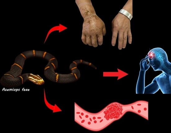
Graphical Abstract

1. Introduction

Commonly known as Fea’s viper, Azemiops feae is a medium-sized, fossorial viperid snake characterized by a secretive and cryptic lifestyle [1,2]. The species inhabits cool, moist montane environments across northern Myanmar, northern Vietnam, and southern China [1,2,3]. In China, its distribution extends from Yunnan in the west to Zhejiang in the east and northward to Shaanxi [1,3]. Azemiops represents an enigmatic lineage within the family Viperidae and is currently placed in the monotypic subfamily Azemiopinae, that its basal to the pitviper clade [2,4,5].
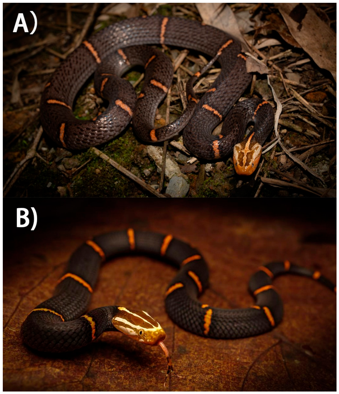
Orlov et al. [2] proposed the recognition of a second species, A. kharini, based primarily on head coloration (white-headed versus black-headed forms; Figure 1) and additional morphological and osteological characters; however, this taxonomic treatment did not include genetic analyses. Subsequent molecular studies demonstrated that specimens attributed to A. kharini are nested within A. feae [6], and intermediate phenotypes combining diagnostic features of both forms have been reported [7]. Accordingly, the Reptile Database currently recognizes A. feae as the sole valid species of the genus.
Given the unresolved taxonomic status of Azemiops and the inability to verify the identity of individual specimens involved in the present study and previously published studies reviewed, we adopt a conservative taxonomic approach and follow the nomenclature of Uetz et al. [3], referring to all Azemiops specimens herein as A. feae. We do not attempt to address or resolve the underlying taxonomic controversies in this study.

2. Results

2.1. Case Report

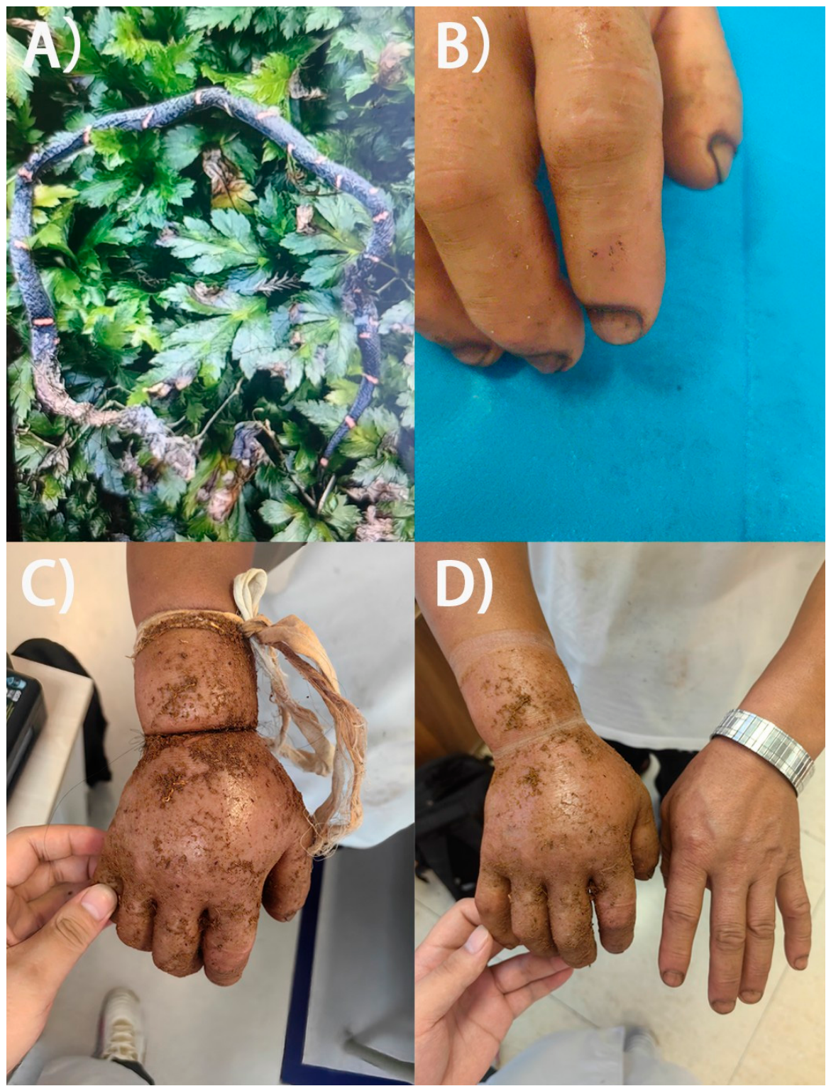
On 19 July 2025, at approximately 16:00, a 53-year-old male was bitten on his right index finger while working in his farmland in Shizhu County, Chongqing Municipality, China. The patient killed and photographed the offending snake at the time of the incident, and the image was later reviewed upon presentation to hospital. Based on dorsal coloration and body shape, the snake was identified as a Fea’s viper (Azemiops feae) (Figure 2A).
Immediately after the bite, the patient experienced acute pain and progressive swelling of the right hand. As first-aid, he applied multiple tight ligatures: a strand of hair at the base of the right index finger, another strand of hair at the right wrist, and a cloth band approximately 10 cm proximal to the wrist (Figure 2C). He also topically applied an herbal poultice (unknown ingredients) mixed with rice wine to the swollen area. Pain and swelling continued to worsen overnight, prompting removal of the ligature at the finger due to severe pain. With no improvement by the following afternoon, the patient sought medical care and arrived at Shizhu Tujia Autonomous County Traditional Chinese Medicine Hospital on July 20 (Day 0), at 16:48 (approximately 24.8 h post-bite).
Upon arrival, the patient was alert and afebrile. Vital signs were stable: temperature 36.7 °C, pulse 80 beats/min, respiratory rate 20 breaths/min, and blood pressure 130/98 mmHg. Examination revealed fang marks on the dorsal surface of the right index finger (Figure 2B), marked swelling of the hand and wrist, restricted finger movement, and significant tenderness. No lymphadenopathy was detected in the right axilla. The patient denied nausea, vomiting, chest tightness, dyspnea, dizziness, headache, abdominal pain, or malaise.
Initial management included removal of all constrictive bands (Figure 2D), wound cleansing, oxygen supplementation, ECG monitoring, and intravenous administration of dexamethasone (10 mg), mannitol (125 mL), vitamin C (2 g) and pantoprazole (40 mg). Tetanus prophylaxis was also administered intramuscularly. Traditional Chinese medicine (TCM) treatments were applied concurrently: small skin incisions between the right index and middle finger, approximately at the depression proximal to the second dorsal interosseous muscle for decompression and external application of a mixture of herbal powders (including Lobelia chinensis, Scutellaria barbata, Paris polyphylla, and others). It must be noted that dexamethasone, mannitol, vitamin C, pantoprazole, and traditional Chinese medicine (TCM) treatments all lack evidence of effectiveness for the treatment of snakebite.
Initial laboratory testing performed at 17:46, approximately 25.5 h post bite, showed normal platelet count and predominantly normal coagulation profile (Table 1), except for mildly reduced fibrinogen (1.92 g/L; reference: 2.0–4.0 g/L). D-dimer was within normal limits (0.29 µg/mL; reference: 0–0.5 µg/mL). At 20:00, 27 h post envenomation, one vial of Gloydius brevicaudus monovalent antivenom (GbMAV; 6000 U/10 mL) was administered. At 20:30, the patient developed neurotoxicity symptoms including palpitations, perioral numbness, drowsiness, and bilateral ptosis. Vital signs and oxygen saturation remained stable, and no rash or airway compromise developed. The patient was observed closely with supplemental low-flow oxygen.
On the second morning (21 July, Day 1), the patient reported marked improvement in swelling and discomfort. Palpitations, numbness, drowsiness, and ptosis had resolved. In the absence of progressive neurotoxic manifestations, no additional antivenom was administered. Supportive therapy continued with intravenous administration of dexamethasone, mannitol, vitamin C, and pantoprazole at the same dosages used on Day 0. TCM therapy, including internal herbal decoctions aimed for “heat-clearing and detoxicating”, was continued as per local practice.
By the third morning (22 July, Day 2), swelling had further improved, and no recurrence of palpitations, numbness, drowsiness, or ptosis was reported. No further antivenom was administered. The dexamethasone dose was reduced to 5 mg, and mannitol and vitamin C were continued in dosages consistent with previous days, and mannitol was discontinued. Repeat laboratory testing at 08:00, approximately 64 h post envenomation revealed mildly elevated white blood cell count (14.27 × 10⁹/L), mildly reduced red blood cell count (4.29 × 10¹²/L), and stable platelets. Coagulation testing showed slight prolongation of thrombin time (21.9 s) and decreased fibrinogen (1.39 g/L), consistent with mild hypofibrinogenemic coagulopathy (Table 1).
On the fourth day (23 July, Day 3), swelling had completely resolved and the skin incision sites had healed. The patient requested discharge, as clinical signs had resolved and laboratory abnormalities on previous day were mild, he was discharged with instructions for rest and follow-up observations on potential recurrence of symptoms.

2.2. Case Report Literature Review

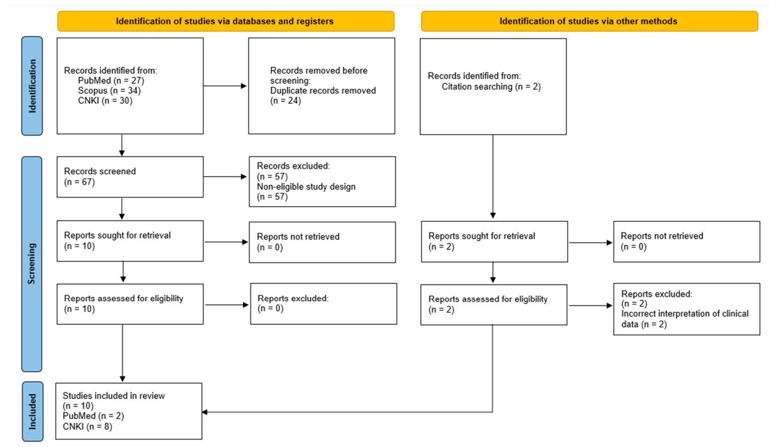
The keyword searches retrieved a total of 91 records, of which 24 were identified as duplicates and removed. Screening of titles and abstracts resulted in the exclusion of a further 57 records that did not report clinical case reports or case series of A. feae envenoming. Citation searching identified two additional publications; however, these were duplicate references that briefly mentioned the identical three cases of A. feae bites without providing any case descriptions and were therefore excluded. Consequently, 10 studies, comprising two publications in English and eight in Chinese, met the inclusion criteria and were included in the review. The study identification, screening, and inclusion process is summarized in a PRISMA flow diagram (Figure 3).
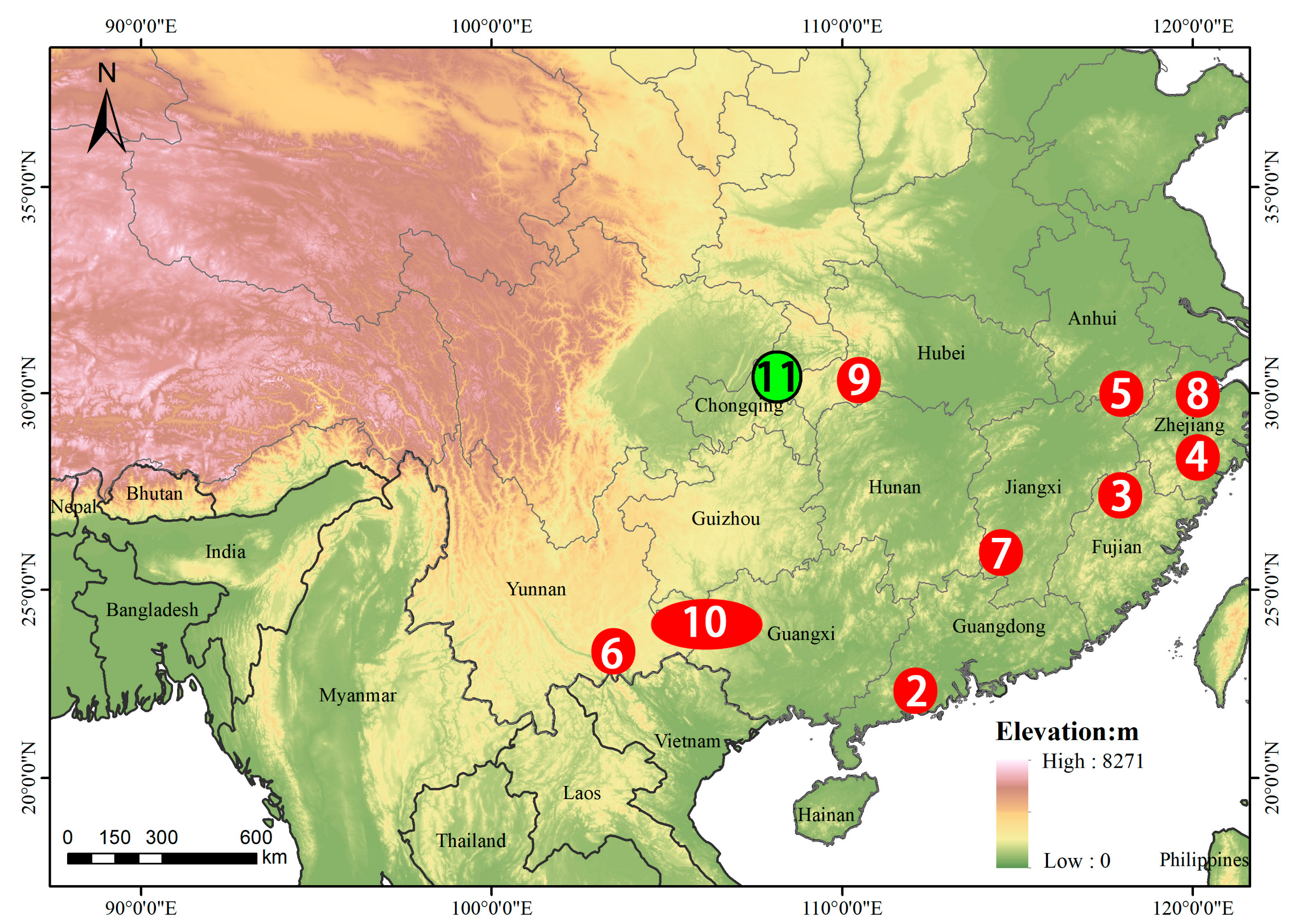
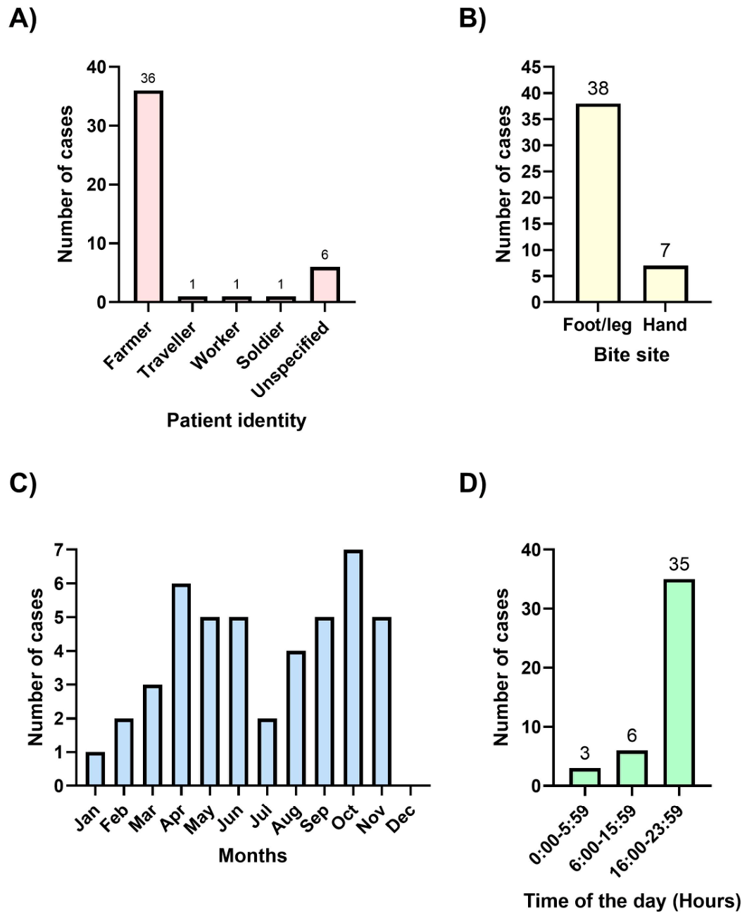
Of the 10 studies included, 9 were single case reports, and 1 was a case series comprising 36 cases of A. feae envenoming. The full text of each report was reviewed, and key epidemiological and clinical information from all published cases (Cases 1–10), together with the single case reported in the present study (Case 11), are summarized in Table 2. The geographic localities of all studies reporting bites by wild snakes are shown in Figure 4, and aggregated epidemiological patterns are presented in Figure 5. The case reported by us is the green dot on Figure 4; GPS 29.998120, 108.113830.
Cases of A. feae envenoming reported in the literature, together with the single case from the present study, were distributed broadly across southern China (Figure 4) and exhibited consistent epidemiological patterns with respect to patient identity, bite site, seasonality, and time of day (Figure 5). Most patients were identified as farmers (36 cases), while only isolated cases involved travellers, workers, or soldiers; patient occupation was unspecified in six cases (Figure 5A). Bites predominantly affected the lower extremities, with 38 cases involving the foot or leg, compared with seven cases involving the hand (Figure 5B). Incidents were recorded throughout the year, with cases reported in all months except December; higher numbers were observed between April and June and again between September and November (Figure 5C). Envenoming occurred across all time periods of the day, with the highest frequency recorded between 16:00 and 23:59 (35 cases), followed by 06:00–15:59 (six cases), and 00:00–05:59 (three cases) (Figure 5D).
Based on the data summarized in Table 2, the clinical presentation of A. feae envenoming was generally characterized by prominent local effects with variable systemic symptoms. Local pain and swelling at the bite site were the most consistent features, reported in nearly all cases across single case reports and the larger case series by Yang [17]. In several cases, local manifestations were mild, with no signs of systemic envenoming [11,13,15,16,17]. Severe tissue damages or necrosis were not reported in any studies.
Systemic manifestations were comparatively uncommon but diverse when present. Signs of suspected neurotoxic envenoming, including dizziness [10,12,17], limb numbness [10], blurred vision [8,12,14], muscle fasciculations [8], and ptosis [12,14,17], were reported sporadically across cases. Other symptoms such as nausea [12,14] and chills [10], were also occasionally documented. Notably, no cases progressed to respiratory failure.
Abnormalities in coagulation parameters were inconsistently reported in earlier case reports; however, among studies that assessed coagulation parameters, mild hypofibrinogenemia (none with fibrinogen dropped below the minimum detection limit) emerged as the most frequently observed abnormality in Guo et al. [9] and Yang [17]. In the largest case series reported by Yang [17], reduced fibrinogen levels were documented in 16 of 36 patients, while other coagulation parameters largely remaining within normal ranges. Renal involvement was rare, with only a single elderly patient exhibiting elevated creatinine levels [14].
Antivenom use varied substantially between cases and institutions. Some patients did not receive antivenom and nonetheless recovered uneventfully [8,10,13,17], whereas others were treated with one or more vials of heterologous monovalent antivenoms. All four commercially available monovalent antivenoms in mainland China had been used to treat A. feae envenoming, including DaMAV Deinagkistrodon acutus pitviper snake venom monovalent used in Fu et al. [14], Guo et al. [9], and Wang and Cheng [12]; GbMAV Gloydius brevicauda pitviper snake venom monovalent in Guo et al. [9], Wang and Cheng [12], Xiong et al. [15], and Yu et al. [16]; BmMAV Bungarus multicinctus elapid snake venom monovalent in Fu et al. [14], Guo et al. [9], Xiong et al. [15], Yang [17], and Yu et al. [16]; and NaMAV Naja atra elapid snake venom monovalent in Chen [11], either alone or in combination. The antivenom administrations were primarily based on local progression, systemic symptoms, or laboratory findings and the types of antivenom available, rather than any standardized criteria. Despite this heterogeneity in management, outcomes were uniformly favourable: all patients recovered, hospitalization durations were generally short (≤8 days in the largest case series by Yang [17]), and no fatalities or permanent sequelae were reported.

3. Discussion

This study expands the limited clinical literature on Azemiops feae envenoming by integrating a new case with previously published reports, allowing a clearer assessment of the epidemiological and clinical characteristics of this poorly known species. Across the available cases, envenoming by wild A. feae was confined to southern China and occurred predominantly in montane regions, consistent with the known distribution and ecological preferences of the species [1,3]. Envenoming events were strongly associated with outdoor occupational activities, particularly farming, reflecting accidental encounters during manual work and mirroring global trends in snakebite epidemiology [18]. Most bites involved the distal lower limbs, consistent with general snakebite patterns reported in China and globally [19,20,21,22]. However, as some studies have documented a higher frequency of bites to the upper limbs [23,24,25], further documentation of envenomation by A. feae is needed to better characterize bite patterns in this poorly known species. Cases occurred throughout the year, with peaks between April and June and between September and November, lower frequency between July and August may be explained by A. feae’s preference for cool and moist conditions and aligns with natural history observations from wild and captive specimens [1]. Envenoming was documented throughout the day, with a distinct nocturnal peak, suggesting that bites most frequently result from nocturnally active individuals than disturbance of resting or concealed snakes. Collectively, these findings indicate that A. feae envenoming represents an occupational and environmentally mediated risk, disproportionately affecting rural populations engaged in agricultural activities within suitable montane habitats during the evening hours.
The clinical features of A. feae envenoming align only partially with the venom composition and activities reported from experimental venom studies. The predominance of local pain and swelling across nearly all reported cases is consistent with the high abundance of phospholipase A2 (PLA2) and presence of other toxins known to induce pain and swelling [26,27], such as snake venom serine proteases (SVSPs) and snake venom metalloproteinases (SVMPs) in A. feae venom [28,29,30,31]. Conversely, the absence of severe tissue necrosis or extensive local tissue destruction accords with experimental findings demonstrating low or absent cytotoxic, myotoxic, and proteolytic tissue-damaging activities in A. feae venom [28,32].
Neurotoxic effects of A. feae venom are likely mediated primarily by azemiopsin, a polypeptide shown to block nicotinic acetylcholine receptors [33]. These alpha-neurotoxic peptides de novo evolved from within the propeptide region of the C-type natriuretic peptide [34]. Genetic analyses suggest that these peptides share a molecular ancestor with the waglerin peptides from Tropidolaemus species, which are also alpha-neurotoxic [35,36,37,38,39]. The reports of neurotoxic manifestations following envenomations by A. feae, including ptosis, blurred vision, limb numbness, and muscle fasciculations, corresponds with in vivo lethality studies in mice and rabbits [28,32,33], and in vitro evidence demonstrating potent inhibition of nicotinic acetylcholine receptors across multiple vertebrate taxa, including humans [40]. However, the generally mild nature of neurological symptoms, together with the absence of respiratory failure in all reported cases, suggests that although neurotoxic components are biologically active, their clinical relevance in human envenoming is typically limited. This is consistent with data showing that, consistent with dietary preferences, A. feae neurotoxicity is highly selective amphibian nicotinic acetylcholine receptors than mammals, being 1.9 times more potent on amphibian receptors than rodents, and 3.44 times more potent on amphibian receptors than on humans [40].
In contrast, the occasional reports of mild hypofibrinogenemia observed in clinical cases are not readily explained by existing venom studies. Experimental investigations have consistently failed to demonstrate coagulotoxic activity of A. feae venom, both in vitro using human plasma and fibrinogen [41], and in vivo in mice and rabbit models [28,32]. While interspecific differences in coagulotoxic venom activity may partly explain discrepancies between animal models and human clinical findings [42], the absence of detectable effects on human plasma and fibrinogen in vitro suggests alternative explanations. One possibility is intraspecific venom variation, particularly in the relative abundance of SVMPs and SVSPs, two toxin families known to act on fibrinogen through non-clotting cleavage or pseudo-procoagulant (thrombin-like) manners [43,44]. Such variation has been documented in Azemiops venoms, with a northern Vietnamese specimen exhibiting a significantly higher abundance of SVSPs compared with Chinese specimens from Sichuan and Zhejiang [28,31]. Consistent with this is the low levels of activity on fibrinogen for the Yunnan sample used in Debono et al. [41]. Importantly, fibrinogen depletion in reported Chinese cases was consistently mild, with no values falling below assay detection limits and no progression to more clinically significant complete coagulopathy. Collectively, these observations indicate that coagulation disturbance is neither a dominant nor a consistent feature of A. feae envenoming. Nevertheless, given its recurrent documentation across multiple studies, routine monitoring of coagulation parameters remains a prudent component of clinical management.
Despite all four commercially available monovalent antivenoms having been used in reported cases of A. feae envenoming [11,14,17], the existing clinical data do not allow reliable assessment of antivenom effectiveness or potency against this species. Some patients recovered uneventfully without receiving antivenom [8,10,13,17], whereas others improved following administration of one or multiple vials of non-species-specific monovalent antivenoms. This heterogeneity, together with the generally mild clinical course observed in many cases, makes it impossible to distinguish spontaneous recovery from treatment-related effects based solely on existing case reports and case series.
Experimental data provide limited but potentially informative insights. Using in vitro ELISA assays, Zheng et al. [28] demonstrated that all four Chinese monovalent antivenoms exhibit immunorecognition of A. feae venom, with Gloydius brevicaudus monovalent pitviper snake antivenom (GbMAV) showing the strongest binding capacity. On this basis, the authors recommended GbMAV as a potential treatment option for A. feae envenoming [28]. Clinically, Bungarus multicinctus monovalent elapid snake antivenom (BmMAV) has been administered in patients with neurotoxic features following A. feae bite [14,17], while Deinagkistrodon acutus monovalent pitviper snake antivenom (DaMAV) has been recommended in Chinese national guidelines for the management of “haemotoxic” envenoming [19]. However, immunorecognition and empiric clinical use do not equate to proven neutralization or clinical effectiveness. Overall, the available evidence remains insufficient to establish the optimal antivenom choice or dosing strategy for A. feae envenoming. Well-designed future studies integrating functional neutralization assays and systematically documented clinical outcomes are needed to evaluate the efficacy and effectiveness of both Chinese and global antivenoms against A. feae venom. As none of the snakes used in the immunizing mixture of the above antivenoms are close relatives of A. feae and there was a similarity of outcomes, this suggests that the antivenoms had minimal, if any, cross-reactivity with the A. feae venom, and that the consistent recoveries were not linked to antivenom usage.

4. Conclusions

In conclusion, this study expands the limited clinical knowledge of A. feae envenoming by integrating a newly documented case with a comprehensive synthesis of previously published reports, many of which are in Chinese and therefore less accessible to the international research community. The new case adds valuable clinical detail to a sparsely documented and understudied species and reinforces that A. feae envenoming is typically characterized by prominent local effects with occasional, generally mild systemic manifestations. By collating and analyzing both English- and Chinese-language sources, this study helps bridge an important language barrier in snakebite research, providing a more complete and globally accessible overview of the epidemiology, clinical features, and treatment practices associated with A. feae envenoming. These findings highlight persistent knowledge gaps in venom activities of A. feae and antivenom efficacy against A. feae envenoming, underscoring the need for future studies combining standardized clinical case reports with experimental venom and antivenom research.

5. Materials and Methods

5.1. Case Report

We report a case of confirmed Azemiops feae envenomation based on medical records of Shizhu Tujia Autonomous County Traditional Chinese Medicine Hospital, Shizhu County, Chongqing Municipality, China.

5.2. Case Report Literature Review

We searched the PubMed and Scopus databases on 1 December 2025 using the keywords “Azemiops” and “Fea’s viper,” and the China National Knowledge Infrastructure (CNKI) using as keywords the Chinese common names of Azemiops “白头蝰” and “黑头蝰”. Titles and abstracts of all retrieved records were screened, and only clinical case reports or case series describing envenoming by confirmed Azemiops feae were included. In addition, the reference lists of all screened articles were manually reviewed to identify further potentially relevant publications. Toxicological studies, including venom composition analyses and in vitro or in vivo venom activity experiments, were excluded from this mini review. The study selection process and numbers of included and excluded records were reported in accordance with the PRISMA-Scoping Review guidelines.

Author Contributions

Conceptualization, Z.Q., Y.T. and B.G.F.; methodology, Z.Q.; investigation, Z.Q. and Y.T.; resources, Q.Z.; data curation, Z.Q., Y.T. and Q.Z.; writing—original draft preparation, Z.Q. and B.G.F.; writing—review and editing, Y.T. and Q.Z.; supervision, B.G.F.; project administration, Z.Q. and B.G.F. All authors have read and agreed to the published version of the manuscript.

Funding

This work in memory of Randy Nielsen was supported by a grant from his family.

Institutional Review Board Statement

The study was conducted in accordance with the Declaration of Helsinki, and approved by the Ethics Committee of Institutional Review Board of the Shizhu Tujia Autonomous County Traditional Chinese Medicine Hospital (protocol code not applicable at the hospital, approval date 9 December 2025).

Informed Consent Statement

Written informed consent was obtained from the patient for publication of the clinical details and accompanying images. All patient-identifying information has been anonymized to protect privacy.

Data Availability Statement

The original contributions presented in this study are included in the article. Further inquiries can be directed to the corresponding authors.

Conflicts of Interest

The authors declare no conflicts of interest.

References

  1. Orlov, N.; Ananjeva, N.; Barabanov, A.; Ryabov, S.; Khalikov, R. Diversity of vipers (Azemiopinae, Crotalinae) in East, Southeast, and South Asia: Annotated checklist and natural history data (Reptilia: Squamata: Serpentes: Viperidae). Faun. Abh. Tierkd. Dresd. 2002, 23, 177–218. [Google Scholar]
  2. Orlov, N.L.; Ryabov, S.A.; Nguyen, T.T. On the taxonomy and the distribution of snakes of the genus Azemiops Boulenger, 1888: Description of a new species. Russ. J. Herpetol. 2013, 20, 110–128. [Google Scholar]
  3. Uetz, P.; Freed, P.; Aguilar, R.; Reyes, F.; Hošek, J. The Reptile Database. Available online: https://reptile-database.reptarium.cz/Azemiops/feae (accessed on 18 December 2025).
  4. Snetkov, P.; Orlov, N. Phylogenetic analysis of old world viperid snakes (Serpentes, Viperidae) based on skeletal morphology. Russ. J. Herpetol. 2017, 24, 22–34. [Google Scholar] [CrossRef]
  5. Alencar, L.R.V.; Quental, T.B.; Grazziotin, F.G.; Alfaro, M.L.; Martins, M.; Venzon, M.; Zaher, H. Diversification in vipers: Phylogenetic relationships, time of divergence and shifts in speciation rates. Mol. Phylogenet. Evol. 2016, 105, 50–62. [Google Scholar] [CrossRef] [PubMed]
  6. Li, J.-N.; Liang, D.; Wang, Y.-Y.; Guo, P.; Huang, S.; Zhang, P. A large-scale systematic framework of Chinese snakes based on a unified multilocus marker system. Mol. Phylogenet. Evol. 2020, 148, 106807. [Google Scholar] [CrossRef]
  7. Teynié, A.; Lottier, A.; David, P. Azemiops feae Boulenger, 1888 (Squamata: Viperidae) et cinq autres additions notables pour la faune des serpents du Laos. Bull. Soc. Herpétol. Fr. 2017, 162, 39–54. [Google Scholar]
  8. Valenta, J.; Stach, Z.; Stourac, P.; Kadanka, Z.; Michalek, P. Neurological symptoms following the Fea’s viper (Azemiops feae) bite. Clin. Toxicol. 2015, 53, 1150–1151. [Google Scholar] [CrossRef]
  9. Guo, S.-Z.; Zhan, S.-M.; Zhuang, S.-F.; Chen, J.; Li, T.-X. Management of an Uncommon Snakebite Envenomation (Azemiops kharini). Am. J. Trop. Med. Hyg. 2025, 112, 1137–1139. [Google Scholar] [CrossRef]
  10. Tang, X.; Huang, Y.; Zhao, J. Case report on a Fea’s viper bite. Fujian Med. J. 1983, 6, 53–54. [Google Scholar] [CrossRef]
  11. Chen, K. The first report of Fea’s viper bite in Pan’an County, Zhejiang Province. J. Snake 2001, 3, 33–34. [Google Scholar]
  12. Wang, G.; Cheng, H. A case report of Fea’s viper bite envenoming. J. Snake 2006, 4, 286–287. [Google Scholar]
  13. Hou, Y.; Lei, C.; Xie, X. Treatment of a patient bitten by a Fea’s viper. Med. J. Natl. Def. Forces Southwest China 2011, 21, 1172. [Google Scholar]
  14. Fu, F.; Zeng, Y.; Chen, Z. A report of a Fea’s viper bite. J. Snake 2013, 25, 65–66. [Google Scholar]
  15. Xiong, K.; Chen, T.; Wang, L. A case report of treatment for a Fea’s viper bite. J. Snake 2016, 28, 94–95. [Google Scholar]
  16. Yu, X.; Zhou, F.; Zhang, T.; Tang, X. A case of treatment for a Fea’s viper bite. J. Snake 2019, 31, 173–174. [Google Scholar]
  17. Yang, X. Clinical Characteristics and Treatment of Patients Bitten by Azemiops kharini. Master’s Thesis, Youjiang Medical University For Nationalities, Baise, China, 2024. [Google Scholar] [CrossRef]
  18. Gutiérrez, J.M.; Calvete, J.J.; Habib, A.G.; Harrison, R.A.; Williams, D.J.; Warrell, D.A. Snakebite envenoming. Nat. Rev. Dis. Primers 2017, 3, 17063. [Google Scholar] [CrossRef] [PubMed]
  19. Lai, R.; Yan, S.; Wang, S.; Yang, S.; Yan, Z.; Lan, P.; Wang, Y.; Li, Q.; Wang, J.; Wang, W.; et al. The Chinese guideline for management of snakebites. World J. Emerg. Med. 2024, 15, 333–355. [Google Scholar] [CrossRef]
  20. Afroz, A.; Siddiquea, B.N.; Chowdhury, H.A.; Jackson, T.N.; Watt, A.D. Snakebite envenoming: A systematic review and meta-analysis of global morbidity and mortality. PLoS Negl. Trop. Dis. 2024, 18, e0012080. [Google Scholar] [CrossRef]
  21. Tekin, R.; Sula, B.; Cakırca, G.; Aktar, F.; Deveci, Ö.; Yolbas, I.; Çelen, M.K.; Bekcibasi, M.; Palancı, Y.; Dogan, E. Comparison of snakebite cases in children and adults. Eur. Rev. Med. Pharmacol. Sci. 2015, 19, 2711–2716. [Google Scholar]
  22. Campbell, B.T.; Corsi, J.M.; Boneti, C.; Jackson, R.J.; Smith, S.D.; Kokoska, E.R. Pediatric snakebites: Lessons learned from 114 cases. J. Pediatr. Surg. 2008, 43, 1338–1341. [Google Scholar] [CrossRef]
  23. Chippaux, J.P. Epidemiology of snakebites in Europe: A systematic review of the literature. Toxicon 2012, 59, 86–99. [Google Scholar] [CrossRef]
  24. Luksić, B.; Bradarić, N.; Prgomet, S. Venomous snakebites in southern Croatia. Coll. Antropol. 2006, 30, 191–197. [Google Scholar]
  25. Dobaja Borak, M.; Babić, Ž.; Caganova, B.; Grenc, D.; Karabuva, S.; Kolpach, Z.; Krakowiak, A.; Kolesnikova, V.; Lukšić, B.; Pap, C.; et al. Viper envenomation in Central and Southeastern Europe: A multicentre study. Clin. Toxicol. 2023, 61, 656–664. [Google Scholar] [CrossRef] [PubMed]
  26. Nielsen, V.G.; Wagner, M.T. Review of the Mechanisms of Snake Venom Induced Pain: It’s All about Location, Location, Location. Int. J. Mol. Sci. 2022, 23, 2128. [Google Scholar] [CrossRef] [PubMed]
  27. Bhardwaj, A.; Muthu, R.; Soundravally, R.; Pillai, A.B.; Bammigatti, C.; Kadhiravan, T. Circulating Secretory Phospholipase A2 Activity following Snakebites and Its Relationship with Envenomation Status and Progression of Local Swelling. Am. J. Trop. Med. Hyg. 2020, 104, 1142–1148. [Google Scholar] [CrossRef]
  28. Zheng, S.-R.; Sun, Y.; Zhao, H.-Y.; Wen, L.; Ji, X.; Gao, J.-F. Differences between Two Groups of Burmese Vipers (Viperidae: Azemiops) in the Proteomic Profiles, Immunoreactivity and Biochemical Functions of Their Venoms. Toxins 2022, 14, 572. [Google Scholar] [CrossRef]
  29. Fry, B.G.; Wüster, W.; Ryan Ramjan, S.F.; Jackson, T.; Martelli, P.; Kini, R.M. Analysis of Colubroidea snake venoms by liquid chromatography with mass spectrometry: Evolutionary and toxinological implications. Rapid Commun. Mass Spectrom. 2003, 17, 2047–2062. [Google Scholar] [CrossRef]
  30. Tsai, I.-H.; Wang, Y.-M.; Huang, K.-F. Structures of Azemiops feae venom phospholipases and cys-rich-secretory protein and implications for taxonomy and toxinology. Toxicon 2016, 114, 31–39. [Google Scholar] [CrossRef]
  31. Babenko, V.V.; Ziganshin, R.H.; Weise, C.; Dyachenko, I.; Shaykhutdinova, E.; Murashev, A.N.; Zhmak, M.; Starkov, V.; Hoang, A.N.; Tsetlin, V.; et al. Novel Bradykinin-Potentiating Peptides and Three-Finger Toxins from Viper Venom: Combined NGS Venom Gland Transcriptomics and Quantitative Venom Proteomics of the Azemiops feae Viper. Biomedicines 2020, 8, 249. [Google Scholar] [CrossRef]
  32. Vest, D.K. Preliminary studies on the venom of the Chinese snake Azemiops feae, Boulenger (Fea’s viper). Toxicon 1986, 24, 510–513. [Google Scholar] [CrossRef] [PubMed]
  33. Utkin, Y.N.; Weise, C.; Kasheverov, I.E.; Andreeva, T.V.; Kryukova, E.V.; Zhmak, M.N.; Starkov, V.G.; Hoang, N.A.; Bertrand, D.; Ramerstorfer, J. Azemiopsin from Azemiops feae viper venom, a novel polypeptide ligand of nicotinic acetylcholine receptor. J. Biol. Chem. 2012, 287, 27079–27086. [Google Scholar] [CrossRef]
  34. Brust, A.; Sunagar, K.; Undheim, E.A.; Vetter, I.; Yang, D.C.; Casewell, N.R.; Jackson, T.N.; Koludarov, I.; Alewood, P.F.; Hodgson, W.C.; et al. Differential evolution and neofunctionalization of snake venom metalloprotease domains. Mol. Cell Proteom. 2013, 12, 651–663. [Google Scholar] [CrossRef]
  35. Debono, J.; Xie, B.; Violette, A.; Fourmy, R.; Jaeger, M.; Fry, B.G. Viper Venom Botox: The Molecular Origin and Evolution of the Waglerin Peptides Used in Anti-Wrinkle Skin Cream. J. Mol. Evol. 2017, 84, 8–11. [Google Scholar] [CrossRef]
  36. Xie, B.; Dashevsky, D.; Rokyta, D.; Ghezellou, P.; Fathinia, B.; Shi, Q.; Richardson, M.K.; Fry, B.G. Dynamic genetic differentiation drives the widespread structural and functional convergent evolution of snake venom proteinaceous toxins. BMC Biol. 2022, 20, 4. [Google Scholar] [CrossRef]
  37. Schmidt, J.J.; Weinstein, S.A.; Smith, L.A. Molecular properties and structure-function relationships of lethal peptides from venom of Wagler’s pit viper, Trimeresurus wagleri. Toxicon 1992, 30, 1027–1036. [Google Scholar] [CrossRef]
  38. Lin, W.W.; Smith, L.A.; Lee, C.Y. A study on the cause of death due to waglerin-I, a toxin from Trimeresurus wagleri. Toxicon 1995, 33, 111–114. [Google Scholar] [CrossRef] [PubMed]
  39. McArdle, J.J.; Lentz, T.L.; Witzemann, V.; Schwarz, H.; Weinstein, S.A.; Schmidt, J.J. Waglerin-1 selectively blocks the epsilon form of the muscle nicotinic acetylcholine receptor. J. Pharmacol. Exp. Ther. 1999, 289, 543–550. [Google Scholar] [CrossRef] [PubMed]
  40. Harris, R.J.; Zdenek, C.N.; Debono, J.; Harrich, D.; Fry, B.G. Evolutionary interpretations of nicotinic acetylcholine receptor targeting venom effects by a clade of Asian Viperidae snakes. Neurotox. Res. 2020, 38, 312–318. [Google Scholar] [CrossRef]
  41. Debono, J.; Bos, M.H.; Coimbra, F.; Ge, L.; Frank, N.; Kwok, H.F.; Fry, B.G. Basal but divergent: Clinical implications of differential coagulotoxicity in a clade of Asian vipers. Toxicol. Vitr. 2019, 58, 195–206. [Google Scholar] [CrossRef] [PubMed]
  42. Fry, B.G.; Morecroft, H.; Chowdhury, A. Context makes the clot: Evolutionary and translational mismatches in snake venom thrombin-like enzyme-induced fibrin-clot formation between human and diverse animal plasmas. Toxicon 2025, 267, 108584. [Google Scholar] [CrossRef] [PubMed]
  43. Sanchez, E.F.; Flores-Ortiz, R.J.; Alvarenga, V.G.; Eble, J.A. Direct fibrinolytic snake venom metalloproteinases affecting hemostasis: Structural, biochemical features and therapeutic potential. Toxins 2017, 9, 392. [Google Scholar] [CrossRef] [PubMed]
  44. Swenson, S.D.; Stack, S.; Markl, F.S. Thrombin-like serine proteinases in reptile venoms. In Handbook of Venoms and Toxins of Reptiles; CRC Press: Boca Raton, FL, USA, 2021; pp. 351–362. [Google Scholar]
Figure 1. Representative colour morphs of the Azemiops feae–kharini complex. (A) “White-headed” form photographed in Guangdong Province, China (photo by: Yong Tang). (B) “Black-headed” form photographed in Yunnan Province, China (photo by: Deming Shen).
Toxins 18 00201 g001
Figure 2. Clinical features of Azemiops feae envenoming reported in this study. (A) Photograph of the killed offending snake later identified as A. feae. (B) Fang marks on the right index finger at the bite site. (C) Right hand showing constrictive first-aid measures using strands of hair and a cloth band, with marked local swelling. (D) Right hand after removal of the hair and cloth band, showing marked local swelling.
Toxins 18 00201 g002
Figure 3. PRISMA flow diagram summarizing the literature search and study selection process for this review. Template adapted from the PRISMA 2020 Flow Diagram (https://www.prisma-statement.org/prisma-2020-flow-diagram). Accessed on 1 March 2026.
Toxins 18 00201 g003
Figure 4. Topographic map showing reported localities of human envenomation by wild Azemiops feae–kharini complex (captive snakebite in Valenta, Stach, Stourac, Kadanka and Michalek [8] not included). Elevation is represented using a colour gradient (0–8271 m), with national, provincial and municipal boundaries labelled. Red circles denote localities of single envenomation cases reported in the literature; the green circle indicates the locality of the single case reported in this study. The dashed circle outlines the study area of the case series reported by Yang [17]. Numbers in each point and the oval circle correspond to the studies summarized in Table 2.
Toxins 18 00201 g004
Figure 5. Epidemiological patterns of Azemiops feae envenoming based on cases reported in the literature and a single case from the present study. The captive bite case described by Valenta et al. [8] is excluded. (A) Social identity of patients. (B) Distribution of bite sites between hand and foot/leg. (C) Monthly distribution of incidents. (D) Time-of-day distribution of incidents. The case presented in Guo et al. [9] is excluded in (D) as time of the day of the incident is missing.
Toxins 18 00201 g005
Table 1. Parameters from coagulation tests performed post Azemiops feae envenomation in Shizhu, Chongqing, China. PT = prothrombin time; TT = thrombin time; APTT = activated partial thromboplastin time; INR = international normalized ratio; FIB = fibrinogen; D-dimer = fibrin degradation product. GbMAV = Gloydius brevicaudus monovalent antivenom. ↑ = above reference range; ↓ = below reference range.
Test25.5 h27 h64 hReference Range
PT (s)11.4GbMAV
(1 vial)
12.510–14
TT (s)19.621.9 ↑14–21
APTT (s)26.525.522–38
INR0.951.040.8–1.25
FIB (g/L)1.92 ↓1.39 ↓2–4
D-dimer (mg/L)0.290.460–0.5
Table 2. Reported clinical manifestations of Azemiops feae-kharini complex envenoming based on this study and published clinical case reports and case series. Abbreviations: DaMAV = Deinagkistrodon acutus monovalent antivenom; GbMAV = Gloydius brevicaudus monovalent antivenom; BmMAV = Bungarus multicinctus monovalent antivenom; NaMAV = Naja atra monovalent antivenom. The date of envenoming is designated as Day 0. Case from this study is shown with green highlight.
No.LocationNo. of CasesPatient ProfileBite SiteClinical ManifestationsAntivenomOutcomeRemarksReference
1Czech
Republic (captive)
143-year-old maleLeft forefingerLocal pain and swelling; muscle fasciculations; blurred visionNoneFull recovery; discharged Day 14Captive snakebiteValenta et al. [8]
2Guangdong, China134-year-old femaleRight anklePain, swelling; finger/toe stiffness; mild hypofibrinogenemia2 vials DaMAV;
2 vials BmMA;
2 vials GbMAV
Recovered; discharged Day 5Patient allergic to GbMAVGuo et al. [9]
3Fujian, China135-year-old maleRight calfPain, swelling; dizziness; limb numbness; chillsNoneRecovered; discharged Day 4Coagulation not tested; possibly allergic to venomTang et al. [10]
4Zhejiang, China147-year-old femaleRight ankleLocal pain and swelling3 vials NaMAVRecovered; discharged Day 8Coagulation not testedChen [11]
5Anhui, China149-year-old maleRight anklePain, swelling; nausea; dizziness; blurred vision; ptosis2 vials DaMAV;
1 vial GbMAV
Recovered; discharged Day 5Coagulation not testedWang and Cheng [12]
6Yunnan, China127-year-old maleRight heelMild local swelling onlyNoneRecovered; discharged Day 7Coagulation not tested, wound incision performedHou et al. [13]
7Jiangxi, China177-year-old maleLeft anklePain, swelling; nausea; blurred vision; ptosis; elevated creatinine2 vials
DaMAV;
2 vialsBmMAV
Improved; discharged Day 21Coagulation not tested, renal involvement suspectedFu et al. [14]
8Zhejiang, China165-year-old femaleRight foot dorsumLocal swelling1 vial
GbMAV;
1 vial
BmMAV
Recovered; discharged Day 1Wound incision performedXiong et al. [15]
9Hubei, China154-year-old maleLeft footPain, swelling2 vials
GbMAV;
2 vials
BmMAV
Recovered; discharged Day 1Coagulation not testedYu et al. [16]
10Yunnan and Guangxi, China3621 males, 15 females; mean age 41 yearsFoot (30 patients), hand (6)Pain (35), swelling (all); mild hypofibrinogenemia (16); dizziness (2); ptosis (1)21 patients treated with antivenom (unspecified type and amount); BmMAV (unspecified amount) for the ptosis caseAll recovered within ≤8 daysCoagulation parameters tested for all patientsYang [17]
11Chongqing, China153-year-old maleRight index fingerPain, swelling; palpitation; numbness around the mouth; drowsiness; ptosis; mild hypofibrinogenemia2 vials
GbMAV
Recovered, discharged Day 5Presented to hospital one day after biteThis study
Disclaimer/Publisher’s Note: The statements, opinions and data contained in all publications are solely those of the individual author(s) and contributor(s) and not of MDPI and/or the editor(s). MDPI and/or the editor(s) disclaim responsibility for any injury to people or property resulting from any ideas, methods, instructions or products referred to in the content.