Aflatoxins and Human Health: Global Exposure, Disease Burden, and One Health Strategies



Abstract
1. Introduction
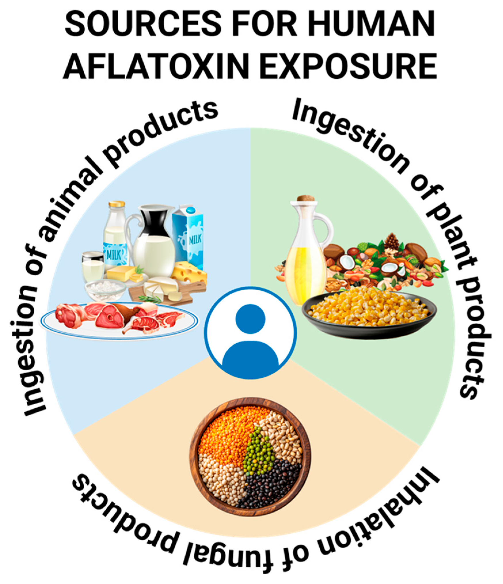
2. Sources of Exposure
2.1. Environmental Reservoirs
2.2. Plant-Based Food Sources
2.3. Animal Sources
2.4. Inhalational Exposure
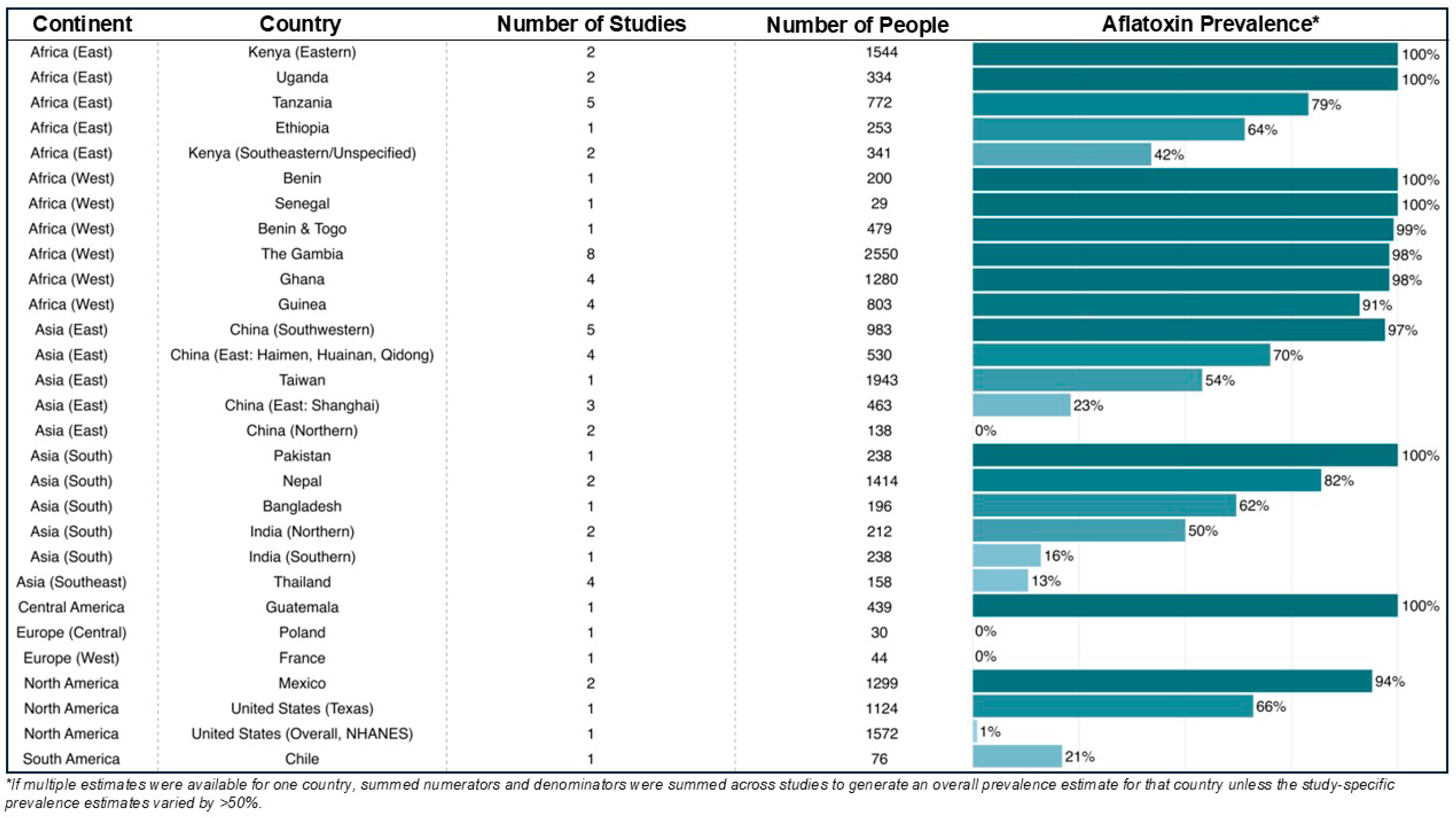
3. Geographic Distribution of Aflatoxins
4. Aflatoxins and Human Disease
4.1. Non-Cancer Outcomes
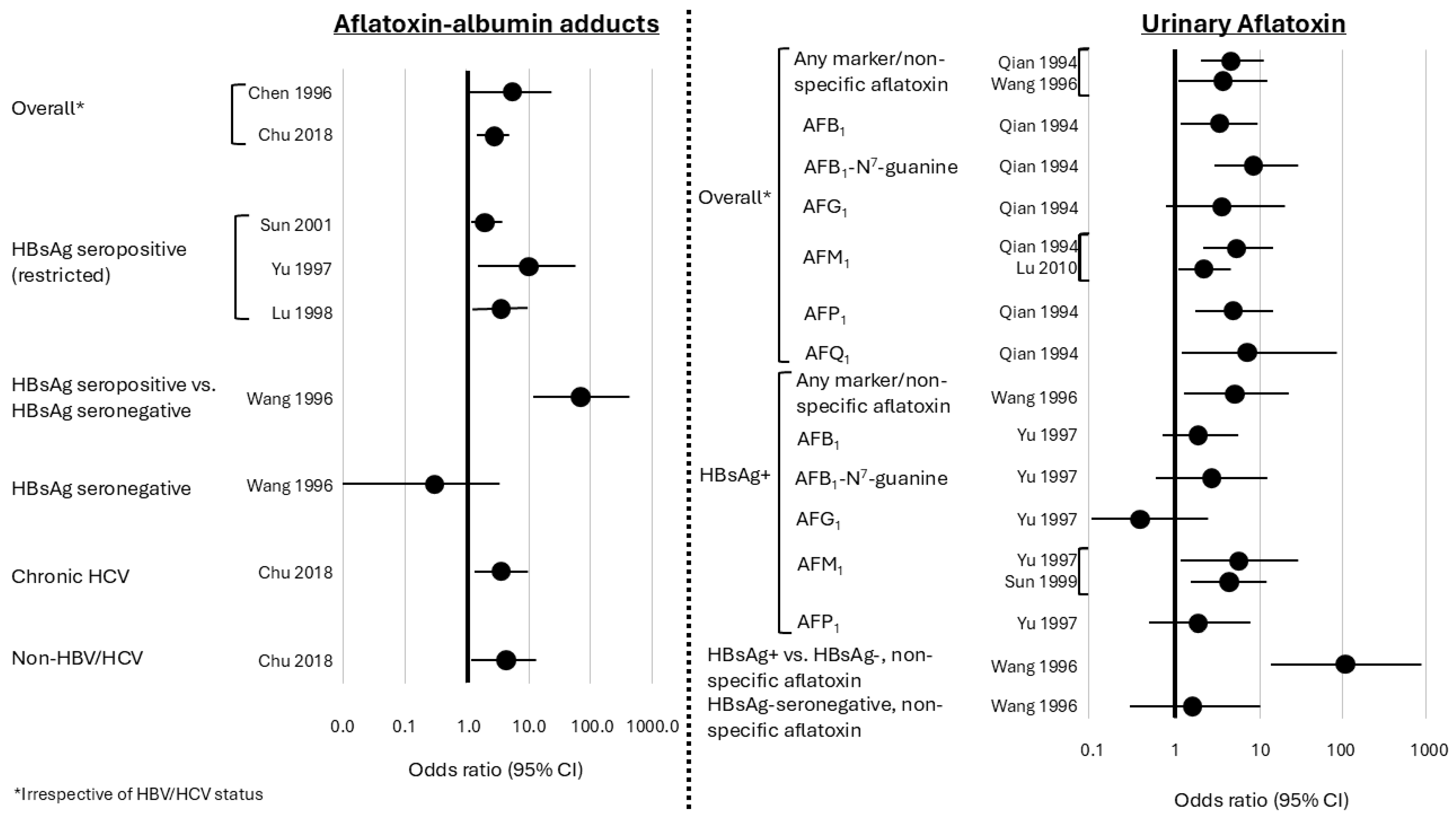
4.2. Liver Cancer
4.3. Gallbladder Cancer
4.4. Other Cancers
5. Biologic Mechanisms for Carcinogenesis
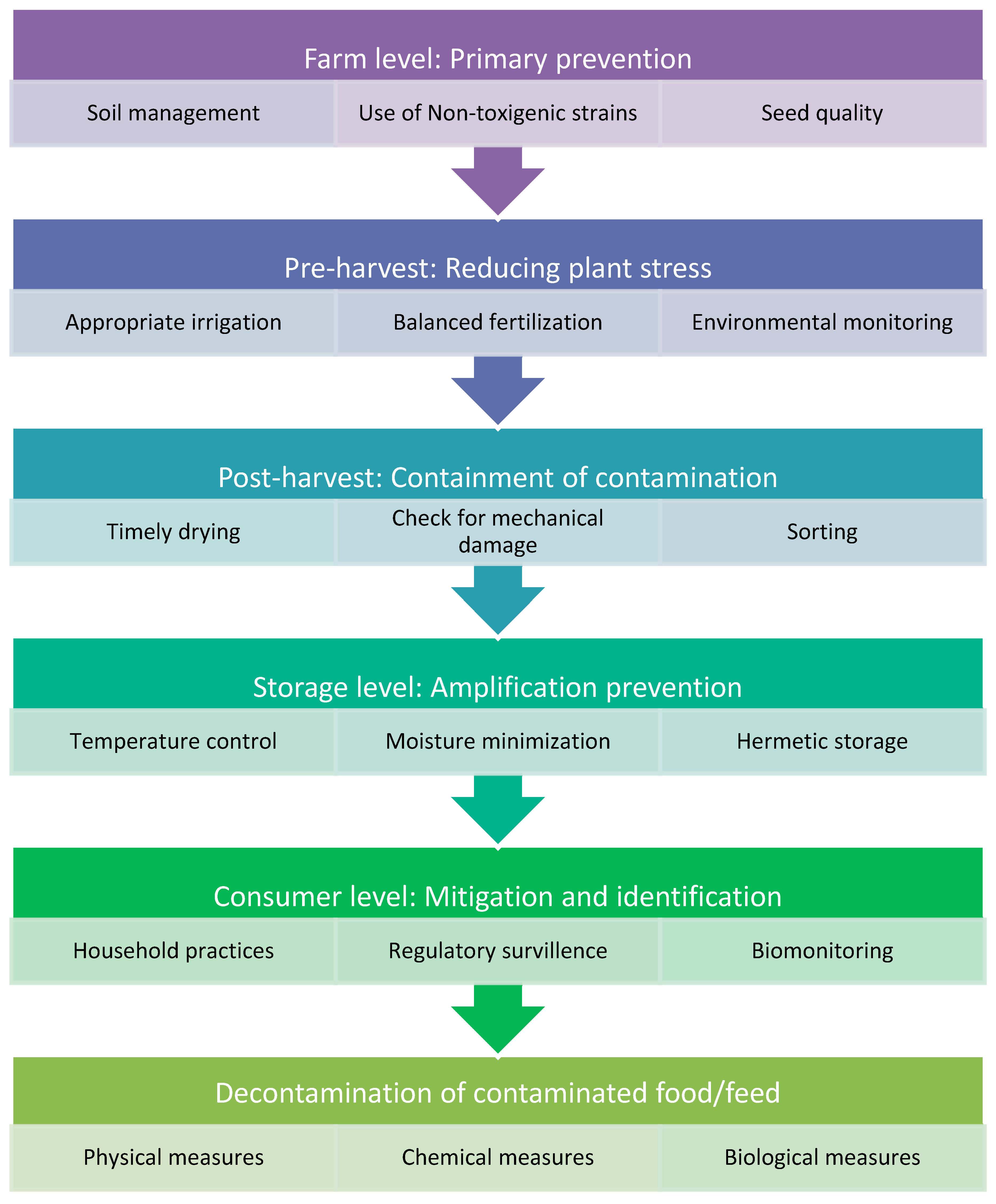
6. Measures to Minimize Aflatoxin Exposure
6.1. Farm-Level Interventions: Primary Prevention at the Source
6.2. Pre-Harvest Level: Reducing Plant Stress and Fungal Entry
6.3. Post-Harvest Level: Containment of Contamination
6.4. Storage Level: Preventing Exponential Amplification
6.5. Consumer-Level Mitigation and Identification of Contamination
6.6. Aflatoxin Decontamination Strategies
6.6.1. Physical Methods
- (a)
- Sorting: Most mycotoxin contamination is concentrated in a small fraction of seeds or kernels. Sorting damaged, discolored, or visibly moldy crops can remove a significant portion of mycotoxins. This low-cost intervention can achieve substantial toxin reduction, particularly in small-scale settings [207]. Methods include manual, mechanical, and electronic sorting:
- Manual selection: Damaged kernels are identified based on size, shape, color, and visible mold.
- Fluorescence sorting: Contaminated maize, cottonseed, and dried figs can be detected under UV light (365 nm) due to bright greenish-yellow fluorescence correlated with aflatoxin presence [208].
- Electronic sorting: Though effective, large-scale industrial application is limited due to cost. Combined electronic and manual sorting is used in the peanut industry to reduce aflatoxin levels [209].
- (b)
- (c)
- Washing: Washing grains can significantly reduce mycotoxin levels. Repeated washing of barley or maize with water reduces mycotoxins by 65–69% [210].
- (d)
- Milling: Removal of grain components during milling can lower mycotoxin content, Fumonisins are distributed unevenly in maize milling fractions, being lower in flaking grits and higher in germ and bran.
- (e)
- Dehulling and polishing: Aflatoxins are predominantly localized in the outer layers of grains. Removal of the hull or bran through dehulling or polishing significantly lowers toxin content, albeit at the expense of some nutritional components.
- (f)
- Heat Treatment: Most mycotoxins are relatively heat-stable under conventional food processing (80–121 °C). Thermal degradation depends on toxin type, moisture, pH, heating time, and temperature. Aflatoxins decompose at 237–306 °C; increased moisture enhances degradation. Roasting peanuts at 150 °C for 30–90 min reduces aflatoxins by 30–60%. Microwave and extrusion cooking have also been effective in degrading mycotoxins while maintaining food quality [212].
- (g)
- Ionizing Radiation: Gamma and X-rays can degrade mycotoxins, a process termed “cold pasteurization” [213]. γ-radiation reduces aflatoxin B1 in peanuts by 75–100% at 1–10 kGy. UV radiation (222–362 nm) can degrade aflatoxin M1 in milk, aflatoxin B1 in dried figs, and peanut oil. Solar energy also contributes to mycotoxin degradation, with up to 70% of aflatoxin B1 in coconut oil destroyed under sunlight [214].
- (h)
- Cold atmospheric plasma: Cold atmospheric plasma (CAP) is an emerging non-thermal technology that degrades aflatoxin through reactive oxygen and nitrogen species generated at atmospheric pressure. CAP disrupts fungal cell walls and breaks key chemical bonds within the aflatoxin molecule, leading to detoxification without significant nutrient loss. Although highly effective and residue free, CAP currently remains limited to industrial scale applications due to cost and infrastructure requirements. Nevertheless, it represents one of the most promising future technologies for large-scale aflatoxin control [215].
- (i)
- Nixtamalization: Nixtamalization is an alkaline cooking process used traditionally in maize tortilla preparation. Nixtamalization is one of the few household cooking methods proven to significantly reduce aflatoxin levels. The alkaline conditions hydrolyze the lactone ring of aflatoxin B1, reducing bioavailability and genotoxicity [216].
- (j)
- Extraction with Solvents: Several solvents can extract mycotoxins from contaminated food and oilseeds, including ethanol, acetone, isopropanol, hexane, and methanol–water mixtures. While effective in removing aflatoxins without toxic byproducts, large-scale applications are limited by cost and disposal issues. For example, 80% isopropanol can remove aflatoxins from cottonseed and peanut meal but also extracts 8–9% of solids [217].
6.6.2. Chemical Methods
- (a)
- Acids: Strong acids such as hydrochloric acid (HCl) and sulfuric acid (H2SO4) can convert aflatoxins aflatoxin B1 and aflatoxin G1 into less toxic hemiacetal forms aflatoxin B2a and aflatoxin G2a through hydration reactions. These treatments can significantly reduce the toxicity of aflatoxins when applied under controlled conditions.
- (b)
- Bases (Ammoniation and Alkalis): Ammoniation is widely employed for the detoxification of aflatoxin-contaminated peanuts, cottonseed, and maize. High-pressure and high-temperature ammoniation treatments can destroy more than 99% of aflatoxin B1. Other alkaline treatments, such as sodium hydroxide, have also been shown to reduce aflatoxin levels effectively, though their efficacy is slightly lower than that of ammoniation [205].
- (c)
- Oxidizing Agents: Oxidizing agents, including ozone (O3) and hydrogen peroxide (H2O2), are used to degrade mycotoxins. Ozone reacts with the C=C double bonds in aflatoxins such as aflatoxin B1, aflatoxin G1, and aflatoxin M1, leading to rapid degradation. Hydrogen peroxide has also been shown to be effective against aflatoxins, zearalenone (ZEN), and deoxynivalenol [220].
- (d)
- Reducing Agents: Reducing agents, such as sodium bisulfite, detoxify aflatoxins by forming sulfonate derivatives, thereby reducing their mutagenic potential. The use of sorbic acid in combination with heat or UV treatments has been shown to further enhance the degradation of aflatoxins [221].
- (e)
- Chlorinating Agents: Chlorine and sodium hypochlorite are effective in degrading aflatoxins and patulin in food products. They are commonly used in the food industry as sanitizing agents and for decontamination of raw materials [222].
- (f)
- Miscellaneous Reagents: Other chemicals, including formaldehyde, potassium permanganate, and sodium borate, have demonstrated detoxification potential. However, their use is limited due to safety concerns and the possibility of producing toxic residues, restricting their application in food and feed [7].
6.6.3. Biological Methods
- (a)
- Microbial degradation: Certain fungi and bacteria can directly degrade mycotoxins. Fungi such as Trichoderma, Phoma, Rhizopus, and Alternaria can degrade aflatoxin B1 by 65–99% within five days. Bacteria such as Acinetobacter calcoaceticus are capable of completely degrading mycotoxins in liquid media, highlighting the potential of microbial systems for targeted detoxification [223].
- (b)
- Fermentation: Fermentation processes with yeasts and bacteria can reduce mycotoxin levels in foods and feeds. Fermentation with Candida intermedia decreases F2 toxin (zearalenone) activity by tenfold [224]. Ethanol fermentation of maize, beer, apple juice, and barley effectively reduces mycotoxins. Lactic acid bacteria and bifidobacteria can degrade aflatoxin B1 and aflatoxin M1 during milk fermentation, thereby improving food safety while maintaining nutritional quality.
- (c)
- Inhibition of absorption in the gastrointestinal tract: Biological binders and probiotics can reduce mycotoxin bioavailability in livestock. Non-nutritive adsorbents such as activated carbon, hydrated sodium calcium aluminosilicate (HSCAS), zeolites, bentonites, and esterified glucomannan prevent systemic absorption of mycotoxins [225]. Lactic acid bacteria and bifidobacteria bind aflatoxins through cell wall interactions, hydrophobic binding, and polysaccharide–peptidoglycan complexes. Binding efficiency varies among bacterial strains; for example, Lacticaseibacillus rhamnosus GG can reduce aflatoxin B1 bioavailability by up to 80%, ultimately lowering aflatoxin M1 excretion in milk [226].
- (d)
- Competitive exclusion: As described above, application of non-aflatoxigenic Aspergillus strains in agricultural fields can competitively suppress toxigenic strains, reducing aflatoxin production at the source. This approach has been successfully implemented in large-scale crop interventions to prevent contamination before harvest.
- (e)
- Preventing adsorption in livestock: In animal production, feed additives such as clay-based binders, aluminosilicates, activated charcoal, Saccharomyces cerevisiae, and lactic acid bacteria are incorporated to reduce intestinal absorption of aflatoxin B1. These biological binders limit systemic absorption and aflatoxin M1 excretion in milk, achieving reductions of 40–80% in dairy systems. The primary mechanism involves binding via cell wall components such as β-glucans and polysaccharides, which sequester toxins in the gastrointestinal tract [218].
- (f)
- Enzymatic degradation: Microbial enzymes such as laccases and peroxidases can structurally modify aflatoxin B1, reducing its ability to form DNA adducts and decreasing carcinogenic potential. Recent advances in computational biology and artificial intelligence have enabled the design of optimized enzymes and predictive modeling of enzyme–toxin interactions, representing a move toward precision detoxification strategies.
6.7. Developments in Biomonitoring
7. International Regulations and Standards for Aflatoxin Control
8. Conclusions
Supplementary Materials
Author Contributions
Funding
Institutional Review Board Statement
Informed Consent Statement
Data Availability Statement
Acknowledgments
Conflicts of Interest
Abbreviations
| NHANES | National Health and Nutrition Examination Survey |
| HPLC | High-performance liquid chromatography |
| LC-MS/MS | Liquid chromatograph–tandem mass spectrometry |
| ELISA | Enzyme-linked immunosorbent assay |
| HPLC-MS/MS | High-performance liquid chromatography–tandem mass spectrometry |
| IDMS | Isotope dilution mass spectrometry |
| HPLC-FLD | High-performance liquid chromatography–fluorescence detector |
| RR | Relative risk |
| CI | Confidence interval |
| HBV | Hepatitis B virus |
| OR | Odds ratio |
| HCC | Hepatocellular carcinoma |
| GBD | Gallbladder cancer |
| ABC | Adenosine triphosphate-binding cassette |
| EHBDC | Extrahepatic bile duct cancer |
| SPIR | Standardized proportional incidence ratio |
| IHBDC | Intrahepatic bile duct cancer |
| SMR | Standardized mortality ratio |
| CYP450 | Cytochrome 450 |
| AFBO | Aflatoxin B1-exo 8,9-epoxide |
| HCV | Hepatitis C virus |
| CAP | Cold atmospheric plasma |
| AI | Artificial intelligence |
| NIH | National Institutes of Health |
References
- Niaz, W.; Iqbal, S.Z.; Ahmad, K.; Majid, A.; Haider, W.; Li, X. Mycotoxins: A comprehensive review of its global trends in major cereals, advancements in chromatographic detections and future prospectives. Food Chem. X 2025, 27, 102350. [Google Scholar] [CrossRef]
- Wild, C.P.; Gong, Y.Y. Mycotoxins and human disease: A largely ignored global health issue. Carcinogenesis 2010, 31, 71–82. [Google Scholar] [CrossRef]
- Yin, S.; Niu, L.; Liu, Y. Recent progress on techniques in the detection of aflatoxin B1 in edible oil: A mini review. Molecules 2022, 27, 6141. [Google Scholar] [CrossRef] [PubMed]
- Winter, G.; Pereg, L. A review on the relation between soil and mycotoxins: Effect of aflatoxin on field, food and finance. Eur. J. Soil. Sci. 2019, 70, 882–897. [Google Scholar] [CrossRef]
- Council for Agricultural Science and Technology (CAST). Mycotoxins: Risks in Plant, Animal, and Human Systems, Task Force Report; Council for Agricultural Science and Technology: Ames, IA, USA, 2003. [Google Scholar]
- Eskola, M.; Kos, G.; Elliott, C.T.; Hajslova, J.; Mayar, S.; Krska, R. Worldwide contamination of food-crops with mycotoxins: Validity of the widely cited ‘FAO estimate’ of 25. Crit. Rev. Food Sci. Nutr. 2020, 60, 2773–2789. [Google Scholar] [CrossRef] [PubMed]
- Kabak, B.; Dobson, A.D.; Var, I. Strategies to prevent mycotoxin contamination of food and animal feed: A review. Crit. Rev. Food Sci. Nutr. 2006, 46, 593–619. [Google Scholar] [CrossRef]
- Kovalsky, P.; Kos, G.; Nahrer, K.; Schwab, C.; Jenkins, T.; Schatzmayr, G.; Sulyok, M.; Krska, R. Co-Occurrence of Regulated, Masked and Emerging Mycotoxins and Secondary Metabolites in Finished Feed and Maize-An Extensive Survey. Toxins 2016, 8, 363. [Google Scholar] [CrossRef]
- Streit, E.; Naehrer, K.; Rodrigues, I.; Schatzmayr, G. Mycotoxin occurrence in feed and feed raw materials worldwide: Long-term analysis with special focus on Europe and Asia. J. Sci. Food Agric. 2013, 93, 2892–2899. [Google Scholar] [CrossRef]
- Mafe, A.N.; Busselberg, D. Mycotoxins in Food: Cancer Risks and Strategies for Control. Foods 2024, 13, 3502. [Google Scholar] [CrossRef]
- Benkerroum, N. Chronic and Acute Toxicities of Aflatoxins: Mechanisms of Action. Int. J. Environ. Res. Public Health 2020, 17, 423. [Google Scholar] [CrossRef]
- IARC Working Group on the Evaluation of Carcinogenic Risks to Humans. Aflatoxins. In Chemical Agents and Related Occupations: A Review of Human Carcinogens; IARC: Lyon, France, 2012; Volume 100F, pp. 225–244.
- Liu, Y.; Chang, C.C.; Marsh, G.M.; Wu, F. Population attributable risk of aflatoxin-related liver cancer: Systematic review and meta-analysis. Eur. J. Cancer 2012, 48, 2125–2136. [Google Scholar] [CrossRef]
- Williams, J.H.; Phillips, T.D.; Jolly, P.E.; Stiles, J.K.; Jolly, C.M.; Aggarwal, D. Human aflatoxicosis in developing countries: A review of toxicology, exposure, potential health consequences, and interventions. Am. J. Clin. Nutr. 2004, 80, 1106–1122. [Google Scholar] [CrossRef] [PubMed]
- Blount, W.P. Turkey “X” Disease. J. Br. Turk. Fed. 1961, 9, 52–61. [Google Scholar]
- Kensler, T.W.; Eaton, D.L. 65 Years on-Aflatoxin Biomarkers Blossoming: Whither Next? Toxins 2024, 16, 496. [Google Scholar] [CrossRef]
- Wannop, C.C. Groundnut Toxicity in Poultry: Turkey X Disease. Br. Vet. J. 1963, 119, 174–177. [Google Scholar] [CrossRef]
- Halver, J.E. Aflatoxicosis and rainbow trout hepatoma. In Mycotoxins in Foodstuffs; Wogan, G.N., Ed.; MIT Press: Cambridge, MA, USA, 1965; pp. 209–234. [Google Scholar]
- Hendrickse, R.G. Of sick turkeys, kwashiorkor, malaria, perinatal mortality, heroin addicts and food poisoning: Research on the influence of aflatoxins on child health in the tropics. Ann. Trop. Med. Parasitol. 1997, 91, 787–793. [Google Scholar] [CrossRef]
- Klich, M.A. Aspergillus flavus: The major producer of aflatoxin. Mol. Plant Pathol. 2007, 8, 713–722. [Google Scholar] [CrossRef]
- Fouché, T.; Claassens, S.; Maboeta, M. Aflatoxins in the soil ecosystem: An overview of its occurrence, fate, effects and future perspectives. Mycotoxin Res. 2020, 36, 303–309. [Google Scholar] [CrossRef]
- Sweeney, M.J.; Dobson, A.D. Mycotoxin production by Aspergillus, Fusarium and Penicillium species. Int. J. Food Microbiol. 1998, 43, 141–158. [Google Scholar] [CrossRef] [PubMed]
- Pickova, D.; Ostry, V.; Malir, F. A recent overview of producers and important dietary sources of aflatoxins. Toxins 2021, 13, 186. [Google Scholar] [CrossRef]
- Lewis, L.; Onsongo, M.; Njapau, H.; Schurz-Rogers, H.; Luber, G.; Kieszak, S.; Nyamongo, J.; Backer, L.; Dahiye, A.M.; Misore, A. Aflatoxin contamination of commercial maize products during an outbreak of acute aflatoxicosis in eastern and central Kenya. Environ. Health Perspect. 2005, 113, 1763–1767. [Google Scholar] [CrossRef] [PubMed]
- Molyneux, R.J.; Mahoney, N.; Kim, J.H.; Campbell, B.C. Mycotoxins in edible tree nuts. Int. J. Food Microbiol. 2007, 119, 72–78. [Google Scholar] [CrossRef] [PubMed]
- Kamika, I.; Mngqawa, P.; Rheeder, J.P.; Teffo, S.L.; Katerere, D.R. Mycological and aflatoxin contamination of peanuts sold at markets in Kinshasa, Democratic Republic of Congo, and Pretoria, South Africa. Food Addit. Contam. Part B 2014, 7, 120–126. [Google Scholar] [CrossRef]
- Bandara, J.; Vithanege, A.; Bean, G. Occurrence of aflatoxins in parboiled rice in Sri Lanka. Mycopathologia 1991, 116, 65–70. [Google Scholar] [CrossRef]
- Toteja, G.S.; Mukherjee, A.; Diwakar, S.; Singh, P.; Saxena, B.N.; Sinha, K.K.; Sinha, A.K.; Kumar, N.; Nagaraja, K.V.; Bai, G.; et al. Aflatoxin B1 contamination in wheat grain samples collected from different geographical regions of India: A multicenter study. J. Food Prot. 2006, 69, 1463–1467. [Google Scholar] [CrossRef]
- Giray, B.; Girgin, G.; Engin, A.B.; Aydin, S.; Sahin, G. Aflatoxin levels in wheat samples consumed in some regions of Turkey. Food Control 2007, 18, 23–29. [Google Scholar] [CrossRef]
- Hathout, A.S.; Abel-Fattah, S.M.; Abou-Sree, Y.H.; Fouzy, A.S.M. Incidence and exposure assessment of aflatoxins and ochratoxin A in Egyptian wheat. Toxicol. Rep. 2020, 7, 867–873. [Google Scholar] [CrossRef] [PubMed]
- Xue, M.; Qu, Z.; Moretti, A.; Logrieco, A.F.; Chu, H.; Zhang, Q.; Sun, C.; Ren, X.; Cui, L.; Chen, Q. Aspergillus mycotoxins: The major food contaminants. Adv. Sci. 2025, 12, 2412757. [Google Scholar] [CrossRef]
- Barbosa-Cánovas, G.V.; Aguilera, J.M.; Chen, X.D.; Hartel, R.W.; Ibarz, A.; Kokini, J.; Marcotte, M.; McCarthy, M.; Niranjan, K.; Peleg, M. Food Engineering Series; Springer: Berlin/Heidelberg, Germany, 2020. [Google Scholar]
- Milićević, D.R.; Milešević, J.; Gurinović, M.; Janković, S.; Đinović-Stojanović, J.; Zeković, M.; Glibetić, M. Dietary exposure and risk assessment of aflatoxin M1 for children aged 1 to 9 years old in Serbia. Nutrients 2021, 13, 4450. [Google Scholar] [CrossRef]
- Chelenga, M.; Matumba, L.; Sitali, M.C.; Kachala, B.; Nambuzi, V.; Mwenifumbo, M.; Gama, A.P.; Mwanza, M.; Monjerezi, M.; Leslie, J.F. Are Aflatoxin Residues in Chicken Products a Real or Perceived Human Dietary Risk? Toxins 2025, 17, 179. [Google Scholar] [CrossRef]
- Zentai, A.; Jozwiak, A.; Suth, M.; Farkas, Z. Carry-Over of Aflatoxin B1 from Feed to Cow Milk-A Review. Toxins 2023, 15, 195. [Google Scholar] [CrossRef] [PubMed]
- Kang’ethe, E.K.; Lang’a, K. Aflatoxin B1 and M1 contamination of animal feeds and milk from urban centers in Kenya. Afr. Health Sci. 2009, 9, 218–226. [Google Scholar] [PubMed]
- Wangia, R.N.; Tang, L.; Wang, J.S. Occupational exposure to aflatoxins and health outcomes: A review. J. Environ. Sci. Health C Environ. Carcinog. Ecotoxicol. Rev. 2019, 37, 215–234. [Google Scholar] [CrossRef]
- Zarba, A.; Hmieleski, R.; Hemenway, D.R.; Jakab, G.J.; Groopman, J.D. Aflatoxin B1—DNA adduct formation in rat liver following exposure by aerosol inhalation. Carcinogenesis 1992, 13, 1031–1033. [Google Scholar] [CrossRef]
- Autrup, J.L.; Schmidt, J.; Autrup, H. Exposure to aflatoxin B1 in animal-feed production plant workers. Environ. Health Perspect. 1993, 99, 195–197. [Google Scholar] [CrossRef]
- Wild, C.; Miller, J.; Groopman, J. Effects of aflatoxins on aflatoxicosis and liver cancer. In Mycotoxin Control in Low-and Middle-Income Countries; International Agency for Research on Cancer: Lyon, France, 2015. [Google Scholar]
- Cardona, T.; LLangantileke, S.; Noomhorm, A. Aflatoxin research on grain in Asia: Its problems and possible solutions. In Mycotoxin Prevention and Control in Foodgrains; FAO: Rome, Italy, 1991. [Google Scholar]
- Kos, J.; Anic, M.; Radic, B.; Zadravec, M.; Janic Hajnal, E.; Pleadin, J. Climate Change-A Global Threat Resulting in Increasing Mycotoxin Occurrence. Foods 2023, 12, 2704. [Google Scholar] [CrossRef]
- Chen, J.G.; Zhu, Y.R.; Qian, G.S.; Wang, J.B.; Lu, J.H.; Kensler, T.W.; Jacobson, L.P.; Munoz, A.; Groopman, J.D. Fifty Years of Aflatoxin Research in Qidong, China: A Celebration of Team Science to Improve Public Health. Toxins 2025, 17, 79. [Google Scholar] [CrossRef]
- Chen, J.G.; Egner, P.A.; Ng, D.; Jacobson, L.P.; Munoz, A.; Zhu, Y.R.; Qian, G.S.; Wu, F.; Yuan, J.M.; Groopman, J.D.; et al. Reduced aflatoxin exposure presages decline in liver cancer mortality in an endemic region of China. Cancer Prev. Res. 2013, 6, 1038–1045. [Google Scholar] [CrossRef] [PubMed]
- Wild, C.P.; Jiang, Y.Z.; Allen, S.J.; Jansen, L.A.; Hall, A.J.; Montesano, R. Aflatoxin-albumin adducts in human sera from different regions of the world. Carcinogenesis 1990, 11, 2271–2274. [Google Scholar] [CrossRef]
- McCoy, L.F.; Scholl, P.F.; Sutcliffe, A.E.; Kieszak, S.M.; Powers, C.D.; Rogers, H.S.; Gong, Y.Y.; Groopman, J.D.; Wild, C.P.; Schleicher, R.L. Human aflatoxin albumin adducts quantitatively compared by ELISA, HPLC with fluorescence detection, and HPLC with isotope dilution mass spectrometry. Cancer Epidemiol. Biomark. Prev. 2008, 17, 1653–1657. [Google Scholar] [CrossRef]
- Scholl, P.F.; Turner, P.C.; Sutcliffe, A.E.; Sylla, A.; Diallo, M.S.; Friesen, M.D.; Groopman, J.D.; Wild, C.P. Quantitative comparison of aflatoxin B1 serum albumin adducts in humans by isotope dilution mass spectrometry and ELISA. Cancer Epidemiol. Biomark. Prev. 2006, 15, 823–826. [Google Scholar] [CrossRef]
- Xia, L.; Wu, H.; Saleem, A.F.; Mupere, E.; Lancioni, C.; Diallo, H.; Potani, I.; Ali, S.A.; Voskuijl, W.; Chisti, M.J.; et al. Aflatoxin exposure and mortality in acutely ill children: Results from the CHAIN network cohort. Bmj Glob. Health 2025, 10, e017375s. [Google Scholar] [CrossRef]
- Gong, Y.; Hounsa, A.; Egal, S.; Turner, P.C.; Sutcliffe, A.E.; Hall, A.J.; Cardwell, K.; Wild, C.P. Postweaning exposure to aflatoxin results in impaired child growth: A longitudinal study in Benin, West Africa. Environ. Health Perspect. 2004, 112, 1334–1338. [Google Scholar] [CrossRef] [PubMed]
- Gong, Y.Y.; Egal, S.; Hounsa, A.; Turner, P.C.; Hall, A.J.; Cardwell, K.F.; Wild, C.P. Determinants of aflatoxin exposure in young children from Benin and Togo, West Africa: The critical role of weaning. Int. J. Epidemiol. 2003, 32, 556–562. [Google Scholar] [CrossRef] [PubMed]
- Wild, C.P.; Jansen, L.A.; Cova, L.; Montesano, R. Molecular dosimetry of aflatoxin exposure: Contribution to understanding the multifactorial etiopathogenesis of primary hepatocellular carcinoma with particular reference to hepatitis B virus. Environ. Health Perspect. 1993, 99, 115–122. [Google Scholar] [CrossRef] [PubMed]
- Turner, P.C.; Moore, S.E.; Hall, A.J.; Prentice, A.M.; Wild, C.P. Modification of immune function through exposure to dietary aflatoxin in Gambian children. Environ. Health Perspect. 2003, 111, 217–220. [Google Scholar] [CrossRef]
- Turner, P.C.; Mendy, M.; Whittle, H.; Fortuin, M.; Hall, A.J.; Wild, C.P. Hepatitis B infection and aflatoxin biomarker levels in Gambian children. Trop. Med. Int. Health 2000, 5, 837–841. [Google Scholar] [CrossRef]
- Wild, C.P.; Yin, F.; Turner, P.C.; Chemin, I.; Chapot, B.; Mendy, M.; Whittle, H.; Kirk, G.D.; Hall, A.J. Environmental and genetic determinants of aflatoxin-albumin adducts in the Gambia. Int. J. Cancer 2000, 86, 1–7. [Google Scholar] [CrossRef]
- Turner, P.C.; Collinson, A.C.; Cheung, Y.B.; Gong, Y.; Hall, A.J.; Prentice, A.M.; Wild, C.P. Aflatoxin exposure in utero causes growth faltering in Gambian infants. Int. J. Epidemiol. 2007, 36, 1119–1125. [Google Scholar] [CrossRef]
- Watson, S.; Moore, S.E.; Darboe, M.K.; Chen, G.; Tu, Y.K.; Huang, Y.T.; Eriksen, K.G.; Bernstein, R.M.; Prentice, A.M.; Wild, C.P.; et al. Impaired growth in rural Gambian infants exposed to aflatoxin: A prospective cohort study. BMC Public Health 2018, 18, 1247. [Google Scholar] [CrossRef]
- Xu, Y.; Moore, S.; Chen, G.; Nshe, P.; Faye-Joof, T.; Prentice, A.; Gong, Y.Y.; Routledge, M. Impact of dietary aflatoxin on immune development in Gambian infants: A cohort study. BMJ Open 2021, 11, e048688. [Google Scholar] [CrossRef]
- Castelino, J.M.; Dominguez-Salas, P.; Routledge, M.N.; Prentice, A.M.; Moore, S.E.; Hennig, B.J.; Wild, C.P.; Gong, Y.Y. Seasonal and gestation stage associated differences in aflatoxin exposure in pregnant Gambian women. Trop. Med. Int. Health 2014, 19, 348–354. [Google Scholar] [CrossRef]
- Diallo, M.S.; Sylla, A.; Sidibe, K.; Sylla, B.S.; Trepo, C.R.; Wild, C.P. Prevalence of exposure to aflatoxin and hepatitis B and C viruses in Guinea, West Africa. Nat. Toxins 1995, 3, 6–9. [Google Scholar] [CrossRef]
- Turner, P.C.; Sylla, A.; Diallo, M.S.; Castegnaro, J.J.; Hall, A.J.; Wild, C.P. The role of aflatoxins and hepatitis viruses in the etiopathogenesis of hepatocellular carcinoma: A basis for primary prevention in Guinea-Conakry, West Africa. J. Gastroenterol. Hepatol. 2002, 17, S441–S448. [Google Scholar] [CrossRef] [PubMed]
- Turner, P.C.; Sylla, A.; Gong, Y.Y.; Diallo, M.S.; Sutcliffe, A.E.; Hall, A.J.; Wild, C.P. Reduction in exposure to carcinogenic aflatoxins by postharvest intervention measures in west Africa: A community-based intervention study. Lancet 2005, 365, 1950–1956. [Google Scholar] [CrossRef]
- Turner, P.C.; Sylla, A.; Kuang, S.Y.; Marchant, C.L.; Diallo, M.S.; Hall, A.J.; Groopman, J.D.; Wild, C.P. Absence of TP53 codon 249 mutations in young Guinean children with high aflatoxin exposure. Cancer Epidemiol. Biomark. Prev. 2005, 14, 2053–2055. [Google Scholar] [CrossRef]
- Jolly, P.; Jiang, Y.; Ellis, W.; Awuah, R.; Nnedu, O.; Phillips, T.; Wang, J.S.; Afriyie-Gyawu, E.; Tang, L.; Person, S.; et al. Determinants of aflatoxin levels in Ghanaians: Sociodemographic factors, knowledge of aflatoxin and food handling and consumption practices. Int. J. Hyg. Environ. Health 2006, 209, 345–358. [Google Scholar] [CrossRef]
- Shuaib, F.M.; Jolly, P.E.; Ehiri, J.E.; Ellis, W.O.; Yatich, N.J.; Funkhouser, E.; Person, S.D.; Williams, J.H.; Qian, G.; Wang, J.S. Socio-demographic determinants of aflatoxin B1-lysine adduct levels among pregnant women in Kumasi, Ghana. Ghana Med. J. 2012, 46, 179–188. [Google Scholar]
- Jolly, P.E.; Shuaib, F.M.; Jiang, Y.; Preko, P.; Baidoo, J.; Stiles, J.K.; Wang, J.S.; Phillips, T.D.; Williams, J.H. Association of high viral load and abnormal liver function with high aflatoxin B1-albumin adduct levels in HIV-positive Ghanaians: Preliminary observations. Food Addit. Contam. Part A Chem. Anal. Control Expo. Risk Assess. 2011, 28, 1224–1234. [Google Scholar] [CrossRef] [PubMed]
- McGlynn, K.A.; Rosvold, E.A.; Lustbader, E.D.; Hu, Y.; Clapper, M.L.; Zhou, T.; Wild, C.P.; Xia, X.L.; Baffoe-Bonnie, A.; Ofori-Adjei, D.; et al. Susceptibility to hepatocellular carcinoma is associated with genetic variation in the enzymatic detoxification of aflatoxin B1. Proc. Natl. Acad. Sci. USA 1995, 92, 2384–2387. [Google Scholar] [CrossRef] [PubMed]
- Mekuria, A.; Xia, L.; Ahmed, T.A.; Bishaw, S.; Teklemariam, Z.; Nedi, T.; Abula, T.; Engidawork, E.; Gong, Y.Y. Contribution of Aflatoxin B(1) Exposure to Liver Cirrhosis in Eastern Ethiopia: A Case-Control Study. Int. J. Gen. Med. 2023, 16, 3543–3553. [Google Scholar] [CrossRef]
- Chen, C.; Mitchell, N.J.; Gratz, J.; Houpt, E.R.; Gong, Y.; Egner, P.A.; Groopman, J.D.; Riley, R.T.; Showker, J.L.; Svensen, E.; et al. Exposure to aflatoxin and fumonisin in children at risk for growth impairment in rural Tanzania. Environ. Int. 2018, 115, 29–37. [Google Scholar] [CrossRef] [PubMed]
- Hoffmann, V.; Jones, K.; Leroy, J.L. The impact of reducing dietary aflatoxin exposure on child linear growth: A cluster randomised controlled trial in Kenya. BMJ Glob. Health 2018, 3, e000983. [Google Scholar] [CrossRef]
- Osoro, E.; Awuor, A.O.; Inwani, I.; Mugo, C.; Hunsperger, E.; Verani, J.R.; Nduati, R.; Kinuthia, J.; Okutoyi, L.; Mwaengo, D.; et al. Association between low maternal serum aflatoxin B1 exposure and adverse pregnancy outcomes in Mombasa, Kenya, 2017–2019: A nested matched case-control study. Matern. Child Nutr. 2024, 20, e13688. [Google Scholar] [CrossRef] [PubMed]
- Nabwire Wangia-Dixon, R.; Xue, K.S.; Alcala, J.; Quach, T.H.T.; Song, X.; Tang, L.; Ombaka, J.; Githanga, D.P.; Anzala, O.A.; Wang, J.S. Nutrition and growth outcomes are affected by aflatoxin exposures in Kenyan children. Food Addit. Contam. Part A Chem. Anal. Control Expo. Risk Assess. 2020, 37, 2123–2134. [Google Scholar] [CrossRef]
- Mshanga, N.; Kassim, N.; Sonto, S.; Martin, H.D.; Pirani, M.; Moore, S.; Auma, C.I.; Kimanya, M.; Gong, Y.Y. A Cross-Sectional Association Between Serum Aflatoxin and Micronutrient Status Among Children Aged 6-24 Months in Rural Tanzania. Matern. Child Nutr. 2025, 21, e70068. [Google Scholar] [CrossRef] [PubMed]
- Kinyenje, E.; Kishimba, R.; Mohamed, M.; Mwafulango, A.; Eliakimu, E.; Kwesigabo, G. Aflatoxicosis outbreak and its associated factors in Kiteto, Chemba and Kondoa Districts, Tanzania. PLoS Glob. Public Health 2023, 3, e0002191. [Google Scholar] [CrossRef]
- Shirima, C.P.; Kimanya, M.E.; Routledge, M.N.; Srey, C.; Kinabo, J.L.; Humpf, H.U.; Wild, C.P.; Tu, Y.K.; Gong, Y.Y. A prospective study of growth and biomarkers of exposure to aflatoxin and fumonisin during early childhood in Tanzania. Environ. Health Perspect. 2015, 123, 173–178. [Google Scholar] [CrossRef]
- Shirima, C.P.; Kimanya, M.E.; Kinabo, J.L.; Routledge, M.N.; Srey, C.; Wild, C.P.; Gong, Y.Y. Dietary exposure to aflatoxin and fumonisin among Tanzanian children as determined using biomarkers of exposure. Mol. Nutr. Food Res. 2013, 57, 1874–1881. [Google Scholar] [CrossRef]
- Lauer, J.M.; Duggan, C.P.; Ausman, L.M.; Griffiths, J.K.; Webb, P.; Wang, J.S.; Xue, K.S.; Agaba, E.; Nshakira, N.; Ghosh, S. Maternal aflatoxin exposure during pregnancy and adverse birth outcomes in Uganda. Matern. Child Nutr. 2019, 15, e12701. [Google Scholar] [CrossRef]
- Tong, Y.; Nakalembe, M.; Mpamani, C.; Nakisige, C.; Namugga, J.; Banturaki, G.; Tonui, P.; Orang’o, O.; Muthoka, K.; Ngeresa, A.; et al. Higher plasma AFB1 concentration is associated with increased risk of HPV 16 and HPV 18 detection and persistence among Ugandan women. Environ. Health 2025, 24, 45. [Google Scholar] [CrossRef]
- Turner, P.C.; Loffredo, C.; Kafrawy, S.E.; Ezzat, S.; Eissa, S.A.; Daly, M.E.; Nada, O.; Abdel-Hamid, M. Pilot survey of aflatoxin-albumin adducts in sera from Egypt. Food Addit. Contam. Part A Chem. Anal. Control Expo. Risk Assess. 2008, 25, 583–587. [Google Scholar] [CrossRef]
- Mahfuz, M.; Hasan, S.M.T.; Alam, M.A.; Das, S.; Fahim, S.M.; Islam, M.M.; Gazi, M.A.; Hossain, M.; Egner, P.A.; Groopman, J.D.; et al. Aflatoxin exposure was not associated with childhood stunting: Results from a birth cohort study in a resource-poor setting of Dhaka, Bangladesh. Public Health Nutr. 2021, 24, 3361–3370. [Google Scholar] [CrossRef] [PubMed]
- Anitha, S.; Raghunadharao, D.; Waliyar, F.; Sudini, H.; Parveen, M.; Rao, R.; Kumar, P.L. The association between exposure to aflatoxin, mutation in TP53, infection with hepatitis B virus, and occurrence of liver disease in a selected population in Hyderabad, India. Mutat. Res. Genet. Toxicol. Environ. Mutagen. 2014, 766, 23–28. [Google Scholar] [CrossRef]
- Yadav, A.; Gupta, P.; Gupta, P.; Patil, A.N.; Das, C.K.; Hooda, H.; Thakur, D.; Sharma, V.; Singh, A.K.; Yadav, T.D.; et al. Aflatoxin exposure is associated with an increased risk of gallbladder cancer. Int. J. Cancer 2025, 156, 322–330. [Google Scholar] [CrossRef] [PubMed]
- Shukla, R.; Ikoma, T.; Tsuchiya, Y.; Asai, T.; Nagar, A.; Kapoor, V.K. Serum Aflatoxin B1-Lysine Adduct Concentration and Gallbladder Cancer: A Case-Control Study. Nutr. Cancer 2025, 77, 619–625. [Google Scholar] [CrossRef]
- Mitchell, N.J.; Riley, R.T.; Egner, P.A.; Groopman, J.D.; Wu, F. Chronic aflatoxin exposure in children living in Bhaktapur, Nepal: Extension of the MAL-ED study. J. Expo. Sci. Environ. Epidemiol. 2017, 27, 106–111. [Google Scholar] [CrossRef] [PubMed]
- Lamichhane, A.; Webb, P.; Andrews-Trevino, J.; Pokharel, A.; Acharya, S.; Shrestha, R.; Davis, D.; Baral, K.; Wang, J.S.; Xue, K.; et al. Dietary determinants of aflatoxin B1-lysine adduct among infants in Nepal. Eur. J. Clin. Nutr. 2022, 76, 1557–1565. [Google Scholar] [CrossRef]
- Ashraf, W.; Rehman, A.; Ahmad, M.U.; Rabbani, M.; Mushtaq, M.H.; Aamir, K.; Xue, K.S.; Wang, J.S. Assessment of aflatoxin B(1)-lysine adduct in children and its effect on child growth in Lahore, Pakistan. Food Addit. Contam. Part A Chem. Anal. Control Expo. Risk Assess. 2022, 39, 1463–1473. [Google Scholar] [CrossRef]
- Gan, L.S.; Skipper, P.L.; Peng, X.C.; Groopman, J.D.; Chen, J.S.; Wogan, G.N.; Tannenbaum, S.R. Serum albumin adducts in the molecular epidemiology of aflatoxin carcinogenesis: Correlation with aflatoxin B1 intake and urinary excretion of aflatoxin M1. Carcinogenesis 1988, 9, 1323–1325. [Google Scholar] [CrossRef]
- Yu, S.Z. Primary prevention of hepatocellular carcinoma. J. Gastroenterol. Hepatol. 1995, 10, 674–682. [Google Scholar] [CrossRef]
- Chen, J.; Ye, Y.; Huang, D.; Pan, D.; Qiu, X.; Lei, L.; Luo, X.; Li, J.; Wu, K.; Xiao, S.; et al. The mediating role of coagulation function on the association of prenatal exposure to aflatoxin B1 and postpartum hemorrhage in Guangxi, China. Environ. Sci. Pollut. Res. Int. 2022, 29, 37543–37555. [Google Scholar] [CrossRef]
- Zhong, Y.; Lu, H.; Lu, X.; He, Z.; Jiang, Y.; Chen, J.; Liabsuetrakul, T. Correlations of Aflatoxin Exposure from Cooking Oil and Dietary Foods During Pregnancy with Birth Weight and Gestational Age at Birth in Guangxi, China. Int. J. Gen. Med. 2024, 17, 1171–1184. [Google Scholar] [CrossRef] [PubMed]
- Tao, P.; Zhi-Ming, L.; Tang-Wei, L.; Le-Qun, L.; Min-Hao, P.; Xue, Q.; Lu-Nam, Y.; Ren-Xiang, L.; Zong-Liang, W.; Lian-Wen, W.; et al. Associated factors in modulating aflatoxin B1-albumin adduct level in three Chinese populations. Dig. Dis. Sci. 2005, 50, 525–532. [Google Scholar] [CrossRef] [PubMed]
- Xue, K.S.; Tang, L.; Sun, G.; Wang, S.; Hu, X.; Wang, J.S. Mycotoxin exposure is associated with increased risk of esophageal squamous cell carcinoma in Huaian area, China. BMC Cancer 2019, 19, 1218. [Google Scholar] [CrossRef]
- Wang, J.S.; Qian, G.S.; Zarba, A.; He, X.; Zhu, Y.R.; Zhang, B.C.; Jacobson, L.; Gange, S.J.; Munoz, A.; Kensler, T.W.; et al. Temporal patterns of aflatoxin-albumin adducts in hepatitis B surface antigen-positive and antigen-negative residents of Daxin, Qidong County, People’s Republic of China. Cancer Epidemiol. Biomark. Prev. 1996, 5, 253–261. [Google Scholar]
- Lu, P.; Kuang, S.; Wang, J. Hepatitis B virus infection and aflatoxin exposure in the development of primary liver cancer. Zhonghua Yi Xue Za Zhi 1998, 78, 340–342. [Google Scholar]
- Szymanska, K.; Chen, J.G.; Cui, Y.; Gong, Y.Y.; Turner, P.C.; Villar, S.; Wild, C.P.; Parkin, D.M.; Hainaut, P. TP53 R249S mutations, exposure to aflatoxin, and occurrence of hepatocellular carcinoma in a cohort of chronic hepatitis B virus carriers from Qidong, China. Cancer Epidemiol. Biomark. Prev. 2009, 18, 1638–1643. [Google Scholar] [CrossRef]
- Koshiol, J.; Gao, Y.T.; Dean, M.; Egner, P.; Nepal, C.; Jones, K.; Wang, B.; Rashid, A.; Luo, W.; Van Dyke, A.L.; et al. Association of aflatoxin and Gallbladder Cancer. Gastroenterology 2017, 153, 488–494.e1. [Google Scholar] [CrossRef]
- Koshiol, J.; Zhu, B.; Wang, R.; Hildesheim, A.; Gao, Y.T.; Egner, P.A.; Yuan, J.M.; Groopman, J.D. Association of aflatoxin with gallbladder cancer in a case-control study nested within a Chinese cohort. Int. J. Cancer 2024, 154, 801–806. [Google Scholar] [CrossRef]
- Chu, Y.J.; Yang, H.I.; Wu, H.C.; Lee, M.H.; Liu, J.; Wang, L.Y.; Lu, S.N.; Jen, C.L.; You, S.L.; Santella, R.M.; et al. Aflatoxin B(1) exposure increases the risk of hepatocellular carcinoma associated with hepatitis C virus infection or alcohol consumption. Eur. J. Cancer 2018, 94, 37–46. [Google Scholar] [CrossRef]
- Chen, S.Y.; Chen, C.J.; Chou, S.R.; Hsieh, L.L.; Wang, L.Y.; Tsai, W.Y.; Ahsan, H.; Santella, R.M. Association of aflatoxin B1-albumin adduct levels with hepatitis B surface antigen status among adolescents in Taiwan. Cancer Epidemiol. Biomark. Prev. 2001, 10, 1223–1226. [Google Scholar]
- Wang, J.S.; Huang, T.; Su, J.; Liang, F.; Wei, Z.; Liang, Y.; Luo, H.; Kuang, S.Y.; Qian, G.S.; Sun, G.; et al. Hepatocellular carcinoma and aflatoxin exposure in Zhuqing Village, Fusui County, People’s Republic of China. Cancer Epidemiol. Biomark. Prev. 2001, 10, 143–146. [Google Scholar]
- Parkin, D.M.; Srivatanakul, P.; Khlat, M.; Chenvidhya, D.; Chotiwan, P.; Insiripong, S.; L’Abbe, K.A.; Wild, C.P. Liver cancer in Thailand. I. A case-control study of cholangiocarcinoma. Int. J. Cancer 1991, 48, 323–328. [Google Scholar] [CrossRef]
- Srivatanakul, P.; Parkin, D.M.; Khlat, M.; Chenvidhya, D.; Chotiwan, P.; Insiripong, S.; L’Abbe, K.A.; Wild, C.P. Liver cancer in Thailand. II. A case-control study of hepatocellular carcinoma. Int. J. Cancer 1991, 48, 329–332. [Google Scholar] [CrossRef]
- Hollstein, M.C.; Wild, C.P.; Bleicher, F.; Chutimataewin, S.; Harris, C.C.; Srivatanakul, P.; Montesano, R. p53 mutations and aflatoxin B1 exposure in hepatocellular carcinoma patients from Thailand. Int. J. Cancer 1993, 53, 51–55. [Google Scholar] [CrossRef] [PubMed]
- Schleicher, R.L.; McCoy, L.F.; Powers, C.D.; Sternberg, M.R.; Pfeiffer, C.M. Serum concentrations of an aflatoxin-albumin adduct in the National Health and Nutrition Examination Survey (NHANES) 1999–2000. Clin. Chim. Acta 2013, 423, 46–50. [Google Scholar] [CrossRef] [PubMed]
- Leroy, J.L.; Sununtnasuk, C.; Garcia-Guerra, A.; Wang, J.S. Low level aflatoxin exposure associated with greater linear growth in southern Mexico: A longitudinal study. Matern. Child Nutr. 2018, 14, e12619. [Google Scholar] [CrossRef]
- Monge, A.; Romero, M.; Groopman, J.D.; McGlynn, K.A.; Santiago-Ruiz, L.; Villalpando-Hernandez, S.; Mannan, R.; Burke, S.M.; Remes-Troche, J.M.; Lajous, M. Aflatoxin exposure in adults in southern and eastern Mexico in 2018: A descriptive study. Int. J. Hyg. Environ. Health 2023, 253, 114249. [Google Scholar] [CrossRef]
- Xue, K.S.; Tang, L.; Shen, C.L.; Pollock, B.H.; Guerra, F.; Phillips, T.D.; Wang, J.S. Increase in aflatoxin exposure in two populations residing in East and West Texas, United States. Int. J. Hyg. Environ. Health 2021, 231, 113662. [Google Scholar] [CrossRef]
- Kroker-Lobos, M.F.; Alvarez, C.S.; Rivera-Andrade, A.; Smith, J.W.; Egner, P.; Torres, O.; Lazo, M.; Freedman, N.D.; Guallar, E.; Graubard, B.I.; et al. Association between aflatoxin-albumin adduct levels and tortilla consumption in Guatemalan adults. Toxicol. Rep. 2019, 6, 465–471. [Google Scholar] [CrossRef]
- Nogueira, L.; Foerster, C.; Groopman, J.; Egner, P.; Koshiol, J.; Ferreccio, C. Gallbladder Cancer Chile Working Group. Association of aflatoxin with gallbladder cancer in Chile. JAMA 2015, 313, 2075–2077. [Google Scholar] [CrossRef] [PubMed]
- Bedard, L.L.; Massey, T.E. Aflatoxin B1-induced DNA damage and its repair. Cancer Lett. 2006, 241, 174–183. [Google Scholar] [CrossRef]
- Blanco, E.; Algranti, E.; Cifuentes, L.A.; Lopez-Carrillo, L.; Mora, A.M.; Rodriguez-Guzman, J.; Rodriguez-Villamizar, L.A.; Veiga, L.H.S.; Canelo-Aybar, C.; Nieto-Gutierrez, W.; et al. Latin America and the Caribbean Code Against cancer 1st edition: Environment, occupation, and cancer. Cancer Epidemiol. 2023, 86, 102381. [Google Scholar] [CrossRef] [PubMed]
- Groopman, J.D.; Smith, J.W.; Rivera-Andrade, A.; Alvarez, C.S.; Kroker-Lobos, M.F.; Egner, P.A.; Gharzouzi, E.; Dean, M.; McGlynn, K.A.; Ramirez-Zea, M. Aflatoxin and the Etiology of Liver Cancer and Its Implications for Guatemala. World Mycotoxin J. 2021, 14, 305–317. [Google Scholar] [CrossRef] [PubMed]
- Solis-Martinez, O.; Monge, A.; Groopman, J.D.; McGlynn, K.A.; Romero-Martinez, M.; Palacios-Rojas, N.; Batis, C.; Lamadrid-Figueroa, H.; Riojas-Rodriguez, H.; Lajous, M. Maize consumption and circulating aflatoxin levels in Mexican middle- and older-aged adults: A cross-sectional analysis. Am. J. Clin. Nutr. 2025, 121, 454–462. [Google Scholar] [CrossRef] [PubMed]
- Bereziartua, A.; Huss, A.; Kers, J.G.; Smit, L.A.M.; Vermeulen, R.; Figueiredo, D.M. Pre-Harvest Aflatoxin Contamination in Crops and Climate Change Factors: A European Overview. Toxins 2025, 17, 344. [Google Scholar] [CrossRef] [PubMed]
- International Agency for Research on Cancer. Aflatoxins. In IARC Monographs on the Evaluation of Carcinogenic Risks to Humans, Volume 56: Some Naturally Occurring Substances: Food Items and Constituents, Heterocyclic Aromatic Amines and Mycotoxins; IARC: Lyon, France, 1993; Volume 56, pp. 245–395. [Google Scholar]
- Mekuria, A.N.; Routledge, M.N.; Gong, Y.Y.; Sisay, M. Aflatoxins as a risk factor for liver cirrhosis: A systematic review and meta-analysis. BMC Pharmacol. Toxicol. 2020, 21, 39. [Google Scholar] [CrossRef] [PubMed]
- Shimakawa, Y.; Lemoine, M.; Njai, H.F.; Bottomley, C.; Ndow, G.; Goldin, R.D.; Jatta, A.; Jeng-Barry, A.; Wegmuller, R.; Moore, S.E.; et al. Natural history of chronic HBV infection in West Africa: A longitudinal population-based study from The Gambia. Gut 2016, 65, 2007–2016. [Google Scholar] [CrossRef]
- Lin, J.; Liang, T.; Huang, Y.; Zuo, C.; Wang, D.; Liu, Y. Co-occurrence of Mycotoxin-Induced Hepatotoxicity in Mice Inhibited by Lycopene: Mitochondrial Impairment and Early Hepatic Fibrosis. Mol. Nutr. Food Res. 2023, 67, e2200671. [Google Scholar] [CrossRef]
- Rushing, B.R.; Selim, M.I. Aflatoxin B1: A review on metabolism, toxicity, occurrence in food, occupational exposure, and detoxification methods. Food Chem. Toxicol. 2019, 124, 81–100. [Google Scholar] [CrossRef]
- Obuseh, F.A.; Jolly, P.E.; Kulczycki, A.; Ehiri, J.; Waterbor, J.; Desmond, R.A.; Preko, P.O.; Jiang, Y.; Piyathilake, C.J. Aflatoxin levels, plasma vitamins A and E concentrations, and their association with HIV and hepatitis B virus infections in Ghanaians: A cross-sectional study. J. Int. AIDS Soc. 2011, 14, 53. [Google Scholar] [CrossRef]
- Tang, L.; Xu, L.; Afriyie-Gyawu, E.; Liu, W.; Wang, P.; Tang, Y.; Wang, Z.; Huebner, H.J.; Ankrah, N.A.; Ofori-Adjei, D.; et al. Aflatoxin-albumin adducts and correlation with decreased serum levels of vitamins A and E in an adult Ghanaian population. Food Addit. Contam. Part A Chem. Anal. Control Expo. Risk Assess. 2009, 26, 108–118. [Google Scholar] [CrossRef]
- Murashiki, T.C.; Munjoma, P.T.; Zinyama-Gutsire, R.B.L.; Mutingwende, I.; Mazengera, L.R.; Duri, K. Aflatoxin B(1) and fumonisin B(1) exposure and adverse birth outcomes in HIV-infected and HIV-uninfected women from Harare, Zimbabwe. Drug Chem. Toxicol. 2025, 48, 842–855. [Google Scholar] [CrossRef] [PubMed]
- Hassen, J.Y.; Debella, A.; Eyeberu, A.; Mussa, I. Level of exposure to aflatoxins during pregnancy and its association with adverse birth outcomes in Africa: A meta-analysis. Int. Health 2024, 16, 577–591. [Google Scholar] [CrossRef]
- Alvito, P.; Pereira-da-Silva, L. Mycotoxin Exposure during the First 1000 Days of Life and Its Impact on Children’s Health: A Clinical Overview. Toxins 2022, 14, 189. [Google Scholar] [CrossRef] [PubMed]
- Andrews-Trevino, J.Y.; Webb, P.; Shively, G.; Rogers, B.L.; Baral, K.; Davis, D.; Paudel, K.; Pokharel, A.; Shrestha, R.; Wang, J.S.; et al. Relatively Low Maternal Aflatoxin Exposure Is Associated with Small-for-Gestational-Age but Not with Other Birth Outcomes in a Prospective Birth Cohort Study of Nepalese Infants. J. Nutr. 2019, 149, 1818–1825. [Google Scholar] [CrossRef]
- Sun, Y.; Yao, Z.; Long, M.; Zhang, Y.; Huang, K.; Li, L. Alveolar Macrophages Participate in the Promotion of Influenza Virus Infection by Aflatoxin B1 at an Early Stage. Toxins 2023, 15, 67. [Google Scholar] [CrossRef] [PubMed]
- Adhikari, M.; Ramjee, G.; Berjak, P. Aflatoxin, kwashiorkor, and morbidity. Nat. Toxins 1994, 2, 1–3. [Google Scholar] [CrossRef]
- Ahmadi, M.; Shahbahrami, R.; Khajeh, F.; Khodaeivandi, S.; Kakavandi, E.; Raziabad, R.H.; Ghanati, K. Aflatoxin B1 and viruses’ combined pathogenesis: A mini systematics review of invitro and invivo studies. Acta Histochem. 2024, 126, 152116. [Google Scholar] [CrossRef]
- Mohsenzadeh, M.S.; Hedayati, N.; Riahi-Zanjani, B.; Karimi, G. Immunosuppression following dietary aflatoxin B1 exposure: A review of the existing evidence. Toxin Rev. 2016, 35, 121–127. [Google Scholar] [CrossRef]
- Allen, S.J.; Wild, C.P.; Wheeler, J.G.; Riley, E.M.; Montesano, R.; Bennett, S.; Whittle, H.C.; Hall, A.J.; Greenwood, B.M. Aflatoxin exposure, malaria and hepatitis B infection in rural Gambian children. Trans. R. Soc. Trop. Med. Hyg. 1992, 86, 426–430. [Google Scholar] [CrossRef]
- Abdulrazzaq, Y.M.; Osman, N.; Yousif, Z.M.; Trad, O. Morbidity in neonates of mothers who have ingested aflatoxins. Ann. Trop. Paediatr. 2004, 24, 145–151. [Google Scholar] [CrossRef]
- Denning, D.W.; Quiepo, S.C.; Altman, D.G.; Makarananda, K.; Neal, G.E.; Camallere, E.L.; Morgan, M.R.; Tupasi, T.E. Aflatoxin and outcome from acute lower respiratory infection in children in The Philippines. Ann. Trop. Paediatr. 1995, 15, 209–216. [Google Scholar] [CrossRef] [PubMed]
- Qian, G.S.; Ross, R.K.; Yu, M.C.; Yuan, J.M.; Gao, Y.T.; Henderson, B.E.; Wogan, G.N.; Groopman, J.D. A follow-up study of urinary markers of aflatoxin exposure and liver cancer risk in Shanghai, People’s Republic of China. Cancer Epidemiol. Biomark. Prev. 1994, 3, 3–10. [Google Scholar]
- Lu, P.X.; Wang, J.B.; Zhang, Q.N.; Wu, Y.; Sun, Y.; Chen, T.Y. Longitudinal study of aflatoxin exposure in the development of primary liver cancer in patients with chronic hepatitis. Zhonghua Yi Xue Za Zhi 2010, 90, 1665–1669. [Google Scholar] [PubMed]
- Sun, Z.; Lu, P.; Gail, M.H.; Pee, D.; Zhang, Q.; Ming, L.; Wang, J.; Wu, Y.; Liu, G.; Wu, Y.; et al. Increased risk of hepatocellular carcinoma in male hepatitis B surface antigen carriers with chronic hepatitis who have detectable urinary aflatoxin metabolite M1. Hepatology 1999, 30, 379–383. [Google Scholar] [CrossRef]
- Chen, C.J.; Wang, L.Y.; Lu, S.N.; Wu, M.H.; You, S.L.; Zhang, Y.J.; Wang, L.W.; Santella, R.M. Elevated aflatoxin exposure and increased risk of hepatocellular carcinoma. Hepatology 1996, 24, 38–42. [Google Scholar] [CrossRef]
- Sun, C.A.; Wang, L.Y.; Chen, C.J.; Lu, S.N.; You, S.L.; Wang, L.W.; Wang, Q.; Wu, D.M.; Santella, R.M. Genetic polymorphisms of glutathione S-transferases M1 and T1 associated with susceptibility to aflatoxin-related hepatocarcinogenesis among chronic hepatitis B carriers: A nested case-control study in Taiwan. Carcinogenesis 2001, 22, 1289–1294. [Google Scholar] [CrossRef][Green Version]
- Wang, L.Y.; Hatch, M.; Chen, C.J.; Levin, B.; You, S.L.; Lu, S.N.; Wu, M.H.; Wu, W.P.; Wang, L.W.; Wang, Q.; et al. Aflatoxin exposure and risk of hepatocellular carcinoma in Taiwan. Int. J. Cancer 1996, 67, 620–625. [Google Scholar] [CrossRef]
- Yu, M.W.; Lien, J.P.; Chiu, Y.H.; Santella, R.M.; Liaw, Y.F.; Chen, C.J. Effect of aflatoxin metabolism and DNA adduct formation on hepatocellular carcinoma among chronic hepatitis B carriers in Taiwan. J. Hepatol. 1997, 27, 320–330. [Google Scholar] [CrossRef] [PubMed]
- Chu, Y.J.; Yang, H.I.; Wu, H.C.; Liu, J.; Wang, L.Y.; Lu, S.N.; Lee, M.H.; Jen, C.L.; You, S.L.; Santella, R.M.; et al. Aflatoxin B(1) exposure increases the risk of cirrhosis and hepatocellular carcinoma in chronic hepatitis B virus carriers. Int. J. Cancer 2017, 141, 711–720. [Google Scholar] [CrossRef] [PubMed]
- Cuthbertson, W.F.; Laursen, A.C.; Pratt, D.A. Effect of groundnut meal containing aflatoxin on Cynomolgus monkeys. Br. J. Nutr. 1967, 21, 893–908. [Google Scholar] [CrossRef] [PubMed]
- Deo, M.G.; Dayal, Y.; Ramalingaswami, V. Aflatoxins and liver injury in the rhesus monkey. J. Pathol. 1970, 101, 47–56. [Google Scholar] [CrossRef]
- Madhavan, T.V.; Tulpule, P.G.; Gopalan, C. Aflatoxin-Induced Hepatic Fibrosis in Rhesus Monkeys. Pathological Features. Arch. Pathol. 1965, 79, 466–469. [Google Scholar]
- Wogan, G.N. Chemical nature and biological effects of the aflatoxins. Microbiol. Mol. Biol. Rev. 1966, 30, 460–470. [Google Scholar] [CrossRef]
- Newberne, P.M.; Butler, W.H. Acute and chronic effects of aflatoxin on the liver of domestic and laboratory animals: A review. Cancer Res. 1969, 29, 236–250. [Google Scholar]
- Krishnamachari, K.A.; Bhat, R.V.; Nagarajan, V.; Tilak, T.B. Hepatitis due to aflatoxicosis. An outbreak in Western India. Lancet 1975, 1, 1061–1063. [Google Scholar] [CrossRef]
- Emerole, G.O. Excretion of aflatoxin B1 as a glutathione conjugate. Eur. J. Drug Metab. Pharmacokinet. 1981, 6, 265–268. [Google Scholar] [CrossRef]
- Harland, E.C.; Cardeilhac, P.T. Excretion of carbon-14-labeled aflatoxin B1 via bile, urine, and intestinal contents of the chicken. Am. J. Vet. Res. 1975, 36, 909–912. [Google Scholar] [CrossRef]
- Sieber, S.M.; Correa, P.; Dalgard, D.W.; Adamson, R.H. Induction of osteogenic sarcomas and tumors of the hepatobiliary system in nonhuman primates with aflatoxin B1. Cancer Res. 1979, 39, 4545–4554. [Google Scholar]
- Venniyoor, A. Cholesterol gallstones and cancer of gallbladder (CAGB): Molecular links. Med. Hypotheses 2008, 70, 646–653. [Google Scholar] [CrossRef]
- Buch, S.; Schafmayer, C.; Volzke, H.; Becker, C.; Franke, A.; von Eller-Eberstein, H.; Kluck, C.; Bassmann, I.; Brosch, M.; Lammert, F.; et al. A genome-wide association scan identifies the hepatic cholesterol transporter ABCG8 as a susceptibility factor for human gallstone disease. Nat. Genet. 2007, 39, 995–999. [Google Scholar] [CrossRef]
- Bustos, B.I.; Perez-Palma, E.; Buch, S.; Azocar, L.; Riveras, E.; Ugarte, G.D.; Toliat, M.; Nurnberg, P.; Lieb, W.; Franke, A.; et al. Variants in ABCG8 and TRAF3 genes confer risk for gallstone disease in admixed Latinos with Mapuche Native American ancestry. Sci. Rep. 2019, 9, 772. [Google Scholar] [CrossRef] [PubMed]
- Fairfield, C.J.; Drake, T.M.; Pius, R.; Bretherick, A.D.; Campbell, A.; Clark, D.W.; Fallowfield, J.A.; Hayward, C.; Henderson, N.C.; Iakovliev, A.; et al. Genome-wide analysis identifies gallstone-susceptibility loci including genes regulating gastrointestinal motility. Hepatology 2022, 75, 1081–1094. [Google Scholar] [CrossRef] [PubMed]
- Joshi, A.D.; Andersson, C.; Buch, S.; Stender, S.; Noordam, R.; Weng, L.C.; Weeke, P.E.; Auer, P.L.; Boehm, B.; Chen, C.; et al. Four Susceptibility Loci for Gallstone Disease Identified in a Meta-analysis of Genome-Wide Association Studies. Gastroenterology 2016, 151, 351–363.e28. [Google Scholar] [CrossRef] [PubMed]
- Teng, M.S.; Yeh, K.H.; Hsu, L.A.; Chou, H.H.; Er, L.K.; Wu, S.; Ko, Y.L. Differential Effects of ABCG5/G8 Gene Region Variants on Lipid Profile, Blood Pressure Status, and Gallstone Disease History in Taiwan. Genes 2023, 14, 754. [Google Scholar] [CrossRef]
- Boekstegers, F.; Marcelain, K.; Barahona Ponce, C.; Baez Benavides, P.F.; Muller, B.; de Toro, G.; Retamales, J.; Barajas, O.; Ahumada, M.; Morales, E.; et al. ABCB1/4 gallbladder cancer risk variants identified in India also show strong effects in Chileans. Cancer Epidemiol. 2020, 65, 101643. [Google Scholar] [CrossRef]
- Mhatre, S.; Wang, Z.; Nagrani, R.; Badwe, R.; Chiplunkar, S.; Mittal, B.; Yadav, S.; Zhang, H.; Chung, C.C.; Patil, P.; et al. Common genetic variation and risk of gallbladder cancer in India: A case-control genome-wide association study. Lancet Oncol. 2017, 18, 535–544. [Google Scholar] [CrossRef]
- Olsen, J.H.; Dragsted, L.; Autrup, H. Cancer risk and occupational exposure to aflatoxins in Denmark. Br. J. Cancer 1988, 58, 392–396. [Google Scholar] [CrossRef] [PubMed]
- Serra, I.; Yamamoto, M.; Calvo, A.; Cavada, G.; Baez, S.; Endoh, K.; Watanabe, H.; Tajima, K. Association of chili pepper consumption, low socioeconomic status and longstanding gallstones with gallbladder cancer in a Chilean population. Int. J. Cancer 2002, 102, 407–411. [Google Scholar] [CrossRef]
- Tsuchiya, Y.; Terao, M.; Okano, K.; Nakamura, K.; Oyama, M.; Ikegami, K.; Yamamoto, M. Mutagenicity and mutagens of the red chili pepper as gallbladder cancer risk factor in Chilean women. Asian Pac. J. Cancer Prev. 2011, 12, 471–476. [Google Scholar] [PubMed]
- Alexandrov, L.B.; Nik-Zainal, S.; Wedge, D.C.; Aparicio, S.A.; Behjati, S.; Biankin, A.V.; Bignell, G.R.; Bolli, N.; Borg, A.; Borresen-Dale, A.L.; et al. Signatures of mutational processes in human cancer. Nature 2013, 500, 415–421. [Google Scholar] [CrossRef]
- Huang, M.N.; Yu, W.; Teoh, W.W.; Ardin, M.; Jusakul, A.; Ng, A.W.T.; Boot, A.; Abedi-Ardekani, B.; Villar, S.; Myint, S.S.; et al. Genome-scale mutational signatures of aflatoxin in cells, mice, and human tumors. Genome Res. 2017, 27, 1475–1486. [Google Scholar] [CrossRef]
- Nepal, C.; Zhu, B.; O’Rourke, C.J.; Bhatt, D.K.; Lee, D.; Song, L.; Wang, D.; Van Dyke, A.L.; Choo-Wosoba, H.; Liu, Z.; et al. Integrative molecular characterisation of gallbladder cancer reveals micro-environment-associated subtypes. J. Hepatol. 2021, 74, 1132–1144. [Google Scholar] [CrossRef]
- Ilic, Z.; Mondal, T.K.; Guest, I.; Crawford, D.R.; Sell, S. Participation of liver stem cells in cholangiocarcinogenesis after aflatoxin B(1) exposure of glutathione S-transferase A3 knockout mice. Tumor Biol. 2018, 40, 1010428318777344. [Google Scholar] [CrossRef] [PubMed]
- Gramantieri, L.; Gnudi, F.; Vasuri, F.; Mandrioli, D.; Fornari, F.; Tovoli, F.; Suzzi, F.; Vornoli, A.; D’Errico, A.; Piscaglia, F.; et al. Aflatoxin B1 DNA-Adducts in Hepatocellular Carcinoma from a Low Exposure Area. Nutrients 2022, 14, 1652. [Google Scholar] [CrossRef]
- Villar, S.; Ortiz-Cuaran, S.; Abedi-Ardekani, B.; Gouas, D.; Nogueira da Costa, A.; Plymoth, A.; Khuhaprema, T.; Kalalak, A.; Sangrajrang, S.; Friesen, M.D.; et al. Aflatoxin-induced TP53 R249S mutation in hepatocellular carcinoma in Thailand: Association with tumors developing in the absence of liver cirrhosis. PLoS ONE 2012, 7, e37707. [Google Scholar] [CrossRef][Green Version]
- Jusakul, A.; Cutcutache, I.; Yong, C.H.; Lim, J.Q.; Huang, M.N.; Padmanabhan, N.; Nellore, V.; Kongpetch, S.; Ng, A.W.T.; Ng, L.M.; et al. Whole-Genome and Epigenomic Landscapes of Etiologically Distinct Subtypes of Cholangiocarcinoma. Cancer Discov. 2017, 7, 1116–1135. [Google Scholar] [CrossRef]
- Nakamura, H.; Arai, Y.; Totoki, Y.; Shirota, T.; Elzawahry, A.; Kato, M.; Hama, N.; Hosoda, F.; Urushidate, T.; Ohashi, S.; et al. Genomic spectra of biliary tract cancer. Nat. Genet. 2015, 47, 1003–1010. [Google Scholar] [CrossRef] [PubMed]
- Wardell, C.P.; Fujita, M.; Yamada, T.; Simbolo, M.; Fassan, M.; Karlic, R.; Polak, P.; Kim, J.; Hatanaka, Y.; Maejima, K.; et al. Genomic characterization of biliary tract cancers identifies driver genes and predisposing mutations. J. Hepatol. 2018, 68, 959–969. [Google Scholar] [CrossRef]
- Sugita-Konishi, Y.; Sato, T.; Saito, S.; Nakajima, M.; Tabata, S.; Tanaka, T.; Norizuki, H.; Itoh, Y.; Kai, S.; Sugiyama, K.; et al. Exposure to aflatoxins in Japan: Risk assessment for aflatoxin B1. Food Addit. Contam. Part A Chem. Anal. Control Expo. Risk Assess. 2010, 27, 365–372. [Google Scholar] [CrossRef]
- Zhou, Z.J.; Ye, Y.H.; Hu, Z.Q.; Hou, Y.R.; Liu, K.X.; Sun, R.Q.; Wang, P.C.; Luo, C.B.; Li, J.; Zou, J.X.; et al. Whole-exome sequencing reveals genomic landscape of intrahepatic cholangiocarcinoma and identifies SAV1 as a potential driver. Nat. Commun. 2024, 15, 9960. [Google Scholar] [CrossRef]
- Kelly, J.D.; Eaton, D.L.; Guengerich, F.P.; Coulombe, R.A., Jr. Aflatoxin B1 activation in human lung. Toxicol. Appl. Pharmacol. 1997, 144, 88–95. [Google Scholar] [CrossRef]
- Donnelly, P.J.; Stewart, R.K.; Ali, S.L.; Conlan, A.A.; Reid, K.R.; Petsikas, D.; Massey, T.E. Biotransformation of aflatoxin B1 in human lung. Carcinogenesis 1996, 17, 2487–2494. [Google Scholar] [CrossRef]
- Marchese, S.; Polo, A.; Ariano, A.; Velotto, S.; Costantini, S.; Severino, L. Aflatoxin B1 and M1: Biological Properties and Their Involvement in Cancer Development. Toxins 2018, 10, 214. [Google Scholar] [CrossRef] [PubMed]
- Hayes, R.B.; van Nieuwenhuize, J.P.; Raatgever, J.W.; ten Kate, F.J. Aflatoxin exposures in the industrial setting: An epidemiological study of mortality. Food Chem. Toxicol. 1984, 22, 39–43. [Google Scholar] [CrossRef] [PubMed]
- Kensler, T.W.; Roebuck, B.D.; Wogan, G.N.; Groopman, J.D. Aflatoxin: A 50-year odyssey of mechanistic and translational toxicology. Toxicol. Sci. 2011, 120, S28–S48. [Google Scholar] [CrossRef]
- Moloi, T.P.; Ziqubu, K.; Mazibuko-Mbeje, S.E.; Mabaso, N.H.; Ndlovu, Z. Aflatoxin B1-induced hepatotoxicity through mitochondrial dysfunction, oxidative stress, and inflammation as central pathological mechanisms: A review of experimental evidence. Toxicology 2024, 509, 153983. [Google Scholar] [CrossRef] [PubMed]
- Li, L.; He, Z.; Shi, Y.; Sun, H.; Yuan, B.; Cai, J.; Chen, J.; Long, M. Role of epigenetics in mycotoxin toxicity: A review. Environ. Toxicol. Pharmacol. 2023, 100, 104154. [Google Scholar] [CrossRef]
- Malvandi, A.M.; Shahba, S.; Mehrzad, J.; Lombardi, G. Metabolic Disruption by Naturally Occurring Mycotoxins in Circulation: A Focus on Vascular and Bone Homeostasis Dysfunction. Front. Nutr. 2022, 9, 915681. [Google Scholar] [CrossRef] [PubMed]
- Huang, S.; Lin, L.; Wang, S.; Ding, W.; Zhang, C.; Shaukat, A.; Xu, B.; Yue, K.; Zhang, C.; Liu, F. Total Flavonoids of Rhizoma Drynariae Mitigates Aflatoxin B1-Induced Liver Toxicity in Chickens via Microbiota-Gut-Liver Axis Interaction Mechanisms. Antioxidants 2023, 12, 819. [Google Scholar] [CrossRef]
- Song, C.; Wang, Z.; Cao, J.; Dong, Y.; Chen, Y. Hesperetin alleviates aflatoxin B1 induced liver toxicity in mice: Modulating lipid peroxidation and ferritin autophagy. Ecotoxicol. Environ. Saf. 2024, 284, 116854. [Google Scholar] [CrossRef]
- Wang, R.; Zhang, Q.; Chen, G.; Kou, R.; Zhang, C.; Wang, Y.; Wang, J.; Huang, Y.; Chen, C. Mechanistic insights into ferroptosis and apoptosis pathways: Synergistic effects of multi-organ toxicity and transgenerational effects induced by co-exposure of epoxiconazole and aflatoxin B1 in zebrafish. J. Adv. Res. 2025, 77, 155–172. [Google Scholar] [CrossRef]
- Zhang, H.J.; Luo, J.Z.; Lan, C.L.; Teng, X.; Ge, B.; Liu, J.Q.; Xie, H.X.; Yang, K.J.; Qin, C.J.; Zhou, X.; et al. Baicalin protects against hepatocyte injury caused by aflatoxin B(1) via the TP53-related ferroptosis Pathway. Ecotoxicol. Environ. Saf. 2024, 281, 116661. [Google Scholar] [CrossRef]
- Zhao, L.; Feng, Y.; Xu, Z.J.; Zhang, N.Y.; Zhang, W.P.; Zuo, G.; Khalil, M.M.; Sun, L.H. Selenium mitigated aflatoxin B1-induced cardiotoxicity with potential regulation of 4 selenoproteins and ferroptosis signaling in chicks. Food Chem. Toxicol. 2021, 154, 112320. [Google Scholar] [CrossRef] [PubMed]
- Zhan, F.; Hu, Y.; Jiang, X.; Fang, Z. The Progress of Ferroptosis of Immune Cells in the Tumor Microenvironment and Its Impact on Tumorigenesis and Development. Immun. Inflamm. Dis. 2026, 14, e70333. [Google Scholar] [CrossRef]
- Ross, R.K.; Yuan, J.M.; Yu, M.C.; Wogan, G.N.; Qian, G.S.; Tu, J.T.; Groopman, J.D.; Gao, Y.T.; Henderson, B.E. Urinary aflatoxin biomarkers and risk of hepatocellular carcinoma. Lancet 1992, 339, 943–946. [Google Scholar] [CrossRef]
- Chen, C.H.; Wang, M.H.; Wang, J.H.; Hung, C.H.; Hu, T.H.; Lee, S.C.; Tung, H.D.; Lee, C.M.; Changchien, C.S.; Chen, P.F.; et al. Aflatoxin exposure and hepatitis C virus in advanced liver disease in a hepatitis C virus endemic area in Taiwan. Am. J. Trop. Med. Hyg. 2007, 77, 747–752. [Google Scholar] [CrossRef] [PubMed]
- Jeannot, E.; Boorman, G.A.; Kosyk, O.; Bradford, B.U.; Shymoniak, S.; Tumurbaatar, B.; Weinman, S.A.; Melnyk, S.B.; Tryndyak, V.; Pogribny, I.P.; et al. Increased incidence of aflatoxin B1-induced liver tumors in hepatitis virus C transgenic mice. Int. J. Cancer 2012, 130, 1347–1356. [Google Scholar] [CrossRef]
- Wild, C.P.; Montesano, R. A model of interaction: Aflatoxins and hepatitis viruses in liver cancer aetiology and prevention. Cancer Lett. 2009, 286, 22–28. [Google Scholar] [CrossRef] [PubMed]
- Gomes, A.L.; Petrus, R.R.; de Sousa, R.L.M.; Fernandes, A.M. Aflatoxins and fumonisins in conventional and organic corn: A comprehensive review. Food Addit. Contam. Part A Chem. Anal. Control Expo. Risk Assess. 2024, 41, 575–586. [Google Scholar] [CrossRef]
- Chen, X.; Abdallah, M.F.; Chen, X.; Rajkovic, A. Current Knowledge of Individual and Combined Toxicities of Aflatoxin B1 and Fumonisin B1 In Vitro. Toxins 2023, 15, 653. [Google Scholar] [CrossRef]
- Edwards, S.G. Influence of agricultural practices on Fusarium infection of cereals and subsequent contamination of grain by trichothecene mycotoxins. Toxicol. Lett. 2004, 153, 29–35. [Google Scholar] [CrossRef]
- Jaime-Garcia, R.; Cotty, P.J. Crop rotation and soil temperature influence the community structure of Aspergillus flavus in soil. Soil Biol. Biochem. 2010, 42, 1842–1847. [Google Scholar] [CrossRef]
- Okun, D.O.; Khamis, F.M.; Muluvi, G.M.; Ngeranwa, J.J.; Ombura, F.O.; Yongo, M.O.; Kenya, E.U. Distribution of indigenous strains of atoxigenic and toxigenic Aspergillus flavus and Aspergillus parasiticus in maize and peanuts agro-ecological zones of Kenya. Agric. Food Secur. 2015, 4, 14. [Google Scholar] [CrossRef]
- Mahuku, G.; Nzioki, H.S.; Mutegi, C.; Kanampiu, F.; Narrod, C.; Makumbi, D. Pre-harvest management is a critical practice for minimizing aflatoxin contamination of maize. Food Control 2019, 96, 219–226. [Google Scholar] [CrossRef]
- Dorner, J.W.; Cole, R.J.; Connick, W.J.; Daigle, D.J.; McGuire, M.R.; Shasha, B.S. Evaluation of biological control formulations to reduce aflatoxin contamination in peanuts. Biol. Control 2003, 26, 318–324. [Google Scholar] [CrossRef]
- Cheli, F.; Pinotti, L.; Novacco, M.; Ottoboni, M.; Tretola, M.; Dell’Orto, V. Mycotoxins in wheat and mitigation measures. Wheat Improvement, Management and Utilization; InTech: London, UK, 2017. [Google Scholar]
- Martin, R.; MacLeod, J.; Caldwell, C. Influences of production inputs on incidence of infection by Fusarium species on cereal seed. Plant Dis. 1991, 75, 784–788. [Google Scholar] [CrossRef]
- Felix D’Mello, J.; Macdonald, A.M.; Postel, D.; Dijksma, W.T.; Dujardin, A.; Placinta, C.M. Pesticide use and mycotoxin production in Fusarium and Aspergillus phytopathogens. Eur. J. Plant Pathol. 1998, 104, 741–751. [Google Scholar] [CrossRef]
- Hell, K.; Cardwell, K.F.; Setamou, M.; Poehling, H.-M. The influence of storage practices on aflatoxin contamination in maize in four agroecological zones of Benin, West Africa. J. Stored Prod. Res. 2000, 36, 365–382. [Google Scholar] [CrossRef]
- Mehan, V.; Ramakrishna, N.; Rao, R.N.; McDonald, D. Preharvest aflatoxin contamination of groundnuts subjected to terminal drought stress in postrainy season. Mycotoxin Res. 1995, 11, 103–109. [Google Scholar] [CrossRef] [PubMed]
- SCOTT, P. Industrial and Farm Detoxification Processes for Mycotoxins. Rev. Méd. Vét. 1998, 149, 543–548. [Google Scholar]
- Atalla, M.M.; Hassanein, N.M.; El-Beih, A.A.; Youssef, Y.A.G. Mycotoxin production in wheat grains by different Aspergilli in relation to different relative humidities and storage periods. Food/Nahrung 2003, 47, 6–10. [Google Scholar] [CrossRef]
- Lane, B.; Woloshuk, C. Impact of storage environment on the efficacy of hermetic storage bags. J. Stored Prod. Res. 2017, 72, 83–89. [Google Scholar] [CrossRef]
- Udomkun, P.; Wiredu, A.N.; Nagle, M.; Müller, J.; Vanlauwe, B.; Bandyopadhyay, R. Innovative technologies to manage aflatoxins in foods and feeds and the profitability of application–A review. Food Control 2017, 76, 127–138. [Google Scholar] [CrossRef]
- Piva, G.; Galvano, F.; Pietri, A.; Piva, A. Detoxification methods of aflatoxins. A review. Nutr. Res. 1995, 15, 767–776. [Google Scholar] [CrossRef]
- Bata, Á.; Lásztity, R. Detoxification of mycotoxin-contaminated food and feed by microorganisms. Trends Food Sci. Technol. 1999, 10, 223–228. [Google Scholar] [CrossRef]
- Shakerardekani, A.; Karim, R.; Mirdamadiha, F. The effect of sorting on aflatoxin reduction of pistachio nuts. J. Food Agric. Environ. 2012, 10, 459–461. [Google Scholar] [CrossRef]
- Hadavi, E. Several physical properties of aflatoxin-contaminated pistachio nuts: Application of BGY fluorescence for separation of aflatoxin-contaminated nuts. Food Addit. Contam. 2005, 22, 1144–1153. [Google Scholar] [CrossRef] [PubMed]
- Dickens, J.; Whitaker, T. Efficacy of electronic color sorting and hand picking to remove aflatoxin contaminated kernels from commercial lots of shelled peanuts. Peanut Sci. 1975, 2, 45–50. [Google Scholar] [CrossRef][Green Version]
- Yener, S.; Köksel, H. Effects of washing and drying applications on deoxynivalenol and zearalenone levels in wheat. World Mycotoxin J. 2013, 6, 335–341. [Google Scholar] [CrossRef]
- Huff, W.E.; Hagler, W.M., Jr. Density segregation of corn and wheat naturally contaminated with aflatoxin, deoxynivalenol and zearalenone. J. Food Prot. 1985, 48, 416–420. [Google Scholar] [CrossRef] [PubMed]
- Samarajeewa, U.; Sen, A.; Cohen, M.; Wei, C. Detoxification of aflatoxins in foods and feeds by physical and chemical methods. J. Food Prot. 1990, 53, 489–501. [Google Scholar] [CrossRef]
- Herzallah, S.; Alshawabkeh, K.; Fataftah, A.A. Aflatoxin decontamination of artificially contaminated feeds by sunlight, γ-radiation, and microwave heating. J. Appl. Poult. Res. 2008, 17, 515–521. [Google Scholar] [CrossRef]
- Refai, M.K.; Niazi, Z.M.; Aziz, N.H.; Khafaga, N.E.M. Incidence of aflatoxin B1 in the Egyptain cured meat basterma and control by γ-irradiation. Food/Nahrung 2003, 47, 377–382. [Google Scholar] [CrossRef]
- García-Ramón, D.F.; Cornelio-Santiago, H.P.; Norabuena, E.; Sumarriva, L.; Alvarez-Chancasanampa, H.; Vega, M.N.; Sotelo-Méndez, A.; Espinoza-Espinoza, L.A.; Pantoja-Tirado, L.R.; Gonzales-Agama, S.H. Effective novel and conventional technologies for decontamination of aflatoxin B1 in foods: A review. Mycotoxin Res. 2025, 41, 301–321. [Google Scholar] [CrossRef] [PubMed]
- Schaarschmidt, S.; Fauhl-Hassek, C. Mycotoxins during the processes of nixtamalization and tortilla production. Toxins 2019, 11, 227. [Google Scholar] [CrossRef]
- Rustom, I.Y. Aflatoxin in food and feed: Occurrence, legislation and inactivation by physical methods. Food Chem. 1997, 59, 57–67. [Google Scholar] [CrossRef]
- Doyle, M.P.; Applebaum, R.S.; Brackett, R.; Marth, E. Physical, chemical and biological degradation of mycotoxins in foods and agricultural commodities. J. Food Prot. 1982, 45, 964–971. [Google Scholar] [CrossRef]
- Tabata, S.; Kamimura, H.; Ibe, A.; Hashimoto, H.; Tamura, Y. Degradation of aflatoxins by food additives. J. Food Prot. 1994, 57, 42–47. [Google Scholar] [CrossRef]
- McKenzie, K.; Sarr, A.; Mayura, K.; Bailey, R.; Miller, D.; Rogers, T.; Norred, W.; Voss, K.; Plattner, R.; Kubena, L. Oxidative degradation and detoxification of mycotoxins using a novel source of ozone. Food Chem. Toxicol. 1997, 35, 807–820. [Google Scholar] [CrossRef]
- Altugˇ, T.; Yousef, A.E.; Marth, E.H. Degradation of aflatoxin B1 in dried figs by sodium bisulfite with or without heat, ultraviolet energy or hydrogen peroxide. J. Food Prot. 1990, 53, 581–583. [Google Scholar] [CrossRef]
- Samarajeewa, U.; Sen, A.; Fernando, S.; Ahmed, E.; Wei, C. Inactivation of aflatoxin B1 in corn meal, copra meal and peanuts by chlorine gas treatment. Food Chem. Toxicol. 1991, 29, 41–47. [Google Scholar] [CrossRef]
- Shantha, T. Fungal degradation of aflatoxin B1. Nat. Toxins 1999, 7, 175–178. [Google Scholar] [CrossRef] [PubMed]
- Sawinsky-Acsadi, J. Utilization of corn containing toxin F-2 by microbiological treatment. Acta Aliment. 1983, 12, 249–263. [Google Scholar]
- Rao, S.N.; Chopra, R. Influence of sodium bentonite and activated charcoal on aflatoxin M1 excretion in milk of goats. Small Rumin. Res. 2001, 41, 203–213. [Google Scholar] [CrossRef]
- Huwig, A.; Freimund, S.; Käppeli, O.; Dutler, H. Mycotoxin detoxication of animal feed by different adsorbents. Toxicol. Lett. 2001, 122, 179–188. [Google Scholar] [CrossRef] [PubMed]
- Gharibzahedi, S.M.T.; Savas, S. Processing and Real-Time Monitoring Strategies of Aflatoxin Reduction in Pistachios: Innovative Nonthermal Methods, Advanced Biosensing Platforms, and AI-Based Predictive Approaches. Foods 2025, 14, 3411. [Google Scholar] [CrossRef] [PubMed]
- Kepka-Borkowska, K.; Chalaskiewicz, K.; Ogluszka, M.; Borkowski, M.; Lepczynski, A.; Pareek, C.S.; Starzynski, R.R.; Lichwiarska, E.; Sultana, S.; Kalra, G.; et al. Current Approaches to Aflatoxin B1 Control in Food and Feed Safety: Detection, Inhibition, and Mitigation. Int. J. Mol. Sci. 2025, 26, 6534. [Google Scholar] [CrossRef]
- Denissenko, M.F.; Koudriakova, T.B.; Smith, L.; O’Connor, T.R.; Riggs, A.D.; Pfeifer, G.P. The p53 codon 249 mutational hotspot in hepatocellular carcinoma is not related to selective formation or persistence of aflatoxin B1 adducts. Oncogene 1998, 17, 3007–3014. [Google Scholar] [CrossRef][Green Version]
- Gouas, D.; Shi, H.; Hainaut, P. The aflatoxin-induced TP53 mutation at codon 249 (R249S): Biomarker of exposure, early detection and target for therapy. Cancer Lett. 2009, 286, 29–37. [Google Scholar] [CrossRef]
- Gouas, D.A.; Shi, H.; Hautefeuille, A.H.; Ortiz-Cuaran, S.L.; Legros, P.C.; Szymanska, K.J.; Galy, O.; Egevad, L.A.; Abedi-Ardekani, B.; Wiman, K.G.; et al. Effects of the TP53 p.R249S mutant on proliferation and clonogenic properties in human hepatocellular carcinoma cell lines: Interaction with hepatitis B virus X protein. Carcinogenesis 2010, 31, 1475–1482. [Google Scholar] [CrossRef]
- Jiang, W.; Wang, X.W.; Unger, T.; Forgues, M.; Kim, J.W.; Hussain, S.P.; Bowman, E.; Spillare, E.A.; Lipsky, M.M.; Meck, J.M.; et al. Cooperation of tumor-derived HBx mutants and p53-249(ser) mutant in regulating cell proliferation, anchorage-independent growth and aneuploidy in a telomerase-immortalized normal human hepatocyte-derived cell line. Int. J. Cancer 2010, 127, 1011–1020. [Google Scholar] [CrossRef]
- Jacobson, T.A.; Bae, Y.; Kler, J.S.; Iyer, R.; Zhang, R.; Montgomery, N.D.; Nunes, D.; Pleil, J.D.; Funk, W.E. Advancing Global Health Surveillance of Mycotoxin Exposures using Minimally Invasive Sampling Techniques: A State-of-the-Science Review. Environ. Sci. Technol. 2024, 58, 3580–3594. [Google Scholar] [CrossRef] [PubMed]
- van Egmond, H.P. Worldwide regulations for mycotoxins. In Mycotoxins and Food Safety; Advances in Experimental Medicine and Biology; Springer: Boston, MA, USA, 2002; Volume 504, pp. 257–269. [Google Scholar] [CrossRef]
- Blanc, M. Sampling: The weak link in the sanitary quality control system of agricultural products. Mol. Nutr. Food Res. 2006, 50, 473–479. [Google Scholar] [CrossRef] [PubMed]
- European Commission. Commission Regulation (EU) 2023/915 of 25 April 2023 on maximum levels for certain contaminants in food and repealing Regulation (EC) No 1881/2006. Off. J. Eur. Union. 2023, 119, 103–157. [Google Scholar]
- Yohannis, E.; Urugo, M.M.; Teka, T.A.; Getachew, P.; Tola, Y.B.; Forsido, S.F.; Kebede, Y.S.; Teferra, T.F. Aflatoxin Contamination in Agri-Food Systems: A Comprehensive Review of Toxicity, Food Security, Economic Impacts, and Sustainable Mitigation Across the Value Chain. Food Sci. Nutr. 2025, 13, e71104. [Google Scholar] [CrossRef] [PubMed]



| Reference | Continent | Country | Age | Year of Blood Collection | N | Assay | Prevalence |
|---|---|---|---|---|---|---|---|
| Hoffmann 2018 [69] | Africa (East) | Kenya | Average 22 months | 2015–2016 | 798 | HPLC | 100% |
| Lauer 2019 [76] | Africa (East) | Uganda | 18–45 years | 2017 | 220 | HPLC | 100% |
| Nabwire Wangia-Dixon 2020 [71] | Africa (East) | Kenya | 6–12 years | 2018 | 746 | HPLC | 100% |
| Kinyenje 2023 [73] * | Africa (East) | Tanzania | 0.42–55 years | 2019 | 26 | LC-MS/MS | 100% |
| Mekuria 2023 [67] * | Africa (East) | Ethiopia | Median 35 years | 2020–2021 | 253 | ELISA | 64% |
| Osoro 2024 [70] * | Africa (East) | Kenya | Mean 29.3 | 2017–2019 | 250 | LC-MS/MS | 40% |
| Mshanga 2025 [72] | Africa (East) | Tanzania | 6–24 months | 2022 | 369 | ELISA | 70% |
| Tong 2025 [77] | Africa (East) | Uganda | Mean 33.2 years | 2020–2021 | 114 | HPLC | 100% |
| Watson 2018 [56] | Africa (West) | The Gambia | 18 months | 2010 | 309 | ELISA | 99% |
| Xu 2021 [57] | Africa (West) | The Gambia | 52 weeks | 2012–2013 | 331 | ELISA | 98% |
| Chen 2022 [88] | Asia (East) | China (Guangxi) | Mean 28 years | 2016–2017 | 320 | ELISA | 100% |
| Zhong 2024 [89] | Asia (East) | China (Guangxi) | Mean 30.1 | 2021–2022 | 126 | HPLC-MS/MS | 100% |
| Mitchell 2017 [83] | Asia (South) | Nepal | 36 months | 2013–2015 | 85 | IDMS | 89% |
| Mahfuz 2021 [79] | Asia (South) | Bangladesh | 36 months | 2010–2014 | 196 | IDMS | 62% |
| Ashraf 2022 [85] | Asia (South) | Pakistan | 1–11 years | 2020 | 238 | HPLC-FLD | 100% |
| Lamichhane 2022 [84] * | Asia (South) | Nepal | 12 months | 2016–2020 | 1329 | HPLC-FLD | 81% |
| Yadav 2025 [81] * | Asia (South) | India (Chandigarh) | Median 41 years | 2021–2022 | 100 | ELISA | 81% |
| Shukla 2025 [82] * | Asia (South) | India (Jaipur) | Median 34–51 years | 2021–2024 | 112 | ELISA | 47% |
| Kroker-Lobos 2019 [107] | Central America | Guatemala | Median 54 | 2016 | 439 | IDMS | 100% |
| Monge 2023 [105] | North America | Mexico (Veracruz, Chiapas, Tamaulipas, Campeche, Yucatán) | ≥40 years (median 55) | 2018–2019 | 952 | IDMS | 92% |
| Nogueira 2015 [108] * | South America | Chile (Santiago, Concepción, Temuco) | 37–79 years | 2012–2013 | 76 | IDMS | 21% |
| Reference | Study Years | Location | Study Design | N Cases | N Controls | Assay | Specimen Type | Covariates | Control Type | OR | 95% CI |
|---|---|---|---|---|---|---|---|---|---|---|---|
| Nogueira 2015 [108] | 2012–2013 | Santiago, Concepción, and Temuco, Chile | Community-based case–control | 36 | 47 | Isotope dilution mass spectrometry | Plasma | Weekly ají rojo consumption | Community controls | 13.2 | 4.3–47.9 |
| 36 | 29 | Gallstone patients | 9.4 | 2.8–37.2 | |||||||
| Koshiol 2017 [95] | 1997–2001 | Shanghai, China | Population-based case–control | 209 | 250 | Isotope dilution mass spectrometry | Plasma | Age, sex | Gallstone patients | 2.7 | 1.7–4.3 |
| Yadav 2025 [81] | 2021–2022 | Chandigarh, India | Hospital-based case–control | 51 | 100 | ELISA | Serum | Age, sex, urban/rural, SES | Gallstone & normal * | 6.8 | 1.3–35.7 |
| Shukla 2025 [82] | 2021–2024 | Jaipur, India | Hospital-based case–control | 45 | 57 | ELISA | Serum | Age, sex | Gallstone patients | 4.1 | 1.7–9.8 |
| 45 | 55 | Liver donors | 16.8 | 4.0–70.2 | |||||||
| Koshiol 2024 [96] | 1986–1989 | Shanghai, China | Case–control nested within residential cohort | 84 | 168 | Isotope dilution mass spectrometry | Serum | Age at sample collection, smoking, alcohol, BMI, education | Cohort participants † | 2.0 | 1.0–3.9 |
| Region | AFB1 Limit (µg/kg) | Total Aflatoxins (µg/kg) | AFM1 in Milk (µg/kg) |
|---|---|---|---|
| Europe | 2–12 | 4–15 | 0.025–0.05 |
| United States | 20 | 20 | 0.5 |
| India | 30 | Not specified | 0.5 |
| China | 0.5–20 | - | 0.5 |
| Brazil | 1–20 | 20 | 0.5 |
| East Africa | 5 | 10 | 0.5 |
Disclaimer/Publisher’s Note: The statements, opinions and data contained in all publications are solely those of the individual author(s) and contributor(s) and not of MDPI and/or the editor(s). MDPI and/or the editor(s) disclaim responsibility for any injury to people or property resulting from any ideas, methods, instructions or products referred to in the content. |
© 2026 by the authors. Licensee MDPI, Basel, Switzerland. This article is an open access article distributed under the terms and conditions of the Creative Commons Attribution (CC BY) license.
Share and Cite
Koshiol, J.; Yadav, A.; Groopman, J.D.; Dutta, U. Aflatoxins and Human Health: Global Exposure, Disease Burden, and One Health Strategies. Toxins 2026, 18, 90. https://doi.org/10.3390/toxins18020090
Koshiol J, Yadav A, Groopman JD, Dutta U. Aflatoxins and Human Health: Global Exposure, Disease Burden, and One Health Strategies. Toxins. 2026; 18(2):90. https://doi.org/10.3390/toxins18020090
Chicago/Turabian StyleKoshiol, Jill, Amit Yadav, John D. Groopman, and Usha Dutta. 2026. "Aflatoxins and Human Health: Global Exposure, Disease Burden, and One Health Strategies" Toxins 18, no. 2: 90. https://doi.org/10.3390/toxins18020090
APA StyleKoshiol, J., Yadav, A., Groopman, J. D., & Dutta, U. (2026). Aflatoxins and Human Health: Global Exposure, Disease Burden, and One Health Strategies. Toxins, 18(2), 90. https://doi.org/10.3390/toxins18020090