Review

Biological Mechanisms of Enterotoxigenic Bacteroides fragilis Toxin: Linking Inflammation, Colorectal Cancer, and Clinical Implications

1
Department of Biomedical Sciences, Division of Microbiology and Virology, University of Sassari, 07100 Sassari, Italy
2
Struttura Complessa Microbiologia e Virologia, Azienda Ospedaliera Universitaria Sassari, 07100 Sassari, Italy
*
Authors to whom correspondence should be addressed.
Toxins 2025, 17(6), 305; https://doi.org/10.3390/toxins17060305
Submission received: 16 May 2025 / Revised: 10 June 2025 / Accepted: 15 June 2025 / Published: 16 June 2025

Abstract

:
Enterotoxigenic Bacteroides fragilis (ETBF) has emerged as a gut microbiome pathogen that can promote intestinal inflammation and contribute to colorectal cancer (CRC). Its principal virulence factor, the Bacteroides fragilis toxin (BFT), is a zinc-dependent metalloprotease that disrupts epithelial barrier integrity, initiates inflammatory signaling pathways, and enhances epithelial proliferation. Although growing evidence supports a link between ETBF and CRC, some inconsistencies across studies highlight the need for further investigation into the molecular mechanisms underpinning BFT-mediated pathogenesis. This review examines the biological structure and activity of BFT, with a focus on its role in epithelial injury, inflammatory responses, and tumorigenesis. In addition, we discuss current challenges in the detection and characterization of ETBF and BFT, including technical limitations in clinical diagnostics and methodological variability across studies. Recent advances in multi-omics technologies, molecular diagnostics, nanobody-based detection platforms, and probiotic intervention are also highlighted as promising avenues for improving ETBF identification and therapeutic targeting. Future research integrating systematic molecular profiling with clinical data is essential to enhance diagnostic accuracy, elucidate pathophysiological mechanisms, and develop effective interventions against ETBF-associated diseases.
Key Contribution: This review provides a detailed analysis of the structure and biological activity of Bacteroides fragilis toxin (BFT), highlighting key unresolved mechanisms in its pathogenesis and current challenges in ETBF detection and therapeutic targeting.

1. Introduction

The human gastrointestinal (GI) tract harbors a vast and diverse population of microorganisms, known as the intestinal microbiota, with an estimated density of 1011 bacteria per gram of feces [1]. This complex ecosystem plays a crucial role in maintaining human health by regulating immune responses, supporting metabolic processes, and providing protection against pathogenic organisms [2,3]. Disruptions in the composition or function of the microbiota, a condition referred to as dysbiosis, have been closely associated with a broad spectrum of diseases, including cardiovascular, neurological, gastrointestinal, and inflammatory disorders [4,5].
The dominant intestinal microbiota consists of two major phyla, Bacteroidetes and Firmicutes [6], with species from the genus Bacteroides accounting for approximately 25–50% of the microbial population [7]. Although Bacteroides fragilis represents only about 0.5–1% of the cultured fecal bacteria [8], it has attracted considerable attention due to its dual role in host interactions, exhibiting both beneficial and pathogenic characteristics [9,10,11,12]. Bacteroides fragilis exists in two main variants: non-toxigenic Bacteroides fragilis (NTBF), which does not produce toxin, and enterotoxigenic Bacteroides fragilis (ETBF), which produces Bacteroides fragilis toxin (BFT) [13,14]. The key difference between these two forms is the ability of ETBF to produce BFT, a feature that transforms a potentially beneficial commensal into a pathogenic organism [8,9], which has been proposed as a potential probiotic due to its anti-inflammatory properties, which are primarily mediated through the stimulation of regulatory T (Treg) cells, suppression of Th2 immune responses, and enhancement of IL-10 secretion, largely driven by the production of capsular components such as polysaccharide A (PSA). Additionally, NTBF contributes to host defense by inhibiting the colonization of pathogens, including Clostridioides difficile, through the activation of Paneth cells and the induction of antimicrobial peptide secretion [15]. In an experimental study, the preventive effects of the B. fragilis ZY-312 strain was evaluated in a murine model of C. difficile infection (CDI). Mice challenged with C. difficile strain VPI 10463 developed severe diarrhea, disruption of intestinal barrier function, and high mortality rates. In contrast, mice pretreated with B. fragilis ZY-312 showed significantly improved survival, preservation of intestinal barrier integrity, evidenced by increased expression of ZO-1 and MUC-2, and enhanced microbial diversity within the gut, thereby preventing pathogen colonization [16].
These contrasting outcomes of ETBF and NTBF on epithelial integrity and immune modulation are summarized in Figure 1.
Furthermore, a study conducted by Chan et al. demonstrated that NTBF can reduce bacteria-driven chronic colitis and tumor development independently of polysaccharide A (PSA) production [17]. In a murine model of antibiotic-associated diarrhea (AAD), the NTBF strain ZY-312 was shown to alleviate diarrhea symptoms, restore intestinal barrier integrity, and promote the reconstruction of the gut microbiota [10]. In addition, in a dextran sulfate sodium (DSS)-induced colitis mouse model, NTBF was shown to promote colonic mucosal regeneration by stimulating interleukin-22 (IL-22) production. This, in turn, activates the STAT3 signaling pathway, thereby enhancing epithelial cell proliferation and strengthening the integrity of the intestinal barrier [18].
In contrast, substantial evidence from epidemiological, molecular, animal model, and clinical studies has underscored the pathogenic role of ETBF strains in the development of gastrointestinal inflammatory conditions, including diarrhea [19,20,21], colitis [19,22], and colorectal cancer [23,24,25]. Additionally, asymptomatic colonization with ETBF (ranging from 6.2% to 20%) appears to be relatively common among adults, complicating its epidemiological characterization and raising concerns about its potential for silent pathogenicity [26,27,28,29].
Despite the existence of previous reviews—such as the systematic review by Scott et al. (2022) that evaluated the role of ETBF in colorectal cancer with a focus on epidemiological heterogeneity and detection challenges, and the mechanistic analysis by Cheng et al. (2020) that explored BFT-induced oncogenic pathways—there remains a lack of a comprehensive and integrative synthesis [30,31]. This review aims to bring together recent findings on the genetic and protein structures of BFT, highlight its roles in chronic inflammation and tumor development, and discuss key challenges in the identification and characterization of ETBF strains. Moreover, emerging multi-omics approaches, and early detection strategies are reviewed to provide insights that may guide future research directions in this field.

2. Molecular and Genetic Structure of Bacteroides fragilis Toxin (BFT)

BFT is the main pathogenicity factor of ETBF. The bft gene encoding this toxin is located within a pathogenicity island (BfPAI) of approximately 6 kilobases, which is typically inserted into a 65-kilobase conjugative transposon (CTn86) on the bacterial chromosome [32]. This island contains both the bft and mpII (another metalloprotease) genes and is characteristically present in ETBF strains [33]. NTBF strains lack the BfPAI; however, some NTBF isolates harbor the flanking regions of CTn86, enabling potential horizontal transfer and acquisition of the pathogenicity island from ETBF strains. Based on chromosomal structure and toxin gene presence, Bacteroides fragilis strains are classified into three patterns as follows: Pattern I: ETBF strains containing both the flanking region and the bft gene; Pattern II: NTBF strains with an intact chromosome, lacking both the flanking region and the bft gene; and Pattern III: NTBF strains harboring the flanking region without the pathogenicity island or the bft gene [32,33].
The bft gene consists of 1191 nucleotides with a GC content of approximately 39%. Three major alleles have been identified—bft-1, bft-2, and bft-3, encoding the BFT-1, BFT-2, and BFT-3 isoforms, respectively. Moreover, a 700-base-pair upstream regulatory region is critical for the optimal production of BFT in ETBF [14,34,35]. The bft gene comprises three isotypic variants, among which bft-2 exhibits the highest pathogenic potential, followed by bft-1 and bft-3 [36,37]. Notably, the simultaneous presence of multiple isoforms within a single strain has not been reported; nonetheless, some strains harbor two homologous copies of a specific isoform. Based on previous global epidemiological studies, including those from Iran, Turkey, and the United States, bft-1 has been identified as the most prevalent isotype of the BFT, whereas BFT-3 has been predominantly identified in East Asian regions, including Iran, Japan, and Vietnam [26,34,37].
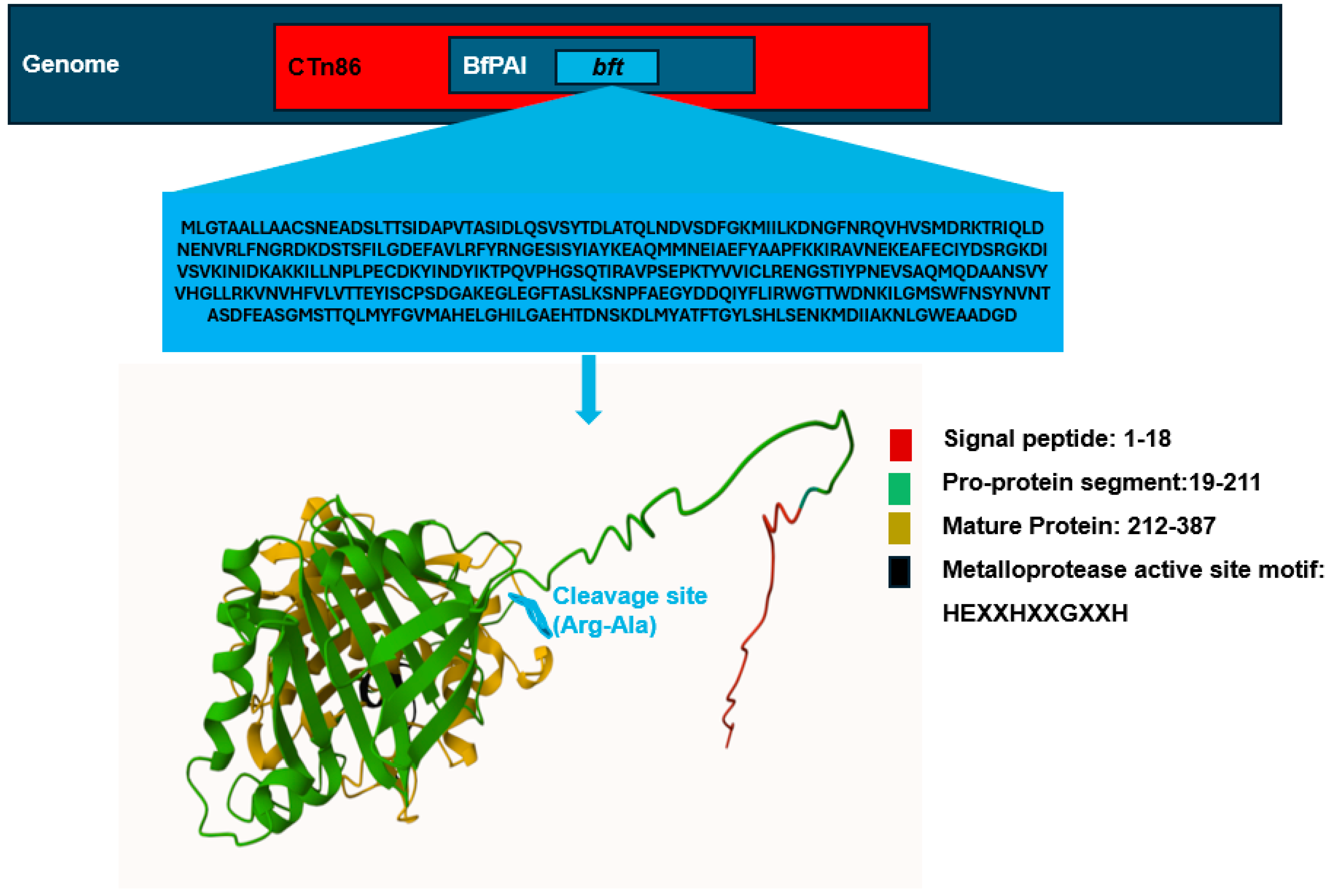
The BFT holotoxin is expressed as a 397-amino-acid protein with an approximate molecular mass of 44.5 kilodaltons [14]. Structurally, the holotoxin is organized into three functional domains: the signal peptide region, the proprotein segment, and the mature toxin [14,35]. Figure 2 provides an overview of the genomic organization of the bft gene, along with the domain architecture and three-dimensional structure of the BFT holotoxin.
The signal peptide domain, composed of 18 amino acids, is essential for directing the holotoxin to the bacterial membrane. Following secretion, the holotoxin exists in a biologically inactive zymogen form (proBFT), in which the ~170-amino-acid pro-domain masks the active site, preventing enzymatic activity.
Proteolytic cleavage at the Arg211–Ala212 junction is required to remove the inhibitory pro-domain and release the mature active toxin [38,39,40]. The 190-amino-acid catalytic domain (CD) of mature BFT contains a characteristic zinc-dependent metalloprotease motif (HEXXHXXGXXH), which is responsible for its enzymatic activity, specifically the degradation of key proteins such as E-cadherin, actin, gelatin, and casein [38,41,42]. This motif, alongside a downstream methionine residue (Met366) embedded in a tight 1,4-β-turn known as the Met-turn, classifies BFT within the metzincin clan of metalloproteases [38,39,43]. The motif includes three histidines that coordinate the catalytic zinc ion and a glutamate residue that functions as a general acid/base during catalysis. Site-directed mutagenesis of critical residues within this core—such as His348, Glu349, His352, Gly355, His358, and Met366—results in a dramatic reduction or complete loss of enzymatic activity, impairing the toxin’s ability to induce morphological alterations in epithelial cells [44]. Intriguingly, its catalytic domain shares high structural similarity with mammalian adamalysins/ADAMs, suggesting a rare horizontal gene transfer event from eukaryotes to B. fragililis [45]. Interestingly, although BFT shares this catalytic architecture with other metzincins, it lacks upstream sequence homology, suggesting that it may represent a unique prototype within the clan [39]. Furthermore, structural integrity of the C-terminal region appears to be essential, as the deletion of even two residues significantly diminishes enzymatic function, while the removal of eight or more leads to complete functional inactivation, despite normal secretion and processing [46]. These findings collectively underscore the critical role of both the zinc-binding motif and the C-terminal architecture in maintaining the proteolytic activity of BFT.
In the BFT-2 isoform, an amphipathic region located at the carboxyl terminus is proposed to facilitate toxin penetration into the host cell membrane and contribute to the formation of ion channels. BFT-1 and BFT-2 exhibit resistance to trypsin and maintain stability across a broad pH range [36,47].
Regulation of BFT expression is mediated, at least in part, by the two-component regulatory system RprXY, which functions as a negative regulator. Overexpression of RprXY has been shown to markedly suppress bft gene expression and reduce BFT production, as demonstrated in experimental models, including Muc2-deficient mice [48]. Recent evidence has revealed that BFT is not freely secreted into the extracellular environment but is packaged with outer membrane vesicles (OMVs). This vesicle-mediated delivery plays a pivotal role in facilitating epithelial disruption and may represent a key mechanism in the pathogenesis of intestinal inflammation [49].

3. ETBF-Associated Diseases and Underlying Mechanisms

Until the 1970s, Bacteroides fragilis was primarily recognized as an opportunistic symbiont within the GI microbiota [50,51]. Notably, Bacteroides fragilis remains the most frequently isolated anaerobic bacterium from human clinical specimens associated with anaerobic infections [52,53]. Although it comprises only approximately 0.5–1% of the normal GI microbiota, translocation to extraintestinal sites can lead to a range of severe infections, including intra-abdominal sepsis, genital tract infections, subcutaneous abscesses, endocarditis, pericarditis, and bacteremia [53,54,55]. In particular, Bacteroides fragilis-associated bacteremia carries a significant mortality rate of approximately 19% [55].
Since the early 1980s, evidence has emerged suggesting that certain strains of Bacteroides fragilis may act beyond the role of an opportunistic pathogen. One of the first pivotal studies in this field was conducted by Myers and colleagues, who isolated Bacteroides fragilis strains with enterotoxin-like activity (BFEL) from the feces of 24- to 48-h-old lambs with diarrheal disease in the Northern Rocky Mountain region of the United States [56]. Pure cultures of Bacteroides obtained from diarrheal lambs induced significant fluid accumulation in lamb and calf intestinal loop models [56]. Three distinct BFEL serogroups were identified among the diarrheal cases. Furthermore, oral inoculation of newborn colostrum-fed lambs with viable BFEL strains resulted in diarrhea, lethargy, and, in one case, death within 32 h post-inoculation, closely resembling the clinical signs observed in natural infections. Subsequent studies successfully isolated ETBF strains from fecal samples of other farm animals, including lambs, calves, and pigs [56,57,58]. In addition, in a follow-up study by Meyer and colleagues, ETBF was isolated from the fecal specimens of 8 out of 44 individuals with diarrhea. In rabbit models, ETBF strains induced mucous and hemorrhagic diarrhea as well as moderate to severe necrotizing colitis, whereas non-toxigenic strains failed to produce such pathological changes. Collectively, these findings underscore the significant role of ETBF in promoting intestinal damage, including diarrhea and colitis [59].
In 2006, Toprak et al. demonstrated for the first time the link between ETBF and colorectal cancer (CRC) [60]. In their study, stool samples from 73 patients with CRC and 59 control individuals were evaluated. After the isolation of Bacteroides fragilis and its identification by conventional methods, the presence of the bft gene was examined using polymerase chain reaction (PCR). The results showed that 38% of isolates from CRC patients carried the bft gene, while this figure in the control group was only 12% (p = 0.009) [60]. Since the 1980s, numerous studies have demonstrated that the frequency of ETBF strains in biopsy, stool, and mucosal samples from patients with diarrhea, inflammatory bowel disease, and colorectal cancer is significantly higher than in healthy individuals [60,61,62,63,64]. These findings suggest a potential role for ETBF in the initiation and progression of intestinal inflammatory disorder. Nevertheless, some studies have reported no clear association between Bacteroides fragilis and disease [65,66]. These inconsistencies may be due to variations in study populations, methodologies, or the failure to distinguish between toxigenic and non-toxigenic strains.
The pathogenesis of BFT, a zinc-dependent metalloprotease, begins with its highly specific binding to a still-unidentified membrane receptor on colonic epithelial cells (CECs). This interaction is strictly dependent on physiological temperature (37 °C) and requires the proteolytically active form of the toxin; catalytically inactive mutants are unable to bind [38,67]. Binding occurs in a polarized fashion—at the apical membrane in differentiated HT29/C1 cells and basolateral membrane in T84 crypt-like cells—suggesting that the receptor redistributes during epithelial differentiation [67]. These features underscore the specificity of BFT–receptor interactions, which serve not only as the initiating step of the pathogenic cascade but also as a trigger for intracellular signaling pathways that compromise epithelial integrity
One of the earliest outcomes of BFT-receptor engagement is the cleavage of E-cadherin, a key cell–cell adhesion molecule at adherens junctions [19]. This cleavage occurs through an ATP-dependent mechanism and rapidly disrupts epithelial barrier integrity [19]. In HT29/C1 cells, the extracellular domain of E-cadherin is shed, followed by γ-secretase-mediated cleavage of the remaining membrane-tethered fragment, producing a 28 kDa cytoplasmic segment that is subsequently degraded via the proteasome [68]. This progressive disassembly of cell–cell contacts facilitate paracellular flux of ions and solutes, contributing to diarrhea.
Simultaneously, BFT exerts broad proteolytic activity against cytoskeletal and extracellular matrix components, including type IV collagen, actin, tropomyosin, myosin, fibrinogen, gelatin, and immune-related proteins such as complement C3 and α1-proteinase inhibitor. Disruption of the actin cytoskeleton induces characteristic morphological changes such as cell rounding and swelling, impairs structural integrity, and weakens epithelial resistance [38,45,69,70].
These structural alterations are closely linked to inflammatory signaling. BFT exposure activates MAPK and NF-κB pathways, promoting the secretion of proinflammatory cytokines like IL-8 and TNF-α [71]. Concurrently, it induces COX-2 and prostaglandin E2 expression, exacerbating fluid secretion and immune cell recruitment—key contributors to mucosal inflammation and secretory diarrhea [72].
Beyond acute epithelial injury, BFT drives tumorigenic processes. Cleavage of E-cadherin leads to β-catenin release, which activates oncogenic transcription, particularly in the context of adenomatous polyposis coli (APC) mutation, upregulating c-Myc and fostering hyperproliferation [30,73,74]. The toxin also upregulates anti-apoptotic factors such as cIAP2 and induces spermine oxidase (SMO), increasing reactive oxygen species (ROS), DNA damage, and compensatory epithelial proliferation. Genotoxicity is further evidenced by the activation of H2AX, a marker of DNA double-strand breaks [75,76].
In APCMin/+ mouse models, colonization with enterotoxigenic B. fragilis (ETBF) induces STAT3 activation and a robust Th17 cytokine profile (IL-17, IL-23, IL-6, TGF-β), generating a pro-carcinogenic inflammatory milieu [77]. Moreover, BFT has been linked to the activation of a STAT3/ZEB2 signaling axis, further compromising epithelial barrier integrity and promoting malignant transformation in CRC cells [25].
In parallel, epigenetic modulation also plays a role in ETBF-driven carcinogenesis. Recent findings indicate that ETBF can promote colorectal cancer cell proliferation through the downregulation of miR-149-3p, mediated by m6A RNA methylation via METTL14. This pathway affects the alternative splicing regulator PHF5A and its downstream target KAT2A. Since miR-149-3p also regulates Th17 cell differentiation, its progressive reduction in patients with IBD and CRC—inversely correlated with ETBF prevalence—underscores the ETBF/miR-149-3p axis as a potential therapeutic target for both inflammatory and neoplastic diseases of the colon [9].

4. Challenges in the Detection of Bacteroides fragilis and Future Research Directions

Bacteroides fragilis is a Gram-negative obligate anaerobic bacterium that resides in the human gastrointestinal tract [78]. Although it plays important roles in maintaining gut homeostasis, certain strains, ETBF, are implicated in inflammatory bowel diseases (IBDs) and CRC [54,78]. Accurate detection and identification of ETBF and its toxin, BFT, remain challenging due to multiple technical and biological factors.
The isolation and detection of Bacteroides fragilis from clinical samples require specialized culture media and strict anaerobic conditions, which complicate and prolong the cultivation process. Furthermore, the high microbial competition present in complex specimens such as feces and colorectal mucosa, along with the relatively low abundance of ETBF during asymptomatic colonization, significantly complicates its isolation and detection. Although selective media such as Bacteroides bile esculin (BBE) agar are commonly employed under anaerobic conditions, overgrowth of other anaerobic organisms, such as enterococci and lactobacilli, can interfere with the isolation of B. fragilis [79].
Traditional biochemical identification methods, such as Rapid ID32A and API 20A kits, are limited by low specificity and often fail to distinguish B. fragilis from closely related species. However, it has been reported that this biochemical scheme is not as accurate as a multiplex PCR identification scheme [80].
While advanced technologies like matrix-assisted laser desorption–ionization time-of-flight mass spectrometry (MALDI-TOF MS) offer higher specificity by analyzing ribosomal protein profiles [81,82], their high cost and technical requirements limit their availability in resource-constrained settings. The Sakamoto study demonstrated that MLSA based on six housekeeping genes (dnaJ, gyrB, hsp60, recA, rpoB, and 16S rRNA) is a valuable method for the identification and classification of Bacteroides species [83]. However, although this method is highly accurate, the need for PCR and sequencing limits its widespread use in all diagnostic laboratories.
Additionally, the confirmation of toxin production by Bacteroides fragilis strains has traditionally relied on biological assays such as the ileal loop test [56,59]. However, this method is costly, time-consuming, and not feasible for routine use in many laboratories.
Alternatively, cell-based diagnostic assays utilizing HT29/C1 cell cultures have been developed and were historically used for BFT detection due to their ability to detect less than 0.5 picomoles of toxin with high specificity and sensitivity [84]. However, this method is now considered outdated due to its labor-intensive procedures, and the availability of more advanced and standardized technologies. Thus, while informative in earlier research, it is no longer regarded as a practical tool for routine clinical diagnostics.
The most common method for identifying Bacteroides fragilis, the bft gene, and its different isotypes is PCR. The 16srRNA gene, nanH, gyrB, and leuB are widely used for the identification of different Bacteroides species [85,86,87]. However, studies have demonstrated that the accuracy of detecting toxigenic strains can be significantly influenced by the design of PCR primers. Some research findings indicate that certain commonly used primers may preferentially detect only specific isotypes of the bft gene, such as bft-1, while exhibiting reduced sensitivity toward bft-2 and bft-3. These observations underscore the importance of designing primers with comprehensive coverage of all bft isotypes to prevent inaccurate estimations of ETBF prevalence in clinical and epidemiological investigations [88]. Moreover, in addition to the proper design of primers for bft detection, the choice of PCR methodology plays a critical role in the sensitivity and accuracy of diagnosing enterotoxigenic ETBF. Comparative studies have demonstrated that, when using purified bacterial DNA, different PCR approaches, including SYBR Green qPCR, TaqMan qPCR, and digital PCR (dPCR), exhibit comparable sensitivity for detecting the toxin gene. However, when applied to clinical stool samples, substantial differences in sensitivity were observed, with SYBR Green qPCR detecting significantly fewer positive cases compared with TaqMan qPCR and dPCR. While TaqMan and dPCR methods identified the bft gene in over 90% of samples, SYBR Green qPCR detected positivity in only about one third of cases [89].
In addition, the type of clinical sample significantly affects detection rates. Biopsy specimens of colorectal mucosa consistently show higher bft positivity than fecal samples [63]; however, ethical considerations, cost, and invasiveness limit their routine clinical application. Other confounding factors include variability in microbial load, host immune status, and geographic differences in ETBF prevalence, indicating that detection strategies must be adapted to population-specific contexts. Furthermore, many previous studies suffer from limitations such as small sample sizes and methodological heterogeneity, including variations in sample collection, DNA extraction, and PCR protocols, as well as reliance on bft gene detection without assessing actual toxin expression or biological activity. Notably, one of the most critical challenges involves the accurate assessment of BFT’s biological activity. While several methods can detect the presence of the bft gene or its protein product, confirming that the active toxin is being produced and is functionally relevant remains technically difficult, particularly in complex clinical samples such as feces or tissue biopsies. These limitations are consistent with findings from the systematic review by Scott et al. (2022), which emphasized the high methodological heterogeneity, poor reproducibility, and risk of bias in many in vitro and in vivo studies assessing ETBF detection and its role in CRC [31]. The lack of standardized protocols and inconsistent reporting across studies continues to hinder the development of reliable diagnostic strategies.
These gaps highlight the need for multicenter studies using standardized protocols and incorporating multi-omics approaches (genomics, transcriptomics, proteomics).
Recent advancements offer promising new directions. For example, monoclonal antibodies and nanobody-based assays have been developed for the sensitive and specific detection of BFT1 and BFT2, enabling the creation of ELISA platforms with detection limits as low as 20–40 ng/mL [90]. Additionally, high affinity nanobodies targeting catalytic and pro-domain regions of BFT provide opportunities for both diagnostic and therapeutic applications [91]. Moreover, recent studies have identified approved drugs capable of inhibiting BFT-3 by binding to an allosteric site within its proenzyme structure, thereby blocking toxin maturation and activity [92]. Moreover, studies have shown that some probiotic Bifidobacterium longum BB536 may be effective in reducing the number of ETBF and other intestinal pathogenic bacteria by improving mucosal barrier function [93,94]. Therefore, investigating the role of non-toxigenic Bacteroides fragilis (NTBF) strains as potential probiotics in reducing ETBF colonization is a topic of considerable interest for future research.
Finally, several critical needs remain unmet. There is a need for multiplex diagnostic platforms capable of simultaneously assessing the presence of the bft gene, its transcript expression, and BFT protein activity. Standardized affordable diagnostic kits suitable for routine clinical use must also be developed. To better understand ETBF pathogenesis, large multicenter studies using standardized methods and integrated omics data remain urgently needed. Furthermore, research efforts should focus on developing targeted strategies for neutralizing BFT activity, including small molecule inhibitors, antibody-based therapies, and competitive inhibitors that can block toxin–receptor interactions. Such approaches may provide new therapeutic options for managing ETBF-associated diseases.

5. Conclusions

Considerable progress has been made in better understanding the role of ETBF and its toxin in gut-associated diseases, yet important challenges remain. While the contribution of BFT to epithelial barrier disruption and inflammation is well recognized, the precise molecular mechanisms—especially the direct interaction with host targets like E-cadherin—require further investigation.
Current diagnostic approaches are often limited by suboptimal sensitivity, specificity, and an inability to confirm active toxin production. Promising strategies, including nanobody-based detection methods, multiplex assays, and targeted therapeutics are being explored to overcome these limitations. The use of probiotics that enhance epithelial barrier function and regulate immune responses offers a complementary non-invasive approach to controlling ETBF colonization. Moving forward, integrative efforts combining advanced diagnostics, toxin-neutralizing strategies, and multi-omics research will be critical to improve clinical management and develop targeted therapies for ETBF-associated diseases.

Author Contributions

L.A.S. conceived and supervised the study; provided resources and funding; and critically reviewed the manuscript. S.J. designed the structure and content of the review; collected and analyzed the literature; drafted the initial manuscript; and implemented revisions. P.M., M.F., I.C. and E.R.S. contributed to literature analysis and interpretation; provided critical suggestions; and supported supervision of the project. All authors have read and agreed to the published version of the manuscript.

Funding

This research was supported by the Regione Autonoma della Sardegna, Legge Regionale 12 December 2022, n. 22, PRIN MUR 2022 n: 2022BP837R to LAS.

Institutional Review Board Statement

Not applicable.

Informed Consent Statement

Not applicable.

Data Availability Statement

No new data were created or analyzed in this study.

Conflicts of Interest

The authors declare no conflict of interest.

Abbreviations

AADAntibiotic-associated diarrhea
BfPAIBacteroides fragilis pathogenicity island
BFTBacteroides fragilis toxin
CRCColorectal cancer
CDIClostridioides difficile infection
CTn86Conjugative transposon 86
DSSDextran sulfate sodium
ETBFEnterotoxigenic Bacteroides fragilis
GIGastrointestinal
IL-10Interleukin-10
IL-22Interleukin-22
MUC-2Mucin 2
NTBFNon-toxigenic Bacteroides fragilis
OMVsOuter membrane vesicles
PSAPolysaccharide A
RprXYTwo-component regulatory system RprXY
STAT3Signal transducer and activator of transcription 3
TregRegulatory T cell
ZEB2Zinc finger E-box binding homeobox 2
ZO-1Zonula occludens-1

References

  1. Sender, R.; Fuchs, S.; Milo, R. Revised Estimates for the Number of Human and Bacteria Cells in the Body. PLoS Biol. 2016, 14, e1002533. [Google Scholar] [CrossRef] [PubMed]
  2. John, H.T.; Thomas, T.C.; Chukwuebuka, E.C.; Ali, A.B.; Anass, R.; Tefera, Y.Y.; Babu, B.; Negrut, N.; Ferician, A.; Marian, P. The Microbiota–Human Health Axis. Microorganisms 2025, 13, 948. [Google Scholar] [CrossRef] [PubMed]
  3. Shao, T.; Hsu, R.; Rafizadeh, D.L.; Wang, L.; Bowlus, C.L.; Kumar, N.; Mishra, J.; Timilsina, S.; Ridgway, W.M.; Gershwin, M.E.; et al. The Gut Ecosystem and Immune Tolerance. J. Autoimmun. 2023, 141, 103114. [Google Scholar] [CrossRef]
  4. Acevedo-Román, A.; Pagán-Zayas, N.; Velázquez-Rivera, L.I.; Torres-Ventura, A.C.; Godoy-Vitorino, F. Insights into Gut Dysbiosis: Inflammatory Diseases, Obesity, and Restoration Approaches. Int. J. Mol. Sci. 2024, 25, 971. [Google Scholar] [CrossRef]
  5. Zhou, W.; Cheng, Y.; Zhu, P.; Nasser, M.I.; Zhang, X.; Zhao, M. Implication of Gut Microbiota in Cardiovascular Diseases. Oxid. Med. Cell. Longev. 2020, 2020, 5394096. [Google Scholar] [CrossRef]
  6. Qin, J.; Li, R.; Raes, J.; Arumugam, M.; Burgdorf, K.S.; Manichanh, C.; Nielsen, T.; Pons, N.; Levenez, F.; Yamada, T.; et al. A Human Gut Microbial Gene Catalogue Established by Metagenomic Sequencing. Nature 2010, 464, 59–65. [Google Scholar] [CrossRef]
  7. Townsend, G.E., 2nd; Han, W.; Schwalm, N.D.; Hong, X.; Bencivenga-Barry, N.A.; Goodman, A.L.; Groisman, E.A.; Ii, G.E.T.; Comstock, L.E. A Master Regulator of Bacteroides Thetaiotaomicron Gut Colonization Controls Carbohydrate Utilization and an Alternative Protein Synthesis Factor. mBio 2020, 11, e03221-19. [Google Scholar] [CrossRef] [PubMed]
  8. Sears, C.L.; Geis, A.L.; Housseau, F. Bacteroides fragilis Subverts Mucosal Biology: From Symbiont to Colon Carcinogenesis. J. Clin. Investig. 2014, 124, 4166–4172. [Google Scholar] [CrossRef]
  9. Cao, Y.; Wang, Z.; Yan, Y.; Ji, L.; He, J.; Xuan, B.; Shen, C.; Ma, Y.; Jiang, S.; Ma, D.; et al. Enterotoxigenic Bacteroides fragilis Promotes Intestinal Inflammation and Malignancy by Inhibiting Exosome-Packaged MiR-149-3p. Gastroenterology 2021, 161, 1552–1566.e12. [Google Scholar] [CrossRef]
  10. Zhang, W.; Zhu, B.; Xu, J.; Liu, Y.; Qiu, E.; Li, Z.; Li, Z.; He, Y.; Zhou, H.; Bai, Y.; et al. Bacteroides fragilis Protects against Antibiotic-Associated Diarrhea in Rats by Modulating Intestinal Defenses. Front. Immunol. 2018, 9, 1040. [Google Scholar] [CrossRef]
  11. Chung, L.; Thiele Orberg, E.; Geis, A.L.; Chan, J.L.; Fu, K.; DeStefano Shields, C.E.; Dejea, C.M.; Fathi, P.; Chen, J.; Finard, B.B.; et al. Bacteroides fragilis Toxin Coordinates a Pro-Carcinogenic Inflammatory Cascade via Targeting of Colonic Epithelial Cells. Cell Host Microbe 2018, 23, 203–214.e5. [Google Scholar] [CrossRef]
  12. He, Q.; Niu, M.; Bi, J.; Du, N.; Liu, S.; Yang, K.; Li, H.; Yao, J.; Du, Y.; Duan, Y. Protective Effects of a New Generation of Probiotic Bacteroides fragilis against Colitis In Vitro and In Vitro. Sci. Rep. 2023, 13, 1584. [Google Scholar] [CrossRef]
  13. Franco, A.A.; Cheng, R.K.; Goodman, A.; Sears, C.L. Modulation of Bft Expression by the Bacteroides fragilis Pathogenicity Island and Its Flanking Region. Mol. Microbiol. 2002, 45, 1067–1077. [Google Scholar] [CrossRef]
  14. Franco, A.A.; Mundy, L.M.; Trucksis, M.; Wu, S.; Kaper, J.B.; Sears, C.L. Cloning and Characterization of the Bacteroides fragilis Metalloprotease Toxin Gene. Infect. Immune 1997, 65, 1007–1013. [Google Scholar] [CrossRef] [PubMed]
  15. Hopkins, M.J.; Macfarlane, G.T. Nondigestible Oligosaccharides Enhance Bacterial Colonization Resistance against Clostridium Difficile in Vitro. Appl. Environ. Microbiol. 2003, 69, 1920–1927. [Google Scholar] [CrossRef] [PubMed]
  16. Deng, H.; Yang, S.; Zhang, Y.; Qian, K.; Zhang, Z.; Liu, Y.; Wang, Y.; Bai, Y.; Fan, H.; Zhao, X.; et al. Bacteroides fragilis Prevents Clostridium Difficile Infection in a Mouse Model by Restoring Gut Barrier and Microbiome Regulation. Front. Microbiol. 2018, 9, 2976. [Google Scholar] [CrossRef] [PubMed]
  17. Chan, J.L.; Wu, S.; Geis, A.L.; Chan, G.V.; Gomes, T.A.M.; Beck, S.E.; Wu, X.; Fan, H.; Tam, A.J.; Chung, L.; et al. Non-Toxigenic Bacteroides fragilis (NTBF) Administration Reduces Bacteria-Driven Chronic Colitis and Tumor Development Independent of Polysaccharide A. Mucosal Immunol. 2019, 12, 164–177. [Google Scholar] [CrossRef]
  18. Zhang, W.; Zhou, Q.; Liu, H.; Xu, J.; Huang, R.; Shen, B.; Guo, Y.; Ai, X.; Xu, J.; Zhao, X.; et al. Bacteroides fragilis Strain ZY-312 Facilitates Colonic Mucosa Regeneration in Colitis via Motivating STAT3 Signaling Pathway Induced by IL-22 from ILC3 Secretion. Front. Immunol. 2023, 14, 1156762. [Google Scholar] [CrossRef]
  19. Kim, W.S.; Hwang, S.; Gwon, S.Y.; Jo, M.; Yoo, S.H.; Hong, J.; Jang, H.N.; Hong, J.E.; Kang, D.H.; Yun, M.; et al. Bacteroides fragilis Toxin Induces Sequential Proteolysis of E-Cadherin and Inflammatory Response in Mouse Intestinal Epithelial Cell Line. Microorganisms 2025, 13, 781. [Google Scholar] [CrossRef]
  20. Ramamurthy, D.; Pazhani, G.P.; Sarkar, A.; Nandy, R.K.; Rajendran, K.; Sur, D.; Manna, B.; Ramamurthy, T. Case-Control Study on the Role of Enterotoxigenic Bacteroides fragilis as a Cause of Diarrhea among Children in Kolkata, India. PLoS ONE 2013, 8, e6062. [Google Scholar] [CrossRef]
  21. Merino, V.R.C.; Nakano, V.; Liu, C.; Song, Y.; Finegold, S.M.; Avila-Campos, M.J. Quantitative Detection of Enterotoxigenic Bacteroides fragilis Subtypes Isolated from Children with and without Diarrhea. J. Clin. Microbiol. 2011, 49, 416–418. [Google Scholar] [CrossRef] [PubMed]
  22. Jo, M.; Hwang, S.; Lee, C.G.; Hong, J.E.; Kang, D.H.; Yoo, S.H.; Kim, W.S.; Yoo, J.Y.; Rhee, K.J. Promotion of Colitis in B Cell-Deficient C57BL/6 Mice Infected with Enterotoxigenic Bacteroides fragilis. Int. J. Mol. Sci. 2024, 25, 364. [Google Scholar] [CrossRef] [PubMed]
  23. Xie, X.; Jiang, D.; Zhou, X.; Ye, X.; Yang, P.; He, Y. Recombinant Bacteroides fragilis Enterotoxin-1 (RBFT-1) Promotes Proliferation of Colorectal Cancer via CCL3-Related Molecular Pathways. Open Life Sci. 2021, 16, 408–418. [Google Scholar] [CrossRef]
  24. Wu, Z.; Yu, M.; Zeng, Y.; Huang, Y.; Zheng, W. LRP11-AS1 Mediates Enterotoxigenic Bacteroides fragilis-Related Carcinogenesis in Colorectal Cancer via the MiR-149-3p/CDK4 Pathway. Cancer Gene Ther. 2024, 32, 184–197. [Google Scholar] [CrossRef] [PubMed]
  25. Yang, J.; Wang, X.; Hu, T.; Huang, H.; Chen, G.; Jin, B.; Zeng, G.; Liu, J. Entero-Toxigenic Bacteroides fragilis Contributes to Intestinal Barrier Injury and Colorectal Cancer Progression by Mediating the BFT/STAT3/ZEB2 Pathway. Cell Cycle 2024, 23, 70–82. [Google Scholar] [CrossRef]
  26. Jasemi, S.; Emaneini, M.; Fazeli, M.S.; Ahmadinejad, Z.; Nomanpour, B.; Sadeghpour Heravi, F.; Sechi, L.A.; Feizabadi, M.M. Toxigenic and Non-Toxigenic Patterns I, II and III and Biofilm-Forming Ability in Bacteroides fragilis Strains Isolated from Patients Diagnosed with Colorectal Cancer. Gut Pathog. 2020, 12, 28. [Google Scholar] [CrossRef]
  27. Sárvári, K.P.; Sóki, J.; Iván, M.; Miszti, C.; Latkóczy, K.; Melegh, S.Z.; Urbán, E. Detection of Enterotoxin and Protease Genes among Hungarian Clinical Bacteroides fragilis Isolates. Anaerobe 2017, 48, 98–102. [Google Scholar] [CrossRef]
  28. Łuczak, M.; Obuch-Woszczatyński, P.; Pituch, H.; Leszczyński, P.; Martirosian, G.; Patrick, S.; Poxton, I.; Wintermans, R.G.F.; Dubreuil, L.; Meisel-Mikołajczyk, F. Search for Enterotoxin Gene in Bacteroides fragilis Strains Isolated from Clinical Specimens in Poland, Great Britain, the Netherlands and France. Med. Sci. Monit. 2001, 7, 222–225. [Google Scholar] [PubMed]
  29. Ulger, N.; Rajendram, D.; Yagci, A.; Gharbia, S.; Shah, H.N.; Gulluoglu, B.M.; Akin, L.M.; Demirkalem, P.; Celenk, T.; Soyletir, G. The Distribution of the Bft Alleles among Enterotoxigenic Bacteroides fragilis Strains from Stool Specimens and Extraintestinal Sites. Anaerobe 2006, 12, 71–74. [Google Scholar] [CrossRef]
  30. Cheng, W.T.; Kantilal, H.K.; Davamani, F. The Mechanism of Bacteroides fragilis Toxin Contributes to Colon Cancer Formation. Malays. J. Med. Sc. 2020, 27, 9–21. [Google Scholar] [CrossRef]
  31. Scott, N.; Whittle, E.; Jeraldo, P.; Chia, N. A Systemic Review of the Role of Enterotoxic Bacteroides fragilis in Colorectal Cancer. Neoplasia 2022, 29, 100797. [Google Scholar] [CrossRef] [PubMed]
  32. Franco, A.A. The Bacteroides fragilis Pathogenicity Island Is Contained in a Putative Novel Conjugative Transposon. J. Bacteriol. 2004, 186, 6077–6092. [Google Scholar] [CrossRef]
  33. Buckwold, S.L.; Shoemaker, N.B.; Sears, C.L.; Franco, A.A. Identification and Characterization of Conjugative Transposons CTn86 and CTn9343 in Bacteroides fragilis Strains. Appl. Environ. Microbiol. 2007, 73, 53–63. [Google Scholar] [CrossRef] [PubMed]
  34. Kato, N.; Liu, C.-X.; Kato, H.; Watanabe, K.; Tanaka, Y.; Yamamoto, T.; Suzuki, K.; Ueno, K. A New Subtype of the Metalloprotease Toxin Gene and the Incidence of the Three Bft Subtypes among Bacteroides fragilis Isolates in Japan. FEMS Microbiol. Lett. 2000, 182, 171–176. [Google Scholar] [CrossRef]
  35. Kling, J. Cloning and Characterization of the Gene for the Metalloprotease Enterotoxin of Bacteroides fragilis. FEMS Microbiol. Lett. 1997, 146, 279–284. [Google Scholar] [CrossRef] [PubMed]
  36. Wu, S.; Dreyfus, L.A.; Tzianabos, A.O.; Hayashi, C.; Sears, C.L. Diversity of the Metalloprotease Toxin Produced by Enterotoxigenic Bacteroides fragilis. Infect. Immun. 2002, 70, 2463–2471. [Google Scholar] [CrossRef]
  37. Chung, G.-T.; Franco, A.A.; Wu, S.; Rhie, G.-E.; Cheng, R.; Oh, H.-B.; Sears, C.L. Identification of a Third Metalloprotease Toxin Gene in Extraintestinal Isolates of Bacteroides fragilis. Infect Immun. 1999, 67, 4945–4949. [Google Scholar] [CrossRef]
  38. Moncrief, J.S.; Obiso, R.; Barroso, L.A.; Kling, J.J.; Wright, R.L.; Van Tassell, R.L.; Lyerly, D.M.; Wilkins, T.D. The Enterotoxin of Bacteroides fragilis Is a Metalloprotease. Infect. Immun. 1995, 63, 175–181. [Google Scholar] [CrossRef]
  39. Gomis-Rüth, F.X. Structural Aspects of the Metzincin Clan of Metalloendopeptidases. Mol. Biotechnol. 2003, 24, 157–202. [Google Scholar] [CrossRef]
  40. Sears, C.L. The Toxins of Bacteroides fragilis. Toxicon 2001, 39, 1737–1746. [Google Scholar] [CrossRef]
  41. Obiso, R.J.; Bevan, D.R.; Wilkins, T.D. Molecular Modeling and Analysis of Fragilysin, the Bacteroides fragilis Toxin. Clin. Infect. Dis. 1997, 2, S153–S155. [Google Scholar] [CrossRef] [PubMed]
  42. Remacle, A.G.; Shiryaev, S.A.; Strongin, A.Y. Distinct Interactions with Cellular E-Cadherin of the Two Virulent Metalloproteinases Encoded by a Bacteroides fragilis Pathogenicity Island. PLoS ONE 2014, 9, e113896. [Google Scholar] [CrossRef]
  43. Gomis-Rüth, F.X. Catalytic Domain Architecture of Metzincin Metalloproteases. J. Biol. Chem. 2009, 284, 15353–15357. [Google Scholar] [CrossRef]
  44. Franco, A.A.; Buckwold, S.L.; Shin, J.W.; Ascon, M.; Sears, C.L. Mutation of the Zinc-Binding Metalloprotease Motif Affects Bacteroides fragilis Toxin Activity but Does Not Affect Propeptide Processing. Infect. Immun. 2005, 73, 5273–5277. [Google Scholar] [CrossRef]
  45. Goulas, T.; Arolas, J.L.; Gomis-Rüth, F.X. Structure, Function and Latency Regulation of a Bacterial Enterotoxin Potentially Derived from a Mammalian Adamalysin/ADAM Xenolog. Proc. Natl. Acad. Sci. USA 2011, 108, 1856–1861. [Google Scholar] [CrossRef]
  46. Sears, C.L.; Buckwold, S.L.; Shin, J.W.; Franco, A.A. The C-Terminal Region of Bacteroides fragilis Toxin Is Essential to Its Biological Activity. Infect. Immun. 2006, 74, 5595–5601. [Google Scholar] [CrossRef] [PubMed]
  47. Van Tassell, R.L.; Lyerly, D.M.; Wilkins, T.D. Purification and Characterization of an Enterotoxin from Bacteroides fragilis. Infect. Immun. 1992, 60, 1343–1350. [Google Scholar] [CrossRef] [PubMed]
  48. Hecht, A.L.; Casterline, B.W.; Choi, V.M.; Bubeck Wardenburg, J. A Two-Component System Regulates Bacteroides fragilis Toxin to Maintain Intestinal Homeostasis and Prevent Lethal Disease. Cell Host Microbe 2017, 22, 443–448.e5. [Google Scholar] [CrossRef]
  49. Zakharzhevskaya, N.B.; Tsvetkov, V.B.; Vanyushkina, A.A.; Varizhuk, A.M.; Rakitina, D.V.; Podgorsky, V.V.; Vishnyakov, I.E.; Kharlampieva, D.D.; Manuvera, V.A.; Lisitsyn, F.V.; et al. Interaction of Bacteroides fragilis Toxin with Outer Membrane Vesicles Reveals New Mechanism of Its Secretion and Delivery. Front. Cell Infect. Microbiol. 2017, 7, 308. [Google Scholar] [CrossRef]
  50. Dornbusch, K.; Nord, C.-E.; Wadström, T. Biochemical Characterization and in Vitro Determination of Antibiotic Susceptibility of Clinical Isolates of Bacteroides fragilis. Scand. J. Infect. Dis. 1974, 6, 253–258. [Google Scholar] [CrossRef]
  51. Nielsen, M.L.; Justesen, T. Anaerobic and Aerobic Bacteriological Studies in Biliary Tract Disease. Scand. J. Gastroenterol. 1976, 11, 437–446. [Google Scholar] [CrossRef] [PubMed]
  52. Kierzkowska, M.; Markowska, K.; Kownacki, J.; Podsiadły, E.; Majewska, A. Anaerobic Bacteraemia—Identification of Bacteroides and Phocaeicola in Blood Samples. Challenges in Antimicrobial Susceptibility Testing. Microbe 2025, 7, 100374. [Google Scholar] [CrossRef]
  53. Jasemi, S.; Emaneini, M.; Ahmadinejad, Z.; Fazeli, M.S.; Sechi, L.A.; Sadeghpour Heravi, F.; Feizabadi, M.M. Antibiotic Resistance Pattern of Bacteroides fragilis Isolated from Clinical and Colorectal Specimens. Ann. Clin. Microbiol. Antimicrob. 2021, 20, 27. [Google Scholar] [CrossRef]
  54. Valguarnera, E.; Wardenburg, J.B. Good Gone Bad: One Toxin Away from Disease for Bacteroides fragilis. J. Mol. Biol. 2020, 432, 765–785. [Google Scholar] [CrossRef] [PubMed]
  55. Wexler, H.M. Bacteroides: The Good, the Bad, and the Nitty-Gritty. Clin. Microbiol. Rev. 2007, 20, 593–621. [Google Scholar] [CrossRef]
  56. Myers, L.L.; Firehammer, B.D.; Shoop, D.S.; Border, M.M. Bacteroides fragilis: A Possible Cause of Acute Diarrheal Disease in Newborn Lambs. Infect. Immune 1984, 44, 241–244. [Google Scholar] [CrossRef]
  57. Border, M.; Firehammer, B.D.; Shoop, D.S.; Myers, L.L. Isolation of Bacteroides fragilis from the Feces of Diarrheic Calves and Lambst. Clin. Microbiol. 1985, 21, 471–473. [Google Scholar] [CrossRef]
  58. Duimstra, J.R.; Myers, L.L.; Collins, J.E.; Benfield, D.A.; Shoop, D.S.; Bradbury, W.C. Enterovirulence of Enterotoxigenic Bacteroides fragilis in Gnotobiotic Pigs. Vet. Pathol. 1991, 28, 514–518. [Google Scholar] [CrossRef]
  59. Myers, L.L.; Shoop, D.S.; Stackhouse, L.L.; Newman, F.S.; Flaherty, R.J.; Letson, G.W.; Sack, A.R.B. Isolation of Enterotoxigenic Bacteroides fragilis from Humans with Diarrheat. J. Clin. Microbiol. 1987, 25, 2330–2333. [Google Scholar] [CrossRef]
  60. Ulger Toprak, N.; Yagci, A.; Gulluoglu, B.M.; Akin, M.L.; Demirkalem, P.; Celenk, T.; Soyletir, G. A Possible Role of Bacteroides fragilis Enterotoxin in the Aetiology of Colorectal Cancer. Clin. Microbiol. Infect. 2006, 12, 782–786. [Google Scholar] [CrossRef]
  61. Matsumiya, Y.; Suenaga, M.; Ishikawa, T.; Kudo, T.; Nakagawa, T.; Okamoto, K.; Tokunaga, M.; Hurtado, C.; Yamada, Y.; Oka, K.; et al. Clinical Significance of Bacteroides fragilis as a Potential Prognostic Factor in Colorectal Cancer. Anaerobe 2023, 84, 102784. [Google Scholar] [CrossRef] [PubMed]
  62. Zamani, S.; Taslimi, R.; Sarabi, A.; Jasemi, S.; Sechi, L.A.; Feizabadi, M.M. Enterotoxigenic Bacteroides fragilis: A Possible Etiological Candidate for Bacterially-Induced Colorectal Precancerous and Cancerous Lesions. Front. Cell Infect. Microbiol. 2020, 9, 449. [Google Scholar] [CrossRef] [PubMed]
  63. Boleij, A.; Hechenbleikner, E.M.; Goodwin, A.C.; Badani, R.; Stein, E.M.; Lazarev, M.G.; Ellis, B.; Carroll, K.C.; Albesiano, E.; Wick, E.C.; et al. The Bacteroides fragilis Toxin Gene Is Prevalent in the Colon Mucosa of Colorectal Cancer Patients. Clin. Infect. Dis. 2015, 60, 208–215. [Google Scholar] [CrossRef]
  64. Viljoen, K.S.; Dakshinamurthy, A.; Goldberg, P.; Blackburn, J.M. Quantitative Profiling of Colorectal Cancer-Associated Bacteria Reveals Associations between Fusobacterium spp., Enterotoxigenic Bacteroides fragilis (ETBF) and Clinicopathological Features of Colorectal Cancer. PLoS ONE 2015, 10, e0119462. [Google Scholar] [CrossRef] [PubMed]
  65. Van, N.; Ahlberg, N.; Chul Jung, B.; Lee, M.H.; Ahn, S.J.; Lee, I.-S.; Kim, Y.S.; Rhee, K.-J. Evaluation of Enterotoxigenic Bacteroides fragilis from Colonic Washings from Patients Undergoing Colonoscopy. J. Exp. Biomed. 2012, 18, 362–368. [Google Scholar]
  66. Lennard, K.S.; Goosen, R.W.; Blackburn, J.M. Bacterially-Associated Transcriptional Remodelling in a Distinct Genomic Subtype of Colorectal Cancer Provides a Plausible Molecular Basis for Disease Development. PLoS ONE 2016, 11, e0166282. [Google Scholar] [CrossRef]
  67. Wu, S.; Shin, J.; Zhang, G.; Cohen, M.; Franco, A.; Sears, C.L. The Bacteroides fragilis Toxin Binds to a Specific Intestinal Epithelial Cell Receptor. Infect. Immun. 2006, 74, 5382–5390. [Google Scholar] [CrossRef]
  68. Wu, S.; Rhee, K.J.; Zhang, M.; Franco, A.; Sears, C.L. Erratum: Bacteroides fragilis Toxin Stimulates Intestinal Epithelial Cell Shedding and γ-Secretase-Dependent E-Cadherin Cleavage. J. Cell Sci. 2007, 120, 1944–1952. [Google Scholar] [CrossRef]
  69. Koshy, S.S.; Montrose, M.H.; Sears, C.L. Human Intestinal Epithelial Cells Swell and Demonstrate Actin Rearrangement in Response to the Metalloprotease Toxin of Bacteroides fragilis. Infect. Immun. 1996, 64, 5022–5028. [Google Scholar] [CrossRef]
  70. Donelli, G.; Fabbri, A.; Fiorentini, C. Bacteroides fragilis Enterotoxin Induces Cytoskeletal Changes and Surface Blebbing in HT-29 Cells. Infect. Immun. 1996, 64, 113–119. [Google Scholar] [CrossRef]
  71. Lee, C.G.; Hwang, S.; Gwon, S.Y.; Park, C.; Jo, M.; Hong, J.E.; Rhee, K.J. Bacteroides fragilis Toxin Induces Intestinal Epithelial Cell Secretion of Interleukin-8 by the E-Cadherin/β-Catenin/NF-ΚB Dependent Pathway. Biomedicines 2022, 10, 827. [Google Scholar] [CrossRef] [PubMed]
  72. Kim, J.M.; Lee, J.Y.; Yoon, Y.M.; Oh, Y.K.; Kang, J.S.; Kim, Y.J.; Kim, K.H. Bacteroides fragilis Enterotoxin Induces Cyclooxygenase-2 and Fluid Secretion in Intestinal Epithelial Cells through NF-ΚB Activation. Eur. J. Immunol. 2006, 36, 2446–2456. [Google Scholar] [CrossRef]
  73. Allen, J.; Hao, S.; Sears, C.L.; Timp, W. Epigenetic Changes Induced by Bacteroides fragilis Toxin. Infect. Immun. 2019, 87, e00447-18. [Google Scholar] [CrossRef]
  74. Li, S.; Liu, J.; Zheng, X.; Ren, L.; Yang, Y.; Li, W.; Fu, W.; Wang, J.; Du, G. Tumorigenic Bacteria in Colorectal Cancer: Mechanisms and Treatments. Cancer Biol. Med. 2021, 18, 147–162. [Google Scholar] [CrossRef]
  75. Goodwin, A.C.; Destefano Shields, C.E.; Wu, S.; Huso, D.L.; Wu, X.Q.; Murray-Stewart, T.R.; Hacker-Prietz, A.; Rabizadeh, S.; Woster, P.M.; Sears, C.L.; et al. Polyamine Catabolism Contributes to Enterotoxigenic Bacteroides fragilis-Induced Colon Tumorigenesis. Proc. Natl. Acad. Sci. USA 2011, 108, 15354–15359. [Google Scholar] [CrossRef] [PubMed]
  76. Sears, C.L. Enterotoxigenic Bacteroides fragilis: A Rogue among Symbiotes. Clin. Microbiol. Rev. 2009, 22, 349–369. [Google Scholar] [CrossRef]
  77. Wu, S.; Rhee, K.J.; Albesiano, E.; Rabizadeh, S.; Wu, X.; Yen, H.R.; Huso, D.L.; Brancati, F.L.; Wick, E.; McAllister, F.; et al. A Human Colonic Commensal Promotes Colon Tumorigenesis via Activation of T Helper Type 17 T Cell Responses. Nat. Med. 2009, 15, 1016–1022. [Google Scholar] [CrossRef] [PubMed]
  78. Patrick, S. A Tale of Two Habitats: Bacteroides fragilis, a Lethal Pathogen and Resident in the Human Gastrointestinal Microbiome. Microbiology 2022, 168, 4. [Google Scholar] [CrossRef]
  79. Ho, P.L.; Ho, L.Y.; Yau, C.Y.; Tong, M.K.; Chow, K.H. A Novel Selective Medium for Isolation of Bacteroides fragilis from Clinical Specimens. J. Clin. Microbiol. 2017, 55, 384–390. [Google Scholar] [CrossRef]
  80. Liu, C.; Song, Y.; McTeague, M.; Vu, A.W.; Wexler, H.; Finegold, S.M. Rapid Identification of the Species of the Bacteroides fragilis Group by Multiplex PCR Assays Using Group- and Species-Specific Primers. FEMS Microbiol. Lett. 2003, 222, 9–16. [Google Scholar] [CrossRef]
  81. Nagy, E. Matrix-Assisted Laser Desorption/Ionization Time-of-Flight Mass Spectrometry: A New Possibility for the Identification and Typing of Anaerobic Bacteria. Future Microbiol. 2014, 9, 217–233. [Google Scholar] [CrossRef] [PubMed]
  82. Nagy, E.; Maier, T.; Urban, E.; Terhes, G.; Kostrzewa, M.; Nord, C.E.; Hedberg, M.; Könönen, E.; Dubreuil, L.; Dosa, E.; et al. Species Identification of Clinical Isolates of Bacteroides by Matrix-Assisted Laser-Desorption/Ionization Time-of-Flight Mass Spectrometry. Clin. Microbiol. Infect. 2009, 15, 796–802. [Google Scholar] [CrossRef] [PubMed]
  83. Sakamoto, M.; Ohkuma, M. Identification and Classification of the Genus Bacteroides by Multilocus Sequence Analysis. Microbiology 2011, 157, 3388–3397. [Google Scholar] [CrossRef]
  84. Weikel, C.S.; Grieco, F.D.; Reuben, J.; Myers, L.L.; Bradley Sack, A.R. Human Colonic Epithelial Cells, HT29/C1, Treated with Crude Bacteroides fragilis Enterotoxin Dramatically Alter Their Morphology. Infect. Immun. 1992, 60, 321–327. [Google Scholar] [CrossRef]
  85. Lee, C.S.; Marion, J.W.; Lee, J. A Novel Genetic Marker for the Rapid Detection of Bacteroides fragilis in Recreational Water as a Human-Specific Faecal Indicator. J. Water Health 2011, 9, 253–264. [Google Scholar] [CrossRef]
  86. Jotwani, R.; Kato, N.; Kato, H.; Watanabe, K.; Ueno, K. Detection of Bacteroides fragilis in Clinical Specimens by Polymerase Chain Reaction Amplification of the Neuraminidase Gene. Curr. Microbiol. 1995, 31, 215–219. [Google Scholar] [CrossRef] [PubMed]
  87. Lee, C.S.; Lee, J. Evaluation of New GyrB-Based Real-Time PCR System for the Detection of B. Fragilis as an Indicator of Human-Specific Fecal Contamination. J. Microbiol. Methods 2010, 82, 311–318. [Google Scholar] [CrossRef]
  88. Aitchison, A.; Frizelle, F.A.; Keenan, J.I. PCR Detection of the Bacteroides fragilis Enterotoxin Gene Relies on Robust Primer Design. J. Clin. Microbiol. 2016, 54, 239–240. [Google Scholar] [CrossRef]
  89. Purcell, R.V.; Pearson, J.; Frizelle, F.A.; Keenan, J.I. Comparison of Standard, Quantitative and Digital PCR in the Detection of Enterotoxigenic Bacteroides fragilis. Sci. Rep. 2016, 6, 34554. [Google Scholar] [CrossRef]
  90. Guo, Y.; Ouyang, Z.; He, W.; Zhang, J.; Qin, Q.; Jiao, M.; Muyldermans, S.; Zheng, F.; Wen, Y. Screening and Epitope Characterization of Diagnostic Nanobody against Total and Activated Bacteroides fragilis Toxin. Front. Immunol. 2023, 14, 1065274. [Google Scholar] [CrossRef]
  91. Mootien, S.; Kaplan, P.M. Monoclonal Antibodies Specific for Bacteroides fragilis Enterotoxins BFT1 and BFT2 and Their Use in Immunoassays. PLoS ONE 2017, 12, e0173128. [Google Scholar] [CrossRef] [PubMed]
  92. Jimenez-Alesanco, A.; Eckhard, U.; Asencio del Rio, M.; Vega, S.; Guevara, T.; Velazquez-Campoy, A.; Gomis-Rüth, F.X.; Abian, O. Repositioning Small Molecule Drugs as Allosteric Inhibitors of the BFT-3 Toxin from Enterotoxigenic Bacteroides fragilis. Protein Sci. 2022, 31, e4427. [Google Scholar] [CrossRef] [PubMed]
  93. Mathipa, M.G.; Thantsha, M.S. Probiotic Engineering: Towards Development of Robust Probiotic Strains with Enhanced Functional Properties and for Targeted Control of Enteric Pathogens. Gut Pathog. 2017, 9, 28. [Google Scholar] [CrossRef] [PubMed]
  94. Odamaki, T.; Sugahara, H.; Yonezawa, S.; Yaeshima, T.; Iwatsuki, K.; Tanabe, S.; Tominaga, T.; Togashi, H.; Benno, Y.; Xiao, J. Effect of the Oral Intake of Yogurt Containing Bifidobacterium Longum BB536 on the Cell Numbers of Enterotoxigenic Bacteroides fragilis in Microbiota. Anaerobe 2012, 18, 14–18. [Google Scholar] [CrossRef]
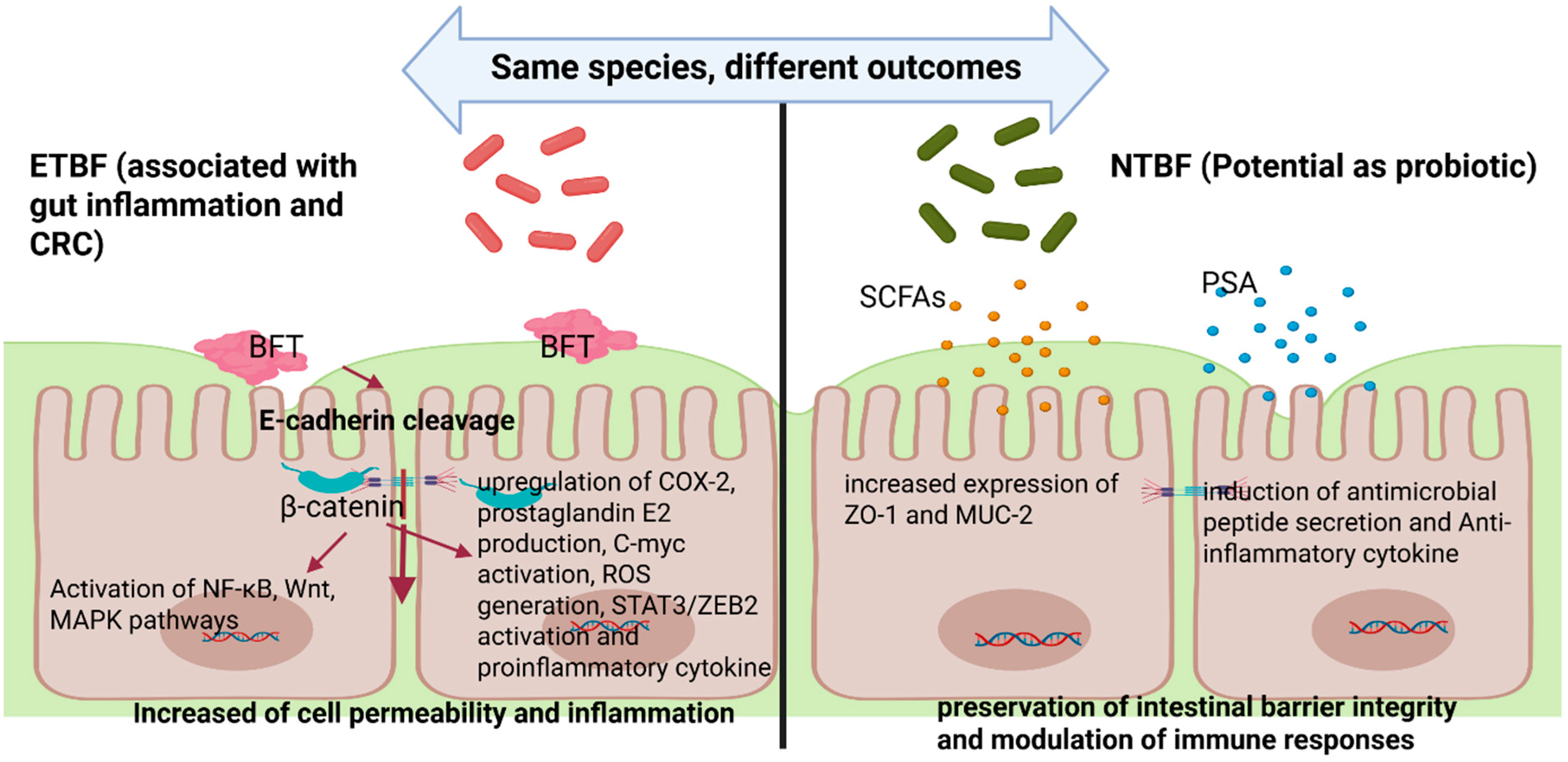
Figure 1. Differential effects of enterotoxigenic Bacteroides fragilis (ETBF) and non-toxigenic Bacteroides fragilis (NTBF) on intestinal epithelial cells. ETBF produces Bacteroides fragilis toxin (BFT), leading to E-cadherin cleavage, activation of β-catenin, upregulation of inflammatory pathways (NF-κB, Wnt, MAPK), COX-2, prostaglandin E2 production, C-myc and STAT3/ZEB2 activation, ROS generation, and proinflammatory cytokine release, ultimately resulting in increased intestinal permeability and inflammation. In contrast, NTBF promotes gut homeostasis through the production of short-chain fatty acids (SCFAs) and polysaccharide A (PSA), upregulation of barrier-protective molecules (ZO-1, MUC-2), induction of antimicrobial peptides, and secretion of anti-inflammatory cytokine.
Toxins 17 00305 g001
Figure 2. Genomic context and structural domains of Bacteroides fragilis toxin (BFT). The bft gene is located within the ~6 kb Bacteroides fragilis pathogenicity island (BfPAI), typically integrated into the 65 kb conjugative transposon CTn86 on the bacterial chromosome. The schematic illustrates the genomic organization of CTn86, BfPAI, and the bft gene flanked by regulatory and accessory elements. The amino acid sequence of the BFT precursor is shown below, with functional domains color coded as follows: signal peptide (residues 1–18, red), pro-domain (residues 19–211, green), and mature toxin (residues 212–387, yellow). The zinc-dependent metalloprotease catalytic motif (HEXXHXXGXXH) is highlighted within the mature domain (black).
Toxins 17 00305 g002
Disclaimer/Publisher’s Note: The statements, opinions and data contained in all publications are solely those of the individual author(s) and contributor(s) and not of MDPI and/or the editor(s). MDPI and/or the editor(s) disclaim responsibility for any injury to people or property resulting from any ideas, methods, instructions or products referred to in the content.