Article

Defence Against Desiccation and Predation in Lophyohylini Casque-Headed Tree Frogs

by
César Alexandre
,
Pedro L. Mailho-Fontana
,
Bianca C. L. F. Távora
,
Marta M. Antoniazzi
and
Carlos Jared
*
Instituto Butantan, Sao Paulo 05503-000, Brazil
*
Author to whom correspondence should be addressed.
Toxins 2025, 17(6), 303; https://doi.org/10.3390/toxins17060303
Submission received: 5 May 2025 / Revised: 6 June 2025 / Accepted: 9 June 2025 / Published: 16 June 2025
(This article belongs to the Collection Evolution of Venom Systems)

Abstract

:
Casque-headed tree frogs (Lophyohylini) can have a very large and distinctive head characterised by hyperossification of their cranial skin. This type of skull was primarily associated with phragmosis, a behaviour in which the frog enters holes backwards and seals them with its head to prevent water loss in challenging environments. Further investigations revealed that hyperossification also gives rise to bony spines interspersed with skin poison glands. These peculiar anatomical features of the head make it challenging for predators to prey on the frogs in phragmosis. When bitten on the head, the bite pressure causes the spines to cross the poison glands, allowing the injection of toxins into the predator’s mouth. We studied the head morphology of different Lophyohylini species along with some characteristics of their cutaneous poison, both in the field and in the laboratory. These frogs exemplify distinct chemical defence strategies, highlighting the differences between venom and poison. Notably, some species can cause self-poisoning in predators by injecting poison (in this case, venom) through their head spines, similar to the use of fangs by snakes.
Key Contribution: Casque-headed tree frogs show a correlation between the phragmotic behavior and the spiny hyperossified skull; some casque-headed species exhibit head movements that favor the injection of poison into the predator’s mouth; casque-headed frogs constitute a distinctive example of animals that can be considered both poisonous and venomous.

1. Introduction

Amphibians, unlike snakes, arachnids, and other venomous groups, lack a toxin injection system composed of venom-producing glands associated with teeth or stings and activated by muscles during attack or defence [1,2,3]. The class Amphibia possesses cutaneous poison glands distributed across the entire surface of their bodies, which are used against predators or microorganisms [3,4,5,6]. Amphibians, in general, are passive in relation to defence against predators. They can exhibit mimetic color to avoid their detection. If detected, they often adopt stereotypical postures, escape, or display aposematic colors, which may confuse the predator or discourage potential attacks [7,8]. In general, the chemical defence system of amphibians depends on the bite of the predator. The predator itself, through the pressure of the bite, activates the amphibian’s cutaneous glands, causing poison to be deposited onto its oral mucosa [9]. In the case of toads (Bufonidae), which possess parotoid macroglands located just behind the eyes, the predator receives jets of poison in its oral mucosa [5]. Through this peculiar passive defence system [3], the predator is responsible for its own poisoning [3,5,9]. Unlike venomous animals, whose active defence aims to protect the individual, the passive defence of amphibians likely extends to the protection of the community: the attacked individual may die, but the predator/aggressor, when it survives the poison effects, is likely to “learn” to avoid similar prey [5,10,11].
In recent years, the distinction between venomous and poisonous animals (or between venom and poison) has increasingly become a target of several debates [3,12,13]. When facing an aggressor, tree frogs and salamanders, in addition to their typical passive chemical defence, are also able to actively inject their poison, which can be classified as venom in this case [3,14,15,16,17,18]. Among the tree frogs, this ability was primarily described in some species from the tribe Lophyohylini (Hylidae: Hylinae: Lophyohylini), widely known as casque-headed tree frogs. This group was well characterised phylogenetically only as recently as 2021 [19]. It consists of several species that can have broad, extensive skulls resembling flattened helmets, mainly covered by prominent bony spines interspersed with poison glands [16,18,19,20,21]. If these animals are attacked and bitten on the head, the pressure from the bite causes the spines to pierce the surrounding poison glands, thereby “injecting” toxins into the predator’s oral mucosa [16]. This unique apparatus displayed by these tree frogs contradicts the traditional classification, enabling their identification as both poisonous and venomous animals [3,16].
The peculiar morphology of casque-headed tree frogs’ skulls, which is often hyperossified and flat-shaped, resembling a lid, has been related to phragmosis [21]. This protective behavior of using a body part to obstruct holes has been documented in ants, spiders, and a few tetrapods, such as armadillos [22,23]. In casque-headed tree frogs, the relationship between phragmosis and their skull shape was traditionally related to reducing water loss due to a decrease in the skin area exposed to the environment. Additionally, the mineralisation of the dermis of the cranial dermis (hyperossification) seems to make the skin more resistant to evaporation [23,24].
With an exclusively Neotropical distribution, they mainly inhabit South America, ranging from semiarid regions to humid forests the Lophyohylini are distributed across 10 genera, currently counting 93 species: Corythomantis (2 species), Dryaderces (2 species), Itapotihyla (monotypic), Nyctimantis (8 species), Osteocephalus (28 species), Osteopilus (8 species), Phyllodytes (16 species), Phytotriades (monotypic), Tepuihyla (9 species), and Trachycephalus (18 species) [19,25]. Three of these species have been the subject of recent toxinological studies: Corythomantis greeningi, from the Brazilian semiarid region [26]; Nyctimantis brunoi, from the coastal shrublands (restinga) of the Brazilian Atlantic Rainforest [27]; and Nyctimantis siemersi, found in flooded environments in open areas of Argentina [18].
In this study, we examined 11 species of Lophyohylini tree frogs, focusing on their natural history, behavior, head morphology and defence mechanisms, including the analysis of the toxicity of their cutaneous secretions, as well as their protein profiles and enzymatic activities, aiming a comprehensive understanding of the strategies composing their antipredator defensive system. The tree frog Boana albomarginata (Hylidae: Hylinae: Cophomantini) was also studied in the same aspects as others, providing a basis for comparison [28]. In addition, we were able to sample two other species of tree frogs from Central America Triprion spatulatus and Triprion spinosus (Hylidae: Hylinae: Hylini), which have a modified skull but belong to another tribe [29].

2. Results

2.1. Natural History Features

During our fieldwork, we observed that more than half of the Lophyohylini species (Figure S1) collected inhabit areas of the Atlantic Rainforest biome (Table 1). We found some of the species in “Restinga” areas, which are coastal shrublands characterised by high solar incidence, salinity and strong winds (Table 1). Other species were collected in environments with even more challenging abiotic characteristics, distributed in the Brazilian semiarid regions (Caatinga), characterised by high solar incidence, very low average rainfall, and restricted seasonal precipitation (Table 1).
Most of the casque-headed tree frogs here analysed, regardless of the biome in which they live, utilize natural shelters, such as tree holes, bromeliads, bamboo cavities, and rock crevices (Figure 1A–C, Figures S2 and S3 and Table 1). Usually, they enter the cavities backwards to perform phragmosis, exposing totally or partially the dorsal head to the environment (Figure 1A–C). Tree holes (Figure 1A) and bromeliads play a prominent role as shelter spots, as these plants retain rainwater between the basis of their leaves (axils) and in the central cup (phytotelma) (Figure 1C). Furthermore, in the semi-arid region (Caatinga), Corythomantis greeningi also utilises rock crevices as shelter (Figure 1B, Figures S3 and S4), which provide a more constant temperature and humidity, thereby maintaining a favorable environment even during drought periods. Phragmosis was not observed in Boana albomarginata, Itapotihyla langsdorffii, Trachycephalus mesophaeus and Trachycephalus typhonius (Table 1).
While inside the shelter, the phragmotic species typically maintain the head flexed towards the chest closing the entrance of the shelter, in a posture named chin-tucking behavior (Figure 1A–C). When kindly disturbed with sticks, these species actively react by attacking the object with the head to protect themselves (Table 1 and Videos S1 and S2). While displaying this defensive behavior inside the natural cavities, or even when manipulated, the frogs can exude great amounts of poison from the skin throughout the body, especially Trachycephalus mesophaeus and Trachycephalus typhonius.

2.2. Head Morphology

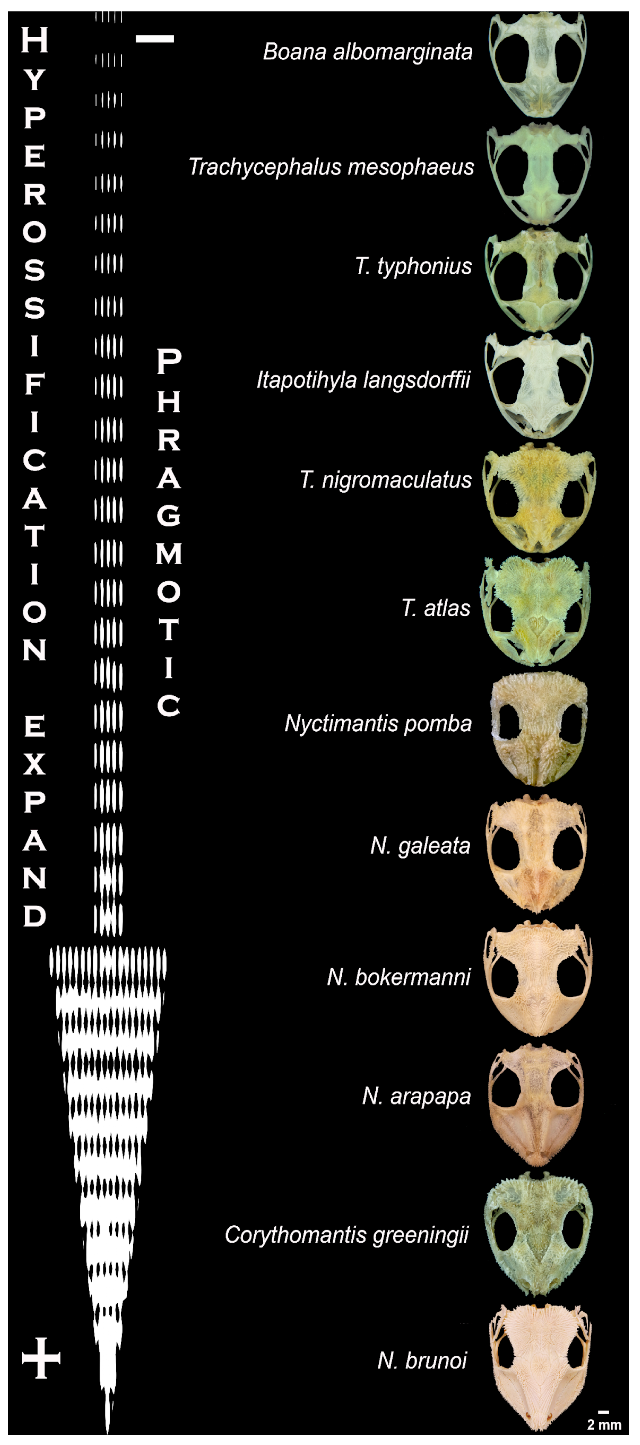
Close observation of the head in different species under a stereomicroscope revealed exposed spines piercing the skin (Figure 1A–C), with exception of Itapotihyla langsdorffii, Trachycephalus mesophaeus, Trachycephalus typhonius and Boana albomarginata. The analysis of the anatomical features of all the skulls reveals that there is a clear difference between the phragmotic frogs in terms of bone area and bone density (i.e., grades of hyperossification) (Figure 2).
The surface of the skulls showed that most of the species have spines arranged side by side (Figure 1D–F and Figure 3). In the snout and particularly in the head posterior region, where they are disposed of as a crown, the spines are more prominent (Figure 3).
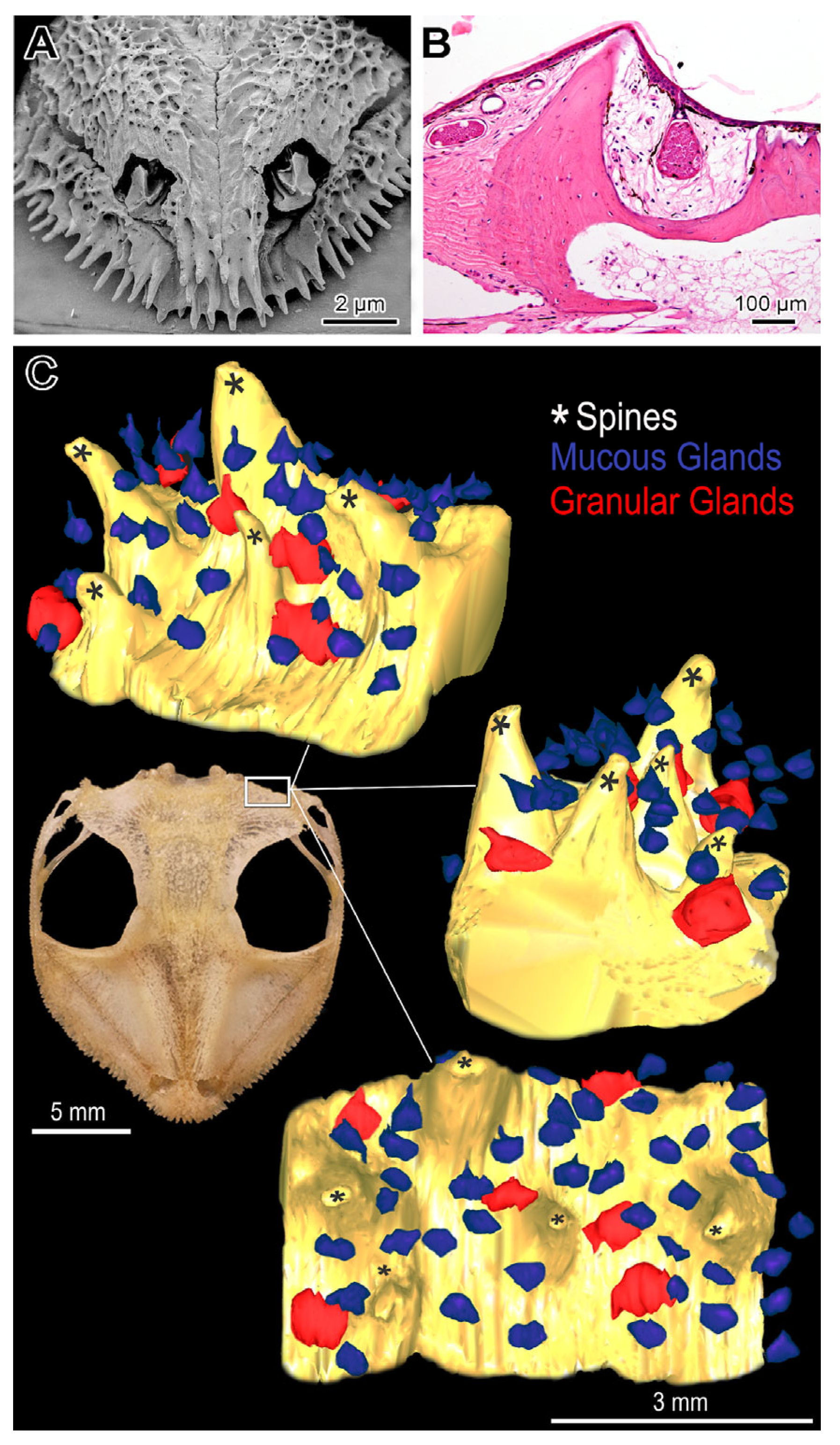
Except for Itapotihyla langsdorffii, Trachycephalus mesophaeus, Trachycephalus typhonius and Boana albomarginata, histological examination of the head demonstrated a clear association between the dermal bony spines and the cutaneous glands (Figure 4). The head of two species of Tripion analysed by histology showed similar morphological characteristics of Lophyohylini species, presenting a clear relationship between cranial ossified spines and poison glands (Figures S5 and S6).

2.3. Biological Activity and Biochemistry of the Cutaneous Poison

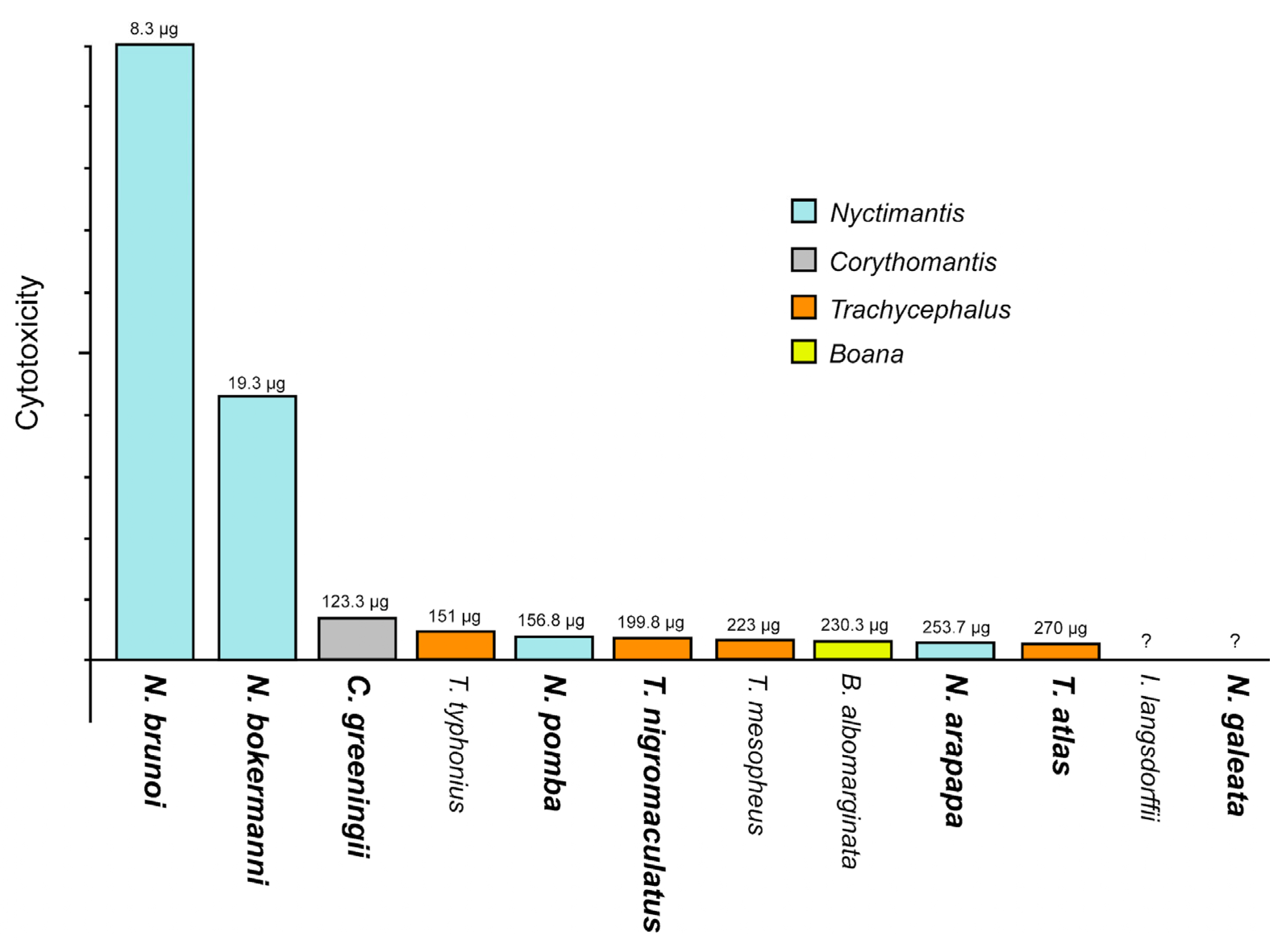
Among the cutaneous secretions of all species, Nyctimantis brunoi exhibited the highest cytotoxicity to fibroblast cells (IC50 8.34 μg/mL), corroborating previously observed in vivo lethality results [16]. The poison of N. bokermanni also demonstrated pronounced cytotoxicity, followed by Corythomantis greeningi, Trachycephalus typhonius, N. pomba, T. nigromaculatus, T. mesophaeus, Boana albomarginata, N. arapapa and T. atlas. Only the poison of Itapotihyla langsdorffii and Nyctimantis galeata did not show cytotoxic activity in all conditions tested (Figure 5).
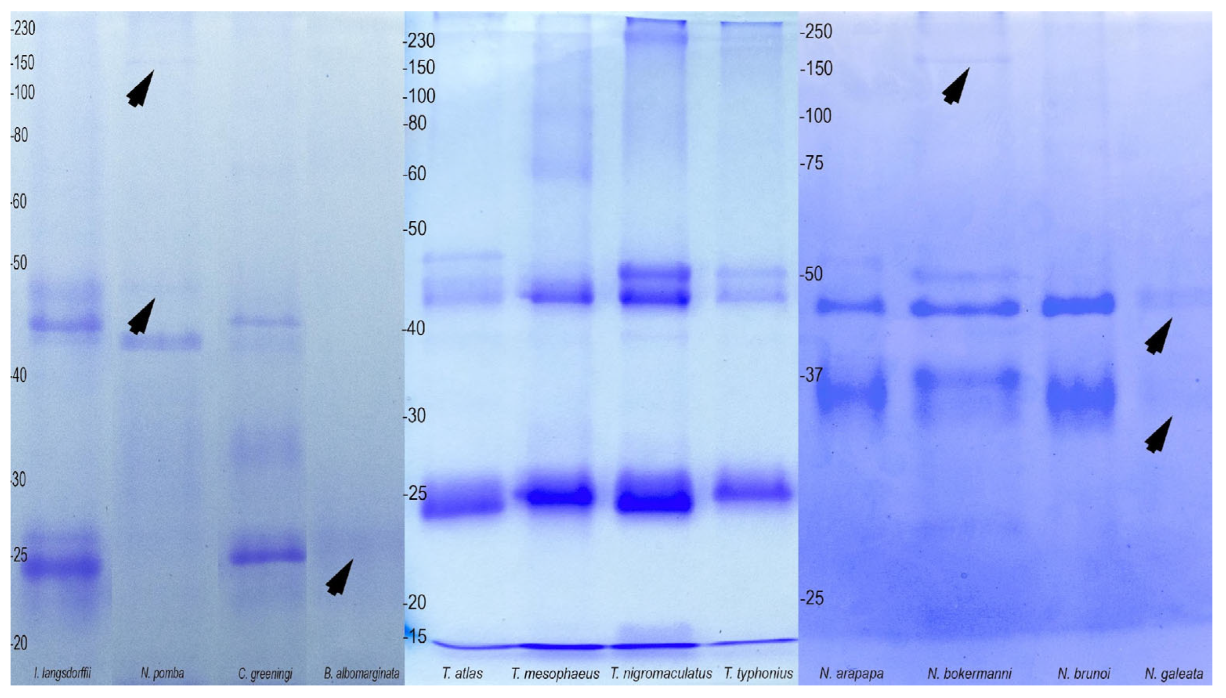
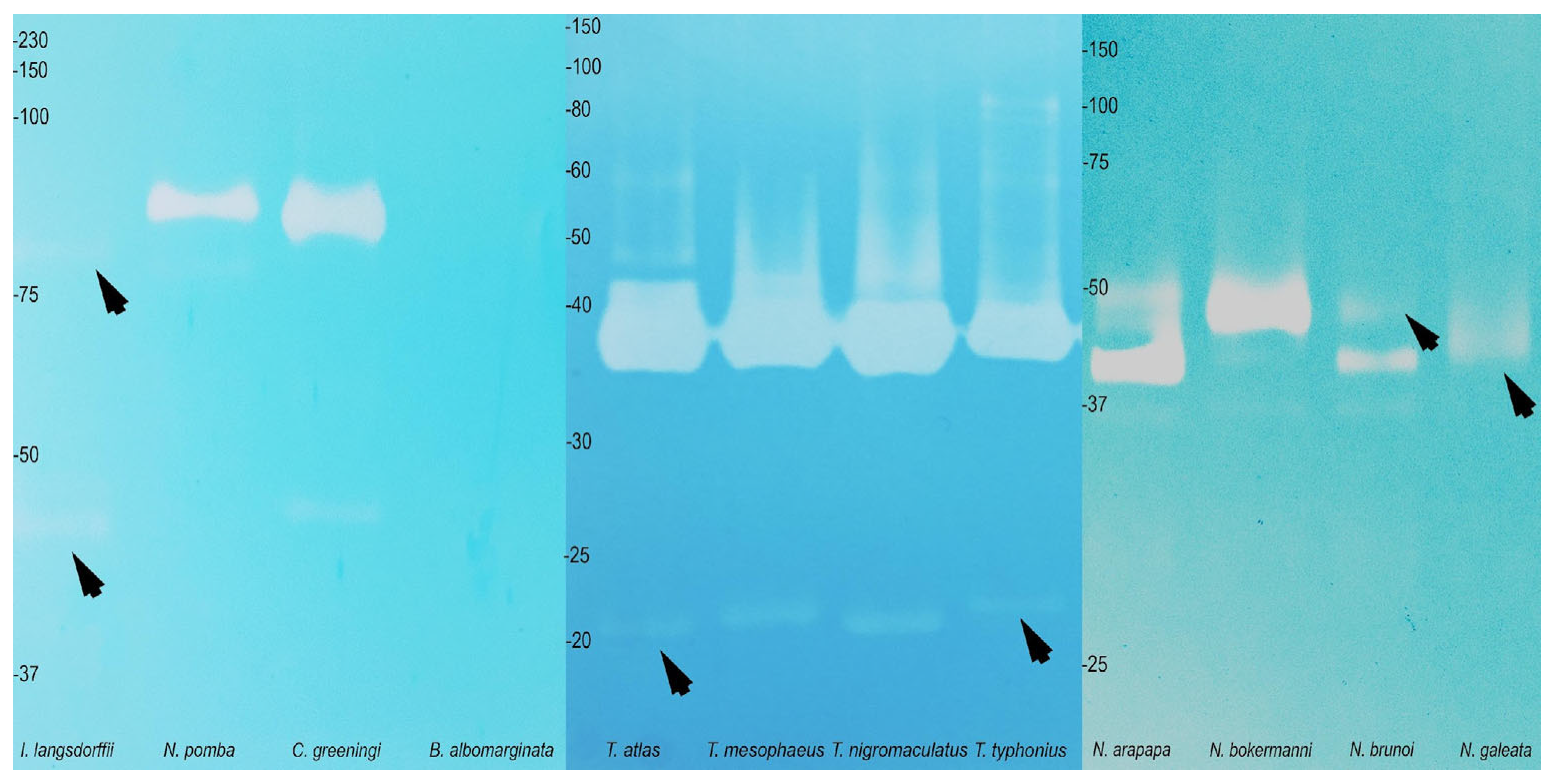
Regarding poison composition, the protein profiles in SDS-PAGE gels were similar among species within each genus. In Nyctimantis, for example, two distinct groups of bands were observed (35 kDa and 45 kDa) (Figure 6). The poison of Trachycephalus exhibited a major band around 25 kDa, along with another group of bands encompassing masses between 40 and 50 kDa (Figure 6). The profile obtained from Trachycephalus was similar to that obtained from the poisons of C. greeningi and I. langsdorffii (Figure 6). Boana albomarginata presented a poor profile, with majority bands below 27 kDa (Figure 6).
In terms of protease activity, zymographic tests performed on SDS-PAGE gels showed gelatinolytic, caseinolytic and fibrinogenolytic activities, particularly in the genus Nyctimantis (Figures S7–S9). The presence of hyaluronidases was observed in the secretions of all Lophyohylini species tested (Figure 7), regardless of the presence of cranial ossification. However, the hyaluronidase activity was particularly weak in Itapotihyla langsdorffii and Nyctimantis galeata and absent in Boana albomarginata (Figure 7).

3. Discussion

Due to the passivity of amphibians when confronted by predators, the front body, which is generally the first part of the animal to be bitten, has developed morphological adaptations strategically aimed at coping with predation. From a toxinological perspective, the development of cutaneous glands and macroglands—such as the parotoids in toads and other groups of anurans and urodeles [5,30]—is noteworthy. From a mechanical perspective, hyperossification is another important adaptation, creating a robust shield against bites. Hyperossification has evolved independently across different lineages at least 25 times [20]. In contrast, species that do not exhibit phragmosis tend to have limited cranial bone extension and less developed or absent spiny projections (Figure 3). Conversely, phragmosis seems to have initially developed as a strategy against desiccation. This strategy appears to have been successful, leading to its use in defence against predators, subsequently to development of spines.
One of the most interesting facts about antipredator defence among the Lophyohylini is the observation in many species of a combination of all these factors, where cranial ossification occurs in parallel with the presence of a significant number of poison glands, combined with phragmosis behavior. Furthermore, the presence of hyaluronidase in the poisonous secretion seems to be a well-established phenomenon within the group, unlike other anurans in which this enzyme in their skin secretion has never been detected. The evolutionary tendency of this combination of factors becomes even stronger when one observes the clear correlation between the area and density of the cranial bone covering, the presence of cranial spines and the occurrence of phragmosis behavior [20,26].
The unusual vertical head movement in some species of casque-headed tree frogs is entirely in line with the behavior of phragmosis. Such movement is possible due to special tendons and muscles in the neck region, allowing the frogs to lower their heads toward their chests (Chin-Tucking behavior) and facilitating better closure of the shelter entrance [21,31], which prevents the animal from being bothered or even captured by a predator. At the same time, phragmosis reduces moisture loss from inside the shelters [32].
Using the “chin-tucking behavior”, at least the species Corythomantis greeningi, Nyctimantis brunoi, and Trachycephalus nigromaculatus have also developed an additional movement analogous to “goring”, investing the head against the attacker [16,26,33]. This movement may favor faster poison injection, forcing the spines to pierce the glands and delivering toxins to the aggressor. The “chin-tucking” behavior is quite evident when holding a specimen in a closed hand. While collecting Corythomantis greeningi in the semiarid region of the Brazilian state of Rio Grande do Norte, one of the authors (CJ) experienced poisoning firsthand. As described in [16], during collection, which was carried out without gloves, the tiny amount of “venom” injected in the hand palm through goring was enough to cause 5 h of intense pain in the entire arm.
Nyctimantis rugiceps from Central America, as well as N. pomba, which inhabits the Brazilian Atlantic Rainforest, are also associated with phragmosis, using bamboo hollows as shelter [34,35]. N. arapapa appears to be the only Nyctimantis that has its entire life cycle restricted to bromeliads, which serve as both shelter and breeding sites [36]. The other species in this genus, such as N. bokermanni, N. brunoi, and N. galeata, reproduce directly in puddles despite using bromeliads for shelter. Moreover, during this study, we corroborated previous observations indicating that Trachycephalus with cranial ossification appears to be associated with xeric environments [37].
The behavior of blocking the entrances to holes and crevices in rocks with a highly ossified head provides the animal with significant protection against water loss. The casque-headed frog Nyctimantis brunoi demonstrated that phragmotic behavior can result in water savings of over 90% [24]. However, at least in Corythomantis greeningi, phragmosis in openings does not appear to be very effective against desiccation [32]. This effectiveness may depend on a combination of factors that greatly vary among the Lophyohylini. Considering morphological adaptations, the most significant factors likely include the density of the cranial ossification in the upper part of the head (where the dermis transforms into bone), the extent of cranial bone extension and density, and the thickness of the head skin [38,39,40].
The biochemical results are likely to reflect the adaptations of casque-headed tree frogs that have evolved in different habitats or suggest distinct biochemical specialisations related to defence against predators. In the genus Trachycephalus, for example, the toxic potential is not correlated with the presence of cranial bony spines. In calls attention to the fact that in some species, the poison is surprisingly potent. For example, compared to the venom of snakes from the genus Bothrops, which are responsible for the highest number of snakebites in Brazil, the poison of N. brunoi is 25-fold more potent [16]. Moreover, together with toxicity, the apparent exclusive presence of hyaluronidase in Lophyohylini among all anurans (and perhaps amphibians), regardless of the presence of cranial ossification, demonstrates the potential of these tree frogs to cause damage to tissues. Such characteristics may facilitate the diffusion through the predator’s body of the other toxic molecules, similar to the mechanism already known for the venom of animals with local effects, such as some snakes, lizards, arachnids, wasps, and even octopuses [41,42]. This supposition is reinforced by the finding of a weak hyaluronidase activity in the species Itapotihyla langsdorffii and Nyctimantis galeata that did not show cytotoxic activity.
Our results reaffirm the toxic potential of Lophyohylini for chemical defence, which, in the case of these anurans, could be classified, regarding the peculiar casque head, as active and endowed with a poison injection system, although more rudimentary compared to the well-known system present in venomous animals. The concurrent use of poison (throughout the body) and venom (through the head) by the casque-headed tree frogs suggests a unique attack and defence system (ADS)—composed of an extensive and robust skull with dermal bony spines and cutaneous glands secreting a cocktail of potent toxins, including hyaluronidases. Moreover, ADS is complemented by special movements of the head and phragmotic behavior. However, this system does not appear to be exclusive to this group. It is also observed in species from other groups of tree frogs, such as the Hylini of the genus Triprion (T. spatulatus, T. petasatus, and T. spinosus) from Mexico and Guatemala, which have been little studied from a toxinological perspective. Thus, it is likely that this system evolved independently in different groups of anurans. In these animals, the ADS shows a varied efficiency in envenomation, ranging from spineless species, such as Itapotihyla langsdorffii, Trachycephalus mesophaeus, Boana albomarginata and T. typhonius, to Corythomantis greeningi and Nyctimantis brunoi, which have largely ossified and spiny heads. The anterior region, represented by the snout, and the posterior region, represented by the crown, are probably the cranial areas with most significant activity within the ADS, as they have the highest concentration of spines. Among the species analysed, C. greeningi, despite being the third most lethal species, has the most effective ADS for envenomation, featuring larger and more prominent spines, along with a significant number of glands.
Although ADS is an essential factor, some species of casque-headed tree frogs that lack spines and do not perform phragmosis also exhibited cytotoxicity and hyaluronidase activity. Therefore, the presence of toxic secretions may have evolved as a characteristic of this group even before the development of spines. In any case, there is a direct association between the presence of spines and ossification in Lophyohylini, which reflects the extension and density of skull bones, characterising hyperossification. As a result, the most extensive and dense skulls are present in species with the highest phragmotic activity.

4. Conclusions

In addition to providing new data that expands our understanding of the behavior and defence mechanisms of anurans, the Lophyohylini serve as an excellent example of the chemical defence strategies employed by amphibians, indirectly illustrating the distinction between venom and poison. While some species rely on completely passive defence, allowing their poison to act through absorption via the oral mucosa at a slower rate, others have developed the ability to actively inject venom, which triggers symptoms rapidly by reaching the bloodstream. Thus, the ‘venomous’ Lophyohylini completely contradicts the typical passive defence strategies of amphibians, including their ability to utilize active injection of toxins through deliberate goring movements. The movement forces the toxins to penetrate the aggressor’s body, functioning similarly to the muscles surrounding the glands in truly venomous animals. In this context, the spines act as piercing agents, fulfilling the same role as teeth or stingers. Finally, the presence of enzymes (hyaluronidases) helps the venom to reach the predator’s tissues and bloodstream rapidly.

5. Materials and Methods

5.1. Animals and Fieldwork

For this study, observations and collections of 11 species of Lophyohylini and one of Cophomantini (Hylidae) were carried out across a significant portion of their geographic distribution, covering approximately 4000 km along the Atlantic coast and inland Brazil. The collected Lophyohylini species included Corythomantis greeningi; Itapotihyla langsdorffii; Nyctimantis arapapa; N. bokermanni; N. brunoi; N. galeata; N. pomba; Trachycephalus atlas; T. mesophaeus; T. nigromaculatus; and T. typhonius (Figure S1). In an additional and speculative manner, for better support, we also included the Cophomantini (Hylidae) Boana albomarginata (Figure S1), and two species of Hylini (Hylidae), Triprion spatulatus and Triprion spinosus. Notably, the species Corythomantis greeningi and Nyctimantis brunoi have been observed and collected by our group on expeditions since 1989. During our recent expeditions, we have successfully observed and collected little-known species considered rare, such as N. galeata [43] and N. bokermanni [44], as well as species with very restricted distributions, such as N. arapapa [31], and even species threatened with extinction, such as N. pomba [34].
Four field trips were carried out, covering the second half of 2019 to March 2022. Different Brazilian phytophysiognomies were covered according to the occurrence of each species, encompassing five states and totaling just over 30 days of work.
Once found, the animals were gently stimulated with a twig to verify basic defensive behaviors. Immediately after collecting, two to five specimens of each species were euthanised by intraperitoneal injection of thiopental (50 mg/kg) in combination with lidocaine (10 mg/mL) to preserve the morphological characteristics closest to those of animals in nature. Immediately afterwards, the specimens were preserved in a buffered paraformaldehyde solution at pH 7.2 for 72 h. After collecting the tissues of interest, the specimens were kept in 70% alcohol in the Structural Biology Laboratory.
Additional specimens were kept in the Vivarium of the Structural Biology Laboratory at the Butantan Institute in 45 L plastic boxes containing dry branches as perches, plastic or rubber tubes for shelter, and water that was frequently replenish. The animals were fed twice a week with varying amounts of crickets (Gryllus assimilis) and cockroaches (Nauphoeta cinerea).
The study was conducted with prior approval from Brazilian Chico Mendes Institute for Biodiversity Conservation (ICMBio-SISBIO-65237-7). All experimental procedures were performed in accordance with the relevant international guidelines and regulations and were approved by the Ethics Committee on Animal Use of Instituto Butantan, CEUA 8465270919 (ID 001887), on 16 October 2019.

5.2. Cranial Anatomy

Two individuals of each species (except Nictymantis pomba) were used to analyse cranial bone anatomy. Soft tissues were extracted using immersion in hydrogen peroxide (40 vol) in addition to sodium hypochlorite (3%) [45]. The corrosion time varied according to the needs of each sample, alternating immersion in the solutions and manual material removal. Cranial structures were identified and named according to classical taxonomy [46]. The term “cranial bone extension” adopted in this paper refers to the area and the bone volume occupied by the skull. The skull is commonly called “hyperossified” when composed of very voluminous bones [20]. The term “bony projections” refers to rounded or pointed protrusions or spines that emerge from the surface of the skull.

5.3. Histology

In all species analysed (except Nictymantis pomba), we histologically studied the posterior region of the skull, here called the “crown”, and the anterior region of the animals’ heads, the “snout”. We verified intraspecific variations and investigated the possible existence of patterns between the genera and aspects of Natural History. Fragments of the tegument in the dorsal region were also evaluated for comparisons between the skull and the back. Cranial decalcification was performed using a 4% EDTA solution for 30 to 65 days ([47], adapted). All samples were dehydrated in ethanol crescent series and embedded in plastic resin (Historesin, Leica Microsystems Nussloch GmbH, Heidelberg, Germany) or paraffin. Sections were obtained using a Leica 2255 microtome (Leica Microsystems, Nussloch GmbH, Heidelberg, Germany), with variable thickness between 0.5 and 6 μm.
In addition to the species collected during fieldwork, we examined the tree frogs Triprion spatulatus and T. spinosus, from Central America (provided by Prof. Edmund D. Brodie Jr. and Prof. Jonathan M. Campbell), which also have a spiny skull even though they do not belong to the Lophyohylini tribe. Cranial bone images of the two species come from the Morpho Source website, with T. spatulatus under ID B9848D79-6233-4503-8832-EB35AF95D9999 and T. spinosus under ID 87602/m4/M11850.
The histological sections were subjected to different staining techniques, using hematoxylin-eosin and toluidine blue-fuchsin to analyse basic morphology in paraffin and resin, respectively. To better understand the composition of the secretion of the different types of glands, different histochemical tests were performed: PAS—periodic acid + Schiff, to reveal sites rich in neutral polysaccharide; alcian blue pH 2.5, to reveal sites rich in acidic polysaccharides; bromophenol blue, to reveal sites rich in proteins; and von Kossa, to reveal sites rich in calcium. The sections were photographed on an Olympus BX51 microscope coupled to an Olympus DP 73 digital camera, and the images were captured using CellSens Standard 1.16 software. To perform the three-dimensional reconstruction of the Attack and Defence System (ADS), a fragment of the periphery of the frontoparietal bone of Nyctimantis arapapa, which has a very extensive and hyperossified cranial region, was used. The reconstruction was performed from 119 serial sections using Reconstruct 1.1.0.0 software (copyright © 1996-2007, John C. Fiala) [48], resulting in a reconstruction equivalent to 5.95 mm2 of the region.

5.4. Collection of Cutaneous Secretion

The species’ secretions were collected by immersing and massaging the specimens in deionised water or PBS 7.4 inside a sterile plastic bag [49]. This method causes mechanical stimulation of the cutaneous glands by gently compressing of different regions of the animals’ bodies.
After extraction, the material was filtered through a “Cell-Strainer 40 μm” filter and separated into aliquots. The protein concentration of the poison was determined using the “NanoVue Plus Spectrophotometer GE Healthcare” optical density reader. To minimize the chance of loss of poison activity, cytotoxicity and zymography experiments were performed immediately after collection, with other aliquots kept in the refrigerator and others in a freezer at −20 °C.

5.5. Cytotoxic Activity

Cytotoxicity tests were performed using a cell line of normal mouse fibroblasts (L929) maintained and tested in culture. The Effective Concentration (EC50) was calculated, which refers to the concentration that kills 50% of a cell population. The cutaneous secretion of the frogs for this test was extracted directly into PBS 7.4.
The cells, kept in liquid nitrogen, underwent a thawing process and were placed in 25 cm2 cell culture bottles containing RPMI-1640 medium supplemented with 10% fetal bovine serum, 2 mM glutamine, 10,000 U/mL penicillin and 10,000 μg/mL streptomycin. The cell line was kept in an incubator at 37 °C, with a humid atmosphere and 5% CO2, until confluence.
For the treatment of cell lines with crude cutaneous secretions from tree frogs, L929 cells were plated at a concentration of 3 × 10⁵ cells/mL (3 × 10⁴ cells/well) of complete RPMI-1640 medium in 96-well cell culture plates. After cell adhesion, the fractions were serially diluted at a ratio of 2 with RPMI-1640 medium and distributed in quadruplicate on the culture plate. The assays were performed with treatments of 6, 24 and 48 h of incubation at 37 °C in a humidified atmosphere with 5% CO2, with each sample of fresh venom, and were repeated three times. As a negative control, wells with cell cultures free from treatment with cutaneous secretion were maintained. After treatment, cell viability was assessed using the MTT assay [3-(4,5-dimethy-2-lthiazolyl)-2,5-diphenyl-2H-tetrazolium bromide [50]. Readings were performed in an ELISA reader (Multiskan EX®) at a wavelength of 540 nm.

5.6. Polyacrylamide Gel Electrophoresis in the Presence of Sodium Dodecyl Sulfate (SDS-PAGE)

To investigate the diversity of proteins in the secretions of the species studied, samples containing 8 μg of proteins were diluted in 15 μL of sample buffer and applied to a 15% polyacrylamide gel (PAGE) containing sodium dodecyl sulphate (SDS) under non-reducing conditions [51]. After the proteins were separated by electrophoresis, the gels were stained with Coomassie Brilliant Blue.

5.7. Zymography

The presence of proteases in the secretions was evaluated by the zymography electrophoresis technique [52], using casein, gelatin, hyaluronic acid and fibrinogen as substrates in 15% SDS-PAGE gels. After the end of electrophoresis, the gels containing casein, gelatin and fibrinogen were processed and stained with Coomassie brilliant blue. Alcian blue was used as dye only for the hyaluronic acid gel. After staining, the gels were immersed in decolorising solutions. As a result, clear areas in the gel indicate the degradation of the substrate used and, therefore, the occurrence of enzymatic activity at a given molecular mass.

Supplementary Materials

The following supporting information can be downloaded at: https://www.mdpi.com/article/10.3390/toxins17060303/s1. Figure S1: Species studied; Figure S2: Nyctimantis brunoi in phragmosis in a bromeliad; Figure S3: Corythomantis greeningi in a rocky crevice during the reproductive station; Figure S4: Corythomantis greeningi lips; Figure S5: Toxin injection system in Triprion spatulatus; Figure S6: Toxin injection system in Triprion spinosus; Figure S7. Gelatinolytic profile in polyacrylamide gel (SDS-PAGE 15%) obtained from the skin secretion of the studied species; Figure S8. Caseinolytic profile in polyacrylamide gel (SDS-PAGE 15%) obtained from the skin secretion of the studied species; Figure S9. Fibronogeniolytic profile in polyacrylamide gel (SDS-PAGE 15%) obtained from the skin secretion of the studied species; Video S1: Defensive behavior associated to phragmosis in Nyctimantis brunoi; Video S2: Defensive behavior associated to phragmosis in Trachycephalus nigromaculatus. References [53,54,55,56,57,58,59,60,61,62,63,64,65,66,67,68,69,70,71,72,73,74,75,76] are cited in the Supplementary Materials.

Author Contributions

Conceptualisation: C.A., P.L.M.-F., M.M.A. and C.J.; methodology: C.A., P.L.M.-F., B.C.L.F.T., M.M.A. and C.J.; investigation: C.A., P.L.M.-F., B.C.L.F.T., M.M.A. and C.J.; visualisation: C.A., P.L.M.-F., B.C.L.F.T., M.M.A. and C.J.; funding acquisition: C.A., M.M.A. and C.J.; project administration: C.A., P.L.M.-F., M.M.A. and C.J.; supervision: C.A., P.L.M.-F., B.C.L.F.T., M.M.A. and C.J.; writing—original draft: C.A., P.L.M.-F., B.C.L.F.T., M.M.A. and C.J.; writing—review & editing: C.A., P.L.M.-F., B.C.L.F.T., M.M.A. and C.J. All authors have read and agreed to the published version of the manuscript.

Funding

This research was funded by the National Council for Scientific and Technological Development (CNPq) #305266/2019-5 (C.J.) and #311357/2020-2 (M.M.A.). The Coordination of Superior Level Staff Improvement (CAPES) financial code 001 also founded this work (C.A.).

Institutional Review Board Statement

The study was conducted with prior approval from Chico Mendes Institute for Biodiversity Conservation (ICMBio-SISBIO-65237-7), and Ethics Committee on Animal Use of the Butantan Institute (CEUAIB-8465270919, 16 October 2019).

Informed Consent Statement

Not applicable.

Data Availability Statement

The original contributions presented in this study are included in the article/Supplementary Materials. Further inquiries can be directed to the corresponding author.

Acknowledgments

This paper is dedicated to the late Professor Werner Bokermann (Parque Zoológico de São Paulo, Brazil), who, in the mid-1980s, first introduced us in the field of the casque-headed tree frogs. The authors thank all members of the Laboratory of Structural Biology at the Instituto Butantan for their competent technical assistance. Our gratitude to Cauê Monticelli from Centro de Conservação de Fauna Silvestre do Estado de São Paulo (CECFAU), who made it possible to analyze the skull and the collect skin secretions from Nyctimantis pomba. We also thank to Butantan Foundation for the technical and logistical support.

Conflicts of Interest

The authors declare no conflicts of interest.

Abbreviations

The following abbreviations are used in this manuscript:
ADSAttack and Defence System
SDS-PAGE JPolyacrylamide gel electrophoresis in the presence of sodium dodecyl sulfate

References

  1. Arbuckle, K. Evolutionary Context of Venom in Animals. Evol. Venom. Anim. Their Toxins 2015, 24, 3–31. [Google Scholar] [CrossRef]
  2. Schendel, V.; Rash, L.D.; Jenner, R.A.; Undheim, E.A.B. The Diversity of Venom: The Importance of Behavior and Venom System Morphology in Understanding Its Ecology and Evolution. Toxins 2019, 11, 666. [Google Scholar] [CrossRef] [PubMed]
  3. Jared, C.; Luiz Mailho-Fontana, P.; Maria Antoniazzi, M. Differences between Poison and Venom: An Attempt at an Integrative Biological Approach. Acta Zool. 2021, 102, 337–350. [Google Scholar] [CrossRef]
  4. Toledo, R.C.; Jared, C. Cutaneous Granular Glands and Amphibian Venoms. Comp. Biochem. Physiol. Part A Physiol. 1995, 111, 1–29. [Google Scholar] [CrossRef]
  5. Jared, C.; Antoniazzi, M.M.; Jordão, A.E.C.; Silva, J.R.M.C.; Greven, H.; Rodrigues, M.T. Parotoid Macroglands in Toad (Rhinella jimi): Their Structure and Functioning in Passive Defence. Toxicon 2009, 54, 197–207. [Google Scholar] [CrossRef]
  6. Mailho-Fontana, P.L.; Antoniazzi, M.M.; Sciani, J.M.; Pimenta, D.C.; Barbaro, K.C.; Jared, C. Morphological and Biochemical Characterization of the Cutaneous Poison Glands in Toads (Rhinella marina Group) from Different Environments. Front. Zool. 2018, 15, 46. [Google Scholar] [CrossRef]
  7. Daly, J.W. The Chemistry of Poisons in Amphibian Skin. Proc. Natl. Acad. Sci. USA 1995, 92, 9–13. [Google Scholar] [CrossRef]
  8. Ferreira, R.B.; Lourenço-de-Moraes, R.; Zocca, C.; Duca, C.; Beard, K.H.; Brodie, E.D. Antipredator Mechanisms of Post-Metamorphic Anurans: A Global Database and Classification System. Behav. Ecol. Sociobiol. 2019, 73, 69. [Google Scholar] [CrossRef]
  9. Regis-Alves, E.; Jared, S.G.S.; Maurício, B.; Mailho-Fontana, P.L.; Antoniazzi, M.M.; Fleury-Curado, M.C.; Brodie, E.D.; Jared, C. Structural Cutaneous Adaptations for Defense in Toad (Rhinella icterica) Parotoid Macroglands. Toxicon 2017, 137, 128–134. [Google Scholar] [CrossRef]
  10. Albuquerque-Pinna, J.; Jeckel, A.M.; Nakamura, D.Y.M.; Bernarde, P.S.; Kocheff, S.; Saporito, R.A.; Grant, T. Defensive Alkaloid Variation and Palatability in Sympatric Poison Frogs. Chemoecology 2024, 34, 83–94. [Google Scholar] [CrossRef]
  11. Smith, K.E.; Halpin, C.G.; Rowe, C. Body Size Matters for Aposematic Prey during Predator Aversion Learning. Behav. Process. 2014, 109, 173–179. [Google Scholar] [CrossRef] [PubMed]
  12. Nelsen, D.R.; Nisani, Z.; Cooper, A.M.; Fox, G.A.; Gren, E.C.K.; Corbit, A.G.; Hayes, W.K. Poisons, Toxungens, and Venoms: Redefining and Classifying Toxic Biological Secretions and the Organisms That Employ Them. Biol. Rev. 2014, 89, 450–465. [Google Scholar] [CrossRef] [PubMed]
  13. Hayes, W.K.; Gren, E.C.K.; Nelsen, D.R.; Corbit, A.G.; Cooper, A.M.; Fox, G.A.; Streit, M.B. It’s a Small World After All: The Remarkable but Overlooked Diversity of Venomous Organisms, with Candidates Among Plants, Fungi, Protists, Bacteria, and Viruses. Toxins 2025, 17, 99. [Google Scholar] [CrossRef]
  14. Brodie, E.D.B., Jr.; Nussbaum, R.A.; Digiovanni, M. Antipredator Adaptations of Asian Salamanders (Salamandridae). Herpetologica 1984, 40, 56–68. [Google Scholar]
  15. Heiss, E.; Natchev, N.; Salaberger, D.; Gumpenberger, M.; Rabanser, A.; Weisgram, J. Hurt Yourself to Hurt Your Enemy: New Insights on the Function of the Bizarre Antipredator Mechanism in the Salamandrid Pleurodeles waltl. J. Zool. 2009, 280, 156–162. [Google Scholar] [CrossRef]
  16. Jared, C.; Mailho-Fontana, P.L.; Antoniazzi, M.M.; Mendes, V.A.; Barbaro, K.C.; Rodrigues, M.T.; Brodie, E.D. Venomous Frogs Use Heads as Weapons. Curr. Biol. 2015, 25, 2166–2170. [Google Scholar] [CrossRef]
  17. Mailho-Fontana, P.L.; Antoniazzi, M.M.; Alexandre, C.; Pimenta, D.C.; Sciani, J.M.; Brodie, E.D.; Jared, C. Morphological Evidence for an Oral Venom System in Caecilian Amphibians. iScience 2020, 23, 101234. [Google Scholar] [CrossRef]
  18. Cajade, R.; Hermida, G.; Piñeiro, J.M.; Regueira, E.; Alcalde, L.; Fusco, L.S.; Marangoni, F. Multiple Anti-Predator Mechanisms in the Red-Spotted Argentina Frog (Amphibia: Hylidae). J. Zool. 2017, 302, 94–107. [Google Scholar] [CrossRef]
  19. Blotto, B.L.; Lyra, M.L.; Cardoso, M.C.S.; Trefaut Rodrigues, M.; Dias, I.R.; Marciano, E., Jr.; Dal Vechio, F.; Orrico, V.G.D.; Brandão, R.A.; Lopes de Assis, C.; et al. The Phylogeny of the Casque-Headed Treefrogs (Hylidae: Hylinae: Lophyohylini). Cladistics 2021, 37, 36–72. [Google Scholar] [CrossRef]
  20. Paluh, D.J.; Stanley, E.L.; Blackburn, D.C. Evolution of Hyperossification Expands Skull Diversity in Frogs. Proc. Natl. Acad. Sci. USA 2020, 117, 8554–8562. [Google Scholar] [CrossRef]
  21. Trueb, L. Evolutionary Relationships of Casque-Headed Tree Frogs with Co-Ossified Skulls (Family Hylidae). Univ. Kans. Publ. Mus. Nat. Hist. 1970, 18, 547–716. [Google Scholar] [CrossRef]
  22. Wheeler, W.M. The Physiognomy of Insects. Q. Rev. Biol. 1927, 2, 1–36. [Google Scholar] [CrossRef]
  23. Ebel, R.; Herrel, A.; Scheyer, T.M.; Keogh, J.S. Review of Osteoderm Function and Future Research Directions. J. Zool. 2024, 325, 1–24. [Google Scholar] [CrossRef]
  24. De Andrade, D.V.; Abe, A.S. Evaporative Water Loss and Oxygen Uptake in Two Casque-Headed Tree Frogs, Aparasphenodon brunoi and Corythomantis greeningi (Anura, Hylidae). Comp. Biochem. Physiol. Part A Physiol. 1997, 118, 685–689. [Google Scholar] [CrossRef] [PubMed]
  25. Frost, D.R. Amphibian Species of the World: An Online Reference, version 6.2; American Museum of Natural History: New York, NY, USA, 2024. [Google Scholar] [CrossRef]
  26. Jared, C.; Antoniazzi, M.M.; Navas, C.A.; Katchburian, E.; Freymüller, E.; Tambourgi, D.V.; Rodrigues, M.T. Head Co-Ossification, Phragmosis and Defence in the Casque-Headed Tree Frog Corythomantis greeningi. J. Zool. 2005, 265, 1–8. [Google Scholar] [CrossRef]
  27. Neto, A.M.; Teixeira, M., Jr. Checklist of the Genus Aparasphenodon Miranda-Ribeiro, 1920 (Anura: Hylidae): Distribution Map, and New Record from São Paulo State, Brazil. Check List 2012, 8, 1303–1307. [Google Scholar] [CrossRef]
  28. Ferro, J.M.; Cardozo, D.E.; Suárez, P.; Boeris, J.M.; Blasco-Zúñiga, A.; Barbero, G.; Gomes, A.; Gazoni, T.; Costa, W.; Nagamachi, C.Y.; et al. Chromosome Evolution in Cophomantini (Amphibia, Anura, Hylinae). PLoS ONE 2018, 13, e0192861. [Google Scholar] [CrossRef]
  29. Faivovich, J.; Pereyra, M.O.; Luna, M.C.; Hertz, A.; Blotto, B.L.; Vásquez-Almazán, C.R.; McCranie, J.R.; Sánchez, D.A.; Baêta, D.; Araujo-Vieira, K.; et al. On the Monophyly and Relationships of Several Genera of Hylini (Anura: Hylidae: Hylinae), with Comments on Recent Taxonomic Changes in Hylids. South Am. J. Herpetol. 2018, 13, 1–32. [Google Scholar] [CrossRef]
  30. Licht, L.E.; Sever, D.M. Structure and Development of the Parotoid Gland in Metamorphosed and Neotenic Ambystoma gracile. Copeia 1993, 1, 116–123. [Google Scholar] [CrossRef]
  31. Pimenta, B.V.S.; Napoli, M.F.; Haddad, C.F.B. A New Species of Casque-Headed Tree Frog, Genus Aparasphenodon Miranda-Ribeiro (Amphibia: Anura: Hylidae), from the Atlantic Rainforest of Southern Bahia, Brazil. Zootaxa 2009, 2123, 46–54. [Google Scholar] [CrossRef]
  32. Jared, C.; Antoniazzi, M.M.; Katchburian, E.; Toledo, R.C.; Freymüller, E. Some Aspects of the Natural History of the Casque-Headed Tree Frog Corythomantis greeningi Boulenger (Hylidae). Ann. Sci. Nat. 1999, 20, 105–115. [Google Scholar] [CrossRef]
  33. Toledo, L.F.; Sazima, I.; Haddad, C.F.B. Behavioural Defences of Anurans: An Overview. Ethol. Ecol. Evol. 2011, 23, 1–25. [Google Scholar] [CrossRef]
  34. Assis, C.D.; Santana, D.J.; Da Silva, F.A.; Quintela, F.M.; Feio, R.N. A New and Possibly Critically Endangered Species of Casque-Headed Tree Frog Aparasphenodon Miranda-Ribeiro, 1920 (Anura, Hylidae) from Southeastern Brazil. Zootaxa 2013, 3716, 583–591. [Google Scholar] [CrossRef]
  35. Duellman, W.E.; Trueb, L. The Systematic Status and Relationships of the Hylid Frog Nyctimantis rugiceps Boulenger. Occas. Pap. Mus. Nat. Hist. Univ. Kans. 1976, 58, 1–14. [Google Scholar]
  36. Lantyer-Silva, A.S.F.; Solé, M.; Zina, J. Reproductive Biology of a Bromeligenous Frog Endemic to the Atlantic Forest: Aparasphenodon arapapa Pimenta, Napoli and Haddad, 2009 (Anura: Hylidae). An. Acad. Bras. Cienc. 2014, 86, 867–880. [Google Scholar] [CrossRef]
  37. Cisneros-Heredia, D.F. Notes on the Natural History of the Casque-Headed Treefrog Trachycephalus jordani (Stejneger & Test, 1891). Herpetozoa 2007, 20, 92–94. [Google Scholar]
  38. Katchburian, E.; Antoniazzi, M.M.; Jared, C.; Faria, F.P.; Souza Santos, H.; Freymüller, E. Mineralized Dermal Layer of the Brazilian Tree-Frog Corythomantis greeningi. J. Morphol. 2001, 248, 56–63. [Google Scholar] [CrossRef] [PubMed]
  39. Navas, C.A.; Jared, C.; Antoniazzi, M.M. Water Economy in the Casque-Headed Tree-Frog Corythomantis greeningi (Hylidae): Role of Behaviour, Skin, and Skull Skin Co-Ossification. J. Zool. 2002, 257, 525–532. [Google Scholar] [CrossRef]
  40. Toledo, R.C.; Jared, C. Cutaneous Adaptations to Water Amphibians Balance. Comp. Biochem. Pysiol. 1993, 105, 593–608. [Google Scholar] [CrossRef]
  41. Bala, E.; Hazarika, R.; Singh, P.; Yasir, M.; Shrivastava, R. A Biological Overview of Hyaluronidase: A Venom Enzyme and Its Inhibition with Plants Materials. Mater. Today Proc. 2018, 5, 6406–6412. [Google Scholar] [CrossRef]
  42. Bordon, K.C.F.; Wiezel, G.A.; Amorim, F.G.; Arantes, E.C. Arthropod Venom Hyaluronidases: Biochemical Properties and Potential Applications in Medicine and Biotechnology. J. Venom. Anim. Toxins Incl. Trop. Dis. 2015, 21, 43. [Google Scholar] [CrossRef]
  43. Dias, I.R.; Silva, G.T.; Solé, M.; de Mira-Mendes, C.V. The Advertisement Call of the Rare Casque-Headed Frog Nyctimantis galeata (Anura: Hylidae) from Its Type Locality, Morro Do Chapéu, Bahia, Brazil. Zootaxa 2020, 4853, 447–450. [Google Scholar] [CrossRef]
  44. Zanette-Silva, L.; Lemos Farias, D.; Ghizoni, I.R. New Records of Aparasphenodon bokermanni (Pombal, 1993) from Santa Catarina, Southern Brazil, and Extension of Genus Range (Anura: Hylidae). Check List 2016, 12, 1–4. [Google Scholar] [CrossRef]
  45. Aggarwal, N.; Gupta, M.; Goyal, P.K.; Kaur, J. An Alternative Approach to Bone Cleaning Methods for Anatomical Purposes. Int. J. Anat. Res. 2016, 4, 2216–2221. [Google Scholar] [CrossRef]
  46. Prado, G.M.; Pombal, J.P., Jr. Espécies de Proceratophrys Miranda-Ribeiro, 1920 com apêndices palpebrais (Anura; Cycloramphidae). Arq. Zool. 2008, 39, 1–85. [Google Scholar] [CrossRef]
  47. Cho, A.; Suzuki, S.; Hatakeyama, J.; Haruyama, N.; Kulkarni, A.B. A Method for Rapid Demineralization of Teeth and Bones. Open Dent. J. 2010, 4, 223–229. [Google Scholar] [CrossRef] [PubMed]
  48. Fiala, J.C. Reconstruct: A Free Editor for Serial Section Microscopy. J. Microsc. 2005, 218, 52–61. [Google Scholar] [CrossRef]
  49. Conceição, K.; Konno, K.; Richardson, M.; Antoniazzi, M.M.; Jared, C.; Daffre, S.; Camargo, A.C.M.; Pimenta, D.C. Isolation and Biochemical Characterization of Peptides Presenting Antimicrobial Activity from the Skin of Phyllomedusa hypochondrialis. Peptides 2006, 27, 3092–3099. [Google Scholar] [CrossRef]
  50. Mossmann, T. Rapid Colorimetric Assay for Cellular Growth and Survival: Application to Proliferation and Cytotoxicity Assays. J. Immunol. Methods 1983, 65, 55–63. [Google Scholar] [CrossRef]
  51. Laemmli, U. Cleavage of Structural Proteins during the Assembly of the Head of Bacteriophage T4. Nature 1970, 227, 680–685. [Google Scholar] [CrossRef]
  52. Heussen, C.; Dowdle, E.B. Electrophoretic Analysis of Plasminogen Activators in Polyacrylamide Gels Containing Sodium Dodecyl Sulfate and Copolymerized Substrates. Anal. Biochem. 1980, 102, 196–202. [Google Scholar] [CrossRef] [PubMed]
  53. Valdujo, P.H.; Camacho, A.; Recoder, R.S.; Teixeira Junior, M.; Ghellere, J.M.B.; Mott, T.; Nunes, P.M.S.; Nogueira, C.; Rodrigues, M.T. Anfíbios Da Estação Ecológica Serra Geral Do Tocantins, Região Do Jalapão, Estados Do Tocantins e Bahia. Biota Neotrop. 2011, 11, 251–261. [Google Scholar] [CrossRef]
  54. Godinho, L.B.; Moura, M.R.; Feio, R.N. New Records and Geographic Distribution of Corythomantis greeningi Boulenger, 1896 (Amphibia: Hylidae). Check List 2013, 9, 148–150. [Google Scholar] [CrossRef]
  55. de Freitas, M.A.; Abegg, A.D.; Dias, I.R.; de Figueiredo Moraes, E.P. Herpetofauna from Serra Da Jibóia, an Atlantic Rainforest Remnant in the State of Bahia, Northeastern Brazil. Herpetol. Notes 2018, 11, 59–72. [Google Scholar]
  56. Arzabe, C.; Loebmann, D. Amphibia, Hylidae, Itapotihyla langsdorffii: Distribution Extension. Check List 2006, 2, 33–34. [Google Scholar] [CrossRef]
  57. Lingnau, R.; Zank, C.; Colombo, P.; Vinciprova, G. Amphibia, Hylidae, Itapotihyla langsdorffii: Distribution Extension. Check List 2006, 2, 38–39. [Google Scholar] [CrossRef]
  58. Airaldi-Wood, K.; Molas, L.P.; González-Soria, L. First Record of Itapotihyla langsdorffii for Caaguazu Department (Paraguay) and Observations on Another Endemic Species of The Atlantic Forest. Kempffiana 2021, 17, 22–29. [Google Scholar]
  59. Lourenço-de-Moraes, R.; Lantyer-Silva, A.S.F.; Toledo, L.F.; Solé, M. Tadpole, Oophagy, Advertisement Call, and Geographic Distribution of Aparasphenodon arapapa Pimenta, Napoli and Haddad 2009 (Anura, Hylidae). J. Herpetol. 2013, 47, 575–579. [Google Scholar] [CrossRef]
  60. Pombal, J.P. New Species of Aparasphenodon (Anura: Hylidae) from Southeastern Brazil. Copeia 1993, 1993, 1088–1091. [Google Scholar] [CrossRef]
  61. Vilela, V.M.d.F.N.; Brassaloti, R.A.; Bertoluci, J. Anurofauna Da Floresta de Restinga Do Parque Estadual Da Ilha Do Cardoso, Sudeste Do Brasil: Composição de Espécies e Uso de Sítios Reprodutivos. Biota Neotrop. 2011, 11, 83–93. [Google Scholar] [CrossRef]
  62. Ruas, D.S.; Mendes, C.V.d.M.; Del-Grande, M.L.; Solé, M. Aparasphenodon brunoi Miranda-Ribeiro, 1920 (Anura: Hylidae): Distribution Extension and Geographic Distribution Map for Bahia State, Brazil. Check List 2013, 9, 858–859. [Google Scholar] [CrossRef]
  63. Pombal, J.P.; Menezes, V.A.; Fontes, A.F.; Nunes, I.; Rocha, C.F.D.; van Sluys, M. A Second Species of the Casque-Headed Frog Genus Corythomantis (Anura: Hylidae) from Northeastern Brazil, the Distribution of C. greeningi, and Comments on the Genus. Bol. Mus. Nac. 2012, 530, 1–14. [Google Scholar]
  64. Magalhães, F.D.M.; Marques, R.; Dos Santos, D.F.; De Magalhães, R.F.; Pezzuti, T.L. Tadpole of Nyctimantis galeata (Anura: Hylidae: Lophyohylini), a Narrow Endemic Casque-Headed Frog from Bahia, Brazil. Zootaxa 2021, 4948, 295–300. [Google Scholar] [CrossRef]
  65. Assis, C.; Feio, R.N. Aparasphenodon pomba. MG Biota 2017, 10, 49–52. [Google Scholar]
  66. Bokermann, W.C.A. Una Nueva Especie de Trachycephalus de Bahia, Brasil (Amphibia, Hylidae). Neotropica 1966, 12, 120–124. [Google Scholar]
  67. Magalhães, A.J.C., Jr.; Barbosa, A.R.; de Azevedo, S.M., Jr.; Pinho, M.S.; Beltrão, M.G.; Alves, B.C.F. Amphibia, Anura, Hylidae, Trachycephalus atlas Bokermann, 1966: Novo Registro e Comentários Sobre a Distribuição Geográfica. Rev. Nord. Zool. 2011, 5, 53–57. [Google Scholar]
  68. Roberto, I.J.; Ribeiro, S.C.; Bezerra, L.; Carneiro, P.B.M. Amphibia, Anura, Hylidae, Trachycephalus atlas Bokermann, 1966: Distribution Extension and Geographic Distribution Map. Check List 2011, 7, 326–327. [Google Scholar] [CrossRef]
  69. Santana, D.; Almeida, R.P.S.; Caldas, F.L.S.; Dias, E.J.D.R.; Faria, R. New Records of Trachycephalus mesophaeus (Hensel, 1867) (Anura: Hylidae) from Atlantic Forest in Sergipe State, Brazil. Herpetol. Notes 2016, 9, 255–260. [Google Scholar]
  70. Gondim-Silva, F.A.T.; Andrade, A.R.S.; Abreu, R.O.; Nascimento, J.S.; Corrêa, G.P.; Menezes, L.; Trevisan, C.C.; Camargo, S.S.; Napoli, M.F. Composition and Diversity of Anurans in the Restinga of the Conde Municipality, Northern Coast of the State of Bahia, Northeastern Brazil. Biota Neotrop. 2016, 16, e20160157. [Google Scholar] [CrossRef]
  71. Dias, I.R.; Vilaça, T.R.A.; Silva, J.R.d.S.; Barbosa, R.S.; Solé, M. Amphibia, Anura, Hylidae, Trachycephalus nigromaculatus Tschudi, 1838: Distribution Extension. Check List 2010, 6, 412–413. [Google Scholar] [CrossRef]
  72. Soares, M.D.L.; Iop, S.; Santos, T.G. dos Expansion of the Geographical Distribution of Trachycephalus typhonius (Linnaeus, 1758) (Anura: Hylidae): First Record for the State of Rio Grande Do Sul, Brazil. Check List 2012, 8, 817–818. [Google Scholar] [CrossRef]
  73. Miranda, D.B.; Venâncio, N.M.; de Albuquerque, S. Rapid Survey of the Herpetofauna in an Area of Forest Management in Eastern Acre, Brazil. Check List 2014, 10, 893–899. [Google Scholar] [CrossRef]
  74. Campião, K.M.; da Silva, I.C.O.; Dalazen, G.T.; Paiva, F.; Tavares, L.E.R. Helminth Parasites of 11 Anuran Species from the Pantanal Wetland, Brazil. Comp. Parasitol. 2016, 83, 92–100. [Google Scholar] [CrossRef]
  75. de Araújo-Silva, C.M.; de Castro Araújo, K.; Ávila, R.W.; Andrade, E.B. Unveiling the Diet Composition of Trachycephalus typhonius (Linnaeus, 1758) from a Relictual Mountain in Brazilian Semiarid. Biol. Bull. 2024, 51, 1181–1189. [Google Scholar] [CrossRef]
  76. Lopes, P.d.N.; Varago, A.; Portilho, J.; Santana, D.J.; Mângia, S. Release Call of Trachycephalus typhonius (Anura, Hylidae) in the Cerrado, Central-Western Brazil. J. Vertebr. Biol. 2024, 73, 24052. [Google Scholar] [CrossRef]
Figure 1. Representative species of casque-headed tree frogs (Lophyohylini) and their phragmotic behavior in nature associated to anatomical aspects of the skull. (A) Nyctimantis brunoi. (B) Corythomantis greeningi. (C) Nyctimantis arapapa. Note the rough head of the different species due to the cranial spines that pierce the skin (see also Figure S2). The inserts from (AC) illustrate the phragmotic behavior in a tree hole, among rock crevices and in the central cup of a bromeliad, respectively. (DF) Skulls of the three species highlighting the differences in shape and in the dermal spines. (D) Nyctimantis brunoi. (E) Corythomantis greeningi. (F) Nyctimantis arapapa.
Toxins 17 00303 g001
Figure 2. Skull bone extension and density of the casque-headed tree frogs (Lophyohylini) and the positive correlation with phragmosis.
Toxins 17 00303 g002
Figure 3. Differences in bone spines proportions in the skull of species studied. The colours represent the same bones in different species.
Toxins 17 00303 g003
Figure 4. Representative scheme of the association between cranial spines and cutaneous glands in casque-headed tree frogs (Lophyohylini). (A) Scanning electron microscopy of the skull of Corythomantis greeningi. Note the prominent spines in the snout. (B) Histological section of the skull of Nyctimantis arapapa evidencing the cutaneous glands among cranial spines. (C) Three-dimensional reconstruction of histological sections from a fragment of Nyctimantis arapapa crown, revealing the skull spines and the cutaneous glands.
Toxins 17 00303 g004
Figure 5. Comparison of cytotoxicity between the poisons of the casque-headed tree frogs. The bars represent the poison toxicity verified in cultured murine fibroblast cells (L929). The numbers above the bars represent the IC50 of each species. Note that the poison of Nyctimantis brunoi was the most toxic, followed by that of Nyctimantis bokermanni, which is 2.3 times less toxic than that of N. brunoi. The other poisons were 14 times (Corythomantis greeningi) to 32 times (Trachycephalus atlas) less toxic than that of N. brunoi. The poisons of Itapothyla langsdorffii and Nyctimantis galeata did not show cytotoxicity. The name of species highlighted in bold have a skull with spines. (?) Cytotoxicity could not be estimated at the doses used in the tests.
Toxins 17 00303 g005
Figure 6. Protein profile in polyacrylamide gel (SDS-PAGE 15%) obtained from the skin secretion of the studied species. The arrows indicate low resolution bands.
Toxins 17 00303 g006
Figure 7. Hyaluronidase profile in polyacrylamide gel (SDS-PAGE 15%) obtained from the skin secretion of the studied species. The arrows indicate low resolution bands.
Toxins 17 00303 g007
Table 1. The studied casque-headed tree frogs and the main individual environmental characteristics.
SpeciesBiomeMicroenvironmentPhragmosisHead AttackCranial Spines
Boana albomarginataAtlantic Rainforest *Not specificNoNoNo
Corythomantis greeeningiiCaatingaRock crevicesYesYesYes
Itapotihyla langsdorffiiAtlantic Rainforest *Not specificNoNoNo
Nyctimantis arapapaAtlantic RainforestBromeliadsYesYesYes
N. bokermanniAtlantic Rainforest *BromeliadsYesYesYes
N. brunoiAtlantic Rainforest *Bromeliads/tree holesYesYesYes
N. galeataCaatingaBromeliadsYesYesYes
N. pombaAtlantic RainforestBambooYesYesYes
Trachycephalus atlasCaatingaBromeliads/palm treesYesYesYes
T. mesophaeusAtlantic RainforestNot specificNoNoNo
T. nigromaculatusAtlantic RainforestBromeliads/tree holesYesYesYes
T. typhoniusSavannahNot specificNoNoNo
* Areas under the influence of marine salinity.
Disclaimer/Publisher’s Note: The statements, opinions and data contained in all publications are solely those of the individual author(s) and contributor(s) and not of MDPI and/or the editor(s). MDPI and/or the editor(s) disclaim responsibility for any injury to people or property resulting from any ideas, methods, instructions or products referred to in the content.