Article

Processing of Clostridium perfringens Enterotoxin by Intestinal Proteases

by
Archana Shrestha
1,
Jessica L. Gonzales
2,
Juliann Beingesser
2,
Francisco A. Uzal
2 and
Bruce A. McClane
1,*
1
Department of Microbiology and Molecular Genetics, University of Pittsburgh, School of Medicine, Pittsburgh, PA 15219, USA
2
California Animal Health and Food Safety Laboratory, School of Veterinary Medicine, University of California Davis, San Bernardino, CA 92408, USA
*
Author to whom correspondence should be addressed.
Toxins 2025, 17(4), 170; https://doi.org/10.3390/toxins17040170
Submission received: 10 February 2025 / Revised: 20 March 2025 / Accepted: 26 March 2025 / Published: 1 April 2025
(This article belongs to the Section Bacterial Toxins)

Abstract

:
C. perfringens type F isolates are a leading cause of food poisoning and antibiotic-associated diarrhea. Type F isolate virulence requires production of C. perfringens enterotoxin [CPE], which acts by forming large pore complexes in host cell plasma membranes. During GI disease, CPE is produced in the intestines when type F strains undergo sporulation. The toxin is then released into the intestinal lumen when the mother cell lyses at the completion of sporulation. Once present in the lumen, CPE encounters proteases. This study examined the in vitro, ex vivo, and in vivo processing of CPE by intestinal proteases and the effects of this processing on CPE activity. Results using purified trypsin or mouse intestinal contents detected the rapid cleavage of CPE to a major band of ~32 kDa and studies with Caco-2 cells showed that this processed CPE still forms large complexes and retains cytotoxic activity. When mouse small intestinal loops were challenged with CPE, the toxin caused intestinal histologic damage, despite rapid proteolytic processing of most CPE to 32 kDa within 15 min. Intestinal large CPE complexes became more stable with longer treatment times. These results indicate that CPE processing involving trypsin occurs in the intestines and the processed toxin retains enterotoxicity.
Key Contribution: During GI disease, C. perfringens enterotoxin (CPE) is released into the intestinal lumen, which is rich in proteases. The study determined that (i) CPE processing involving trypsin occurs rapidly in vitro, ex vivo, and in the intestines, (ii) this processed CPE is present in CPE large pore complexes, and (iii) this CPE retains cytotoxic and enterotoxic activity.

1. Introduction

Clostridium perfringens is a major human gastrointestinal pathogen [1,2,3,4,5,6]. Annually, this bacterium causes one million and five million food poisoning cases in the USA and the European Union, respectively [3,7]. The vast majority of C. perfringens human foodborne disease cases are caused by type F strains [8], with type F food poisoning ranking as the second most common bacterial foodborne disease in the USA [3,7]. Type F strains also cause 5–15% of all cases of nonfoodborne human gastrointestinal disease, most notably including antibiotic-associated diarrhea [9,10,11].
The production of C. perfringens enterotoxin, a single polypeptide of 35 kDa, is essential for type F strain enteric virulence [9,12]. In human enterocyte-like Caco-2 cells, CPE action starts when this toxin binds to receptors, which include certain members of the claudin family of ~20–27 kDa proteins located in epithelial or endothelial tight junctions [13,14,15]. The binding of CPE to these claudin receptors forms an ~90 kDa small complex that contains CPE plus both a receptor and a nonreceptor claudin [9]. Approximately 6 CPE small complexes oligomerize into an ~425–500 kDa large complex that initially corresponds to a prepore [16]. Each CPE molecule sequestered in this prepore then extends a β-hairpin to create a β-barrel pore that is selectively permeable to cations, including calcium [9,17]. With prolonged treatment times, a secondary large CPE complex of ~550–660 kDa containing CPE, receptor and nonreceptor claudins, and the tight junction protein occludin can sometimes be detected in Caco-2 cells [9]. Calcium influx into CPE pore-containing Caco-2 cells then activates calpain [9], which kills these cells via either classical caspase-3 mediated apoptosis [when a lower CPE dose treatment causes limited calpain activation] or necroptosis [when a higher CPE dose treatment causes strong calpain activation] [9].
In the small intestine, CPE also binds to claudin receptors on villus tip cells, followed by the formation of a large CPE complex [9,18]. The ability to form this large CPE complex appears to be important for CPE-induced intestinal damage based on results from studies using several CPE variants blocked at various steps in CPE action [9]. These experiments revealed that only CPE variants capable of forming large complexes in Caco-2 cells can produce intestinal histologic damage in mouse small intestinal loops. Other studies have strongly suggested that this CPE-induced intestinal histologic damage is responsible for intestinal fluid and electrolyte losses that clinically manifest as diarrhea [9,12].
During GI disease, CPE is produced when type F isolates sporulate in the intestines [1,9,19]. Once made, CPE is not secreted but, instead, accumulates in the cytoplasm of the mother cell [12,19]. When sporulation is completed, the mother cell lyses to free its mature spore, concomitantly releasing CPE into the intestinal lumen [12]. After discharge into the lumen, CPE encounters a complex environment replete with proteases. These include pancreatic proteases such as trypsin, chymotrypsin, and elastase but also intestinal cell proteases such as mesotrypsin and microbial proteases produced by the intestinal microbiota [20,21,22,23,24,25]. Adding to the complexity of intestinal protease activity, there are also host dietary and microbial protease inhibitors present and some normal microbiota producing protease-degrading enzymes [21].
Many bacterial protein toxins produced in the intestines, including several C. perfringens toxins [1], are cleaved by intestinal proteases. A number of previous studies have reported that, in vitro, CPE can also be processed using purified trypsin or chymotrypsin [26,27,28,29]. Specifically, purified trypsin was shown to remove the first 25 amino acids [~3 kDa] from the N-terminus of CPE to generate an ~32 kDa CPE fragment. Results with purified chymotrypsin were less definitive. Initial studies reported chymotrypsin cleaves CPE, but only if the toxin is reduced and S-carboxymethylated [30,31]. Under these conditions, a later study [26] reported that chymotrypsin cleaves CPE at multiple points. Paradoxically, that study concluded that purified chymotrypsin removes the first 36 N-terminal amino acids from CPE to generate a CPE fragment of ~31 kDa [26], although the diversity and relative abundance of CPE fragments generated by chymotrypsin treatment were never addressed.
The extent of CPE processing, if any, in the complicated protease environment of the intestines has not yet been examined, despite its potential disease relevance given that proteolytic cleavage affects the toxicity of other C. perfringens toxins [1]. Therefore, this study evaluated the effects of mouse small intestinal contents on CPE proteolytic processing and compared these ex vivo results to the in vitro effects of purified trypsin or chymotrypsin on CPE to discern the involvement of these two proteases in CPE proteolytic processing by intestinal contents. Studies were then extended to evaluate CPE proteolytic processing and toxicity in the intestines using mouse small intestinal loops; these analyses also provided new insights regarding the formation kinetics and stability of the large CPE complex in vivo.

2. Results

2.1. Further Examining Purified Trypsin or Chymotrypsin Effects on CPE Processing In Vitro

The current study first sought to confirm previous reports [27,28,29] that, in vitro, purified trypsin reduces the molecular mass of CPE by ~3 kDa. Consistent with these reports, a slight [~3 kDa] reduction in CPE molecular mass was detected [Figure 1A] via either CPE Western blotting or Coomassie blue staining of 12% acrylamide gels after electrophoresis of the toxin, which was incubated with purified trypsin for 10 min at 37 °C. The extent of this rapid CPE processing did not increase when using a 10-fold higher trypsin concentration. CPE Western blotting of these samples revealed that this in vitro trypsin treatment caused a reduction in immunoreactivity relative to CPE incubated in the absence of trypsin. Since the level of CPE present after trypsin treatment showed a slight decrease upon Coomassie blue staining and previous studies [27,29] reported that trypsin removes N-terminal CPE sequences in vitro, these results indicate that the N-terminal CPE sequences removed by purified trypsin contribute to one or more epitopes recognized by the CPE polyclonal antiserum used in this study.
As described in the introduction, the effects of purified chymotrypsin on CPE are less clear but reportedly require S-carboxmethylation and reduction [30,31]. Therefore, our current study performed both CPE Western blotting and Coomassie blue staining after SDS-PAGE of samples containing CPE incubated with or without purified chymotrypsin but without S-carboxymethylation [Figure 1B]. After a 10 min incubation at 37 °C, Coomassie blue staining of these gels showed that purified chymotrypsin had rapidly processed CPE to a single band only slightly [~1 kDa] smaller than CPE. The extent of this processing did not increase when using a 10-fold higher chymotrypsin concentration. CPE Western blotting of similar gels found the same results and also detected a reduction in immunoreactivity for this CPE fragment vs. CPE after chymotrypsin treatment. Since the level of CPE present after chymotrypsin treatment showed a slight decrease when visualized by Coomassie blue staining, the CPE region removed by chymotrypsin must also contribute to one or more epitopes recognized by the CPE polyclonal antiserum used in this study.

2.2. Effects of Mouse Intestinal Contents [MICs] on CPE Processing Ex Vivo

Since intestinal contents contain a mix of trypsin, chymotrypsin, and other host or microbial proteases [20,21], this study next examined the effects of MICs from 3 different mice on CPE processing ex vivo. When analyzed via CPE Western blotting, these three MICs all rapidly processed CPE in a similar manner to a major species that displayed only a small [~3 kDa] reduction in molecular mass compared to native CPE [Figure 2A]. As also observed in Figure 1 using purified trypsin or chymotrypsin, the intensity of the CPE Western blot signal decreased upon MIC treatment.
The next CPE Western blot experiment [Figure 2B] compared the head-to-head migration of CPE on 12% acrylamide gels containing SDS after the toxin had been incubated with purified trypsin, purified chymotrypsin, or MICs. This analysis revealed that ex vivo incubation of CPE with MICs generated a CPE fragment that migrates similarly to the CPE fragment produced by in vitro incubation of CPE with purified trypsin. By comparison, the CPE fragment generated via treatment with purified chymotrypsin in Figure 2B migrated more slowly than the trypsin- or MIC-generated CPE fragments on the same Western blot. These results are consistent with the involvement of trypsin in CPE processing with MICs. On these Western blots in Figure 2B, there was again some decrease in CPE species signal intensity after treatment with MICs.
When this experiment was repeated with Coomassie blue staining [Figure 2C], several protein bands were visible in the MIC-only samples [no CPE present]. Subtracting these background MIC bands from lanes run with samples where both CPE and MICs were present together supports similar CPE processing as detected by Western blotting, i.e., the major CPE fragment was ~3 kDa smaller after MIC processing. These results are also consistent with similar CPE processing with MICs and trypsin. Finally, compared to CPE alone, there was only a slight decrease in staining intensity for the MIC-generated CPE fragment, as shown in Figure 2C; combining this result with the strong decrease in Western blot signal intensity visible in Figure 2A,B after MIC treatment indicates that, as with purified trypsin or chymotrypsin treatment, the MICs disrupt one or more epitopes in CPE.

2.3. Effects of Trypsin- or Chymotrypsin-Specific Inhibitors on CPE Processing by MIC

To investigate further the involvement of trypsin in the MIC-generated CPE processing shown in Figure 2, the trypsin-specific inhibitor TLCK was employed. To verify the specificity of this inhibitor, we first showed [Figure 3A] that [i] preincubating purified trypsin with TLCK blocked CPE processing, but [ii] this inhibitor did not block CPE processing by purified chymotrypsin.
For the studies shown in Figure 2, Coomassie blue staining of gels was performed to detect any protease-generated CPE fragments that might lack epitopes and thus would not be visualized by CPE Western blotting. In addition, to simplify these analyses, a lower MIC volume was used than for the experiments in Figure 2C to avoid the presence of detectable levels of MIC proteins upon Coomassie blue staining, as confirmed by results of the MIC-only lane shown in Figure 3B, where no protein bands are visible. With the MIC preparations used in Figure 3B, a major CPE band ~3 kDa less than CPE was again detected, along with a minor CPE band reduced in size by ~7 kDa.
Preincubating MICs with TLCK prior to the addition of CPE caused a slight increase in the size of the main CPE fragment and a distinct increase in the staining intensity of this CPE fragment compared to the main CPE fragment generated by the MICs in the absence of TLCK [Figure 3B]. In addition, a decrease in staining intensity was noted for the MIC-generated minor CPE band when TLCK was present; this reduction in minor CPE band levels likely explains the increased staining intensity for the prominent CPE band in the samples shown in Figure 3B when both MICs and TLCK were present. These results indicate that TLCK can effectively impact normal CPE processing by MICs and thereby further support the involvement of trypsin in CPE processing by MICs.
Similar experiments were repeated using the chymotrypsin inhibitor TPCK. Preincubation of purified chymotrypsin with TPCK blocked subsequent CPE processing, while preincubation of purified trypsin with TPCK had no subsequent effect on CPE processing, verifying inhibitor specificity [Figure 3A]. Preincubation of this MIC preparation with TPCK still produced a major ~32 kDa CPE fragment similar to that produced by MICs alone [no inhibitor] and a minor ~28 kDa CPE band was also visible at the same staining intensity as with MICs alone [Figure 3B]. Therefore, these results do not provide support for a chymotrypsin requirement in normal MIC processing of CPE.
Conceivably, chymotrypsin’s effects on CPE processing might become detectable in the absence of trypsin activity, so MICs were also preincubated with both TPCK and TLCK inhibitors. Compared to the effects of trypsin inhibitor alone, no difference in MIC processing of CPE was observed in the presence of both inhibitors. Lastly, the observation of some CPE processing in the presence of both TLCK and TPCK suggested that other MIC proteases can also process CPE in the absence of trypsin. Consistent with this possibility, pan-protease inhibitors completely blocked CPE processing by MICs [Figure 3B].

2.4. Analysis of MICs’ Effects on Large CPE Complex Formation in Caco-2 Cells

As described in the Introduction, evidence indicates that large CPE complex formation is important for CPE pore formation, CPE-induced cytotoxicity, and CPE-induced intestinal damage. Therefore, the current study next assessed [Figure 4A] whether ex vivo exposure of CPE to MICs affects the subsequent formation of large CPE complexes in human enterocyte-like Caco-2 cells. For this evaluation, CPE was added to Caco-2 cells that were incubated in either HBSS containing MICs or HBSS alone; lysates of these cells were then electrophoresed on 6% acrylamide gels containing SDS to detect large CPE complex formation [large CPE complexes are SDS-resistant [32]]. In the absence of MICs, large CPE complex formation was detectable at the 15 min time-point and the amount of this complex then increased further between 15 and 30 min of that treatment. Importantly, the presence of MICs had no effect on large CPE complex formation levels at either time-point.

2.5. Effects of Preincubating CPE with MICs on CPE-Induced Cytotoxicity in Caco-2 Cells

We next evaluated [Figure 4B] whether MICs’ effects on CPE processing impact CPE-induced cytotoxicity in Caco-2 cells. This experiment involved preincubating CPE with HBSS that did or did not contain MICs ex vivo for 10 min at 37 °C [as in Figure 2] and then applying these samples to Caco-2 cells for 1 h at 37 °C. The results of this experiment indicated that preincubating CPE with MICs did not significantly affect the subsequent cytotoxic activity of CPE for Caco-2 cells [Figure 4B]. As a control, a 1 h treatment of Caco-2 cells with HBSS containing the same concentration of MICs alone [no CPE] did not induce cytotoxicity, indicating that the cytotoxicity observed in Caco-2 cells treated with both MICs and CPE was attributable to the toxin. Consistent with these cytotoxicity results, CPE Western blotting [Figure 4C] detected similar levels of large CPE complex formation in these same Caco-2 cells, whether they were treated with CPE that had or had not been preincubated with MICs.

2.6. Analyses of Large CPE Complex Formation Kinetics and Stability in the Mouse Small Intestine

We previously reported [18] that CPE also forms a large complex in the mouse small intestine. However, the time-course of large CPE complex formation in vivo, as well as any stability changes to these intestinal large CPE complexes over time, have not been examined. While lacking, information regarding the kinetics and stability of large CPE complexes in the intestines would be useful for designing and interpreting experiments to assess the proteolytic processing of CPE in vivo. Therefore, the current study first CPE-challenged mouse small intestinal loops for 15 min, 30 min, or 4 h [Figure 5]. Intestinal contents from these mice then were [to promote CPE dissociation from the complex] or were not boiled, followed by CPE Western blotting after electrophoresis on 6% acrylamide gels containing SDS. In these CPE Western blot analyses, immunoreactive material was present in mouse small intestinal loops treated with CPE in buffer, but not in loops treated with buffer alone, confirming the specificity of the Western blot for CPE detection.
This experiment [Figure 5A] detected appreciable in vivo formation of large CPE complexes after only 15 min of CPE treatment of mouse small intestinal loops, indicating that both CPE binding [which is required for large CPE complex formation [9]] and large CPE complex formation begin quickly in the intestines. However, even without sample boiling, substantial amounts of noncomplexed CPE were also detectable in CPE Western blots of these 15 min time-point intestinal content samples. This noncomplexed CPE could be free, unbound CPE present in the lumen and/or CPE bound to the intestines in CPE small complex, which dissociates in SDS [32]. When the 15 min intestinal content samples were boiled, the ratio of free to large CPE complex-associated toxin increased, indicating that some CPE had dissociated from the large CPE complex.
Similar Western blot analyses were also performed with mouse small intestinal loops treated for 30 min [Figure 5B] or 4 h [Figure 5C] with the same CPE dose. For intestinal contents from loops treated with CPE for 30 min, the ratio of free CPE to large complex-associated CPE in nonboiled samples was much lower than for samples from loops treated with CPE for 15 min, indicating that nearly all CPE present in these 30 min CPE treatment samples was now associated with the large CPE complex and, by extension, nearly all CPE had bound to the intestines by 30 min. However, boiling these 30 min samples still caused the appearance of substantial free CPE on these Western blots, i.e., similar to the samples from mice treated with CPE for 15 min, boiling induced significant dissociation of CPE from large CPE complex in intestinal contents from mice subjected to a 30 min CPE treatment.
Like the 30 min CPE treatment results, little free CPE was detected in nonboiled samples from loops treated with the toxin for 4 h. However, in contrast to the 15 min or 30 min CPE treatment results, boiling caused only limited CPE dissociation from the large CPE complex present in mouse intestinal contents from loops treated for 4 h with CPE. This indicates that the large CPE complex formed in the small intestine physically changes over time to become more stable, i.e., more resistant to dissociation by heat and SDS.

2.7. Evaluating Whether the Large CPE Complex Formed in CPE-Treated Mouse Small Intestine Contains Full-Length or Processed CPE

Since Figure 5 demonstrates rapid large CPE complex formation in mouse small intestinal loops, it was important to distinguish whether the CPE associated with this in vivo large CPE complex had been proteolytically processed by intestinal proteases or if it was still intact due to rapid intestinal binding and/or protection from proteases once sequestered within the large complex. This question was addressed by treating mouse small intestinal loops with CPE for 15 min, 30 min, or 4 h and then subjecting these samples to CPE Western blot on [i] a 6% acrylamide gel with SDS [no sample boiling] to confirm large CPE complex formation in these samples or [ii] on a 12% SDS-containing polyacrylamide gel with sample boiling in order to promote complex dissociation to better visualize the size of CPE fragments.
The results shown in Figure 6A confirm those in Figure 5 by showing that large CPE complex formation became detectable within 15 min of the loop challenge. After 30 min of the loop challenge, the vast majority of CPE was associated with the large CPE complex. The experiments shown in Figure 6B then provided new information indicating that, even after only 15 min of CPE treatment, much of the toxin bound to the intestines had already been proteolytically processed. Specifically, on CPE Western blots of these 12% acrylamide gels with SDS and sample boiling, much of the CPE in the intestines electrophoresed as a smaller band than native CPE and co-migrated with CPE processed ex vivo by MICs. Similarly to the ex vivo results in Figure 3, a minor fraction of the dissociated CPE had been processed further to a size ~7 kDa less than CPE at all time-points evaluated in this experiment. Since nearly all CPE present in the 30 min or 4 h samples, and a substantial portion of the CPE in the 15 min samples, was associated with the large CPE complex, and these results indicate that much of the CPE present in the large complex formed in mouse small intestinal loops has been proteolytically processed.

2.8. Assessing Whether the Proteolytically Processed CPE Associated with the Large CPE Complex Formed in the Small Intestine Retains Enterotoxicity

Since Figure 6 represents the first definitive indication that much of the CPE associated with the large CPE complex formed in the intestines undergoes proteolytic processing, histologic analyses were performed on these intestines to confirm that this processed CPE retains enterotoxicity, i.e., the ability to cause intestinal damage.
These histological analyses detected little or no intestinal damage after a 15 min CPE challenge of mouse small intestinal loops. However, by 30 min of CPE challenge, a time by which much of the CPE in the loops had been processed and was almost completely associated with the large CPE complex [Figure 6], CPE began to cause small intestinal damage, including villus blunting and epithelial desquamation [Figure 7]. This damage became more severe with time [Figure 7].

3. Discussion

Proteases in the intestines process several of the C. perfringens toxins produced in the intestines, e.g., beta, iota, and epsilon toxins [1,33,34,35,36]. Previous studies [26,27,28,29] have examined the in vitro effects of purified trypsin or chymotrypsin on CPE and reported that these proteases cleave CPE. Using amino acid sequencing, mass spectrometry and Western blot approaches, several studies [27,28,29,31] reported that purified trypsin removes the first 25 N-terminal amino acids [~3 kDa] from CPE. The current SDS-PAGE results using purified trypsin are consistent with this conclusion. The current study also determined that trypsin treatment decreased Western blot immunoreactivity using a polyclonal CPE antiserum. This result indicates that the first 25 amino acids of CPE contribute to one or more epitopes, which is consistent with a previous epitope mapping study [28] that showed that a CPE25-319 fragment lost a non-neutralizing epitope present in native CPE.
Previous studies [26,30,31] concluded that CPE is insensitive to purified chymotrypsin unless the toxin is reduced and S-carboxymethylated. However, when CPE is reduced and S-carboxymethylated, it was reported [26] that purified chymotrypsin removes the first 36 N-terminal amino acids from CPE. This conclusion has been confusing since, (i) after the S-carboxymethylation and reduction of CPE, chymotrypsin cleavage was also detected at several other N-terminal amino acids preceding CPE amino acid 38 [26], while (ii) the size or relative abundance of CPE fragment[s] produced by these chymotrypsin cleavage conditions was not evaluated in this study.
In response, the current study evaluated purified chymotrypsin cleavage of CPE, in the absence of S-carboxymethylation, by using Western blotting and Coomassie blue staining after SDS-PAGE. A predominant CPE fragment was identified that is only ~1 kDa smaller than CPE and larger than the CPE fragment produced by trypsin. Since trypsin removes only the first 25 N-terminal amino acids from CPE, this result indicates that, even without S-carboxymethylation, i.e., under relatively physiological conditions, purified chymotrypsin does affect CPE but not by the removal of the 36 N-terminal amino acids.
Results using purified trypsin or chymotrypsin individually are informative, but the intestinal lumen contains multiple other host and microbial proteases and some of these proteases have been shown to process other C. perfringens toxins. For example, serine proteases initiate the processing of C. perfringens epsilon toxin, but carboxypeptidase then further processes that toxin [34]. Consequently, it was unclear whether [i] chymotrypsin or trypsin effects on CPE predominate in the intestines, [ii] these two proteases act synergistically to process CPE beyond their individual cleavage effects, or [iii] other host or microbial proteases in the intestines can, alone or in combination with trypsin or chymotrypsin, cleave CPE beyond the individual effects observed for trypsin or chymotrypsin. Therefore, to better understand CPE processing during type F intestinal disease, the current study evaluated CPE processing by MICs ex vivo and in the small intestine.
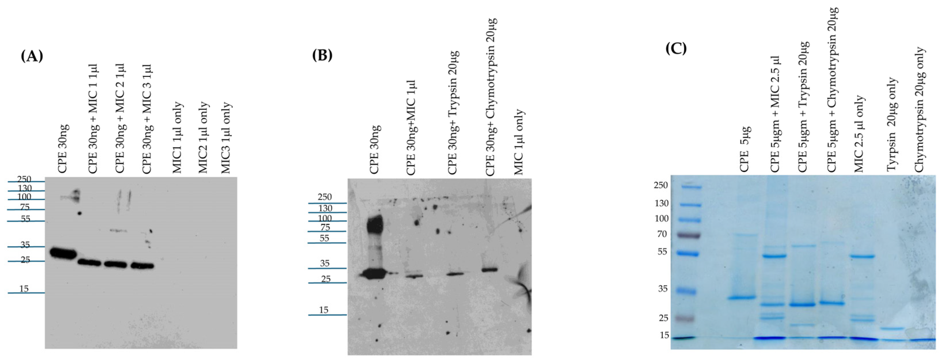
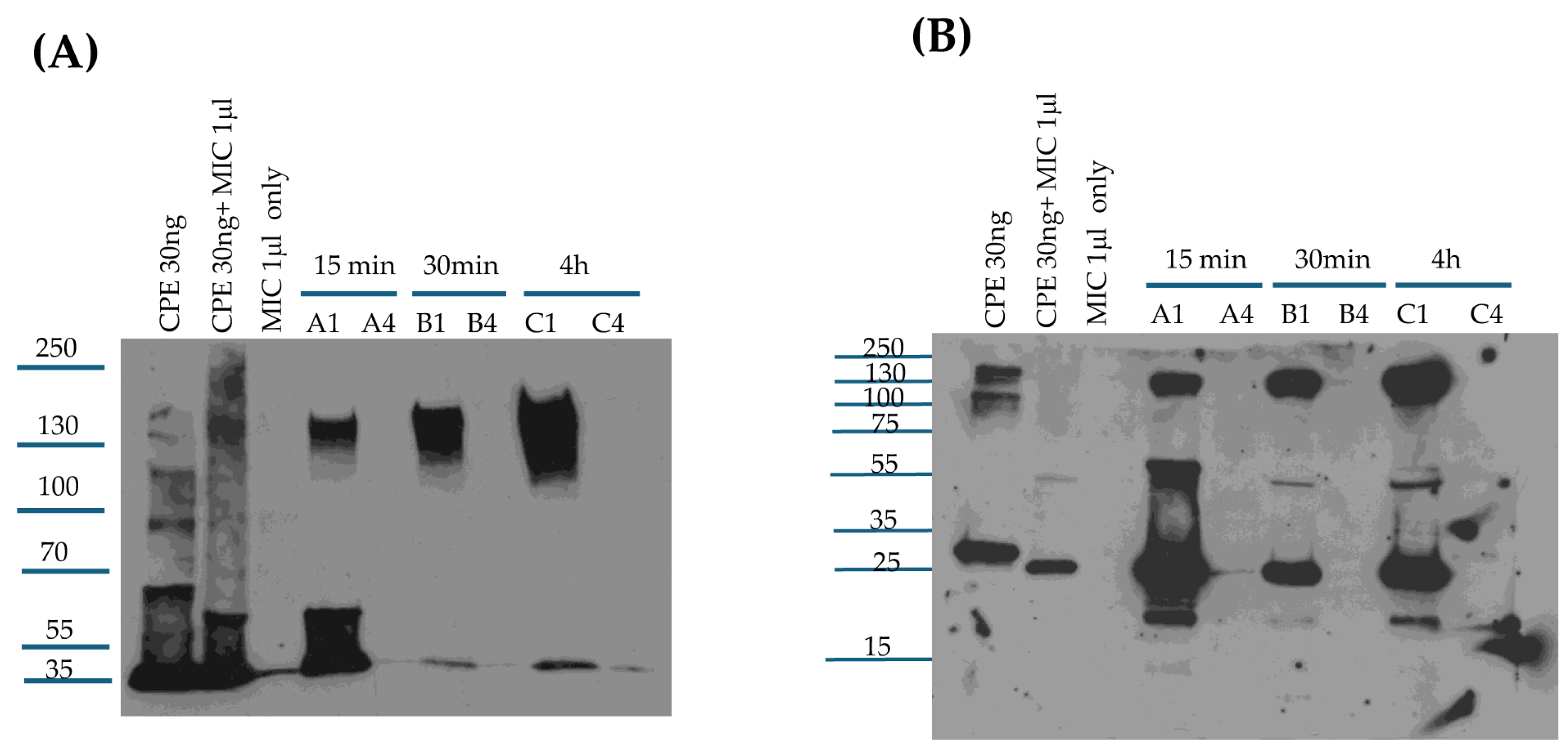
The current results indicate that, when incubated ex vivo with MICs or added to the small intestine, CPE is mainly cleaved to a fragment matching the CPE fragment size generated by purified trypsin and ~3 kDa less than native CPE. With some MIC preparations, and in the tested mouse small intestinal loops, a minor CPE band ~7 kDa smaller than CPE was also observed, suggesting there may be some animal-to-animal variability in CPE processing. It should also be noted that this minor ~28 kDa CPE fragment is too small to be cytotoxic based upon results of previous CPE deletion analysis studies [9].
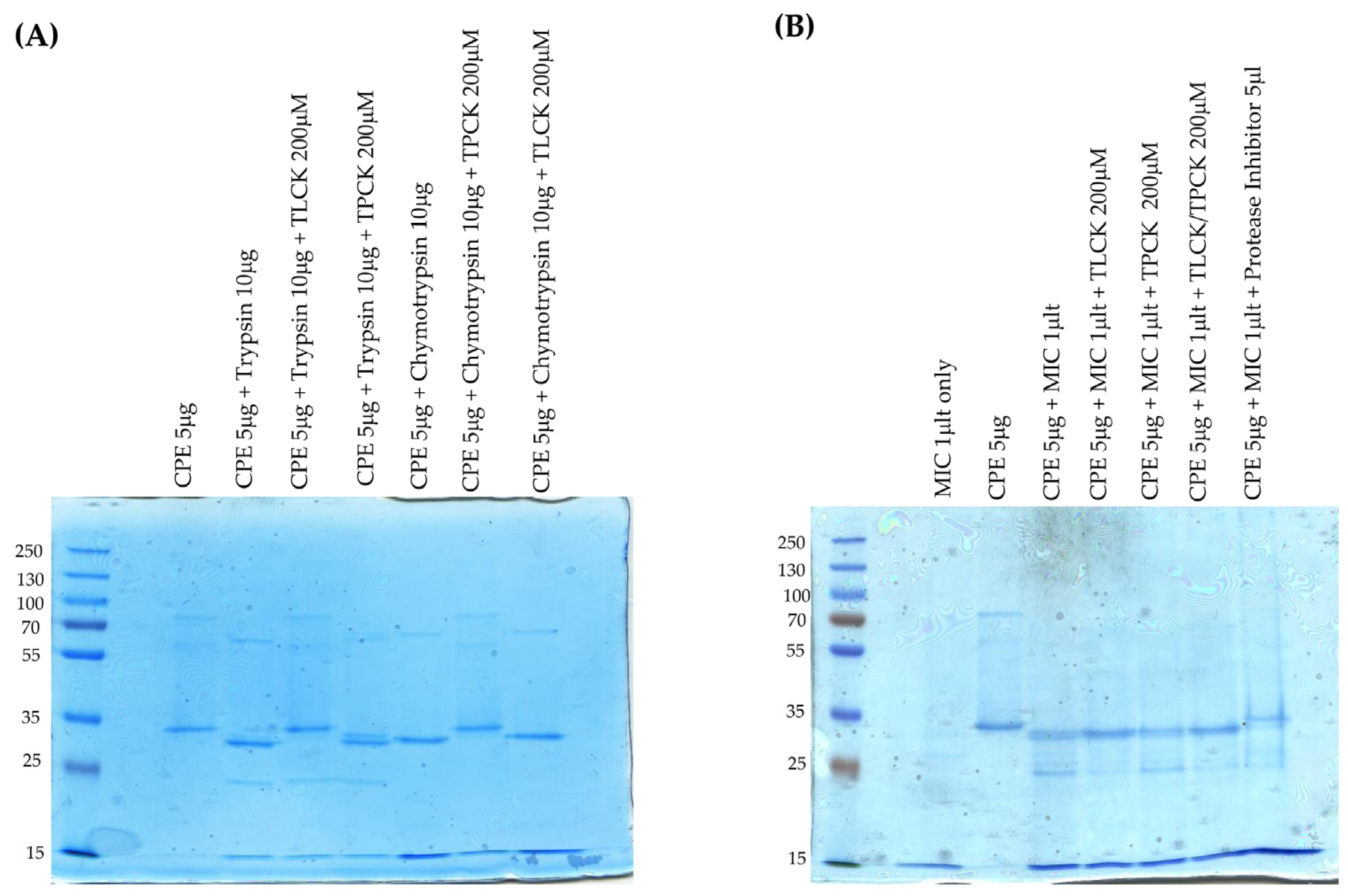
An important role for trypsin in CPE processing ex vivo received support from the ability of the trypsin inhibitor TLCK to affect this CPE processing by MICs. When MICs were preincubated with TLCK, there was a small increase in the size and staining intensity of the main CPE fragment, and the staining intensity of the minor CPE also visibly decreased. Preincubation of MICs with a chymotrypsin inhibitor did not affect normal CPE processing by MICs. When both trypsin and chymotrypsin inhibitors were present, CPE processing occurred similarly as in the presence of TLCK alone. Combining the pan-protease and trypsin-specific inhibitor results indicates that other host or microbial proteases in the intestines can affect CPE processing in the absence of trypsin, although, as mentioned, trypsin appears necessary and sufficient for processing CPE into the major fragment of ~32 kDa. These inhibitor results also indicate that the minor processed CPE band of ~28 kDa observed with some MIC preparations, and produced in the intestine, also involves trypsin activity. Of note, CPE structural biology studies [9] showed that the N-terminal 34 amino acids of CPE [which includes the N-terminal trypsin cleavage site] have no detectable electron density via X-ray crystallography. Since this CPE region is disordered, it may be more accessible to intestinal proteases, including trypsin, when the toxin is free in solution.
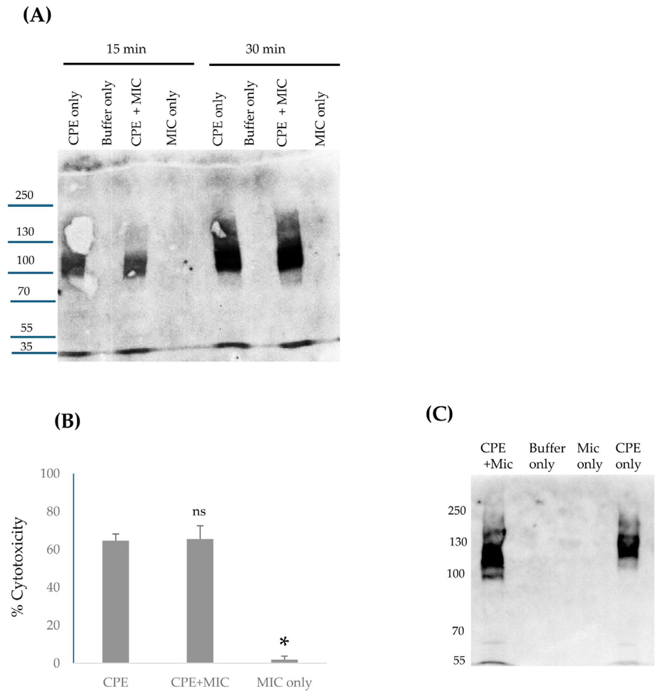
Kinetically, the current study determined that CPE processing by MICs ex vivo is very rapid, reaching completion within 10 min. This rapid processing likely provides a major explanation for the detection of processed CPE in intestinal loops treated with CPE for only 15 min, i.e., considerable amounts of CPE may be proteolytically processed even before this toxin binds to the intestines or becomes sequestered in the large CPE complex. However, it remains possible that some CPE may be processed once it becomes localized in the large CPE complex given that CPE binding and large CPE complex formation also occur quickly in vivo, becoming detectable by the 15 min treatment time-point in mouse small intestinal loops. Consistent with the possibility that some CPE cleavage might occur after the toxin becomes sequestered in the large complex, a previous study [37] showed that a portion of CPE in the large complex remains exposed on the membrane surface, i.e., CPE antibodies react with CPE that is present in the large complex in intestinal brush border membranes. If some processing of CPE localized in the large complex does occur, this might involve the unstructured N-terminus of CPE remaining outside the large complex and therefore being accessible to trypsin. It is also possible that CPE remains more susceptible to proteases when transiently located in the small complex or before the prepore to pore shift. Given the absence of structural information concerning CPE complexes, these issues will await future studies.
Intestinal proteases vary in their effects on the toxicity of C. perfringens toxins produced in the intestines [1,33,34]. For some toxins [e.g., epsilon toxin], this processing has a toxicity-activating effect, but for other toxins [e.g., beta toxin], proteolytic cleavage is detrimental, with a toxin-inactivating effect. The current study first explored this issue in vitro. When CPE and MICs were preincubated together and that mix was then added to Caco-2 cells, this experiment revealed that CPE preincubated with MICs exhibits similar Caco-2 cell cytotoxicity as does CPE preincubated without MICs, i.e., proteolytic processing by MICs did not substantially affect CPE cytotoxicity under the tested conditions. Consistent with the MIC-processed CPE retaining cytotoxic activity in vitro, the current study also demonstrated that, while much of the CPE associated with the large complex formed in the intestines is proteolytically processed, it retains intestinal enterotoxicity, i.e., in the presence of intestinal proteases, CPE still causes the intestinal damage important for type F disease [9,12,38].
Beyond providing important insights into CPE proteolytic processing under intestinal conditions, the current study also offers new information regarding large CPE complex formation and stability in vivo. The availability of mouse small intestinal loops treated for different times with CPE allowed this study to analyze large CPE complex formation kinetics and large CPE complex stability in vivo. CPE Western blot studies showed that large CPE complex formation and, by extension, CPE binding [since it is required for large CPE complex formation [9]] occur rapidly in the intestines. These Western blot results further indicate that by 30 min, the vast majority of CPE injected into mouse small intestinal loops had already bound and become sequestered in large CPE complex. However, a significant percentage of the CPE present in large CPE complex after this 30 min treatment was still susceptible to dissociation by sample boiling prior to SDS-PAGE. In contrast, after 4 h of CPE treatment, very little CPE could be released from the large complex by sample boiling. Collectively, these results indicate that the large CPE complex formed in vivo becomes more physically stable with time. Further studies are needed to better analyze how the formation and composition of the large CPE complex influence stability of this complex in vivo. It would also be worthwhile in the future to examine whether the strain, age, or diet of mice influence CPE complex formation or stability in vivo.

4. Materials and Methods

4.1. Proteases and Inhibitors

Purified trypsin [Bovine Pancreas TPCK-treated Trypsin] was purchased from Thermo Scientific (Rockford, IL, USA), while purified chymotrypsin [Alpha Chymotrypsin TLCK-treated] was obtained from Worthington Biochemical Corporation (Lakewood, NJ, USA). N-Tosyl-L-phenylalanine chloromethyl ketone [Synonym: TPCK] chymotrypsin inhibitor was purchased from Sigma (St. Louis, MO, USA), as was N-a-tosyl-L-lysine chloromethyl ketone [Synonym: TLCK] trypsin inhibitor. Protease Inhibitor Cocktail III Mammalian [AEBSF HCl: 100 mM, Aprotinin: 80 uM, Bestatin: 5 mM, E-64: 1.5 mM, Leupeptin: 2 mM, Pepstatin A: 1 mM] was purchased from RPI (Mt Prospect, IL, USA) [Research Products International].

4.2. Clostridium Perfringens Enterotoxin [CPE]

As described previously [39], CPE was purified from type F strain NCTC8238 [ATCC 12916] using sequential ammonium sulfate precipitations and gel filtration chromatography. This purified CPE was homogeneous as assessed by SDS-PAGE.

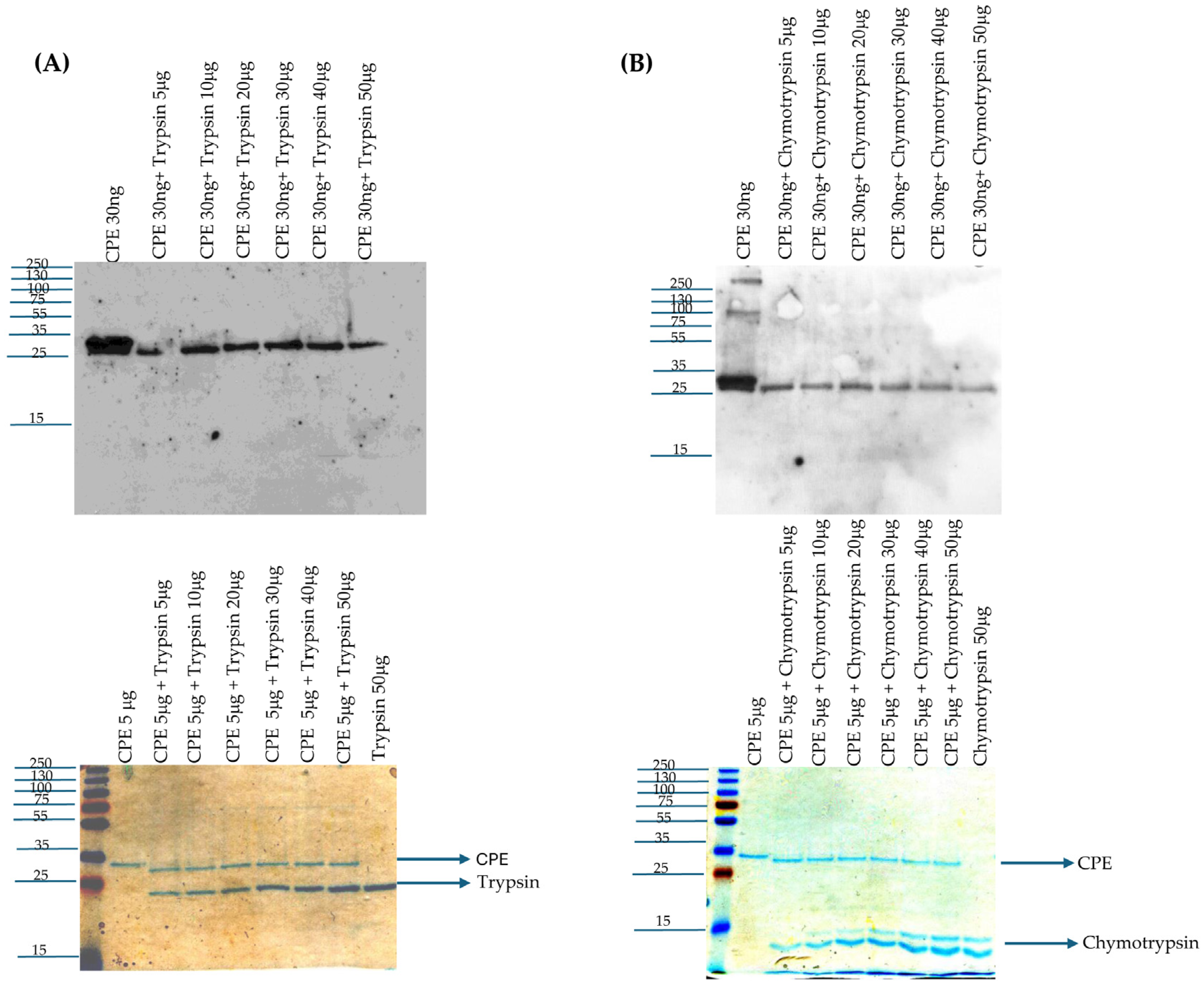
4.3. Treatment of CPE with Purified Trypsin In Vitro

Purified CPE [30 ng] was treated with 5, 10, 20, 30, 40, or 50 μg of trypsin in PBS [total volume 25 μL] for 10 min at 37 °C. After this incubation, 5 μL of 5x loading buffer (which contains both SDS and β-mercaptoethanol) was added to each sample and the samples were then loaded onto a 12% acrylamide gel containing SDS. After electrophoresis, the separated proteins on these gels were electrotransferred onto nitrocellulose membranes [0.45 μm pore size, Biorad (Hercules, CA, USA)], which were then blocked with 5% milk in Tris-buffer saline with 0.1% Tween-20 [Fisher Scientific, Fair Lawn, NJ, USA]. The blocked membranes were incubated overnight at 4 °C with a 1:1000 dilution of polyclonal CPE antibody [40]. On the next day, the blot was incubated with a 1:10,000 dilution of goat anti-rabbit IgG antibody conjugated with horseradish peroxidase [Invitrogen (Eugene, OR, USA)] at RT for 1 h with gentle shaking. The blot was developed using SuperSignal West Pico PLUS Chemiluminescent Substrate [Thermo Scientific (Rockford, IL, USA)].
The above experiment was also performed using a reaction containing 5 μg of CPE in PBS [total volume 25 μL] with 5–50 μg of trypsin in PBS that was incubated for 10 min at 37 °C, followed by electrophoresis, as above, and staining the gels with Coomassie blue [G-250, Biorad (Hercules, CA, USA)].

4.4. Treatment of CPE with Purified Chymotrypsin In Vitro

Purified CPE [30 ng] was treated with 5, 10, 20, 30, 40, or 50 μg of chymotrypsin in PBS [total volume 25 μL] for 10 min at 37 °C. After this treatment, CPE Western blotting was performed as described above for trypsin treatment. Similarly, the above experiment was repeated using a reaction containing 5 μg of CPE in 25 μL of PBS with 5–50 μg of purified chymotrypsin that was incubated for 10 min at 37 °C, followed by electrophoresis [as described above] and staining the gels with Coomassie blue stain.

4.5. Preparation of MICs

Small intestinal lumen contents were obtained from healthy ~4-month-old BALB/c mice after they had been euthanized by means of CO2 asphyxiation for colony management. Equal numbers of male and female mice were used. After euthanasia, small intestinal lumen contents were immediately harvested from excised small intestine by gentle squeezing and stored at −80 °C. For use in experiments, each intestinal content sample was thawed, weighed, and mixed with PBS buffer at a ratio of 1:1. The mixture was then centrifuged for 5 min and supernatants [now designated as MIC] were collected, aliquoted, and stored at −80 °C until use in the current study. Typically, one MIC preparation was used for each experimental repetition.

4.6. Comparison of CPE Processing by MICs from Different Mice

To evaluate the ex vivo effects of MICs from different mice on CPE, 30 ng of native CPE was incubated in 25 μL of PBS with 1 μL of MICs from three different mice [2 female and 1 male] for 10 min at 37 °C. After incubation, 5 μL of 5x loading buffer was added to each sample and the samples were then boiled for 5 min before loading onto a 12% acrylamide gel containing SDS. After electrophoresis, separated proteins were electrotransferred onto a 0.45 μm pore size nitrocellulose membrane and processed for CPE Western blot analysis as described earlier.

4.7. Comparison of CPE Processing by MICs, Purified Trypsin, or Purified Chymotrypsin

For CPE Western blot analyses, 30 ng of native CPE was added to PBS containing 1 μL of MICs, 20 μg of purified trypsin, or 20 μg of purified chymotrypsin [reaction volumes 25 μL] for 10 min at 37 °C. Identical MIC, trypsin, or chymotrypsin samples were similarly incubated without CPE. After this incubation, all samples were treated with 5 μL of 5x loading buffer and this sample was then boiled for 5 min; the trypsin- or chymotrypsin-treated samples were not boiled to reduce CPE aggregation. All samples were then electrophoresed on 12% acrylamide gels containing SDS and processed for CPE Western blotting, as described earlier. For Coomassie blue staining analyses, 5 μg of native CPE was incubated in PBS [reaction volume of 25 μL] containing 2.5 μL of MICs, 20 μg of purified trypsin, or 20 μg of purified chymotrypsin. The samples were then processed and electrophoresed on a 12% acrylamide gel containing SDS as described above for CPE Western blots. After electrophoresis, gels were stained with Coomassie blue.

4.8. Effects of Protease Inhibitors on MIC Processing of CPE Ex Vivo

Purified trypsin [10 μg] was preincubated with 200 μM TLCK or 200 μM TPCK in PBS [total reaction volume was 20 μL], while 10 μg of purified chymotrypsin was preincubated with 200 μM TPCK or 200 μM TLCK in PBS [total reaction volume was 20 μL]. The control sample contained only 20 μL of PBS. All samples were preincubated at 37 °C for 30 min. After this preincubation, 5 μg of CPE in 5 μL of PBS was added to each sample before incubation for 10 min at 37 °C. The incubated samples were then treated with 5 μL of 5x loading buffer and electrophoresed on 12% acrylamide gels containing SDS, followed by staining these gels with Coomassie blue. For MIC treatments, MICs [1 μL] in PBS [total reaction volume of 20 μL] were preincubated with 200 μM TLCK, 200 μM TPCK, 200 μM each of TCPK and TLCK, or 5 μL of Protease Inhibitor Cocktail III. As a control, 1 μL of MICs was preincubated in 20 μL of PBS. These preincubations were performed at 37 °C for 30 min. After this preincubation, 5 μL of PBS containing 5 μg of CPE was added to each sample and the samples were incubated for 10 min at 37 °C. These samples were then treated with 5 μL of 5x loading buffer, boiled for 5 min, and then electrophoresed on 12% acrylamide gels containing SDS, followed by staining the gels with Coomassie blue staining.

4.9. Caco-2 Cell Culture

Authenticated Caco-2 cells were purchased from ATCC. Genetic information about these cells (lot number 61777387) is available at https://www.atcc.org/products/htb-37., as accessed on 15 March 2025. These Caco-2 cells were grown in Eagle’s essential medium [Lonza (Walkersville, MD, USA)] supplemented with 10% fetal bovine serum [Gibco (Grand Island, NY, USA)], 1% MEM nonessential amino acids [Cytiva (Logan, UT, USA)], 100 μg/mL of penicillin-streptomycin solution [Corning (Manassas, VA, USA)], and 1% glutamine [Corning]. These cultures were maintained at 37 °C in a 5% CO2 atmosphere.

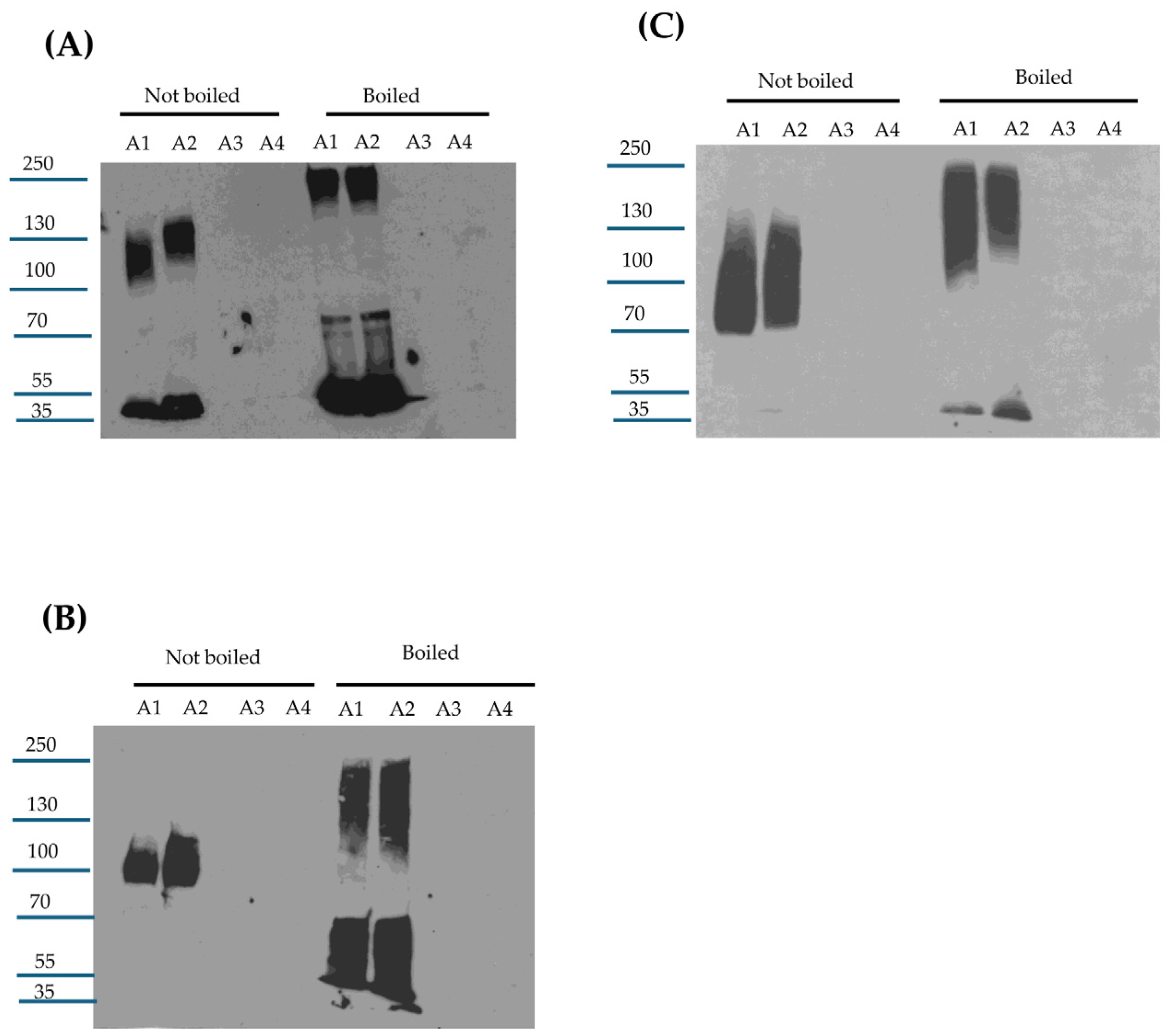
4.10. MICs’ Effects on Large CPE Complex Formation by Caco-2 Cells

Caco-2 cells were grown in 6-well cell culture dishes for 5–6 days. Cultures were then treated for 15 min at 37 °C with CPE [0.5 μg/mL] in the presence of 1 μL of MICs in 1 mL of HBSS; the MICs were first added into the culture, immediately followed by the addition of CPE. Other Caco-2 cell wells were treated with 1 mL of HBSS alone or with 1 mL of HBSS containing 1 μL of MICs [no CPE]. After a 15 or 30 min of incubation, each sample was gently collected using a cell scrapper and lysed with RIPA buffer [Boston BioProducts, Inc (Millford, MA, USA).] in the presence of Benzonase Nuclease [EMD Millipore (Burlington, MA, USA)] and Protease Inhibitor Cocktail III at RT for 30 min with gentle absorption. The samples were centrifuged and the lysates were collected in sterile tubes. A 1 μL aliquot of lysate from each treatment was then mixed with 24 μL of HBSS buffer plus 5 μL of 5x loading buffer and the total 30 μL final volume was loaded onto a 6% SDS PAGE gel and processed for CPE Western blotting as already described.

4.11. Effects of MIC Pretreatment on CPE Cytotoxicity and Large CPE Complex Formation for Caco-2 Cells

HBSS containing native CPE [3 μg] in the presence or absence of 2.5 μL of MICs, HBSS containing 2.5 μL of MICs, or HBSS alone [total sample volumes of 25 μL] was preincubated at 37 °C for 10 min. After that preincubation, samples were adjusted with HBSS to a 1 μg/mL CPE concentration, with a similar dilution of the MIC alone [no CPE] sample. Each sample [1 mL] was then added onto confluent Caco-2 cell cultures for 1 h at 37 °C. After this treatment, culture supernatants were collected, centrifuged and the lysates were processed for LDH-release, as an indicator of cytotoxicity, using the CyQUANT LDH Cytotoxicity Assay Kit [Invitrogen].
Adherent cells in these cultures were gently collected using a cell scrapper and lysed with RIPA buffer with Benzonase Nuclease and Protease Inhibitor Cocktail III at RT for 30 min. The samples were centrifuged and a 1 μL aliquot of lysate from each treatment was then mixed with 24 μL of HBSS buffer plus 5 μL of 5x loading buffer; the total final volume was then loaded onto a 6% acrylamide gel with SDS and processed for CPE Western blotting as already described.

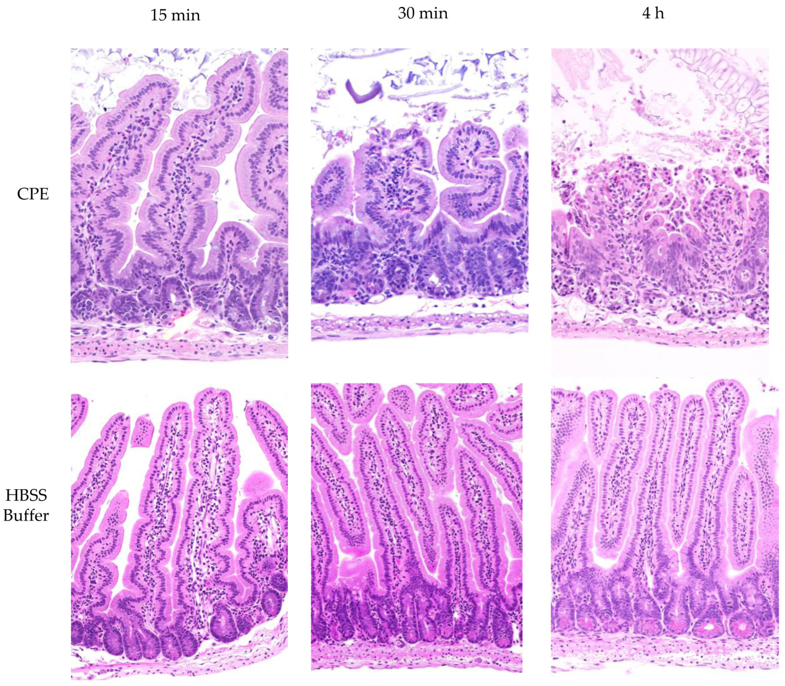
4.12. CPE Treatment of Mouse Small Intestinal Loops

To assess the effects of intestinal proteases on in vivo CPE processing, large CPE complex formation and enterotoxicity, the mouse small intestinal loop model was used. Balb/C mouse small intestine segments [~10 cm] were surgically ligated under general anesthesia, as previously described [18]. These intestinal loop experiments were conducted using 6 groups of mice [n = 6 per group, with equal numbers of male and female mice]. Each loop was challenged with 1 mL of HBSS that did or did not contain 100 μg of purified CPE. After 15 min, 30 min, or 4 h of incubation, the mice were euthanized. Samples from these treated intestinal loops were collected, fixed in 10% buffered formalin at pH 7.2 for 72 h and then processed into 4 μm sections before staining with hematoxylin and eosin [H&E]. These samples were examined by a pathologist in a blinded fashion. Contents of the intestinal loops were collected by gentle squeezing and stored at −80 °C until their use in the experiments.

4.13. Analysis of Large CPE Complex Formation and Stability In Vivo

Intestinal contents from control or CPE-treated mice, as described above, were centrifuged and half of the supernatant volume was boiled for 5 min while the other half was not. These samples were then electrophoresed on 12% acrylamide gels with SDS or on 6% acrylamide gel with SDS and processed for CPE Western blotting as described above for Caco-2 cells. Samples run on 12% acrylamide gels with SDS were boiled for 5 min before electrophoresis and Western blotting.

4.14. Statistical Analyses

The statistical significance of CPE cytotoxicity results was assessed by means of one-way ANOVA with Dunnett’s multiple-comparison test.

Author Contributions

Conceptualization, A.S., F.A.U. and B.A.M.; Methodology, A.S., J.L.G. and J.B.; Validation, A.S. and F.A.U.; Formal analysis, A.S., F.A.U. and B.A.M.; Investigation, A.S., J.L.G., J.B., F.A.U. and B.A.M.; Data curation, A.S.; Writing—original draft, A.S., F.A.U. and B.A.M.; Writing—review and editing, A.S., F.A.U. and B.A.M.; Project administration, F.A.U. and B.A.M.; Funding acquisition, B.A.M. All authors have read and agreed to the published version of the manuscript.

Funding

This work was generously supported by grant RO1AI019844-41 from the National Institute of Allergy and Infectious Diseases. The content is solely the responsibility of the authors and does not necessarily represent the official views of the National Institutes of Health.

Institutional Review Board Statement

The experiments involving mice intestinal contents were reviewed and approved by the University of California Davis Institutional Animal Care and Use Committee as IACUC protocol 23482, approved 18 July 2023. The experiments involving mouse small intestinal loops were reviewed and approved by the University of California Davis Institutional Animal Care and Use Committee as IACUC as protocol 23182, approved 2 February 2023.

Informed Consent Statement

Not applicable.

Data Availability Statement

The original contributions presented in this study are included in this article. Further inquiries can be directed to the corresponding author(s).

Conflicts of Interest

No potential conflict of interest was reported by the authors.

References

  1. Mehdizadeh Gohari, I.; Navarro, M.A.; Li, J.; Shrestha, A.; Uzal, F.A.; McClane, B.A. Pathogenicity and virulence of Clostridium perfringens. Virulence 2021, 12, 723–753. [Google Scholar] [PubMed]
  2. Ba, X.; Jin, Y.; Ning, X.; Gao, Y.; Li, W.; Li, Y.; Wang, Y.; Zhou, J. Clostridium perfringens in the intestine: Innocent bystander or serious threat? Microorganisms 2024, 12, 1610. [Google Scholar] [CrossRef] [PubMed]
  3. Kiu, R.; Hall, L.J. An update on the human and animal enteric pathogen Clostridium perfringens. Emerg. Microbes Infect. 2018, 7, 141. [Google Scholar]
  4. Ou, L.; Ye, B.; Sun, M.; Qi, N.; Li, J.; Lv, M.; Lin, X.; Cai, H.; Hu, J.; Song, Y.; et al. Mechanisms of intestinal epithelial cell damage by Clostridium perfringens. Anaerobe 2024, 87, 102856. [Google Scholar]
  5. Grenda, T.; Jarosz, A.; Sapala, M.; Grenda, A.; Patyra, E.; Kwiatek, K. Clostridium perfringens-opportunistic foodborne pathogen, its diversity and epidemiological significance. Pathogens 2023, 12, 768. [Google Scholar] [CrossRef] [PubMed]
  6. Da Silva Felicio, M.T.; Hald, T.; Liebana, E.; Allende, A.; Hugas, M.; Nguyen-The, C.; Johannessen, G.S.; Niskanen, T.; Uyttendaele, M.; McLauchlin, J. Risk ranking of pathogens in ready-to-eat unprocessed foods of non-animal origin (FoNAO) in the EU: Initial evaluation using outbreak data (2007–2011). Int. J. Food Microbiol. 2015, 195, 9–19. [Google Scholar]
  7. Scallan, E.; Hoekstra, R.M.; Angulo, F.J.; Tauxe, R.V.; Widdowson, M.; Roy, S.; Jones, J.L.; Griffin, P.M. Foodborne illness acquired in the United States-major pathogens. Emerg. Infect. Dis. 2011, 17, 7–15. [Google Scholar]
  8. Lahti, P.; Heikinheimo, A.; Johansson, T.; Korkeala, H. Clostridium perfringens type A isolates carrying the plasmid-borne enterotoxin gene (genotypes IS1151-cpe or IS1470-like-cpe) are a common cause of food poisonings. J. Clin. Microbiol. 2008, 46, 371–373. [Google Scholar]
  9. Shrestha, A.; Mehdizadeh Gohari, I.; Li, J.; Navarro, M.; Uzal, F.A.; McClane, B.A. The biology and pathogenicity of Clostridium perfringens type F: A common human enteropathogen with a new(ish) name. Microbiol. Mol. Biol. Rev. 2024, 88, e0014023. [Google Scholar]
  10. Carman, R.J. Clostridium perfringens in spontaneous and antibiotic-associated diarrhoea of man and other animals. Rev. Med. Microbiol. 1997, 8 (Suppl. S1), S43–S45. [Google Scholar]
  11. Larcombe, S.; Hutton, M.L.; Lyras, D. Involvement of bacteria other than Clostridium difficile in antiboitic-associated diarrhoea. Trends Microbiol. 2016, 24, 463–476. [Google Scholar] [PubMed]
  12. McClane, B.A.; Robertson, S.L.; Li, J. Clostridium perfringens. In Food Microbiology: Fundamentals and Frontiers, 4th ed.; Doyle, M.P., Buchanan, R.L., Eds.; ASM Press: Washington, DC, USA, 2013; pp. 465–489. [Google Scholar]
  13. Katahira, J.; Inoue, N.; Horiguchi, Y.; Matsuda, M.; Sugimoto, N. Molecular cloning and functional characterization of the receptor for Clostridium perfringens enterotoxin. J. Cell Biol. 1997, 136, 1239–1247. [Google Scholar] [PubMed]
  14. Katahira, J.; Sugiyama, H.; Inoue, N.; Horiguchi, Y.; Matsuda, M.; Sugimoto, N. Clostridium perfringens enterotoxin utilizes two structurally related membrane proteins as functional receptors in vivo. J. Biol. Chem. 1997, 272, 26652–26658. [Google Scholar] [PubMed]
  15. Shrestha, A.; McClane, B.A. Human claudin-8 and -14 are receptors capable of conveying the cytotoxic effects of Clostridium perfringens enterotoxin. MBio 2013, 4, e00594-12. [Google Scholar]
  16. Robertson, S.L.; Smedley, J.G., Jr.; Singh, U.; Chakrabarti, G.; Van Itallie, C.M.; Anderson, J.M.; McClane, B.A. Compositional and stoichiometric analysis of Clostridium perfringens enterotoxin complexes in Caco-2 cells and claudin 4 fibroblast transfectants. Cell Microbiol. 2007, 9, 2734–2755. [Google Scholar]
  17. Benz, R.; Popoff, M.R. Clostridium perfringens enterotoxin: The toxin forms highly cation-selective channels in lipid bilayers. Toxins 2018, 10, 341. [Google Scholar] [CrossRef]
  18. Caserta, J.A.; Robertson, S.L.; Saputo, J.; Shrestha, A.; McClane, B.A.; Uzal, F.A. Development and application of a mouse intestinal loop model to study the in vivo action of Clostridium perfringens enterotoxin. Infect. Immun. 2011, 79, 3020–3027. [Google Scholar]
  19. Duncan, C.L. Time of enterotoxin formation and release during sporulation of Clostridium perfringens type A. J. Bacteriol. 1973, 113, 932–936. [Google Scholar]
  20. Rao, S.; Grover, M. Intestinal Proteases. Curr. Opin. Gastro 2023, 39, 472–478. [Google Scholar]
  21. Caminero, A.; Guzman, M.; Libertucci, J.; Lomax, A.E. The emerging roles of bacterial proteases in intestinal diseases. Gut Microbes 2023, 15, 2181922. [Google Scholar]
  22. Biancheri, P.; Sabtino, A.D.; Corazza, G.R.; MacDonald, T.T. Proteases and the gut barrier. Cell Tissue Res. 2013, 351, 269–280. [Google Scholar] [PubMed]
  23. Rao, M.B.; Tanksale, A.M.; Ghatge, M.S.; Deshpande, V.V. Molecular and biotechnological aspects of microbial proteases. Microbiol. Mol. Biol. Rev. 1998, 62, 597–635. [Google Scholar] [PubMed]
  24. Hammers, D.; Carothers, K.; Lee, S. The role of bacterial proteases in microbe and host-microbe interactions. Curr. Drug Targets 2022, 23, 222–239. [Google Scholar]
  25. Hou, J.J.; Ding, L.; Yang, T.; Yang, Y.F.; Jin, Y.P.; Zhang, X.P.; Ma, A.H.; Qin, Y.H. The proteolytic activity in inflammatory bowel disease: Insight from gut microbiota. Microb. Pathog. 2024, 188, 106560. [Google Scholar]
  26. Granum, P.E.; Richardson, M. Chymotrypsin treatment increases the activity of Clostridium perfringens enterotoxin. Toxicon 1991, 29, 445–453. [Google Scholar]
  27. Granum, P.E.; Whitaker, J.R.; Skjelkvale, R. Trypsin activation of enterotoxin from Clostridium perfringens type A. Biochim. Biophys. Acta 1981, 668, 325–332. [Google Scholar]
  28. Hanna, P.C.; Wieckowski, E.U.; Mietzner, T.A.; McClane, B.A. Mapping functional regions of Clostridium perfringens type A enterotoxin. Infect. Immun. 1992, 60, 2110–2114. [Google Scholar]
  29. Ogbu, C.P.; Kapoor, S.; Vecchio, A.J. Structural basis of Clostridium perfringens enterotoxin activation and oligomerization by trypsin. Toxins 2023, 15, 637. [Google Scholar] [CrossRef]
  30. Richardson, M.; Granum, P.E. The amino acid sequence of the enterotoxin from Clostridium perfringens type A. FEBS Lett. 1985, 182, 479–484. [Google Scholar]
  31. Richardson, M.; Granum, P.E. Sequence of the amino-terminal part of enterotoxin from Clostridium perfringens type A: Identification of points of trypsin activation. Infect. Immun. 1983, 40, 943–949. [Google Scholar]
  32. Wieckowski, E.U.; Wnek, A.P.; McClane, B.A. Evidence that an ~50kDa mammalian plasma membrane protein with receptor-like properties mediates the amphilicity of specifically-bound Clostridium perfringens enterotoxin. J. Biol. Chem. 1994, 269, 10838–10848. [Google Scholar] [CrossRef] [PubMed]
  33. Rioseco, M.M.; Beingesser, J.; Uzal, F.A. Freezing or adding trypsin inhibitor to equine intestinal contents extends the lifespan of Clostridium perfringens beta toxin for diagnostic purposes. Anaerobe 2012, 18, 357–360. [Google Scholar] [CrossRef]
  34. Freedman, J.C.; Li, J.; Uzal, F.A.; McClane, B.A. Proteolytic processing and activation of Clostridium perfringens epsilon toxin by caprine small intestinal contents. MBio 2014, 5, e01994. [Google Scholar] [CrossRef] [PubMed]
  35. Miyata, S.; Matsushita, O.; Minami, J.; Katayama, S.; Shimamoto, S.; Okabe, A. Cleavage of a C-terminal peptide is essential for heptamerization of Clostridium perfringens epsilon-toxin in the synaptosomal membrane. J. Biol. Chem. 2001, 276, 13778–13783. [Google Scholar] [CrossRef]
  36. Gibert, M.; Petit, L.; Raffestin, S.; Okabe, A.; Popoff, M.R. Clostridium perfringens iota-toxin requires activation of both binding and enzymatic components for cytopathic activity. Infect. Immun. 2000, 68, 3848–3853. [Google Scholar] [CrossRef]
  37. Kokai-Kun, J.F.; McClane, B.A. Evidence that region(s) of the Clostridium perfringens enterotoxin molecule remain exposed on the external surface of the mammalian plasma membrane when the toxin is sequestered in small or large complex. Infect. Immun. 1996, 64, 1020–1025. [Google Scholar] [CrossRef]
  38. McDonel, J.L.; Chang, L.W.; Pounds, J.L.; Duncan, C.L. The effects of Clostridium perfringens enterotoxin on rat and rabbit ileum: An electron microscopy study. Lab. Investig. 1978, 39, 210–218. [Google Scholar] [PubMed]
  39. McDonel, J.L.; McClane, B.A. Production, purification and assay of Clostridium perfringens enterotoxin. Method. Enzym. 1988, 165, 94–103. [Google Scholar]
  40. McClane, B.A.; Strouse, R.J. Rapid detection of Clostridium perfringens type A enterotoxin by enzyme-linked immunosorbent assay. J. Clin. Microbiol. 1984, 19, 112–115. [Google Scholar] [CrossRef]
Figure 1. Effects of purified trypsin or chymotrypsin on CPE processing in vitro. (A) trypsin treatment. Top panel: Thirty nanograms of CPE was incubated with 0, 5, 10, 20, 30, 40, or 50 μg of trypsin in PBS buffer for 10 min at 37 °C. After this incubation, samples were electrophoresed without boiling on a 12% acrylamide gel containing SDS and then CPE Western blotted. Bottom panel: Five micrograms of CPE was treated with 0, 5, 10, 20, 30, 40, or 50 μg of trypsin in PBS buffer for 10 min at 37 °C. The samples were then electrophoresed as described above, followed by staining with Coomassie blue [bottom panel]. (B) Chymotrypsin treatment. Top panel: Thirty nanograms of CPE was treated with 0, 5, 10, 20, 30, 40, or 50 μg of chymotrypsin in PBS buffer for 10 min at 37 °C. After incubation, samples were electrophoresed without boiling on a 12% acrylamide gel containing SDS and then CPE Western blotted. Bottom panel: Five micrograms of CPE was treated with 0, 5, 10, 20, 30, 40, or 50 μg of chymotrypsin in PBS buffer for 10 min at 37 °C. The samples were then electrophoresed as described above, followed by staining with Coomassie blue [bottom panel]. Numbers to the left of the gels show the size of the protein standards by molecular mass [in kDa]. The results shown in both panels are representative of three repetitions for each gel.
Toxins 17 00170 g001
Figure 2. Effects of mouse intestinal contents on CPE. (A) MICs’ effects on CPE processing. MICs were prepared from 3 different mice [2 female and 1 male]. To evaluate their effects on CPE, 30 ng of native CPE was or was not mixed with 1 μL of MICs in PBS and incubated at 37 °C for 10 min. After this incubation, samples were boiled, electrophoresed on a 12% acrylamide gel, and then processed for CPE Western blot analysis. The same amount of MIC alone [no CPE] in PBS was similarly CPE Western blotted to assure that the observed immunoreactivity was due to the presence of CPE. (B) Head-to-head CPE Western blot comparison of the effects of MICs, purified trypsin, or chymotrypsin on CPE processing. MIC alone [no CPE] was also included as a specificity control. Before electrophoresis and CPE Western blotting as described for panel A, MICs and MICs plus CPE samples, but not CPE plus trypsin or chymotrypsin samples, were boiled. (C) Head-to-head Coomassie blue staining comparison of the effects of MICs, purified trypsin, or chymotrypsin on CPE processing. Samples were prepared and electrophoresed as in panels A and B except that 5 μg of CPE and/or 2.5 μL of MICs were added to samples [when specified]. Also electrophoresed were equivalent concentrations of MICs, purified trypsin, or purified chymotrypsin in the absence of CPE, for comparison. Samples with MICs were boiled but other samples were not. The results in Panel A, B and C are representative results of three repetitions. Numbers to the left of gels or blots indicate the size of standard proteins [in kDa].
Toxins 17 00170 g002
Figure 3. Effects of protease inhibitors on CPE processing. (A) Ten micrograms of purified trypsin was preincubated for 30 min at 37 °C with or without 200 μM TLCK or 200 μM TPCK in PBS; ten micrograms of purified chymotrypsin was similarly preincubated with or without 200 μM TPCK or 200 μM TLCK in PBS. After this preincubation, 5 μg of CPE was added to each sample and these samples were further incubated for 10 min at 37 °C. The incubated samples were then electrophoresed on 12% acrylamide gels containing SDS; after electrophoresis the gels were stained with Coomassie blue. (B) One microliter of MICs was preincubated for 30 min at 37 °C with [i] 200 μM TLCK, [ii] 200 μM TPCK [iii] 200 μM of both TCPK and TLCK, and [iv] 5 μL of resuspended Protease Inhibitor Cocktail III in PBS. As a control, 1 μL of MICs was similarly preincubated in PBS only. After preincubation, 5 μg of CPE was added to each sample and the samples were further incubated for 10 min at 37 °C. The incubated samples were then boiled and electrophoresed on 12% acrylamide gels containing SDS, followed by staining the gels with Coomassie blue. The results shown are representative of three repetitions.
Toxins 17 00170 g003
Figure 4. Effects of MICs on large CPE complex formation and CPE-induced cytotoxicity in Caco-2 cells. (A) MICs’ effects on large CPE complex formation. MICs in HBSS were added to confluent Caco-2 cells, followed immediately by the addition of 0.5 μg/mL of CPE for 15 or 30 min. Other control wells received the same amount of MICs in HBSS [no CPE] or HBSS alone. After incubation, the cells were harvested gently and lysed with RIPA buffer in the presence of Benzonase Nuclease and a Protease Inhibitor cocktail at RT with gentle rotation for 30 min. Lysates were centrifuged and 1 μL of each supernatant was mixed with 24 μL of HBSS plus 5 μL of 5x loading buffer. This mixture was loaded without boiling onto a 6% acrylamide gel with SDS. After electrophoresis and transfer, blots were processed for CPE Western blotting. The results shown are representative of three Western blots. Numbers to the left of gels indicate the size of standard proteins [in kDa]. (B) Effects of preincubating CPE with MICs on CPE-induced cytotoxicity. Before their addition to Caco-2 cells, HBSS containing 3 μg of CPE, 3 μg of CPE with 2.5 μL of MICs, 2.5 μL of MICs, or HBSS buffer alone was preincubated at 37 °C for 10 min. The samples were then collected and adjusted with HBSS to 1 μg/mL of CPE, or diluted similarly for MICs alone, and added onto confluent Caco2 cells for 1 h at 37 °C. After incubation, the samples were gently collected and spun down and the lysates were processed for LDH release, as a marker for cytotoxicity, using the CyQUANT LDH Cytotoxicity Assay Kit. The results shown are the means of 3 repetitions. Error bars show SD. The cytotoxicity difference between CPE vs. CPE plus MICs was not statistically significant [p < 0.05], but * indicates that treatment with MICs only was significantly [p < 0.05] different from CPE treatment. (C) Effects of preincubating CPE with MICs on large CPE complex formation by panel B Caco-2 cells. Panel B Caco-2 cells were lysed and processed for CPE Western blotting as described in Panel A. The results shown are representative of three repetitions.
Toxins 17 00170 g004
Figure 5. Analyses of in vivo large CPE complex formation kinetics and large CPE complex stability. Mouse small intestinal loops were treated with HBSS containing 100 μg/mL of CPE or HBSS alone for 15 min, 30 min, or 4 h. The small intestinal contents were then collected, electrophoresed on a 6% acrylamide gel with SDS, and processed for CPE Western blot analysis with or without sample boiling [as indicated]: (A) 15 min treatment, (B) 30 min treatment, and (C) 4 h treatment. Note that the large CPE complex migrates anomalously on 6% acrylamide gels with SDS [16], so its migration vs. standard proteins [as indicated on left of gel in kDa] does not accurately reflect the size of this complex. Shown are representative Western CPE blots for three repetitions for each time-point.
Toxins 17 00170 g005
Figure 6. Analysis of CPE processing in vivo. (A) Confirmation of large CPE complex formation in mouse small intestinal loops. Loops A1, B1, and C1 received 100 μg/mL of CPE in HBSS for 15 min, 30 min, or 4 h, respectively. Loops A4, B4, and C4 received only HBSS [no CPE] for 15 min, 30 min, or 4 h, respectively. Intestinal contents were then electrophoresed, without boiling, on a 6% acrylamide gel with SDS and CPE Western blotted. Markers to the left of blots show the migration of protein standards in kDa, although, as mentioned in the legend for Figure 5, the large CPE complex migrates anomalously on 6% acrylamide gels with SDS, so its migration vs. standard proteins does not accurately reflect its true size. Also shown is the migration of CPE alone, CPE treated with MICs ex vivo, and MIC alone for comparison. (B) Analysis of CPE size after treatment of mouse small intestinal loops. After loops were treated as described for panel A, intestinal contents were then electrophoresed, with sample boiling, on 12% acrylamide gels with SDS before CPE Western blotting. Markers to the left of blots show the migration of protein standards in kDa. Also shown is the migration of CPE alone, CPE treated with MICs ex vivo, and MICs alone for comparison. Gels shown in panels A and B are representative of three repetitions.
Toxins 17 00170 g006
Figure 7. CPE processed in the small intestines retains toxicity. Mouse small intestinal loops were challenged with HBSS containing 100 μg/mL of CPE [top row] or HBSS alone [bottom panel] for 15 min, 30 min, or 4 h, as indicated. After these treatments, samples from the intestinal loops were processed routinely for the production of 4 μm thick hematoxylin and eosin [H&E]-stained sections. The results shown are representative of 3 mice for each challenge.
Toxins 17 00170 g007
Disclaimer/Publisher’s Note: The statements, opinions and data contained in all publications are solely those of the individual author(s) and contributor(s) and not of MDPI and/or the editor(s). MDPI and/or the editor(s) disclaim responsibility for any injury to people or property resulting from any ideas, methods, instructions or products referred to in the content.

