Article

Self-Sufficient Aflatoxin Decontamination System: MOF-Based Composite Membrane with Peroxidase-Mimic and Controlled H2O2 Generation

1
School of Food Science and Pharmaceutical Engineering, Nanjing Normal University, Nanjing 210023, China
2
Inner Mongolia Mengniu Dairy (Group) Limited by Share Limited, Hohhot 010020, China
3
Yangzhou Center for Food and Drug Control, Yangzhou 225002, China
4
China National Center for Food Safety Risk Assessment, Beijing 100000, China
5
Key Laboratory of Biotoxin Analysis & Assessment, State Administration for Market Regulation, Nanjing Institute of Product Quality Inspection, Nanjing 210019, China
*
Authors to whom correspondence should be addressed.
These authors contributed equally to this work.
Toxins 2025, 17(10), 516; https://doi.org/10.3390/toxins17100516
Submission received: 21 August 2025 / Revised: 23 September 2025 / Accepted: 3 October 2025 / Published: 20 October 2025
(This article belongs to the Special Issue Detection, Biosynthesis and Control of Mycotoxins (4th Edition))

Abstract

Aflatoxin B1 (AFB1) and its metabolite aflatoxin M1 (AFM1) are stable and carcinogenic mycotoxins that are commonly found in dairy products, posing serious food safety concerns. However, conventional degradation methods face limited degradation efficiency and high energy demand. Here, we develop an innovative polyvinylidene fluoride (PVDF) composite membrane incorporating Fe/Co-based metal-organic frameworks (MOF) (Named Fe/Co-MIL-88B(NH2)) and CaO2 for targeted aflatoxin removal from milk. This system integrates two synergistic mechanisms: (1) hierarchical porous MOF structures enabling superior aflatoxin adsorption capacity and peroxidase-like catalytic activity, and (2) CaO2 acts as a controllable-release H2O2 donor, supplying a steady flux of reactive oxygen species without the addition of exogenous H2O2. Moreover, the PVDF membrane with mechanical stability offers uniform immobilization of active components, which prevents the aggregation of nanozymes. As a result, the integrated membrane achieves high degradation efficiency for AFB1 and AFM1, exceeding 95% within 60 min. By eliminating external oxidant addition and minimizing collateral nutrient damage, the technology demonstrates remarkable operational stability (>10 cycles) and milk quality preservation capability. This breakthrough establishes an efficient and reusable detoxification method, providing new opportunities for mycotoxin mitigation in dairy products through spatiotemporal control of reactive oxygen species.
Key Contribution: The manuscript introduces a novel PVDF composite membrane incorporating Fe/Co-MIL-88B(NH2) and CaO2, achieving over 95% degradation of AFB1 and AFM1 in milk within 60 min. This innovation ensures high operational stability (>10 cycles) and minimal nutrient damage, preserving milk quality. This breakthrough offers an efficient, reusable, and sustainable detoxification method for aflatoxins in dairy products.

1. Introduction

Aflatoxins are a group of carcinogenic secondary metabolites predominantly produced by Aspergillus flavus and Aspergillus parasiticus, posing severe threats to global food safety. Among them, aflatoxin B1 (AFB1) has been classified as a Group 1 carcinogen by IARC due to its extreme hepatotoxicity and mutagenicity [1,2]. Due to its high chemical stability, AFB1 easily remains in conventional foods such as corn, peanuts, and their products. After being ingested by animals, it is oxidized by P450 into the hydroxyl metabolite aflatoxin M1(AFM1) [3]. This metabolite is subsequently secreted into milk, resulting in contamination of dairy products with reported detection rates exceeding 60% in some regions [4,5]. These toxicological effects of AFB1 and AFM1 raise particular concern due to their presence in widely consumed dairy commodities [5,6]. Thus, the efficient degradation of aflatoxins, especially AFB1and AFM1, is essential for protecting public health and ensuring the safety of agricultural and dairy products.
Traditional methods for aflatoxin degradation primarily involve ozone treatment [7], ultraviolet degradation [8,9], and fermentation degradation using fungi or bacteria [10]. However, these methods typically require a large amount of energy consumption and have low applicability [11,12]. For example, after 60 min of ozone treatment, the content of AFM1 in milk and whey concentrate was reduced to 18.9% and 9.9%, respectively [13]. These limitations put an urgent need for the development of low-energy-consumption and high-efficiency strategies for aflatoxin degradation.
In this context, nanozymes have garnered increasing attention for the application in food safety due to their exceptional catalytic activity, stability, and functional versatility [14,15]. Particularly, nanozymes with peroxidase-like activity have demonstrated extensive utility in biology, chemistry, and medicine owing to their ability to activate H2O2 and generate reactive oxygen species under mild conditions [16]. Metal organic frameworks (MOFs) represent a novel class of composite materials bridging inorganic chemistry and polymer science, characterized by well-defined porous structures formed through coordination interactions between metal ions/clusters and organic molecules [17,18]. This unique structural design endows MOFs with exceptional properties unattainable in conventional metals or polymers, such as ultra-high surface area, tunable porosity, and outstanding compatibility [19,20,21]. Zhang et al. synthesized a Co-MOF catalyst and investigated its catalytic degradation of tetracycline (TC) using peroxymonosulfate (PMS) activation, achieving a removal rate exceeding 90% within 30 min [22]. Therefore, MOFs, with their multiple unique advantages, serve as an ideal nanozyme platform for degrading mycotoxins, significantly enhancing degradation efficiency [23,24].
Although MOF-based nanozymes with peroxidase-like activity have been widely applied in degrading organic pollutants in wastewater, their application in food toxin degradation remains limited due to the reliance on the addition of exogenous H2O2 to drive catalytic oxidation. Furthermore, excessive residual H2O2 could impact the food quality because of the potential oxidation of other nutrient substances [15]. Moreover, the inherent nanoparticulate nature of nanozymes makes them easy to aggregate in aqueous solutions, which results in reduced catalytic activity, poor recyclability, and secondary pollution [25]. These limitations underscore the need to develop controlled nanozyme-based methods capable of achieving high adsorption and degradation efficiency toward efficient aflatoxin removal, without relying on the addition of external H2O2.
To address these challenges, we propose a novel strategy that integrates in situ H2O2 generation and MOF-based nanozyme with peroxidase-like activity into a composite membrane system for the efficient degradation of aflatoxins. Inspired by previous work on the degradation of mycotoxins using MOF materials and their composites with mycelial membranes (with degradation efficiencies reaching 90~98.6%) [26,27,28,29], we anticipate that the degradation efficiency of the constructed Fe/Co-MIL-88B(NH2)/CaO2/PVDF composite membrane toward AFB1 and AFM1 will reach approximately 95%. This strategy provides a new method for the degradation and reliable analysis of aflatoxins.

2. Results and Discussion

2.1. Principles of Aflatoxin Degradation

We propose a novel strategy for the degradation of aflatoxins that integrates the in situ generation of H2O2 and MOF-based nanozyme with peroxidase-like activity within a composite membrane system. Specifically, a bimetallic MOF, Fe/Co-MIL-88B(NH2), has been synthesized and incorporated into a polyvinylidene fluoride (PVDF) membrane together with CaO2 to fabricate the Fe/Co-MIL-88B(NH2)/CaO2/PVDF composite membrane (Scheme 1). In this integrated system, CaO2 enables controlled in situ release of H2O2, which eliminates the reliance on external addition of H2O2 and inevitable oxidative damage. Moreover, Fe/Co-MIL-88B(NH2) with peroxidase-like activity facilitates the catalysis of H2O2 to generate reactive oxygen species for aflatoxin degradation. Furthermore, the PVDF membrane not only offers structural integrity but also ensures the uniform immobilization of MOF particles, avoiding the problem of nanoparticle aggregation and poor recyclability. This Fe/Co-MIL-88B(NH2)/CaO2/PVDF composite membrane achieves a high degradation efficiency for AFB1 (1 mg/L), exceeding 95% within 60 min, outperforming other similar methods in toxin degradation. This work demonstrates an efficient approach for aflatoxin removal, providing valuable prospects for the development of advanced detoxification strategies in the food industry.

2.2. Characterization of MOF

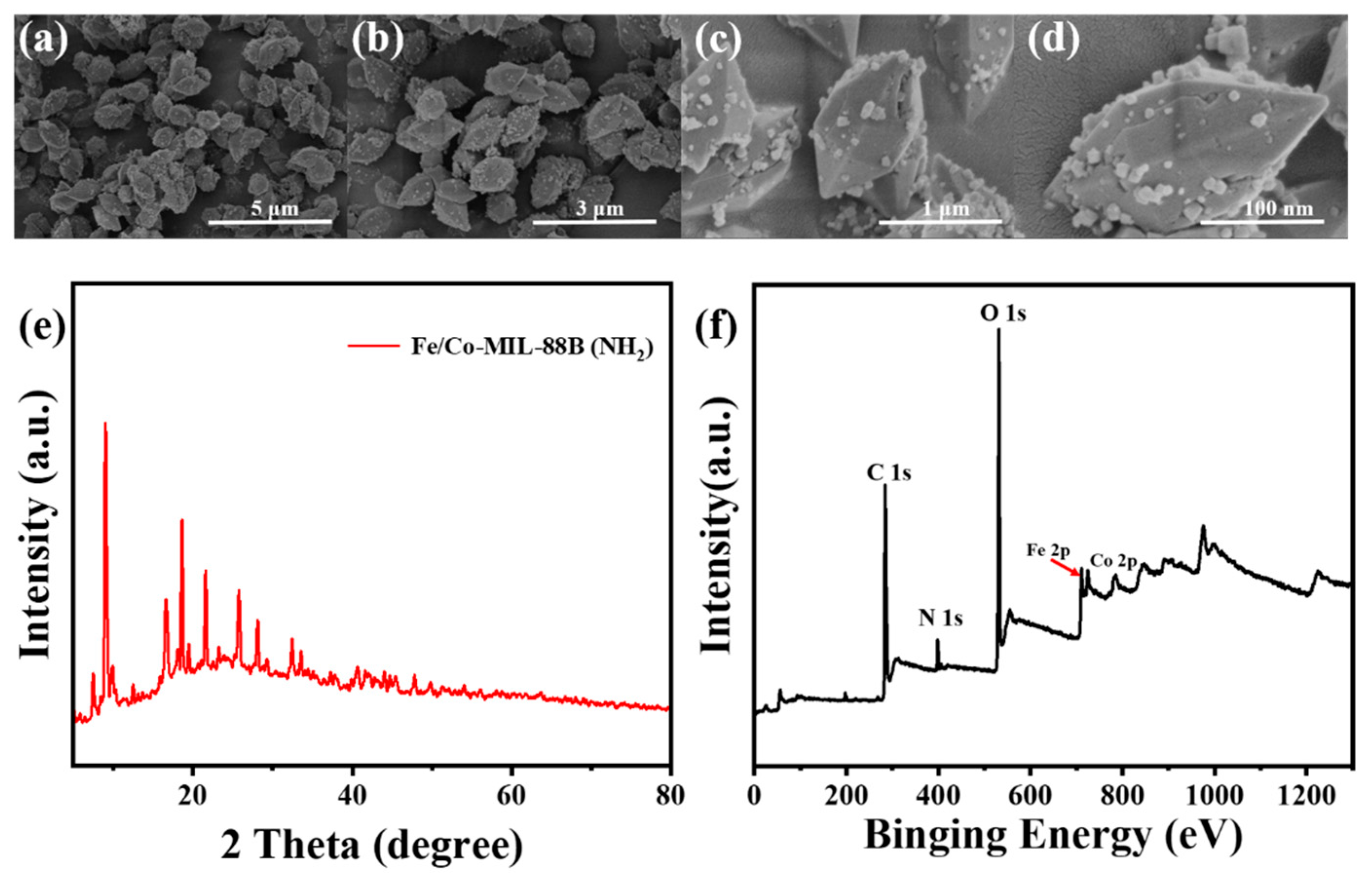
The PXRD pattern of the as-synthesized MOF is consistent with the simulated pattern of MIL-88B, confirming the successful synthesis (Figure 1e). The diffraction peaks observed at 9.2°, 10.1°, 16.5°, and 18.6° represent typical (002), (101), (103), and (200) planes of the MIL-88B structure, respectively [30,31,32,33]. The clear and distinct characteristic peaks associated with each crystalline plane further corroborate the high crystallinity of the synthesized Fe/Co-MIL-88B(NH2).
The elemental composition and chemical state of MOF are subsequently investigated by XPS (Figure 1f). The characteristic peaks at 284 eV, 398 eV, and 531 eV belong to C 1s, N 1s, and O 1s, respectively [34]. The Fe 2p spectrum displays peaks at 710.6 eV and 724.3 eV, assigned to Fe 2p3/2 and Fe 2p1/2, respectively (Figure S1). Additionally, the presence of the satellite peak at 717.1 eV is indicative of Fe3+, confirming that Fe exists in the Fe (III) oxidation state within Fe/Co-MIL-88B(NH2) [35]. The Co 2p spectrum reveals peaks at 783.3 eV and 795.1 eV (Figure S2), corresponding to Co 2p3/2 and Co 2p1/2, respectively [36].
The SEM images reveal that Fe/Co-MIL-88B (NH2) exhibits a regular octahedral double-cone morphology, with small particle attachments observed on the surface (Figure 1a–d). This morphology closely aligns with the previously reported structure [37], further verifying the successful synthesis of Fe/Co-MIL-88B (NH2).

2.3. Characterization of Membranes

Figure 2a–c depicts SEM images of the pristine PVDF membrane, while Figure 2d–f displays those of the Fe/Co-MIL-88B(NH2)/CaO2/PVDF composite membrane. A significant morphological transformation is observed with the structure evolving from the dense, non-porous architecture of PVDF membrane to the reticulated porous network of composite membrane. The presence of Fe/Co-MIL-88B(NH2) particles on the surface of the composite membrane is discernible, indicating successful surface loading. Furthermore, CaO2 particles are observed within the membrane cross-section, corroborating the successful preparation of the Fe/Co-MIL-88B(NH2)/CaO2/PVDF composite membrane.
The functional groups present in different membranes were examined using FT-IR spectroscopy. As shown in Figure 3a, the Fe/Co-MIL-88B(NH2)/CaO2/PVDF composite membrane displays two peaks at 1642 cm−1 and 1427 cm−1, corresponding to the asymmetric and symmetric vibrations of coordinated carboxyl groups, respectively, indicating successful coordination of the organic ligands with metal centers. The peak at 831 cm−1 corresponds to the out-of-plane bending vibration of aromatic C-H in organic ligands, while the low-frequency peak at 478 cm−1 is assigned to Fe-O vibrations, confirming the presence of Fe-carboxylate coordination in the MOF structure. These spectral features are consistent with the structural characteristics of MIL-88B [38]. Additionally, the absorption bands 1159 cm−1 at and 890 cm−1 are attributed to the C-F stretching vibration in the PVDF matrix and the presence of Ca2+ binding, respectively [39,40], further confirming the fabrication of composite membrane.
The original PVDF membrane and Fe/Co-MIL-88B(NH2)/CaO2/PVDF composite membrane are analyzed by PXRD characterization. In Figure 3b, diffraction peaks observed at 20.5° and 42.7° in both membranes correspond to the semicrystalline structure of PVDF [41]. Additionally, the sharp peaks at 29.5°, 31.7°, 35.6°, and 45.4° appear exclusively in the composite membrane, indicating the presence of crystalline phases attributed to the incorporation of Fe/Co-MIL-88B(NH2). This phenomenon may be attributed to the shift in diffraction peaks of Fe/Co-MIL-88B(NH2) [33].
The elemental composition of the membranes was analyzed by XPS. As illustrated in Figure 3c, compared to the original PVDF membrane, the composite membrane exhibits additional signals related to Fe, Co, and Ca elements, confirming the successful incorporation of Fe/Co-MIL-88B(NH2) and CaO2.
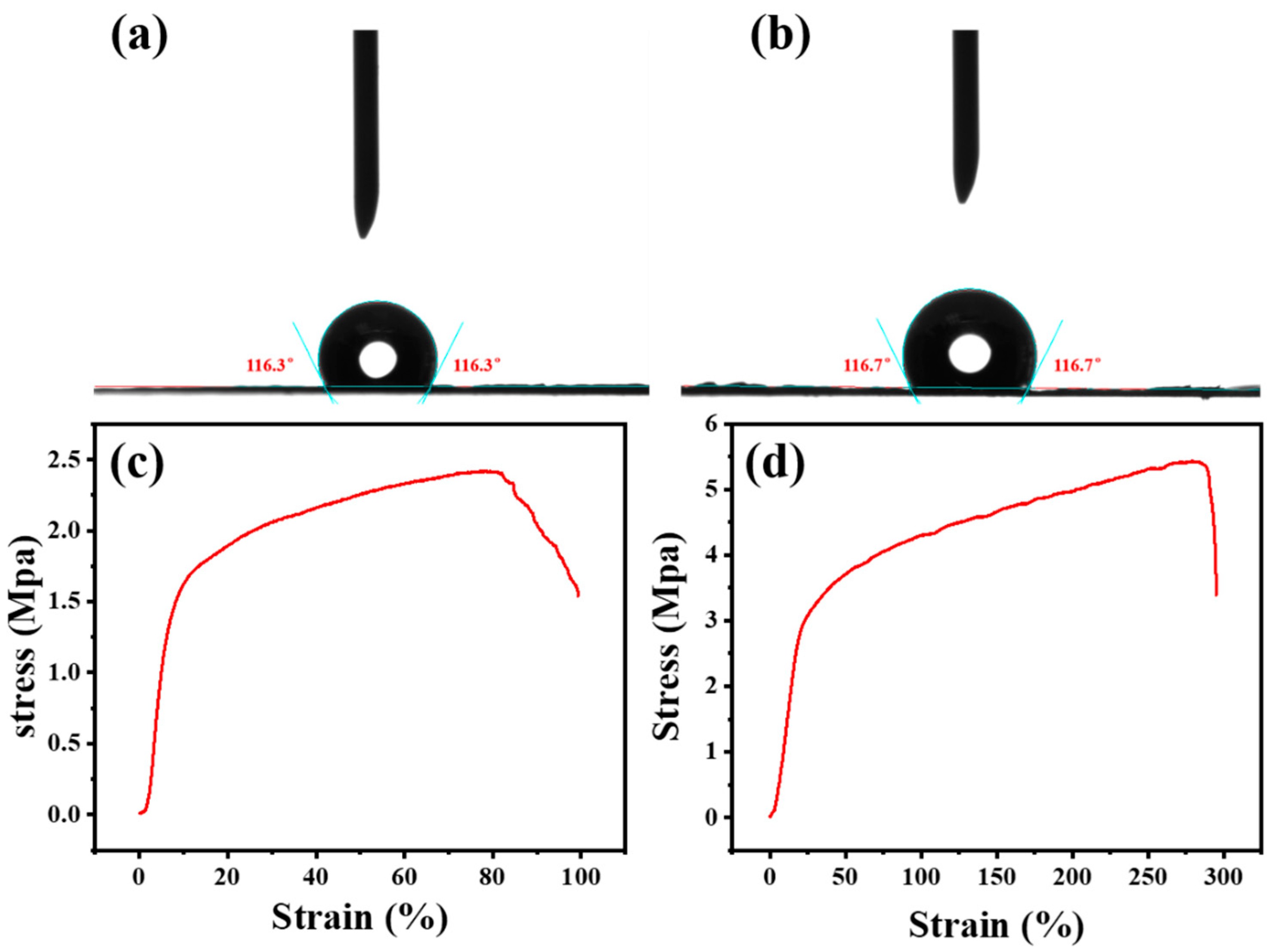
To assess the alteration in surface wettability of the composite membrane, static contact angle measurements are conducted on both the original PVDF membrane and the Fe/Co-MIL-88B(NH2)/CaO2/PVDF composite membrane (Figure 4a,b). The contact angle of the original PVDF membrane is measured at 116.3°, while that of the composite membrane is 116.7°. The minimal changes in water contact angle demonstrate that the incorporation of Fe/Co-MIL-88B(NH2) and CaO2 has little influence on surface wettability. Moreover, both membranes exhibit contact angles greater than 90°, indicating a hydrophobic characteristic. This inherent hydrophobicity is conducive to maintaining membrane stability in aqueous environments. Additionally, the relatively smooth surface of these membranes also contributes to the high water contact angles as the surface roughness is known to influence wettability [42].

2.4. Mechanical Properties of the Membrane

The mechanical properties of the original PVDF membrane and composite membrane are evaluated. As shown in Figure 4c,d and Table S1, the tensile strength, elongation at break, and elastic modulus of the original PVDF membrane are measured to be 2.42 MPa, 99.27%, and 35.5 MPa, respectively. Due to the incorporation of Fe/Co-MIL-88B(NH2) and CaO2, the composite membrane exhibits significant enhancement in mechanical properties, with elongation at break increasing nearly threefold to 294.98% and the tensile strength doubling to 5.43 MPa. In contrast, the elastic modulus of the composite membrane reduces to 15.87 MPa, implying a reduction in stiffness. These results indicate that the prepared Fe/Co-MIL-88B(NH2)/CaO2/PVDF composite membrane exhibits significantly improved flexibility, albeit at the expense of reduced rigidity, compared to the original PVDF membrane.

2.5. Degradation Condition Experiment

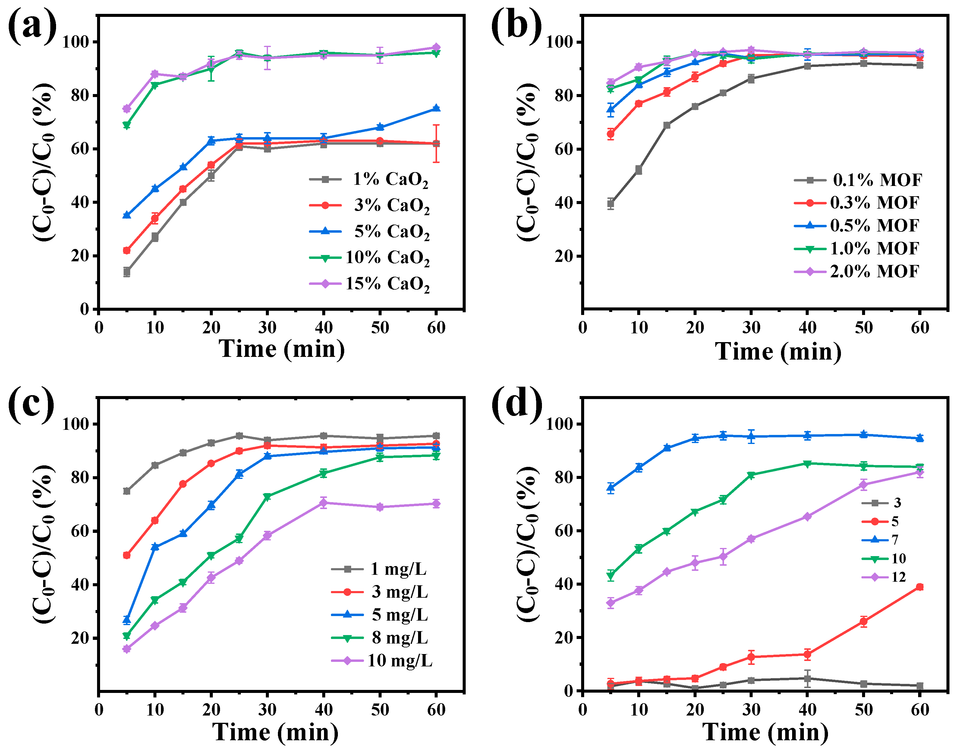
The degradation performance of the Fe/Co-MIL-88B(NH2)/CaO2/PVDF composite membrane under various conditions is systematically evaluated using a controlled variable method, including changes in CaO2 content, Fe/Co-MIL-88B(NH2) content, AFB1 concentration, and pH value. As depicted in Figure 5a, the degradation rate of AFB1 gradually increases with increasing CaO2 content. However, when the CaO2 content exceeds 10% to 15%, the degradation rate reaches a plateau, suggesting that 10% CaO2 content may represent a saturation threshold and optimal loading level, given the confines of membrane volume. Figure 5b illustrates the impact of varying Fe/Co-MIL-88B(NH2) content in the composite membrane on degradation rate. The degradation rate rises with higher Fe/Co-MIL-88B(NH2) content, and it remains consistent at approximately 95% across different MOF contents, though differences become insignificant beyond a content exceeding 0.5%. Therefore, the optimal composition for fabricating composite membranes appears to be 10% CaO2 and 0.5% Fe/Co-MIL-88B(NH2).
The degradation rate of AFB1 gradually decreases with escalating AFB1 concentration (Figure 5c). Specifically, when the concentration of AFB1 increases from 1 mg/L to 10 mg/L, the degradation rate declines from 95% to approximately 70%. At different pH values, the degradation rate varies (Figure 5d). The composite membrane exhibits the highest degradation rate of AFB1 under neutral conditions, followed by alkaline conditions, and the lowest under acidic conditions. Particularly, at the pH of 3, the composite membrane shows almost no degradation capability toward AFB1. These findings indicate that the composite membrane demonstrates favorable degradability under neutral and mildly alkaline environments. The reduced efficiency under acidic conditions may be attributed to the inhibitory effect of excess H+ ions on the enzyme-mimetic activity of Fe/Co-MIL-88B(NH2).
To further evaluate the degradation performance of the Fe/Co-MIL-88B(NH2)/CaO2/PVDF composite membrane, the biodegradation experiment toward AFM1 (1mg/L) is carried out under the optimal degradation conditions of 10% CaO2 content, 5% Fe/Co-MIL-88B(NH2) content, and a neutral environment, as described in Section 2.5. The results, presented in Figures S3 and S4, demonstrate that the composite membrane achieves a degradation rate of 96% for AFM1 within 1 h, confirming the good degradability of the composite membrane for aflatoxin.

2.6. Comparison of Degradation Performance of Different Membrane Compositions

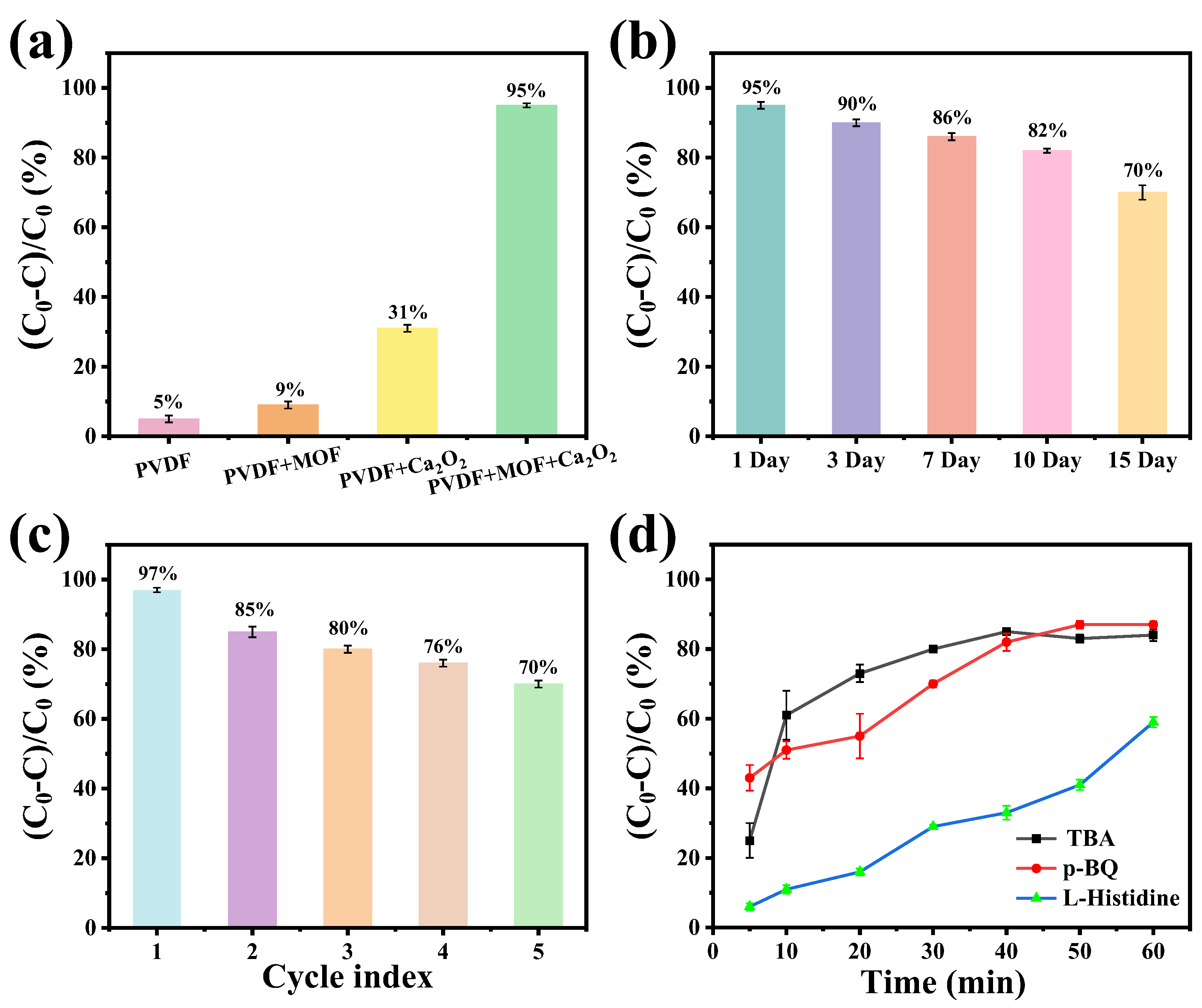
To investigate the influence of membrane compositions on AFB1 degradation, different material combinations are added during membrane synthesis, including single PVDF, PVDF + Fe/Co-MIL-88B(NH2), PVDF + CaO2, and PVDF + CaO2 + Fe/Co-MIL-88B(NH2), respectively (Figure 6a). Neither the PVDF membrane nor the Fe/Co-MIL-88B(NH2)/PVDF composite membrane showed any degradation activity toward AFB1. The degradation rate of the PVDF + CaO2 composite membrane reaches only approximately 30%, which may be attributed to the oxidative effect of H2O2 generated from the decomposition of CaO2 [11,43]. Notably, with the addition of the Fe/Co-MIL-88B(NH2) and CaO2, the degradation rate reaches 95%, indicating the synergistic effect between CaO2 and Fe/Co-MIL-88B(NH2) for effective degradation of AFB1.

2.7. Stability and Cyclability of Membranes

The degradation stability of the membrane is crucial for its practical application. Therefore, the stability and recyclability of the membrane are investigated separately. As depicted in Figure 6b, the Fe/Co-MIL-88B(NH2)/CaO2/PVDF composite membrane exhibits commendable stability during the 15-day test period, with the degradation rate remaining above 75% after 15 days. The decline in degradation rate may be attributed to the reduction in CaO2 content within the Fe/Co-MIL-88B(NH2)/CaO2/PVDF composite membrane during the reaction, leading to a decrease in hydrogen peroxide yield. The degradation rate remains above 80% even after five consecutive cycles, highlighting the excellent recyclability of the membrane (Figure 6c). These results confirm that the Fe/Co-MIL-88B(NH2)/CaO2/PVDF composite membrane possesses outstanding stability and holds strong potential for long-term practical applications.

2.8. Comparison of Metal Leaching Performance

The degradation of pure Fe/Co-MIL-88B(NH2) resulted in Fe and Co concentrations of 1272 µg/L and 2533 µg/L, respectively, in the post-reaction solution. In contrast, the degradation of Fe/Co-MIL-88B(NH2)/CaO2/PVDF composite membrane yielded Fe and Co concentrations below the detection limit, indicating negligible metal leaching. This significantly lower metal concentration is well below the safety limit. It shows that the use of Fe/Co-MIL-88B(NH2)/CaO2/PVDF composite membrane can effectively prevent the overflow of metal ions and improve safety in food use [44].

2.9. Degradation Mechanism

In Figure 6d, the active substances were studied by adding active quenchers to the degradation reaction system. Specifically, p-benzoquinone (p-BQ), L-histidine, and tert-butyl alcohol (TBA) are added, respectively, each corresponding to O2•−, 1O2, and ·OH [45]. It can be found that L-histidine has the greatest impact on the degradation rate, while the other two groups (p-BQ, TBA) have relatively minor effects. These results indicate that 1O2 active substance plays an important role in the degradation reaction, with O2•− and ·OH playing secondary roles.

2.10. Degradation Products

The degradation products of AFB1 are analyzed via LC-MS/MS, revealing that the degradation of AFB1 is primarily attributed to alterations in its furan ring and methoxy group. The LC-MS/MS results indicate three distinct degradation pathways. As shown in Figure 7a, Pathway I involves the conversion of P1 AFB1 (313 m/z) to P2 (329 m/z) through epoxidation of terminal double bonds. Pathway II undergoes hydroxylation and structural modification to generate P3 (m/z 301). Subsequently, functional group cleavage combined with additional hydroxylation leads to the formation of P4 (m/z 274) [46]. Pathway III begins with the formation of P5 (m/z 340) from P1 through oxygen addition to the terminal furan ring and the benzene ring. Subsequent double-bond addition on the furan ring and demethylation of the benzene ring side chain generate P6 (m/z 318). Finally, the loss of CO results in the formation of P7 (m/z 284) [47].
The degradation products of AFM1 were further analyzed by LC-MS/MS, revealing two potential pathways (Figure 7b). In pathway I, AFM1 (P1, m/z 329) undergoes hydration and methoxylation at the terminal furan double bond to generate P2 (m/z 340) [48]. In pathway II, hydroxylation of P1 introduces two additional hydroxyl groups, leading to the formation of P3 (m/z 363). This modification likely results from hydration or reductive alteration of double bonds and/or carbonyl groups. P3 is then transformed into P4 (m/z 303) through dehydration and oxidative rearrangement, accompanied by the loss of hydroxyl groups and the generation of a carbonyl functionality [49].
These metabolites were identified with lower toxicity compared to the parent aflatoxin, suggesting that the degradation process reduces potential health risks. The LC-MS spectra of the degradation products described above are provided in Supporting Information Figures S4 and S5. It should be noted that this work focuses on demonstrating the possible degradation pathways, while the quantitative percentage of each degradation product was not determined.

2.11. Safety Test of Composite Membrane

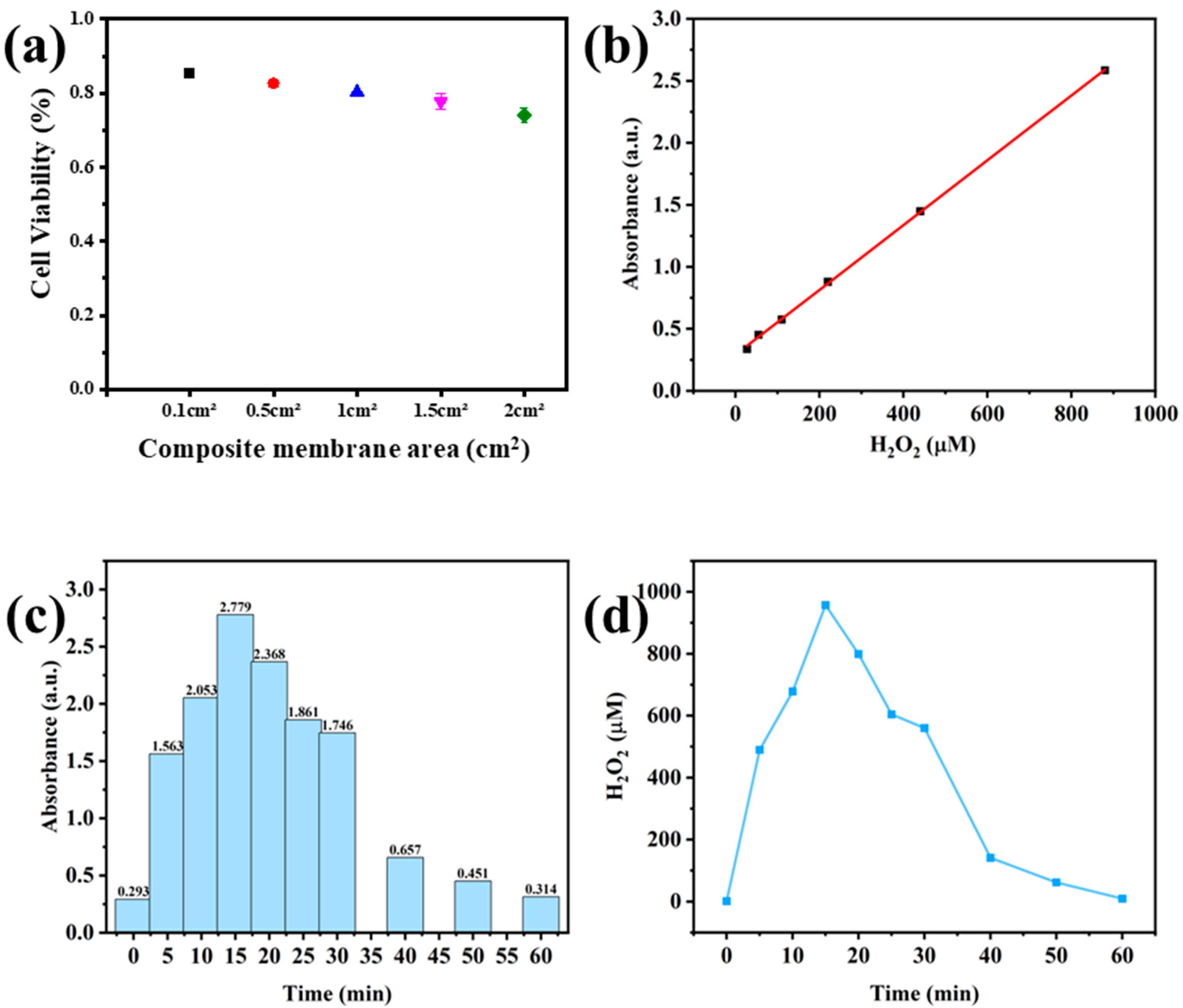
The cytotoxicity of the composite membrane is assessed through cytotoxicological assays, and cell viability is determined via MTT assays. As depicted in Figure 8a, cell survival rates consistently exceed 70% across various areas of the composite membrane. This suggests that the composite membrane exhibits low toxicity and poses no harm to the human body [50]. Examination of the degradation products revealed substantial disruption of the lactone ring, yielding non-toxic small-molecule byproducts [51].
Hydrogen peroxide concentration is monitored over time using UV-Vis spectrophotometry at 352 nm. A pre-established calibration curve (Figure 8b) is employed to convert absorbance readings to H2O2 concentrations. As depicted in Figure 8c,d, a substantial decrease in hydrogen peroxide levels is observed after 15 min of the reaction. After 60 min, the residual H2O2 concentration in the solution is approximately 8 µM, significantly lower than the maximum permissible residue limit of 0.5 mg/L for hydrogen peroxide in food products, as established by the U. S. Food and Drug Administration (FDA) [52,53]. Furthermore, the H2O2 concentration in the reaction solution is significantly lower than the typical 5 mM concentration commonly employed in degradation experiments, which often necessitates exogenous addition [54]. Such findings further affirm the safety of the membrane for use in food applications.

2.12. Actual Test of the Composite Membrane

The practical performance of the Fe/Co-MIL-88B(NH2)/CaO2/PVDF composite membrane is evaluated by introducing AFB1/AFM1 into milk to simulate real-world contamination scenarios. As shown in Figure S6, the degradation efficiencies of AFB1 and AFM1 in milk treated with Fe/Co-MIL-88B(NH2)/CaO2/PVDF composite membrane exceed 95%, indicating an effective toxin removal. Compared to existing methodologies, including enzymatic degradation and chemical treatments, this composite membrane achieves higher removal efficiency while maintaining milk quality and safety standards (Table S2). This superior performance is attributed to the synergistic catalytic activity of the Fe/Co-MIL-88B(NH2)/CaO2 component, which facilitates oxidative degradation of aflatoxins under mild conditions. Meanwhile, the initial concentration of the AFB1 and AFM1 used in this study was 1 mg/mL, and experiments confirmed that the constructed composite membrane achieved a degradation rate exceeding 95% for this concentration within 1 h. After treatment by this system, the residual toxin concentration can theoretically be reduced to below 50 μg/mL. The European Commission has set the maximum residue levels (MRLs) for AFB1 and AFM1 in edible foods and milk at 0.5 to 50 ng/mL [55]. The results indicate that this degradation technology can reduce the residual concentration to well below the EU’s maximum residue threshold, sufficiently demonstrating that its efficient toxin removal capability in practical applications meets and significantly exceeds conventional detection requirements. These findings highlight the practical relevance of the membrane for aflatoxin detoxification in milk and provide a basis for further application in food safety.
The results of quality assessments on raw and composite membrane-treated milk samples, conducted using National Food Safety Standard Methods, are summarized in Table S3. The protein, lipid, and lactose content of all three milk brands showed only minor variations following composite membrane treatment. These results further underscore the dependable applicability of the Fe/Co-MIL-88B(NH2)/CaO2/PVDF composite membrane under practical operating conditions.

3. Conclusions

In summary, we have developed a novel ultrathin composite membrane by synergistically integrating Fe/Co-MIL-88B(NH2) and CaO2 within a PVDF matrix for efficient and safe aflatoxin degradation from milk. This composite membrane simultaneously utilizes the porous structure and peroxidase-like catalytic activity of Fe/Co-MIL-88B(NH2), as well as the in situ controlled generation of H2O2 enabled by CaO2, effectively converting aflatoxins in milk into non-toxic small molecules. Moreover, the controlled release of H2O2 avoids the reliance on additional exogenous H2O2 and oxidative damage to other nutrients. Furthermore, by incorporating PVDF, the composite membrane eliminates the potential risk of metal ion leaching and nanoparticle aggregation, addressing the concern with the use of MOF-based nanozyme. As a result, the Fe/Co-MIL-88B(NH2)/CaO2/PVDF composite membrane achieves a superior degradation efficiency of AFB1 and AFM1, exceeding 95% within 60 min, and demonstrates outstanding stability, repeatability, and compactness. This work offers an efficient and reusable strategy for aflatoxin degradation, demonstrating substantial potential for practical application in dairy product processing.

4. Experimental Section

4.1. Materials

N, N-dimethylformamide (DMF), Ferric (III) chloride hexahydrate (FeCl3·6H2O), cobalt(II) nitrate hexahydrate (Co(NO3)2·6H2O), 2-Aminoterephthalic acid (H2(NH2)BDC), AFB1, and AFM1 are obtained from Aladdin Biochemical Technology Co., Ltd. (Shanghai, China). H2O2 (3%) and CaO2 are purchased from Yuanye Biochemical Technology Co., Ltd. (Shanghai, China). PVDF powders (Solef 1010), tert-butyl alcohol (TBA, 99%), p-Benzoquinone (p-BQ, 99%), L-histidine (99%), Dimethyl sulfoxide (DMSO), and 3-(4,5-dimethylthiazol-2-yl)-2,5-diphenyltetrazolium bromide (MTT) are obtained from Macklin Biochemical Technology Co., Ltd. (Shanghai, China). Potassium iodide (KI) was obtained from Sigma-Aldrich (St. Louis, MO, USA). The whole milk (3.5% fat) is obtained from the Mengniu Dairy. All reagents can be used directly without purification; the aqueous solution is ultra-pure water with a resistivity of 18.2 MΩ.cm.

4.2. Preparation of Fe/Co-MIL-88B(NH2)

Fe/Co-MIL-88B (NH2) is synthesized via a solvothermal method as in previous reports [37]. Specifically, 0.82 g FeCl3·6H2O, 0.90 g Co(NO3)2·6H2O and 0.53 g H2(NH2)BDC are dissolved in 31.81 g of DMF. The mixture is stirred for 1 h and then poured into a 100 mL Teflon-lined reactor. The reactor is heated at 120 °C for 24 h in an oven. After the reaction, the powdered material is naturally cooled to room temperature, then washed five times with DMF, centrifuged, and dried at 60 °C for 8 h to obtain Fe/Co-MIL-88B (NH2) nanozyme.

4.3. Membrane Preparation

Fe/Co-MIL-88B(NH2)/CaO2/PVDF composite membranes are prepared using the non-solvent induced phase separation (NIPS) method [21] with modification. Initially, powdered Fe/Co-MIL-88B (NH2) and CaO2 are uniformly dispersed in DMF solvent for 10 min using ultrasonic vibration to prevent significant aggregation of particles in the system. Subsequently, PVDF powder is added to the above solution and dissolved by mechanical stirring at 50 °C for 4 h to form a casting solution. Finally, the casting solution is degassed at a constant temperature for 4 h to eliminate bubbles. The Fe/Co-MIL-88B(NH2)/CaO2/PVDF composite membrane is then cast onto a glass plate using a membrane coater.
After casting, the membrane is immersed in deionized water and washed several times with deionized water to remove residual solvent. It is then stored in deionized water and replaced with deionized water every 12 h.

4.4. Characterization

The morphology and structure of Fe/Co-MIL-88B (NH2) and the membrane are observed by scanning electron microscope (SEM, JEOL Ltd, JSM-5610LV, Japan, procuring Country: Nanjing, China). The Fourier-transform infrared (FT-IR) spectrum of Fe/Co-MIL-88B (NH2) was recorded using an FT-IR spectrometer (Bruker Corporation, Vertex 70, Germany, procuring Country: Nanjing, China). The spectrum of the composite membrane was recorded using an attenuated total reflection FT-IR (ATR-FTIR, Thermo Nicolet Corporation, Nexus 670, USA, procuring Country: Nanjing, China). The powder X-ray diffraction (PXRD) patterns are measured by a Siemens X-ray diffractometer (XRD, SIEMENS AG, D5000, Germany, procuring Country: Nanjing, China) (D5000). The X-ray photoelectron spectroscopy (XPS) spectrum is recorded by an Escalab250Xi photoelectron spectrometer (ThermoScientific, USA, procuring Country: Nanjing, China).
The concentration of metal ions in the degraded solution is detected by inductively coupled plasma mass spectrometry (ICP-MS, Agilent Technologies Inc., Agilent 7850 (MS), USA, procuring Country: Nanjing, China).
The surface hydrophilicity of the membrane is measured using a contact angle analyzer (DataPhysics Instruments Co., Ltd, Dataphysics DCA T21, Germany, procuring Country: Nanjing, China). The mechanical properties of the membrane are tested by an electronic automatic universal testing machine (INSTRON CORPORATION, CMT6103/ZWICK/Instron 5969, USA, procuring Country: Nanjing, China).

4.5. Degradability Test

At room temperature, the degradation reaction is carried out in a 50 mL centrifuge tube. A 1 cm2 Fe/Co-MIL-88B(NH2)/CaO2/PVDF composite membrane is immersed in 20 mL of an aqueous AFB1 or AFM1 solution (1 mg/mL) for 60 min of degradation. At specific times, 0.5 mL of the mixture is collected and filtered through a 0.22 μm polyethersulfone (PES) syringe filter, and then the AFB1 and AFM1 concentration is subsequently determined. The degradation rate (denoted as R) is calculated using the following equation:
R ( % ) =   ( C 0 C )   /   C 0 × 100 %
where C0 and C (mg/L) represent the AFB1 concentration in the aqueous solution at the initial reaction and the end of the reaction, respectively.

4.6. Metal Leaching Test

4 mg of Fe/Co-MIL-88B(NH2) is added to 20 mL of solution containing AFB1 (1 mg/L), while 227 μL of H2O2 (3%) solution is added for the degradation reaction. Collect 10 mL of reaction solution after membrane and Fe/Co-MIL-88B(NH2) degradation of toxins, centrifuge at 8000 rpm for 10 min, and analyze by ICP-MS. To demonstrate the safety of the composite membrane, the heavy metal content in the degradation solution is analyzed using ICP-MS, confirming that no nanoparticles from the composite membrane leaked into the solution.

4.7. Analytical Method

The sample is tested using high-performance liquid chromatography (HPLC, Shimadzu Co., Ltd., Japan) and a liquid chromatograph mass spectrometer (LC-MS, LC-MS-8050, Shimadzu Co., Ltd., Japan, procuring Country: Nanjing, China).
A Hypersil GOLD aQ C18 column (2.5 μm × 4.6 mm × 250 mm, Thermo Scientific™) is used to determine the concentration of AFB1 with HPLC (Shimadzu Co., Ltd., Japan, procuring Country: Nanjing, China). The ultra-pure water/methanol (50:50 v/v) is used as the mobile phase. The flow rate is 1 mL/min with a temperature of 35 °C. The excitation wavelength is 360 nm, and the emission wavelength is 440 nm. The total running time of HPLC analysis is 15 min, and the injection volume is 10 μL. The limit of detection (LOD) of the method is 0.1 ng/mL. The peak area is used to calculate the removal rate of AFB1.
The same C18 column is used to determine the concentration of AFM1 with HPLC using the same mobile phase, flow rate, and temperature. The excitation wavelength is 365 nm, and the emission wavelength is 435 nm. The total running time of HPLC analysis is 15 min, and the injection volume is 10 μL. The peak area is used to calculate the removal rate of AFM1.
The degradation products were determined by LC-MS. The mobile phase consists of 50% 0.1% formic acid water and 50% methanol with a flow rate of 0.3 mL/min. The chromatographic separation was carried out on a C18 column (ACQUITY UPLC BEH, 1.7 μm, 2.1 × 50 mm) with an injection volume of 10 μL. Mass spectrometry was performed in positive ion mode using an ESI source with the capillary voltage of 4.0 kV, the source temperature of 300 °C, the desolvation temperature of 526 °C, and a desolvation gas flow rate of 10 L/min. The mass spectrometer was operated in full scan mode with a scan range of m/z 100–1000 or m/z 100–500.

4.8. Safety Experiment

Cytotoxicological experiments are carried out by the Methyl Thiazolyl Tetrazolium (MTT) method, and HepG2 cells are selected as experimental objects to determine cell viability. HepG2 cells are seeded into 48-well plates and cultured in a carbon dioxide incubator for 24 h. Separately, the membrane samples of different areas (0.1 cm2, 0.5 cm2, 1.0 cm2, 1.5 cm2, and 2.0 cm2) are incubated in 200 μL of culture medium for 24 h to obtain membrane-conditioned medium. After incubation, the supernatant is collected and applied to the HepG2 cells. Subsequently, 90 μL of fresh medium and 10 μL of MTT solution (5 mg/mL) are added to the supernatant, and the mixture is incubated for 4 h. At the end, the supernatant is taken, and 100 μL of DMSO is added to each well. The plate is then placed on a low-speed shaker for 10 min to ensure that the crystals are completely dissolved. Finally, the absorbance of each well was measured at 490 nm using a microplate reader due to the instrumental equipment, as 490 nm is also commonly used in MTT assays. All measurements were performed consistently under identical conditions to ensure reliable comparisons.
The cell viability is calculated as follows:
Cell Viability (%) = (A1A2)/(A0A1)
A1 represents the absorbance of the cells after incubating with different sample solutions, A0 represents the absorbance of the cells after incubating with the medium, and A2 represents the absorbance of the blank group.
To monitor residual hydrogen peroxide levels, a 1 cm2 Fe/Co-MIL-88B(NH2)/CaO2/PVDF composite membrane is immersed in 20 mL of a 1 mg/L AFB1 solution. Iodometric titration is used to quantify the H2O2 concentration. Every 5 to 10 min, 3 mL of the reaction solution is withdrawn, filtered through a 0.22 μm PES syringe filter to remove the composite membrane, and analyzed. The filtrate is treated with 3 mL of 0.1 M potassium hydrogen phthalate (KHP) and 3 mL of 0.4 M potassium iodide (KI), stirred, and allowed to react for 30 min. The absorbance of the resulting solution at 352 nm is measured using UV-Vis spectrophotometry.

4.9. Actual Test

20 mL of milk without AFB1 or AFM1 contamination is placed into a 50 mL centrifuge tube and centrifuged at 8000 rpm at −4 °C for 15 min. Subsequently, AFB1, AFM1, and a 1 cm2 Fe/Co-MIL-88B(NH2)/CaO2/PVDF composite membrane are added to the centrifuge tube, and the mixture is allowed to react for 60 min. Additionally, 4 mL of milk is mixed with 0.4 g of sodium chloride and 12 mL of ethyl acetate on a shaker at 300 rpm for 10 min. The mixture is then centrifuged for 10 min at 8000 rpm to separate the organic layer. The resulting 6 mL supernatant was evaporated under a nitrogen flow at 40 °C and subsequently re-dissolved in a mixture of 1 mL of 15% acetonitrile/H2O (V:V) and 0.1% formic acid. The concentration of AFB1 and AFM1 after degradation is determined following filtration through a 0.22 μm PES syringe filter.
To assess the potential impact of composite membrane treatment on milk quality, 15 mL of treated milk is analyzed for protein, lipid, and lactose content using standardized methods outlined by the National Food Safety Standards. Untreated milk samples serve as a control group. Three different commercially available milk brands sourced from local supermarkets in Nanjing are selected for this study.

Supplementary Materials

The following supporting information can be downloaded at https://www.mdpi.com/article/10.3390/toxins17100516/s1: Figure S1: High-resolution XPS spectra of Fe 2p. The spectra is shown for qualitative observation; no quantitative values are implied; Figure S2: High-resolution XPS spectra of Co 2p. The spectra is shown for qualitative observation; no quantitative values are implied. Figure S3: Actual photos of different film samples. (c) PVDF, (b) PVDF+CaO2, (c) PVDF+Fe/Co-MIL-88B(NH2), and (d) PVDF+CaO2+Fe/Co-MIL-88B(NH2); Figure S4: LC-MS spectra for the AFB1 degradation; Figure S5: LC-MS spectra for the AFM1 degradation; Figure S6: Actual test of membrane; Table S1: Mechanical properties of membrane; Table S2: Comparison of different methods for AFB1 degradation; Table S3: Effect of composite membrane-treated milk samples. References [56,57,58,59,60] are cited in the Supplementary Materials.

Author Contributions

X.C.: Writing—original draft, Validation, Methodology, Investigation, Formal analysis. W.Z.: Writing—original draft, Investigation, Formal analysis. X.Z.: Writing—original draft, Software, Methodology, Investigation, Formal analysis. J.Z.: Writing—original draft, Data curation. J.Y.: Writing—review and editing, Methodology, Investigation. H.W.: Methodology, Investigation. X.M.: Writing—review and editing, Methodology, Investigation. C.Z.: Writing—review and editing. L.W.: Writing—review and editing, Visualization, Validation, Supervision, Resources, Funding acquisition, Formal analysis. All authors have read and agreed to the published version of the manuscript.

Funding

This research was funded by the Jiangsu Basic Research Center for Synthetic Biology Grant, grant number: BK20233003; the Open Research Projects of the Key Laboratory of Biotoxin Analysis & Assessment, the State Administration for Market Regulation and the Key Laboratory of Catering Food Safety and the Systematic Monitoring for Jiangsu Province Market Regulation, grant number: BAA202401; the Key University Science Research Project of Jiangsu Province, grant number: 25KJA550001.

Institutional Review Board Statement

Not applicable.

Informed Consent Statement

Not applicable.

Data Availability Statement

The original contributions presented in this study are included in the article/Supplementary Materials. Further inquiries can be directed to the corresponding authors.

Conflicts of Interest

Author Xiaofei Cheng was employed by the company Inner Mongolia Mengniu Dairy (Group) Limited by Share Limited. The remaining authors declare that the research was conducted in the absence of any commercial or financial relationships that could be construed as a potential conflict of interest.

References

  1. Ji, J.; Xie, W. Detoxification of Aflatoxin B1 by magnetic graphene composite adsorbents from contaminated oils. J. Hazard. Mater. 2020, 381, 120915. [Google Scholar] [CrossRef]
  2. Ren, Z.; Luo, J.; Wan, Y. Enzyme-Like Metal-Organic Frameworks in Polymeric Membranes for Efficient Removal of Aflatoxin B1. ACS Appl. Mater. Inter. 2019, 11, 30542–30550. [Google Scholar] [CrossRef]
  3. Zhang, Y.; Sun, Y.; Man, Y.; Jiang, X.; Zhao, R.; Xiang, G.; He, L. Construction of a controllable and dispersed Fe3O4-based catalyst using ZIFs as a spatial support for highly catalytic degradation of aflatoxin B1. Appl. Catal. B Environ. Energy 2022, 318, 121818. [Google Scholar] [CrossRef]
  4. Cao, H.; Liang, D.; Tang, K.; Sun, Y.; Xu, Y.; Miao, M.; Zhao, Y. SERS and MRS signals engineered dual-mode aptasensor for simultaneous distinguishment of aflatoxin subtypes. J. Hazard. Mater. 2024, 462, 132810. [Google Scholar] [CrossRef]
  5. Turna, N.; Wu, F. Aflatoxin M1 in milk: A global occurrence, intake, & exposure assessment. Trends Food Sci. Tech. 2021, 110, 183–192. [Google Scholar]
  6. Guo, L.; Zhang, J.; Bao, Y.; Zhang, Y.; Zhang, D.; Ma, X.; Zhang, J. Label-free and highly sensitive detection of aflatoxin B1 by Ag IANPs via surface-enhanced Raman spectroscopy. Food Chem. 2024, 458, 140231. [Google Scholar] [CrossRef] [PubMed]
  7. Romero, A.; Sartori, A.; Caetano-Silva, M.; Alencar, S.; Calori, M.; Augusto, P. Ozone processing of peanut “milk”: Degradation of aflatoxins, impact on quality attributes and the potential effect on peanut allergens. J. Clean. Prod. 2023, 405, 136950. [Google Scholar] [CrossRef]
  8. Nguyen, T.; Palmer, J.; Loo, T.; Shilton, A.; Petcu, M.; Newson, H.; Flint, S. Investigation of UV light treatment (254 nm) on the reduction of aflatoxin M1 in skim milk and degradation products after treatment. Food Chem. 2022, 390, 133165. [Google Scholar] [CrossRef] [PubMed]
  9. Wang, S.; Herrera-Balandrano, D.; Shi, X.; Chen, X.; Liu, F.; Laborda, P. Occurrence of aflatoxins in water and decontamination strategies: A review. Water Res. 2023, 232, 119703. [Google Scholar] [CrossRef] [PubMed]
  10. Liu, S.; Zhou, Y.; Feng, Y.; Peng, Q.; Li, Y.; He, C.; Fang, Z.; Xiao, Y.; Fang, W. A cost-saving, safe, and highly efficient natural mediator for laccase application on aflatoxin detoxification. Food Chem. 2024, 455, 139862. [Google Scholar] [CrossRef]
  11. Nguyen, T.; Flint, S.; Palmer, J. Control of aflatoxin M1 in milk by novel methods: A review. Food Chem. 2020, 311, 125984. [Google Scholar] [CrossRef]
  12. Song, C.; Yang, J.; Wang, Y.; Ding, G.; Guo, L.; Qin, J. Mechanisms and transformed products of aflatoxin B1 degradation under multiple treatments: A review. Crit. Rev. Food Sci. 2024, 64, 2263–2275. [Google Scholar] [CrossRef] [PubMed]
  13. Sert, D.; Mercan, E. Effects of ozone treatment to milk and whey concentrates on degradation of antibiotics and aflatoxin and physicochemical and microbiological characteristics. LWT Food Sci. Technol. 2021, 144, 111226. [Google Scholar] [CrossRef]
  14. Ding, S.; Barr, J.; Lyu, Z.; Zhang, F.; Wang, M.; Tieu, P.; Li, X.; Engelhard, M.; Feng, Z.; Beckman, S.; et al. Effect of Phosphorus Modulation in Iron Single-Atom Catalysts for Peroxidase Mimicking. Adv. Mater. 2024, 36, e2209633. [Google Scholar] [CrossRef] [PubMed]
  15. Zeng, R.; Gao, Q.; Xiao, L.; Wang, W.; Gu, Y.; Huang, H.; Tan, Y.; Tang, D.; Guo, S. Precise Tuning of the D-Band Center of Dual-Atomic Enzymes for Catalytic Therapy. J. Am. Chem. Soc. 2024, 146, 10023–10031. [Google Scholar] [CrossRef]
  16. Shan, J.; Jin, X.; Zhang, C.; Huang, M.; Xing, J.; Li, Q.; Cui, Y.; Niu, Q.; Chen, X.; Wang, X. Metal natural product complex Ru-procyanidins with quadruple enzymatic activity combat infections from drug-resistant bacteria. Acta Pharm. Sin. B 2024, 14, 2298–2316. [Google Scholar] [CrossRef]
  17. Deng, Y.; Guo, M.; Zhou, L.; Huang, Y.; Srivastava, S.; Kumar, A.; Liu, J. Prospects, advances and biological applications of MOF-based platform for the treatment of lung cancer. Biomater. Sci 2024, 12, 3725–3744. [Google Scholar]
  18. Sun, Y.; Ding, S.; Zhao, X.; Sun, D.; Yang, Y.; Chen, M.; Zhu, C.; Jiang, B.; Gu, Q.; Liu, H.; et al. Self-reinforced MOF-based nanogel alleviates osteoarthritis by Long-Acting Drug Release. Adv. Mater. 2024, 36, e2401094. [Google Scholar] [CrossRef]
  19. Kirchon, A.; Feng, L.; Drake, H.; Joseph, E.; Zhou, H. From fundamentals to applications: A toolbox for robust and multifunctional MOF materials. Chem. Soc. Rev. 2018, 47, 8611–8638. [Google Scholar] [CrossRef]
  20. Tibbetts, I.; Kostakis, G. Recent Bio-Advances in Metal-Organic Frameworks. Molecules 2020, 25, 1291. [Google Scholar] [CrossRef]
  21. Tansell, A.; Jones, C.; Easun, T. MOF the beaten track: Unusual structures and uncommon applications of metal-organic frameworks. Chem. Cent. J. 2017, 11, 100. [Google Scholar] [CrossRef]
  22. Zhang, S.; Qin, S.; Xu, G.; Xie, W.; Xu, N.; Jiang, N.; Xu, Y.; Hu, X.; Su, Z. Co-based MOF as an efficient catalyst by peroxymonosulfate activation for degradation of tetracycline: Synthesis and performance. Crystengcomm 2024, 26, 3744–3753. [Google Scholar] [CrossRef]
  23. Choi, G.; Mandal, M.; Jung, H.; Panda, J.; Kwon, Y.; Zhang, K.; Vivek, E.; Shon, M.; Ravi, K.; Baek, K.; et al. Post-synthetic modifications (PSM)-induced defects in hybrid metal-organic frameworks (MOFs) to unleash potential in gas separation membrane applications. J. Mater. Sci. Technol. 2024, 201, 95–118. [Google Scholar] [CrossRef]
  24. Li, X.; Zheng, H.; Chen, J.; Xu, M.; Bai, Y.; Liu, T. MIL-101 (Fe)@Ag Rapid Synergistic Antimicrobial and Biosafety Evaluation of Nanomaterials. Molecules 2022, 27, 3497. [Google Scholar] [CrossRef] [PubMed]
  25. Chen, X.; He, D.; Shentu, J.; Yang, S.; Yang, Y.; Wang, Y.; Zhang, R.; Wang, K.; Qian, J.; Long, L. Smartphone-assisted colorimetric and near-infrared ratiometric fluorescent sensor for on-spot detection of H2O2 in food samples. Chem. Eng. J. 2023, 472, 144900. [Google Scholar] [CrossRef]
  26. Pan, J.; Yang, X.; Zhou, J.; Cheng, W.; Cheng, K. Novel ZIF-8/ZnS hollow polyhedral heterostructures derived from ZIF-8 with enhanced photocatalytic activity for degradation of aflatoxin B1. Prog. Nat. Sci. 2023, 33, 575–580. [Google Scholar] [CrossRef]
  27. Yuan, X.; Zhu, W.; Tang, K.; Zhang, P.; Lou, W. Closed-loop peroxide supply strategy: Engineering photo-nanozyme cascade systems for ultrafast degradation of mycotoxins in water. J. Environ. Chem. Eng. 2025, 13, 117909. [Google Scholar] [CrossRef]
  28. Zhu, X.; Wei, J.; Xu, S.; Zhu, Y.; Shen, W.; Wu, L. Metal-organic framework incorporated fungal mycelium membrane for synergistic mycotoxin degradation via adsorption, oxidation, and photocatalysis. Food Chem. 2025, 480, 143861. [Google Scholar] [CrossRef]
  29. Yuan, X.; Chen, X.; Xiong, J.; Cao, Y.; Ta, N.; Gao, R.; Lou, W. Engineering the primary and second coordination sphere of metal-organic framework boosts the peroxidase-like activity. Chem. Eng. J. 2025, 522, 166966. [Google Scholar] [CrossRef]
  30. Zheng, H.; Zhou, Y.; Wang, D.; Zhu, M.; Sun, X.; Jiang, S.; Fan, Y.; Zhang, D.; Zhang, L. Surface-functionalized PVDF membranes by facile synthetic Cu-MOF-74 for enhanced contaminant degradation and antifouling performance. Colloid. Surf. A 2022, 651, 129640. [Google Scholar] [CrossRef]
  31. Hong, W.; Li, C.; Tang, T.; Xu, H.; Yu, Y.; Liu, G.; Wang, F.; Lei, C.; Zhu, H. The photocatalytic activity of the SnO2/TiO2/PVDF composite membrane in rhodamine B degradation. New J. Chem. 2021, 45, 2631–2642. [Google Scholar] [CrossRef]
  32. Li, X.; Yu, Z.; Shao, L.; Feng, X.; Zeng, H.; Liu, Y.; Long, R.; Zhu, X. Self-cleaning photocatalytic PVDF membrane loaded with NH2-MIL-88B/CDs and Graphene oxide for MB separation and degradation. Opt. Mater. 2021, 119, 111368. [Google Scholar] [CrossRef]
  33. Li, L.; Zhang, H.; Zhang, M.; Wang, T.; Hou, X. MIL-88B(Fe)/cellulose microspheres as sorbent for the fully automated dispersive pipette extraction towards trace sulfonamides in milk samples prior to UPLC-MS/MS analysis. Anal. Chim. Acta 2022, 1232, 340420. [Google Scholar] [CrossRef]
  34. Wu, Y.; Fang, Y.; Fu, J.; He, L.; Kabtamu, D.; Matovic, L.; Li, F.; Li, J. Optimized scalable synthesis and granulation of MIL-88B(Fe) for efficient arsenate removal. J. Environ. Chem. Eng. 2022, 10, 108556. [Google Scholar] [CrossRef]
  35. Feng, J.; Wang, H.; Ma, Z. Ultrasensitive amperometric immunosensor for the prostate specific antigen by exploiting a Fenton reaction induced by a metal-organic framework nanocomposite of type Au/Fe-MOF with peroxidase mimicking activity. Microchim. Acta 2020, 187, 95. [Google Scholar] [CrossRef]
  36. Zhang, Q.; Xu, J.; Ma, X.; Xu, J.; Yun, Z.; Zuo, Q.; Wang, L. A novel Fe-based Bi-MOFs material for photocatalytic degradation of tetracycline: Performance, mechanism and toxicity assessment. J. Water Process Eng. 2021, 44, 102364. [Google Scholar] [CrossRef]
  37. Han, M.; Ren, M.; Li, Z.; Qu, L.; Yu, L. A two-dimensional thin Co-MOF nanosheet as a nanozyme with high oxidase-like activity for GSH detection. New J. Chem. 2022, 46, 10682–10689. [Google Scholar] [CrossRef]
  38. Jiang, Q.; Xiao, Y.; Hong, A.; Gao, Z.; Shen, Y.; Fan, Q.; Feng, P.; Zhong, W. Bimetallic Metal-Organic Framework Fe/Co-MIL-88(NH2) Exhibiting High Peroxidase-like Activity and Its Application in Detection of Extracellular Vesicles. ACS Appl. Mater. Inter. 2022, 14, 41800–41808. [Google Scholar] [CrossRef] [PubMed]
  39. Soetaredjo, F.; Santoso, S.; Lunardi, V.; Kurniawan, A.; Shuwanto, H.; Lie, J.; Foe, K.; Irawaty, W.; Yuliana, M.; Putro, J.; et al. Highly efficient degradation of organic pollutant mixtures by a Fe(III)-based MOF-catalyzed Fenton-like process in subcritical water. J. Mol. Liq. 2022, 347, 117989. [Google Scholar] [CrossRef]
  40. Wang, W.; Shi, Y.; Zhang, P.; Zhang, Z.; Xu, X. Metal-organic framework ZnL-PVDF supramolecular ultrafiltration membrane for enhanced separation performance. J. Phys. Chem. Solids 2022, 171, 110865. [Google Scholar] [CrossRef]
  41. Meng, J.; Xie, Y.; Gu, Y.; Yan, X.; Chen, Y.; Guo, X.; Lang, W. PVDF-CaAlg nanofiltration membranes with dual thin-film-composite (TFC) structure and high permeation flux for dye removal. Sep. Purif. Technol. 2021, 255, 117739. [Google Scholar] [CrossRef]
  42. Singh, N.; Madhav, H.; Yadav, S.; Jaiswar, G. Impact of vanadium-, sulfur-, and dysprosium-doped zinc oxide nanoparticles on various properties of PVDF/functionalized-PMMA blend nanocomposites: Structural, optical, and morphological studies. J. Appl. Polym. Sci. 2019, 136, 47116. [Google Scholar] [CrossRef]
  43. Shen, M.; Singh, R. Decomposing Aflatoxins in Peanuts Using Advanced Oxidation Processes by UV and H2O2. Food Bioprocess Tech. 2022, 15, 1647–1657. [Google Scholar] [CrossRef]
  44. Guo, R.; Li, Y.; Chen, Y.; Liu, Y.; Niu, B.; Gou, J.; Cheng, X. Efficient degradation of sulfamethoxazole by CoCu LDH composite membrane activating peroxymonosulfate with decreased metal ion leaching. Chem. Eng. J. 2021, 417, 127887. [Google Scholar] [CrossRef]
  45. Chen, D.; Bai, Q.; Ma, T.; Jing, X.; Tian, Y.; Zhao, R.; Zhu, G. Stable metal-organic framework fixing within zeolite beads for effectively static and continuous flow degradation of tetracycline by peroxymonosulfate activation. Chem. Eng. J. 2022, 435, 134916. [Google Scholar] [CrossRef]
  46. Faraji, A.R.; Gil, A.; Farahanipour, A.; Tehrani, E.; Khoramdareh, N.B.; Dashtabadi, E.; Saeedi, S. Synergic removal of Aflatoxin B1 in oily matrices by focusing on the peroxidase-like nanozymes-driven strategies: Mechanisms and intermediate toxicity, nutritional impact, advances and challenges. Trends Food Sci. Technol. 2025, 163, 105135. [Google Scholar] [CrossRef]
  47. Stanley, J.; Patras, A.; Pendyala, B.; Vergne, M.; Bansode, R. Performance of a UV-A LED system for degradation of aflatoxins B1 and M1 in pure water: Kinetics and cytotoxicity study. Sci. Rep. 2020, 10, 13473. [Google Scholar] [CrossRef] [PubMed]
  48. Nguyen, T.; Palmer, J.; Pedley, J.; Petcu, M.; Newson, H.L.; Keener, K.; Flint, S. The effect of variations in cold plasma conditions on the detoxification of Aflatoxin M1 and degradation products. Int. Dairy. J. 2025, 160, 106103. [Google Scholar] [CrossRef]
  49. Kousar, K.; Naseer, F.; Abduh, M.; Kakar, S.; Gul, R.; Anjum, S.; Ahmad, T. Green synthesis of hyaluronic acid coated, thiolated chitosan nanoparticles for CD44 targeted delivery and sustained release of Cisplatin in cervical carcinoma. Front. Pharmacol. 2023, 13, 1073004. [Google Scholar] [CrossRef]
  50. Nicolás-Vázquez, I.; Méndez-Albores, A.; Moreno-Martínez, E.; Miranda, R.; Castro, M. Role of Lactone Ring in Structural, Electronic, and Reactivity Properties of Aflatoxin B1: A Theoretical Study. Arch. Environ. Con Tox 2010, 59, 393–406. [Google Scholar] [CrossRef]
  51. Nascimento, C.; Santos, P.; Pereira, E.; Rocha, F. Recent advances on determination of milk adulterants. Food Chem. 2017, 221, 1232–1244. [Google Scholar] [CrossRef]
  52. Peng, T.; Ye, S.; Liu, R.; Qu, J. Colorimetric and fluorescent dual-signals probes for naked-eye detection of hydrogen peroxide and applications in milk samples and in vivo. Spectrochim. Acta A 2023, 297, 122757. [Google Scholar] [CrossRef]
  53. Belousov, A.; Parkhacheva, A.; Suleimanov, E.; Fukina, D.; Markov, A.; Vorotyntsev, A.; Koroleva, A.; Zhizhin, E.; Shafiq, I. Design of visible light-responsive CsM0.25W1.75O6 (M = Ni, Co, Mn, Cu) β-pyrochlore oxides with enhanced photocatalytic activity towards a set of pollutants. Ceram. Int. 2024, 50, 45334–45352. [Google Scholar] [CrossRef]
  54. Pacheco-Alvarez, M.O.A.; Sevillano-Arredondo, R.M.; Serrano, O.; Peralta-Hernandez, J.M. Copper-PANI-graphite HB2 composite for eco-friendly efficient degradation of textile dyes: Advancements in wastewater treatment enhanced by solar radiation. Chemosphere 2024, 366, 143537. [Google Scholar] [CrossRef]
  55. Beitollahi, H.; Tajik, S.; Dourandish, Z.; Zhang, K.; Le, Q.; Jang, H.; Shokouhimehr, M. Recent advances in the aptamer-based electrochemical biosensors for detecting aflatoxin B1 and its pertinent metabolite aflatoxin M1. Sensors 2022, 20, 3256. [Google Scholar] [CrossRef]
  56. Kurup, A.H.; Patras, A.; Pendyala, B.; Vergne, M.J.; Bansode, R.R. Evaluation of ultraviolet-light (UV-A) emitting diodes technology on the reduction of spiked aflatoxin B1 and aflatoxin M1 in whole milk. Food Bioprocess Technol 2021, 15, 165–176. [Google Scholar] [CrossRef]
  57. Bodbodak, S.; Hesari, J.; Peighambardoust, S.; Mahkam, M. Selective decontamination of aflatoxin M1 in milk by molecularly imprinted polymer coated on the surface of stainless steel plate. Int. J. Dairy Technol 2018, 71, 868–878. [Google Scholar] [CrossRef]
  58. Sangare, L.; Zhao, Y.; Folly, Y.; Chang, J.; Li, J.; Selvaraj, J.; Liu, Y. Aflatoxin B1 Degradation by a Pseudomonas Strain. Toxins 2014, 6, 3028–3040. [Google Scholar] [CrossRef]
  59. Du, X.; Zheng, M.; Zhang, H.; Qiu, Y.; Ji, F.; Nie, Z.; Xia, Y. New application of a dye-decolorizing peroxidase immobilized on magnetic nanoparticles for efficient simultaneous degradation of two mycotoxins. Food Chem. 2025, 463, 141341. [Google Scholar] [CrossRef]
  60. Zhou, L.; Duan, X.; Dai, J.; Ma, Y.; Yang, Q.; Hou, X. A covalent-organic framework-based platform for simultaneous smartphone detection and degradation of aflatoxin B1. Talanta 2024, 278, 126505. [Google Scholar] [CrossRef]
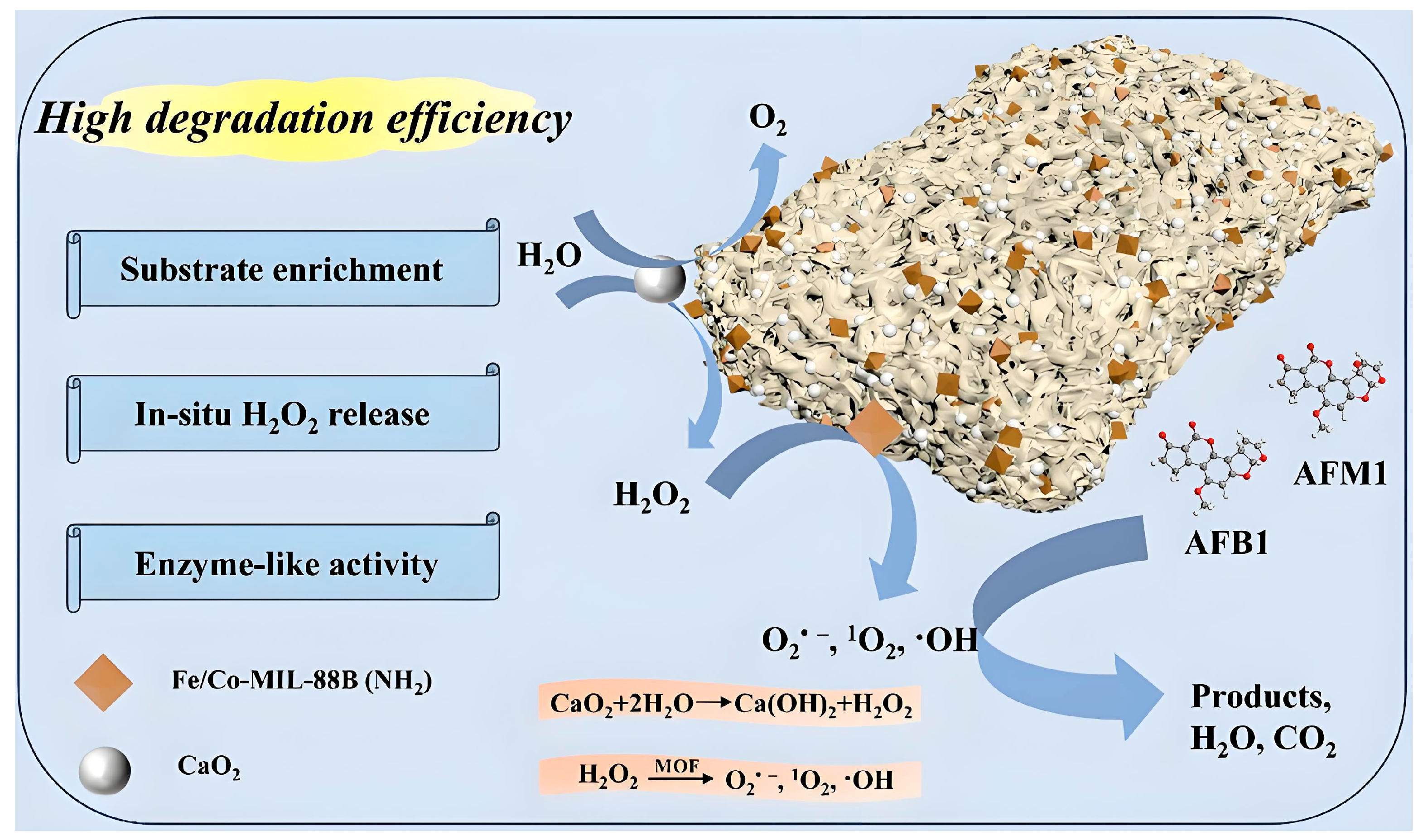
Scheme 1. The schematic illustration of the fabricated Fe/Co-MIL-88B(NH2)/CaO2/PVDF composite membrane for aflatoxin degradation.
Toxins 17 00516 sch001
Figure 1. (ad) SEM images, (e) PXRD pattern, and (f) XPS spectra of Fe/Co-MIL-88B(NH2). The pattern and spectra are shown for qualitative observation; no quantitative values are implied.
Toxins 17 00516 g001
Figure 2. (a,b) SEM images with the top-down view and (c) the SEM image with the cross-section view of the PVDF membrane. (d,e) SEM images with the top-down view and (f) the SEM image with the cross-section view of the Fe/Co-MIL-88B(NH2)/CaO2/PVDF composite membrane.
Toxins 17 00516 g002
Figure 3. (a) The FT-IR spectra, (b) PXRD patterns, and (c) full-scan XPS spectra of the PVDF membrane and the Fe/Co−MIL−88B(NH2)/Ca2O2/PVDF composite membrane. These spectra are shown for qualitative observation; no quantitative values are implied.
Toxins 17 00516 g003
Figure 4. The static contact angles of (a) the PVDF membrane and (b) the Fe/Co-MIL-88B(NH2)/CaO2/PVDF composite membrane. The stress–strain measurements of (c) the PVDF membrane and (d) the Fe/Co-MIL-88B(NH2)/CaO2/PVDF composite membrane.
Toxins 17 00516 g004
Figure 5. The degradation rate of the Fe/Co-MIL-88B(NH2)/CaO2/PVDF composite membrane toward AFB1 with different (a) CaO2 content, (b) Fe/Co-MIL-88B(NH2) content, (c) AFB1 concentration, and (d) pH values.
Toxins 17 00516 g005
Figure 6. AFB1 degradation changes in different (a) membrane compositions (MOF stands for Fe/Co-MIL-88B(NH2)), (b) Fe/Co-MIL-88B(NH2)/CaO2/PVDF stability testing, and (c) Fe/Co-MIL-88B(NH2)/CaO2/PVDF cyclable test. (d) Degradation rate under different quencher agents.
Toxins 17 00516 g006
Figure 7. (a) AFB1 degradation pathway. (b) AFM1 degradation pathway.
Toxins 17 00516 g007
Figure 8. (a) HepG2 cell survival after incubation with different membrane areas. (b) The linear relationship of H2O2 concentration and UV-Vis absorption intensity. (c) The absorbance of the solution at 352 nm at different times and (d) the calculated hydrogen peroxide concentration at different times.
Toxins 17 00516 g008
Disclaimer/Publisher’s Note: The statements, opinions and data contained in all publications are solely those of the individual author(s) and contributor(s) and not of MDPI and/or the editor(s). MDPI and/or the editor(s) disclaim responsibility for any injury to people or property resulting from any ideas, methods, instructions or products referred to in the content.