Article

Venom Proteomics of Trimeresurus gracilis, a Taiwan-Endemic Pitviper, and Comparison of Its Venom Proteome and VEGF and CRISP Sequences with Those of the Most Related Species

1
Institute of Wildlife Conservation, National Pingtung University of Science and Technology, Pingtung 912301, Taiwan
2
Institute of Biological Chemistry, Academia Sinica, Taipei 11529, Taiwan
3
Institute of Biochemical Sciences, National Taiwan University, Taipei 106319, Taiwan
4
Department of Biological Science and Technology, National Pingtung University of Science and Technology, Pingtung 912301, Taiwan
*
Author to whom correspondence should be addressed.
These authors contributed equally to this work.
Toxins 2023, 15(7), 408; https://doi.org/10.3390/toxins15070408
Submission received: 28 May 2023 / Revised: 17 June 2023 / Accepted: 20 June 2023 / Published: 22 June 2023
(This article belongs to the Special Issue Omics Approaches to Study Toxins)

Abstract

:
Trimeresurus gracilis is an endemic alpine pitviper in Taiwan with controversial phylogeny, and its venom proteome remains unknown. In this study, we conducted a proteomic analysis of T. gracilis venom using high-performance liquid chromatography-tandem mass spectrometry and identified 155 toxin proteoforms that belong to 13 viperid venom toxin families. By searching the sequences of trypsin-digested peptides of the separated HPLC fractions against the NCBI database, T. gracilis venom was found to contain 40.3% metalloproteases (SVMPs), 15.3% serine proteases, 6.6% phospholipases A2, 5.0% L-amino acid oxidase, 4.6% Cys-rich secretory proteins (CRISPs), 3.2% disintegrins, 2.9% vascular endothelial growth factors (VEGFs), 1.9% C-type lectin-like proteins, and 20.2% of minor toxins, nontoxins, and unidentified peptides or compounds. Sixteen of these proteoforms matched the toxins whose full amino-acid sequences have been deduced from T. gracilis venom gland cDNA sequences. The hemorrhagic venom of T. gracilis appears to be especially rich in PI-class SVMPs and lacks basic phospholipase A2. We also cloned and sequenced the cDNAs encoding two CRISP and three VEGF variants from T. gracilis venom glands. Sequence alignments and comparison revealed that the PI-SVMP, kallikrein-like proteases, CRISPs, and VEGF-F of T. gracilis and Ovophis okinavensis are structurally most similar, consistent with their close phylogenetic relationship. However, the expression levels of some of their toxins were rather different, possibly due to their distinct ecological and prey conditions.
Key Contribution: As part of the effort to conserve an endemic Taiwanese alpine pitviper species, this study analyzed the venom proteome of T. gracilis for the first time and compared its toxin sequences with the corresponding toxins from closely related pitviper genera such as Ovophis, Protobothrops, and Crotalus. Our results suggest that antivenom prepared with stronger antigenicity against pitvipers’ PI-SVMPs should be a better choice to treat T. gracilis envenoming. Characterization of the T. gracilis venom proteome and the structures and functions of its major toxins may improve our understanding of the pathophysiology of T. gracilis envenoming and aid the preparation and selection of antivenom for its snakebite treatment.

1. Introduction

Taiwan is a mountainous island, with two thirds of its territory covered by mountain forests. Today, alpine organisms worldwide face various threats such as climate change, air pollution, and human development; thus, research on alpine organisms is urgently needed. Among the more than 200 mountains in Taiwan higher than 3000 m, some glacial refuges and relic species have considerably high conservation value [1,2,3]. Trimeresurus gracilis Oshima, 1920 [4] is an endemic medium-sized pit viper distributed mainly at altitudes above 2000 m in central Taiwan. Remarkably, the taxonomy and genus name of T. gracilis have been controversial [5]. Phylogenetic analyses based on the mitochondrial and nuclear gene sequences of various pitvipers revealed that T. gracilis is phylogenetically close to Ovophis okinavensis in central Ryukyu [6,7,8]. However, T. gracilis gives live birth to its offspring, whereas O. okinavensis is among the few egg-laying pit viper species. Moreover, the prey ecology of T. gracilis and O. okinavensis is rather different [9,10], and how this affects their venom proteomes remains to be further explored and clarified.
T. gracilis snakebites mainly elicit hemorrhagic symptoms in patients, including local tissue damage (myonecrosis, dermal necrosis, edema, hemorrhage, and blistering) and systemic coagulopathy [11], although T. gracilis envenoming cases are rare. To date, no specific antivenom is available for T. gracilis envenomation, and the venom proteome has not been resolved. T. gracilis-envenomed patients have been treated with local “bivalent hemotoxic-antivenom” against Viridovipera stejnegeri and Protobothrops mucrosquamatus, but this antivenom failed to relieve the local lesions of the patients before they received surgical intervention [11].
Previously, we cloned and sequenced T. gracilis venom proteins belonging to three major toxin families: an acidic phospholipase A2 (PLA2) and a Lys49-homolog of PLA2 [12], ten venom serine proteases (SVSPs) [13], and five metalloproteinases (SVMPs) including PII-class and its disintegrin (DIS) domain [14]. We have shown that the amino acid sequences of some representative toxins, including Lys49-PLA2, several SVSP variants, and the PI-class metalloprotease of T. gracilis, are highly similar to those of the corresponding venom toxins of O. okinavensis [15,16]. The taxonomy of T. gracilis and its relationship with other eastern Asian pitvipers, meanwhile, remains puzzling, and this species and those under the genus Gloydius have been shown to be the most likely Asian sisters of New World pitvipers [7,17,18,19]. Thus, it is not surprising that PIII-SVMP and some SVSP variants expressed in T. gracilis venom bear high structural similarities to the corresponding toxins from New World pitvipers [13,14].
Venomic studies may provide important insights into the pathophysiology of envenoming and the molecular evolution of venom toxin multigene families [20]. Our aim was to investigate the venom proteome and full sequences of not only the major but also the secondary or minor toxin families of T. gracilis in order to better understand the composition and evolution of its venom and to treat snakebites effectively. In the present study, T. gracilis venom proteomics were studied using high-performance liquid chromatography (HPLC) and liquid chromatography-tandem mass spectrometry (LC-MS/MS). We also cloned and sequenced the venom gland cDNAs encoding Cys-rich secretory proteins (CRISPs) and vascular endothelial growth factors (VEGFs). These T. gracilis toxins were compared with other pitviper toxins using a BLAST search and sequence alignments. Our results may provide a deeper understanding of the venom composition of T. gracilis and the evolutionary relationships between T. gracilis and other related pitvipers, such as the east Asian Ovophis, Protobothrops, and the New World Crotalus.

2. Results

2.1. Chromatographic and Electrophoretic Profiling of T. gracilis Venom

Using a C18 reverse-phase column, crude T. gracilis venom was separated into 39 peptide/protein fractions by HPLC (Figure 1A). These fractions were collected and analyzed using sodium dodecyl sulfate-polyacrylamide gel electrophoresis (SDS-PAGE) under reducing conditions (Figure 1B). The first 13 fractions (eluted within the first 55 min) failed to show any bands on the gel. Notably, most medium-to high-MW venom proteins (5000–260,000) were eluted between 55 and 130 min and collected in fractions 14–39 (Figure 1A). As expected, most of the small peptides or proteins eluted earlier than large proteins, and the basic variants of the venom toxins eluted earlier than the acidic variants of the same toxin family. However, SDS-PAGE results revealed that most of the HPLC peaks contained multiple proteins or subunits rather than a single purified protein (Figure 1B).

2.2. T. gracilis Venom Proteomic Analysis

Mass spectrometric analyses of the proteins in the HPLC peaks (particularly the broader ones) revealed that they may contain several proteins from different toxin families (Table 1 and Supplementary Table S1). Overall, 155 toxin proteoforms were identified in the HPLC fractions, except for peaks 1–5 and 7. Sixteen of these proteoforms were T. gracilis venom proteins whose full amino-acid sequences have been deduced from venom gland transcriptome, including three SVMPs, six SVSPs, two PLA2s, two CRISPs, one DIS, and two VEGFs (Supplementary Table S2). By measuring the peak area under the curve of the HPLC chromatogram, the relative abundances of low-molecular-weight peptides, nontoxins (e.g., keratins), and unidentified compounds in the venom together accounted for 20.1%, which were mainly from the first 13 fractions. The identified peptides/proteins were categorized into 13 toxin families and sorted quantitatively (Supplementary Table S2).
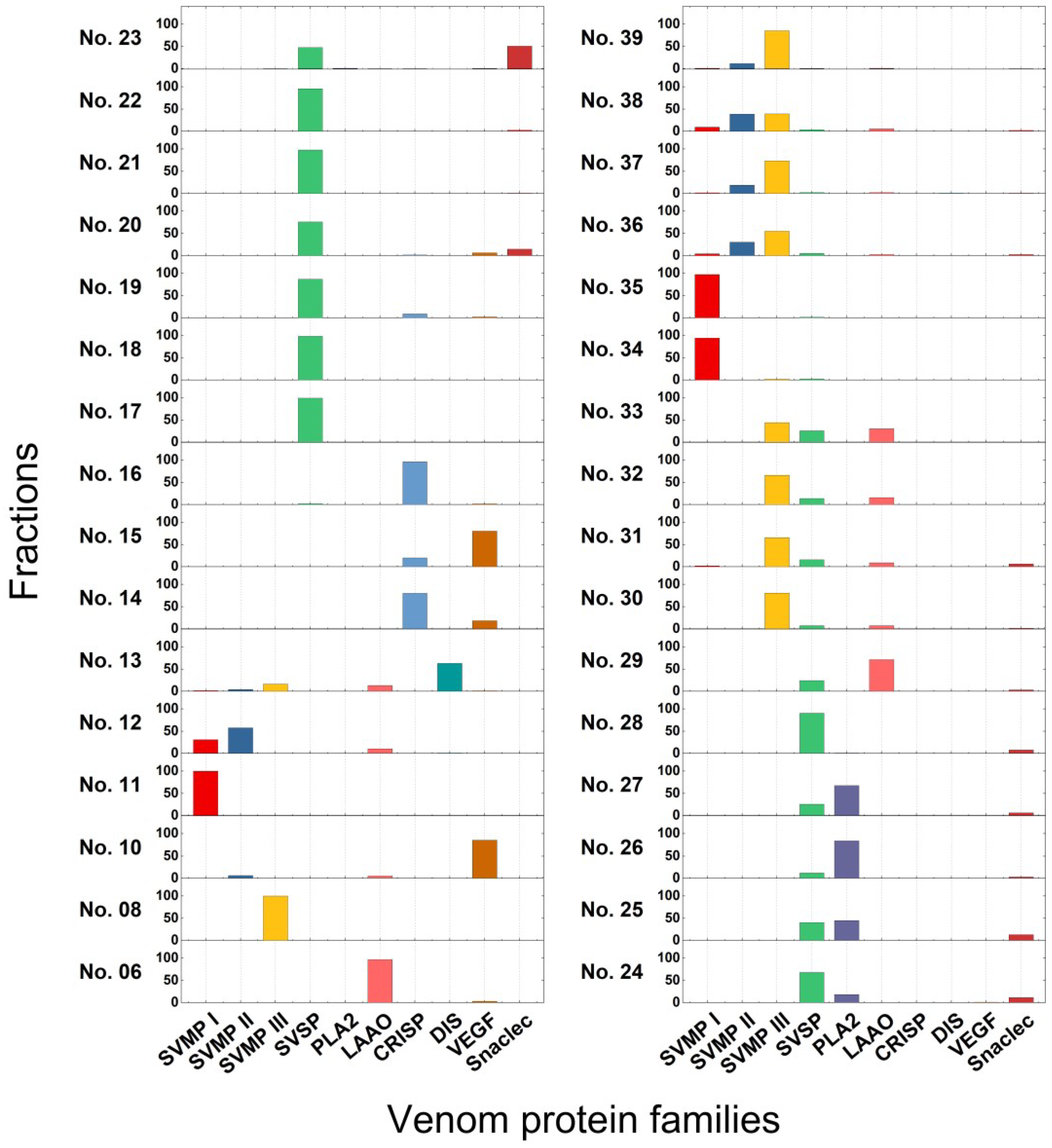
The relative abundances of individual proteins in each chromatographic fraction were calculated and consolidated, as detailed in the Materials and Methods section at the end of this paper, and the T. gracilis venom proteome is summarized in a pie chart (Figure 1C). Of these, SVMPs (40.3%) are the most abundant toxin family and were dominant in fractions 30–39, with a 22.5 kDa PI-SVMP eluted mainly in fractions 34–35 (Figure 2). SVSPs (15.3%) are also abundant and dominant in fractions 17–24, 28, followed by PLA2s (6.6%), which were mainly eluted in fractions 25–27; LAAOs (5.0%), which were dominant in fraction 29; CRISPs (4.6%), which were dominant in fractions 14 and 16; DISs (3.2%), which were derived from PII-SVMP precursors and were dominant in fraction 13; VEGFs (2.9%), which were dominant in fractions 10 and 15; and C-type lectin-like proteins (snaclecs, 1.9%), which were dominant in fraction 23 (Figure 1C and Figure 2). Less abundant toxins in T. gracilis venom included nerve growth factors (NGF, 0.07%), phospholipase B (PLB, 0.06%), hyaluronidases (HYA, 0.01%), 5′-nucleotidases (5′NT, 0.01%), and cystatins (<0.01%) (Supplementary Tables S1 and S2).
The Tgc-SVMPs comprises PI-class (75.7%), PII-class (5.3%), and PIII-class (19.0%) enzymes, represented by 40 proteoforms, and matched to the published SVMP sequences of T. gracilis [14] or other species. Notably, some peptides detected in fractions 6–13 appeared to be hydrolyzed fragments of SVMPs or LAAOs, which possibly resulted from autodegradation during experimental handling of the samples. Our results also showed that 54 SVSP proteoforms were detected and partially matched previously published sequences of Tgc-SVSPs [13] or SVSPs from other species. The LAAO, CRISP, DIS, VEGF, and snaclec families have 9, 9, 2, 5, and 13 proteoforms, respectively. The PLA2 family is dominated by acidic PLA2s, with 13 proteoforms detected that partially matched with previously published sequences of Tgc-E6 [12] or acidic PLA2s from other species.

2.3. Two Cysteine-Rich Secretory Proteins (CRISPs) of T. gracilis Venom

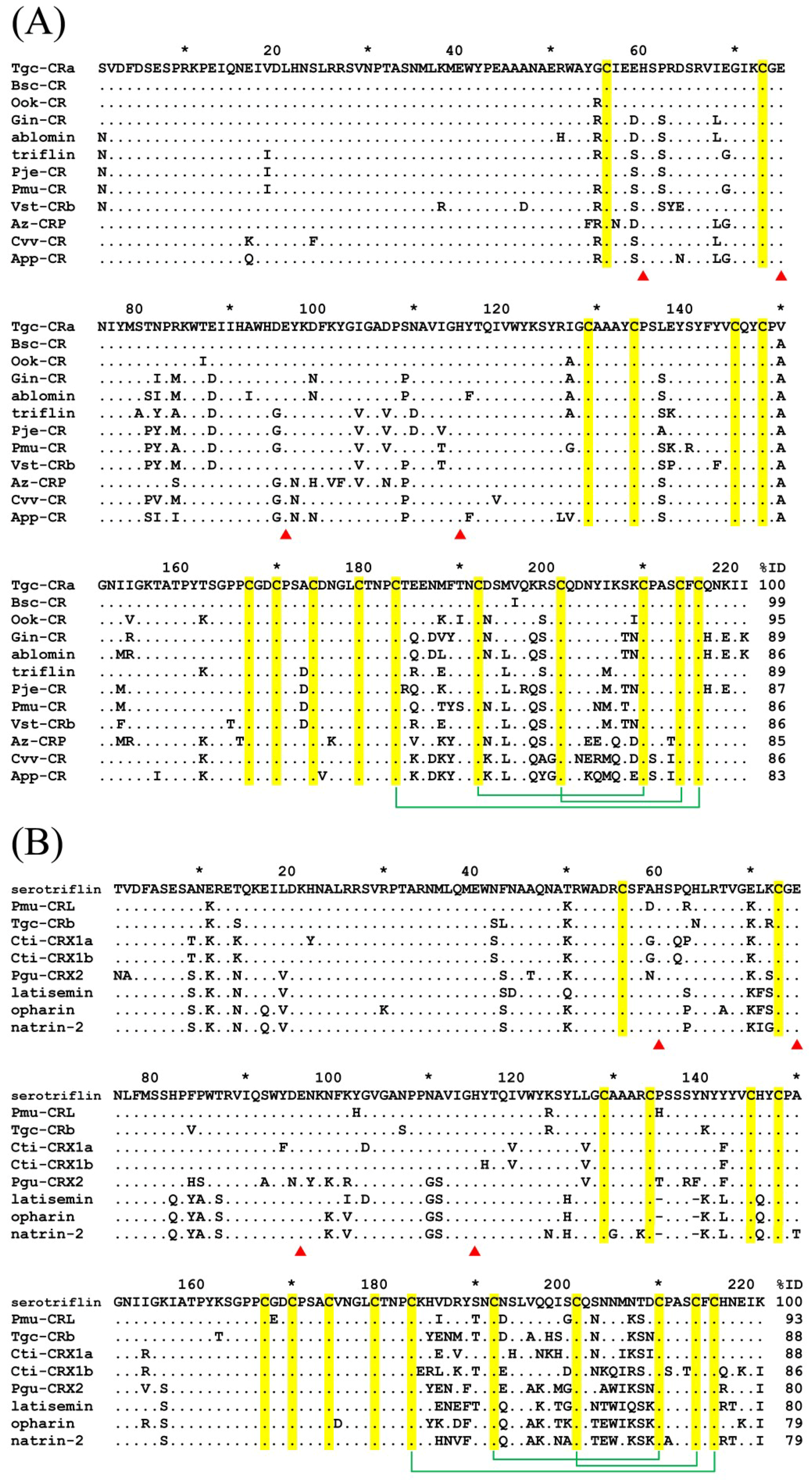
The cDNAs encoding the two novel CRISPs (Tgc-CRa and Tgc-CRb) were cloned and fully sequenced from venom glands and submitted to GenBank under the accession numbers ACE73569.1 (gi|190195325) and ACE73570.1 (gi|190195327), respectively. Tgc-CRa and Tgc-CRb appeared to be paralogs, with only 67% sequence similarity. They were aligned with possible orthologous snake venom CRISPs retrieved using BLASTp (Figure 3A and Figure 3B, respectively). We are not able to find any venom-CRISP sequences of other Trimeresurus species in databanks. All the pitviper venom CRISPs contain 221 amino acid residues (Figure 3A,B), whereas those from true vipers may contain 220 residues [21]. The 16 Cys residues and sequences in their N-terminal half, including the pathogenesis-related protein-1 (PR-1) domain [22], are highly conserved. Tgc-CRa is acidic, and its sequence is 95% identical to that of Ook-CR [15] and >99% similar to the venom CRISPs of Bothriechis schlegelii and Protobothrops species (Figure 3A). In contrast, Tgc-CRb is basic and most similar to serotriflin from the blood of P. flavoviridis [23] and a serotriflin-like protein (i.e., Pmu-CRL) from P. mucrosquamatus; it is also similar to some basic CRISPs of elapid venom (Figure 3B), and these CRISPs are possibly also expressed in tissues other than venom glands. As shown in Table 1, Tables S1 and S2, Tgc-CRa and Tgc-CRb were eluted in the HPLC fractions 14 and 16, respectively (Table 1, Figure 2), and the content of Tgc-CRa was higher than that of Tgc-CRb.

2.4. Three VEGFs Are Expressed in the T. gracilis Venom Gland

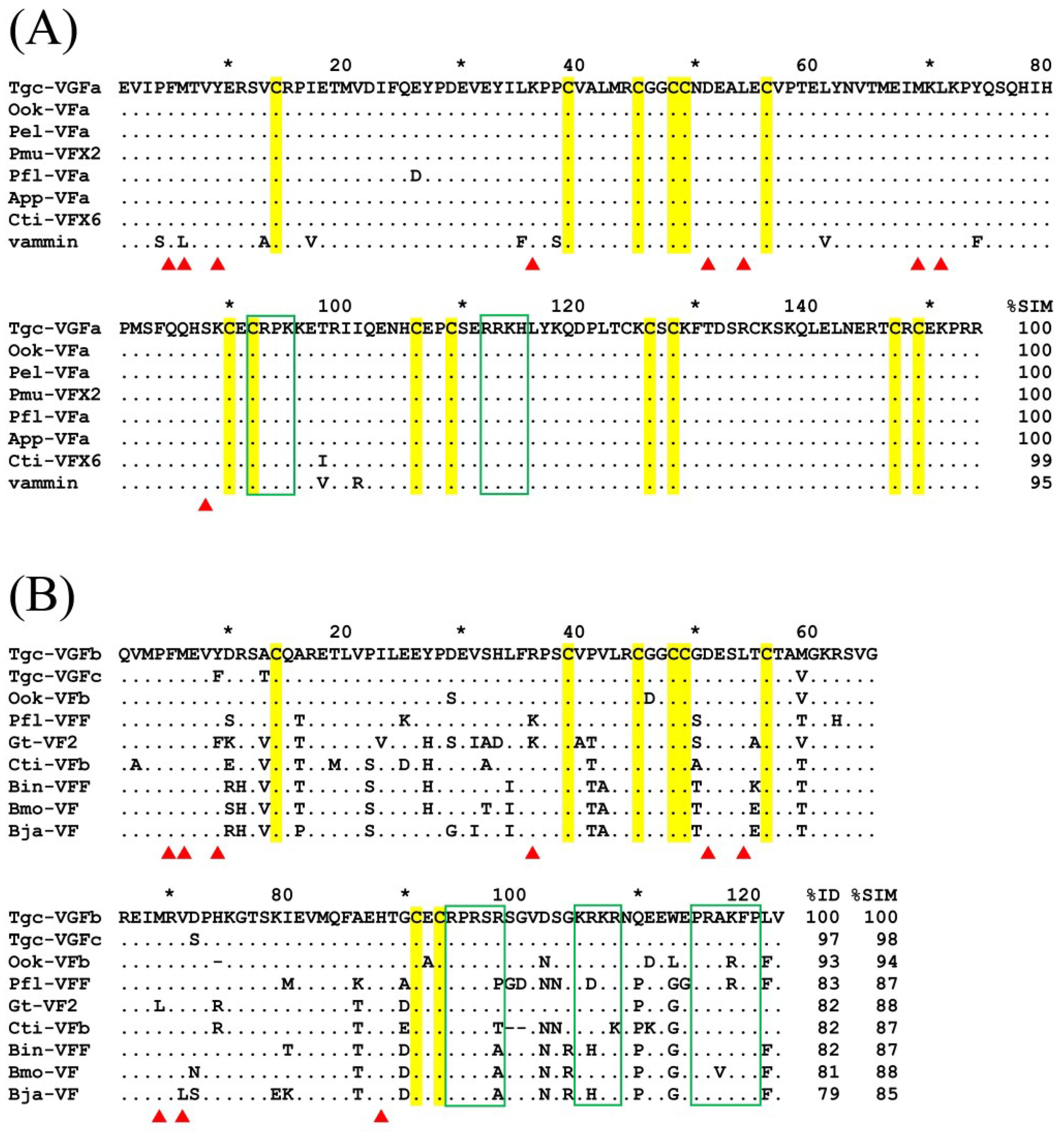
Both the tissue and the venom-types of VEGFs have distinct biochemical properties and are common components of most viperid venom [24,25]. Here, we cloned and sequenced the cDNAs encoding three distinct VEGFs from T. gracilis venom glands, which were deposited to GenBank with accession numbers OQ614863–OQ614865, for Tgc-VGFa, Tgc-VGFb, and Tgc-VGFc, respectively. Their sequences were aligned with possible orthologous snake venom VEGFs retrieved from a BLAST search, respectively (Figure 4A,B). Thus far, NCBI databases do not contain any venom VEGF sequences from other Trimeresurus species. Apparently, the 154-residue Tgc-VGFa is identical to or >99% similar to the VEGFs expressed in the venom of O. okinavensis, Protobothrops, and some New World pitvipers, and those present in the venom of true vipers (subfamily Viperinae) (Figure 4A). Both Tgc-VGFb and Tgc-VGFc contain 122 residues and are approximately 98% similar to each other; both are 93% identical to the sequence of the Ook-VGF protein (Figure 4B) and eluted in fractions 10 and 15, respectively (Table 1, Figure 2). Both types of VEGFs contain conserved receptor-binding residues, and their C-terminal residues contain potentially basic regions responsible for binding heparin (Figure 4A,B).

3. Discussion

3.1. T. gracilis Venom Proteome

The major toxin families expressed in the venom of most pit vipers are metalloproteases, phospholipase A2, serine proteases, and snaclecs [26,27], which are also present in the T. gracilis venom. At least 12 different protein families have been identified in T. gracilis venom, with eight major families (SVMPs, SVSPs, PLA2s, LAAOs, CRISPs, DISs, VEGFs, and snaclecs) comprising 79.8% of all the venom components (Figure 1C). Additionally, low levels of NGFs, PLBs, HYAs, 5′NTs, and cystatins were identified (Supplementary Table S2). Other unidentified components of T. gracilis venom may mainly include low-molecular-weight peptide families, such as bradykinin-potentiating peptides, C-type natriuretic peptides, and tripeptidyl SVMP inhibitors [28]. To further verify the presence of these peptides in the T. gracilis venom, we searched for both trypsin-digested and non-trypsin-digested sequences by mass spectrometry using the non-redundant NCBI database. We detected two proteoforms, bradykinin-potentiating and natriuretic peptides, in fractions 4–6 by partially matching previously published sequences of O. okinavensis and Bothrops atrox (see Supplementary Table S3). Other minor venom enzymes, such as glutaminyl cyclase, aminopeptidase, and phosphodiesterase [26], are expected to be present in T. gracilis venom, but this has not been confirmed.
Among the 10 Tgc-SVSP variants [13], relatively high levels of Tgc-KN1, Tgc-KN4, Tgc-PAH1/2, and Tgc-PA3 were detected by proteomic analysis (Table 1; Supplementary Table S2). Among the five reported Tgc-SVMPs [14], Tgc-MP (PI class), Tgc-PIIc, and Tgc-PIII were clearly present in the venom (Table 1). In addition, two CRISP variants (Tgc-CRa and Tgc-CRb) and two venom-type VEGF variants (Tgc-VGFb and Tgc-VGFc) were also identified (Table 1). Venom VEGFs usually comprise 2–5% of the pit viper venom proteome [29], and Tgc-VGFb and VGFc contribute 2.9% of the venom proteome (Figure 1C). We previously cloned and isolated an acidic PLA2 (Tgc-E6W30) from Tgc venom (collected much earlier, not from Mt. Daxue) with a total yield of approximately 6% (w/w) [12], which is consistent with the relative abundance of acidic PLA2s in the T. gracilis venom proteome (6.6%; Supplementary Table S2). Other minor acidic PLA2 variants, or possibly an E6A30-PLA2, may also be present in the Tgc-venom analyzed in the present study, which could be highly similar to the acidic PLA2s isolated from O. okinavensis and G. brevicaudus (formerly G. halys or G. blomhoffii) (Table 1 and Table S1). A proteoform eluted by HPLC in fraction 26 (Supplementary Table S1) was assigned as Tgc-K49 by searching on the NCBI database, but it is more likely to be an acidic PLA2 variant for three reasons. (1) Basic K49-PLA2 homolog usually eluted earlier than acidic PLA2s from the RP-HPLC column in 0.1% TFA, but this proteoform was eluted in fraction 26 like other acidic PLA2s (eluted in fractions 22–28). (2) Venom content of K49-PLA2 homologs is usually higher than those of the enzymatically active PLA2s, but this proteoform was detected only once and based on two peptides which match a mutated region (residues 70–100) in the Tgc-K49 sequence, and the region happens to be highly similar to the corresponding regions in Tgc-E6W30 and Ook-E6A30 PLA2s [12]. (3) The high number of acidic PLA2s proteoforms detected (Table 1 and Supplementary Table S2) strongly suggests the presence of more than one acidic PLA2 isoforms in T. gracilis venom and this proteoform is likely a E6A30-PLA2.

3.2. Comparison of Venom Composition and Toxicity among Closely Related Species

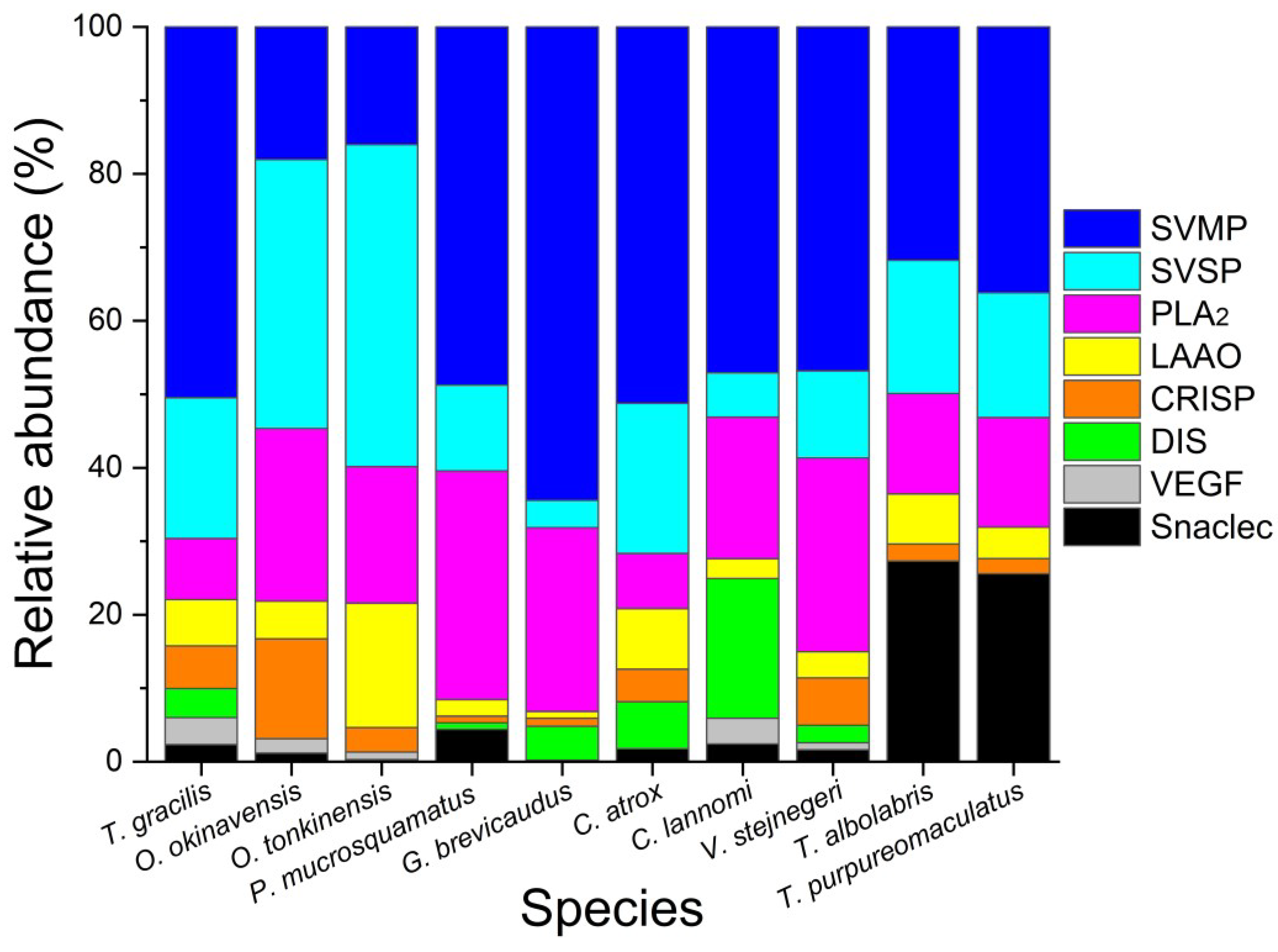
Tgc-MP (a PI-SVMP) is probably as hemorrhagic as okinalysin because of their 95% sequence similarity [14,16]. By acting synergistically with other venom components, abundant Tgc-MP may play a crucial role in the pathophysiology of T. gracilis envenoming. One of the prominent pathologies associated with the hemorrhagic PI-SVMPs is the development of extensive blistering [30], which may become a reservoir of venom toxins that can continuously damage the local tissues. Another distinct function of a number of PI-SVMPs is their ability to activate potent inflammatory responses directly [31]. Our results reveal high structural similarities between the venom proteins of T. gracilis and O. okinavensis; not only are the full sequences of their venom PLA2s, PI-SVMPs, CRISPs, and VEGFs most similar, but also the tryptic peptide sequences of their LAAO, PLB, and HYA match each other (Supplementary Table S2). Although T. gracilis is phylogenetically closely related with O. okinavensis, the toxicity of O. okinavensis venom (LD50 11 μg/g mouse, via intravenous injection; [32]) is much weaker than that of T. gracilis venom (LD50 3 μg/g mouse, via intraperitoneal injection; Tsai et al., unpublished data). The difference in their lethality could be partly explained by the differential expression of their venom toxins, i.e., SVSPs are dominant in O. okinavensis venom [15,33], whereas SVMPs are dominant in T. gracilis venom. T. gracilis venom promotes hemorrhage, hypotension, and impaired blood coagulation, which is consistent with mammalian predation by adult T. gracilis. O. okinavensis venom is comprised of overwhelmingly abundant SVSPs and fewer SVMPs (Figure 5), apparently representing a hybrid strategy optimized mainly for frogs [16], in addition to small mammals.
The venom proteomes of many Asian and New World pit viper species have recently been reported [27,37,38]. We are able to compare the venom proteome of T. gracilis with those of hemorrhagic and phylogeographically related pit viper species [7,8], as shown in Figure 5. SVMPs are the most prominent toxin family in the venom of T. gracilis, G. brevicaudus, P. mucrosquamatus, C. atrox, C. lannomi, and V. stejnegeri; however, the proportions of their PI-, PII-, and PIII-SVMPs are rather diverse (Supplementary Figure S1). Snaclecs are dominant in the venom of T. albolabris and T. purpureomaculatus compared to other species in Figure 5, and the venom proteome of both arboreal species are not similar to that of T. gracilis. Of note, both T. gracilis and C. atrox venom are most abundant in SVMPs and SVSPs, followed by acidic PLA2s, while SVSPs are the most abundant family in Ovophis venom [15,33] that appears to lack DISs (Figure 5). It is also recognized that T. gracilis is the Asian sister of the New World pitvipers [6,7,8] and T. gracilis and C. atrox share high sequence similarities in their PIII-SVMPs and some SVSP variants [13,14]. Possibly because of similar diet ecology in adults, T. gracilis and C. atrox share the venom proteome with grossly similar proportions of the major toxin families (Figure 5), and their lethalities to mice are close, as LD50 of C. atrox venom is 5.0 μg/g mouse for intraperitoneal injection or 2.72 μg/g mouse for intravenous injection [39,40]. Nevertheless, the results of comparing the proteomic data from different studies may be confounded by the variations in the protein detection method, ages of the snakes, or other factors (summarized in Supplementary Table S4), and need to be explained with caution. For example, conditions of pre-treatment by trypsin could be inconsistent in the proteomic studies, in-solution tryptic digestion provided a higher number of proteins identified, and a larger sequence coverage for bottom-up proteomic studies, as compared to using in-gel digestion [41].

3.3. Sequence Comparison of T. gracilis Venom CRISPs

CRISPs are generally not abundant in snake venom, but are widely distributed taxonomically. The presence of CRISP toxins with high degrees of sequence similarity in all snakes suggests earlier diversification of CRISPs before the divergence between Viperidae and the remaining Colubroids [42,43]. The 19-residue signal peptides of CRISPs are highly conserved and favorable for cDNA cloning and sequencing using PCR. In the present study, two venom CRISPs, Tgc-CRa and Tgc-CRb, were fully sequenced for the first time, and a single CRISP transcript was identified in the O. okinavensis transcriptome (Figure 3). Tgc-CRa is highly similar to CRISPs identified in the venom of O. okinavensis, Gloydius and Protobothrops (Figure 3A). Their C-terminal Cys-rich domain (CRD) contained three highly conserved disulfide bridges and a proline bracket [44] (Figure 3A,B). Triflin (from P. flavoviridis) and ablomin (from G. blomhoffi) are L-type Ca2+-channel antagonists of arterial smooth muscle contractions that promote vasodilation and hypotension. CRISPs purified from Bothrops venom species may induce inflammatory responses and interfere with complement pathways, generating bioactive fragments (C3a, C4a, and C5a) and anaphylatoxins [45]. Similar to triflin, both Ook-CRa and Tgc-CRa contain hydrophobic residues at Phe189, Met195, Tyr205, and Phe215, which were shown by crystallographic studies to obstruct the target ion channels, and the highly conserved Glu186 and Phe189 are the most likely functional residues [46]. In contrast to most of the known venom CRISP sequences, an N-glycosylation site was present at N48 in serotriflin and N44 in Pgu-CRX2 (Figure 3B). In both Tgc-CRb and serotriflin, Phe189 is replaced by Tyr189, and the “Pro-bracket” regions 84–90 show low similarities to those of Tgc-CRa and triflin; thus, they are unlikely to bind identical ion-channels.

3.4. Sequence Comparison of T. gracilis Venom VEGFs

We deduced that the protein sequences of three Tgc-VEGF variants, Tgc-VGFa of 154 amino acid residues, appeared to be tissue type-specific variants and similar to human VEGF-A (Figure 4A), whereas Tgc-VGFb and Tgc-VGFc of 122 residues were snake venom types (Figure 4B), which are strongly hypotensive toxins [29]. As pointed out previously [25], the structures of tissue-type VEGFs (or VEGF-A) are highly conserved among venomous snakes and even among all vertebrates (Figure 4A), whereas those of venom-type VEGFs (also annotated as VEGF-F) are highly diversified in the regions around the receptor-binding loops and C-terminal putative coreceptor-binding regions (Figure 4B) and show different affinities to heparin [29]. Ook-VFa (AB852007.1), 154 residues long, is the most highly expressed VEGF in O. okinavensis venom [15]. In contrast, T. gracilis venom contains mainly Tgc-VGFb and Tgc-VGFc but not Tgc-VGFa (Table 1), and Tgc-VGFb is 93% identical to Ook-VFb (Figure 4B). As expected, Tgc-VGFb and Tgc-VGFc may increase vascular permeability, cause hypotension, and facilitate the spread and transport of toxin molecules, particularly when synergized with the KN subtype of SVSP [24,29].

4. Conclusions

Our proteomic profiling of T. gracilis venom was facilitated by using both comprehensive and more specific or restricted databases resulting from extensive cDNA sequencing of the three major toxin families (PLA2, SVSP, and SVMP) and the resolution of full sequences of T. gracilis venom CRISPs and VEGFs in the present study. Our results demonstrate that T. gracilis venom toxins qualitatively resemble those of O. okinavensis rather than other Trimeresurus and Protobothrops species, which is consistent with the close phylogeographic linking of T. gracilis and O. okinavensis [7,19]. The differential expression of venom proteins of T. gracilis and O. okinavensis (Figure 5) can be explained by the adaptation of both species to different environments and prey ecologies. Being an Asian sister of the New World pit vipers, T. gracilis retains some ancient venom genes (e.g., PIII-SVMP) that bear high sequence similarity to the corresponding toxin genes of some hemorrhagic Crotalus species [14]. In contrast to most Crotalus venom, T. gracilis venom lacks crotamine-like myotoxins and highly diversified PII-SVMPs, but is rich in hemorrhagic PI-SVMPs and VEGF-F. The relatively abundant Tgc-MP, Tgc-KN1 and Tgc-KN4, and Tgc-VGFb and Tgc-VGFc could explain the tissue damage, hypotension, and coagulopathy observed in T. gracilis envenoming. It has been demonstrated that using pan-specific effective antivenoms immunized with venoms from only a few species of pitvipers could treat the envenoming by other pitvipers if the immunizing venoms contain toxin families that are representative of the species to which the antivenom is targeted [47]. Our results suggest that antivenom prepared with stronger antigenicity against pitvipers’ PI-SVMPs could be a better candidate to treat T. gracilis envenoming. It is also possible to test whether the antivenom against hemorrhagic Crotalus venom (or adding it to the Taiwan bivalent hemotoxic-antivenom) could effectively treat T. gracilis envenoming. Further studies on the genome and taxonomy of T. gracilis and the pharmacology of its venom toxins are required to clarify its conservation status as well as its venom pathophysiology.

5. Materials and Methods

5.1. Chemicals

All the chemicals and reagents used were of analytical grade. Bovine serum albumin (BSA), formic acid (FA), dithiothreitol (DTT), and iodoacetamide (IAM) were purchased from Sigma-Aldrich (Burlington, MA, USA). Ammonium bicarbonate (AMBIC) was purchased from J.T. Baker (Phillipsburg, NJ, USA). Protein Assay dye was purchased from Bio-Rad (Hercules, CA, USA). The ExcelBand™ 3-color broad-range protein marker (5–245 kDa) was purchased from Smobio (Hsinchu, Taiwan). C18 RP-HPLC column (250 × 4.6 mm, 5 μm particle) was obtained from Thermo Scientific™ BioBasic™ (Waltham, MA, USA). HPLC grade acetonitrile (ACN) was purchased from Honeywell (Charlotte, NC, USA). Trifluoroacetic acid (TFA) was purchased from Acros Organics (Geel, Belgium). Sequence-grade modified trypsin was purchased from Promega (Madison, WI, USA).

5.2. Animals and Venom

We collected four adult T. gracilis (3 females and 1 male) samples from Mount Daxue, Central Taiwan, from September 2020 to December 2022. The corresponding author of this study is responsible for the taxonomic identification of snakes. According to Lin and Tu [9], snakes with a snout-vent length (SVL) larger than 22 cm were considered adults. The mean ± SEM (range) of the SVL and body mass of each collected snake was 50.3 ± 9.46 (44.0–64.0) cm and 64.0 ± 14.1 (48.0–79.0) g, respectively. Venom samples were collected manually at intervals of 14 d or more after venom collection or snake feeding. The wet venom yield per first collection of each snake was 47.0 ± 28.9 (10.7–81.2) mg. Crude venoms from the four T. gracilis specimens were pooled in equal proportion, lyophilized in an FD-series freeze-dryer and CES-series centrifugal evaporator (Panchum Scientific Corp., Kaohsiung, Taiwan), and stored at −80 °C until analysis.

5.3. Determination of Venom Protein Concentration

The lyophilized venom samples were dissolved with ultrapure water and centrifuged 10,000× g at 4 °C for 10 min, and the protein concentration of the supernatant was determined in triplicate using a protein assay dye (Bio-Rad, Hercules, CA, USA) with bovine serum albumin as calibration standard.

5.4. Reverse-Phase High-Performance Liquid Chromatography

T. gracilis venom containing 1.0 mg venom proteins was reconstituted in 20 μL ultrapure water and subjected to C18 reverse-phase fractionation using an HPLC system (Chromaster 5160 Pump and Chromaster 5410 UV detector, Hitachi, Tokyo, Japan). The C18 column was pre-equilibrated with 0.1% TFA in water (Buffer A) and eluted with 0.1% TFA in ACN (Buffer B) at a flow rate of 1.0 mL/min, using a linear gradient of 5% B for 5 min, 5–10% B for 5 min, 10–20% B for 30 min, 20–30% B for 5 min, 30–60% B for 90 min, and 60–70% B for 5 min. The protein elution was monitored at 215 nm and fractions were collected manually, lyophilized, and stored at −80 °C until use.

5.5. Sodium Dodecyl Sulfate-Polyacrylamide Gel Electrophoresis

Protein fractions collected during RP-HPLC were further analyzed by SDS-PAGE according to the method described by Laemmli [48]. The ExcelBand™ 3-color broad-range protein marker (5–245 kDa) was used as a calibration standard. Approximately 5 μg of protein from each fraction was loaded to the 12.5% polyacrylamide gel under reducing conditions and electrophorized at 110 V for 2 h. The gels were stained with Coomassie Brilliant Blue R-250 (Bio-Rad) and de-stained for visualization.

5.6. In-Solution Tryptic Digestion and Peptide Identification by Mass Spectrometry

After lyophilization, 10 μg of protein from each fraction was reduced with DTT, alkylated with IAM, and hydrolyzed with trypsin at an enzyme:substrate ratio of 1:25. The resulting peptides were desalted using MIcroSpin™ columns according to the manufacturer’s protocol (Cytiva, Amersham, UK). The samples were lyophilized, reconstituted in 5% ACN/0.1% FA in water, and subjected to nanoscale electrospray ionization liquid chromatography-tandem mass spectrometry (nano-ESI-LCMS/MS) using a Dionex Ultimate 3000 RSLC system (Thermo Scientific, Waltham, MA, USA) coupled with a Q Exactive mass spectrometer (Thermo Scientific). Samples were loaded in a C18 column (75 μm × 150 mm, 2 μm, 100 Å) (Thermo Scientific Acclaim™ PepMap™, Waltham, MA, USA) at a flow rate of 0.25 μL/min. The injection volume was 5 μL per sample and the mobile phase was 0.1% FA in water (Solution A) and 0.1% FA in 95% ACN (Solution B). The gradient applied was: 1% B for 5.5 min, 1–30% B for 39.5 min, 30–60% B for 3 min, 60–80% B for 2 min, 80% B for 10 min, 80–1% B for 5 min, and 1% B for 5 min. The ion polarity was set to positive ionization mode. Spectra were acquired in MS/MS mode with an MS scan range of 200–3000 m/z and an MS/MS scan range of 50–3000 m/z. The 10 most intense ions from the MS scan were subjected to fragmentation for MS/MS spectra. Data were analyzed with PEAKS Studio 10.5 (Bioinformatics Solutions Inc., Waterloo, ON, Canada), and the peptide-mass-finger-printing results were searched based on the non-redundant NCBI database of Serpentes (taxid:8570). Carbamidomethyl was used for static modification, and oxidation was used for dynamic modification. Protein/peptide identification was validated using the following filters: protein false discovery rate (FDR) ≥1% and unique peptides ≥1; the protein/peptide found was based on the identity of partial sequences. Keratin peptides were eliminated from further analyses. The relative abundance (%) of individual proteins in each chromatographic fraction was determined following previous methods [49,50]:
Relative abundance of protein Q (%) = (mean spectral intensity of protein Q in fraction R/total mean spectral intensity in fraction R) × AUC of fraction R from HPLC (%)
The area under the curve (AUC) for each collected fraction was automatically integrated and determined from the HPLC chromatogram using Chromaster software (Hitachi, Tokyo, Japan). For each protein identified in the individual fraction, the number of spectra, the number of unique peptides, the “mean spectral intensity of protein Q in fraction R”, and “total mean spectral intensity in fraction R”, as well as other detailed data, are provided in Supplementary Table S1.

5.7. Molecular Cloning and Sequence Determination of CRISPs and VEGFs

T. gracilis venom cDNAs were prepared from venom gland mRNAs as described previously [12]. To amplify and clone the cDNAs encoding venom CRISPs, PCR was conducted using SuperTaq DNA polymerase with a pair of mixed-base oligonucleotide primers [51] designed according to the highly conserved cDNA regions in the nucleotide database. Primer 1 was designed in the sense direction: TTCA(A/C)AACA(A/G)(C/T)AGAAATG, and primer 2 was designed in the antisense direction: GATGCTACA(T/C)AG(T/G)CTTGTG [52]. DNA fragments of approximately 1.0 kb were amplified by PCR, as shown by electrophoresis of the products on a 1% agarose gel, and harvested.
The abbreviation of T. gracilis (Tgc) was used to name novel peptides. cDNAs encoding both the venom and serum types of Tgc-VEGFs were cloned using different sets of degenerate primers for PCR amplification. Specific primer pairs were designed based on the conserved nucleotide sequences previously used for venom VEGFs [25,53]. The PCR-amplified DNA products were analyzed on a 1% agarose gel and harvested. After treatment with polynucleotide kinase, the amplified cDNAs were inserted into the pGEM-T easy vector (Promega Corp., Madison, WI, USA) and transformed in Escherichia coli strain JM109. White transformants and cDNA clones were selected. The DNA Sequencing System (model 373A) and TaqDye-Deoxy terminator-cycle sequencing kit (PE Applied Biosystems, Waltham, MA, USA) were used to determine the nucleotide sequences. The protein sequences of T. gracilis venom CRISPs and VEGFs were deduced from their nucleotide sequences.

5.8. BLAST Analyses and Sequence Alignments

Protein-to-protein BLAST (BLASTp) was used to retrieve the most similar sequences for each of the novel Tgc-CRISP and Tgc-VEGF variants, using the non-redundant NCBI database (http://www.ncbi.nlm.nih.gov (accessed on 21 March 2023)). The retrieved toxin homologs were from a broad selection of pitviper genera, and preferably those have been purified and characterized. The sequences were aligned using Clustal X2 [54] and MUSCLE [55] in MEGA X [56]; gaps were introduced to optimize the comparison, and % identities or % similarities of the sequences were calculated using the sequence manipulation suite [57].

Supplementary Materials

The following supporting information can be downloaded from https://www.mdpi.com/article/10.3390/toxins15070408/s1. Table S1: Detailed data for the toxin proteoforms identified by RP-HPLC profiling of Trimeresurus gracilis (Tgc) venom using nano-ESI-LCMS/MS. Table S2: Relative abundances of different toxin families identified in Trimeresurus gracilis (Tgc) venom. Table S3: Potential bradykinin-potentiating peptides and natriuretic peptides in Trimeresurus gracilis venom identified by searching for non-trypsin-digested sequences detected by mass spectrometry. Table S4: Comparison of sample size, age and gender, and venom proteomic analysis methods used to study nine pitviper species, based on published data: Ovophis okinanvensis and O. tonkinensis [33], Gloydius brevicaudus [34], Protobothrops mucrosquamatus and Viridovipera stejnegeri [35], Crotalus atrox [36], C. lannomi [37], T. albolabris and T. purpureomaculatus [38]. Figure S1: Schematic comparison of the relative abundances of three classes of SVMPs in the venom of Trimeresurus gracilis and related pitvipers based on their venom proteomic data: Ovophis okinanvensis and O. tonkinensis [33], Gloydius brevicaudus [34], Protobothrops mucrosquamatus and Viridovipera stejnegeri [35], Crotalus atrox [36], and C. lannomi [37].

Author Contributions

Conceptualization, I.-H.T. and T.-S.T.; methodology, T.-C.T., Y.-Y.C., I.-H.T. and T.-S.T.; software, T.-C.T. and Y.-Y.C.; validation, I.-H.T. and T.-S.T.; formal analysis, T.-C.T., Y.-Y.C., I.-H.T. and T.-S.T.; investigation, T.-C.T., I.-H.T. and T.-S.T.; resources, I.-H.T. and T.-S.T.; data curation, T.-S.T.; writing—original draft preparation, I.-H.T. and T.-S.T.; writing—review and editing, I.-H.T. and T.-S.T.; visualization, T.-C.T. and Y.-Y.C. and T.-S.T.; supervision, T.-S.T.; project administration, T.-S.T.; funding acquisition, I.-H.T. and T.-S.T. All authors have read and agreed to the published version of the manuscript.

Funding

This research was funded by the Academia Sinica (to I.H.T.), the Ministry of Science and Technology (grant numbers 109-2621-B-020-001, 110-2621-B-020-001), the Council of Agriculture, the Kaohsiung City Government, the National Pingtung University of Science and Technology (grant number NPUST-111005), and the Kaohsiung Chang Gung Memorial Hospital and National Pingtung University of Science and Technology Joint Research Program (grant number CGMH-NPUST-2022-CORPG8M0111).

Institutional Review Board Statement

The study was approved (approval numbers: NPUST-108-079, Date: 10 February 2020; NPUST-109-121, Date: 4 February 2021; NPUST-110-100, Date: 27 September 2021) by the Animal Care and Use Committee of the National Pingtung University of Science and Technology.

Informed Consent Statement

Not applicable.

Data Availability Statement

Data are contained within the article or Supplementary Material.

Acknowledgments

Our special thanks go to Jue-Liang Hsu and his laboratory members for their assistance in HPLC and proteomic analyses. We also thank Ying-Ming Wang for cloning and sequence analyses of VEGFs and CRISPs, Jun-Wei Chang, Tong-Yu Ke, Mo Han Ruan and Jui-Hsiang Fan for helping in the collection of snake specimens and venom samples.

Conflicts of Interest

The authors declare no conflict of interest.

References

  1. Catalan, J.; Ninot, J.M.; Aniz, M.M. The high mountain conservation in a changing world. In High Mountain Conservation in a Changing World; Catalan, J., Ninot, J.M., Aniz, M.M., Eds.; Springer Nature: Cham, Switzerland, 2017; pp. 3–36. [Google Scholar] [CrossRef] [Green Version]
  2. Lai, J.S.; Luei, K.Y. Two new Hynobius (Caudata: Hynobiidae) salamanders from Taiwan. Herpetologica 2008, 64, 63–80. [Google Scholar] [CrossRef]
  3. Schulz, H.M.; Li, C.F.; Thies, B.; Chang, S.C.; Bendix, J. Mapping the montane cloud forest of Taiwan using 12 year MODIS-derived ground fog frequency data. PLoS ONE 2017, 12, e0172663. [Google Scholar] [CrossRef] [PubMed] [Green Version]
  4. Oshima, S. Notes on the venomous snakes from the islands of Formosa and Riu Kiu. Annu. Rep. Inst. Sci. Gov. Formosa 1920, 2, 1–99. Available online: https://www.biodiversitylibrary.org/item/105191#page/13/mode/1up (accessed on 19 June 2023).
  5. Zaher, H.; Murphy, R.W.; Arredondo, J.C.; Graboski, R.; Machado-Filho, P.R.; Mahlow, K.; Montingelli, G.G.; Quadros, A.B.; Orlov, N.L.; Wilkinson, M.; et al. Large-scale molecular phylogeny, morphology, divergence-time estimation, and the fossil record of advanced caenophidian snakes (Squamata: Serpentes). PLoS ONE 2019, 14, e0216148. [Google Scholar] [CrossRef] [Green Version]
  6. Castoe, T.A.; Parkinson, C.L. Bayesian mixed models and the phylogeny of pitvipers (Viperidae: Serpentes). Mol. Phylogenet. Evol. 2006, 39, 91–110. [Google Scholar] [CrossRef] [PubMed]
  7. Malhotra, A.; Creer, S.; Pook, C.E.; Thorpe, R.S. Inclusion of nuclear intron sequence data helps to identify the Asian sister group of New World pitvipers. Mol. Phylogenet. Evol. 2010, 54, 172–178. [Google Scholar] [CrossRef]
  8. Wüster, W.; Peppin, L.; Pook, C.E.; Walker, D.E. A nesting of vipers: Phylogeny and historical biogeography of the Viperidae (Squamata: Serpentes). Mol. Phylogenet. Evol. 2008, 49, 445–459. [Google Scholar] [CrossRef]
  9. Lin, C.F.; Tu, M.C. Food habits of the Taiwanese mountain pitviper, Trimeresurus gracilis. Zool. Stud. 2008, 47, 697–703. [Google Scholar]
  10. Mori, A.; Toda, M.; Ota, H. Winter activity of the Hime-habu (Ovophis okinavensis) in the humid subtropics: Foraging on breeding anurans at low temperatures. In Biology of the Vipers; Schuett, G.W., Höggren, M., Douglas, M.E., Greene, H.W., Eds.; Eagle Mountain Publishing LC: Eagle Mountain, UT, USA, 2002; pp. 329–344. [Google Scholar]
  11. Tsai, T.S.; Chan, Y.Y.; Huang, S.M.; Chuang, P.C. Case report: Symptoms and prognosis of Trimeresurus gracilis envenomation. Am. J. Trop. Med. Hyg. 2022, 106, 1281–1284. [Google Scholar] [CrossRef]
  12. Tsai, I.H.; Tsai, T.S.; Wang, Y.M.; Tu, M.C.; Chang, H.C. Cloning and characterization of Trimeresurus gracilis venom phospholipases A2: Comparison with Ovophis okinavensis venom and the systematic implications. Toxicon 2012, 59, 151–157. [Google Scholar] [CrossRef]
  13. Tsai, T.S.; Wang, Y.M.; Tsai, I.H. Sequence determination and bioinformatic comparison of ten venom serine proteases of Trimeresurus gracilis, a Taiwanese endemic pitviper with controversial taxonomy. Toxicon 2022, 206, 28–37. [Google Scholar] [CrossRef]
  14. Tsai, T.S.; Tsai, I.H. Full sequencing and comparison of five venom metalloproteases of Trimeresurus gracilis: The PI-enzyme is most similar to okinalysin but the PIII-enzyme is most similar to Crotalus venom enzymes. Toxicon 2023, 225, 107053. [Google Scholar] [CrossRef]
  15. Aird, S.D.; Watanabe, Y.; Villar-Briones, A.; Roy, M.C.; Terada, K.; Mikheyev, A.S. Quantitative high-throughput profiling of snake venom gland transcriptomes and proteomes (Ovophis okinavensis and Protobothrops flavoviridis). BMC Genom. 2013, 14, 790. [Google Scholar] [CrossRef] [Green Version]
  16. Komori, Y.; Murakami, E.; Uchiya, K.; Nonogaki, T.; Nikai, T. Okinalysin, a novel P-I metalloproteinase from Ovophis okinavensis: Biological properties and effect on vascular endothelial cells. Toxins 2014, 6, 2594–2604. [Google Scholar] [CrossRef] [Green Version]
  17. Alencar, L.R.V.; Quental, T.B.; Grazziotin, F.G.; Alfaro, M.L.; Martins, M.; Venzon, M.; Zaher, H. Diversification in vipers: Phylogenetic relationships, time of divergence and shifts in speciation rates. Mol. Phylogenet. Evol. 2016, 105, 50–62. [Google Scholar] [CrossRef]
  18. Yang, Z.M.; Guo, Q.; Ma, Z.R.; Chen, Y.; Wang, Z.Z.; Wang, X.M.; Wang, Y.M.; Tsai, I.H. Structures and functions of crotoxin-like heterodimers and acidic phospholipases A2 from Gloydius intermedius venom: Insights into the origin of neurotoxic-type rattlesnakes. J. Proteom. 2015, 112, 210–223. [Google Scholar] [CrossRef]
  19. Malhotra, A.; Thorpe, R.S. A phylogeny of four mitochondrial gene regions suggests a revised taxonomy for Asian pitvipers (Trimeresurus and Ovophis). Mol. Phylogenet. Evol. 2004, 32, 83–100. [Google Scholar] [CrossRef]
  20. Modahl, C.M.; Mackessy, S.P. Full-length venom protein cDNA sequences from venom-derived mRNA: Exploring compositional variation and adaptive multigene evolution. PLoS Negl. Trop. Dis. 2016, 10, e0004587. [Google Scholar] [CrossRef] [Green Version]
  21. Ramazanova, A.S.; Starkov, V.G.; Osipov, A.V.; Ziganshin, R.H.; Filkin, S.Y.; Tsetlin, V.I.; Utkin, Y.N. Cysteine-rich venom proteins from the snakes of Viperinae subfamily–molecular cloning and phylogenetic relationship. Toxicon 2009, 53, 162–168. [Google Scholar] [CrossRef]
  22. Matsunaga, Y.; Yamazaki, Y.; Hyodo, F.; Sugiyama, Y.; Nozaki, M.; Morita, T. Structural divergence of cysteine-rich secretory proteins in snake venoms. J. Biochem. 2009, 145, 365–375. [Google Scholar] [CrossRef]
  23. Aoki, N.; Sakiyama, A.; Kuroki, K.; Maenaka, K.; Kohda, D.; Deshimaru, M.; Terada, S. Serotriflin, a CRISP family protein with binding affinity for small serum protein-2 in snake serum. Biochim. Biophys. Acta 2008, 1784, 621–628. [Google Scholar] [CrossRef] [PubMed]
  24. Chen, Y.L.; Tsai, I.H.; Hong, T.M.; Tsai, S.H. Crotalid venom vascular endothelial growth factors has preferential affinity for VEGFR-1. Characterization of Protobothrops mucrosquamatus venom VEGF. Thromb. Haemost. 2005, 93, 331–338. [Google Scholar] [CrossRef] [PubMed] [Green Version]
  25. Yamazaki, Y.; Matsunaga, Y.; Tokunaga, Y.; Obayashi, S.; Saito, M.; Morita, T. Snake venom vascular endothelial growth factors (VEGF-Fs) exclusively vary their structures and functions among species. J. Biol. Chem. 2009, 284, 9885–9891. [Google Scholar] [CrossRef] [PubMed] [Green Version]
  26. Tasoulis, T.; Isbister, G.K. A review and database of snake venom proteomes. Toxins 2017, 9, 290. [Google Scholar] [CrossRef] [Green Version]
  27. Tasoulis, T.; Isbister, G.K. A current perspective on snake venom composition and constituent protein families. Arch. Toxicol. 2023, 97, 133–153. [Google Scholar] [CrossRef]
  28. Leonardi, A.; Sajevic, T.; Pungercar, J.; Krizaj, I. Comprehensive study of the proteome and transcriptome of the venom of the most venomous european viper: Discovery of a new subclass of ancestral snake venom metalloproteinase precursor-derived proteins. J. Proteome Res. 2019, 18, 2287–2309. [Google Scholar] [CrossRef]
  29. Ferreira, I.G.; Pucca, M.B.; Oliveira, I.S.; Cerni, F.A.; Jacob, B.; Arantes, E.C. Snake venom vascular endothelial growth factors (svVEGFs): Unravelling their molecular structure, functions, and research potential. Cytokine Growth Factor Rev. 2021, 60, 133–143. [Google Scholar] [CrossRef]
  30. Gutierrez, J.M.; Rucavado, A.; Chaves, F.; Diaz, C.; Escalante, T. Experimental pathology of local tissue damage induced by Bothrops asper snake venom. Toxicon 2009, 54, 958–975. [Google Scholar] [CrossRef]
  31. Dawson, C.A.; Ainsworth, S.; Albulescu, L.-O.; Casewell, N.R. Snake venom metalloproteinases. In Handbook of Venoms and Toxins of Reptiles, 2nd ed.; Mackessy, S.P., Ed.; CRC Press: Boca Raton, FL, USA, 2021; pp. 363–380. [Google Scholar]
  32. Sadahiro, S.; Yamauchi, K.; Kondo, S.; Konda, H.; Murata, R. Immunological studies on snake venom I. Comparison of venoms from Genus Trimeresurus inhabiting the Ryukyu islands. Jpn. J. Bacteriol. 1965, 20, 21–26, (In Japanese with English Abstract). [Google Scholar] [CrossRef]
  33. Tan, C.H.; Palasuberniam, P.; Tan, K.Y. Snake venom proteomics, immunoreactivity and toxicity neutralization studies for the Asiatic Mountain Pit Vipers, Ovophis convictus, Ovophis tonkinensis, and Hime Habu, Ovophis okinavensis. Toxins 2021, 13, 514. [Google Scholar] [CrossRef]
  34. Gao, J.F.; Wang, J.; He, Y.; Qu, Y.F.; Lin, L.H.; Ma, X.M.; Ji, X. Proteomic and biochemical analyses of short-tailed pit viper (Gloydius brevicaudus) venom: Age-related variation and composition-activity correlation. J. Proteom. 2014, 105, 307–322. [Google Scholar] [CrossRef]
  35. Villalta, M.; Pla, D.; Yang, S.L.; Sanz, L.; Segura, A.; Vargas, M.; Chen, P.Y.; Herrera, M.; Estrada, R.; Cheng, Y.F.; et al. Snake venomics and antivenomics of Protobothrops mucrosquamatus and Viridovipera stejnegeri from Taiwan: Keys to understand the variable immune response in horses. J. Proteom. 2012, 75, 5628–5645. [Google Scholar] [CrossRef]
  36. Calvete, J.J.; Fasoli, E.; Sanz, L.; Boschetti, E.; Righetti, P.G. Exploring the venom proteome of the western diamondback rattlesnake, Crotalus atrox, via snake venomics and combinatorial peptide ligand library approaches. J. Proteome Res. 2009, 8, 3055–3067. [Google Scholar] [CrossRef] [Green Version]
  37. Neri-Castro, E.; Zarzosa, V.; Colis-Torres, A.; Fry, B.G.; Olvera-Rodriguez, A.; Jones, J.; Reyes-Velasco, J.; Zamudio, F.; Borja, M.; Alagon, A.; et al. Proteomic and toxicological characterization of the venoms of the most enigmatic group of rattlesnakes: The long-tailed rattlesnakes. Biochimie 2022, 202, 226–236. [Google Scholar] [CrossRef]
  38. Anita, S.; Sadjuri, A.R.; Rahmah, L.; Nugroho, H.A.; Mulyadi; Trilaksono, W.; Ridhani, W.; Safira, N.; Bahtiar, H.; Maharani; et al. Venom composition of Trimeresurus albolabris, T. insularis, T. puniceus and T. purpureomaculatus from Indonesia. J. Venom. Anim. Toxins Incl. Trop. Dis. 2022, 28, e20210103. [Google Scholar] [CrossRef]
  39. LD50men. Available online: https://web.archive.org/web/20120413182323/http://www.venomdoc.com/LD50/LD50men.html (accessed on 20 May 2023).
  40. De Roodt, A.R.; Desio, M.A.; Lanari, L.C.; Lago, N.R.; Goñi, F.M.; Dozoretz, D.; Calderón, L.; Regner, P.; de Oliveira, V.C.; Damin, C. Paraspecific neutralization of the venom form adults and young Crotalus atrox by paraspecific South American Antivenoms. In Proceedings of the 1st International Electronic Conference on Toxins Session Poster, Online, 16–31 January 2021. [Google Scholar] [CrossRef]
  41. Choksawangkarn, W.; Sriswasdi, S.; Kalpongnukul, N.; Wongkongkathep, P.; Saethang, T.; Chanhome, L.; Laoungbua, P.; Khow, O.; Sumontha, M.; Chaiyabutr, N.; et al. Combined proteomic strategies for in-depth venomic analysis of the beaked sea snake (Hydrophis schistosus) from Songkhla Lake, Thailand. J. Proteom. 2022, 259, 104559. [Google Scholar] [CrossRef]
  42. Fry, B.G.; Wuster, W.; Ryan Ramjan, S.F.; Jackson, T.; Martelli, P.; Kini, R.M. Analysis of Colubroidea snake venoms by liquid chromatography with mass spectrometry: Evolutionary and toxinological implications. Rapid Commun. Mass Spectrom. 2003, 17, 2047–2062. [Google Scholar] [CrossRef]
  43. Tadokoro, T.; Modahl, C.M.; Maenaka, K.; Aoki-Shioi, N. Cysteine-rich secretory proteins (CRISPs) from venomous snakes: An overview of the functional diversity in a large and underappreciated superfamily. Toxins 2020, 12, 175. [Google Scholar] [CrossRef] [Green Version]
  44. Kini, R.M. Proline brackets and identification of potential functional sites in proteins: Toxins to therapeutics. Toxicon 1998, 36, 1659–1670. [Google Scholar] [CrossRef]
  45. Bernardes, C.P.; Menaldo, D.L.; Zoccal, K.F.; Boldrini-Franca, J.; Peigneur, S.; Arantes, E.C.; Rosa, J.C.; Faccioli, L.H.; Tytgat, J.; Sampaio, S.V. First report on BaltCRP, a cysteine-rich secretory protein (CRISP) from Bothrops alternatus venom: Effects on potassium channels and inflammatory processes. Int. J. Biol. Macromol. 2019, 140, 556–567. [Google Scholar] [CrossRef]
  46. Shikamoto, Y.; Suto, K.; Yamazaki, Y.; Morita, T.; Mizuno, H. Crystal structure of a CRISP family Ca2+-channel blocker derived from snake venom. J. Mol. Biol. 2005, 350, 735–743. [Google Scholar] [CrossRef] [PubMed]
  47. Sousa, L.F.; Nicolau, C.A.; Peixoto, P.S.; Bernardoni, J.L.; Oliveira, S.S.; Portes-Junior, J.A.; Mourao, R.H.; Lima-dos-Santos, I.; Sano-Martins, I.S.; Chalkidis, H.M.; et al. Comparison of phylogeny, venom composition and neutralization by antivenom in diverse species of Bothrops complex. PLoS Negl. Trop. Dis. 2013, 7, e2442. [Google Scholar] [CrossRef] [PubMed] [Green Version]
  48. Laemmli, U.K. Cleavage of structural proteins during the assembly of the head of bacteriophage T4. Nature 1970, 227, 680–685. [Google Scholar] [CrossRef] [PubMed]
  49. Tan, C.H.; Tan, K.Y.; Tan, N.H. A protein decomplexation strategy in snake venom proteomics. Methods Mol. Biol. 2019, 1871, 83–92. [Google Scholar] [CrossRef] [PubMed]
  50. Tan, K.Y.; Wong, K.Y.; Tan, N.H.; Tan, C.H. Quantitative proteomics of Naja annulifera (sub-Saharan snouted cobra) venom and neutralization activities of two antivenoms in Africa. Int. J. Biol. Macromol. 2020, 158, 605–616. [Google Scholar] [CrossRef]
  51. Mullis, K.B.; Faloona, F.A. Specific synthesis of DNA in vitro via a polymerase-catalyzed chain reaction. Methods Enzymol. 1987, 155, 335–350. [Google Scholar] [CrossRef]
  52. Tsai, I.H.; Wang, Y.M.; Huang, K.F. Structures of Azemiops feae venom phospholipases and Cys-rich-secretory protein and implications for taxonomy and toxinology. Toxicon 2016, 114, 31–39. [Google Scholar] [CrossRef]
  53. Junqueira de Azevedo, I.L.; Farsky, S.H.; Oliveira, M.L.; Ho, P.L. Molecular cloning and expression of a functional snake venom vascular endothelium growth factor (VEGF) from the Bothrops insularis pit viper. A new member of the VEGF family of proteins. J. Biol. Chem. 2001, 276, 39836–39842. [Google Scholar] [CrossRef] [Green Version]
  54. Larkin, M.A.; Blackshields, G.; Brown, N.P.; Chenna, R.; McGettigan, P.A.; McWilliam, H.; Valentin, F.; Wallace, I.M.; Wilm, A.; Lopez, R.; et al. Clustal W and Clustal X version 2.0. Bioinformatics 2007, 23, 2947–2948. [Google Scholar] [CrossRef] [Green Version]
  55. Edgar, R.C. MUSCLE: Multiple sequence alignment with high accuracy and high throughput. Nucleic Acids Res. 2004, 32, 1792–1797. [Google Scholar] [CrossRef] [Green Version]
  56. Kumar, S.; Stecher, G.; Li, M.; Knyaz, C.; Tamura, K. MEGA X: Molecular Evolutionary Genetics Analysis across computing platforms. Mol. Biol. Evol. 2018, 35, 1547–1549. [Google Scholar] [CrossRef]
  57. Sequence Manipulation Suite. Available online: https://www.bioinformatics.org/sms2/ident_sim.html (accessed on 20 May 2023).
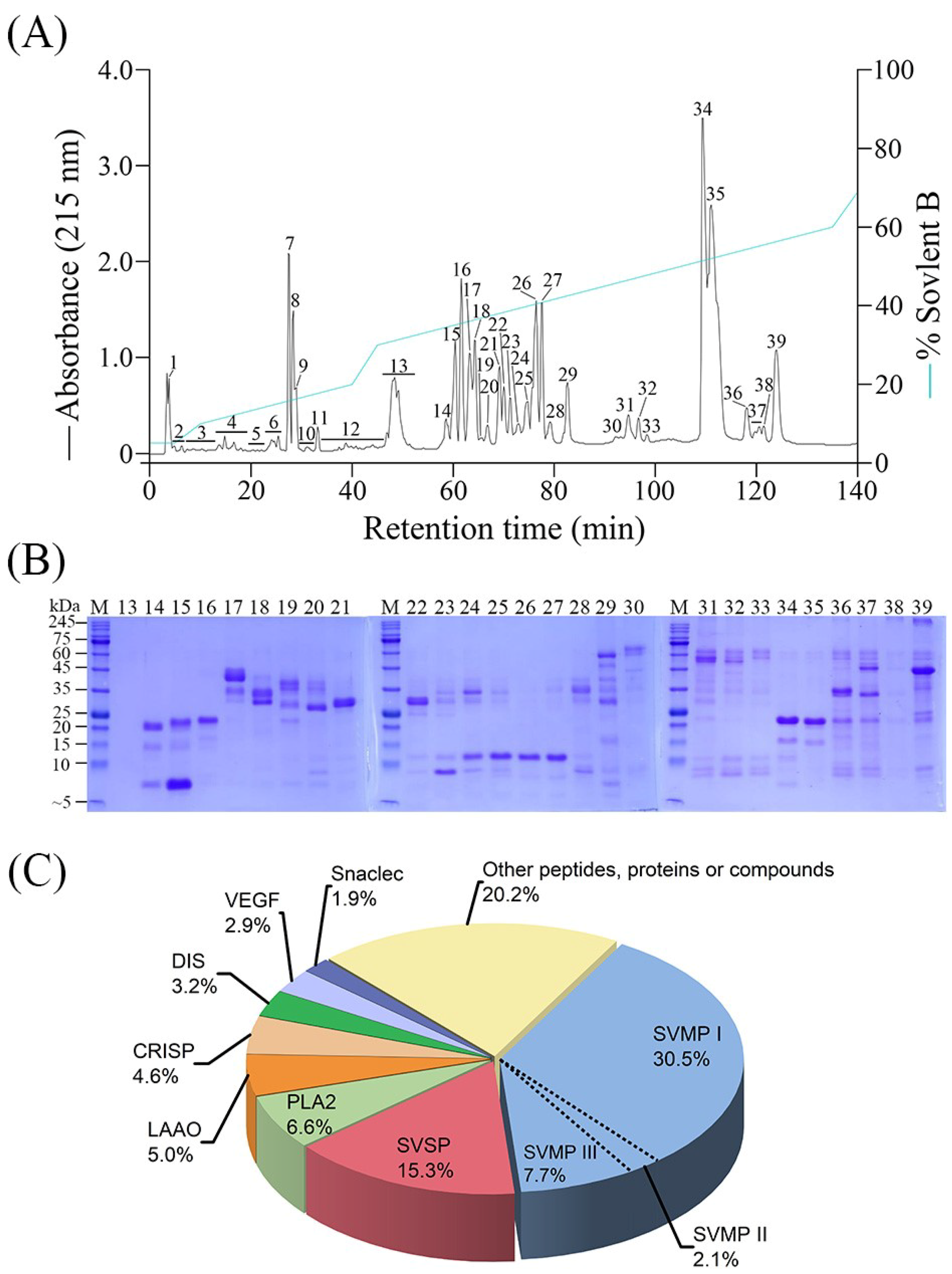
Figure 1. Venomic analysis of adult Trimeresurus gracilis from Mt. Daxue, Taiwan. (A) Reversed phase high performance liquid chromatography (RP-HPLC) profile of T. gracilis pooled venom (1.0 mg). The underline below numbers indicates that several (minor) fractions are collected together for mass spectrometry (MS) analysis. (B) Sodium dodecyl sulfate-polyacrylamide gel electrophoresis (SDS-PAGE) analyses of the HPLC peaks under reducing condition. Gel lanes loaded with the first 13 fractions failed to show any bands. (C) Pie chart representing relative abundance (in percentage of total venom components) of different toxin families based on the results of MS analysis. SVMP, snake venom metalloproteinase; SVSP, snake venom serine protease; PLA2, phospholipase A2; LAAO, L-amino acid oxidase; CRISP, cysteine-rich secretory protein; DIS, disintegrin; VEGF, vascular endothelial growth factor; snaclec, C-type lectin-like protein. The fraction “Other peptides, proteins or compounds” include nerve growth factor (0.07%), phospholipase B (0.06%), hyaluronidase (0.01%), 5′-nucleotidase (0.01%), cystatins (<0.01%), low molecular weight or small and hydrolized peptides, nontoxins (e.g., keratins), and other unidentified compounds.
Toxins 15 00408 g001
Figure 2. Comparisons on the relative abundance (%) of eight Trimeresurus gracilis toxin-families detected in the HPLC fractions. SVMP, snake venom metalloproteinase; SVSP, snake venom serine protease; PLA2, phospholipase A2; LAAO, L-amino acid oxidase; CRISP, cysteine-rich secretory protein; DIS, disintegrin; VEGF, vascular endothelial growth factor; snaclec, C-type lectin-like protein.
Toxins 15 00408 g002
Figure 3. Sequence alignments of snake venom Cys-rich secretory proteins (CRISPs). Conserved Cys residues are highlighted in yellow, potential Ca2+ binding residues are indicated by red triangles, and gaps are shown by hyphens. * indicates a marker count from the average of adjacent numbers. Three pairs of C-terminal disulfide bridges are shown in green. (A) The acidic CRISP homologs retrieved by BLAST. Accession numbers and species are: Tgc-CRa, ACE73569.1; Bsc-CR, ACE73559.1 (Bothriechis schlegelii); Ook-CR, BAN82147.1 (Ovophis okinavensis); Gin-CR, UQT19685.1 (Gloydius intermedius); ablomin, UQT19680.1 (G. blomhoffii); triflin, Q8JI39.1 (Protobothrops flavoviridis); Pje-CR, Q7ZZN9.1 (P. jerdonii); Pmu-CR, XP_015678374.1 (P. mucrosquamatus); Vst-CRb, ACE73573.1 (Viridovipera stejnegeri); Az-CRP, ACE73558.1 (Azemiops feae); Cvv-CR, ACE73566.1 (Crotalus v. viridis); and App-CR, Q7ZTA0.1 (Agkistrodon p. piscivorus). (B) The basic CRISPs and CRISPs homologous to Tgc-CRb. Accession numbers and species are: Serotriflin, P0CB15 (P. flavoviridis); Pmu-CRL, XP_015678372 (P. mucrosquamatus); Tgc-CRb, ACE73570.1; Cti-CRX1a, XP_039185543.1 (C. tigris); Cti-CRX1b, XP_039185562.1 (C. Tigris); Pgu-CRX2, XP_034288380.1 (Pantherophis guttatus blood); latisemin, Q8JI38.1 (Laticauda semifasciata); opharin, ACN93671.1 (Ophiophagus hannah); and natrin-2, Q7ZZN8.1 (Naja atra).
Toxins 15 00408 g003
Figure 4. Sequence alignments of VEGF family proteins expressed in viperid venom glands. Conserved Cys residues are highlighted in yellow, amino acid residues potentially involved in VEGF-receptor-binding are marked with red triangles below the sequences, and possible heparin-binding regions are boxed with green lines. * indicates a marker count from the average of adjacent numbers. (A) Tgc-VGFa and homologs retrieved by BLASTp. Accession numbers and species are: Tgc-VGFa, OQ614863; Ook-VFa, BAN89442.1 (Ovophis okinavensis); Pel-VFa, BAP39940.1 (Protobothrops elegans); Pmu-VFX2, XP_015673445.1 (P. mucrosquamatus); Pfl-VFa, BAD38845.1 (P. flavoviridis); App-VFa, C0K3N4.1 (Agkistrodon p. piscivorus); Cti-VFX6, XP_039198673.1 (Crotalus tigris); and Vammin, C0K3N5.1 (Vipera ammodytes ammodytes). (B) Tgc-VGFb and Tgc-VGFc homologs retrieved by BLASTp. Accession numbers and species are: Tgc-VGFb, OQ614864; Tgc-VGFc, OQ614865; Ook-VFb, BAN82145.1 (O. okinavensis); Pfl-VFF, P67862.1 (P. flavoviridis); Gt-VF2, BAO57712.1 (Gloydius tsushimaensis); Cti-VFb, XP_039218052.1 (Crotalus tigris); Bin-VFF, Q90X24.1 (Bothrops insularis); Bmo-VF, ATU85531.1 (B. moojeni); and Bja-VF, KAG5858117.1 (B. jararaca).
Toxins 15 00408 g004
Figure 5. Comparison of the venom proteome of Trimeresurus gracilis to those of other related pitvipers. The abundance of eight key toxin families (relative to the sum of their total abundances) in each venom species were calculated based on published data, respectively: Ovophis okinavensis and O. tonkinensis [33], Gloydius brevicaudus [34], Protobothrops mucrosquamatus and Viridovipera stejnegeri [35], Crotalus atrox [36] and C. lannomi [37], T. albolabris, and T. purpureomaculatus [38]. SVMP, snake venom metalloproteinase; SVSP, snake venom serine protease; PLA2, phospholipase A2; LAAO, L-amino acid oxidase; CRISP, cysteine-rich secretory protein; DIS, disintegrin; VEGF, vascular endothelial growth factor; snaclec, C-type lectin-like protein.
Toxins 15 00408 g005
Table 1. Trimeresurus gracilis (Tgc) venom proteome as profiled by reversed phase high performance liquid chromatography (RP-HPLC) and nanoscale electrospray ionization liquid chromatography-tandem mass spectrometry (nano-ESI-LC-MS/MS). Minor components (<0.1% of total venom components) are not displayed.
HPLC Fraction
/Toxin Family
Protein (Proteoform) NameDatabase
Accession
(NCBI)
SpeciesProtein
Score
Relative Abundance (%)
Fraction 6
LAAOL-amino oxidasegi|538260091Ovophis okinavensis69.701.91
Fraction 8
SVMP IIIMetalloprotease PIII [Tgc-PIII] *gi|335892636Trimeresurus gracilis34.410.11
Fraction 10
VEGFTgc-VGFbOQ614864Trimeresurus gracilis75.040.28
Fraction 11
SVMP IMetalloprotease PI [Tgc-MP]gi|335892630Trimeresurus gracilis50.200.63
Fraction 12
SVMP IIMetalloprotease precursor H4, partialgi|7340946Deinagkistrodon acutus82.920.91
SVMP IMetalloprotease PI [Tgc-MP]gi|335892630Trimeresurus gracilis71.950.49
LAAOChain A Amine oxidasegi|1186227927Bothrops atrox132.930.16
Fraction 13
DISMetalloprotease PIIb [gracilisin] **gi|335892632Trimeresurus gracilis99.533.13
LAAOChain A Amine oxidasegi|1186227927Bothrops atrox93.750.65
SVMP IIIP-III_metalloproteasegi|547223066Ovophis okinavensis202.130.31
SVMP IIIMetalloproteinase type III 12bgi|1041577317Agkistrodon conanti183.990.22
SVMP IISnake venom metalloprotease precursorgi|2035122236Bothrops jararaca127.910.17
SVMP IIIMetalloprotease PIII [Tgc-PIII]gi|335892636Trimeresurus gracilis147.800.17
SVMP IIIMetalloproteinase (type III) 1agi|818935191Crotalus adamanteus130.400.11
Fraction 14
CRISPSerotriflingi|1002598708Protobothrops mucrosquamatus190.380.46
CRISPCysteine-rich seceretory protein Og-CRPb, partial [Tgc-CRb]gi|190195327Trimeresurus gracilis347.510.28
VEGFTgc-VGFbOQ614864Trimeresurus gracilis207.390.21
CRISPCRiSP-Sut-27gi|476539526Suta fasciata52.480.12
Fraction 15
VEGFTgc-VGFcOQ614865Trimeresurus gracilis1211.16
VEGFTgc-VGFbOQ614864Trimeresurus gracilis195.330.80
CRISPCysteine-rich seceretory protein Dr-CRPKgi|190195321Daboia russelii155.560.23
VEGFCadam10_VEGF-1gi|1178170176Crotalus adamanteus85.20.20
CRISPCysteine-rich seceretory protein Og-CRPa [Tgc-CRa]gi|190195325Trimeresurus gracilis287.810.18
CRISPCysteine-rich secretory protein, partialgi|2205501413Malpolon monspessulanus109.710.13
Fraction 16
CRISPCysteine-rich seceretory protein Dr-CRPKgi|190195321Daboia russelii176.090.88
CRISPCysteine-rich seceretory protein Bs-CRPgi|190195305Bothriechis schlegelii364.810.69
CRISPCysteine-rich secretory protein, partialgi|2205501413Malpolon monspessulanus128.480.61
CRISPCysteine-rich secretory protein TRI1gi|123898155Trimorphodon biscutatus53.330.26
Fraction 17
SVSPSerine proteinase 12agi|1180525223Agkistrodon contortrix contortrix118.971.34
SVSPThrombin-like enzyme LmrSP-3gi|1714612439Lachesis muta rhombeata59.050.75
SVSPAncrod=thrombin-like alpha-fibrinogenasegi|247212Akistrodon rhodostoma89.780.56
SVSPVenom thrombin-like enzyme, partialgi|118430266Deinagkistrodon acutus109.050.34
Fraction 18
SVSPThrombin-like enzyme collinein-4gi|1109550140Crotalus durissus collilineatus97.450.39
SVSPSerine proteinase 8bgi|1041578893Sistrurus tergeminus120.130.39
SVSPThrombin-like enzyme bhalternin; Fibrinogen-clotting enzymegi|298351882Bothrops alternatus107.850.32
SVSPSnake venom serine protease pallasegi|158514815Gloydius halys147.70.22
SVSPAncrod-like proteingi|1334616Calloselasma rhodostoma104.210.20
SVSPSerine endopeptidasegi|1333445426Crotalus tigris157.20.18
SVSPSerine proteinase 2gi|1041577225Agkistrodon conanti120.950.18
SVSPAgkihpingi|484358552Gloydius halys125.980.16
SVSPPlasminogen-activator subtype serine protease (PA1/2) [Tgc-PAH1/2]gi|2289393718/2289393720Trimeresurus gracilis304.560.14
SVSPSnake venom serine protease precursorgi|2035122138Bothrops jararaca172.040.14
Fraction 21
SVSPKallikrein-like serine protease (KN4) [Tgc-KN4]gi|2289393712Trimeresurus gracilis277.880.74
SVSPKallikrein-like serine protease (KN1) [Tgc-KN1]gi|2289393706Trimeresurus gracilis156.880.65
SVSPSerine proteinase 1gi|1041577231Agkistrodon conanti130.590.17
Fraction 22
SVSPKallikrein-like serine protease (KN4) [Tgc-KN4]gi|2289393712Trimeresurus gracilis219.860.50
SVSPKallikrein-like serine protease (KN1) [Tgc-KN1]gi|2289393706Trimeresurus gracilis151.810.43
SVSPSerine proteinase 1gi|1041577231Agkistrodon conanti104.590.23
SVSPSnake venom serine protease serpentokallikrein-2 isoform X1gi|1002585685Protobothrops mucrosquamatus147.930.14
Fraction 23
SnaclecC-type lectin LmsL; Galactose-specific lectin; Mutinagi|34922643Lachesis stenophrys220.070.22
SnaclecGalactose binding lectingi|538260107Ovophis okinavensis204.870.16
SnaclecGalactose binding lectin, partialgi|538259813Protobothrops flavoviridis168.470.16
SnaclecChain B Galactose-specific lectingi|33357350Crotalus atrox208.840.14
SVSPSerine proteinase 8cgi|1041578891Sistrurus tergeminus103.020.12
Fraction 24
SVSPKallikrein-like serine protease (KN1) [Tgc-KN1]gi|2289393706Trimeresurus gracilis172.270.12
SVSPPlasminogen-activator subtype serine protease (PA3) [Tgc-PA3]gi|2289393722Trimeresurus gracilis154.610.11
Fraction 25
SVSPKallikrein-like serine protease (KN1) [Tgc-KN1]gi|2289393706Trimeresurus gracilis130.750.27
PLA2Acidic phospholipase A2 [Tgc-E6]gi|59727071Trimeresurus gracilis266.650.23
SVSPThrombin-like enzymegi|38146946Gloydius shedaoensis108.670.14
SVSPThrombin-like enzyme halystasegi|3122187Gloydius blomhoffii100.350.14
PLA2Phospholipase A2 precursorgi|743759444Protobothrops tokarensis66.770.13
PLA2Acidic phospholipase A2gi|129420Gloydius blomhoffii117.080.13
PLA2Phospholipase A2 type IIEgi|384110782Dispholidus typus83.970.13
SVSPSerine proteinase 19bgi|1041577233Agkistrodon conanti112.350.12
PLA2Phospholipase A2gi|584481356Ovophis makazayazaya92.740.11
Fraction 26
PLA2Acidic phospholipase A2 [Tgc-E6]gi|59727071Trimeresurus gracilis444.111.70
PLA2Phospholipase A2 isozyme CTs-A3, partialgi|37785867Viridovipera stejnegeri136.61.08
PLA2Phospholipase A2, partialgi|538259861Protobothrops flavoviridis147.330.90
SVSPKallikrein-like serine protease (KN4) [Tgc-KN4]gi|2289393712Trimeresurus gracilis135.890.25
SVSPKallikrein-like serine protease (KN1) [Tgc-KN1]gi|2289393706Trimeresurus gracilis143.160.19
Fraction 27
PLA2Acidic phospholipase A2 [Tgc-E6]gi|59727071Trimeresurus gracilis300.990.79
PLA2Phospholipase A2, partialgi|538259861Protobothrops flavoviridis121.880.53
PLA2Phospholipase A2 isozyme CTs-A3, partialgi|37785867Viridovipera stejnegeri117.830.40
SVSPKallikrein-like serine protease (KN1) [Tgc-KN1]gi|2289393706Trimeresurus gracilis102.540.20
SVSPKallikrein-like serine protease (KN4) [Tgc-KN4]gi|2289393712Trimeresurus gracilis121.040.17
SVSPPlasminogen-activator subtype serine protease (PA3) [Tgc-PA3]gi|2289393722Trimeresurus gracilis1440.15
Fraction 28
SVSPPlasminogen-activator subtype serine protease (PA3) [Tgc-PA3]gi|2289393722Trimeresurus gracilis137.280.28
SVSPSerine proteinase 19bgi|1041577233Agkistrodon conanti108.990.27
SVSPPlasminogen-activator subtype serine protease (PA1/2) [Tgc-PAH1/2]gi|2289393718/2289393720Trimeresurus gracilis134.510.19
SVSPKallikrein-like serine protease (KN4) [Tgc-KN4]gi|2289393712Trimeresurus gracilis102.150.17
SVSPKallikrein-like serine protease (KN1) [Tgc-KN1]gi|2289393706Trimeresurus gracilis96.40.13
Fraction 29
LAAOL-amino-acid oxidasegi|347602330Vipera ammodytes ammodytes212.560.66
LAAOChain A Ahp-laaogi|48425312Gloydius halys199.240.51
LAAOL-amino acid oxidasegi|538260091Ovophis okinavensis275.320.42
SVSPKallikrein-like serine protease (KN4) [Tgc-KN4]gi|2289393712Trimeresurus gracilis111.550.16
SVSPPlasminogen-activator subtype serine protease (PA3) [Tgc-PA3]gi|2289393722Trimeresurus gracilis138.90.13
LAAOBATXLAAO1gi|1127252627Bothrops atrox176.60.12
SVSPKallikrein-like serine protease (KN1) [Tgc-KN1]gi|2289393706Trimeresurus gracilis119.140.12
Fraction 30
SVMP IIIMetalloproteinase, partialgi|297593822Echis carinatus sochureki50.520.31
SVMP IIIMetalloproteinase-disintegrin-like atrolysin-A, partialgi|1663479917Protobothrops mucrosquamatus183.980.25
SVMP IIIBATXSVMPIII16gi|1127252547Bothrops atrox74.010.11
Fraction 31
SVMP IIIMetalloprotease PIII [Tgc-PIII]gi|335892636Trimeresurus gracilis307.130.46
SVMP IIIMetalloproteinase-disintegrin-like atrolysin-A, partialgi|1663479917Protobothrops mucrosquamatus236.990.40
Fraction 32
SVMP IIIMetalloprotease PIII [Tgc-PIII]gi|335892636Trimeresurus gracilis202.570.37
SVMP IIIMetalloproteinase-disintegrin-like atrolysin-A, partialgi|1663479917Protobothrops mucrosquamatus162.60.12
LAAOL-amino acid oxidasegi|538260091Ovophis okinavensis181.720.11
Fraction 34
SVMP IP-II_metalloprotease ***gi|547223068Ovophis okinavensis217.815.79
SVMP IMetalloprotease PI [Tgc-MP]gi|335892630Trimeresurus gracilis362.993.88
SVMP IP-II metalloprotease, partial ***gi|547223015Protobothrops flavoviridis189.920.20
SVMP IIIMDC-6dgi|1829138061Crotalus atrox167.810.22
SVSPKallikrein-like serine protease (KN4) [Tgc-KN4]gi|2289393712Trimeresurus gracilis165.770.11
Fraction 35
SVMP IMetalloprotease PI [Tgc-MP]gi|335892630Trimeresurus gracilis360.0919.26
SVSPKallikrein-like serine protease (KN1) [Tgc-KN1]gi|2289393706Trimeresurus gracilis119.160.15
SVSPKallikrein-like serine protease (KN4) [Tgc-KN4]gi|2289393712Trimeresurus gracilis105.690.11
Fraction 36
SVMP IIIMetalloproteinase, partialgi|1773624963Dispholidus typus62.960.38
SVMP IITgc-PIIcOK482650Trimeresurus gracilis308.730.10
SVMP IIMetalloproteinase type II 6cgi|1041579264Sistrurus miliarius barbouri127.080.10
Fraction 37
SVMP IISnake venom metalloprotease precursorgi|2035122167Bothrops jararaca106.310.16
SVMP IITgc-PIIcOK482650Trimeresurus gracilis206.130.12
SVMP IIIMetalloproteinase type III 9bgi|1041579226Sistrurus miliarius barbouri75.230.11
Fraction 38
SVMP IITgc-PIIcOK482650Trimeresurus gracilis220.30.11
Fraction 39
SVMP IIIMetalloprotease P-III 3, partialgi|675402421Protobothrops flavoviridis137.470.76
SVMP IIIMetalloproteinase (type III) 6agi|1180525232Agkistrodon contortrix contortrix222.510.68
SVMP IIIMetalloproteinase type III 12bgi|1041577317Agkistrodon conanti206.170.60
SVMP IIIP-III_metalloproteasegi|547223066Ovophis okinavensis318.960.51
SVMP IISnake venom metalloprotease precursorgi|2035122236Bothrops jararaca111.170.42
SVMP IIIMetalloproteinase type III 5agi|1041579244Sistrurus miliarius barbouri230.380.33
SVMP IIISnake venom metalloprotease precursorgi|2035122126Bothrops jararaca115.290.19
* Updated toxin names are indicated in square brackets. ** The sequence matches gracilisin [14]. *** The sequence matches Tgc-MP [14] and okinalysin [16].
Disclaimer/Publisher’s Note: The statements, opinions and data contained in all publications are solely those of the individual author(s) and contributor(s) and not of MDPI and/or the editor(s). MDPI and/or the editor(s) disclaim responsibility for any injury to people or property resulting from any ideas, methods, instructions or products referred to in the content.