Article

Growth, Toxin Content and Production of Dinophysis Norvegica in Cultured Strains Isolated from Funka Bay (Japan)

1
Coastal and Inland Fisheries Division, Fisheries Technology Institute, Japan Fisheries Research and Education Agency, 2-12-4 Fukuura, Kanazawa-ku, Yokohama 236-8648, Japan
2
Department of Integrative Agriculture, College of Agriculture and Veterinary Medicine, United Arab Emirates University, Al Ain P.O. Box 15551, Abu Dhabi, United Arab Emirates
3
Faculty of Marine Resources and Environment, Tokyo University of Marine Science and Technology, 4-5-7 Konan, Minato-ku, Tokyo 108-8477, Japan
4
Seafood Safety and Technology Division, Fisheries Technology Institute, Japan Fisheries Research and Education Agency, 2-12-4 Fukuura, Kanazawa-ku, Yokohama 236-8648, Japan
5
Headquarters, Hokkaido Research Organization, Kita 19 Nishi 11, Kita-ku, Sapporo 060-0819, Japan
6
Hakodate Fisheries Research Institute, Hokkaido Research Organization, 20-5 Benten-cho, Hakodate 040-0051, Japan
7
Department of Marine Systems, Tallinn University of Technology, Akadeemia tee 15A, 12618 Tallinn, Estonia
8
Environmental Conservation Division, Fisheries Technology Institute, Japan Fisheries Research and Education Agency, 2-17-5 Maruishi, Hatsukaichi 739-0452, Japan
9
Department of Aquatic Bioscience, Graduate School of Agricultural and Life Sciences, The University of Tokyo 1-1-1 Yayoi, Bunkyo, Tokyo 113-8657, Japan
*
Author to whom correspondence should be addressed.
Toxins 2023, 15(5), 318; https://doi.org/10.3390/toxins15050318
Submission received: 28 February 2023 / Revised: 7 April 2023 / Accepted: 28 April 2023 / Published: 1 May 2023

Abstract

:
The successful cultivation of Dinophysis norvegica Claparède & Lachmann, 1859, isolated from Japanese coastal waters, is presented in this study, which also includes an examination of its toxin content and production for the first time. Maintaining the strains at a high abundance (>2000 cells per mL−1) for more than 20 months was achieved by feeding them with the ciliate Mesodinium rubrum Lohmann, 1908, along with the addition of the cryptophyte Teleaulax amphioxeia (W.Conrad) D.R.A.Hill, 1992. Toxin production was examined using seven established strains. At the end of the one-month incubation period, the total amounts of pectenotoxin-2 (PTX2) and dinophysistoxin-1 (DTX1) ranged between 132.0 and 375.0 ng per mL−1 (n = 7), and 0.7 and 3.6 ng per mL−1 (n = 3), respectively. Furthermore, only one strain was found to contain a trace level of okadaic acid (OA). Similarly, the cell quota of pectenotoxin-2 (PTX2) and dinophysistoxin-1 (DTX1) ranged from 60.6 to 152.4 pg per cell−1 (n = 7) and 0.5 to 1.2 pg per cell−1 (n = 3), respectively. The results of this study indicate that toxin production in this species is subject to variation depending on the strain. According to the growth experiment, D. norvegica exhibited a long lag phase, as suggested by the slow growth observed during the first 12 days. In the growth experiment, D. norvegica grew very slowly for the first 12 days, suggesting they had a long lag phase. However, after that, they grew exponentially, with a maximum growth rate of 0.56 divisions per day (during Days 24–27), reaching a maximum concentration of 3000 cells per mL−1 at the end of the incubation (Day 36). In the toxin production study, the concentration of DTX1 and PTX2 increased following their vegetative growth, but the toxin production still increased exponentially on Day 36 (1.3 ng per mL−1 and 154.7 ng per mL−1 of DTX1 and PTX2, respectively). The concentration of OA remained below detectable levels (≤0.010 ng per mL−1) during the 36-day incubation period, with the exception of Day 6. This study presents new information on the toxin production and content of D. norvegica, as well as insights into the maintenance and culturing of this species.
Key Contribution: This work describes the successful cultivation, toxin content, and production of the cosmopolitan Dinophysis norvegica responsible for diarrhetic shellfish poisoning (DSP) in sub-temperate cold marine waters. The toxin content for seven strains isolated from Japan as well as toxin production for one of the strains in a 36-day culture are reported.

1. Introduction

Harmful algal blooms (HABs) are caused by several species of microalgae in freshwater, marine, and brackish environments. These events may lead to important impacts on the ecosystem and on a socioeconomic level, as well as human illnesses [1,2,3]. There has been a marked increase in the occurrence of HABs worldwide [1,2,3,4,5]. The rise has been associated with climate change and intensified anthropogenic activities, notably, eutrophication, transport of species with maritime activities, alteration of natural habitats, and growth of the aquaculture industry [4,6,7,8,9,10,11,12,13,14].
In marine waters, dinoflagellates include the majority of toxin-producing HAB species and are responsible for several human poisoning syndromes, including Ciguatera fish poisoning (CFP), neurotoxic shellfish poisoning (NSP), paralytic shellfish poisoning (PSP), and diarrhetic shellfish poisoning (DSP) [15]. For example, gastrointestinal poisoning in humans is caused by the consumption of shellfish contaminated with DSP toxins [16,17]. These toxins are produced by dinoflagellates from the genera Dinophysis, Phalacroma, and Prorocentrum. Ten species of Dinophysis and two species of Phalacroma are known to produce lipophilic diarrhetic shellfish toxins, or DSTs, i.e., okadaic acid (OA) and its analogs such as dinophysistoxins (DTX), principally DTX1, DTX2, and DTX3, in addition to bioactive pectenotoxins, PTX [17,18,19,20,21,22,23,24,25,26,27,28].
Typically, Dinophysis spp. do not attain high cell densities, but form dense patches of populations, which sets them apart from other HAB species and makes their monitoring and prediction of shellfish contaminations with DSTs more difficult, especially since molecular tools have been hard to develop due to insufficient resolution to differentiate between the species [21,29,30,31,32,33,34,35,36].
Despite the availability of extensive studies, little is known about the ecophysiology, bloom mechanisms, and toxin production of the Dinophysis species due to difficulties in establishing and maintaining cultures [21,37,38,39]. The discovery of mixotrophy in Dinophysis species [37,38,39,40] and plastids of cryptophyte origin [40,41,42,43,44,45,46,47] led to the first success in establishing cultures of Dinophysis acuminata Claparède & Lachmann, 1859 [48]. Seven species were subsequently cultured based on feeding Dinophysis spp. with the ciliate Mesodinium rubrum grown with the cryptophyte Teleaulax sp., namely, D. fortii Pavillard, 1924 [49]; D. acuta Ehrenberg, 1839 [50]; D. sacculus F. Stein, 1883 [51]; D. tripos [52]; D. cf. ovum (F.Schütt) T.H.Abé [53]; D. caudata Saville-Kent, 1881 [54]; and D. infundibulum J. Schiller, 1928 [55]. Mainly growth and, in some cases, toxin production in the established cultures have been reported. A few studies have investigated the effects of temperature, prey, and irradiance on the growth and toxin production of Dinophysis spp. in these cultures [33,48,56,57,58,59,60,61,62,63,64,65].
The global expansion of Dinophysis species associated with climate change and aquaculture activities has resulted in difficulties for fisheries and the aquaculture industry through the long-term closure of shellfish-producing areas [14,66,67]. Among the toxigenic species, six of the Dinophysis species are widely distributed across the globe, including D. norvegica [36,68,69]. D. norvegica is usually reported in the northern hemisphere, which is the cold-temperature region, for example, including the coastal waters around Scotland and Norway, the Baltic Sea, and the Arctic Sea [29,30,70,71,72,73,74,75,76]. Recently, the species was reported for the first time at very low occurrence in oceanic samples in the southern hemisphere, in the southern Argentine Sea [77]. It forms dense blooms in the Baltic Sea and eastern Canada, with mild DSP outbreaks [72,78,79,80]. The earliest information from 1989 based on cells isolated from field samples showed the production of either OA or DTX1, or both, in Japan, Norway, and Spain [17], and a high content of OA in eastern Canada [81]. More recently, LC-MS have shown the production of PTX2, PTX12, and traces of OA by strains from Norway [22]. In the Baltic Sea, D. norvegica produces OA, PTX2, and PTX2SA [82], leading to the contamination of blue mussels and flounders with OA [83,84]. One recent study reported the production of Dihydrodinophysistoxin-1 in isolated cells from environmental samples and cultures of D. norvegica from the Gulf of Maine, USA [85], with a complete absence of OA, DTX1, and DTX2 following analyses with LC-MS/MS. In Japan, high levels of PTX2 have been reported for the first time in cells of D. norvegica isolated from field samples [86]. In a later study, PTX2 was confirmed as the dominant toxin in D. norvegica, although some of the isolated cells had trace levels of OA and DTX1 [87]. In the present study, we report the successful cultivation of D. norvegica isolated from Japanese waters for the first time. The toxin productions in seven strains of D. norvegica are provided, as well as the information on the growth and toxin production of one strain (DN16062021FUN-06) during a 36-day culture experiment.

2. Results

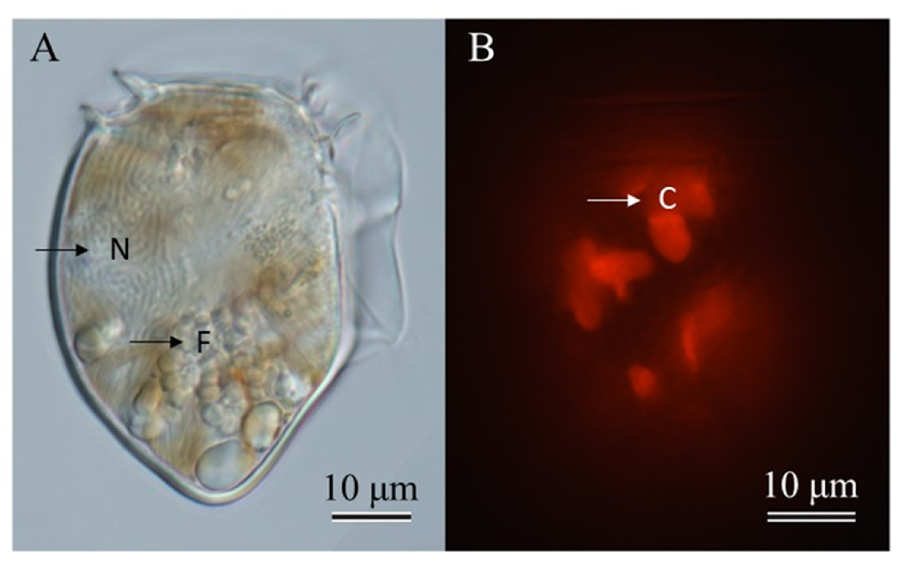
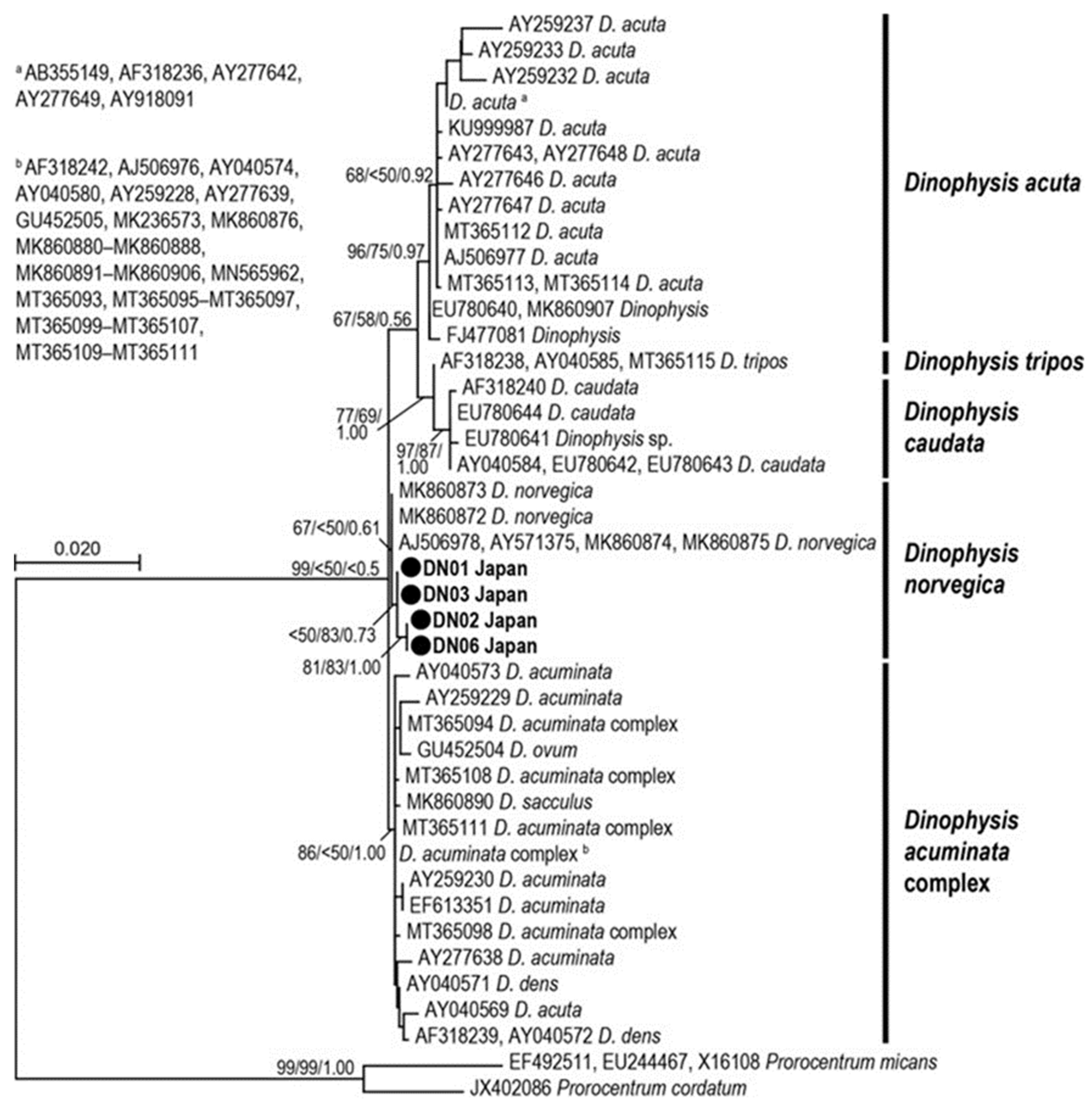
2.1. Species Identification

Dinophysis norvegica cells are generally large, ovoid, and robust. The posterior end tapers to a triangular shape (Figure 1). The micrographs showed the large nucleus which occupied the upper half of the cell and food vacuoles at the lower part (Figure 1A) and numerous chloroplasts (Figure 1B). Dinophysis acuta closely resembles D. norvegica based on morphology; therefore, they may be misidentified. These species can be distinguishable by their size (although it overlaps) and the location of the widest position: D. acuta is larger and widest below the mid-section, whereas D. norvegica is smaller and widest in the middle region of the cell [88,89]. A phylogenetic analysis based on the D1/D2 region (735 bp) supported that the strains isolated from Funka Bay, Japan, belong to the D. norvegica clade (Figure 2), and are closely related to the strains from Canada, Norway, and the USA (Atlantic Ocean).

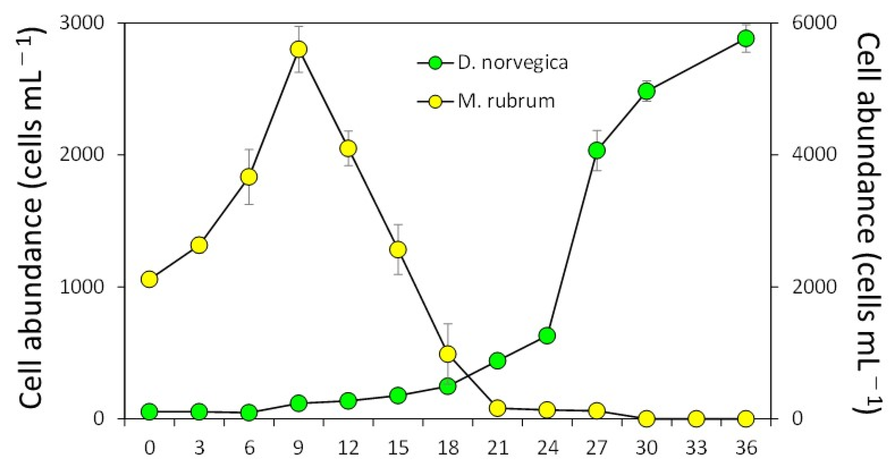
2.2. Feeding Behavior and Growth of Dinophysis Norvegica from Funka Bay in Culture Experiments

Only seven cultures of 48 single-cell isolates grew with the addition of the ciliate Mesodinium rubrum from the Oita Prefecture (Japan) as the prey species. However, clonal strains were successfully established, and the isolation success was 14.6% (7/48). Maximum cell densities of cultured strains during the 36 days of incubation ranged from 1057 cells per mL−1 to 3050 cells per mL−1 (mean of 2020 ± 702 cells per mL−1; n =7). Similar to the case of other Dinophysis species, Dinophysis norvegica was able to feed on the ciliate M. rubrum and grow.
In the growth experiment (12.5 °C, 12:12 light/dark cycle and irradiance of 100 μmol m−2 s−1), the ciliate M. rubrum grew exponentially during the first 9 days, reaching 5600 ± 346 cells per mL−1 (mean ± SD, n = 3) (Figure 3). The information on the experimental conditions is available in Section 4.1. After that, cell abundances of M. rubrum decreased sharply and disappeared by Day 30, probably due to the active consumption by D. norvegica and natural death. Dinophysis norvegica grew, but it showed very slow growth for the first 12 days, suggesting that they have a long lag phase. After that, they grew exponentially, with a maximum growth rate of 0.56 divisions per day−1 (from Day 24 to Day 27), reaching a maximum concentration of 2883 ± 104 cell per mL−1 at the end of incubation (Day 36), and the growth rates in every third day from Day 0 to Day 30 were −0.06 to 0.56 divisions per day−1 (0.18 ± 0.18) (Figure 3).

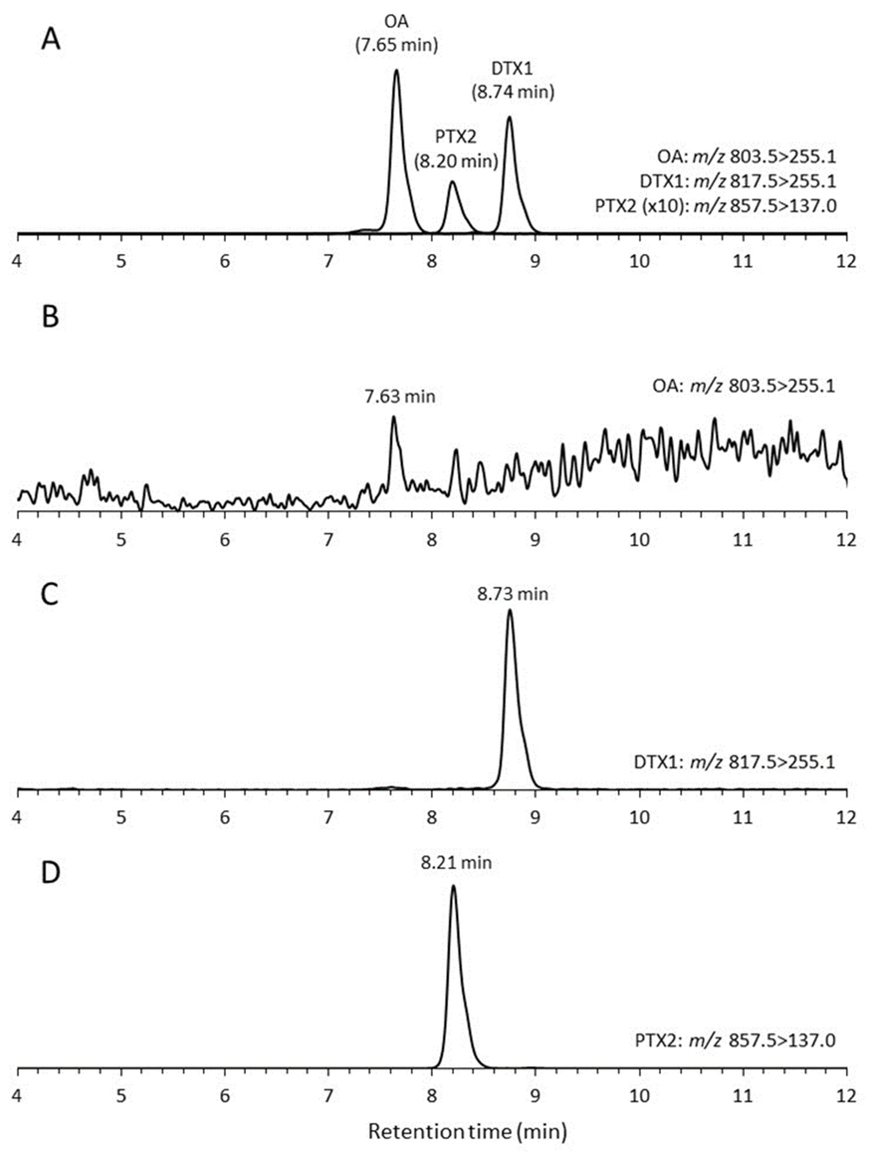
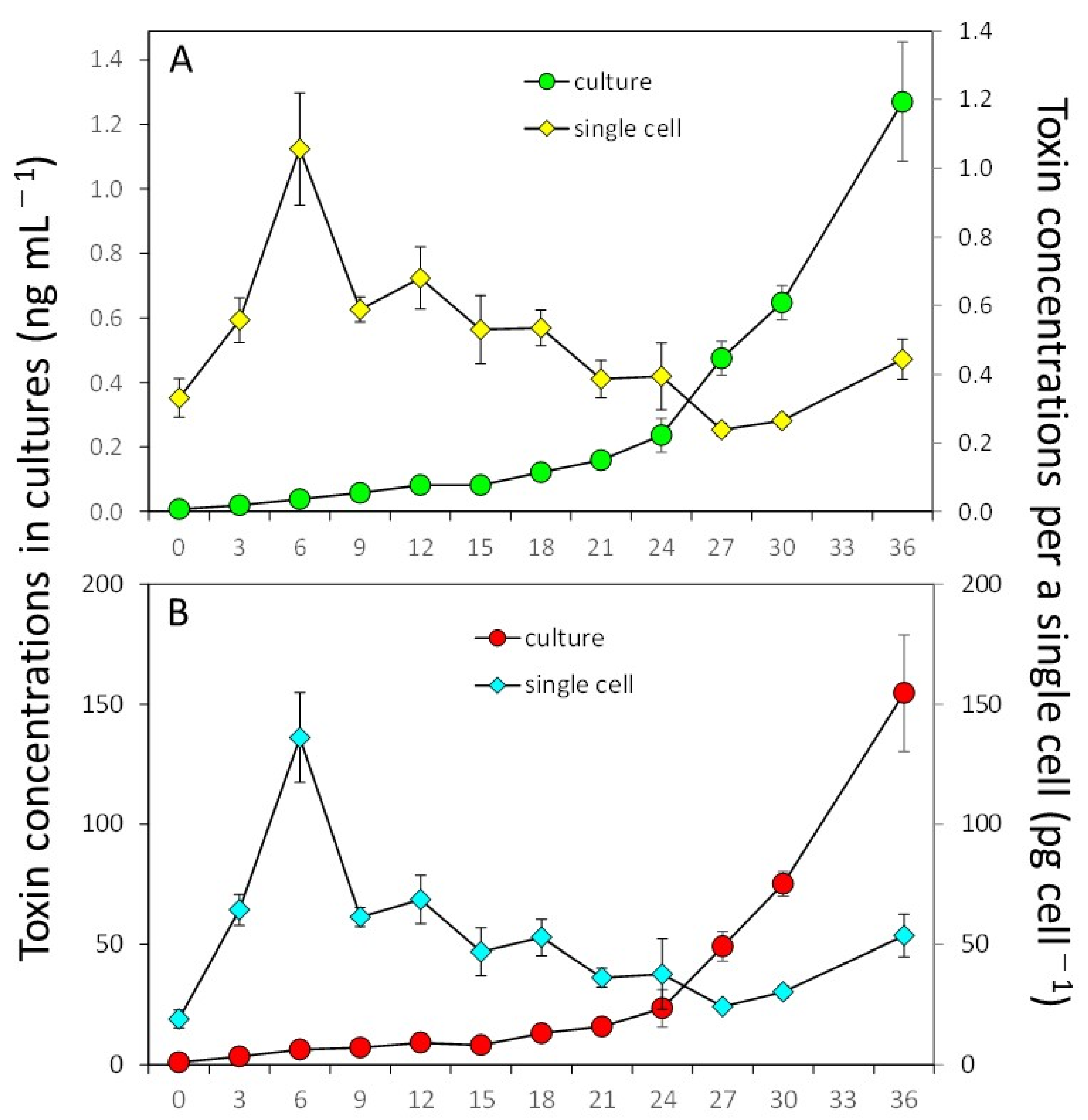
2.3. Toxin Production

Low levels of DTX1 were found, but only in cells of three strains of D. norvegica at 0.5 pg per cell−1 for DN16062021FUN-05, 0.7 pg per cell−1 for DN16062021FUN-08, and 1.2 pg per cell−1 for DN16062021FUN-06. All seven strains, however, produced PTX2 with cell quotas varying from 60.6 pg per cell−1 to 152.4 pg per cell−1 (Table 1). In cultures, the toxin concentration ranged from 0.705 ng per mL−1 to 3.55 ng per mL−1 in the case of DTX1 and from 126 ng per mL−1 to 375 ng per mL−1 in the case of PTX2 (Table 1). All strains of Dinophysis norvegica did not produce OA, except for strain DN16062021FUN-08, in which we detected a trace level of OA. Thus, the PTX2 showed clear peaks in all D. norvegica strains, and the DTX1 was detected in enough concentrations to be quantified from three strains, DN16062021FUN-05, DN16062021FUN-06, and DN16062021FUN-08. OA was lower than the detection limit concentration in all strains, but there was a small peak with a signal-to-noise ratio of 4 at the same retention time as OA in the MRM chromatograms of strain DN16062021FUN-08 (Figure 4). It may be possible that OA could be detected from samples with high cell density and concentration.
Strain DN16062021FUN-06 of D. norvegica produced DTX1 and PTX2 throughout the 36-day culture experiment (Figure 5). Both productions of DTX1 and PTX2 started to increase from 0.092 ± 0.009 pg per cell−1 on Day 12 to 1.280 ± 0.185 pg per cell−1 on Day 36, and from 9.3 ± 0.5 pg per cell−1 on Day 12 to 154.7 ± 24.2 pg per cell−1 on Day 36, respectively. The specific toxin production rates for DTX1 and PTX2 during the exponential growth phase were 0.291 ± 0.020 pg per mL−1 per day−1 and 0.291 ± 0.023 pg per mL−1 per day−1, respectively. The net toxin production rate (Rtox) during the exponential growth phase was 0.001 ± 0.0003 ng per mL−1 per day−1 for DTX1 and 0.13 ± 0.03 ng per mL−1 per day−1 for PTX2 in the exponential phase. DTX2 concentrations that were above the detection limit were detected from Day 18 to Day 36 and ranged from 0.132 to 1.28 ng per mL−1. For PTX2, the concentrations above the detection limit were measured from Day 3 until the end of the experiment, Day 36, and were in the range of 3.4 to 154.7 ng per mL−1.

3. Discussion

Similar to the case of other Dinophysis species [49,50,51,52,53,54,55], Dinophysis norvegica could feed on the ciliate M. rubrum by inserting the peduncle into the cells. The ciliate became immobile just after insertion, and their cilia were shed from the cell within 1–5 min. The cytoplasm of the prey was actively ingested through the peduncle. It took 45–100 min until the whole cell content of M. rubrum was consumed by D. fortii and D. tripos [49,52]. Dinophysis acuminata, D. caudata, and D. fortii, isolated from Japanese coastal waters, displayed growth rates of 0.50–1.03 divisions per day−1, reaching maximum concentrations of 2200–11,000 cells per mL−1 at temperatures ranging from 18 to 25 °C [36]. In this study, D. norvegica showed a similar growth rate at a lower temperature (12.5 °C) than other species. The culture strains were maintained successfully at high densities (>2000 cells per mL−1) for more than 20 months. This is an advancement compared to previous findings, where clonal cultures of D. norvegica isolated from Funka Bay and Lake Notoroko in Hokkaido, Japan, grew well (>1000 cells per mL−1) in the first incubation (24/96, 25%). However, when they were reinoculated into fresh Mesodinium cultures, no further growth was confirmed, and thus, it was not possible to establish their cultures (0/96, 0%) [52]. This led to the conclusion that the predator and the prey from different regions may be incompatible and cause failure in culturing [52]. In the growth experiment using the established cultures, D. norvegica had a much longer log phase (12 days) (Figure 3) than D. acuminata, D. caudata, and D. fortii (3–4 days) [49,54,55]. D. norvegica may become unstable when reinoculated into fresh Mesodinium cultures because of sudden environmental changes such as changes in pH. It is suggested that the reinoculation of D. norvegica cells with a relatively high concentration (>250 cells per mL−1) would achieve the successful maintenance of the cultures for long periods. The ranges of water temperatures and salinity in which this species appears in southern Hokkaido, including Funka Bay, were 2–16 °C and 24.3–33.9 PSU during 2016–2020 (https://www.hro.or.jp/list/fisheries/research/central/section/kankyou/kaidoku/j12s220000000dgw.html, accessed on 2 February 2023). The influence of the temperature (12.5 °C) on the success of establishing cultures is still unclear in this study.
The establishment of strains in the toxic Dinopshysis species is important for investigating toxin production and understanding how it is influenced by changes in physical–chemical conditions. Until now, information on the concentration of toxins produced by D. norvegica has been available based on isolated and pooled cells from field samples from two locations in Norway (Sogndal and Flødevigen Bay) and in Japan (Yakumo and Saroma) [19,22,87]. Similar to the previous records based on isolated cells, OA concentrations in cultured strains remained below the detection limit [19,22,87]. The maximum DTX1 concentrations in cultured strains were lower than reported based on cells from field samples (1.16 pg per cell−1 vs. 14 pg per cell−1) [19,22,87]. Interestingly the PTX2 maximum concentrations were much higher in the cultured strains (152 pg per cell−1), than in the cells collected from the field in Yakumo Japan (89 pg per cell−1) and 1.7 pg per cell−1 from Flødevigen Bay in Norway [22,87]. From 1987 until 2022, 43 HABs associated with D. norvegica were recorded globally, the majority of them associated with DSP [90]. A study based on Scottish shellfish farms estimated that a 1% increase in the toxins produced by Dinophysis spp. can lead to a 0.66% reduction in shellfish production, resulting in an estimated annual loss of GBP 1.37 million [91]. Additional studies on strains in culture may therefore enhance our understanding of not only the variability in toxin production between strains from the same and different geographical locations but also the impact of changes in physicochemical variables. [69,92].

4. Materials and Methods

4.1. Isolation and Establishment of Clonal Cultures

The ciliate Mesodinium rubrum and cryptophyte Teleaulax amphioxeia were isolated from Inokushi Bay (32.7998 N, 131.8923 E) in Oita Prefecture, Japan, at the end of February 2007 [49]. To sustain the M. rubrum culture, a mixture of 50 mL of the culture (7.0–9.0 × 10³ cells per mL−1) and 100 mL of a modified f/2 medium [93,94] was prepared, along with 25–100 µL of T. amphioxeia culture (containing 0.5–2.0 × 10⁴ cells) as a food source. The culture medium was prepared based on autoclaved natural seawater collected from Tokyo Bay (35.3460 N, 139.6570 E) with the addition of 1/3 nitrate, phosphate, metals, and 1/10 vitamins. Before autoclaving, the salinity of the seawater was adjusted to 30 practical salinity units (PSU). The ciliate culture was maintained at a temperature of 18 °C under a photon irradiance of 100 μmol m−2 s−1, provided by cool-white fluorescent lamps, with a 12:12 h light/dark cycle.
Cells of Dinophysis norvegica (48 cells in total) were isolated by micropipetting from a seawater sample collected from Funka Bay, Japan (42.28 N, 140.35 E), in June 2021 and incubated in individual wells of a 48-well microplate (Iwaki, Japan). Each D. norvegica cell was grown in 1.0 mL of the culture medium, containing ca. 1.0 × 10³ cells of M. rubrum as the prey species. Dinophysis cells were incubated under the same light conditions as those for the M. rubrum culture, but at 12.5 °C. After one month of incubation, seven strains grew well and were established as clonal strains. The established cultures in each strain were maintained by inoculating small aliquots (0.1 mL) into 2.9 mL of fresh M. rubrum culture (approximately 2 × 10³ cells per mL−1) in 12-well microplates. These microplates were then incubated for a month under the same conditions as mentioned above, without the addition of Teleaulax culture. After one month of incubation, 1 mL of culture from each strain was sampled for toxin analysis. Cell abundances were estimated on 100 μL of cultures (in triplicates) inoculated and fixed with Lugol’s solution (2%) into a 96-well plate (Iwaki, Japan). Cells were counted under the inverted microscope (Nikon TE-300). Additionally, the orange autoflorescence derived from the chloroplasts of M. rubrum in D. norvegica cells was observed using an epifluorescence microscope under blue light excitation ((Zeiss Axioskop 2 (Carl Zeiss, Göttingen, Germany)) equipped with a digital camera (Axiocam 305 color (Carl Zeiss, Göttingen, Germany)).

4.2. Growth Experiments

The ciliate Mesodinium rubrum culture grown until the late exponential growth phase (ca. 5.0 × 10³ cells per mL−1) was diluted with the fresh culture media when it reached the late exponential growth phase to obtain an initial cell abundance of ca. 2.0 × 10³ cells per mL−1, and 7.5 mL aliquots of the mixed culture were inoculated into the wells of 6-well microplates (Iwaki, Japan). Next, 125 µL of a D. norvegica culture (strain DN16062021FUN-06) containing 375 cells was added into the M. rubrum culture to obtain an initial concentration of 50 cells per mL−1. The growth experiment was conducted for 36 days under the same light and temperature conditions used for maintaining the culture of D. norvegica. In the growth experiment, 1 mL of the cultures (triplicate) was sampled for toxin analysis every three days except for Day 33. D. norvegia and Mesodinium cell abundances were obtained as described in the previous paragraph. The specific growth rate (µ, divisions per day−1) of D. norvegica was determined during the exponential growth phase according to [95].

4.3. Sequences of 28S rDNA (D1-D2 Region)

Genomic DNA was extracted from several cells in each strain with 5% Chelex buffer in four of the established strains [96]. The reaction mixture for PCR amplification was prepared by adding 1 μL of template DNA, 1 μM of both D1/D2 primer sets [97], 0.2 mM of each dNTP, 1× PCR buffer, 1.5 mM Mg2+, 1U KOD-Plus-Ver.2 (TOYOBO, Osaka, Japan), and RNA-free dH2O to achieve a final volume of 25 μL. The amplification was carried out using a thermal cycler (PC-808, ASTEC, Fukuoka, Japan). PCR amplification was performed with the following cycling conditions: 2 min at 94 °C, 30 cycles at 94 °C for 15 s, 55 °C for 30 s, and 68 °C for 40 s. The PCR products were transformed into DH5α cells (Promega, Madison, WI, USA) after ligation into the pGEM T-Easy Vector (Promega). After color selection, plasmid DNA was purified and the DNA sequences were determined using M13 Reverse and U19 primers with a Dynamic ET terminator cycle sequencing kit (GE Healthcare, Little Chalfont, UK). The sequences were then analyzed on a DNA sequencer (ABI3730, Applied Biosystems, Foster City, CA, USA). A BLAST search was performed to determine closely related species, and their GenBank sequences were obtained for phylogenetic analyses. The sequences were aligned using AliView [98], and identical sequences were compiled into a single sequence. All newly obtained sequences were deposited into the DDBJ databank (accession numbers: LC760478-LC760481).
A phylogenetic tree was constructed based on maximum likelihood (ML) using MEGA version 10 [99] with the best substitution model selected: Kimura 2-parameter model plus gamma distribution (G = 0.86). Bootstrap support (BS) values of ML and neighbor-joining (NJ) analyses for the trees were estimated using 500 replicates each. For the posterior probabilities (PP) of Bayesian inference, the best model substitution calculated by Akaike information criterion in jModelTest version 2.1.10 [100] was TIM plus gamma (G = 0.9710), and the effective sample size was calculated using Bayesian Evolutionary Analysis Sampling Trees (BEAST) and Tracer. Bayesian inference was conducted using MrBayes version 3.2.5 [101] based on the Bayesian information criteria calculated by jModelTest. A total of 3,077,000 Markov chain Monte Carlo generations were used with 4 chains and trees sampled every 1000 generations with PP estimated with 25% generations burn-in. Convergence of the chains was reconfirmed when the average standard deviations of the split frequencies were below 0.01 after calculations. Sequences from Prorocentrum micans Ehrenberg, 1834, and Prorocentrum cordatum (Ostenfeld) J.D.Dodge, 1976, were used as outgroup.

4.4. DSP Toxin Analysis

The samples were frozen at −30 °C until the toxins were extracted by solid-phase extraction (SPE). The SPE of toxins was modified compared to a previous method [86,102,103,104]. The MonoSpin C18 centrifuge cartridge column (GL Science Inc., Tokyo, Japan) was loaded with 1 mL of the thawed samples and conditioned with 1.0 mL each of methanol and distilled water. The SPE column was rinsed with 0.5 mL distilled water, and the toxins were extracted with 0.1 mL methanol. The methanol eluates were subjected to LC-MS/MS analysis using a previously established method. [105]. A Nexera-20XR series liquid chromatograph (Shimadzu, Kyoto, Japan) was coupled to a QTRAP 4500 mass spectrometer (SCIEX, MA, USA) of a hybrid triple quadrupole/linear ion trap. Separations were performed on LC columns (internal diameter [i.d.], 100 mm × 2.1 mm) packed with 1.9 μm Hypersil GOLD C8 (Thermo Fisher Scientific Inc., Waltham, MA, USA) and maintained at 30 °C. Eluent A was composed of water, while eluent B was a mixture of 95% acetonitrile and 5% water, which contained both 2 mM ammonium formate and 50 mM formic acid. Toxin elutions were carried out from the column with 50% B at a flow rate of 0.3 mL per min−1. LC-MS/MS analysis was performed using multiple-reaction monitoring (MRM) with negative-mode ionization. The target parent ions and fragment ions in Q1 and Q3 were used for each toxin, as follows: OA, m/z 803.5 > 255.1; DTX1, m/z 817.5 > 255.1; PTX2, m/z 857.5 > 137.0; PTX1 and PTX11, m/z 873.5 > 137.0; PTX2 Seco acid (PTX2 SA), m/z 875.5 > 137.0. The lowest detection limits of OA/DTX1 and PTX2 were 0.1 and 1.2 ng per mL−1. The LC-MS/MS method was used to analyze 100 cells of the toxic plankton, which showed levels equivalent to 0.1 pg/cell of OA (and DTX1) and 1.2 pg/cell of PTX2. During the growth experiments, the specific toxin production rate (µtox, pg per cell−1 per day−1) and net toxin production rate (Rtox) were calculated for the exponential growth phase, using the previously published equations [106].

Author Contributions

Conceptualization, S.N.; Investigation, T.K. and M.N.; Methodology, H.U. and S.N.; Data analyses, H.U., L.B., N.N., R.M., S.N., and W.M.L.; writing—original draft, L.B., S.S., and S.N.; writing—review and editing: S.S., L.B., and S.N. All authors have read and agreed to the published version of the manuscript.

Funding

This work was funded by JST/JICA, Science and Technology Research Partnership for Sustainable Development (JPMJSA1705) [SN], a Grant-in-Aid for Scientific Research (Kiban-B) by the Japan Society for the Promotion of Science (21H02274) [SN, NN, RM, TK], (18KK0182) [SN], and the Estonian Research Council (PSG735) [SS].

Data Availability Statement

Data are contained within the article. Access to sequencing data is described in Section 4.3.

Acknowledgments

We thank Toshiyuki Suzuki and Hiroshi Oikawa, Fisheries Technology Institute, Japan Fisheries Research and Education Agency, for their encouragement during this study. Also, Mitsunori Iwataki, Graduate School of Agricultural and Life Sciences, The University of Tokyo is thanked for lending the light microscope.

Conflicts of Interest

The authors declare no conflict of interest.

References

  1. Smayda, T.J. Novel and nuisance phytoplankton blooms in the sea: Evidence for a global epidemic. In Toxic Mar. Plankton; Granéli, E., Sundström, B., Edler, L., Anderson, D.M., Eds.; Elsevier: New York, NY, USA, 1990; pp. 29–40. [Google Scholar]
  2. Hallegraeff, G.M. A review of harmful algal blooms and their apparent global increase. Phycologia 1993, 32, 79–99. [Google Scholar] [CrossRef]
  3. Van Dolah, F.M. Marine algal toxins: Origins, health effects, and their increased occurrence. Environ. Health Persp. 2000, 108, 133–141. [Google Scholar] [CrossRef]
  4. Zingone, A.; Oksfeldt Enevoldsen, H. The diversity of harmful algal blooms: A challenge for science and management. Ocean Coast. Manag. 2000, 43, 725–748. [Google Scholar] [CrossRef]
  5. Allen, J.I.; Anderson, D.; Burford, M.; Dyhrman, S.; Flynn, K.; Glibert, P.M.; Granéli, E.; Heil, C.; Sellner, K.; Smayda, T.; et al. Global Ecology and Oceanography of Harmful Algal Blooms, Harmful Algal Blooms in Eutrophic Systems; GEOHAB Report 4IOC and SCOR; GEOHAB: Paris, France; Baltimore, MD, USA, 2000; pp. 1–74. [Google Scholar]
  6. Anderson, D.M.; Glibert, P.M.; Burkholder, J.M. Harmful algal blooms and eutrophication: Nutrient sources, composition, and consequences. Estuaries 2002, 25, 562–584. [Google Scholar] [CrossRef]
  7. Edwards, M.; Johns, D.G.; Leterme, S.C.; Svendsen, E.; Richardson, A.J. Regional climate change and harmful algal blooms in the northeast Atlantic. Limnol. Oceanogr. 2006, 51, 820–829. [Google Scholar] [CrossRef]
  8. Hégaret, H.; Shumway, S.E.; Wikfors, G.H.; Pate, S.; Burkholder, J.M. Potential transfer of harmful algae through relocation of bivalve molluscs. Mar. Ecol. Prog. Ser. 2008, 361, 169–181. [Google Scholar] [CrossRef]
  9. Heisler, J.; Gilbert, P.M.; Burkholder, J.M.; Anderson, D.M.; Cochlan, W.; Dennison, W.C.; Dortch, Q.; Gobler, C.J.; Heil, C.A.; Humphries, E.; et al. Eutrophication and harmful algal blooms: A scientific consensus. Harmful Algae 2008, 8, 3–13. [Google Scholar] [CrossRef]
  10. Moore, S.K.; Trainer, V.L.; Mantua, N.J.; Parker, M.S.; Laws, E.A.; Backer, L.C.; Fleming, L.E. Impacts of climate variability and future climate change on harmful algal blooms and human health. Environ. Health 2008, 7, S4. [Google Scholar] [CrossRef]
  11. Roger, A.D.; Laffoley, D. d’A. International Earth System Expert Workshop on Ocean Stresses and Impacts; Summary Report; IPSO: Oxford, UK, 2011; 18p. [Google Scholar]
  12. Fu, F.X.; Tatters, A.O.; Hutchins, D.A. Global change and the future of harmful algal blooms in the ocean. Mar. Ecol. Prog. Ser. 2012, 470, 207–233. [Google Scholar] [CrossRef]
  13. Glibert, P.M.; Allen, J.I.; Artioli, Y.; Beusen, A.; Bouwman, L.; Harle, J.; Holmes, R.; Holt, J. Vulnerability of coastal ecosystems to changes in harmful algal bloom distribution to climate change: Projections based on model analysis. Glob. Chang. Biol. 2014, 20, 3845–3858. [Google Scholar] [CrossRef]
  14. Hallegraeff, G.M.; Anderson, D.M.; Belin, C.; Dechroui Bottein, M.-Y.; Bresnan, E.; Chinain, M.; Enevoldsen, H.; Iwataki, M.; Karlson, B.; McKenzie, C.H.; et al. Perceived global increase in algal blooms is attributable to intensified monitoring and emerging bloom impacts. Commun. Earth Environ. 2021, 2, 117. [Google Scholar] [CrossRef]
  15. Burkholder, J.M. Implication of harmful microalgae and heterotrophicdinoflagellates in management of sustainable marine fisheries. Biol. App. 1998, 8, S37–S62. [Google Scholar]
  16. Yasumoto, T.; Oshima, Y.; Yamaguchi, M. Occurrence of a new type of shellfish poisoning in the Tohoku district. Bull. Jpn. Soc. Sci. Fish. 1978, 44, 1249–1255. [Google Scholar] [CrossRef]
  17. Lee, J.S.; Igarashi, T.; Fraga, S.; Dahl, E.; Hovgaard, P.; Yasumoto, T. Determination of diarrhetic shellfish toxins in various dinoflagellate species. J. Appl. Phycol. 1989, 1, 147–152. [Google Scholar] [CrossRef]
  18. Jun, J.H.; Sim, C.S.; Lee, C.O. Cytotoxic compounds from a two-sponge association. J. Nat. Prod. 1995, 58, 1722–1726. [Google Scholar]
  19. Terao, K.; Ito, E.; Yanagi, T.; Yasumoto, T. Histopatholgical studies on marine toxin poisoning. 1. Ultrastructural changes in the small intestine and liver of suckling mice induced by dinophysistoxin-1 and pectenotoxin-1. Toxicon 1986, 24, 1141–1151. [Google Scholar] [CrossRef] [PubMed]
  20. Mackenzie, L.A. A Long-Term Time Series of Dinophysis acuminata Blooms and Associated Shellfish Toxin Contamination in Port Underwood, Marlborough Sounds, New Zealand. Toxins 2019, 11, 7421. [Google Scholar] [CrossRef]
  21. Maestrini, S.Y. Bloom dynamics and ecophysiology of Dinophysis spp. In Physiological Ecology of Harmful Algal Blooms; Anderson, D.M., Cembella, A.D., Hallegraeff, G.M., Eds.; NATO ASI Series; Springer-Verlag: Berlin, Germany, 1998; Volume G 41, pp. 243–265. [Google Scholar]
  22. Miles, C.O.; Wilkins, A.L.; Samdal, I.A.; Sandvik, M.; Petersen, D.; Quilliam, M.A.; Naustvoll, L.J.; Jensen, D.J.; Cooney, J.M. A novel pectenotoxin, PTX-12, in Dinophysis spp. and shellfish from Norway. Chem. Res. Toxicol. 2004, 17, 1423–1433. [Google Scholar] [CrossRef]
  23. Ito, E.; Suzuki, T.; Oshima, Y.; Yasumoto, T. Studies on diarrhetic activity on pectenotoxin-6 in the mouse and rat. Toxicon 2008, 15, 707–716. [Google Scholar] [CrossRef]
  24. Reguera, B.; Pizarro, G. Planktonic Dinoflagellates Which Produce Polyether Toxins of the Old—DSP Complex. In Seafood and Freshwater Toxins: Pharmacology, Physiology and Detection, 2nd ed.; Botana, L.M., Ed.; Taylor & Francis: London, UK, 2008; pp. 257–284. [Google Scholar]
  25. Tubaro, A.; Sosa, S.; Bornancin, A.; Hungerford, J. Pharmacology and toxicology of diarrheic shellfish toxins. In Seafood and Freshwater Toxins. Pharmacology, Physiology and Detection, 2nd ed.; Botana, L.M., Ed.; CRC Press: Boca Raton, FL, USA, 2008; pp. 229–253. [Google Scholar]
  26. Vilariño, N.; Espiña, B. Pharmacology of pectenotoxins. In Seafood and Freshwater Toxins. Pharmacology, Physiology and Detection, 2nd ed.; Botana, L.M., Ed.; CRC Press: Boca Raton, FL, USA, 2008; pp. 361–369. [Google Scholar]
  27. Lawrence, J.; Loreal, H.; Toyofuku, H.; Hess, P.; Iddya, K.; Ababouch, L. Assessment and Management of Biotoxin Risks in Bivalve Molluscs; FAO Fisheries and Aquaculture Technical Paper 551; FAO: Rome, Italy, 2011; p. 337. [Google Scholar]
  28. Moestrup, Ø.; Akselmann-Cardella, R.; Fraga, S.; Hoppenrath, M.; Iwataki, M.; Komárek, J.; Larsen, J.; Lundholm, N.; Zingone, A. 2009 Onwards. IOC-UNESCO Taxonomic Reference List of Harmful Micro Algae. Available online: http://www.marinespecies.org/hab (accessed on 25 April 2022). [CrossRef]
  29. Edvardsen, B.; Shalchian-abrizi, K.; Jakobsen, K.S.; Medlin, L.K.; Dahl, E.; Brubak, S.; Paasche, E. Genetic variability and molecular phylogeny of Dinophysis species (Dinophyceae) from Norwegian waters inferred from single cell analyses of rDNA. J. Phycol. 2003, 39, 395–408. [Google Scholar] [CrossRef]
  30. Hart, M.C.; Green, D.H.; Bresnan, E.; Bolch, C.J. Large ribosomal RNA gene variation and sequence heterogeneity of Dinophysis (Dinophyceae) species from Scottish coastal waters. Harmful Algae 2007, 6, 271–287. [Google Scholar] [CrossRef]
  31. Raho, N.; Pizarro, G.; Escalera, L.; Reguera, B.; Marín, I. Morphology, toxin composition and molecular analysis of Dinophysis ovum Schütt, a dinoflagelate of the “Dinophysis acuminata complex”. Harmful Algae 2008, 7, 839–848. [Google Scholar] [CrossRef]
  32. Raho, N.; Rodríguez, F.; Reguera, B.; Marín, I. Are the mitochondrial cox1 and cob genes suitable markers for species of Dinophysis Ehrenberg? Harmful Algae 2013, 28, 64–70. [Google Scholar] [CrossRef]
  33. Rodríguez, F.; Escalera, L.; Reguera, B.; Rial, P.; Riobó, P.; de Jesús da Silva, T. Morphological variability, toxinology and genetics of the dinoflagellate Dinophysis tripos (Dinophysiaceae, Dinophysiales). Harmful Algae 2012, 13, 26–33. [Google Scholar] [CrossRef]
  34. Taylor, M.; McIntyre, L.; Ritson, M.; Stone, J.; Bronson, R.; Bitzikos, O.; Rourke, W.; Galanis, E.; Team, O. Outbreak of diarrhetic shellfish poisoning associated with mussels, British Columbia, Canada. Mar. Drugs 2013, 11, 1669–1676. [Google Scholar] [CrossRef]
  35. Stern, R.F.; Amorim, A.L.; Bresnan, E. Diversity and plastid types in Dinophysis acuminata complex (Dinophyceae) in Scottish waters. Harmful Algae 2014, 39, 223–231. [Google Scholar] [CrossRef]
  36. Nagai, S.; Sildever, S.; Suzuki, T.; Nishitani, G.; Basti, L.; Kamiyama, T. Successful cultivation and growth characteristics of the dinoflagellate Dinophysis. In Dinoflagellates: Morphology, Life History and Ecological Significance; Suba Rao, D.V., Ed.; Nova Science Publishers: New York, NY, USA, 2020; pp. 129–166. [Google Scholar]
  37. Sampayo, M.A.de.M. Trying to cultivate Dinophysis spp. In Toxic Phytoplankton Blooms in the Sea; Smayda, T.J., Shimizu, Y., Eds.; Elsevier: Amsterdam, The Netherlands, 1993; pp. 807–810. [Google Scholar]
  38. Jacobson, D.M.; Andersen, R.A. The discovery of mixotrophy in photosynthetic species of Dinophysis (Dinophyceae): Light and electron microscopical observations of food vacuoles in Dinophysis acuminata, D. norvegica and two heterotrophic dinophysoid dinoflagellates. Phycologia 1994, 33, 97–110. [Google Scholar] [CrossRef]
  39. Nishitani, G.; Miyamura, K.; Imai, I. Trying to cultivation of Dinophysis caudata (Dinophyceae) and the appearance of small cells. Plankton Biol. Ecol. 2003, 50, 31–36. [Google Scholar]
  40. Nishitani, G.; Sugioka, H.; Imai, I. Seasonal distribution of species of the toxic dinoflagellate genus Dinophysis in Maizuru Bay (Japan), with comments on their autofluorescence and attachment of picophytoplankton. Harmful Algae 2002, 1, 253–264. [Google Scholar] [CrossRef]
  41. Schnepf, E.; Elbrächter, M. Cryptophycean-like double membrane-bound chloroplast in the dinoflagellate, Dinophysis Ehrenb.: Evolutionary, phylogenetic and toxicological implications. Bot. Acta 1988, 101, 196–203. [Google Scholar] [CrossRef]
  42. Lucas, I.A.N.; Vesk, M. The fine structure of two photosynthetic species of Dinophysis (Dinophysiales, Dinophyceae). J. Phycol. 1990, 26, 345–357. [Google Scholar] [CrossRef]
  43. Hewes, C.D.; Mitchell, B.G.; Moisan, T.A.; Vernet, M.; Reid, F.M.H. The phycobilin signature of chloroplasts from three dinoflagellate species: A microanalytic study of Dinophysis caudata, D. fortii, and D. acuminata (Dinophysiales, Dinophyceae). J. Phycol. 2002, 34, 945–954. [Google Scholar] [CrossRef]
  44. Takishita, K.; Koike, K.; Maruyama, T.; Ogata, T. Molecular evidence for plastid robbery (kleptoplastidy) in Dinophysis, a dinoflagellate causing diarrhetic shellfish poisoning. Protist 2002, 153, 293–302. [Google Scholar] [CrossRef]
  45. Hackett, J.D.; Maranda, L.; Yoon, H.S.; Bhattacharya, D. Phylogenetic evidence for the cryptophyte origin of the plastid of Dinophysis (Dinophysiales, Dinophyceae). J. Phycol. 2003, 39, 440–448. [Google Scholar] [CrossRef]
  46. Janson, S.; Granéli, E. Genetic analysis of the psbA gene from single cells indicates a cryptomonad origin of the plastid in Dinophysis (Dinophyceae). Phycologia 2003, 42, 473–477. [Google Scholar] [CrossRef]
  47. Janson, S. Molecular evidence that plastids in the toxin-producing dinoflagellate genus Dinophysis originate from the free-living cryptophyte Teleaulax amphioxeia. Environ. Microbiol. 2004, 6, 1102–1106. [Google Scholar] [CrossRef]
  48. Park, M.G.; Kim, S.; Kim, H.S.; Myung, G.; Kang, Y.G.; Yih, W. First successful culture of the marine dinoflagellate Dinophysis acuminata. Aquat. Microb. Ecol. 2006, 45, 101–106. [Google Scholar] [CrossRef]
  49. Nagai, S.; Nitshitani, G.; Tomaru, Y.; Sakiyama, S.; Kamiyama, T. Predation by the toxic dinoflagellate Dinophysis fortii on the ciliate Myrionecta rubra and observation of sequestration of ciliate chloroplasts. J. Phycol. 2008, 44, 909–922. [Google Scholar] [CrossRef] [PubMed]
  50. Jaén, D.; Mamán, L.; Domínguez, R.; Martín, E. First report of Dinophysis acuta in culture. Harmful Algal News 2009, 39, 1–2. [Google Scholar]
  51. Riobó, P.; Reguera, B.; Franco, J.M.; Rodríguez, F. First report of the toxin profile of Dinophysis sacculus Stein from LC-MS analysis of laboratory cultures. Toxicon 2013, 76, 221–224. [Google Scholar] [CrossRef]
  52. Nagai, S.; Suzuki, T.; Kamiyama, T. Successful cultivation of the toxic dinoflagellate Dinophysis tripos (Dinophyceae). Plankton Benthos Res. 2013, 8, 171–177. [Google Scholar] [CrossRef]
  53. Mafra, L.L., Jr.; Nagai, S.; Uchida, H.; Tavares, C.P.S.; Escobar, B.P.; Suzuki, T. Harmful effects of Dinophysis to the ciliate Mesodinium rubrum: Implications for prey capture. Harmful Algae 2016, 59, 82–90. [Google Scholar] [CrossRef]
  54. Nishitani, G.; Nagai, S.; Sakiyama, S.; Kamiyama, T. Successful cultivation of the toxic dinoflagellate Dinophysis caudata (Dinophyceae). Plankton Benthos Res. 2008, 3, 78–85. [Google Scholar] [CrossRef]
  55. Nishitani, G.; Nagai, S.; Takano, Y.; Sakiyama, S.; Baba, K.; Kamiyama, T. Growth characteristics and phylogenetic analysis of the marine dinoflagellate Dinophysis infundibulus (Dinophyceae). Aquat. Microb. Ecol. 2008, 52, 209–221. [Google Scholar] [CrossRef]
  56. Kim, S.; Kang, Y.G.; Kim, H.S.; Yih, W.; Coats, D.W.; Park, M.G. Growth and grazing responses of the mixotrophic dinoflagellate Dinophysis acuminata as functions of light intensity and prey concentration. Aquat. Microb. Ecol. 2008, 51, 301–310. [Google Scholar] [CrossRef]
  57. Kim, M.; Nam, S.W.; Shin, W.; Coats, D.W.; Park, M.G. Dinophysis caudata (Dinophyceae) sequesters and retains plastids from the mixotrophic ciliate prey Mesodinium rubrum. J. Phycol. 2012, 48, 569–579. [Google Scholar] [CrossRef]
  58. Kamiyama, T.; Suzuki, T. Production of dinophysis-1 and pectenotoxin-2 by a culture of Dinophysis acuminata (Dinophyceae). Harmful Algae 2009, 8, 312–317. [Google Scholar] [CrossRef]
  59. Hackett, J.D.; Tong, M.M.; Kulis, D.M.; Fux, E.; Hess, P.; Bire, R.; Anderson, D.M. DSP toxin production de novo in cultures of Dinophysis acuminata (Dinophyceae) from North America. Harmful Algae 2009, 8, 873–879. [Google Scholar] [CrossRef]
  60. Kamiyama, T.; Nagai, S.; Suzuki, T.; Miyamura, K. Effect of temperature on production of okadaic acid, dinophysistoxin-1, and pectenotoxin-2 by Dinophysis acuminata in culture experiments. Aquat. Microb. Ecol. 2010, 60, 193–202. [Google Scholar] [CrossRef]
  61. Tong, M.; Kulis, D.M.; Fux, E.; Smith, J.L.; Hess, P.; Zhou, Q.; Anderson, D.M. The effects of growth phase and light intensity on toxin production by Dinophysis acuminata from northeastern United States. Harmful Algae 2011, 10, 254–264. [Google Scholar] [CrossRef]
  62. Nagai, S.; Suzuki, T.; Nishikawa, T.; Kamiyama, T. Differences in the production and excretion kinetics of okadaic acid, dinophysistoxin-1, and pectenotoxin-2 between cultures of Dinophysis acuminata and Dinophysis fortii isolated from western Japan. J. Phycol. 2011, 47, 1326–13337. [Google Scholar] [CrossRef] [PubMed]
  63. Basti, L.; Uchida, H.; Kanamori, M.; Matsushima, R.; Suzuki, T.; Nagai, S. Mortality and pathology of Japanese scallop, Patinopecten (Mizuhopecten) yessoensis, and noble scallop, Mimachlamys nobilis, fed monoclonal culture of PTX-producer, Dinophysis caudata. In Marine and Freshwater Harmful Algae, Proceedings of the 16th International Conference on Harmful Algae, Wellington, New Zealand from 27 to 31 October 2014; MacKenzie, A.L., Ed.; Cawthron Institute, Nelson, New Zealand and International Society for the Study of Harmful Algae: Wellington, New Zealand, 2015; pp. 105–108. [Google Scholar]
  64. Basti, L.; Uchida, H.; Matsushima, R.; Watanabe, R.; Suzuki, T.; Yamatogi, T.; Nagai, S. Influence of temperature on growth and production of Pectenotoxin-2 by a monoclonal culture of Dinophysis caudata. Mar. Drugs 2015, 13, 7124–7137. [Google Scholar] [CrossRef] [PubMed]
  65. Basti, L.; Suzuki, T.; Uchida, H.; Kamiyama, T.; Nagai, S. Thermal acclimation affects growth and lipophilic toxin production in a strain of cosmopolitan harmful alga Dinophysis acuminata. Harmful Algae 2018, 73, 119–128. [Google Scholar] [CrossRef] [PubMed]
  66. Gobler, J.C.; Dohrty, O.M.; Hattenrath-Lehmann, T.K.; Griffith, A.W.; Kang, Y.; Litaker, R.W. Ocean warming since 1982 has expanded the niche of toxic algal blooms in the North Atlantic and North Pacific oceans. PNAS 2017, 114, 4975–4980. [Google Scholar] [CrossRef] [PubMed]
  67. Guillotreau, P.; Le Bihan, V.; Morineau, B.; Pardo, S. The vulnerability of shellfish farmers to HAB events: An optimal matching analysis of closure decrees. Harmful Algae 2021, 101, 101968. [Google Scholar] [CrossRef]
  68. Reguera, B.; Velo-Suárez, L.; Raine, R.; Park, M. Harmful Dinophysis species: A review. Harmful Algae 2012, 14, 87–106. [Google Scholar] [CrossRef]
  69. Reguera, B.; Riobó, P.; Rodríguez, F.; Díaz, P.; Pizarro, G.; Paz, B.; Franco, J.M.; Blanco, J. Dinophysis toxins: Causative organisms, distribution and fate in shellfish. Mar. Drugs 2014, 12, 394–461. [Google Scholar] [CrossRef]
  70. Carpenter, E.J.; Janson, S.; Boje, R.; Pollenhne, F.; Chang, J. The dinoflagellate Dinophysis norvegica: Biological and ecological observation in the Baltic Sea. Eur. J. Phycol. 1995, 30, 1–9. [Google Scholar] [CrossRef]
  71. Okolodkov, Y.B.; Dodge, J.D. Biodiversity and biogeography of planktonic dinoflagellates in the Arctic Ocean. J. Exp. Mar. Biol. Ecol. 1996, 202, 19–27. [Google Scholar] [CrossRef]
  72. Meyer-Harms, B.; Pollenhe, F. Alloxanthin in Dinophysis norvegica (Dinophysiales, Dinophyceae) from the Baltic Sea. J. Phycol. 1998, 34, 280–285. [Google Scholar] [CrossRef]
  73. Bresnan, E.; Fryer, R.; Hart, M.; Percy, L. Correlation between Algal Presence in Water and Toxin Presence in Shellfish. Fish. Res. Serv. Contract Rep. 2005, 4, 5. [Google Scholar]
  74. Jansen, S.; Riser, C.W.; Wassmann, P.; Bathmann, U. Copepod feeding behavior and egg production during a dinoflagellate bloom in the North Sea. Harmful Algae 2006, 5, 102–112. [Google Scholar] [CrossRef]
  75. Smayda, T.J. Harmful Algal Bloom Communities in Scottish Coastal Waters: Relationship to Fish Farming and Regional Comparisons—A Review; Scottish Executive Environmental Group: Edinburgh, UK, 2006. [Google Scholar]
  76. Whyte, C.; Swan, S.; Davidson, K. Changing wind patterns linked to unusually high Dinophysis blooms around the Shetland Islands, Scotland. Harmful Algae 2014, 39, 365–373. [Google Scholar] [CrossRef]
  77. Fabro, E.; Almandoz, G.O.; Ferrario, M.; Tillmann, U.; Cembella, A.; Krock, B. Distribution of Dinophysis species and their association with lipophilic phycotoxins in plankton from the Argentine Sea. Harmful Algae 2016, 59, 31–41. [Google Scholar] [CrossRef]
  78. Shahi, N.; Nayak, B.B.; Mallik, S.K. Dinophysis norvegica: First report of the toxic temperate water Dinophysis in Manori creek of Mumbai water. Harmful Algae News 2010, 42, 14–15. [Google Scholar]
  79. Hernández-Becerril, D.U. La Diversidad del Fitoplancton Marino de México. Un Acercamiento Actual. In Planctología Mexicana; Barreiro, M.T., Meave, M.E., Signoret, M., Figueroa, M.G., Eds.; Sociedad Mexicana de Planctología (SOMPAC); Universidad Autónoma Metropolitana: México City, México, 2003; pp. 1–17. [Google Scholar]
  80. Suba Rao, D.V.; Pan, Y.; Zitko, V.; Bugden, G.; Mackelgan, K. Diarrhetic shellfish poisoning (DSP) associated with a subsurface bloom of Dinophysis norvergica in Bedford Basin, eastern Canada. Mar. Ecol. Prog. Ser. 1993, 97, 117–126. [Google Scholar] [CrossRef]
  81. Cembella, A.D. Occurrence of okadaic acid, a major diarrhetic shellfish toxin, in natural populations of Dinophysis spp. from the east of North America. J. Appl. Phycol. 1989, 1, 307–310. [Google Scholar] [CrossRef]
  82. Goto, H.; Igarashi, T.; Watai, M.; Yasumoto, T.; Gomez, O.V.; Valdivia, G.L.; Noren, F.; Gisselson, L.A.; Granéli, E. Worldwide occurrence of pectenotoxins and yessotoxins in shellfish and phytoplankton. In Proceedings of the Ninth International Conference on Harmful Algal Blooms, Tasmania, Australia, 7–11 February 2000; p. 20 (Abstract). [Google Scholar]
  83. Pimiä, V.; Kankaanpaä, H.; Kononen, K. The first observation of okadaic acid in Mytilus edulis from the Gulf of Finland. Boreal Environ. Res. 1998, 2, 381–385. [Google Scholar]
  84. Sipiä, V.; Kankaanpaä, H.; Meriluoto, J. The first observation of okadaic acid in flounder in the Baltic Sea. Sarsia 2000, 85, 471–475. [Google Scholar] [CrossRef]
  85. Deeds, J.R.; Stutts, W.L.; Celiz, M.D.; mac Leod, J.; Hamilton, A.E.; Lewis, B.J.; Miller, D.W.; Kanwit, K.; Smith, J.L.; Kulis, D.M.; et al. Dihydrodinophysistoxin-1 produced by Dinophysis norvegica in the Gulf of Main, USA and its accumulation in shellfish. Toxins 2020, 12, 533. [Google Scholar] [CrossRef]
  86. Suzuki, T.; Miyazono, A.; Baba, K.; Sugawara, R.; Kamiyama, T. LC-MS/MS analysis of okadaic acid analogues and other lipophilic toxins in single-cell isolates of several Dinophysis species collected in Hokkaido, Japan. Harmful Algae 2009, 8, 233–238. [Google Scholar] [CrossRef]
  87. Uchida, H.; Watanabe, R.; Matsushima, R.; Oikawa, H.; Nagai, S.; Kamiyma, T.; Baba, K.; Miyazono, A.; Kosaka, K.; Kaga, S.; et al. Toxin profiles of okadaic acid analogues and other lipophilic toxins in Dinophysis from Japanese coastal waters. Toxins 2018, 10, 457. [Google Scholar] [CrossRef] [PubMed]
  88. Balech, E. Some Norwegian Dinophysis species (Dinoflagellata). Sarsia 1976, 61, 75–94. [Google Scholar] [CrossRef]
  89. Taylor, F.J.R.; Fukuyo, Y.; Larsen, J. Taxonomy of harmful dinoflagellates. In Manual on Harmful Marine Microalgae; Hallegraeff, G.M., Anderson, D.M., Cembella, A.D., Eds.; IOC Manuals and Guides No. 33; UNESCO: Paris, France, 1995; pp. 283–317. [Google Scholar]
  90. IOC-UNESCO. The Harmful Algal Event Database (HAEDAT). Available online: http://haedat.iode.org/index.php (accessed on 18 February 2023).
  91. Martino, S.; Gianella, F.; Davidson, K. An approach for evaluating the economic impacts of harmful algal blooms: The effects of blooms of toxic Dinophysis spp. on the productivity of Scottish shellfish farms. Harmful Algae 2020, 99, 101912. [Google Scholar] [CrossRef]
  92. Séchet, V.; Sibat, M.; Billien, G.; Carpentier, L.; Rovillon, G.A.; Raimbault, V.; Malo, F.; Gaillard, S.; Perrière-Rumebe, M.; Hess, P.; et al. Characterization of toxin-producing strains of Dinophysis spp. (Dinophyceae) isolated from French coastal waters, with a particular focus on the D. acuminata-complex. Harmful Algae 2021, 107, 101974. [Google Scholar] [CrossRef]
  93. Guillard, R.P.L. Culture of phytoplankton for feeding marine invertebrates. In Culture of Marine Inveretbrate Animals; Smith, W.L., Chanley, M.H., Eds.; Springer: Boston, MA, USA, 1975. [Google Scholar]
  94. Nagai, S.; Matsuyama, Y.; Oh, S.-J.; Itakura, S. Effect of nutrients and temperature on encystment of the toxic dinoflagellate Alexandrium tamarense (Dinophyceae) isolated from Hiroshima Bay, Japan. Plankton Biol. Ecol. 2004, 51, 103–109. [Google Scholar]
  95. Guillard, R.R.L.; Kilham, P.; Jackson, T.A. Kinetics of silicon-limited growth in the marine diatom Thalassiosira pesudonana Hasle and Heimdal (=Cyclotella nana Husted). J. Phycol. 1973, 9, 233–237. [Google Scholar]
  96. Nagai, S.; Yamamoto, K.; Hata, N.; Itakura, S. Study of DNA extraction methods for use in loop-mediated isothermal amplification detection of single resting cysts in the toxic dinoflagellates Alexandrium tamarense and A. catenella. Mar. Genom. 2012, 7, 51–56. [Google Scholar] [CrossRef]
  97. Scholin, C.A.; Herzog, M.; Sogin, M.; Anderson, D.M. Identification of group- and strain-specific genetic markers for globally distributed Alexandrium (Dinophyceae). II. Sequence analysis of a fragment of the LSU rRNA gene. J. Phycol. 1994, 30, 999–1011. [Google Scholar] [CrossRef]
  98. Larsson, A. AliView: A fast and lightweight alignment viewer and editor for large data sets. Bioinformatics 2014, 30, 3276–3278. [Google Scholar] [CrossRef]
  99. Kumar, S.; Stecher, G.; Li, M.; Knyaz, C.K.T. Mega X: Molecular evolutionary genetics analysis across computing platforms. Mol. Biol. Evol. 2018, 35, 1547–1549. [Google Scholar] [CrossRef] [PubMed]
  100. Darriba, D.; Taboada, G.; Doallo, R.; Posada, D. jModelTest 2: More models, new heuristics and parallel computing. Nat. Methods 2012, 9, 772. [Google Scholar] [CrossRef] [PubMed]
  101. Ronquist, F.; Teslenko, M.; van der Mark, P.; Ayres, D.L.; Darling, A.; Höhna, S.; Larget, B.; Liu, L.; Suchard, M.A.; Huelsenbeck, J.P. MrBayes 3.2: Efficient Bayesian phylogenetic inference and model choice across a large model space. Syst. Biol. 2012, 61, 539–542. [Google Scholar] [CrossRef] [PubMed]
  102. Suzuki, T.; Mitsuya, T.; Imai, M.; Yamasaki, M. DSP toxin contents in Dinophysis fortii and scallops collected at Mutsu Bay Japan. J. Appl. Phycolology 1997, 8, 509–515. [Google Scholar] [CrossRef]
  103. Suzuki, T.; Mitsuya, T.; Matsubara, H.; Yamasaki, M. Determination of pectenotoxin-2 after solid-phase extraction from seawater and from the dinoflagellate Dinophysis fortii by liquid chromatography with electrospray mass spectrometry and ultraviolet detection: Evidence of oxidation of pectenotoxin-2 to pectenotoxin-6 in scallops. J. Chromatography A 1998, 815, 155–160. [Google Scholar]
  104. Ajani, P.A.; Henriquez-Nunez, H.F.; Verma, A.; Nagai, S.; Uchida, H.; Tesoriero, M.J.; Farrell, H.; Zammit, A.; Brett, S.; Murray, S.A. Mapping the development of a Dinophysis bloom in a shellfish aquaculture area using a novel molecular qPCR assay. Harmful Algae 2022, 116, 102253. [Google Scholar] [CrossRef]
  105. Suzuki, T.; Quilliam, M. LC-MS/MS analysis of Diarrhetic Shellfish Poisoning (DSP) toxins, okadaic acid and dinophysistoxin analogues, and other lipophilic toxins. Anal. Sci. 2011, 27, 571–584. [Google Scholar] [CrossRef] [PubMed]
  106. Albinsson, M.E.; Negri, A.P.; Blackburn, S.I.; Bolch, C.J.S. Bacterial Community Affects Toxin Production by Gymnodinium catenatum. PLoS ONE 2014, 9, e104623. [Google Scholar] [CrossRef]
Figure 1. Micrographs of a vegetative cell of Dinophysis norvegica in culture in a bright field (A) and with fluorescence under blue light excitation (B). Plastids of D. norvegica emit red autofluoresecence. Cells in the early exponential growth phase were used. Scale bar is 10 µm, C: chloroplast, F: food vacuole, N: nucleus.
Toxins 15 00318 g001
Figure 2. Maximum likelihood (ML) tree of Dinophysis norvegica inferred from D1/D2 region (735 b.p.). Bootstrap supports of ML and neighbor joining (NJ) and posterior probabilities (PP) of Bayesian inference are indicated at node (ML/NJ/PP). Culture strains obtained in this study are in bold.
Toxins 15 00318 g002
Figure 3. Growth of strain DN16062021FUN-06 of Dinophyis norvegica in culture with the ciliate Mesodinium rubrum.
Toxins 15 00318 g003
Figure 4. The MRM chromatograms of lipophilic shellfish toxins by LC-MS/MS analysis. (A): The MRM chromatograms of lipophilic shellfish toxins standards OA, DTX1, and PTX2 100 ng per mL−1. (B): A chromatogram of OA MRM transition (m/z 803.5 > 255.1) from Dinophysis norvegica strain DN16062021FUN-08 extract. (C): A chromatogram of DTX1 MRM transition (m/z 817.5 > 255.1) from strain DN16062021FUN-08 extract. (D): A chromatogram of PTX2 MRM transition (m/z 857.5 > 137.0) from strain DN16062021FUN-08 extract.
Toxins 15 00318 g004
Figure 5. Toxin production of strain DN16062021FUN-06 of Dinophysis norvegica in culture with the ciliate Mesodinium rubrum. (A): Dinophysistoxin-1, (B): Pectenotoxin-2.
Toxins 15 00318 g005
Table 1. Toxin production in seven cultured strains of Dinophysis norvegica.
StrainsConcentration in
Culture (ng per mL−1)
Number
of Cells
Cell Quota
(pg per mL−1)
OADTX1PTX2OADTX1PTX2
DN16062021FUN-01ND (<0.1)ND (<0.1)1371807ND (<0.01)ND (<0.01)75.8
DN16062021FUN-02ND (<0.1)ND (<0.1)1262080ND (<0.01)ND (<0.01)60.6
DN16062021FUN-03ND (<0.1)ND (<0.1)1452333ND (<0.01)ND (<0.01)62.1
DN16062021FUN-05ND (<0.1)1.443752850ND (<0.01)0.5131.6
DN16062021FUN-06ND (<0.1)3.553163050ND (<0.01)1.2103.6
DN16062021FUN-07ND (<0.1)ND (<0.1)1611057ND (<0.02)ND (<0.02)152.4
DN16062021FUN-08ND (<0.1)0.705132936ND (<0.02) 0.7137.0
OA: Okadaic acid; DTX1: Dinophysistoxin-1; PTX2: Pectenotoxin-2; ND: Not detected (limit of detection); After one month of incubation, 1 mL of each culture was sampled for toxin analysis.
Disclaimer/Publisher’s Note: The statements, opinions and data contained in all publications are solely those of the individual author(s) and contributor(s) and not of MDPI and/or the editor(s). MDPI and/or the editor(s) disclaim responsibility for any injury to people or property resulting from any ideas, methods, instructions or products referred to in the content.

