Article

Intraspecific Diversity and Pathogenicity of Bacillus thuringiensis Isolates from an Emetic Illness

by
Jintana Pheepakpraw
1,2,
Thida Kaewkod
1,
Maytiya Konkit
3,
Sasiprapa Krongdang
4,
Kanyaluck Jantakee
1,
Rueankaew Praphruet
5,
Sakunnee Bovonsombut
1,6,
Aussara Panya
1,
Yingmanee Tragoolpua
1,
Niall A. Logan
7 and
Thararat Chitov
1,6,*
1
Department of Biology, Faculty of Science, Chiang Mai University, Chiang Mai 50200, Thailand
2
Doctor of Philosophy Program in Applied Microbiology (International Program), Faculty of Science, Chiang Mai University, Chiang Mai 50200, Thailand
3
Division of Microbiology, Faculty of Science and Technology, Nakhon-Pathom Rajabhat University, Nakhon-Pathom 73000, Thailand
4
Faculty of Science and Social Sciences, Burapha University, Sa Kaeo Campus, Sa Kaeo 27160, Thailand
5
Institute of Product Quality and Standardization, Maejo University, Chiang Mai 50290, Thailand
6
Environmental Science Research Center (ESRC), Faculty of Science, Chiang Mai University, Chiang Mai 50200, Thailand
7
Formerly of Glasgow Caledonian University, Glasgow G4 0BA, UK
*
Author to whom correspondence should be addressed.
Toxins 2023, 15(2), 89; https://doi.org/10.3390/toxins15020089
Submission received: 1 December 2022 / Revised: 10 January 2023 / Accepted: 12 January 2023 / Published: 18 January 2023
(This article belongs to the Special Issue Foodborne Toxins: Advanced Detection and Toxicity)

Abstract

:
This study describes an emetic food-borne intoxication associated with a Bacillus cereus group species and the characterization of the bacterial isolates from the incident in aspects of molecular tying, genetic factors, cytotoxicity, and pathogenic mechanisms relating to emetic illness. Through the polyphasic identification approach, all seven isolates obtained from food and clinical samples were identified as Bacillus thuringiensis. According to multilocus sequence typing (MLST) analysis, intraspecific diversity was found within the B. thuringiensis isolates. Four allelic profiles were found, including two previously known STs (ST8 and ST15) and two new STs (ST2804 and ST2805). All isolates harbored gene fragments located in the cereulide synthetase (ces) gene cluster. The heat-treated culture supernatants of three emetic B. thuringiensis isolates, FC2, FC7, and FC8, caused vacuolation and exhibited toxicity to Caco-2 cells, with CC50 values of 56.57, 72.17, and 79.94 µg/mL, respectively. The flow cytometry with the Annexin V/PI assay revealed both apoptosis and necrosis mechanisms, but necrosis was the prominent mechanism that caused Caco-2 cell destruction by FC2, the most toxic isolate.
Key Contribution: This study found that Bacillus thuringiensis was linked to emetic food-borne illness. Some virulent factors and the pathogenicity of the outbreak strains were determined.

1. Introduction

Bacillus cereus has long been recognized as a food-borne pathogen, one capable of producing two distinct types of toxins: diarrheagenic and emetic toxins [1,2]. Some other species that are closely related to B. cereus, known as the B. cereus group [2], were also occasionally found involved in food-borne outbreaks, such as B. thuringiensis and, more rarely, B. mycoides [3]. It is possible that some of the B. cereus group species might have been misidentified as B. cereus at the time of the outbreaks [3] or that B. cereus group species other than B. cereus have become emerging food-borne pathogens [4]. In addition, the potential of the B. cereus group species to cause food-borne illnesses has been increasingly recognized in recent years [5].
Bacillus thuringiensis, a species closely related to B. cereus, is better known for its ability to produce insecticidal toxins. B. thuringiensis has been widely used as a biocontrol agent [6,7,8,9] and its toxin genes have been transferred to genetically modified crops [10,11]. Members of the B. thuringiensis species have occasionally been found in food [12,13,14]. Enterotoxin genes, commonly present in B. cereus, are also widely distributed among B. thuringiensis isolates [5,15,16]. This species has been implicated in food-borne illnesses, but most of the Bacillus food-borne outbreak cases involved B. cereus [3,17]. There has been some indication of the presence of pathogenicity factors in some B. thuringiensis outbreak strains. In the epidemiology study by Bonis et al. in 2021 [18], B. thuringiensis strains retrieved from food-borne outbreaks in France during 2007–2017 were collected, and their phenotypic, genotypic, and genome characteristics were investigated. Virulence factors, including lecithinase activity, hemolysis activity, and virulence genes (cytK2, nheABC, and hblCDA), were found to be widely distributed (97–100%) in the outbreak strains and could be associated with the gastrointestinal symptoms in the outbreak history records. These virulent factors are mostly related to the diarrheal type of illness [18]. Little is known about this species’ association with emetic illness and the pathogenicity factors associated with it.
Following an incident of emetic food-borne outbreak in Glasgow, UK, Bacillus thuringiensis was isolated from food and vomit samples. The incident occurred in December 2007 and involved two female children, ages three and four, from one family. The meal was prepared using fresh pasta, smoked mackerel, and mussels in a garlic butter sauce, all purchased from a supermarket. Within a few hours of purchase, the pasta was cooked in boiling water, the smoked mackerel was combined with the mussels in a garlic butter sauce, and the sauce portion was reheated. The meal was served immediately after preparation. Approximately 4 h after consumption, the older child started vomiting, and several further episodes of vomiting occurred over the next 3 h. The younger child vomited only once, about 5 h after the consumption of the meal. There were no other symptoms. The children had eaten similar-sized portions, but their parents, who had eaten larger portions of the same food, did not show any symptoms of illness.
Specimens of the meal leftovers (the sauce portion) and vomit from one child were examined for microbiological content and possible toxin residue. The level of presumptive B. cereus group colonies was recorded as less than 1.0 × 10² cfu/g in food and 8.0 × 10² cfu/g in the vomit sample. These strains were subsequently identified as B. thuringiensis. Neither coagulase-positive staphylococci nor staphylococcal enterotoxins were found in the samples.
This paper describes further characterization of the B. thuringiensis isolates involved in the emetic outbreak and their heat-stable toxin. It particularly focuses on their biochemical characteristics, diversity, genetic factors associated with emetic toxin production, and cytotoxicity.

2. Results and Discussion

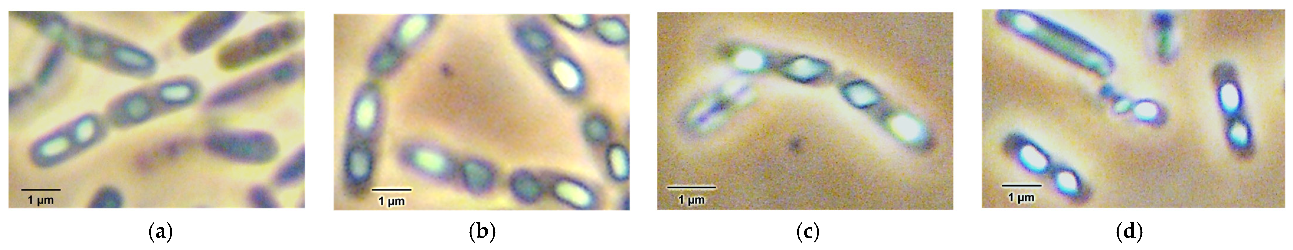
2.1. Morphological and Biochemical Characterization of Food-Borne Outbreak Isolates

All the B. thuringiensis isolates from food and the vomit samples were Gram-positive, spore-forming rods with unswollen sporangia, bearing roughly bipyramidal toxin crystals (Figure 1). They could grow at 45 °C and below, but not at 48 °C or 50 °C. The API profiles of the isolates varied, but they all showed the typical biochemical reaction patterns for B. cereus/B. thuringiensis species (Table S1). All isolates were able to degrade starch and utilize salicin.
Cases of B. cereus emetic illness are usually associated with rice and pasta dishes [18,19,20,21,22,23], but the organisms involved in this small food-borne outbreak were isolated from the mackerel and mussels in the garlic butter sauce, not from the pasta. However, an extensive study by Messelhäusser et al. (2014) [24] showed that emetic toxin-producing strains belonging to the B. cereus group were not only found in foods with high carbohydrate content, such as pasta or rice, as was earlier found. These observations have implications for the breadth of sampling that should be carried out in the investigation of emetic outbreaks. The isolates had biochemical characteristics typical of B. cereus/B. thuringiensis, but it was interesting to note that the present isolates could degrade starch and utilize salicin, unlike many emetic B. cereus strains previously characterized [25,26,27]. This recalls the comment made by Thorsen et al. (2006) [28] that not all emetic toxin-producing B. cereus strains are unable to hydrolyze starch or produce acid from salicin.

2.2. Molecular Identification and Typing of Isolates from Emetic Illness

2.2.1. Identification of Isolates Using 16S rRNA Gene Sequence Analysis

The 16S rRNA sequences of the B. thuringiensis isolates, two from food (FC1 and FC2; obtained from mussels and smoked mackerel in garlic butter sauce), and five from the vomit sample of one patient (FC6, FC7, FC8, FC9, and FC10), were determined. The 16S rRNA Blast results against strains in the NCBI database confirmed their identity as B. thuringiensis (Table 1).
The sequences of the B. thuringiensis isolates were also compared with those of three reference strains: B. thuringiensis IAM 12077 (or ATCC 10792, B. thuringiensis type strain); B. cereus ATCC 14579 (B. cereus type strain); and B. cereus F4810/72 (a reference B. cereus emetic strain, also known as strain B0358 in the Logan Bacillus Collection at Glasgow). Based on the 16S rRNA fragment, the FC isolates were also closely related to the B. thuringiensis and B. cereus strains tested.
B. thuringiensis is known as one of the closest species to B. cereus. These two species are closely related genetically, and their 16S rRNA sequence identities are more than 99% [29]. Polyphasic identification is therefore useful in this case. As expected from the Blast results, many of the B. cereus strains were also among the closest relatives. Because all FC isolates produced parasporal crystals, they were therefore confirmed to be B. thuringiensis by their genetic characteristics, which are also supported by the biochemical profiles.

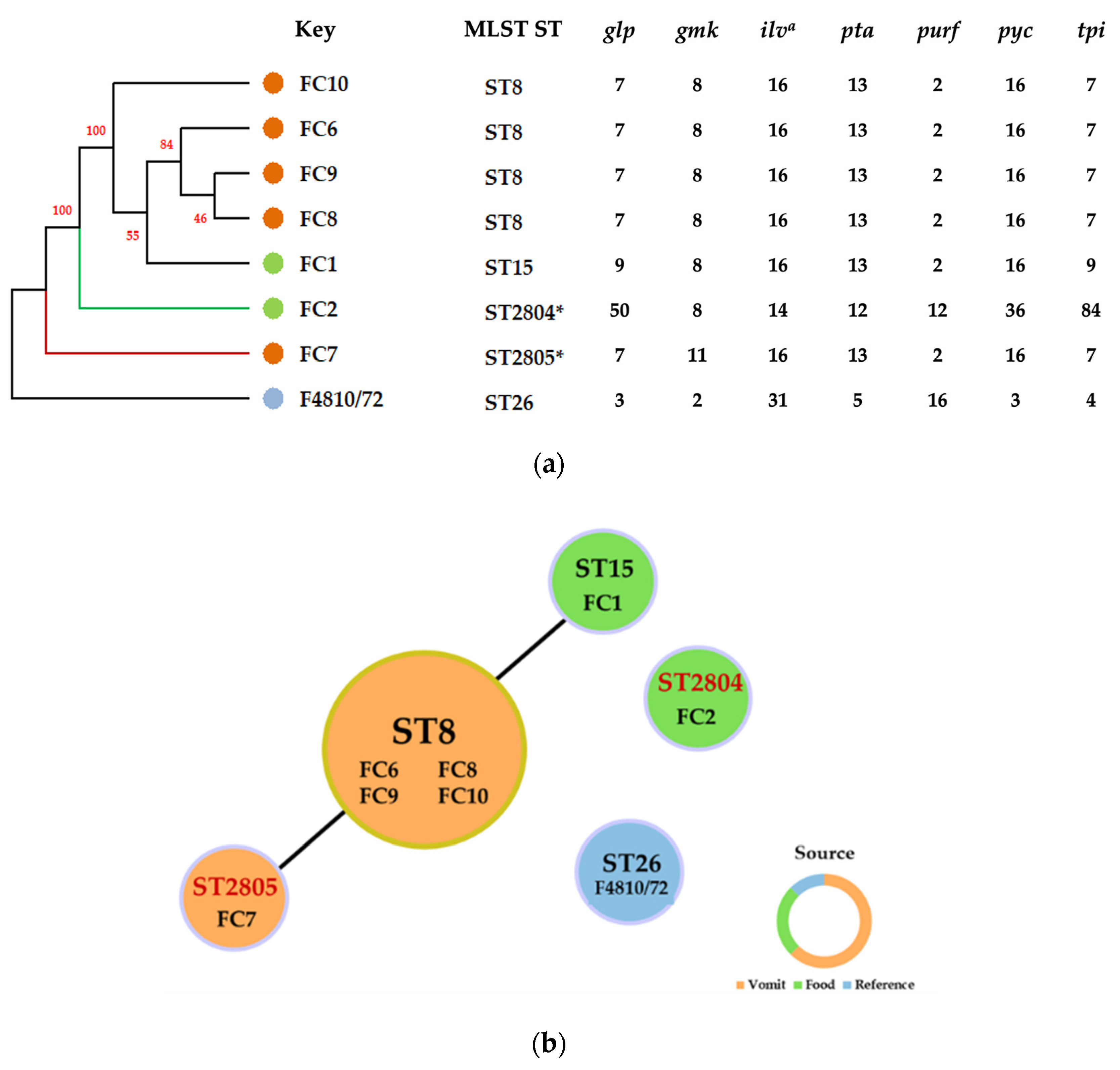
2.2.2. Molecular Typing of Isolates Using Multilocus Sequence Typing (MLST) Analysis

We further examined the genetic diversity and relatedness of the food poisoning isolates using multilocus sequence typing (MLST), which is highly discriminatory for microbial strains. Based on Yang et al. (2017) [30], seven housekeeping genes for the B. cereus group (glpF, gmk, ilvD, pta, pur, pycA, and tpi) were used in the analysis. A sequence type, ST26, to which B. cereus F4810/72 belongs, was also included in the analysis.
Each amplified fragment of the housekeeping genes was sequenced, and the processed sequences were submitted to the MLST database (see Section 4.2.2 for details). The dendrogram constructed using UPGMA with allelic profiles of STs is presented in Figure 2a. The phylogenetic analysis based on sequence alignment revealed that the B. thuringiensis isolates belonged to different clonal groups. The isolates FC1, FC6, FC8, FC9, and FC10 were arranged in one clade, and FC2 and FC7 were separated from this clade and were distinct from each other. All of the B. thuringiensis isolates were separated from B. cereus F4810/72 (Figure 2a).
Using the goeBURST algorithm, one group and two singletons were defined. FC1, FC6, FC7, FC8, FC9, and FC10 (Clonal Complex 8; CC8) were assigned to Group 1, and FC2 and F4810/72 were designated as singletons. Overall, the bacterial strains, including emetic B. cereus F4810/72, were resolved into five allelic profiles of STs (Figure 2b), three of which have previously been assigned and two of which are new.
MLST typing analyzes a set of seven housekeeping genes. This method has been successfully used for different applications, including the typing of food-borne isolates [30]. It has also been used for typing of environmental strains of B. cereus group [31]. The established B. cereus MLST database (PubMLST; http://pubmlst.org/bcereus/info/primers.shtml, accessed on 1 December 2022) allows comparisons of a set of isolates to the existing sequence type and the identification of a new sequence type, under both of which techniques we found our strains belonged.
B. cereus F4810/72, a classical emetic strain that was used as a reference in this study, has the allelic profile ST26 [32]. This ST was also the most commonly identified ST for B. cereus strains [33]. The fact that the B. thuringiensis isolates belonged to different STs clearly confirmed that there was an intraspecific genetic diversity of strains involved in the emetic illness case recorded in this study. Because B. thuringiensis was not commonly present in food, and the occurrence of emetic strains with direct evidence of clinical symptoms was even more unusual (Hoffmaster et al., 2008) [33], this finding is worthy of note as it may indicate various sources of contamination or the possibility of gene transfer.
Since B. thuringiensis strains have been used as biopesticides, there have been concerns raised about the use of this organism for this purpose due to the potential enterotoxin production of this species. Extensive use of these organisms, especially in the form of spores, may cause B. thuringiensis to be more widely spread in the environment and have more frequent implications for food-borne illnesses. Cases of diarrheal disease have been linked to biopesticide strains [34]. Recently, B. thuringiensis found in salad was suspected to be the cause of a food-borne outbreak in the EU [29]. Biggel et al. (2022) [34] suggested that many food isolates of B. thuringiensis had their origins in biopesticide strains and belong to previously observed sequence types (i.e., ST8, ST15, ST16, or ST23) [35]. Similarly, our MLST results showed that many of the food-borne outbreak B. thuringiensis isolates belong to ST8 (four isolates from vomit) and ST15 (one food isolate). These sequence types, which might have been linked to biopesticide strains, have been frequently found in fresh vegetable samples [29,36,37]. Moreover, it is interesting to record the new ST genotypes found in this study and their association with emetic illness. Although we cannot be certain about the original source of the strains belonging to these STs, these data could be useful in future analyses of epidemiological data and assessments of the ecological and biological roles of B. thuringiensis isolates. Moreover, the new STs might indicate an emerging pathogenic strain and could be useful in the monitoring of food safety and the control of disease in relation to this rare emetic food-borne pathogen.

2.3. Detection and Characterization of Genes Associated with Emetic Traits in B. thuringiensis Isolates

2.3.1. Detection of Non-Ribosomal Peptide Synthetase (NRPS)/Cereulide Synthetase Genes

Genetic factors that are associated with the Bacillus emetic traits, i.e., emetic toxin production capability or cereulide synthetase, were investigated in the B. thuringiensis isolates through PCR amplification of fragments of the NRPS/cereulide synthetase (ces) genes. The expected amplified regions in the ces gene cluster (based on the sequence of strain F4810/72, Genbank accession no. DQ360825.1 [38]) are shown in Figure 3a. The PCR results are shown in Figure 3b. Although the amplified gene fragments were achieved with high quantity and specificity with strain F4810/72, the primer pairs had different efficacies when applied to B. thuringiensis isolates. The primers BEF/BER, CER1/EMT1, and EM1-F/EM1-R yielded amplicons of expected sizes from all B. thuringiensis isolates, even though nonspecific amplifications were also observed in some reactions with primers BEF/BER and EM1F/EM1R. However, primers CesF1/CesR2 gave positive results only with FC2 (food isolate) and FC8 (clinical isolate).
The four pairs of primers used in this investigation were evaluated by different researchers as being specific to B. cereus emetic strains or to the ces gene. Even though these sets of primers were independently designed, their specific targets were at different sites on the ces gene. The BEF/BER and CER1/EMT primers specifically target cesA, whereas the EM1F/EM1R and CesF1/CesR2 primers specifically target cesB (Figure 3a). Primers BEF/BER, CER1/EMT, and EM1F/EM1R were validated by Toh et al. (2004) [39], Horwood et al. (2004) [40], and Ehling-Schulz et al. (2004) [41], respectively, to be associated with cereulide-producing strains. The CesF1/CesR2 primers were described by Ehling-Schulz et al. (2005b) [42] as specific to the valine activation module of the cereulide synthetase gene, and disruption of this gene resulted in a non-cereulide-producing mutant. The differences in the amplification patterns observed in our study for the B. thuringiensis isolates and B. cereus F4810/72, as seen in Figure 3b, may be due to the variation of the nucleotides within and around these fragments.
Failure to amplify the ces fragment by CesF1/CesR2 primers in many B. thuringiensis isolates, apart from FC2 and FC8, is not unexpected. Kim et al. (2010) [43] observed that this pair of primers could yield a negative result with some emetic toxin-producing B. cereus strains. Our experience showed that this pair of primers required more specific reaction conditions than the others. On the other hand, failure to amplify the gene fragment may be a result of non-conserved DNA sequences around this region of the cesB gene in emetic toxin-producing Bacillus, especially with sequence diversity within members of the B. cereus group having been previously observed [44]. This observation in our study suggested that using these four pairs of primers offers a more sensitive option for identifying the presence of fragments of cesA and cesB, the two consecutive genes encoding CesA and CesB proteins that are required for cereulide production [45]. Further investigations into the genetic features of the isolates are desirable, and in this study, we explored the sequence of some of these gene fragments.

2.3.2. Sequence Analysis of B. thuringiensis Amplified Gene Fragments

Selected gene fragments amplified from the DNA of the B. thuringiensis isolates using the primers BEF/BER, CER1/EMT1, and EM1-F/EM1-R were sequenced (the sequences are deposited in the NCBI BioProject database PRJNA188745). They were highly similar (with 98–99% identity) or identical to the ces gene found in B. cereus F4810/72 (Genbank accession no. DQ360825.1 [38] and the crs (another abbreviation for cereulide synthetase) gene from B. cereus no. 55 (Genbank accession no. AB248763.2 [46]). Fragments generated using primers CER1/EMT1 were also comparable (with 99–100% identity) to the B. cereus NRPS gene (Genbank accession no. AY331260.1 [40]). They also had 99–100% identity with the cesA gene found in toxigenic B. pumilus NR 19/5 and B. licheniformis NR 5106 (Genbank accession no. AM493712.1 and AM493711.1, respectively [47]). In addition, fragments yielded from primers EM1-F/EM1-R also had 96–97% identity to the cesB gene found in an emetic toxin-producing strain of B. pumilus (strain NIOB133; Genbank accession no. EU289221.1 [48]. The positions on the B. thuringiensisces gene, which were different from those on the ces gene of B. cereus and other Bacillus species, had mismatches, insertions, or deletions of nucleotides.
From the biochemical and genetic characterization, it is clear that the outbreak isolates do not represent a single strain. The fact that they all harbored fragments that are parts of the ces gene cluster indicated that this gene may be distributed among B. thuringiensis strains in certain geographical locations [26,49]. Gene transfer is possible given that the ces gene in some strains is located on a megaplasmid [44,46,50] and mobile genetic elements (MGEs) have been found to be associated with the ces gene cluster [44]. More extensive studies on ces distribution in B. thuringiensis would help us understand how widely this feature is spread in the natural environment. It would also lead to a better assessment of its impact on food safety.

2.4. Cytotoxicity of Emetic B. thuringiensis Isolates to Caco-2 Cells

According to the clinical symptoms and the results from the genetic characterization presented above, we presumed that some of the B. thuringiensis isolates would display similar biological activity to that of emetic B. cereus, which can produce the emetic toxin cereulide, a heat-stable cyclic peptide [51,52]. Because of the molecular nature of the emetic toxin, which has a very low molecular weight (1.2 KDa), it is difficult to detect it using an immunological method or a simple analytical chemistry method [53]. Therefore, biological assays, especially cytotoxicity assays, have been extensively used for the detection of the emetic toxin [54,55,56].
In this study, the cytotoxicity of the heat-treated culture supernatants of the outbreak isolates was tested on human colorectal adenocarcinoma (Caco-2) cells. The isolates tested included B. thuringiensis FC2, FC7, and FC8, which represented sequence types ST2804, 2805, and ST8, respectively. A preliminary cytotoxicity test showed that the heat-treated supernatants of the B. thuringiensis isolates were cytotoxic to Caco-2 cells (a non-emetic strain control, B. cereus DSM 4384, did not show cytotoxic activity in the same experiment) (Figure S1). Further investigation of the degree of cytotoxicity (expressed as half-maximal cytotoxic concentration, CC50) of isolates FC2, FC7, and FC8 showed that the reduction of Caco-2 viability induced by the heat-treated culture supernatants of these isolates was in a dose-dependent manner (Figure 4a). According to the CC50 values, the FC2 isolate showed a higher degree of cytotoxicity to Caco-2 cells than FC7 and FC8 (Figure 4a). Vacuolation was observed in Caco-2 cells treated with the heat-treated supernatants from all three isolates (Figure 4b).
It is difficult to identify the isolate(s) responsible for the illness, as the MLST results (Section 2.2.2) indicated a diversity among the outbreak isolates. The cytotoxicity results suggested a possible role of FC2 in the emetic illness, as it was more toxic to Caco-2 cells than the other isolates, although the contribution of the other isolates to the illness may not be excluded.

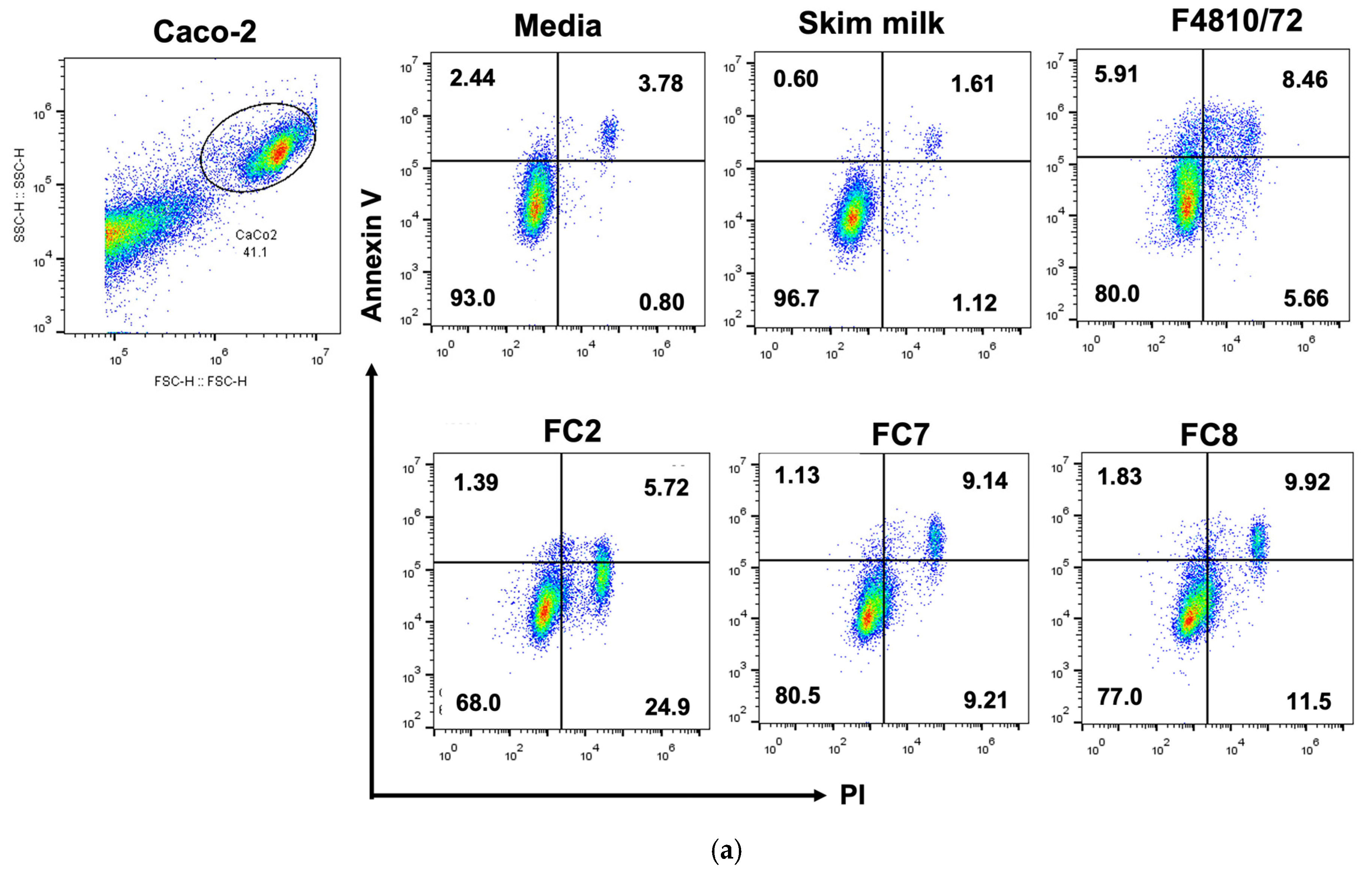
2.5. Annexin V/PI Apoptosis Detection

To clarify the cell death pathway caused by the heat-stable toxin in the supernatants from each bacterial isolate, we performed annexin V/PI staining to discriminate the apoptosis and necrosis mechanisms of cell death through flow cytometry. Caco-2 cells were treated for 48 h with the cell-free supernatants of FC2, FC7, and FC8 at a concentration of 50% (v/v) (which had 56.62, 62.70, 64.91 µg/mL total protein, respectively). Skim milk and the culture supernatant of F4810/72 were used as negative and positive controls, respectively.
From the results, B. cereus F4810/72 appeared to cause both apoptotic cell death (14.85 ± 1.49%) and necrotic cell death (5.91 ± 0.32%) (Figure 5a,b). For cell death through the apoptotic mechanism, 6.07 ± 0.81% was detected as early apoptosis and 8.78 ± 0.69% as late apoptosis. Virtanen et al., (2008) [57] have reported that cereulide from B. cereus F4810/72 increased necrotic cell death in porcine fetal Langerhans and beta-cells within 2 days. Similarly, Hoornstra et al. (2013) [58] also observed necrotic cell death caused by cereulide from B. cereus F4810/72 in other mammalian cells (PBMC, HaCaT, PK-15, and L-929). Our testing of the effect of heat-stable toxins in the culture supernatant of B. cereus F4810/72 with Caco-2 cells also pointed to cell destruction through necrosis. However, apoptosis seemed to be a major mechanism of cell destruction in Caco-2 cells.
As for the B. thuringiensis FC2, FC7, and FC8 isolates, the heat-treated cell-free supernatants caused cell death through both the apoptosis and necrosis pathways, as observed with the reference emetic strain B. cereus F4810/72. However, necrosis was a major pathway in the FC2 isolate, which had the highest rate of necrosis cell death (23.47 ± 2.92%) among the three B. thuringiensis isolates tested (Figure 5b). For FC7 and FC8, cell death rates through the necrosis mechanism were similar to those of apoptosis, which was mainly early apoptosis. These results suggest that the B. thuringiensis isolates from this emetic outbreak, although they could cause a similar symptom to that typically recognized as being caused by B. cereus, might have some differences in the toxin entity from that of B. cereus F4810/72.

3. Conclusions

In this study, we report a food-borne emetic illness associated with B. thuringiensis and present the results of the investigation on the diversity and pathogenicity of the isolates from this food-borne outbreak incident. The biochemical and genetic profiles suggested an appreciable diversity among the isolates. The multilocus sequence typing (MLST) analysis revealed that the B. thuringiensis isolates did not represent one strain. Some isolates belonged to the existing sequence types (ST8 and ST15) that have been linked to biopesticide strains, whereas some belonged to two new STs (designated ST2804 and ST2805). All food and clinical isolates harbored gene fragments located in the cereulide synthetase (ces) gene cluster, which indicated the likelihood that an emetic toxin of a similar nature to cereulide might have been implicated in the illness. The heat-stable toxin in the supernatant of B. thuringiensis FC2, FC7, and FC8, which represented ST2804, ST2805, and ST8, respectively, reduced the viability of Caco-2 in a dose-dependent manner. According to the CC50 values, the FC2 isolate derived from the food involved in the illness showed a higher degree of cytotoxicity to Caco-2 cells than did FC7 and FC8. The flow cytometry with Annexin V/PI staining revealed that cell destruction by all three food-poisoning B. thuringiensis isolates occurred through both apoptosis and necrosis pathways, but for FC2, necrosis was likely the main mechanism that caused Caco-2 cell destruction.

4. Materials and Methods

4.1. Bacterial Strains/Isolates and Identification

The isolates from the food-borne outbreak comprised two from food: FC1 and FC2 (obtained from the leftover pasta sauce: mussels and smoked mackerel in garlic butter sauce) and five from the vomit of one patient: FC6, FC7, FC8, FC9, and FC10. They were isolated on Polymyxin Pyruvate Egg-Yolk Mannitol Bromothymol-Blue Agar (PEMBA), which was incubated at 37 °C for 48 h. The isolates were examined using phase-contrast microscopy and biochemically characterized using the API 20E and API 50 CHB systems (BioMérieux, Marcy-l’Étoile, France). Growth at 40 °C, 42 °C, 45 °C, 48 °C, and 50 °C, was also observed in Tryptone Soy Broth (Oxoid, Basingstoke, UK) cultures incubated in water baths. B. cereus F4810/72 (also known as strain B0358 in the Logan Bacillus Collection at Glasgow) was used as a reference emetic toxin-producing strain. All bacterial cultures were maintained on Tryptone Soy Agar (TSA) (Oxoid, Basingstoke, UK), stored at 4 °C, or in lyophilized form.

4.2. Identification and Typing of B. thuringiensis Isolates from Emetic Food-Borne Illness

4.2.1. Identification of Isolates Using 16S rRNA Gene Sequencing

Chromosomal DNA from the pure cultures of the emetic food poisoning isolates was extracted using the CTAB/phenol-chloroform extraction method described by Griffiths et al. (2000) [59]. The 16S rRNA gene was amplified using primers 27F (5′-AGAGTTTGATCMTGGCTCAG-3′) and 1492R (5′-CGGTTACCTTGTTACGACTT-3′) [60] at a final concentration of 0.1 μM. The PCR was performed with an initial denaturation for 2 min at 94 °C, 35 cycles of denaturation for 30 s at 94 °C, primer annealing for 30 s at 55 °C, extension for 30 s at 72 °C, and a final extension for 7 min at 72 °C (Lane, 1991) [61]. The PCR products were purified using a PCR purification kit (Sigma-Aldrich, Darmstadt, Germany). Sequencing was performed by Celemics (Seoul, Republic of Korea). The low-quality bases in the sequences were trimmed using Bioedit software version 7.05.3 [62]. The processed sequences were subjected to Basic Local Alignment Search Tool (BLAST) analysis against the 16S rRNA sequences in the National Center for Biotechnology Information (NCBI) database to find the closest relatives.

4.2.2. Typing of Isolates Using Multilocus Sequence Typing (MLST) Analysis

The primers for the housekeeping genes (glpF, gmk, ilvD, pta, pur, pycA, and tpi) were used for multilocus sequence typing (MLST) according to Yang, et al. (2017) [30] (ilvD2 was also used if the ilvD locus failed to find an allelic profile). The sequencing of the amplified genes was performed by Macrogen (Seoul, Republic of Korea).
Sequences were analyzed using BioEdit software version 7.1.9 (Abbott, Carlsbad, CA, USA). All allelic profiles and sequence types (STs) detected in the study were assigned according to the online MLST database for B. cereus (https://pubmlst.org/organisms/ bacillus-cereus, accessed on 1 December 2022). Grouping tools were used to establish the molecular type, clonal complexes (CCs), singletons, and the relationship between established groups. All STs were displayed with BURST [63], using the characteristic profiles for each isolate as the input data. Minimal expansion trees were also generated using the geoBURST algorithm [63] performed with Phyloviz 2.0 [64] to define CCs and display models. A phylogenetic tree of the related sequences of the seven loci of the housekeeping genes was generated with UPGMA based on the method of Kimura-2-parameter [65] and performed with 1000 bootstrap replicates in Mega X [66].

4.3. Genetic Analysis of B. thuringiensis Isolates

4.3.1. DNA Extraction

DNA samples were prepared from overnight cultures of Bacillus strains grown on Trypticase Soy Agar (TSA) slants (Oxoid, Basingstoke, UK). Cells were scraped from the surface of the agar slant into TE buffer (10 mM Tris, 1 mM EDTA, pH 8.0), and the DNA was extracted using a CTAB-based method for mini-preparation of DNA [67].

4.3.2. Polymerase Chain Reaction (PCR) Amplification of Cereulide Synthetase Gene

Amplification of the cereulide synthetase (ces) gene was carried out using four pairs of oligonucleotide primers: BEF/BER [39], CER1/EMT1 [40], EM1-F/EM1-R [41], and CesF1/CesR2 [42]. The annealing temperatures for the above primers were 55 °C, 58 °C, 61 °C, and 58 °C, respectively (optimized in this study for amplification of these fragments on B. thuringiensis DNA). The internal transcribed spacer (its) gene was used for an internal control, which was amplified using primers ITS-F1/ITS-R1 [68] with an annealing temperature of 50 °C. Each of the 50 mL-reactions contained 1 × reaction buffer, 1.5 mM MgCl2, 0.2 mM dNTPs, 20 pmoles of each primer, 1 μL DNA template, and 1 unit of DNA polymerase (Phusion High-Fidelity DNA Polymerase, New England Biolab, Ipswich, MA, USA). Amplification was carried out in the PTC 200 thermal cycler (MJ Research, Waltham, MA, USA), using the following steps: 5 min initial denaturation at 95 °C; 35 cycles of PCR amplification for 45 s at 95 °C, 45 s at 50–61 °C (depending on the primers, see above), 1.5 min at 72 °C; and 10 min final extension at 72 °C.

4.3.3. DNA Sequencing and Sequence Similarity Analysis

Purified PCR products from selected B. thuringiensis isolates amplified using primers CER1/EMT1, BEF/BER, and EM1-F/EM1-R were sequenced using an Applied Biosystems genetic analyzer (performed by First Base Laboratory, Selangor, Malaysia). The nucleotide sequences of the PCR fragments were analyzed for their similarities to sequences published in the NCBI database using the Blast program. In addition, they were compared to the sequence of reference strains using the Blast 2 Sequences program.

4.4. Cytotoxicity Assay

4.4.1. Preparation of Heat-Treated Supernatants from Bacterial Isolates

The overnight bacterial cultures (0.1% (v/v)) were inoculated in 50 mL of 2.5% skim milk medium (SMM) (Oxoid, Basingstoke, UK). The cultures were incubated for 24 h at 25 °C in an orbital shaking incubator (shaker speed: 150 rpm). After incubation, the supernatant portion of each bacterial culture was collected after centrifugation at 3000× g for 10 min, and the cell pellet was discarded. The supernatant was heated in an autoclave for 15 min at 121 °C, left to cool, and filtered through a 0.2 µm membrane filter [56].

4.4.2. Cell Culture

The human colorectal adenocarcinoma cell line (Caco-2) (ATCC HTB-37) was cultured in Dulbecco’s Modified Eagle Medium (DMEM; Gibco; Thermo Fisher Scientific, Waltham, MA, USA) was supplemented with 10% heat-inactivated fetal bovine serum (Gibco; Thermo Fisher Scientific, Waltham, MA, USA) and Pen–Strep (100 units/mL penicillin and 100 μg/mL streptomycin) (Caisson, Smithfield, UT, USA). The cells were incubated at 37 °C in a 5% CO2 incubator.

4.4.3. Cytotoxicity Assay

The cytotoxicity of the heat-treated supernatant from the bacterial isolates was tested using the MTT cytotoxicity assay [56] and the PrestoBlue cytotoxicity assay [69]. In brief, Caco-2 cells were plated at 105 cells/well in 96-well plates for 48 h before the experiment. The Caco-2 cells were treated with 100 µL of the supernatant from the bacterial isolates prepared at various concentrations for 48 h. For the PrestoBlue cytotoxicity test, after 48 h of treatment, the PrestoBlue™ reagent (Thermo Fisher Scientific, Waltham, MA, USA) was added to measure cell viability, and the cells were incubated for 30 min at 37 °C in a 5% CO2 incubator. Absorbance was read at 570/600 nm using a Bio Tek Synergy HTX microplate reader (Agilent Technologies, Santa Clara, CA, USA). The percentages of cell viability in relation to that of the non-treatment control were calculated using the following equation.
% Cell viability = [(OD570 − OD600) treated cells/(OD570 − OD600) non-treated cells] × 100
Half-maximal cytotoxic concentration (CC50) values were then calculated using Graph Pad Prism 9.4.1 software (GraphPad Software, Inc., San Diego, CA, USA). Protein concentrations of the heat-treated supernatants were measured using Bradford reagent (Thermo Fisher Scientific, Waltham, MA, USA) according to the manufacturer’s protocol.

4.4.4. Annexin V/PI Assay

Apoptotic stages were evaluated by conducting an Annexin V/PI assay and analyzed using a flow cytometer. Caco-2 cells were seeded at 5 × 10⁴ cells/mL in 6-well culture plates, using 2 mL per well, and treated with the supernatant from the bacterial isolates at 50% concentration or 1/2 dilution (having total proteins of 56.62 μg/mL, 62.70 μg/mL, 64.91 μg/mL for FC2, FC7, and FC8, respectively). After 48 h of treatment, the Caco-2 cells were collected and stained with Annexin V/PI, according to the Annexin V/PI kit protocol (ImmunoTool, Friesoythe, Germany). Briefly, the cell pellets were trypsinized and resuspended in 100 µL of ice-cold 1 × binding buffer. Annexin V-APC and PI were added to the tubes at the dilution of 1:100. The cell suspension was then mixed and incubated for 15 min in the dark. Samples were analyzed on a BD Accuri C6 Flow Cytometer and FlowJo software (BD Biosciences, San Jose, CA, USA).

Supplementary Materials

The following are available online at: https://www.mdpi.com/article/10.3390/toxins15020089/s1, Table S1: Positive biochemical characteristics of the emetic outbreak B. thuringiensis isolates tested using the API 20 and API 50 CH galleries, Figure S1: Caco-2 cell viability after treatment with heat-treated culture supernatants of emetic food-borne outbreak isolates.

Author Contributions

Conceptualization, T.C., Y.T., A.P. and J.P.; methodology, T.C., Y.T., A.P., J.P., T.K., M.K., S.K., K.J. and R.P.; software, M.K., S.K., T.C. and J.P.; validation, T.C., Y.T., A.P., T.K., M.K., S.K. and N.A.L.; formal analysis, T.C., J.P., T.K., M.K., S.K., K.J. and R.P.; investigation, T.C., A.P. and J.P.; resources, T.C., Y.T., S.B. and A.P.; data curation, T.C., M.K., S.K., T.K, R.P. and J.P.; writing—original draft preparation, T.C., T.K., M.K., S.K., R.P. and J.P.; writing—review and editing, T.C., Y.T., A.P., S.K. and N.A.L.; visualization, J.P., S.K., T.K., M.K. and K.J.; supervision, T.C., Y.T., A.P. and S.B.; project administration, T.C.; funding acquisition, T.C. All authors have read and agreed to the published version of the manuscript.

Funding

This research work was partially funded by Chiang Mai University and J.P. was partially funded by Chiang Mai University Graduate School.

Institutional Review Board Statement

Not applicable.

Informed Consent Statement

Not applicable.

Data Availability Statement

Not applicable.

Acknowledgments

The authors would like to express our gratitude to A.G. Williams, A.D. Sutherland, S.E. Withers, and W.J.J. Finlay (formerly at Glasgow Caledonian University, UK) for their assistance and advice in the preliminary investigation of the outbreak, and to Chutipa Chiawpanit and Methi Wathikthinnakon (Chiang Mai University, Thailand) for assistance in data analysis. We thank Janjira Wanchana (Maejo University, Thailand) for her assistance with maintenance of the bacterial cultures and Nattakorn Yuayuan (Chiang Mai University) for his technical assistance.

Conflicts of Interest

The authors declare no conflict of interest.

References

  1. Stenfors Arnesen, L.P.; Fagerlund, A.; Granum, P.E. From soil to gut: Bacillus cereus and its food poisoning toxins. FEMS Microbiol. Rev. 2008, 32, 579–606. [Google Scholar] [CrossRef] [Green Version]
  2. Ehling-Schulz, M.; Lereclus, D.; Koehler, T.M. The Bacillus cereus group: Bacillus species with pathogenic potential. Microbiol. Spectr. 2019, 7, 1–60. [Google Scholar] [CrossRef]
  3. McIntyre, L.; Bernard, K.; Beniac, D.; Isaac-Renton, J.L.; Naseby, D.C. Identification of Bacillus cereus group species associated with food poisoning outbreaks in British Columbia, Canada. Appl. Environ. Microbiol. 2008, 74, 7451–7453. [Google Scholar] [CrossRef] [Green Version]
  4. Ceuppens, S.; Boon, N.; Uyttendaele, M. Diversity of Bacillus cereus group strains is reflected in their broad range of pathogenicity and diverse ecological lifestyles. FEMS Microbiol. Ecol. 2013, 84, 433–450. [Google Scholar] [CrossRef] [Green Version]
  5. Logan, N.A. Bacillus and relatives in food-borne illness. J. Appl. Microbiol. 2012, 112, 417–429. [Google Scholar] [CrossRef]
  6. Helgason, E.; Økstad, O.A.; Caugant, D.A.; Johansen, H.A.; Fouet, A.; Mock, M.; Hegna, I.; Kolstø, A.B. Bacillus anthracis, Bacillus cereus, and Bacillus thuringiensis—One species on the basis of genetic evidence. Appl. Environ. Microbiol. 2000, 66, 2627–2630. [Google Scholar] [CrossRef] [Green Version]
  7. Bravo, A.; Likitvivatanavong, S.; Gill, S.S.; Soberón, M. Bacillus thuringiensis: A story of a successful bioinsecticide. Insect Biochem. Mol. Biol. 2011, 41, 423–431. [Google Scholar] [CrossRef] [Green Version]
  8. Jeong, H.; Jo, S.H.; Hong, C.E.; Park, J.M. Genome sequence of the endophytic bacterium Bacillus thuringiensis strain KB1, a potential biocontrol agent against phytopathogens. Microbiol. Resour. Announce. 2016, 21, e00279-16. [Google Scholar] [CrossRef] [Green Version]
  9. Valtierra de Luis, D.; Villanueva, M.; Berry, C.; Caballero, P. Potential for Bacillus thuringiensis and other bacterial toxins as biological control agents to combat dipteran pests of medical and agronomic importance. Toxins 2020, 12, 773. [Google Scholar] [CrossRef]
  10. Sanchis, V.; Bourguet, D. Bacillus thuringiensis: Applications in agriculture and insect resistance management. A review. Agron. Sustain. 2008, 28, 11–20. [Google Scholar] [CrossRef]
  11. Abbas, M.S.T. Genetically engineered (modified) crops (Bacillus thuringiensis crops) and the world controversy on their safety. Egypt J. Biol. Pest Control 2018, 28, 52. [Google Scholar] [CrossRef]
  12. Rosenquist, H.; Smidt, L.; Andersen, S.R.; Jensen, G.B.; Wilcks, A. Occurrence and significance of Bacillus cereus and Bacillus thuringiensis in ready-to-eat food. FEMS Microbiol. Lett. 2005, 250, 129–136. [Google Scholar] [CrossRef] [PubMed] [Green Version]
  13. Frederiksen, K.; Rosenquist, H.; Jørgensen, K.; Wilcks, A. Occurrence of natural Bacillus thuringiensis contaminants and residues of Bacillus thuringiensis-based insecticides on fresh fruits and vegetables. Appl. Environ. Microbiol. 2006, 72, 3435–3440. [Google Scholar] [CrossRef] [Green Version]
  14. Jovanovic, J.; Ornelis, V.F.M.; Madder, A.; Rajkovic, A. Bacillus cereus food intoxication and toxicoinfection. Compr. Rev. Food Sci. Food Saf. 2021, 20, 3719–3761. [Google Scholar] [CrossRef]
  15. Kim, J.B.; Choi, O.K.; Kwon, S.M.; Cho, S.H.; Park, B.J.; Jin, N.Y.; Yu, Y.M.; Oh, D.H. Prevalence and toxin characteristics of Bacillus thuringiensis isolated from organic vegetables. J. Microbiol. Biotechnol. 2017, 27, 1449–1456. [Google Scholar] [CrossRef]
  16. Johler, S.; Kalbhenn, E.M.; Heini, N.; Brodmann, P.; Gautsch, S.; Bağcioğlu, M.; Contzen, M.; Stephan, R.; Ehling-Schulz, M. Enterotoxin production of Bacillus thuringiensis isolates from biopesticides, foods, and outbreaks. Front. Microbiol. 2018, 9, 1915. [Google Scholar] [CrossRef] [Green Version]
  17. Jackson, S.G.; Goodbrand, R.B.; Ahmed, R.; Kasatiya, S. Bacillus cereus and Bacillus thuringiensis isolated in a gastroenteritis outbreak investigation. Lett. Appl. Microbiol. 1995, 21, 103–105. [Google Scholar] [CrossRef]
  18. Bonis, M.; Felten, A.; Pairaud, S.; Dijoux, A.; Maladen, V.; Mallet, L.; Radomski, N.; Duboisset, A.; Arar, C.; Sarda, X.; et al. Comparative phenotypic, genotypic, and genomic analyses of Bacillus thuringiensis associated with food-borne outbreaks in France. PLoS ONE 2021, 19, 16. [Google Scholar] [CrossRef]
  19. Schwenk, V.; Riegg, J.; Lacroix, M.; Märtlbauer, E.; Jessberger, N. Enteropathogenic potential of Bacillus thuringiensis isolates from soil, animals, food and biopesticides. Foods 2020, 9, 1484. [Google Scholar] [CrossRef]
  20. Mahler, H.; Pasi, A.; Kramer, J.M.; Schulte, P.; Scoging, A.C.; Bär, W.; Krähenbühl, S. Fulminant liver failure in association with the emetic toxin of Bacillus cereus. N. Engl. J. Med. 1997, 336, 1142–1148. [Google Scholar] [CrossRef]
  21. Dierick, K.; Coillie, E.V.; Swiecicka, I.; Meyfroidt, G.; Devlieger, H.; Meulemans, A.; Hoedemaekers, G.; Fourie, L.; Heyndrickx, M.; Mahillon, J. Fatal family outbreak of Bacillus cereus-associated food poisoning. J. Clin. Microbiol. 2005, 43, 4277–4279. [Google Scholar] [CrossRef] [PubMed] [Green Version]
  22. Naranjo, M.; Denayer, S.; Botteldoorn, N.; Delbrassinne, L.; Veys, J.; Waegenaere, J.; Sirtaine, N.; Driesen, R.B.; Sipido, K.R.; Mahillon, J.; et al. Sudden death of a young adult associated with Bacillus cereus food poisoning. J. Clin. Microbiol. 2011, 49, 4379–4381. [Google Scholar] [CrossRef] [PubMed] [Green Version]
  23. Rodrigo, D.; Rosell, C.M.; Martinez, A. Risk of Bacillus cereus in relation to rice and derivatives. Foods 2021, 10, 302. [Google Scholar] [CrossRef]
  24. Messelhäusser, U.; Frenzel, E.; Blöchinger, C.; Zucker, R.; Kämpf, P.; Ehling-Schulz, M. Emetic Bacillus cereus are more volatile than thought: Recent food-borne outbreaks and prevalence studies in Bavaria (2007–2013). Biomed. Res. Int. 2014, 2014, 465603. [Google Scholar] [CrossRef] [Green Version]
  25. Logan, N.A.; Berkeley, R.C. Identification of Bacillus strains using the API system. J. Gen. Microbiol. 1984, 130, 1871–1882. [Google Scholar] [CrossRef] [Green Version]
  26. Ehling-Schulz, M.; Svensson, B.; Guinebretiere, M.H.; Lindbäck, T.; Andersson, M.; Schulz, A.; Fricker, M.; Christiansson, A.; Granum, P.E.; Märtlbauer, E.; et al. Emetic toxin formation of Bacillus cereus is restricted to a single evolutionary lineage of closely related strains. Microbiology 2005, 151, 183–197. [Google Scholar] [CrossRef] [Green Version]
  27. Yang, Y.; Foster, J.T.; Yi, M.; Zhan, L.; Zhang, Y.; Zhou, B.; Jiang, J.; Mei, L. Phenotypic homogeneity of emetic Bacillus cereus isolates in China. Lett. Appl. Microbiol. 2021, 73, 646–651. [Google Scholar] [CrossRef] [PubMed]
  28. Thorsen, L.; Hansen, B.M.; Nielsen, K.F.; Hendriksen, N.B.; Phipps, R.K.; Budde, B.B. Characterization of emetic Bacillus weihenstephanensis, a new cereulide-producing bacterium. Appl. Environ. Microbiol. 2006, 72, 5118–5121. [Google Scholar] [CrossRef] [PubMed] [Green Version]
  29. de Bock, T.; Zhao, X.; Jacxsens, L.; Devlieghere, F.; Rajkovic, A.; Spanoghe, P.; Höfte, M.; Uyttendaele, M. Evaluation of B. thuringiensis-based biopesticides in the primary production of fresh produce as a food safety hazard and risk. Food Control 2021, 130, 108390. [Google Scholar] [CrossRef]
  30. Yang, Y.; Yu, X.; Zhan, L.; Chen, J.; Zhang, Y.; Zhang, J.; Chen, H.; Zhang, Z.; Zhang, Y.; Lu, Y.; et al. Multilocus sequence type profiles of Bacillus cereus isolate from infant formula in China. Food Microbiol. 2017, 62, 46–50. [Google Scholar] [CrossRef]
  31. Liu, Y.; Lai, Q.; Du, J.; Shao, Z. Genetic diversity, and population structure of the Bacillus cereus group bacteria from diverse marine environments. Sci. Rep. 2017, 7, 689. [Google Scholar] [CrossRef] [PubMed] [Green Version]
  32. Carroll, L.M.; Wiedmann, M.; Mukherjee, M.; Nicholas, D.C.; Mingle, L.A.; Dumas, N.B.; Cole, J.A.; Kovac, J. Characterization of emetic and diarrheal Bacillus cereus strains from a 2016 food-borne outbreak using whole-genome sequencing: Addressing the microbiological, epidemiological, and bioinformatic challenges. Front. Microbiol. 2019, 10, 144. [Google Scholar] [CrossRef] [PubMed] [Green Version]
  33. Hoffmaster, A.R.; Novak, R.T.; Marston, C.K.; Gee, J.E.; Helsel, L.; Pruckler, J.M.; Wilkins, P.P. Genetic diversity of clinical isolates of Bacillus cereus using multilocus sequence typing. BMC Microbiol. 2008, 8, 191. [Google Scholar] [CrossRef] [Green Version]
  34. Biggel, M.; Jessberger, N.; Kovac, J.; Johler, S. Recent paradigm shifts in the perception of the role of Bacillus thuringiensis in food-borne disease. Food Microbiol. 2022, 105, 104025. [Google Scholar] [CrossRef]
  35. Raymond, B.; Federici, B.A. In defense of Bacillus thuringiensis, the safest and most successful microbial insecticide available to humanity—A response to EFSA. FEMS Microbiol. Ecol. 2017, 93, fix084. [Google Scholar] [CrossRef] [Green Version]
  36. Frentzel, H.; Juraschek, K.; Pauly, N.; Kelner-Burgos, Y.; Wichmann-Schauer, H. Indications of biopesticidal Bacillus thuringiensis strains in bell pepper and tomato. Int. J. Food Microbiol. 2020, 321, 108542. [Google Scholar] [CrossRef] [PubMed]
  37. Wang, K.; Shu, C.; Soberón, M.; Bravo, A.; Zhang, J. Systematic characterization of Bacillus Genetic Stock Center Bacillus thuringiensis strains using Multi-Locus Sequence Typing. J. Invertebr. Pathol. 2018, 155, 5–13. [Google Scholar] [CrossRef]
  38. Ehling-Schulz, M.; Fricker, M.; Grallert, H.; Rieck, P.; Wagner, M.; Scherer, S. Cereulide synthetase gene cluster from emetic Bacillus cereus: Structure and location on a mega virulence plasmid related to Bacillus anthracis toxin plasmid pXO1. BMC Microbiol. 2006, 6, 20. [Google Scholar] [CrossRef] [Green Version]
  39. Toh, M.; Moffitt, M.C.; Henrichsen, L.; Raftery, M.; Barrow, K.; Cox, J.M.; Marquis, C.P.; Neilan, B.A. Cereulide, the emetic toxin of Bacillus cereus, is putatively a product of nonribosomal peptide synthesis. J. Appl. Microbiol. 2004, 97, 992–1000. [Google Scholar] [CrossRef]
  40. Horwood, P.F.; Burgess, G.W.; Oakey, H.J. Evidence for non-ribosomal peptide synthetase production of cereulide (the emetic toxin) in Bacillus cereus. FEMS Microbiol. Lett. 2004, 236, 319–324. [Google Scholar] [CrossRef]
  41. Ehling-Schulz, M.; Fricker, M.; Scherer, S. Identification of emetic toxin producing Bacillus cereus strains by a novel molecular assay. FEMS Microbiol. Lett. 2004, 232, 189–195. [Google Scholar] [CrossRef] [PubMed] [Green Version]
  42. Ehling-Schulz, M.; Vukov, N.; Schulz, A.; Shaheen, R.; Andersson, M.; Märtlbauer, E.; Scherer, S. Identification and partial characterization of the nonribosomal peptide synthetase gene responsible for cereulide production in emetic Bacillus cereus. Appl. Environ. Microbiol. 2005, 71, 105–113. [Google Scholar] [CrossRef] [Green Version]
  43. Kim, J.B.; Kim, J.M.; Park, Y.B.; Han, J.A.; Lee, S.H.; Kwak, H.S.; Hwang, I.G.; Yoon, M.H.; Lee, J.B.; Oh, D.H. Evaluation of various PCR assays for the detection of emetic toxin producing Bacillus cereus. J. Microbiol. Biotechnol. 2010, 20, 1107–1113. Available online: https://pubmed.ncbi.nlm.nih.gov/20668404/ (accessed on 1 December 2022). [PubMed]
  44. Mei, X.; Xu, K.; Yang, L.; Yuan, Z.; Mahillon, J.; Hu, X. The genetic diversity of cereulide biosynthesis gene cluster indicates a composite transposon Tnces in emetic Bacillus weihenstephanensis. BMC Microbiol. 2014, 14, 149. [Google Scholar] [CrossRef] [Green Version]
  45. Ehling-Schulz, M.; Frenzel, E.; Gohar, M. Food-bacteria interplay: Pathometabolism of emetic Bacillus cereus. Front. Microbiol. 2015, 6, 704. [Google Scholar] [CrossRef] [PubMed] [Green Version]
  46. Miyamoto, T.; Itoh, Y.; Hayashi, S.; Sadakari, K.; Kamikado, H.; Honjoh, K.; Kobayashi, H. Bacillus cereus crs1, crs2 Genes for Cereulide Synthetase 1, Cereulide Synthetase 2, Complete cds. Genbank Nucleotide Database Accession no. AB 248763.2, National Center for Biotechnology Information, U.S. National Library of Medicine, U.S.A. Available online: http://www.ncbi.nlm.nih.gov/nuccore/AB248763.2 (accessed on 1 December 2022).
  47. Nieminen, T.; Rintaluoma, N.; Andersson, M.; Taimisto, A.-M.; Ali-Vehmas, T.; Seppälä, A.; Priha, O.; Salkinoja-Salonen, M. Toxinogenic Bacillus pumilus and Bacillus licheniformis from mastitic milk. Vet. Microbiol. 2007, 124, 329–339. [Google Scholar] [CrossRef]
  48. Parvathi, A.; Krishna, K.; Jose, J.; Joseph, N.; Nair, S. Biochemical and molecular characterization of Bacillus pumilus isolated from coastal environment in Cochin, India. Braz. J. Microbiol. 2009, 40, 269–275. Available online: https://www.ncbi.nlm.nih.gov/pubmed/24031357 (accessed on 1 December 2022). [CrossRef] [Green Version]
  49. Agata, N.; Ohta, M.; Mori, M. Production of an emetic toxin, Cereuide, is associated with a specific class of Bacillus cereus. Curr. Microbiol. 1996, 33, 67–69. [Google Scholar] [CrossRef] [PubMed]
  50. Hoton, F.M.; Andrup, L.; Swiecicka, I.; Mahillon, J. The cereulide genetic determinants of emetic Bacillus cereus are plasmid-borne. Microbiology 2005, 151, 2121–2124. [Google Scholar] [CrossRef]
  51. Isobe, M.; Ishikawa, T.; Suwan, S.; Agata, N.; Ohta, M. Synthesis and activity of cereulide, a cyclic dodecadepsipeptide ionophore as emetic toxin from Bacillus cereus. Bioorg. Med. Chem. 1995, 5, 2855–2858. [Google Scholar] [CrossRef]
  52. Alonzo, D.A.; Magarvey, N.A.; Schmeing, T.M. Characterization of cereulide synthetase, a toxin-producing macromolecular machine. PLoS ONE 2015, 10, e0128569. [Google Scholar] [CrossRef] [PubMed] [Green Version]
  53. Ramarao, N.; Tran, S.L.; Marin, M.; Vidic, J. Advanced methods for detection of Bacillus cereus and its pathogenic factors. Sensors 2020, 20, 2667. [Google Scholar] [CrossRef] [PubMed]
  54. Hughes, S.; Bartholomew, B.; Hardy, J.; Kramer, J. Potential application of a HEp-2 cell assay in the investigation of Bacillus cereus emetic-syndrome food poisoning. FEMS Microbiol. Lett. 1988, 52, 7–11. [Google Scholar] [CrossRef]
  55. Beattie, S.A.; Williams, A. Detection of toxigenic strains of Bacillus cereus and other Bacillus spp. with an improved cytotoxicity assay. Lett. Appl. Microbiol. 1999, 28, 221–225. [Google Scholar] [CrossRef]
  56. Finlay, W.J.; Logan, N.A.; Sutherland, A.D. Semiautomated metabolic staining assay for Bacillus cereus emetic toxin. Appl. Environ. Microbiol. 1999, 65, 1811–1812. [Google Scholar] [CrossRef] [PubMed] [Green Version]
  57. Virtanen, S.M.; Roivainen, M.; Andersson, M.A.; Ylipaasto, P.; Hoornstra, D.; Mikkola, R.; Salkinoja-Salonen, M.S. In vitro toxicity of cereulide on porcine pancreatic langerhans islets. Toxicon 2008, 51, 1029–1037. [Google Scholar] [CrossRef]
  58. Hoornstra, D.; Andersson, M.A.; Teplova, V.V.; Mikkola, R.; Uotila, L.M.; Andersson, L.C.; Roivainen, M.; Gahmberg, C.G.; Salkinoja-Salonen, M.S. Potato crop as a source of emetic Bacillus cereus and cereulide-induced mammalian cell toxicity. Appl. Environ. Microbiol. 2013, 79, 3534–3543. [Google Scholar] [CrossRef] [Green Version]
  59. Griffiths, R.I.; Whiteley, A.S.; O’Donnell, A.G.; Bailey, M.J. Rapid method for coextraction of DNA and RNA from natural environments for analysis of ribosomal DNA and rRNA-based microbial community composition. Appl. Environ. Microbiol. 2000, 66, 5488–5491. [Google Scholar] [CrossRef] [Green Version]
  60. Rajoka, M.S.R.; Mehwish, H.M.; Siddiq, M.; Haobin, Z.; Zhu, J.; Yan, L.; Shao, D.; Xu, X.; Shi, J. Identification, characterization, and probiotic potential of Lactobacillus rhamnosus isolated from human milk. LWT-Food Sci. Technol. 2017, 84, 271–280. [Google Scholar] [CrossRef]
  61. Lane, D.J. 16S/23S rRNA Sequencing. In Nucleic Acid Techniques in Bacterial Systematic; Stackebrandt, E., Goodfellow, M., Eds.; John Wiley and Sons: New York, NY, USA, 1991; pp. 115–175. [Google Scholar] [CrossRef]
  62. Hall, T.A. BioEdit: A User-Friendly Biological Sequence Alignment Editor and Analysis Program for Window 95/98/NT. In Nucleic Acids Symposium Series; Oxford University Press: London, UK, 1999; Volume 41, pp. 95–98. [Google Scholar] [CrossRef]
  63. Francisco, A.P.; Bugalho, M.; Ramirez, M.; Carriço, J.A. Global optimal eBURST analysis of multilocus typing data using a graphic matroid approach. BMC Bioinform. 2009, 10, 152. [Google Scholar] [CrossRef] [Green Version]
  64. Nascimento, M.; Sousa, A.; Ramirez, M.; Francisco, A.P.; Carriço, J.A.; Vaz, C. PHYLOViZ 2.0: Providing scalable data integration and visualization for multiple phylogenetic inference methods. J. Bioinform. 2017, 33, 128–129. [Google Scholar] [CrossRef] [PubMed] [Green Version]
  65. Kimura, M.A. Simple method for estimating evolutionary rates of base substitutions through comparative studies of nucleotide sequences. J. Mol. Evol. 1980, 16, 111–120. [Google Scholar] [CrossRef] [PubMed]
  66. Kumar, S.; Stecher, G.; Li, M.; Knyaz, C.; Tamura, K. MEGA X: Molecular evolutionary genetics analysis across computing platforms. Mol. Biol. Evol. 2018, 35, 1547–1549. [Google Scholar] [CrossRef] [PubMed]
  67. Ausubel, F.M.; Brent, R.; Kingston, R.E.; Moore, D.D.; Seidman, J.G.; Smith, J.A.; Struhl, K. Purification and concentration of DNA from aqueous solution. In Short Protocols in Molecular Biology; John Wiley & Sons, Inc.: New York, NY, USA, 1995. [Google Scholar]
  68. Yang, I.C.; Shih, D.Y.C.; Huang, T.P.; Huang, Y.P.; Wang, J.Y.; Pan, T.M. Establishment of a novel multiplex PCR assay and detection of toxigenic strains of the species in the Bacillus cereus group. J. Food Prot. 2005, 68, 2123–2130. [Google Scholar] [CrossRef]
  69. Xu, M.; McCanna, D.J.; Sivak, J.G. Use of the viability reagent PrestoBlue in comparison with alamarBlue and MTT to assess the viability of human corneal epithelial cells. J. Pharmacol. Toxicol. Methods 2015, 71, 1–7. [Google Scholar] [CrossRef]
Figure 1. Phase-contrast micrographs of some B. thuringiensis isolates obtained from food and clinical (vomit) samples associated with the emetic illness: (a) FC1 (food isolate); (b) FC2 (food isolate); (c) FC7 (clinical isolate); (d) FC8 (clinical isolate). The pictures show cells containing ellipsoidal spores and bipyramidal toxin crystals.
Toxins 15 00089 g001
Figure 2. Multilocus sequence typing (MLST) analysis of the Bacillus thuringiensis group isolates: (a) Comparison of UPGMA-based dendrograms of eight isolates and the MLST profiles. The two new MLST profiles were submitted to the PubMLST database and are marked with an asterisk (*). (b) goeBURST analysis performed with Phyloviz2.0, representing the clustering of the B. thuringiensis food poisoning isolates and one reference emetic B. cereus strain into 5 sequence types (STs). The ST identification, according to the MLST database, and the isolate/strain belonging to each ST are indicated inside each circle. The circle size is proportional to the number of isolates belonging to the same ST type. The new STs are shown in red.
Toxins 15 00089 g002
Figure 3. (a) A diagram illustrating the relative positions of the target sites of primers BEF/BER, CER1/EMT1, EM1F/EM1R, and CesF1/CesR2 on the cesA and cesB genes. The positions shown in the diagram are based on the sequence derived from B. cereus strain F4810/72 (Genbank accession no. DQ360825.1). (b) Specific amplicons (indicated by arrows) obtained from PCR using different primer sets.
Toxins 15 00089 g003
Figure 4. Concentration-response curves for heat-stable toxin in the culture supernatants of the B. thuringiensis isolates (FC2, FC7, and FC8) involved in the emetic food-borne outbreak (a) and the effect of the heat-treated culture supernatants of the isolates (50% (v/v)) on Caco-2 cells (b). The data are presented with SD (n = 3). The curves were generated with Graph Pad Prism 9.4.1 using a nonlinear regression curve-fitting algorithm.
Toxins 15 00089 g004
Figure 5. Results from flow cytometric analysis of Caco-2 cells treated with culture supernatants of B. cereus F4810/72 (B0358) and B. thuringiensis FC2, FC7, and FC8 isolates, showing that the strains induced apoptosis and necrosis cell death in Caco-2. The cells were treated with 50% (v/v) of the culture supernatants for 48 h before staining with annexin V/PI. (a) Four distinct cell phenotypes are distinguishable by the quadrant: viable (lower left quadrant), early apoptosis (upper left quadrant), late apoptosis (upper right quadrant), and necrosis (lower right quadrant). (b) The bar graphs show the percentages (as means ± SD) of cells treated with the culture supernatants from the test strains in the early apoptosis, late apoptosis, and necrosis phases, which were statistically significantly different (***, p < 0.005) from the control (untreated cells).
Toxins 15 00089 g005aToxins 15 00089 g005b
Table 1. Closest strains to the FC isolates involved in the emetic food-borne illness, retrieved from the Blast results of their 16S rRNA sequences against strains in the NCBI nucleotide database.
IsolateAccession No.
(FC Isolate)
Closest RelativeAccession No.
(Closest Relative)
Percent IdentityPercent Coverage
FC1ON351569 Bacillus thuringiensis HER1410CP050183.1100.0093
FC2ON351570 Bacillus thuringiensis GCU1MN590524.199.9397
Bacillus thuringiensis 41(7) ilKY323326.1100.0097
FC6ON351571 Bacillus thuringiensis HER1410CP050183.199.9399
FC7ON351572 Bacillus thuringiensis HER1410CP050183.1100.0098
FC8ON351573 Bacillus thuringiensis HER1410CP050183.1100.0099
FC9ON351574 Bacillus thuringiensis HER1410CP050183.1100.0097
FC10ON351575 Bacillus thuringiensis LAA3OM585510.1100.0095
Disclaimer/Publisher’s Note: The statements, opinions and data contained in all publications are solely those of the individual author(s) and contributor(s) and not of MDPI and/or the editor(s). MDPI and/or the editor(s) disclaim responsibility for any injury to people or property resulting from any ideas, methods, instructions or products referred to in the content.

