Abstract
Vermicompost was used for humic acid (HA) preparation, and the adsorption of aflatoxin B1 (AFB1) was investigated. Two forms of HA were evaluated, natural HA and sodium-free HA (SFHA). As a reference, a non-commercial zeolitic material was employed. The adsorbents were characterized by attenuated total reflectance-Fourier transform infrared spectroscopy (ATR-FTIR), energy-dispersive X-ray spectroscopy (EDS), zeta potential (ζ-potential), scanning electron microscopy (SEM), and point of zero charge (pHpzc). The adsorbent capacity of the materials when added to an AFB1-contaminated diet (100 µg AFB1/kg) was evaluated using an in vitro model that simulates the digestive tract of chickens. Characterization results revealed the primary functional groups in HA and SFHA were carboxyl and phenol. Furthermore, adsorbents have a highly negative ζ-potential at the three simulated pH values. Therefore, it appears the main influencing factors for AFB1 adsorption are electrostatic interactions and hydrogen bonding. Moreover, the bioavailability of AFB1 in the intestinal section was dramatically decreased when sorbents were added to the diet (0.2%, w/w). The highest AFB1 adsorption percentages using HA and SFHA were 97.6% and 99.7%, respectively. The zeolitic material had a considerable adsorption (81.5%). From these results, it can be concluded that HA and SFHA from vermicompost could be used as potential adsorbents to remove AFB1 from contaminated feeds.
Key Contribution:
Humic acid and sodium-free humic acid are highly effective in the adsorption of AFB1 and could be used as potential adsorbents to remove aflatoxins from contaminated feeds.
1. Introduction
Some toxigenic fungi can synthesize mycotoxins as secondary metabolites; mycotoxins have a wide range of chemical structures and a low molecular weight and threaten human and animal health [1]. The most common way to consume mycotoxins is through food contamination; however, exposure can also happen when spores are directly contacted or inhaled [2]. Of all the mycotoxins, aflatoxin B1 (AFB1) is the most harmful. Two closely related fungi, Aspergillus flavus and Aspergillus parasiticus, produce primarily aflatoxins. For instance, A. togoensis only synthesizes AFB1; A. flavus and A. pseudotamarii synthesize AFB1 and aflatoxin B2 (AFB2), while A. aflatoxiformans, A. arachidicola, A. austwickii, A. cerealis, A. luteovirescens, A. minisclerotigenes, A. mottae, A. nomius, A. novoparasiticus, A. parasiticus, A. pipericola, A. pseudocaelatus, A. pseudonomius, A. sergii, and A. transmontanensis produce AFB1, AFB2, aflatoxin G1 (AFG1), and aflatoxin G2 (AFG2) [3]. The toxic effects of AFB1 on poultry are well known. Low production and a high vulnerability to illness are hazards of AFB1 in poultry [2]. Hepatotoxic consequences include reduced liver-to-body weight ratios, changes in liver enzymes, abnormal blood-clotting pattern, and histological abnormalities, such hepatocellular necrosis and biliary hyperplasia [4].
The detoxification of mycotoxin-contaminated grains can be accomplished using a variety of procedures, including physical removal, chemical conversion to less toxic products, enzymatic detoxification, and microbial degradation, among others [5]. A better approach to reducing the harmful effects of mycotoxins on animal health would be to include more natural active ingredients in the feed. In the study of mycotoxin binders, substances derived from plants play a significant role [5,6]. Additionally, decontamination procedures must be integrated into animal diets in a simple, affordable, and safe manner Physical methods of removing or inactivating mycotoxins are less expensive and easier to use than chemical methods [7]. The effectiveness of adsorbents is related to their structure, charge distribution, and surface area. The shape and polarity of mycotoxins also affect their binding affinity [8].
Humic substances (HS), mainly composed of humic acids (HA), fulvic acids (FA), and humins, are heterogeneous macromolecules with numerous negatively charged functional groups, primarily carboxyl and phenol [9]. These groups are thought to be potential pollutant-binding sites, such as metallic species, herbicides, and pesticides [10], as well as complex formations with metal cations [11]. Recently, our research group studied an extract of HA from vermicompost under different experimental conditions to clarify its mechanism of action [12,13,14]. In previous in vitro and in vivo studies, several sources of HS containing mixtures of HA and FA have been used as mycotoxin binders [15]; however, purified HA from vermicompost has never been tested against AFB1. HAs extracted from vermicompost are still in an early humification process and are considered immature; this factor may cause structural differences compared to HA extracted from lignites, leonardites, or other HA-aged sources.
Attenuated total reflectance-Fourier transform infrared spectroscopy (ATR-FTIR), energy-dispersive X-ray spectroscopy (EDS), zeta potential (ζ-potential), scanning electron microscopy (SEM), and point of zero charge (pHpzc) are some important techniques for characterizing mycotoxins binders [16]. These techniques involve little material and are easily repeatable, non-destructive, and reasonably straightforward. These techniques may help to characterize the main chemical components of HA extracted from vermicompost, which may help to elucidate the possible mechanisms of action of HA as potential aflatoxin binder. As a result, the purpose of this study was to prepare and characterize HA from vermicompost and evaluate its adsorption capacity for AFB1 in an in vitro poultry digestive model.
2. Results and Discussion
2.1. Characterization
2.1.1. ATR-FTIR
Figure 1 shows the ATR-FTIR spectra of HA, SFHA, and the zeolite. The main FTIR bands and their corresponding assignments are shown in Table 1. In general, both HA adsorbents have a wide variety of functional groups, and their spectra showed high intensity for five principal bands: (A) 3238–3267 cm−1 associated with OH-stretching vibrations, (B) 2926–2927 cm−1 related to aliphatic groups, (C) 1571–1957 cm−1 associated with carboxyl, amide, and aromatic vibrations, (E) 1412–1420 cm−1 associated with carboxyl, aromatic, and phenol vibrations, and (G) 1120–1123 cm−1 associated with C-O carbonyl. Furthermore, HA shows two distinctive bands at (I) 846 cm−1 and (J) 620 cm−1 assigned to the aromatic and aliphatic groups. The lack of certain bands in the FTIR spectra indicates a lower content of functional groups. Therefore, the absence of some bands in the SFHA spectra indicates HA has higher quantities of aromatic and aliphatic groups. On the other hand, SFHA exhibits three specific bands at (D) 1508 cm−1 (carboxyl, amide, and aromatic groups), (F) 1215 cm−1 (carboxyl vibrations), and (H) 1030 cm−1 (aromatic groups). The band located between 1100 cm−1 and 1035 cm−1 corresponds to the Si-O stretching of silicate [17]. Its presence is attributed to impurities of aluminosilicates not eliminated during the HA extraction process from soils. However, the non-commercial zeolitic material also exhibited the distinctive band associated with Al3+ -OH at (A) 3621 cm−1. Significant water absorption at the B (3387 cm−1) and C bands (1630 cm−1) proved the zeolite was also hydrated. These bands (B and C) are commonly associated with water molecules linked with Na+ and Ca2+ in the channels and cages of the zeolitic material. The band at (D) 1000 cm−1 corresponds to the stretching vibration of T–O in TO4 tetrahedra (T = Si and Al). Furthermore, the bands at (E) 793 cm−1 and (H) 444 cm−1 are related to the stretching vibration of O–T–O and the bending of T–O bonds, respectively [17]. Finally, the absorption band at (F) 600 cm−1 is associated with the presence of heulandite [18]. These results are in accordance with those obtained by other researchers [19,20,21,22,23]. In general, HA contains a wide variety of acidic functional groups, such as carboxylic, carbonyl, hydroxyl, and phenolic (hydrophilic domains), as well as methyl, aliphatic, and aromatic moieties (hydrophobic domains). These functional groups are considered potential sites for binding pollutants [11,24], and for building complexes with certain metal cations [10]. Other studies indicate OH, C-O, C=O, and phenolic groups form hydrogen bonds with certain pollutants [17,25,26]. For instance, Vázquez-Durán et al [27] indicated OH could establish hydrogen bonds with the oxygen atoms in the methoxy, carbonyl, and ether groups of the AFB1 molecule.
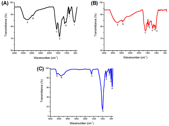
Figure 1.
Comparative Fourier transform infrared spectra of: (A) humic acids, (B) sodium-free humic acids, and (C) the inorganic mycotoxin binder (zeolite).

Table 1.
Band assignments of the vibrational frequencies in the humic acids and the inorganic mycotoxin binder (zeolite).
2.1.2. SEM
The surface morphology and microstructure of the sorbents were evaluated using a series of acquired SEM images (Figure 2). The surface morphology of the zeolite is amorphous, and large clumps cover the pores. On the other hand, HA and SFHA had rough and uneven surfaces with aggregates of different shapes and sizes. Specifically for HA, most of the aggregated particles were approximately 163.42 ± 20.14 μm in size. Moreover, grooves on the particle surface with the trace of granules were also observed. These results are consistent with the granular structure of several HAs extracted from various sources [28,29]. HA’s physical and chemical compositions differ considerably depending on its source, environmental conditions, and extraction procedure [20,24]. Several variables, including concentration, pH, ionic strength, charge density, acidic group ionization degree, and intermolecular interactions, influence the structural conformation of HA [30,31]. The HA structure is protonated at low pHs (below 4), resulting in a more condensed structure due to the establishment of H-bonds and van der Waals interactions. However, at pH levels between 4 and 7, HA adopts a more expanded and scattered macrostructure due to intra and intermolecular repulsion. Furthermore, at pH greater than 11, a condensed structure can also be observed [32]. According to this research, certain HA molecules engage in supramolecular interactions through dispersive forces, such as hydrogen bonds, van der Waals interactions, and π–π interactions [28,33].
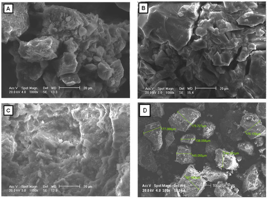
Figure 2.
Structure of (A) humic acids, (B) sodium-free humic acids, and (C) the zeolite under SEM at ×1000. Measurements (D) of humic acids at ×100.
2.1.3. EDS
Figure 3 shows the energy-dispersive X-ray spectroscopy (EDS) spectra of the HA, SFHA, and the zeolite. Moreover, the elemental compositions of HA, SFHA, and the zeolite are shown in Table 2. Results indicated the principal elements in HA and SFHA were C with 35.42% and 43.13%, and O with 20.32% and 46.43%, respectively. The Na content of SFHA considerably dropped, decreasing from 27.68% to 1.78% due to its reduction by washing. Other minor elements were involved in HA materials, such as Al, Si, K, P, S, and Cl. Meanwhile, Mg and Fe were not detected. Parameters are found within published ranges in the literature [34,35]. Information on the elemental composition of HA is not particularly conclusive due to their complexity and a wide range of other factors [36]. In addition, the main elements in the zeolite were Si (40.93%), C (31.99%), O (14.13%), and Al (7.46%), and minor contents of Na (0.25%), K (1.80%), Mg (1.12%), Ca (1.45%), and Fe (0.87%). Furthermore, Cl, P, and S were not detected. Similar chemical compositions for this kind of material have been reported by other researchers [37,38,39].
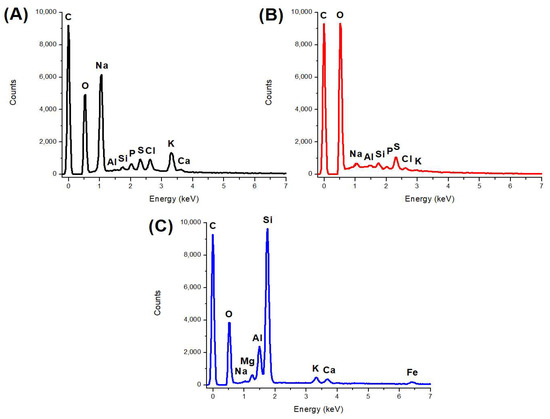
Figure 3.
Representative energy-dispersive X-ray spectroscopy (EDS) spectra of (A) humic acids, (B) sodium-free humic acids, and (C) the zeolite.
Table 2.
The elemental composition (%) of humic acids (HA), sodium-free humic acids (SFHA) and the zeolite.
2.1.4. ζ-Potential
In colloidal systems, the ζ-potential is frequently employed to track the behavior of particles suspended in a liquid. Additionally, the magnitude of the ζ-potential indicates the strength of the electrostatic attraction or repulsion between particles; therefore, it may be used to describe the surface of charged particles [40]. Figure 4 shows the relationship between pH and ζ-potential of HA, SFHA, and the zeolite. The three adsorbents generally show the same behavior: as the pH increases (from 2 to 11), the ζ-potential value becomes more negative. The pH values used for the determination of ζ-potential were 2, 5, and 7, each one according to the simulated compartment (crop, proventriculus, and intestine) in the in vitro digestive model. At pH 2, values of −46.44 mV and −30.75 mV were observed; at pH 5 of −50.16 mV and −49.2 mV; and at pH 7 of −54.46 mV and −40.93 mV for HA and SFHA, respectively. As a result, at all three pH values tested, HA was more negative, followed by SFHA and the zeolite. Our findings are consistent with those reported by Hamza et al [41] who state as the pH rises, the acidic functional groups of HA deprotonate, resulting in a more negative surface. Moreover, Deng and Bai [42] also found when pH exceeds 1.9, HA has a negative ζ-potential. Omar et al. (2014) and Coles and Yong [43] also reported ζ-potential values of HA of −20 mV at pH 3 and −44 mV at pH 10. Finally, Loosli et al. [44] reported strong negative charge with ζ-potential values ranging from −30.2 mV at pH 3 to −69.0 mV at pH 11. Even at low pH, HAs are negatively charged because of the dissociation of their acidic functional groups (mainly carboxylic and phenolic hydroxyl) [26,45]. Instead of aromatic components, variations in acid group conformation can be used to explain how the interactions are pH-dependent [32]. The first decrease (pH 3) of ζ-potential corresponds to the dissociation of carboxylic acid groups, while the second (pH 6) corresponds to the start of ionization of phenolic acid groups [46]. Our findings show the three adsorbents have a highly negative ζ-potential at the three simulated pH levels of the in vitro model, particularly at pH 7. As a result, the AFB1 molecule and the HA particle surface might be attracted by electrostatic forces since HAs are anionic polyelectrolytes that can interact with the positively charged AFB1 molecules [47].
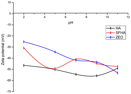
Figure 4.
Relationship between zeta potential (ζ-potential) and pH of humic acids, sodium-free humic acids, and the zeolite. Mean of five replicates ± standard error.
2.1.5. pHpzc
Understanding the surface charge of the particles is greatly aided by the pHpzc, which stands for the point of zero charge where the sum of positive and negative charges is equal. Figure 5 shows the pHpzc of HA, SFHA, and the zeolite. It has been hypothesized that the surface of the adsorbent will be positively charged if pH < pHpzc and negatively charged if pH ˃ pHpzc [47]. From the curves, the pHpzc for HA, SFHA, and the zeolite were 10.4, 2.2, and 8.8, respectively. According to Coles and Yong [43], the pHpzc of HA was less than 0.5. Moreover, Gjessing [48] reported pHpzc values for HA ranging from 1.2 to 1.8, which is very similar to the value of SFHA in our study. Furthermore, Giasuddin et al [49] reported the HA’s surface charge was negative over a pH range of 5 to 9.3. However, the presence of organic matter in the HA determines whether the pHpzc decreases or increases [43].
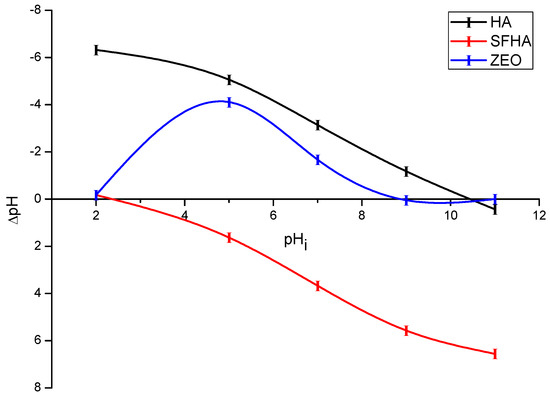
Figure 5.
Point of zero charge (pHpzc) of humic acids, sodium-free humic acids, and the zeolite. Mean of five replicates ± standard error.
Interestingly, compared to the pHpzc of HA, which was 10.4, the SFHA reduced their pHpzc value to 2.2 after washing. This significant change in the pHpzc value could be attributed to a change in organic matter composition or to the purification process used to reduce the sodium salts in the HA. In this context, AFB1 adsorption is expected to be significant because SFHA has a negatively charged surface in all compartments simulated in the in vitro study. HA and the zeolite, on the other hand, have a positive surface charge on all three gastrointestinal sections, indicating electrostatic interactions do not govern AFB1 adsorption. It is well known that other mechanisms, such as weak electrostatic interactions and moderate electron donor–acceptor attraction, contribute to the adsorption of aflatoxins into inorganic binders [37].
2.2. In Vitro Digestive Model
Figure 6 depicts the percentage of AFB1 adsorption of the three tested sorbents. At the end of the in vitro model (the intestinal section), HA and SFHA adsorbents had 97.6% and 99.7% AFB1 uptake, respectively. Using the zeolite, a moderate sorption uptake of 81.5% was reached. In contrast, controls (without the addition of sorbent materials) show a marked lack of AFB1 adsorption (<3%). Following our findings, it was reported an in vitro model with 100 mg/mL of HA inclusion at 20 ng AFB1/g achieved 90.50% of AFB1 adsorption [50]. Moreover, Ye et al. [51] investigated the AFB1 adsorption capacity in the presence of sodium humate inclusions, various pH levels, interaction times, and AFB1 concentrations. The highest AFB1 adsorption percentages reported were 88.12% at pH 7 and 76.36% at pH 8. Furthermore, Vázquez-Durán et al. [27], using a dynamic in vitro model to assess the AFB1 adsorption capacity of a non-commercial zeolite at 5% (w/w) inclusion, reported an adsorption percentage of 75.5%, which is consistent with this research.
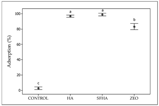
Figure 6.
The adsorption capacity of humic acids (HA), sodium-free humic acids (SFHA), and the zeolite against AFB1 using an in vitro digestive poultry model. Mean values ± standard error. a–c Means with different letter are statistically different (Tukey p ≤ 0.05).
2.3. The Mechanism for AFB1 Adsorption onto HA
Because HA possesses highly hydrophobic surfaces and a wide variety of negatively charged functional groups [9], interactions between HA and AFB1 may involve different mechanisms. Tan [52] lists seven potential ways HA might bind to gaseous, liquid, and solid components: (i) physical forces, (ii) chemical forces, (iii) hydrogen bonds, (iv) hydrophobic interactions, (v) electrostatic interactions, (vi) coordination reactions, and (vii) ligand exchange. The most important interactions between HA and AFB1 are electrostatic interactions and hydrogen bonding. Nevertheless, other interactions could be considered, such as π–π stacking [53] and hydrophobic interactions (due to the many bond indices related to hydrophobic groups, including CH2, CH3, and C=C) [54] (Figure 7). Although this phenomenon is still not fully understood, aromatic structures and functional groups, such as OH and COOH, contributed to HA’s high AFB1 adsorption capacity [55]. Several techniques have been proposed to investigate the adsorption of different molecules onto HA. For example, physical modelling (Langmuir isotherm), kinetic modelling (Elovich kinetic model), surface complexation modelling, and Ligand and charge distribution model, among others [54]. However, to elucidate the nature of the molecular interactions between HA and AFB1, future theoretical simulations using density functional theory (DFT) may be considered [56].
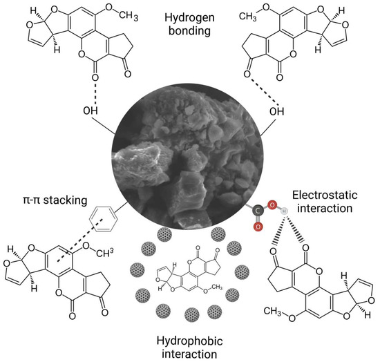
Figure 7.
The hypothetical mechanism by which humic acids bind the AFB1 molecule.
Nevertheless, because in vitro tests cannot fully simulate the conditions of a bird’s digestive tract to determine the effectiveness of HA derived from vermicompost in reducing the harmful effects of AFB1, in vivo experiments must be conducted. Data on the effectiveness of HA extracted from vermicompost to reduce the impact of AFB1 in broilers are still meager. However, previous research has demonstrated the effectiveness of various humic substances as mycotoxins binders in in vivo trials. For instance, the addition of oxihumate (3.5 g/kg) was found to have a protective effect against liver, stomach, and heart damage in diets contaminated with AFB1, and a significative decrease in several serum, and hematological biochemical indicators linked to aflatoxin toxicity was also seen in broilers [57]. Adding HA to chicken feed in amounts ranging from 0.2% to 0.4% (w/w) improved feed efficiency, reduced liver and bursa damage, and improved serum biochemical profiles associated with aflatoxin toxicity [58]. Additionally, adding HA (0.3% w/w) decreased AFB1 residues in the liver and enhanced broiler antibody production against Newcastle disease [59].
3. Conclusions
In this study, two forms of HAs extracted from vermicompost were prepared, further characterized, and tested in AFB1 adsorption experiments using an in vitro poultry digestive model. Despite differences in the microstructure, elemental composition, surface functional groups, pHpzc, and ζ-potential, both adsorbents demonstrated significant AFB1 adsorption capacities when using an in vitro poultry digestive model. As a result, it can be concluded HA derived from vermicompost is highly effective in the adsorption of AFB1. However, more in vivo studies will enhance our comprehension of HA efficacy to reduce the toxic effects of AFB1 in poultry. There is now research being conducted in this area.
4. Materials and Methods
4.1. Humic Acids
As previously described [12], HAs were extracted and isolated from a vermicompost. HAs were extracted with a sodium hydroxide solution (1M NaOH) in compost:alkali ratio of 1:4, then stirred for 2 h. A Whatman grade 40 filter paper was used to filter the suspension after it had been at room temperature for 24 h. The supernatant was then separated by decantation after the filtrate had been centrifuged for 15 min at 3500× g. The HA-containing supernatant was acidified with 10% HCl and agitated continuously until pH 2 was achieved, allowing the HA to precipitate. Centrifugation at 3500× g for 15 min separated HA from FA. Finally, the precipitate (HA) was normalized with 1M NaOH until pH 10 was achieved, then oven-dried at 60 °C. A black powder was produced as a result. To produce the second adsorbent material, hereinafter referred to as sodium-free humic acids (SFHA), HAs were redispersed in deionized water and neutralized with 10% HCl until pH 7 was reached, then washed (centrifugation and redispersion) ten times to remove excess sodium and subsequently oven-dried at 60 °C.
4.2. Characterization of HA
4.2.1. ATR-FTIR
Using a Fourier transform infrared spectrophotometer Frontier SP8000 (Perkin Elmer, Waltham, MA, USA) equipped with an attenuated total reflection (ATR) attachment (DuraSamplIR II, Smiths Detection, Warrington, UK), functional groups on the surface of the adsorbent materials were analyzed. Samples were deposited on the ATR diamond crystal, and spectra were collected in transmittance mode by combining 32 scans with a resolution of 4 cm−1 in the 4000–400 cm−1 region.
4.2.2. Scanning Electron Microscopy (SEM)
Utilizing a scanning electron microscope (JSM-6010LA, Jeol Inc., MA, USA), the adsorbents’ size and morphology was examined. A thin gold coating was applied to the samples to improve electron conductivity and image quality. Using a 20 kV accelerating voltage, microscopy was carried out. Secondary electron imaging mode was used to capture the images at a 1000× magnification.
4.2.3. Energy-Dispersive X-ray Spectroscopy (EDS)
Using an energy-dispersive X-ray spectrometer with an environmental scanning electron microscope (Phillips XL30, EDS-ESEM, Eindhoven, The Netherlands), the multi-element analysis was carried out. A high-performance X Trace micro-spot X-ray source was used to analyze each sample three times, and an attached XFlash® 6/10 silicon drift detector was used to quantify the X-ray fluorescence spectrum it produced (Bruker Nano GmbH, Berlin, Germany).
4.2.4. Zeta Potential (ζ-Potential)
The ZetaSizer Pro (Malvern Instruments, Worcestershire, UK) was used to determine zeta potential. The samples used for the measurements (20 mg dissolved in 10 mL distilled water) were adjusted to various pH levels using either HCl (0.1 M) or NaOH (0.1 M). To minimize the effects of viscosity and scattering, 100 µL of the aqueous phase were collected and diluted with 2 mL deionized water. Then, diluted samples were examined in a disposable capillary cell DTS1070 at room temperature with a 120-s equilibration time. Each measurement included eleven runs and three replicates of each sample to obtain a consistent result. The ZS Xplorer software was used to examine the results.
4.2.5. Point of Zero Charge (pHpzc)
In accordance with the instructions of Zavala-Franco et al [37], the pHpzc was measured. Briefly, equal quantities of sorbents were introduced to a series of flasks filled with distilled water at various pH levels (2, 5, 7, 9, and 11). The pH of the supernatant was measured after samples were agitated at 250 rpm for 195 min. The pH was measured using a combination glass electrode (Conductronic PC-45, Puebla, Mexico). The plot of ∆pH against pH was used to determine the pHpzc.
4.3. In Vitro Adsorption Studies
Preparation of the AFB1-Contaminated Diet
Aflatoxin (100 µg AFB1/mL) was made as a main stock in dimethyl sulfoxide. After that, distilled water was used to dilute the AFB1 solution to 1 µg AFB1/mL. An experimental maize-soybean meal diet was made to closely match the nutritional needs of broiler chickens, as suggested by the National Research Council [60]. There were no antibiotics or anticoccidial drugs in the diet (Table 3; adapted from Solís-Cruz et al. [61]). To achieve 100 µg AFB1/kg, the diet was contaminated with 0.5 mL of the AFB1 solution. Thereafter, five samples were chosen at random, and the content of AFB1 was determined using the immunoaffinity column clean-up and liquid chromatography with fluorescence detection methodology. Levels of B-aflatoxins (AFB1 and AFB2), total fumonisins (FB1, FB2, and FB3), and ochratoxin A (OTA) were also determined in the diet using monoclonal antibody-based affinity columns (VICAM Science Technology, Watertown, MA, USA) and fluorescence detection. In general, the experimental diet had no detectable levels of B-aflatoxins and total fumonisins; assayed contents of these mycotoxins were below the detection limits of the immunoaffinity column techniques employed (<1 ng/g and <0.016 mg/kg, respectively). OTA was present at a level of 7 ng/g.
Table 3.
Ingredient composition of the experimental poultry diet.
4.4. In Vitro Digestive Model
The AFB1 adsorptive capacity of the tested materials was assessed using a previously described in vitro gastrointestinal poultry model [62] with minor modifications. The assay was carried out with one control (zeolite) and two different treatments (HA and SFHA). This model simulated the physiological conditions of broiler chicken crop, proventriculus, and intestine. Every tube was incubated at 40 °C while being shaken at 19 rpm at an angle of 30°. For each compartment, the pH, enzymes, and time windows were adjusted. In the beginning, 5 g of the AFB1-contaminated feed and 10 mg of each adsorbent material were placed in polypropylene tubes (50 mL). Each tube received 10 mL of 0.03 M HCl to imitate the crop environment (pH reached values ~5.2). For 30 min, the tubes were incubated. A pH range of 1.4 to 2.0 was attained by adding 2.5 mL of 1.5 M HCl and 3000 U of pepsin (Merck KGaA, Darmstadt, Germany) per gram of feed in each tube following the incubation time. For a further 45 min, each tube was incubated. To simulate the third and final gastrointestinal compartment, 6.84 mg of 8×-pancreatin (Merck KGaA, Darmstadt, Germany) per gram of feed was added to 6.5 mL of 1.0 M NaHCO3. The pH in this region was reached, between 6.4 and 6.8. Tubes were incubated for an additional 120 min. The entire in vitro digesting process took 195 min. Afterward, the supernatant from all tubes was collected and stored at −20 °C for further analysis after centrifuging them all at 7000× g for 30 min. To determine the real AFB1 concentrations in each tube, controls (without the addition of adsorbent materials) were also prepared. The entire experiment was carried out in quintuplicate. The adsorption percentage of AFB1 for each tested material was calculated as follows:
where is the concentration of AFB1 in the control (ng/mL); and is the concentration of AFB1 in the supernatant of the treatments (ng/mL).
4.5. Aflatoxin Assay
Using monoclonal anti-body-based immunoaffinity columns (Afla-B, VICAM Science Technology, Watertown, MA, USA), AFB1 was removed from the supernatants and then utilized for ultraperformance liquid chromatography (UPLC). A modified version of the procedure that Hernández-Ramirez et al [63] previously described was employed. A UPLC system (Waters ACQUITY H-class) was used, equipped with a quaternary solvent manager and a reverse phase column (2.1 mm × 100 mm, 1.7 µm particles). A mobile phase of water, methanol, and acetonitrile (64:18:18) was used to elute AFB1. Samples (1 µL) from the anti-body-based immunoaffinity columns were injected and eluted with a flow rate of 700 µL/min. A fluorescence detector with settings of 365 nm excitation and 429 nm emission was used to detect the toxin. The AFB1 concentration was estimated using a calibration curve with a standard reference (AFB1, Merck KGaA, Darmstadt, Germany).
4.6. Method Validation
The performance of the clean-up procedure was tested by measuring the percentage of AFB1 recovery using the UPLC methodology, spiking four replicates of the experimental poultry diet with six different aflatoxin contents over the range of 8 to 250 ng AFB1/g, attaining a toxin recovery of 92% with a standard deviation of 3.4, standard error of 1.7, and a coefficient variation value of 4.4%. Moreover, the validation of the UPLC method was performed based on the guidelines for single-laboratory validation of analytical methods for trace-level concentrations of organic chemicals elaborated by the AOAC/FAO/IAEA/IUPAC [64]. The following parameters were evaluated: limit of detection (LOD), limit of quantification (LOQ), and linearity. For linearity, a six-point calibration curve was plotted at concentrations over the range of 10 to 1000 ng AFB1/L. In general, detection and quantification limits were found to be 2.0 and 6.7 ng AFB1/L, respectively. The linearity estimated with the coefficient of determination (R2) was 0.9984. These results indicated the methodology used was acceptable.
4.7. Experimental Design and Statistical Analysis
Data was subjected to one-way analysis of variance (one way-ANOVA) as a completely randomized design. Significant differences among the means were determined by the Tukey test. A value of p = 0.05 was used to detect significant differences between treatments.
Author Contributions
Conceptualization, S.G.-R.; methodology, J.d.D.F.-C. and A.V.-D.; software, J.A.M.-G.; validation, A.V.-D.; formal analysis, J.A.M.-G. and A.M-A.; investigation, B.S.-C., M.d.J.N.-R., D.H.-P., R.M.-G. and B.M.H.; resources, G.T.-I.; data curation, M.d.J.N.-R., B.S.-C. and D.H.-P.; writing—original draft preparation, J.A.M.-G. and A.M.-A.; writing—review and editing, S.G.-R., G.T.-I. and X.H.-V.; visualization, S.G.-R. and A.M.-A.; supervision, A.M.-A.; project ad-ministration, M.d.L.Á. All authors have read and agreed to the published version of the manuscript.
Funding
This research was partially supported by the Mexican Council of Science and Technology (CONACYT), the UNAM-PAPIIT grant numbers PDCPN_2017_4777 and IA101253, and by USDA-NIFA Sustainable Agriculture Systems, Grant No. 2019-69012-29905. Title of Project: Empowering US Broiler Production for Transformation and Sustainability USDA-NIFA (Sustainable Agriculture Systems): No. 2019-69012-29905.
Institutional Review Board Statement
Not applicable.
Informed Consent Statement
Not applicable.
Data Availability Statement
The datasets generated for this study are available on request to the corresponding author.
Conflicts of Interest
The authors declare the research was conducted in the absence of any commercial or financial relationships that could be construed as a potential conflict of interest.
References
- Coulombe, R.A. Biological Action of Mycotoxins. J. Dairy Sci. 1993, 76, 880–891. [Google Scholar] [CrossRef]
- Zain, M.E. Impact of Mycotoxins on Humans and Animals. J. Saudi Chem. Soc. 2011, 15, 129–144. [Google Scholar] [CrossRef]
- Frisvad, J.C.; Hubka, V.; Ezekiel, C.N.; Hong, S.-B.; Nováková, A.; Chen, A.J.; Arzanlou, M.; Larsen, T.O.; Sklenář, F.; Mahakarnchanakul, W.; et al. Taxonomy of Aspergillus Section Flavi and Their Production of Aflatoxins, Ochratoxins and Other Mycotoxins. Stud. Mycol. 2019, 93, 1–63. [Google Scholar] [CrossRef]
- Rawal, S.; Kim, J.E.; Coulombe, R. Aflatoxin B1 in Poultry: Toxicology, Metabolism and Prevention. Res. Vet. Sci. 2010, 89, 325–331. [Google Scholar] [CrossRef]
- Popescu, R.G.; Avramescu, S.; Marin, D.E.; Țăranu, I.; Georgescu, S.E.; Dinischiotu, A. The Reduction of the Combined Effects of Aflatoxin and Ochratoxin A in Piglet Livers and Kidneys by Dietary Antioxidants. Toxins 2021, 13, 648. [Google Scholar] [CrossRef]
- Wang, Y.; Liu, F.; Zhou, X.; Liu, M.; Zang, H.; Liu, X.; Shan, A.; Feng, X. Alleviation of Oral Exposure to B1-Induced Renal Dysfunction, Oxidative Stress, and Cell Apoptosis in Mice Kidney by Curcumin. Antioxidants 2022, 11, 1082. [Google Scholar] [CrossRef] [PubMed]
- Fouad, A.; Ruan, D.; El-Senousey, H.; Chen, W.; Jiang, S.; Zheng, C. Harmful Effects and Control Strategies of Aflatoxin B1 Produced by Aspergillus Flavus and Aspergillus Parasiticus Strains on Poultry: Review. Toxins 2019, 11, 176. [Google Scholar] [CrossRef] [PubMed]
- Kabak, B.; Dobson, A.D.W.; Var, I. Strategies to Prevent Mycotoxin Contamination of Food and Animal Feed: A Review. Crit. Rev. Food Sci. Nutr. 2006, 46, 593–619. [Google Scholar] [CrossRef]
- Piccolo, A. The Supramolecular Structure of Humic Substances: A Novel Understanding of Humus Chemistry and Implications in Soil Science. In Advances in Agronomy; Elsevier: Amsterdam, The Netherlands, 2002; Volume 75, pp. 57–134. ISBN 978-0-12-000793-6. [Google Scholar]
- Guo, X.; Liu, H.; Wu, S. Humic Substances Developed during Organic Waste Composting: Formation Mechanisms, Structural Properties, and Agronomic Functions. Sci. Total Environ. 2019, 662, 501–510. [Google Scholar] [CrossRef]
- Jiménez-González, M.A.; Álvarez, A.M.; Carral, P.; Almendros, G. Chemometric Assessment of Soil Organic Matter Storage and Quality from Humic Acid Infrared Spectra. Sci. Total Environ. 2019, 685, 1160–1168. [Google Scholar] [CrossRef]
- Maguey-Gonzalez, J.A.; Michel, M.A.; Baxter, M.F.A.; Solis-Cruz, B.; Hernandez-Patlan, D.; Merino-Guzman, R.; Hernandez-Velasco, X.; Latorre, J.D.; Hargis, B.M.; Tellez, G.; et al. Effects of Humic Acids on Recovery of Salmonella Enterica Serovar Enteritidis. Ann. Anim. Sci. 2018, 18, 387–399. [Google Scholar] [CrossRef]
- Maguey-Gonzalez, J.A.; Michel, M.A.; Baxter, M.F.A.; Tellez, G.; Moore, P.A.; Solis-Cruz, B.; Hernández-Patlan, D.; Merino-Guzman, R.; Hernandez-Velasco, X.; Latorre, J.D.; et al. Effect of Humic Acids on Intestinal Viscosity, Leaky Gut and Ammonia Excretion in a 24 Hr Feed Restriction Model to Induce Intestinal Permeability in Broiler Chickens. Anim. Sci. J. 2018, 89, 1002–1010. [Google Scholar] [CrossRef] [PubMed]
- Maguey-González, J.A.; Gómez-Rosales, S.; de Lourdes Angeles, M.; López-Hernández, L.H.; Rodríguez-Hernández, E.; Solís-Cruz, B.; Hernández-Patlán, D.; Merino-Gúzman, R.; Téllez-Isaías, G. Effects of Humic Acids on the Recovery of Different Bacterial Strains in an in Vitro Chicken Digestive Model. Res. Vet. Sci. 2022, 145, 21–28. [Google Scholar] [CrossRef] [PubMed]
- De Lourdes Angeles, M.; Gómez-Rosales, S.; Téllez-Isaias, G. Mechanisms of Action of Humic Substances as Growth Promoters in Animals. In Humus and Humic Substances—Recent Advances; IntechOpen: London, UK, 2022; ISBN 978-1-80356-212-4. [Google Scholar]
- Vázquez-Durán, A.; de Jesús Nava-Ramírez, M.; Téllez-Isaías, G.; Méndez-Albores, A. Removal of Aflatoxins Using Agro-Waste-Based Materials and Current Characterization Techniques Used for Biosorption Assessment. Front. Vet. Sci. 2022, 9, 897302. [Google Scholar] [CrossRef] [PubMed]
- Zhang, J.; Dai, J.; Wang, R.; Li, F.; Wang, W. Adsorption and Desorption of Divalent Mercury (Hg2+) on Humic Acids and Fulvic Acids Extracted from Typical Soils in China. Colloids Surf. A Physicochem. Eng. Asp. 2009, 335, 194–201. [Google Scholar] [CrossRef]
- Mozgawa, W.; Sitarz, M.; Rokita, M. Spectroscopic Studies of Different Aluminosilicate Structures. J. Mol. Struct. 1999, 511–512, 251–257. [Google Scholar] [CrossRef]
- Alberts, J.J.; Filip, Z. Metal Binding in Estuarine Humic and Fulvic Acids: FTIR Analysis of Humic Acid-Metal Complexes. Environ. Technol. 1998, 19, 923–931. [Google Scholar] [CrossRef]
- Manzak, A.; Kurşun, C.; Yıldız, Y. Characterization of Humic Acid Extracted from Aqueous Solutions with Polymer Inclusion Membranes. J. Taiwan Inst. Chem. Eng. 2017, 81, 14–20. [Google Scholar] [CrossRef]
- Baglieri, A.; Vindrola, D.; Gennari, M.; Negre, M. Chemical and Spectroscopic Characterization of Insoluble and Soluble Humic Acid Fractions at Different PH Values. Chem. Biol. Technol. Agric. 2014, 1, 9. [Google Scholar] [CrossRef]
- Ribeiro, J.S.; Ok, S.S.; Garrigues, S.; de la Guardia, M. FTIR Tentative Characterization of Humic Acids Extracted from Organic Materials. Spectrosc. Lett. 2001, 34, 179–190. [Google Scholar] [CrossRef]
- Domínguez-Negrete, A.; Gómez-Rosales, S.; Angeles, M.d.L.; López-Hernández, L.H.; Reis-de Souza, T.C.; López-García, Y.; Zavala-Franco, A.; Téllez-Isaias, G. Effect of the Addition of Humic Substances as Growth Promoter in Broiler Chickens Under Two Feeding Regimens. Animals 2019, 9, 1101. [Google Scholar] [CrossRef] [PubMed]
- Rupiasih, N.N.; Vidyasagar, P.B. Analytical Study of Humic Acid from Various Sources Commonly Used as Fertilizer: Emphasis on Heavy Metal Content. Int. J. Des. Nat. Ecodynamics 2009, 4, 32–46. [Google Scholar] [CrossRef]
- Xu, D.; Xu, Z.; Zhu, S.; Cao, Y.; Wang, Y.; Du, X.; Gu, Q.; Li, F. Adsorption Behavior of Herbicide Butachlor on Typical Soils in China and Humic Acids from the Soil Samples. J. Colloid Interface Sci. 2005, 285, 27–32. [Google Scholar] [CrossRef]
- Vermeer, A.W.P.; van Riemsdijk, W.H.; Koopal, L.K. Adsorption of Humic Acid to Mineral Particles. 1. Specific and Electrostatic Interactions. Langmuir 1998, 14, 2810–2819. [Google Scholar] [CrossRef]
- Vázquez-Durán, A.; de Jesús Nava-Ramírez, M.; Hernández-Patlán, D.; Solís-Cruz, B.; Hernández-Gómez, V.; Téllez-Isaías, G.; Méndez-Albores, A. Potential of Kale and Lettuce Residues as Natural Adsorbents of the Carcinogen Aflatoxin B1 in a Dynamic Gastrointestinal Tract-Simulated Model. Toxins 2021, 13, 771. [Google Scholar] [CrossRef]
- Prado, A.G.S.; Pertusatti, J.; Nunes, A.R. Aspects of Protonation and Deprotonation of Humic Acid Surface on Molecular Conformation. J. Braz. Chem. Soc. 2011, 22, 6. [Google Scholar] [CrossRef]
- Yang, F.; Zhang, S.; Cheng, K.; Antonietti, M. A Hydrothermal Process to Turn Waste Biomass into Artificial Fulvic and Humic Acids for Soil Remediation. Sci. Total Environ. 2019, 686, 1140–1151. [Google Scholar] [CrossRef]
- Tarasevich, Y.I.; Dolenko, S.A.; Trifonova, M.Y.; Alekseenko, E.Y. Association and Colloid-Chemical Properties of Humic Acids in Aqueous Solutions. Colloid J. 2013, 75, 207–213. [Google Scholar] [CrossRef]
- Sarlaki, E.; Paghaleh, A.S.; Kianmehr, M.H.; Vakilian, K.A. Chemical, Spectral and Morphological Characterization of Humic Acids Extracted and Membrane Purified from Lignite. Chem. Chem. Technol. 2020, 14, 353–361. [Google Scholar] [CrossRef]
- Klučáková, M.; Věžníková, K. Micro-Organization of Humic Acids in Aqueous Solutions. J. Mol. Struct. 2017, 1144, 33–40. [Google Scholar] [CrossRef]
- Baigorri, R.; Fuentes, M.; González-Gaitano, G.; García-Mina, J.M. Analysis of Molecular Aggregation in Humic Substances in Solution. Colloids Surf. A Physicochem. Eng. Asp. 2007, 302, 301–306. [Google Scholar] [CrossRef]
- Giovanela, M.; Crespo, J.S.; Antunes, M.; Adamatti, D.S.; Fernandes, A.N.; Barison, A.; da Silva, C.W.P.; Guégan, R.; Motelica-Heino, M.; Sierra, M.M.D. Chemical and Spectroscopic Characterization of Humic Acids Extracted from the Bottom Sediments of a Brazilian Subtropical Microbasin. J. Mol. Struct. 2010, 981, 111–119. [Google Scholar] [CrossRef]
- Liu, X.; Hou, Y.; Li, Z.; Yu, Z.; Tang, J.; Wang, Y.; Zhou, S. Hyperthermophilic Composting of Sewage Sludge Accelerates Humic Acid Formation: Elemental and Spectroscopic Evidence. Waste Manag. 2020, 103, 342–351. [Google Scholar] [CrossRef] [PubMed]
- Doskočil, L.; Burdíková-Szewieczková, J.; Enev, V.; Kalina, L.; Wasserbauer, J. Spectral Characterization and Comparison of Humic Acids Isolated from Some European Lignites. Fuel 2018, 213, 123–132. [Google Scholar] [CrossRef]
- Zavala-Franco, A.; Hernández-Patlán, D.; Solís-Cruz, B.; López-Arellano, R.; Tellez-Isaias, G.; Vázquez-Durán, A.; Méndez-Albores, A. Assessing the Aflatoxin B1 Adsorption Capacity between Biosorbents Using an In Vitro Multicompartmental Model Simulating the Dynamic Conditions in the Gastrointestinal Tract of Poultry. Toxins 2018, 10, 484. [Google Scholar] [CrossRef]
- Díaz-Nava, C.; Olguín, M.T.; Solache-Ríos, M. Water Defluoridation by Mexican Heulandite–clinoptilolite. Sep. Sci. Technol. 2002, 37, 3109–3128. [Google Scholar] [CrossRef]
- Ostrooumov, M.; Cappelletti, P.; de’Gennaro, R. Mineralogical Study of Zeolite from New Mexican Deposits (Cuitzeo Area, Michoacan, Mexico). Appl. Clay Sci. 2012, 55, 27–35. [Google Scholar] [CrossRef]
- Lin, D.-Q.; Brixius, P.J.; Hubbuch, J.J.; Thömmes, J.; Kula, M.-R. Biomass/Adsorbent Electrostatic Interactions in Expanded Bed Adsorption: A Zeta Potential Study: Study of Zeta Potential in Expanded Bed Absorption. Biotechnol. Bioeng. 2003, 83, 149–157. [Google Scholar] [CrossRef]
- Hamza, Z.; El-Hashash, M.; Aly, S.; Hathout, A.; Soto, E.; Sabry, B.; Ostroff, G. Preparation and Characterization of Yeast Cell Wall Beta-Glucan Encapsulated Humic Acid Nanoparticles as an Enhanced Aflatoxin B1 Binder. Carbohydr. Polym. 2019, 203, 185–192. [Google Scholar] [CrossRef]
- Deng, S.; Bai, R.B. Aminated Polyacrylonitrile Fibers for Humic Acid Adsorption: Behaviors and Mechanisms. Environ. Sci. Technol. 2003, 37, 5799–5805. [Google Scholar] [CrossRef]
- Coles, C.A.; Yong, R.N. Humic Acid Preparation, Properties and Interactions with Metals Lead and Cadmium. Eng. Geol. 2006, 85, 26–32. [Google Scholar] [CrossRef]
- Loosli, F.; Le Coustumer, P.; Stoll, S. TiO2 Nanoparticles Aggregation and Disaggregation in Presence of Alginate and Suwannee River Humic Acids. PH and Concentration Effects on Nanoparticle Stability. Water Res. 2013, 47, 6052–6063. [Google Scholar] [CrossRef] [PubMed]
- Illés, E.; Tombácz, E. The Role of Variable Surface Charge and Surface Complexation in the Adsorption of Humic Acid on Magnetite. Colloids Surf. A Physicochem. Eng. Asp. 2003, 230, 99–109. [Google Scholar] [CrossRef]
- Alvarez-Puebla, R.A.; Garrido, J.J. Effect of PH on the Aggregation of a Gray Humic Acid in Colloidal and Solid States. Chemosphere 2005, 59, 659–667. [Google Scholar] [CrossRef]
- Ramales-Valderrama, R.; Vázquez-Durán, A.; Méndez-Albores, A. Biosorption of B-Aflatoxins Using Biomasses Obtained from Formosa Firethorn [Pyracantha Koidzumii (Hayata) Rehder]. Toxins 2016, 8, 218. [Google Scholar] [CrossRef] [PubMed]
- Volk, B.G. Physical and Chemical Characteristics of Aquatic Humus; Gjessing, E.T., Ed.; Ann Arbor Science Publishers Inc.: Ann Arbor, MI, USA, 1976. [Google Scholar] [CrossRef]
- Giasuddin, A.B.M.; Kanel, S.R.; Choi, H. Adsorption of Humic Acid onto Nanoscale Zerovalent Iron and Its Effect on Arsenic Removal. Environ. Sci. Technol. 2007, 41, 2022–2027. [Google Scholar] [CrossRef]
- Abd El-Shafea, Y. In vivo and in vitro evaluation of efficacy of humic acid against aflatoxins. Al-Azhar J. Pharm. Sci. 2014, 49, 133–154. [Google Scholar] [CrossRef]
- Ye, S.-Q.; Lv, X.-Z.; Zhou, A.-G. In Vitro Evaluation of the Efficacy of Sodium Humate as an Aflatoxin B1 Adsorbent. Aust. J. Basic Appl. Sci. 2009, 3, 1296–1300. [Google Scholar]
- Tan, K.H. Humic Matter in Soil and the Environment: Principles and Controversies; CRC Press: Boca Raton, FL, USA, 2003; ISBN 978-0-429-22322-8. [Google Scholar]
- Tikhonov, V.; Drozdova, O.; Karpukhin, M.; Demin, V. Participation of Cadmium (Ll) and Copper (Ll) Ions in Intermolecular Forces of Humic Acids in Solutions. IOP Conf. Ser. Earth Environ. Sci. 2019, 368, 012053. [Google Scholar] [CrossRef]
- Islam, M.A.; Morton, D.W.; Johnson, B.B.; Angove, M.J. Adsorption of Humic and Fulvic Acids onto a Range of Adsorbents in Aqueous Systems, and Their Effect on the Adsorption of Other Species: A Review. Sep. Purif. Technol. 2020, 247, 116949. [Google Scholar] [CrossRef]
- Zhou, Q.; Ouyang, S.; Ao, Z.; Sun, J.; Liu, G.; Hu, X. Integrating Biolayer Interferometry, Atomic Force Microscopy, and Density Functional Theory Calculation Studies on the Affinity between Humic Acid Fractions and Graphene Oxide. Environ. Sci. Technol. 2019, 53, 3773–3781. [Google Scholar] [CrossRef] [PubMed]
- Vázquez-Durán, A.; Téllez-Isaías, G.; Hernández-Rodríguez, M.; Ruvalcaba, R.M.; Martínez, J.; Nicolás-Vázquez, M.I.; Aceves-Hernández, J.M.; Méndez-Albores, A. The Ability of Chlorophyll to Trap Carcinogen Aflatoxin B1: A Theoretical Approach. Int. J. Mol. Sci. 2022, 23, 6068. [Google Scholar] [CrossRef] [PubMed]
- Van Rensburg, C.J.; Van Rensburg, C.E.J.; Van Ryssen, J.B.J.; Casey, N.H.; Rottinghaus, G.E. In Vitro and In Vivo Assessment of Humic Acid as an Aflatoxin Binder in Broiler Chickens. Poult. Sci. 2006, 85, 1576–1583. [Google Scholar] [CrossRef] [PubMed]
- Ghahri, H.; Habibian, R.; Fam, M.A. Evaluation of the Efficacy of Esterified Glucomannan, Sodium Bentonite, and Humic Acid to Ameliorate the Toxic Effects of Aflatoxin in Broilers. Turk. J. Vet. Anim. Sci. 2010, 34, 385–391. [Google Scholar] [CrossRef]
- Arafat, R.Y.; Khan, S.H. Saima Evaluation of Humic Acid as an Aflatoxin Binder in Broiler Chickens. Ann. Anim. Sci. 2017, 17, 241–255. [Google Scholar] [CrossRef]
- National Research Council; Board on Agriculture; Subcommittee on Poultry Nutrition. Nutrient Requirements of Poultry: Ninth Revised Edition, 1994; National Academies Press: Cambridge, MA, USA, 1994; ISBN 978-0-309-04892-7. [Google Scholar]
- Solís-Cruz, B.; Hernández-Patlán, D.; Beyssac, E.; Latorre, J.D.; Hernandez-Velasco, X.; Merino-Guzman, R.; Tellez, G.; López-Arellano, R. Evaluation of Chitosan and Cellulosic Polymers as Binding Adsorbent Materials to Prevent Aflatoxin B1, Fumonisin B1, Ochratoxin, Trichothecene, Deoxynivalenol, and Zearalenone Mycotoxicoses Through an In Vitro Gastrointestinal Model for Poultry. Polymers 2017, 9, 529. [Google Scholar] [CrossRef]
- Latorre, J.D.; Hernandez-Velasco, X.; Kuttappan, V.A.; Wolfenden, R.E.; Vicente, J.L.; Wolfenden, A.D.; Bielke, L.R.; Prado-Rebolledo, O.F.; Morales, E.; Hargis, B.M.; et al. Selection of Bacillus Spp. for Cellulase and Xylanase Production as Direct-Fed Microbials to Reduce Digesta Viscosity and Clostridium Perfringens Proliferation Using an in Vitro Digestive Model in Different Poultry Diets. Front. Vet. Sci. 2015, 2, 25. [Google Scholar] [CrossRef]
- Hernández-Ramírez, J.O.; Merino-Guzmán, R.; Téllez-Isaías, G.; Vázquez-Durán, A.; Méndez-Albores, A. Mitigation of AFB1-Related Toxic Damage to the Intestinal Epithelium in Broiler Chickens Consumed a Yeast Cell Wall Fraction. Front. Vet. Sci. 2021, 8, 677965. [Google Scholar] [CrossRef]
- Alder, L.; Hill, A.; Holland, P.T.; Lantos, J.; Lee, S.M.; MacNeil, J.D.; O’Rangers, J.; van Zoonen, P.; Ambrus, A. Guidelines for single-laboratory validation of analytical methods for trace-level concentrations of organic chemicals. In Principles and Practices of Method Validation; Fajgelj, A., Ambrus, Á., Eds.; Royal Society of Chemistry: Cambridge, UK, 2000; pp. 179–187. ISBN 978-0-85404-783-3. [Google Scholar]


Disclaimer/Publisher’s Note: The statements, opinions and data contained in all publications are solely those of the individual author(s) and contributor(s) and not of MDPI and/or the editor(s). MDPI and/or the editor(s) disclaim responsibility for any injury to people or property resulting from any ideas, methods, instructions or products referred to in the content. |
© 2023 by the authors. Licensee MDPI, Basel, Switzerland. This article is an open access article distributed under the terms and conditions of the Creative Commons Attribution (CC BY) license (https://creativecommons.org/licenses/by/4.0/).