Systematic Review

Knowledge about Snake Venoms and Toxins from Colombia: A Systematic Review

by
Jaime Andrés Pereañez
1,2,*,
Lina María Preciado
1 and
Paola Rey-Suárez
1,3
1
Research Group in Toxinology, Pharmaceutical, and Food Alternatives, Pharmaceutical and Food Sciences Faculty, University of Antioquia, Medellín 50010, Colombia
2
Research Group in Pharmaceutical Promotion and Prevention, University of Antioquia, Medellín 50010, Colombia
3
Centro de Investigación en Recursos Naturales y Sustentabilidad, Universidad Bernardo O’Higgins, Santiago 8320000, Chile
*
Author to whom correspondence should be addressed.
Toxins 2023, 15(11), 658; https://doi.org/10.3390/toxins15110658
Submission received: 10 October 2023 / Revised: 8 November 2023 / Accepted: 10 November 2023 / Published: 15 November 2023
(This article belongs to the Section Animal Venoms)

Abstract

:
Colombia encompasses three mountain ranges that divide the country into five natural regions: Andes, Pacific, Caribbean, Amazon, and Orinoquia. These regions offer an impressive range of climates, altitudes, and landscapes, which lead to a high snake biodiversity. Of the almost 300 snake species reported in Colombia, nearly 50 are categorized as venomous. This high diversity of species contrasts with the small number of studies to characterize their venom compositions and natural history in the different ecoregions. This work reviews the available information about the venom composition, isolated toxins, and potential applications of snake species found in Colombia. Data compilation was conducted according to the PRISMA guidelines, and the systematic literature search was carried out in Pubmed/MEDLINE. Venom proteomes from nine Viperidae and three Elapidae species have been described using quantitative analytical strategies. In addition, venoms of three Colubridae species have been studied. Bioactivities reported for some of the venoms or isolated components—such as antibacterial, cytotoxicity on tumoral cell lines, and antiplasmodial properties—may be of interest to develop potential applications. Overall, this review indicates that, despite recent progress in the characterization of venoms from several Colombian snakes, it is necessary to perform further studies on the many species whose venoms remain essentially unexplored, especially those of the poorly known genus Micrurus.
Key Contribution: This review highlights the available information about biological effects, proteomic analyses, and potential uses of snake venoms and toxins from Colombia.

1. Introduction

Snake venom is a complex biological secretion of specialized glands in certain snake species used to subdue prey or as a defense mechanism [1]. Venoms mainly comprise enzymes, proteins without enzymatic activity, peptides, organic chemical compounds, trace molecules such as ions (iron, calcium, zinc, cobalt, potassium or sodium), and citrate, among other components [1]. Snakebite envenoming occurs when snakes accidentally inject venom into humans, causing various pathophysiological effects, some of which can be life-threatening [2,3,4]. Nevertheless, snake venoms are also rich sources of bioactive compounds, which have led to the development of some therapeutic drugs [1,5].
Among more than 4000 snake species reported worldwide, around 20% are venomous [6,7]. Colombia is a megadiverse country, with over 270 snake species recognized [8]. Nevertheless, snakes from the Viperidae and Elapidae families and a reduced number of the Colubridae can inject venom; therefore, they are considered of medical importance [9,10]. In Colombia, between 4000 and 5500 snakebites have been reported annually in recent years [10], most of them inflicted by Bothrops spp.; however, the most severe cases are attributed to snakes from the genus Micrurus spp., Lachesis spp., and Colombian rattlesnake Crotalus durissus cumanensis [9,10]. Studies about diversity and abundance in snake venom proteomes report two dominant protein families for elapid venom: three-finger toxins (3FTxs) and phospholipases A2 (PLA2s). In contrast, the most abundant protein families for the Viperidae family are PLA2s, snake venom serine proteases (SVSPs), and snake venom metalloproteases (SVMPs) [11,12].
Snake venom composition can vary significantly between species and even among individuals within a species, age, and geographical location, among other aspects [1,13]. Therefore, it is crucial to characterize the venom composition for species of a country or a geographical area within a country. In addition, it is essential to characterize the snake venoms functionally, to correlate the signs and symptoms observed in envenomed patients, and to contribute to improving antivenom production [3]. The first study about snake venoms from Colombia was conducted in the beginning of the 1990s [14]. In more recent years, the composition of a number of these venoms have been characterized by proteomic profiling studies, and the isolation and characterization of some of their toxins have been achieved [14].
This systematic review aims to summarize and discuss the available information on biological effects, proteomic composition, and potential use of snake venoms and toxins from Colombia.

2. Materials and Methods

This review was conducted and reported according to the PRISMA (Preferred Reporting Items for Systematic Review and Met-Analysis) guidelines.

2.1. Eligibility Criteria, Search Strategies, and Information Sources

Published articles about whole snake venom characterization, composition by proteomic strategies, isolation and characterization of snake toxins, and bioprospecting uses were included. As exclusion criteria, we eliminated reviews, conference abstracts, editorials, and articles on antivenoms, natural or synthetic snake venom inhibitors, clinical aspects of envenomings, other toxinology topics, publications not related to snake venoms or toxins from Colombia, and articles without full-text access.
A systematic search using the described strategies was carried out in Pubmed/MEDLINE. Searches were limited without a start date through 31 July 2023, to identify published articles about whole snake venom characterization, composition by proteomic strategies, isolation and characterization of toxins, and their bioprospecting uses. The following search terms were used: (“Toxins, Biological”[Mesh] AND “Colombia”) OR (“Snake Venoms”[Mesh] AND “Proteomics”[Mesh] AND “Colombia”). This review was not registered in the final version.

2.2. Study Selection

Search strategies were imported and merged into reference management software, ZOTERO®. Then, titles and abstracts were assessed for inclusion. Full texts of relevant articles were retrieved and independently assessed by two authors.

2.3. Data Collection Process and Data Items

Data were extracted independently by two of the authors and compared using standardized data extraction forms. Discrepancies were discussed. Data extracted included title, authors, journal, and relevant comments.

2.4. Graphics

Graphics were built with Python (Version 3.10).

3. Results and Discussion

From the systematic literature search, 512 manuscripts were retrieved. However, after reviewing all articles, 467 were excluded because they either were not related to snake venoms and toxins from Colombia (n = 399), dealt with clinical aspects of snakebites (n = 8), or were associated with other topics in toxinology (antivenoms, n = 2; Lonomia identification, n = 14; scorpion venoms, n = 17; snake venom/toxin inhibitors, n = 17 and spider venoms, n = 9). In addition, it was impossible to access the full text of one manuscript. A total of 45 articles were chosen, and from their list of references, 7 were included (Figure 1).

3.1. Proteomic Profiles of Snake Venoms from Colombia

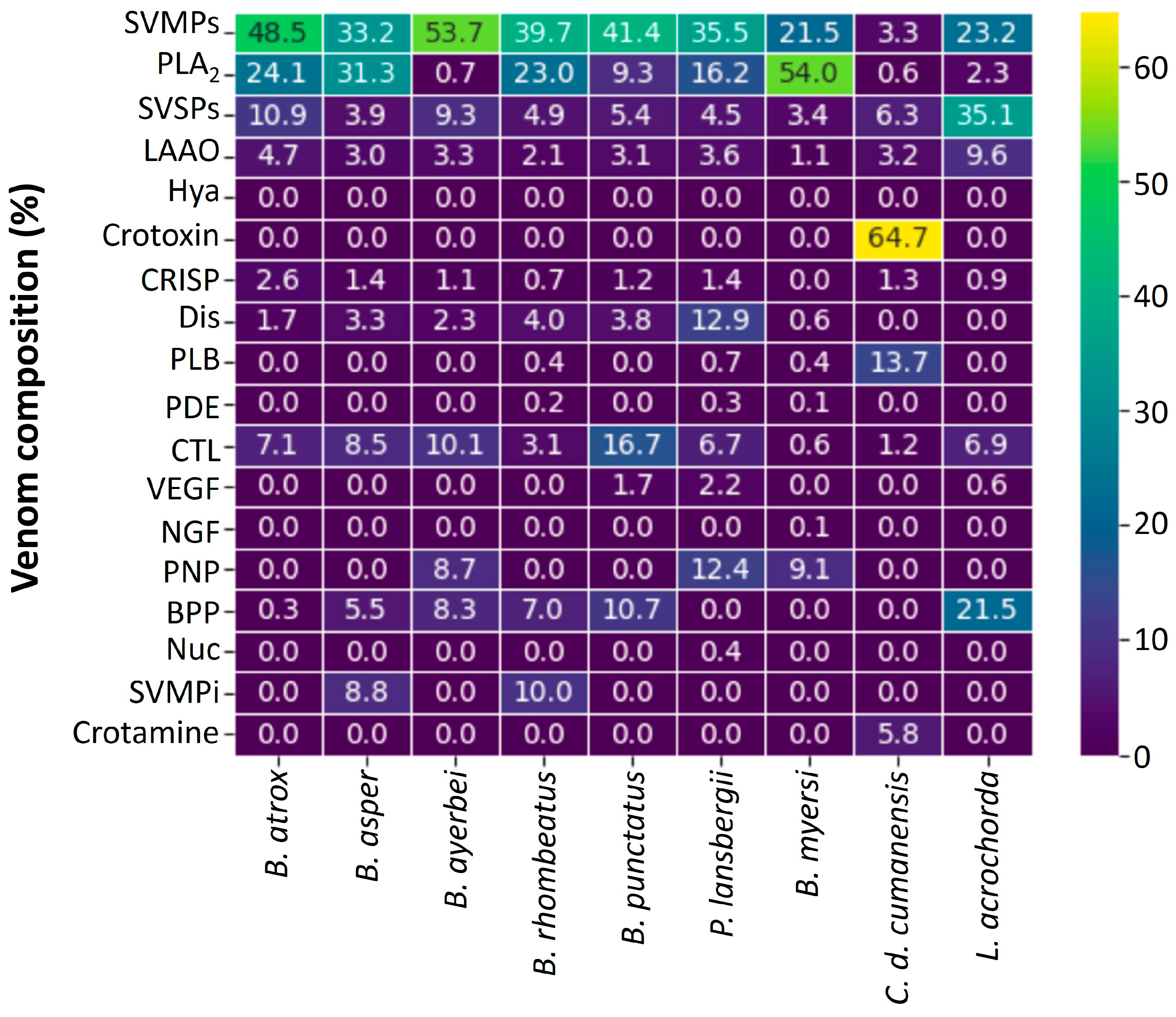
Proteomic techniques have been used to unveil the venom compositions, and to estimate the relative abundances of protein families for the Viperidae species shown in Figure 2. The venoms of B. asper [15,16], B. atrox [17], B. punctatus [18], B. ayerbei [16], B. rhombeatus [15], Bothrocophias myersi [19], Porthidium lansbergii [20], Lachesis acrochorda [21], and C. d. cumanensis [22] have been characterized by proteomic profiling. Proteins identified in these venoms belong to between seven to twelve families, with a median of 9. In general, the most abundant components are PLA2s, SVMPs, SVSPs, and L-amino acid oxidases (LAAOs). However, there were exceptions. For example, the venom of C. d. cumanensis has a high abundance of crotoxin (64.71%), in similarity with other South American rattlesnakes [23,24].
All Bothrops spp. venoms characterized until now have higher proportions of SVMPs than PLA2s [15,16,17,18]. This same pattern was observed in the venoms of P. lansbergii and L. acrochorda [20,21]. In contrast, the venom of B. myersi has a superior percentage of PLA2s. In addition, the venom of this species is the most complex, with twelve protein families [19]. Special characteristics of L. acrochorda venom are their high amounts of SVSPs and bradykinin-potentiating peptides (BBPs, 35.1% and 25.5%, respectively), even higher than PLA2s. Moreover, this venom showed the highest abundance of LAAOs (9.6%) [21].
C-type lectins are more abundant in the venom of B. punctatus (16.7%) [18], and they were identified in all Viperidae venoms. Conversely, the venom of P. lansbergii has the highest amounts of disintegrins [20], whereas these toxins were not identified in the venom of L. acrochorda [21]. A particular finding is the presence of Hyaluronidases, the spreading factor of venoms, which were only detected in the venom of B. myersi at a trace proportion (0.01%) [19]. However, this result might be explained because, in some venoms, not all proteins were successfully identified or assigned to known protein families.
The venom of C. d. cumanensis differs from all others studied thus far because this species is the only viper in Colombia that causes neurotoxicity. This effect is explained by the presence of high amounts crotoxin (64.7%) in the venom, which induces diaphragm-flaccid paralysis in snakebites inflicted by this Colombian rattlesnake [9]. In similarity to venoms from several other rattlesnakes, C. d. cumanensis venom also contains crotamine, a small basic polypeptide with myotoxic and cell-penetrating activities [25].
In addition to those shown in Figure 2, other proteomic studies have been performed using a “bottom-up whole venom shotgun profiling” approach, which can identify the proteins present in the venoms, but does not generate a reliable quantification of their relative abundance discussed in [26]. Nevertheless, the qualitative information provided by such analyses is valuable. Jiménez-Charris et al. [27] characterized B. asper venom from the Gorgona island and two ecoregions from Valle del Cauca state (Pacific and western). In the venom from Gorgona island, the following protein families were identified: SVMPs, PLA2s, CTL, CRISP, LAAO, SVSP, PLB, NGF, and Glutaminyl cyclase (GCY). In terms of qualitative composition, the venom of Gorgona Island is comparable to that of the Pacific ecoregion. The main difference was the presence of Nuc in the latter snake venom. In contrast, in the venom from the western ecoregion, no PLB, GCY, and Nuc were identified. Despite these differences, the three venoms studied are comparable with B. asper venoms from other regions of Colombia [15] in terms of protein family presence.
Another study by Montoya-Gómez et al. [28] described the qualitative protein family composition of L. acrochorda venom from Valle del Cauca, and detected SVMPs, PLA2s, SVSPs, LAAO, CTL, and NGF. This venom was very different from that reported by Madrigal et al. [21], which contained SVMPs, PLA2s, SVSPs, LAAO, Nuc, Hya, PDE, CTL, CRISP, BPP, and VEGF. The variations between the two venoms were attributed to the different proteomic approaches used, as well as the source of venoms from specimens kept in captivity and from wild animals for the studies of Madrigal et al. [21] and Montoya-Gómez et al. [28], respectively.
On the other hand, the composition of three venoms from the Elapidae family (coral snakes) have also been studied by proteomic profiling (Figure 3).
Proteomic analyses of Micrurus spp. venoms have revealed a dichotomic compositional pattern, with some species containing more PLA2s than 3FTxs, while in others, 3FTx predominate [32]. Micrurus species inhabiting South America tend to express the 3FTx-predominant venom phenotype, while the PLA2-rich pattern is observed in species inhabiting North America. In contrast, species found in Central America and northern South America present either of the two venom patterns [32]. Micrurus spp. from Colombia conform to the venom compositional pattern described. M. mipartitus from Antioquia state (northwestern Colombia) is rich in 3FTxs (61.1%), but it also contains 29.0% of PLA2s [29]. Instead, the venom of M. dumerilii from Antioquia is predominant in PLA2s (52.0%), compared to 28.1% of 3FTxs [30]. Similar findings were reported for the M. lemniscatus helleri venom from Amazonas (the southernmost state from Colombia), with 62.5% of PLA2s and 21.1% of 3FTxs [31]. The other components identified in Micrurus spp. venoms from Colombia were always below 10.0%.

3.2. Biological Activities of Viperidae and Elapidae Venoms from Colombia

The lethal activity of Viperidae venoms has been determined in mouse assays. The most studied venom is B. asper from different geographical regions in the country. The LD50 (95% confidence interval, µg/mouse) values by intraperitoneal (i.p.) route, for B. asper from Antioquia, Cauca, Valle del Cauca (Pacific ecoregion), and Gorgona Island are 67.1 (60.1–74.1) [33], 100.9 (83.2–122.8) [16], 112.8 (96.6–129.6) [27], and 118.2 (86.5–158.0) [27], respectively. It is also important to consider that the average of the LD50 (µg/mouse) of B. asper venoms from different ecoregions from Antioquia state is 65.3 (51.0–89.0) [3]. On the other hand, the LD50 (µg/mouse) values for B. atrox (Meta); B. ayerbei (Cauca); B. punctatus (Antioquia); and B. rhombeatus (Cauca), are 81.4 (80.2–83.6) [33], 50.1 (37.4–58.3) [16], 47.0 (36.0–61.0) [34] and 54.9 (36.0–83.8) µg/mouse [35], respectively. Therefore, the most lethal venoms from Bothrops spp. tested until now are B. ayerbei, B. rhombeatus, and B. punctatus. However, these results are not completely comparable because the biological assays use different mice strains.
The LD50 (95% confidence interval, µg/mouse) values by i.p. route of the other Viperidae venoms are: L. acrochorda (Valle del Cauca) 290.0 (260.0–323.0) [28]; L. acrochorda (Chocó) 130.0 (106.0–160.0) [34]; P. nasutum (Antioquia) 62.0 (51.0–74.0) [35]; P. lansbergii (Atlántico), yellow and gray morphs 98.0 (87.7–109.5) and 93.9 (730–128.6), respectively [36]; B. myersi (Valle del Cauca) 128.4 (101.1–156.6) [19]; and C. d. cumanensis (pool Colombia) 1.8 (1.2–2.5) µg/mouse [34].
In contrast, the LD50 (95% confidence interval, µg/mouse) for the venom of M. dumerilii (Antoquia) is 23.6 (15.0–38.0) [30], and for M. mipartitus (Antioquia) is 9.0 (6.6–12.1) [34].
Several studies have characterized other biological activities of Viperidae venoms from Colombia (Figure 4).
Edema-forming activity (in the mouse footpad assay) has been demonstrated for all venoms, and there is a good correlation with the effects observed in the snakebites inflicted for the species described in Figure 4. Edema can range from mild to severe. A mild effect is induced by B. schelegelii [37] and C. d. cumanensis [34,38] venoms, while a moderate effect is caused by B. myersi venom [19], and a severe effect is triggered by Bothrops spp., Porthidum spp., and Lachesis spp. venoms [9,16,20,34,39,40,41].
Myotoxicity has been evaluated by quantifying the activity of creatine kinase (CK) in the plasma of mice injected with venoms or toxins. All Viperidae venoms have demonstrated myotoxicity in mouse assays [16,19,34,36,41], in agreement with the clinical finding of moderate to severe CK activity increase in the plasma of envenomed patients [9,19,38,39,40].
The hemorrhagic activity of snake venoms is attributed to the action of SVMPs [42]. All Viperidae venoms tested until now have hemorrhagic activity; however, their potency is variable. Bothrops spp. [16,27,34], Porthidium spp. [20,34], L. acrochorda [28,34], and B. myersi venoms are more hemorrhagic than B. schelegelii venom [34,37]. In contrast, C. d. cumanensis is only weakly hemorrhagic [34]. In addition, Viperidae snake venoms from Colombia induce hemostatic disorders related to their pro-coagulant activity demonstrated in vitro, their fibrinogenolytic activity displayed in vivo, and the alteration of coagulation times [16,19,20,27,34,36]. These venoms are characterized by a massive consumption of fibrinogen that can only be recovered by antivenom injection [9,39,43,44].
Venoms from Micrurus spp. are mainly neurotoxic; however, they have been tested for their capacity to induce other activities. The venom of M. dumerilii induced a conspicuous myotoxic, cytotoxic, and anticoagulant effect, and it was mildly edematogenic and proteolytic, whereas it lacked hemorrhagic activity [30]. In contrast, M. mipartitus venom caused weak anticoagulant and myotoxic effects and lacked hemorrhagic activity [29]. The activities described correlate with signs and symptoms observed in snakebites inflicted by coral snakes because they do not induce local tissue effects, but they cause systemic neurotoxicity, leading to a life-threatening flaccid paralysis of the diaphragm [9,45].

3.3. Comments on Specific Genus/Species

Several studies have been performed to isolate and characterize toxins and, furthermore, try to correlate the results with the effects observed in patients who suffer a snakebite. In addition, other studies have been carried out to identify venom variations at different levels.

3.3.1. Bothrops spp.

As mentioned above, venom from B. asper is the most studied. In this context, different toxins from Bothrops spp. have been isolated. Posada Arias et al. [46] isolated and characterized an acidic PLA2 from the B. asper venom (Antioquia). The toxin was named BaCol PLA2. This protein induced apoptosis, indirect hemolysis, and anticoagulant activity in vitro, and produced edema and myotoxicity in mice. Another PLA2 was identified by Pereañez et al. [47]. This basic enzyme induced a conspicuous myotoxic effect and a moderate edema. In vitro, the toxin was cytotoxic and weakly anticoagulant. The biological effects described for these PLA2s contribute to the signs and symptoms observed in patients bitten by B. asper [9,39].
Other studies characterized proteins from B. atrox venom (Meta). Núñez et al. [48] isolated a PLA2 homolog (a PLA2 without enzyme activity, due to amino acid changes at the active site and Ca2+-binding loop [49]). This toxin lacked anticoagulant activity; nevertheless, it induced myotoxicity and edema. From B. atrox venom (Meta), Patiño et al. [50] characterized a hemorrhagic SVMP, which also caused myotoxicity and degraded fibrinogen. The two toxins may play a role in myotoxic, hemorrhage, and clotting disorders observed in patients who suffer a snakebite perpetrated by B. atrox [9].
Saldarriaga et al. [33] studied the ontogenic variation in B. atrox (Meta) and B. asper (Antioquia) venoms. The study analyzed venoms of <0.5, 1, 2, and 3 years of both species. A conspicuous ontogenetic variability was observed in venoms from both species. Venoms from newborn and juvenile specimens showed higher lethal, hemorrhagic, edema-forming, and coagulant activities, whereas 3-year-old specimens showed higher PLA2 activity. Other differences were evidenced in the molecular masses of proteins expressed. A predominance of proteins with high molecular mass was observed in the venoms from specimens of <1 year of age, with a change towards proteins having lower molecular mass as snakes aged.

3.3.2. Lachesis acrochorda

Ángel-Camilo et al. [51] reported the cardiovascular effects induced by L. acrochorda venom (Cauca). In vitro, the venom was not cytotoxic to neutrophils and platelets but triggered human plasma coagulation and platelet aggregation. Ex vivo, venom increased the magnitude of spontaneous contractions of the isolated right atrium of rats. In contrast, venom relaxed KCl- or phenylephrine-induced contractions in isolated rat aorta. In addition, venom caused hypotension and bradycardia in rats. It also detected hemorrhage in pulmonary and renal tissues. The authors suggested that the presence of SVMPs and SVSPs in the venom may explain these cardiovascular effects. Nevertheless, the hypotensive action of this venom can also be attributed to the abundant BPP’s (21.5%) [21], the highest proportion reported in proteomic studies performed on Colombian Viperidae venoms (Figure 2).
Otero et al. [41] compared the lethal, hemorrhagic, edema-forming, myotoxic, coagulant, defibrinating, proteolytic, and indirect hemolytic activities of Lachesis spp. from Costa Rica, Colombia, and Brazil. At that moment, L. acrochorda from Antioquia and Chocó was classified as L. muta muta. All venoms tested showed the biological activities mentioned above. Even though significant differences were observed in specific pharmacological activities between some of the venoms, the authors concluded that there was no consistent pattern of variation suggesting a divergence of one venom from the others.

3.3.3. Crotalus durissus cumanensis

The venom of this species has been studied to evaluate ontogenic and geographical variations, and some of its toxins have been isolated. Cespedes et al. [52] compared the venoms of the mother (Tolima), father (Guajira), and offspring (six specimens) in their capacity to induce lethal, edema-forming, defibrinating, hemolytic and coagulant activities. All venoms induced edema, but none produced a hemorrhagic effect. Venom of the mother was more lethal, hemolytic, coagulant, and defibrinating than the father’s venom. In contrast, venoms from young snakes were comparable to that obtained from the mother, but the coagulant effect was stronger in offspring venoms. Electrophoretic profiles of all the venoms were not significantly different. However, chromatographic profiles revealed differences for the father’s venom, while the mother’s and offspring venoms were similar. The authors concluded that the venom variability in C. d. cumanensis did not appear to be associated with age and gender. On the other hand, Arévalo-Páez et al. [53] found that venoms from both adult and juvenile snakes showed neurotoxic activity on chick biventer cervicis nerve-muscles preparations, but this effect developed more rapidly with juvenile than adult venoms.
Regarding geographical variability, Rodriguez-Vargas et al. [54] identified differences between C. d. cumanensis from different eco-regions of Colombia. The main finding was the presence of crotamine only in venoms from the Caribbean region. In contrast, the venom of Magdalena Medio was the most lethal and coagulant, and it showed the highest PLA2 and hyaluronidase activities.
The neurotoxicity of C. durissus sub-species is attributed to the presence of the crotoxin complex, a heterodimer, with an acidic sub-unit (crotapotin) and a basic PLA2 named CB [55,56]. Pereañez et al. [57] isolated the CB sub-unit from C. d. cumanensis (Meta) venom. The N-terminal sequence of this protein showed high identity with CBs from other South American rattlesnakes. In addition, the enzyme induced a conspicuous myotoxicity and moderate edema, and it caused human plasma anticoagulation. Two SVSPs (Cdc SI, and Cdc SII) have also been isolated from C. d. cumanensis (Meta) venom [58]. The N-terminal sequences of the two toxins suggested that they belong to the family of thrombin-like enzymes. These toxins showed coagulant activity on human plasma and fibrinogen, moderate edema induction, and increased vascular permeability and defibrinogenation. Nevertheless, they lack hemorrhagic and myotoxic activities. From the same venom, a LAAO was purified [59]. This enzyme lacked cytotoxic activity on mouse myoblasts (C2C12) and peripheral blood mononuclear cells but showed antibacterial activity (see below). Studies on isolated C. d. cumanensis venom components indicate that CB from the crotoxin complex contributes to systemic myotoxicity and edema observed in patients envenomed by this species, and the two SVSPs contribute to clotting disorders [9].

3.3.4. Bothriechis schelegelii

Otero et al. [34] and Prezotto-Neto et al. [37] reported the biological activities of B. schlegelii venom from Antioquia (Figure 4). In addition, Montealegre-Sánchez et al. [60] reported some individual variability in this species by studying the venoms of two females and one male collected in Valle del Cauca State, the southwest region of Colombia. Venoms showed differences in electrophoretic and chromatographic profiles. The venoms displayed indirect hemolytic, edematogenic, and procoagulant activities, with differences between the male and females. Further, none of the venoms caused hemorrhage at the tested doses (20–80 µg), which differs from the findings of Otero et al. [34] and Prezotto-Neto et al. [37].
A LAAO was purified from B. schelegelii venom (Antioquia) [61]. This enzyme lacked cytotoxic activity on mouse myoblasts (C2C12) and peripheral blood mononuclear cells. In addition, this protein showed antibacterial activity (see below).

3.3.5. Porthidium spp.

Jiménez-Charris et al. [62] tested the systemic alterations triggered by P. lansbergii venom (Caribbean region). After intraperitoneal injection, envenomed mice showed hypodynamic condition, clonic head movements, bradypnea, and thoracoabdominal imbalance. Histological examinations showed that the venom caused brain and lung hemorrhage, and the liver evidenced parenchymal alterations with abundant extravasated erythrocytes. Kidneys showed tubular necrosis; furthermore, increased plasma creatinine was observed. After 12 h, envenomed mice showed increased alkaline phosphatase and alanine aminotransferase enzymatic activities. In contrast, aspartate aminotransferase and lactate dehydrogenase increased at seven h and returned to near baseline by 12 h. These results suggest that P. lansbergii induces systemic hemorrhage that can trigger hypovolemic shock, which, together with kidney injury, can contribute to acute renal injury observed in patients who suffer P. lansbergii snakebites [9].
From the same venom, two PLA2 enzymes were characterized, one basic (Pllans-I) and another acidic (Pllans-II) [63]. The basic PLA2 caused myotoxicity, edema, and lethality (by intracerebroventricular injection) in mice; in vitro, it showed cytotoxic and anticoagulant activities. In contrast, the acidic enzyme lacked all these activities, except for the induction of moderate edema. The authors also reported a synergism between two PLA2s for the myotoxic effect. Another acidic PLA2 (PnPLA2) was purified by Vargas et al. [64] from the venom of P. nasutum (Antioquia). This enzyme was not cytotoxic on murine skeletal muscle myoblast C2C12. Nonetheless, it inhibited platelet aggregation. In addition, PnPLA2 showed antibacterial activity (see below).

3.3.6. Micrurus spp.

Renjifo et al. [65] tested the neurotoxic activity of M. mipartitus and M. dissoleucus venoms on chick biventer cervicis nerve-muscle preparations. The venom of M. mipartitus induced inhibition of nerve-mediated twitches and blocked the contractile response to exogenous acetylcholine (Ach), which indicated a postsynaptic mode of action. In contrast, M. dissoleucus venom did not cause complete inhibition of nerve-mediated twitches and inhibited the contractile response to exogenous Ach. In addition, both venoms showed myotoxic activity on chick biventer cervicis nerve-muscle preparations, confirmed by histological examination, with vacuolization, edema, and necrotic cell infiltration.
The most studied venoms of Micrurus spp. From Colombia are those of M. mipartitus and M. dumerilii, and some components that contribute to their toxic effects have been isoalated. In M. mipartitus venom, the most abundant toxin is a 3FTx named Mipartoxin-I, which showed a potent lethal effect in mice and blocked the postsynaptic nicotinic receptor on both avian and mouse nerve-muscle preparations [66], in agreement with findings of Renjifo et al. [65]. Another lethal protein is MmipPLA2, which is also myotoxic in mice [67]. Recently, it was reported that the lethal effect of the whole venom was completely neutralized when a mixture of antibodies raised against the mentioned toxins was used [68]. From the venom of M. mipartitus, a LAAO has also been isolated, named MmipLAAO [69], which is not likely to be related to the lethal effect of the whole venom, but it showed antimicrobial activity (see below).
From the venom of M. dumerilii, a PLA2 was isolated and named MdumPLA2, which was not lethal but strongly myotoxic and moderately edematogenic [67]. This toxin has been cloned and expressed in Escherichia coli [70]. The recombinant enzyme showed catalytic, anticoagulant, edematogenic, and myotoxic activities. In addition, it was used as an immunogen to produce antibodies in rabbits, which neutralized the PLA2 activity of the recombinant toxin and a moderate percentage of the myotoxic activity of M. dumerilii whole venom. The authors proposed that including recombinant proteins in the immunizing mixtures may be a strategy to improve antivenom production against Micrurus spp. venom.
M. dumerilii venom also contains a 3FTx named Clarkitoxin-I-Mdum, which is not lethal [71]. More recently, a lethal fraction of this venom was partially purified, which contains two 3FTx and one PLA2. It was demonstrated that antibodies produced against this fraction neutralized the lethal effect induced by M. dumerilii whole venom [72].

3.4. Colubrid Venoms

The venoms of the Colubridae family have been underexplored in Colombia. Only three species have been studied: Erythrolamprus bizona, Pseudoboa neuwiedii, and Leptodeira annulata.
L. annulata venom induced a partial neuromuscular blockade in chick biventer cervicis neuromuscular preparations in vitro, accompanied by morphological alterations, presumably attributable to the pronounced proteolytic activity mediated by SVMPs [73], as recorded on substrates such as against elastin-Congo red, fibrin, fibrinogen, gelatin, and blue skin powder. In contrast, the venom did not show esterase activity towards the substrate BapNA, indicating the absence of SVSPs, and consistent with the absence of thrombin-like activity (no coagulation in citrated plasma or purified fibrinogen). Likewise, this venom did not induce platelet aggregation and LAAO activity. Furthermore, the venom elicited myonecrosis and elevated serum CK concentrations, and its PLA2 activity was confirmed through attenuation in the presence of a specific PLA2 inhibitor [74].
On the other hand, the venom of P. neuwiedii induced adverse effects on neurotransmission in chick biventer cervicis neuromuscular preparations in vitro. It produced a moderate blockade and reduced muscle contractures when exposed to exogenously added acetylcholine and potassium chloride. Additionally, it caused mild muscle damage [75]. This venom contains primarily SVMPs, CRISPs, and PLA2 enzymes, as well as less abundant components such as C-type lectin-like protein (CLP), phospholipase B (PLB), and vascular endothelial growth factor (VEGF). Notably, no serine proteinases (SVSPs) were found in the venom. From an enzymatic perspective, the venom exhibits high proteolytic activity on substrates like casein, azocasein, and gelatin. This proteolytic activity can potentially affect coagulation in vivo by degrading fibrinogen through the action of SVMPs. The PLA2 activity in the venom was comparable to that of B. atrox venom [76].
The venom of E. bizona exhibited high proteolytic activity compared to the venom of P. neuwiedii, with very low PLA2 and amidolytic activities. Additionally, this venom provoked a partial neuromuscular blockade that was not accompanied by alterations in twitch height. This suggests that the blockade likely resulted from myotoxicity rather than a neurotoxic origin, although there could be a masked post-sympathetic effect [75].
In conclusion, these venoms exhibited diverse enzymatic and biological activities, with local effects primarily mediated by SVMPs and PLA2 enzymes, and none showed activities related to serine proteinases.

3.5. Potential Applications of Colombian Snake Venoms

Snake venoms have a recognized therapeutic potential and have been extensively studied for developing new drugs. Captopril (antihypertensive), tirofiban and Eptifibatide (antiplatelet), and Batroxobin (thrombolytic) are examples of approved drugs derived from snake venoms [1]. Screenings on Colombian snake venoms have demonstrated biological activities such as antibacterial, antiplasmodial, and cytotoxic properties on tumoral cell lines. These studies will be summarized below.

3.5.1. Antibacterial Activity

Vargas et al. [64] reported the isolation of an acidic PLA2 (PnPLA2) from P. nasutum venom with a dose-dependent bactericidal activity against Staphylococcus aureus. The minimum inhibitory concentration (MIC) and Minimal Bactericidal Concentration (MBC) were 32 μg/mL. This PLA2 was not cytotoxic to murine skeletal muscle myoblasts C2C12, in contrast to basic enzymes isolated from other viperid snake venoms, suggesting its potential pharmacological applications [64].
Further work by Vargas et al. [59] reported an L-amino acid oxidase (CdcLAAO) from C. d. cumanensis venom with antibacterial activity against S. aureus (Gram-positive) and Acinetobacter baumannii (Gram-negative) bacteria, with MICs of 8 μg/mL and 16 μg/mL for S. aureus and A. baumannii, respectively. Scanning electron microscopy revealed morphological alterations in bacterial cells treated with CdcLAAO for 24 h, consistent with membrane damage and debris deposition on the cell surface. Interestingly, CdcLAAO did not exhibit cytotoxic activity on the mouse myoblast cell line C2C12 and peripheral blood mononuclear cells.
Later, Vargas et al. [61] described the antibacterial activity of BsLAAO, a LAAO isolated from B. schlegelii venom. This toxin showed an inhibitory effect against S. aureus with a MIC of 4 μg/mL and MBC of 8 μg/mL. Against A. baumannii, it showed a MIC of 2 μg/mL and MBC of 4 μg/mL. This activity was inhibited by catalase, indicating that antimicrobial activity was due to H2O2 production. BsLAAO did not show cytotoxic activity against mouse myoblast cell line C2C12 or peripheral blood mononuclear cells. The existence of a window of concentrations in which LAAOs exert bactericidal action but are harmless to human cells demonstrates the potential antimicrobial applications of these toxins. To establish their inhibitory mechanism and eventual therapeutic uses, it is necessary to carry out further studies.
Finally, the antibacterial effect of a purified LAAO (MipLAAO) from M. mipartitus venom was demonstrated. It showed a potent bactericidal effect on S. aureus (MIC: 2 µg/mL), but not on E. coli [69].

3.5.2. Antiplasmodial Activity

Two studies have reported the antiplasmodial activity of snake venoms from Colombia and their isolated toxins. Quintana et al. [77] showed the antiplasmodial activity of the whole venom of C. d. cumanensis, a fraction containing crotoxin, and purified crotoxin B against Plasmodium falciparum in vitro. The whole venom was active against the parasite at concentrations of 0.17 ± 0.03 μg/mL, crotoxin complex fraction at 0.76 ± 0.17 μg/mL, and Crotoxin B at 0.6 ± 0.04 μg/mL. Crude venom and crotoxin of C. d. cumanensis are strongly neurotoxic. However, the PLA2 subunit of crotoxin, Crotoxin B, shows negligible neurotoxicity, even at doses as high as 700 mg/kg in mice, suggesting the potential antiplasmodial activity of this PLA2.
Further work by Quintana et al. [78] studied the antiplasmodial effect of the whole venom and two fractions purified by ion-exchange chromatography from Bothrops asper venom (fraction V contained a catalytically active PLA2, and fraction VI contained a PLA2 homolog devoid of enzymatic activity). The whole venom, as well as its fractions V and VI, were active against cultures of P. falciparum at concentrations of 0.13 ± 0.01 μg/mL, 1.42 ± 0.56 μg/mL, and 22.89 ± 1.22 μg/mL, respectively. Assays of cytotoxic activity on peripheral blood mononuclear cells found that fraction V had higher toxicity than whole venom and fraction VI, the latter showing a more selective antiplasmodial potential [78].

3.5.3. Cytotoxicity on Tumoral Cell Lines

Several studies have established the antitumoral activity of different toxins isolated from Colombian venoms, mainly from the Porthidium genera. The first study was reported by Bonilla-Porras et al. [79]. A zinc-metalloproteinase (Nasulysin-1) purified from P. nasutum venom showed specific apoptosis-inducing activity in acute lymphocytic leukemia and chronic myeloid leukemia cells, without affecting the viability of human lymphocyte cells. In addition, Nasulysin-1 at a concentration of 20 μg/mL induced loss of the mitochondrial membrane potential, activated the apoptosis-inducing factor, the protease caspase-3, and induced chromatin condensation and DNA fragmentation, all markers of apoptosis. These results suggested the potential of this metalloproteinase as a therapeutic agent for treating leukemia [80].
On the other hand, Jiménez-Charris et al. [80] reported the antitumoral and angiostatic potential effects of an acidic Asp49–PLA2 (Pllans–II) from P. langbergii snake venom on HeLa cells in vitro. This toxin exhibited dose-dependent cytotoxicity and cell cycle arrest in the G1 phase on cervical carcinoma HeLa cells without effects on normal epithelial and endothelial cells. Pllans–II induced both early and late apoptosis on HeLa cells through the modulation of essential gene mediators of apoptosis through extrinsic pathways [80]. Later, Montoya-Gómez et al. reported that Pllans–II induced cell death in a cervical cancer cell line. This toxin showed a dose-dependent cytotoxic effect on cancer cells and an insignificant effect on normal endothelial cells [81].
A disintegrin with antitumoral activity has also been Isolated from P. lansbergii venom. This toxin, named Lansbermin-I, has the RGD motif in its sequence. Lansbermin-I showed potent inhibition of ADP and collagen-induced platelet aggregation on human plasma and displayed inhibitory effects on the adhesion and migration of breast cancer cell lines without affecting nontumorigenic breast and lung cells. Additionally, Lansbermin-I inhibited in vitro angiogenesis on human endothelial (HUVEC) cells [82]. These reports are very promising for developing an antitumoral agent derived from P. lansbergii venom.
Finally, the cytotoxic effect of M. mipartitus snake venom and a purified LAAO (MipLAAO) on human peripheral blood lymphocytes and Jurkat cells was reported. M. mipartitus venom and MipLAAO induced morphological changes in the cell nucleus/DNA, mitochondrial membrane potential, intracellular reactive oxygen species levels, and cellular apoptosis markers in a dose-dependent manner, without affecting human peripheral blood lymphocytes [83]. This is the only report that shows a potential application for Micrurus venom from Colombia.

3.5.4. Distribution of the Species Described in This Systematic Review

Figure 5 shows the geographical distribution of the venoms of the Viperidae and Elapidae families described in this systematic review.

3.5.5. Reported Findings after the Final Date of the Systematic Search

During the reviewing process of this manuscript, three venom proteomes of coral snakes were reported [84]. The studied venoms were M. helleri (Putumayo), M. medemi (Meta), and M. sangilensis (Santander). All venoms were rich in PLA2s, with relative contents of 40.63%, 43.14%, and 30.40% for M. helleri, M. medemi, and M. sangilensis, respectively. The second family of proteins was the 3FTXs in all venoms, with percentages between 14.10 and 17.69%. An intriguing finding was the content of SVMPs, which were between 9.63% and 13.10%. Nevertheless, the proteolytic activity of the venoms was lower than trypsin, and the hemorrhagic activity was not tested. Therefore, these results need further studies. The other venom components, such as L-amino acid oxidase, Phospholipase B-like, and serine protease, were below 10%.

4. Conclusions

Research on Colombian snake venoms has thus far reported the quantitative proteomic composition of nine Viperidae species (B. atrox, B. asper, B. ayerbei, B. rhombeatus, B. punctatus, P. lansbergii, B. myersi, C. d. cumanensis, and L. acrochorda). The most abundant toxins in those venoms are PLA2s and SVPMs, with exceptions for L. acrochorda, in which the most abundant toxins are SVSPs. These results correlate with the pathophysiological effects observed in Viperidae envenomings in Colombia, including edema, hemorrhage, hemostatic disorders, and myotoxicity. Furthermore, neurotoxicity is the main effect observed in crotalic envenomings due to the high abundance of neurotoxic PLA2 (crotoxin).
On the other hand, three venoms from Colombian coral snakes have been studied at the compositional level. The venom of M. mipartitus showed 3FTxs as the main component. In contrast, the venom of M. dumerilii and M. lemniscatus helleri demonstrated a pattern dominated by PLA2s. The main effect reported for Micrurus venoms is neurotoxicity, an effect that correlates with the biological activities of their main toxins.
The venoms of only three Colubridae species have been studied: Erythrolamprus bizona, Pseudoboa neuwiedii, and Leptodeira annulata. These showed diverse enzymatic and biological activities, with local effects mediated by SVMPs and PLA2 enzymes.
Although 15 venoms of snake species from Colombia have been characterized, there is a need to perform further studies on the many uncharacterized species, especially those from the Micrurus genus. In addition, it is important to further explore the potential applications of Colombian snake venoms. To this date, antibacterial, antitumoral, and antiplasmodial activities have been reported. It is crucial to expand research to evaluate the possible therapeutic applications of Colombian snake venom towards developing novel pharmaceutical drugs.
Finally, the information summarized in this review can be used by toxinologists, biologists, herpetologists, and physicians in Colombia, and in other countries where the species described in this study also inhabit, with an interest in understanding the venoms’ composition, their biological activities, and the pathophysiology of snakebite envenomings.

Author Contributions

Conceptualization, J.A.P.; methodology, J.A.P., L.M.P. and P.R.-S.; software, J.A.P.; validation, J.A.P., L.M.P. and P.R.-S.; investigation, J.A.P., L.M.P. and P.R.-S.; resources, J.A.P.; data curation, J.A.P. and L.M.P.; writing—original draft preparation, J.A.P., L.M.P. and P.R.-S.; writing—review and editing, J.A.P., L.M.P. and P.R.-S.; visualization, J.A.P.; supervision, J.A.P.; project administration, J.A.P.; funding acquisition, J.A.P. All authors have read and agreed to the published version of the manuscript.

Funding

This research was funded by Universidad de Antioquia (CIQF-319). The APC was funded by Universidad de Antioquia.

Acknowledgments

The authors thank the Universidad de Antioquia and all Colombian researchers who study snake venoms. We would also like to thank Bruno Lomonte (Instituto Clodomiro Picado, University of Costa Rica) for critical reading of the manuscript.

Conflicts of Interest

The authors declare no conflict of interest.

References

  1. Oliveira, A.L.; Viegas, M.F.; da Silva, S.L.; Soares, A.M.; Ramos, M.J.; Fernandes, P.A. The Chemistry of Snake Venom and Its Medicinal Potential. Nat. Rev. Chem. 2022, 6, 451–469. [Google Scholar] [CrossRef]
  2. Warrell, D.A. Snake Bite. Lancet 2010, 375, 77–88. [Google Scholar] [CrossRef] [PubMed]
  3. Gutiérrez, J.M.; Calvete, J.J.; Habib, A.G.; Harrison, R.A.; Williams, D.J.; Warrell, D.A. Snakebite Envenoming. Nat. Rev. Dis. Primers 2017, 3, 17063. [Google Scholar] [CrossRef] [PubMed]
  4. Kasturiratne, A.; Wickremasinghe, A.R.; de Silva, N.; Gunawardena, N.K.; Pathmeswaran, A.; Premaratna, R.; Savioli, L.; Lalloo, D.G.; de Silva, H.J. The Global Burden of Snakebite: A Literature Analysis and Modelling Based on Regional Estimates of Envenoming and Deaths. PLoS Med. 2008, 5, e218. [Google Scholar] [CrossRef] [PubMed]
  5. Pereañez Jiménez, J.A.; Vargas Muñoz, L.J. Toxinas de serpientes con alto potencial terapéutico y su uso en la biomedicina. Iatreia 2009, 22, 382–391. [Google Scholar] [CrossRef]
  6. O’Shea, M. Snakes of the World: A Guide to Every Family; Princeton University Press: Princeton, NJ, USA, 2023; ISBN 978-0-691-24066-4. [Google Scholar]
  7. Uetz, P.; Freed, P.; Aguilar, R.; Reyes, F.; Hošek, J. The Reptile Database. Available online: http://www.reptile-database.org/ (accessed on 24 August 2023).
  8. Lynch, J.D. El contexto de las serpientes de Colombia con un análisis de las amenazas en contra de su conservación. Rev. Acad. Colomb. Cienc. Exactas Físicas Nat. 2012, 36, 435–449. [Google Scholar]
  9. Otero-Patiño, R. Snake Bites in Colombia. In Clinical Toxinology in Australia, Europe, and Americas; Vogel, C.-W., Seifert, S.A., Tambourgi, D.V., Eds.; Toxinology; Springer: Dordrecht, The Netherlands, 2018; pp. 3–50. ISBN 978-94-017-7438-3. [Google Scholar]
  10. León-Núñez, L.J.; Camero-Ramos, G.; Gutiérrez, J.M. Epidemiology of Snakebites in Colombia (2008–2016). Rev. Salud Publica 2020, 22, 280–287. [Google Scholar] [CrossRef]
  11. Tasoulis, T.; Pukala, T.L.; Isbister, G.K. Investigating Toxin Diversity and Abundance in Snake Venom Proteomes. Front. Pharmacol. 2021, 12, 768015. [Google Scholar] [CrossRef]
  12. Tasoulis, T.; Isbister, G.K. A Current Perspective on Snake Venom Composition and Constituent Protein Families. Arch. Toxicol. 2023, 97, 133–153. [Google Scholar] [CrossRef]
  13. Casewell, N.R.; Jackson, T.N.W.; Laustsen, A.H.; Sunagar, K. Causes and Consequences of Snake Venom Variation. Trends Pharmacol. Sci. 2020, 41, 570–581. [Google Scholar] [CrossRef]
  14. Pereañez, J.A.; Preciado, L.M.; Romero, L.E. Toxinology in Colombia: Contributions of programa de ofidismo/escorpionismo and other research groups. Vitae 2019, 26, 120–134. [Google Scholar] [CrossRef]
  15. Mora-Obando, D.; Salazar-Valenzuela, D.; Pla, D.; Lomonte, B.; Guerrero-Vargas, J.A.; Ayerbe, S.; Gibbs, H.L.; Calvete, J.J. Venom Variation in Bothrops asper Lineages from North-Western South America. J. Proteom. 2020, 229, 103945. [Google Scholar] [CrossRef]
  16. Mora-Obando, D.; Guerrero-Vargas, J.A.; Prieto-Sánchez, R.; Beltrán, J.; Rucavado, A.; Sasa, M.; Gutiérrez, J.M.; Ayerbe, S.; Lomonte, B. Proteomic and Functional Profiling of the Venom of Bothrops ayerbei from Cauca, Colombia, Reveals Striking Interspecific Variation with Bothrops asper Venom. J. Proteom. 2014, 96, 159–172. [Google Scholar] [CrossRef]
  17. Núñez, V.; Cid, P.; Sanz, L.; De La Torre, P.; Angulo, Y.; Lomonte, B.; Gutiérrez, J.M.; Calvete, J.J. Snake Venomics and Antivenomics of Bothrops atrox Venoms from Colombia and the Amazon Regions of Brazil, Perú and Ecuador Suggest the Occurrence of Geographic Variation of Venom Phenotype by a Trend towards Paedomorphism. J. Proteom. 2009, 73, 57–78. [Google Scholar] [CrossRef]
  18. Fernández Culma, M.; Andrés Pereañez, J.; Núñez Rangel, V.; Lomonte, B. Snake Venomics of Bothrops punctatus, a Semiarboreal Pitviper Species from Antioquia, Colombia. PeerJ 2014, 2, e246. [Google Scholar] [CrossRef]
  19. Pereañez, J.A.; Preciado, L.M.; Fernández, J.; Camacho, E.; Lomonte, B.; Castro, F.; Cañas, C.A.; Galvis, C.; Castaño, S. Snake Venomics, Experimental Toxic Activities and Clinical Characteristics of Human Envenomation by Bothrocophias myersi (Serpentes: Viperidae) from Colombia. J. Proteom. 2020, 220, 103758. [Google Scholar] [CrossRef]
  20. Jiménez-Charris, E.; Montealegre-Sanchez, L.; Solano-Redondo, L.; Mora-Obando, D.; Camacho, E.; Castro-Herrera, F.; Fierro-Pérez, L.; Lomonte, B. Proteomic and Functional Analyses of the Venom of Porthidium lansbergii lansbergii (Lansberg’s Hognose Viper) from the Atlantic Department of Colombia. J. Proteom. 2015, 114, 287–299. [Google Scholar] [CrossRef]
  21. Madrigal, M.; Sanz, L.; Flores-Díaz, M.; Sasa, M.; Núñez, V.; Alape-Girón, A.; Calvete, J.J. Snake Venomics across Genus Lachesis. Ontogenetic Changes in the Venom Composition of Lachesis stenophrys and Comparative Proteomics of the Venoms of Adult Lachesis melanocephala and Lachesis acrochorda. J. Proteom. 2012, 77, 280–297. [Google Scholar] [CrossRef]
  22. Quintana-Castillo, J.C.; Vargas, L.J.; Segura, C.; Estrada-Gómez, S.; Bueno-Sánchez, J.C.; Alarcón, J.C. Characterization of the Venom of C. d. cumanesis of Colombia: Proteomic Analysis and Antivenomic Study. Toxins 2018, 10, 85. [Google Scholar] [CrossRef]
  23. Calvete, J.J.; Sanz, L.; Cid, P.; de la Torre, P.; Flores-Díaz, M.; Dos Santos, M.C.; Borges, A.; Bremo, A.; Angulo, Y.; Lomonte, B.; et al. Snake Venomics of the Central American Rattlesnake Crotalus simus and the South American Crotalus durissus Complex Points to Neurotoxicity as an Adaptive Paedomorphic Trend along Crotalus Dispersal in South America. J. Proteome Res. 2010, 9, 528–544. [Google Scholar] [CrossRef]
  24. Boldrini-França, J.; Corrêa-Netto, C.; Silva, M.M.S.; Rodrigues, R.S.; De La Torre, P.; Pérez, A.; Soares, A.M.; Zingali, R.B.; Nogueira, R.A.; Rodrigues, V.M.; et al. Snake Venomics and Antivenomics of Crotalus durissus Subspecies from Brazil: Assessment of Geographic Variation and Its Implication on Snakebite Management. J. Proteom. 2010, 73, 1758–1776. [Google Scholar] [CrossRef] [PubMed]
  25. Batista da Cunha, D.; Pupo Silvestrini, A.V.; Gomes da Silva, A.C.; Maria de Paula Estevam, D.; Pollettini, F.L.; de Oliveira Navarro, J.; Alves, A.A.; Remédio Zeni Beretta, A.L.; Annichino Bizzacchi, J.M.; Pereira, L.C.; et al. Mechanistic Insights into Functional Characteristics of Native Crotamine. Toxicon 2018, 146, 1–12. [Google Scholar] [CrossRef] [PubMed]
  26. Calvete, J.J.; Lomonte, B.; Saviola, A.J.; Calderón Celis, F.; Ruiz Encinar, J. Quantification of Snake Venom Proteomes by Mass Spectrometry-Considerations and Perspectives. Mass. Spectrom. Rev. 2023. [Google Scholar] [CrossRef]
  27. Jiménez-Charris, E.; Montoya-Gómez, A.; Torres, J.K.; Gómez-Díaz, M.; Bolívar-García, W. First Functional and Proteomic Analysis of Bothrops asper Snake Venom from Gorgona Island-Colombia, and Its Comparative Characterization with Two Colombian Southwest Ecoregions. Biochimie 2022, 194, 19–27. [Google Scholar] [CrossRef]
  28. Montoya-Gómez, A.; Osorno-Valencia, D.; Gómez-Díaz, M.; Bolívar-García, W.; Jiménez-Charris, E. Proteomic and Functional Analyses of Lachesis acrochorda Snake Venom from the Valle Del Cauca Department of Colombia. Acta Trop. 2023, 241, 106895. [Google Scholar] [CrossRef] [PubMed]
  29. Rey-Suárez, P.; Núñez, V.; Gutiérrez, J.M.; Lomonte, B. Proteomic and Biological Characterization of the Venom of the Redtail Coral Snake, Micrurus mipartitus (Elapidae), from Colombia and Costa Rica. J. Proteom. 2011, 75, 655–667. [Google Scholar] [CrossRef]
  30. Rey-Suárez, P.; Núñez, V.; Fernández, J.; Lomonte, B. Integrative Characterization of the Venom of the Coral Snake Micrurus dumerilii (Elapidae) from Colombia: Proteome, Toxicity, and Cross-Neutralization by Antivenom. J. Proteom. 2016, 136, 262–273. [Google Scholar] [CrossRef] [PubMed]
  31. Sanz, L.; Quesada-Bernat, S.; Ramos, T.; Casais-E-Silva, L.L.; Corrêa-Netto, C.; Silva-Haad, J.J.; Sasa, M.; Lomonte, B.; Calvete, J.J. New Insights into the Phylogeographic Distribution of the 3FTx/PLA2 Venom Dichotomy across Genus Micrurus in South America. J. Proteom. 2019, 200, 90–101. [Google Scholar] [CrossRef] [PubMed]
  32. Lomonte, B.; Rey-Suárez, P.; Fernández, J.; Sasa, M.; Pla, D.; Vargas, N.; Bénard-Valle, M.; Sanz, L.; Corrêa-Netto, C.; Núñez, V.; et al. Venoms of Micrurus Coral Snakes: Evolutionary Trends in Compositional Patterns Emerging from Proteomic Analyses. Toxicon 2016, 122, 7–25. [Google Scholar] [CrossRef]
  33. Saldarriaga, M.M.; Otero, R.; Núñez, V.; Toro, M.F.; Díaz, A.; Gutiérrez, J.M. Ontogenetic Variability of Bothrops atrox and Bothrops asper Snake Venoms from Colombia. Toxicon 2003, 42, 405–411. [Google Scholar] [CrossRef]
  34. Otero, R.; Guillermo Osorio, R.; Valderrama, R.; Augusto Giraldo, C. Pharmacologic and enzymatic effects of snake venoms from Antioquia and Choco (Colombia). Toxicon 1992, 30, 611–620. [Google Scholar] [CrossRef] [PubMed]
  35. Mora-Obando, D.; Pla, D.; Lomonte, B.; Guerrero-Vargas, J.A.; Ayerbe, S.; Calvete, J.J. Antivenomics and in vivo Preclinical Efficacy of Six Latin American Antivenoms towards South-Western Colombian Bothrops asper Lineage Venoms. PLoS Negl. Trop. Dis. 2021, 15, e0009073. [Google Scholar] [CrossRef] [PubMed]
  36. De Arco-Rodríguez, B.; Montealegre-Sánchez, L.; Solano-Redondo, L.; Castro-Herrera, F.; Ortega, J.G.; Castillo, A.; Vargas-Zapata, C.; Jiménez-Charris, E. Phylogeny and Toxicological Assessments of Two Porthidium lansbergii lansbergii Morphotypes from the Caribbean Region of Colombia. Toxicon 2019, 166, 56–65. [Google Scholar] [CrossRef]
  37. Prezotto-Neto, J.P.; Kimura, L.F.; Alves, A.F.; Gutiérrez, J.M.; Otero, R.; Suárez, A.M.; Santoro, M.L.; Barbaro, K.C. Biochemical and Biological Characterization of Bothriechis schlegelii Snake Venoms from Colombia and Costa Rica. Exp. Biol. Med. 2016, 241, 2075–2085. [Google Scholar] [CrossRef]
  38. Cañas, C.A. Biological and Medical Aspects Related to South American Rattlesnake Crotalus durissus (Linnaeus, 1758): A View from Colombia. Toxins 2022, 14, 875. [Google Scholar] [CrossRef] [PubMed]
  39. Cañas, C.A.; Castro-Herrera, F.; Castaño-Valencia, S. Clinical Syndromes Associated with Viperidae Family Snake Envenomation in Southwestern Colombia. Trans. R. Soc. Trop. Med. Hyg. 2021, 115, 51–56. [Google Scholar] [CrossRef] [PubMed]
  40. Cañas, C.A.; Vallejo, A. Envenomation by Bothrops punctatus in Southwestern Colombia. Toxicon 2016, 124, 94–96. [Google Scholar] [CrossRef]
  41. Otero, R.; Furtado, M.F.; Gonçalves, C.; Núñez, V.; García, M.E.; Osorio, R.G.; Romero, M.; Gutiérrez, J.M. Comparative Study of the Venoms of Three Subspecies of Lachesis muta (Bushmaster) from Brazil, Colombia and Costa Rica. Toxicon 1998, 36, 2021–2027. [Google Scholar] [CrossRef]
  42. Gutiérrez, J.M.; Escalante, T.; Rucavado, A.; Herrera, C. Hemorrhage Caused by Snake Venom Metalloproteinases: A Journey of Discovery and Understanding. Toxins 2016, 8, 93. [Google Scholar] [CrossRef]
  43. Otero-Patiño, R.; Segura, A.; Herrera, M.; Angulo, Y.; León, G.; Gutiérrez, J.M.; Barona, J.; Estrada, S.; Pereañez, A.; Quintana, J.C.; et al. Comparative Study of the Efficacy and Safety of Two Polyvalent, Caprylic Acid Fractionated [IgG and F(Ab’)2] Antivenoms, in Bothrops asper Bites in Colombia. Toxicon 2012, 59, 344–355. [Google Scholar] [CrossRef]
  44. Otero, R.; Gutiérrez, J.; Beatriz Mesa, M.; Duque, E.; Rodríguez, O.; Luis Arango, J.; Gómez, F.; Toro, A.; Cano, F.; María Rodríguez, L.; et al. Complications of Bothrops, Porthidium, and Bothriechis Snakebites in Colombia. A Clinical and Epidemiological Study of 39 Cases Attended in a University Hospital. Toxicon 2002, 40, 1107–1114. [Google Scholar] [CrossRef] [PubMed]
  45. Cañas, C.A.; Castro-Herrera, F.; Castaño-Valencia, S. Envenomation by the Red-Tailed Coral Snake (Micrurus mipartitus) in Colombia. J. Venom. Anim. Toxins Incl. Trop. Dis. 2017, 23, 9. [Google Scholar] [CrossRef] [PubMed]
  46. Posada Arias, S.; Rey-Suárez, P.; Pereáñez, J.A.; Acosta, C.; Rojas, M.; Delazari Dos Santos, L.; Ferreira, R.S.; Núñez, V. Isolation and Functional Characterization of an Acidic Myotoxic Phospholipase A2 from Colombian Bothrops asper Venom. Toxins 2017, 9, 342. [Google Scholar] [CrossRef] [PubMed]
  47. Pereañez, J.A.; Quintana, J.C.; Alarcón, J.C.; Núñez, V. Isolation and Functional Characterization of a Basic Phospholipase A2 from Colombian Bothrops asper Venom. Vitae 2014, 21, 38–48. [Google Scholar] [CrossRef]
  48. Núñez, V.; Arce, V.; Gutiérrez, J.M.; Lomonte, B. Structural and Functional Characterization of Myotoxin I, a Lys49 Phospholipase A2 Homologue from the Venom of the Snake Bothrops atrox. Toxicon 2004, 44, 91–101. [Google Scholar] [CrossRef] [PubMed]
  49. Lomonte, B. Lys49 Myotoxins, Secreted Phospholipase A2-like Proteins of Viperid Venoms: A Comprehensive Review. Toxicon 2023, 224, 107024. [Google Scholar] [CrossRef]
  50. Patiño, A.C.; Pereañez, J.A.; Núñez, V.; Benjumea, D.M.; Fernandez, M.; Rucavado, A.; Sanz, L.; Calvete, J.J. Isolation and Biological Characterization of Batx-I, a Weak Hemorrhagic and Fibrinogenolytic PI Metalloproteinase from Colombian Bothrops atrox Venom. Toxicon 2010, 56, 936–943. [Google Scholar] [CrossRef]
  51. Angel-Camilo, K.L.; Guerrero-Vargas, J.A.; de Carvalho, E.F.; Lima-Silva, K.; de Siqueira, R.J.B.; Freitas, L.B.N.; de Sousa, J.A.C.; Mota, M.R.L.; Santos, A.A.D.; da Neves-Ferreira, A.G.C.; et al. Disorders on Cardiovascular Parameters in Rats and in Human Blood Cells Caused by Lachesis acrochorda Snake Venom. Toxicon 2020, 184, 180–191. [Google Scholar] [CrossRef]
  52. Céspedes, N.; Castro, F.; Jiménez, E.; Montealegre, L.; Castellanos, A.; Cañas, C.A.; Arévalo-Herrera, M.; Herrera, S. Biochemical Comparison of Venoms from Young Colombian Crotalus durissus cumanensis and Their Parents. J. Venom. Anim. Toxins Incl. Trop. Dis. 2010, 16, 268–284. [Google Scholar] [CrossRef]
  53. Arévalo-Páez, M.; Rada-Vargas, E.; Betancur-Hurtado, C.; Renjifo, J.M.; Renjifo-Ibáñez, C. Neuromuscular Effect of Venoms from Adults and Juveniles of Crotalus durissus cumanensis (Humboldt, 1811) from Guajira, Colombia. Toxicon 2017, 139, 41–44. [Google Scholar] [CrossRef]
  54. Rodríguez-Vargas, A.; Vega, N.; Reyes-Montaño, E.; Corzo, G.; Neri-Castro, E.; Clement, H.; Ruiz-Gómez, F. Intraspecific Differences in the Venom of Crotalus durissus cumanensis from Colombia. Toxins 2022, 14, 532. [Google Scholar] [CrossRef] [PubMed]
  55. Pereañez, J.A.; Gómez, I.D.; Patiño, A.C. Relationship between the Structure and the Enzymatic Activity of Crotoxin Complex and Its Phospholipase A2 Subunit: An in Silico Approach. J. Mol. Graph. Model. 2012, 35, 36–42. [Google Scholar] [CrossRef] [PubMed]
  56. Sampaio, S.C.; Hyslop, S.; Fontes, M.R.M.; Prado-Franceschi, J.; Zambelli, V.O.; Magro, A.J.; Brigatte, P.; Gutierrez, V.P.; Cury, Y. Crotoxin: Novel Activities for a Classic Beta-Neurotoxin. Toxicon 2010, 55, 1045–1060. [Google Scholar] [CrossRef] [PubMed]
  57. Pereañez, J.A.; Núñez, V.; Huancahuire-Vega, S.; Marangoni, S.; Ponce-Soto, L.A. Biochemical and Biological Characterization of a PLA2 from Crotoxin Complex of Crotalus durissus cumanensis. Toxicon 2009, 53, 534–542. [Google Scholar] [CrossRef] [PubMed]
  58. Patiño, A.C.; Pereañez, J.A.; Gutiérrez, J.M.; Rucavado, A. Biochemical and Biological Characterization of Two Serine Proteinases from Colombian Crotalus durissus cumanensis Snake Venom. Toxicon 2013, 63, 32–43. [Google Scholar] [CrossRef]
  59. Vargas, L.J.; Quintana, J.C.; Pereañez, J.A.; Núñez, V.; Sanz, L.; Calvete, J. Cloning and Characterization of an Antibacterial L-Amino Acid Oxidase from Crotalus durissus cumanensis Venom. Toxicon 2013, 64, 1–11. [Google Scholar] [CrossRef]
  60. Montealegre-Sánchez, L.; Montoya-Gómez, A.; Jiménez-Charris, E. Individual Variations in the Protein Profiles and Functional Activities of the Eyelash Palm Pit-Viper (Bothriechis schlegelii) Venom from the Colombian Southwest Region. Acta Trop. 2021, 223, 106113. [Google Scholar] [CrossRef]
  61. Vargas Muñoz, L.J.; Estrada-Gomez, S.; Núñez, V.; Sanz, L.; Calvete, J.J. Characterization and cDNA Sequence of L-Amino Acid Oxidase with Antibacterial Activity. Int. J. Biol. Macromol. 2014, 69, 200–207. [Google Scholar] [CrossRef]
  62. Jiménez-Charris, E.; González-Duque, D.; Moreno, M.C.; Solano-Redondo, L.; Montoya-Gómez, A.; Montealegre-Sánchez, L.; Buriticá, E. Evaluation of the Systemic Alterations Triggers by Porthidium lansbergii lansbergii Snake Venom. Acta Trop. 2021, 222, 106047. [Google Scholar] [CrossRef]
  63. Jiménez-Charris, E.; Montealegre-Sánchez, L.; Solano-Redondo, L.; Castro-Herrera, F.; Fierro-Pérez, L.; Lomonte, B. Divergent Functional Profiles of Acidic and Basic Phospholipases A2 in the Venom of the Snake Porthidium Lansbergii Lansbergii. Toxicon 2016, 119, 289–298. [Google Scholar] [CrossRef]
  64. Vargas, L.J.; Londoño, M.; Quintana, J.C.; Rua, C.; Segura, C.; Lomonte, B.; Núñez, V. An Acidic Phospholipase A2 with Antibacterial Activity from Porthidium nasutum Snake Venom. Comp. Biochem. Physiol. B Biochem. Mol. Biol. 2012, 161, 341–347. [Google Scholar] [CrossRef] [PubMed]
  65. Renjifo, C.; Smith, E.N.; Hodgson, W.C.; Renjifo, J.M.; Sanchez, A.; Acosta, R.; Maldonado, J.H.; Riveros, A. Neuromuscular Activity of the Venoms of the Colombian Coral Snakes Micrurus dissoleucus and Micrurus mipartitus: An Evolutionary Perspective. Toxicon 2012, 59, 132–142. [Google Scholar] [CrossRef] [PubMed]
  66. Rey-Suárez, P.; Floriano, R.S.; Rostelato-Ferreira, S.; Saldarriaga-Córdoba, M.; Núñez, V.; Rodrigues-Simioni, L.; Lomonte, B. Mipartoxin-I, a Novel Three-Finger Toxin, Is the Major Neurotoxic Component in the Venom of the Redtail Coral Snake Micrurus mipartitus (Elapidae). Toxicon 2012, 60, 851–863. [Google Scholar] [CrossRef]
  67. Rey-Suárez, P.; Núñez, V.; Saldarriaga-Córdoba, M.; Lomonte, B. Primary Structures and Partial Toxicological Characterization of Two Phospholipases A2 from Micrurus mipartitus and Micrurus dumerilii Coral Snake Venoms. Biochimie 2017, 137, 88–98. [Google Scholar] [CrossRef] [PubMed]
  68. Cardona-Ruda, A.; Rey-Suárez, P.; Núñez, V. Anti-Neurotoxins from Micrurus mipartitus in the Development of Coral Snake Antivenoms. Toxins 2022, 14, 265. [Google Scholar] [CrossRef] [PubMed]
  69. Rey-Suárez, P.; Acosta, C.; Torres, U.; Saldarriaga-Córdoba, M.; Lomonte, B.; Núñez, V. MipLAAO, a New L-Amino Acid Oxidase from the Redtail Coral Snake Micrurus mipartitus. PeerJ 2018, 6, e4924. [Google Scholar] [CrossRef]
  70. Romero-Giraldo, L.E.; Pulido, S.; Berrío, M.A.; Flórez, M.F.; Rey-Suárez, P.; Nuñez, V.; Pereañez, J.A. Heterologous Expression and Immunogenic Potential of the Most Abundant Phospholipase A2 from Coral Snake Micrurus dumerilii to Develop Antivenoms. Toxins 2022, 14, 825. [Google Scholar] [CrossRef]
  71. Rey-Suárez, P.; Saldarriaga-Córdoba, M.; Torres, U.; Marin-Villa, M.; Lomonte, B.; Núñez, V. Novel Three-Finger Toxins from Micrurus dumerilii and Micrurus mipartitus Coral Snake Venoms: Phylogenetic Relationships and Characterization of Clarkitoxin-I-Mdum. Toxicon 2019, 170, 85–93. [Google Scholar] [CrossRef]
  72. Gómez-Robles, J.; Rey-Suárez, P.; Pereañez, J.A.; Lomonte, B.; Núñez, V. Antibodies against a Single Fraction of Micrurus dumerilii Venom Neutralize the Lethal Effect of Whole Venom. Toxicol. Lett. 2023, 374, 77–84. [Google Scholar] [CrossRef]
  73. Torres-Bonilla, K.A.; Schezaro-Ramos, R.; Floriano, R.S.; Rodrigues-Simioni, L.; Bernal-Bautista, M.H.; Alice da Cruz-Höfling, M. Biological Activities of Leptodeira annulata (Banded Cat-Eyed Snake) Venom on Vertebrate Neuromuscular Preparations. Toxicon 2016, 119, 345–351. [Google Scholar] [CrossRef]
  74. Torres-Bonilla, K.A.; Panunto, P.C.; Pereira, B.B.; Zambrano, D.F.; Herrán-Medina, J.; Bernal, M.H.; Hyslop, S. Toxinological Characterization of Venom from Leptodeira annulata (Banded Cat-Eyed Snake; Dipsadidae, Imantodini). Biochimie 2020, 174, 171–188. [Google Scholar] [CrossRef]
  75. Torres-Bonilla, K.A.; Floriano, R.S.; Schezaro-Ramos, R.; Rodrigues-Simioni, L.; da Cruz-Höfling, M.A. A Survey on Some Biochemical and Pharmacological Activities of Venom from Two Colombian Colubrid Snakes, Erythrolamprus bizona (Double-Banded Coral Snake Mimic) and Pseudoboa neuwiedii (Neuwied’s False Boa). Toxicon 2017, 131, 29–36. [Google Scholar] [CrossRef] [PubMed]
  76. Torres-Bonilla, K.A.; Andrade-Silva, D.; Serrano, S.M.T.; Hyslop, S. Biochemical Characterization of Venom from Pseudoboa neuwiedii (Neuwied’s False Boa; Xenodontinae; Pseudoboini). Comp. Biochem. Physiol. C Toxicol. Pharmacol. 2018, 213, 27–38. [Google Scholar] [CrossRef] [PubMed]
  77. Quintana, J.C.; Chacón, A.M.; Vargas, L.; Segura, C.; Gutiérrez, J.M.; Alarcón, J.C. Antiplasmodial Effect of the Venom of Crotalus durissus cumanensis, Crotoxin Complex and Crotoxin B. Acta Trop. 2012, 124, 126–132. [Google Scholar] [CrossRef]
  78. Castillo, J.C.Q.; Vargas, L.J.; Segura, C.; Gutiérrez, J.M.; Pérez, J.C.A. In Vitro Antiplasmodial Activity of Phospholipases A2 and a Phospholipase Homologue Isolated from the Venom of the Snake Bothrops asper. Toxins 2012, 4, 1500–1516. [Google Scholar] [CrossRef]
  79. Bonilla-Porras, A.R.; Vargas, L.J.; Jimenez-Del-Rio, M.; Nuñez, V.; Velez-Pardo, C. Purification of Nasulysin-1: A New Toxin from Porthidium nasutum Snake Venom That Specifically Induces Apoptosis in Leukemia Cell Model through Caspase-3 and Apoptosis-Inducing Factor Activation. Toxicon 2016, 120, 166–174. [Google Scholar] [CrossRef] [PubMed]
  80. Jiménez-Charris, E.; Lopes, D.S.; Gimenes, S.N.C.; Teixeira, S.C.; Montealegre-Sánchez, L.; Solano-Redondo, L.; Fierro-Pérez, L.; de Melo Rodrigues Ávila, V. Antitumor Potential of Pllans-II, an Acidic Asp49-PLA2 from Porthidium lansbergii lansbergii Snake Venom on Human Cervical Carcinoma HeLa Cells. Int. J. Biol. Macromol. 2019, 122, 1053–1061. [Google Scholar] [CrossRef]
  81. Montoya-Gómez, A.; Rivera Franco, N.; Montealegre-Sanchez, L.I.; Solano-Redondo, L.M.; Castillo, A.; Mosquera-Escudero, M.; Jiménez-Charris, E. Pllans-II Induces Cell Death in Cervical Cancer Squamous Epithelial Cells via Unfolded Protein Accumulation and Endoplasmic Reticulum Stress. Molecules 2022, 27, 6491. [Google Scholar] [CrossRef]
  82. Montealegre-Sánchez, L.; Gimenes, S.N.C.; Lopes, D.S.; Teixeira, S.C.; Solano-Redondo, L.; de Melo Rodrigues, V.; Jiménez-Charris, E. Antitumoral Potential of Lansbermin-I, a Novel Disintegrin from Porthidium lansbergii lansbergii Venom on Breast Cancer Cells. Curr. Top. Med. Chem. 2019, 19, 2069–2078. [Google Scholar] [CrossRef]
  83. Bedoya-Medina, J.; Mendivil-Perez, M.; Rey-Suarez, P.; Jimenez-Del-Rio, M.; Núñez, V.; Velez-Pardo, C. L-Amino Acid Oxidase Isolated from Micrurus mipartitus Snake Venom (MipLAAO) Specifically Induces Apoptosis in Acute Lymphoblastic Leukemia Cells Mostly via Oxidative Stress-Dependent Signaling Mechanism. Int. J. Biol. Macromol. 2019, 134, 1052–1062. [Google Scholar] [CrossRef]
  84. Rodríguez-Vargas, A.; Franco-Vásquez, A.M.; Bolívar-Barbosa, J.A.; Vega, N.; Reyes-Montaño, E.; Arreguín-Espinosa, R.; Carbajal-Saucedo, A.; Angarita-Sierra, T.; Ruiz-Gómez, F. Unveiling the Venom Composition of the Colombian Coral Snakes Micrurus helleri, M. medemi, and M. sangilensis. Toxins 2023, 15, 622. [Google Scholar] [CrossRef]
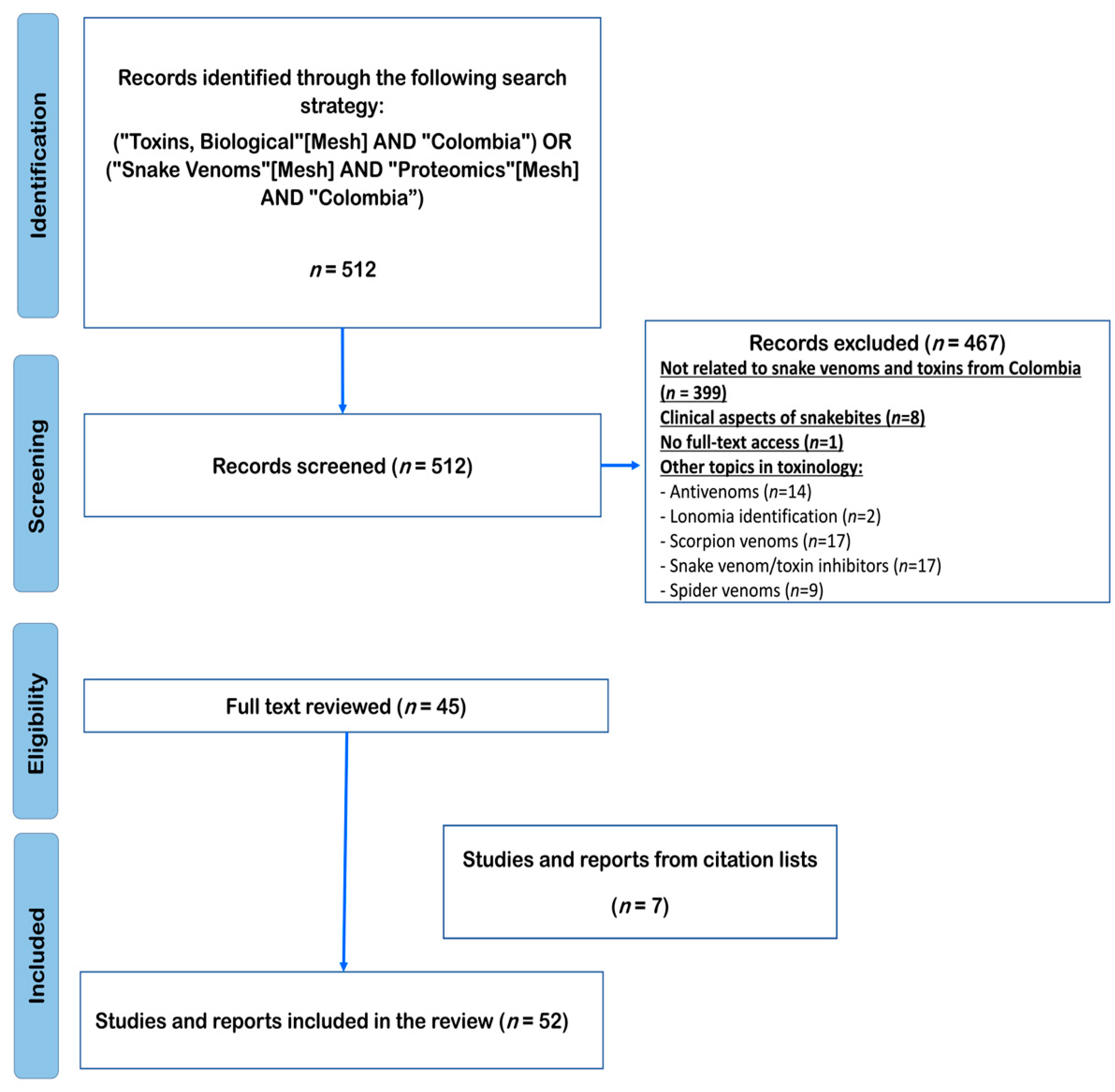
Figure 1. PRISMA flow diagram for the literature search strategy.
Toxins 15 00658 g001
Figure 2. Quantitative venom proteomes from Viperidae family. Venom localities: B. atrox (Meta) [17], B. asper (Cauca) [15,16], B. ayerbei (Cauca) [16], B. rhombeatus (Cauca) [15], B. punctatus (Antioquia) [18], P. lansbergii (Caribbean) [20], B. myersi (Valle del Cauca) [19], C. d. cumanensis (pool from Meta, Tolima, Cundinamarca, and Magdalena), L. acrochorda (pool from Antioquia and Chocó). Abbreviations for protein family names: PLA2s: phospholipase A2; SVMPs: metalloproteinase; LAAO: L-amino acid oxidase; CTL: C-type lectin/lectin-like; CRISP: cysteine-rich secretory protein; Dis: Disintegrins; SVSPs: serine proteinase; Nuc: nucleotidase; PDE: phosphodiesterase; Hya: hyaluronidase; NGF: nerve growth factor; PLB: phospholipase B; PNP: peptides and/or nonproteinaceous components; BPP: bradykinin-potentiating peptide. The unknown fractions were not considered in this graph.
Toxins 15 00658 g002
Figure 3. Quantitative venom proteomes from the Elapidae family. Venom localities: M. mipartitus (Antioquia) [29], M. dumerilii (Antioquia) [30], and M. lemniscatus helleri (Amazonas) [31]. Abbreviations for protein family names: 3FTx: three-finger toxins; PLA2s: phospholipase A2; SVMPs: metalloproteinase; LAAO: L-amino acid oxidase; CTL: C-type lectin/lectin-like; SVSPs: serine proteinase; Nuc: nucleotidase; PDE: phosphodiesterase; Hya: hyaluronidase; Kun: Kunitz-type inhibitors; PLB: phospholipase B; PNP: peptides and/or nonproteinaceous components. The unknown fractions were not considered in this graph.
Toxins 15 00658 g003
Figure 4. Biological activities reported for Colombian snake venoms from the Viperidae family. B. atrox (Meta) [33]; B. asper (Cauca and Antioquia) [16,34]; B. ayerbei (Cauca) [16]; B. rhombeatus (Cauca) [16]; B. punctatus (Antioquia) [18]; P. lansbergii (Atlántico) [20]; B. myersi (Valle del Cauca) [19]; P. nasutum (Antioquia) [34]; B. schlegelii (Antioquia) [34,37]; L. acrochorda (Antioquia) [34]; C. d. cumanensis (Meta) [34].
Toxins 15 00658 g004
Figure 5. Geographical distribution of snake venoms of Viperidae (blue) and Elapidae (yellow) families described in this systematic review.
Toxins 15 00658 g005
Disclaimer/Publisher’s Note: The statements, opinions and data contained in all publications are solely those of the individual author(s) and contributor(s) and not of MDPI and/or the editor(s). MDPI and/or the editor(s) disclaim responsibility for any injury to people or property resulting from any ideas, methods, instructions or products referred to in the content.