Review

Towards Understanding the Function of Aegerolysins

1
Department of Molecular Biology and Nanobiotechnology, National Institute of Chemistry, SI-1000 Ljubljana, Slovenia
2
Department of Biology, Biotechnical Faculty, University of Ljubljana, SI-1000 Ljubljana, Slovenia
*
Author to whom correspondence should be addressed.
Toxins 2022, 14(9), 629; https://doi.org/10.3390/toxins14090629
Submission received: 13 August 2022 / Revised: 4 September 2022 / Accepted: 9 September 2022 / Published: 11 September 2022

Abstract

:
Aegerolysins are remarkable proteins. They are distributed over the tree of life, being relatively widespread in bacteria and fungi, but also present in some insects, plants, protozoa, and viruses. Despite their abundance in cells of certain developmental stages and their presence in secretomes, only a few aegerolysins have been studied in detail. Their function, in particular, is intriguing. Here, we summarize previously published findings on the distribution, molecular interactions, and function of these versatile aegerolysins. They have very diverse protein sequences but a common fold. The machine learning approach of the AlphaFold2 algorithm, which incorporates physical and biological knowledge of protein structures and multisequence alignments, provides us new insights into the aegerolysins and their pore-forming partners, complemented by additional genomic support. We hypothesize that aegerolysins are involved in the mechanisms of competitive exclusion in the niche.
Key Contribution: Aegerolysin proteins are involved in various interactions by recognizing a molecular receptor in the target organism. Formation of pores with various larger non-aegerolysin-like protein partners is one of the possible responses of the aegerolysin-producing organism in competitive exclusion of other organisms from the ecological niche.

Graphical Abstract

1. Introduction

The aegerolysin family (Pfam 06355) is a lesser-known protein family that has received increasing attention in recent years. The aegerolysin family consists of proteins that are biochemically characterized as β-structured proteins and share some common features: similar small molecular weights (15–20 kDa), low isoelectric points, and stability in a wide pH range [1]. Because they are non-core proteins, without a member of this protein family in each of the sequenced fungi, their distribution among fungal species is inconsistent, and different numbers of homologs have been reported for species within the same genus [2,3,4]. They are not only relatively widespread in fungi and bacteria, but also identified in few plants, protozoa, viruses, and insects [1,2].
In recent years, several reviews of this protein family have been published, but none of them included data on the ecology of the organisms producing them. In particular, their function is enigmatic, although some authors suggest a role in the development of the organism [1,2,3]. However, some of them function as two-component cytolysins that exhibit membrane permeabilization activity together with another non-aegerolysin-like protein [2,5]; these act together to perforate natural and artificial lipid membranes [1,2,5]. The aegerolysin-like proteins provide membrane lipid selectivity and recruit partner protein molecules to form a pore complex inserted into the membrane [2,5].
Despite the limited scientific knowledge about the function of aegerolysins, several potential applications are already emerging. Most commonly, some fungal aegerolysins serve as probes for the detection, labeling, and imaging of specific membrane lipids, lipid rafts, cancer cells, invertebrates, or parasites [4,6,7,8,9,10,11]. In high concentrations, they can induce both artificial lipid vesicles as well as live cells, such as blood cells or neuroblastoma cells, to bend and bud [10]. A role of some aegerolysins in combating obesity and related metabolic disorders has been recognized [10]. Their genes and expression may serve as markers for the progression of fruiting body differentiation during mushrooms cultivation [10] or as biomarkers to detect fungal exposure and progression of infectious disease [3,4]. In addition, antibodies produced against aegerolysins can serve as immuno-diagnostic tools [4]. Due to their variable sequence, aegerolysins serve as tools to identify of fungal phytopathogen isolates compared to some closely related species where the internal transcribed spacer barcoding method has failed [4]. Strong promoters regulating aegerolysin genes can promote the secretion of heterologous proteins from fungi in concomitant multi-gene expression [4]. Certain aegerolysins that combine with larger protein partners to form pore-forming complexes can be used to selectively eliminate insect pests [4,10] or to treat certain types of cancer cells [4,10].
Currently, a total of 2303 sequenced genomes of fungal species are deposited in the largest public database of fungal genomes [12]. Aegerolysins are encoded in the genomes of Dikarya fungi but are absent in some clades, e.g., Saccharomycotina [4]. Fungal genomes contain 949 protein sequences with the recognized aegerolysin domain PF06355 [12]. In 31,332 genomes in a bacterial database, there were 169 hits for aegerolysins [13]. Despite their presence in cells at certain developmental stages and their presence in secretomes, few aegerolysins have been studied in detail.
Here we provide a complete overview of the lifestyles of organisms (fungi, bacteria, insects, and viruses) in which aegerolsyins have been identified not only by genome sequencing, but also with at least some previously described functional properties. Their putative structures were predicted using the recently introduced successful prediction tool AlphaFold2 to further improve the knowledge of their function [14]. The presence of putative bicomponent partner proteins or additional aegerolysin gene copies was also investigated.

2. Aegerolysins

We have collected (experimental) published data on 23 different aegerolysins and their variants. In total, they were characterized from 18 different species belonging to different kingdoms of tree of life. Twelve of these aegerolysins belong to fungi, four to bacteria, and one to insects and viruses. In fungi, they were characterized from four mushrooms (Agaricomycotina) from the order Agaricales: Pleurotus ostreatus, P. eryngii, Agrocybe aegerita, and Moniliophthora pernicious, as well as in the ordo Polyporales: Lignosus rhinocerotis (Table 1). The origin of these aegerolysins were also four filamentous Eurotimycetes from the ordo Eurotiales: Aspergillus fumigatus, A. niger, A. terreus, and A. oryzae (Table 1). Two species belonged to the Sordariomycetes, ordo Hypocreales: Beauveria bassiana, and Trichoderma atroviride, and another to the Dothideomycetes, ordo Pleosporales: Alternaria geisen (Table 1). There were four bacterial species, two belonging to Firmicutes: Bacillus thuringiensis and Clostridium bifermentans, and another two to Proteobacteria: Pseudomonas aeruginosa and Alcaligenes faecalis (Table 1). Another species belongs to Insecta—Lepidoptera, Noctuidae: Pseudoplusia includes, and another to Varidnaviria, Ascoviridae: Trichoplusia ni ascovirus 2c (Table 1).

2.1. Fungal Aegerolysins from Basidiomycota—Agaricomycotina

2.1.1. Aegerolysins from Pleurotus ostreatus

The oyster mushroom or hiratake, Pleurotus ostreatus (Jacq. ex Fr.) P. Kumm. (1871) (Agaricales), is widely distributed in many temperate and subtropical forests. As a saprotroph, a white-rot wood fungus, it is a primary decomposer of wood, especially deciduous trees such as beech. It is also known to paralyze nematodes and consume prey [15]. As a common edible mushroom, it is grown commercially for food throughout the world.
Pleurotolysin A (PlyA, PriA), ostreolysin (OlyA), ostreolysin A6 (OlyA6), recombinant ostreolysin (rOlyA), are aegerolysins from the mushroom P. ostreatus. According to genome sequences of P. ostreatus strains PC9 and PC15, they are encoded by the same locus [52,53,54,55,56].
In 1979, it was discovered that P. ostreatus encodes the hemolytic protein pleurotolysin with a size of 15 kDa, and these first experiments showed the importance of sphingomyelin (SM) for Ply binding [57]. A clone containing aegerolysin gene was the most abundant expressed sequence tag (EST) in the primordial and the fruit body library [58]. Then, 25 years later, in 2004, it was revealed that this Ply protein is actually composed of subunits A and B [59]. Component A (PlyA), a 15 kDa aegerolysin, binds to SM/cholesterol (Chol)-rich membranes and then recruits a 59 kDa membrane attack complex/perforin (MACPF)-domain containing protein partner, PlyB, which then leads to transmembrane pore formation and cell lysis [59]. The combination of X-ray diffraction and cryo-electron microscopy has shown that PlyA paired with PlyB forms a 39-heteromeric membrane-embedded pore with a 13-fold symmetry structure (PDB ID: 4V2T) (Figure 1) [60]. PlyA has a β-sandwich structure similar to the actinoporin family (PF06369) of pore-forming proteins [60]. For adequate binding of PlyA, the Chol content should be more than 30 mol% [59]. PlyA/PlyB nanopores have been recently proposed as sequencing tools [61].
In 2002, another group of researchers isolated a very similar 15 kDa protein from the same mushroom and named it ostreolysin (Oly) [63]. The primary structure of Oly is 79% (29 amino acids substitution) identical to PlyA [11]. The first experiments with the native isolate of Oly showed that it is cytolytic for erythrocytes and various cell lines [64,65]. Since the expression of Oly occurs during the formation of primordia and fruiting bodies of the mushroom, it has been speculated that it plays a role in the fruiting bodies of the mushroom [63,66,67]. Biochemical experiments in 2004 showed that Oly also specifically recognizes membranes enriched in SM and Chol [68,69], and that binding of a protein is abolished after pretreatment of cells with methyl-β-cyclodextrin or lysophospholipids [70,71]. However, as in the case of Ply, it was later revised that native isolate of Oly (OlyA) was contaminated with PlyB, which was responsible for the observed cytolytic effects [72].
Ostreolysin A6 (OlyA6), which has 78% identity (31 amino acids substitution) with OlyA [11], was expressed as recombinant protein in 2013 [72]. First experiments showed that OlyA6 specifically bind to SM/Chol-rich lipid membranes and has very similar binding properties to PlyA [72]. Recently, OlyA6 was confirmed to bind to SM/Chol lipid complexes and not to free SM (PDB ID: 6MYJ) [73]. OlyA6 was later fused to the fluorescent protein mCherry, and was used as a lipid raft marker [74]. OlyA6-mCherry was shown to be non-toxic to vertebrate cells and, due to its stability and small size, it became an important tool for studying lipid rafts [74]. OlyA6-mCherry stained lipid rafts in plasma membranes in live and fixed Madin-Darby canine kidney (MDCK) cells did not colocalize with other raft markers, that sense individual raft-residing lipids, and did not bind to intracellular membranes, and its binding was abolished by methyl-β-cyclodextrin or sphingomyelinase pretreatment [74]. However, it has been observed that excessive concentrations of OlyA6 or Oly6-mCherry can lead to membrane vesiculation [72,74,75]. In addition, OlyA6-mCherry applied to live MDCK cells was internalized via caveolin-dependent pathway and was detected near the Golgi complex after 90 min [74]. Similar to the combination of PlyA and PlyB or OlyA and PlyB, the mixtures of OlyA6 and PlyB form transmembrane pores, leading to cell lysis [72]. The combination of lipid raft sensing OlyA6 and PlyB can be used as anticancer treatment for cancers with increased concentration of lipid rafts in their membranes as is the case in urothelial cancer cells [76]. Recently, it was revealed that OlyA6 has another high affinity lipid receptor, ceramide phosphoethanolamine (CPE), which is specific for cell membranes of invertebrates and some bacteria [77,78,79]. OlyA6 binds 1000-fold more strongly to vesicles composed of CPE and Chol than to vesicles composed of SM/Chol [80], and it also binds to membranes with physiological concentrations (5 mol%) of CPE [78,79]. In addition to labelling lipid rafts, OlyA6 can also be used as a marker to label CPE in insect cells and tissues [79,80,81]. Furthermore, OlyA6/PlyB protein mixtures permeabilize lipid vesicles containing ≤5 mol% CPE and are toxic to CPE-containing Sf9 cells, as well as to the larvae of Colorado potato beetle (Leptinotarsa decemlineata Say, 1824) and western corn rootworm (Diabrotica virgifera virgifera LeConte, 1868), and thus have been proposed as new environmentally friendly biopesticides [10,79] with low toxicity for non-target organisms [82] and no toxicity for humans after ingestion due to their fast decomposition by mammalian digestive enzymes [83]. It must be noted, however, that the intravenous application of the native OlyA6/PlyB isolate in rodents resulted in a cardiorespiratory arrest and can induce severe histopathological changes in kidneys, lungs, and heart [84,85]. The half lethal dose (LD50) in mice is 1.17 mg/kg [86]. Very recently, another lipid receptor of OlyA6, ceramide aminoethylphosphonate (CAEP), a CPE analogue of marine invertebrates such as mollusks and cnidarians, was identified [87]. Moreover, OlyA6/PlyB complexes triggered strong lysis of CAEP–lipid containing vesicles.
In 2017, another aegerolysin from P. ostreatus was produced in recombinant form and named recombinant ostreolysin (rOlyA) [88], which has an amino acid substitution at position 50 (valine to isoleucine) compared to OlyA6. rOlyA has not been used as a lipid raft marker, but it has been shown to have anti-proliferative and pro-apoptotic effects against colon cancer cells or can be used the treatment of metabolic disorders, which was attributed to its lipid raft binding properties [88]. rOlyA induced apoptosis of human and mouse cancer cells in vitro but was not toxic to normal intestinal cells [88]. After endocytosis, rOlyA colocalized with β-III tubulin, which probably impaired microtubule dynamics and led to cell death. These results were confirmed by in vivo mouse models in which rOlyA reduced tumor cell growth. Further, through its lipid raft binding and interfering with signaling cascades, rOlyA can induce browning of white preadipocytes used for energy storage in vitro, resulting in a brown-like phenotype with increased mitochondrial biogenesis. rOlyA has therefore been proposed as a candidate for regulating metabolic disorders such as obesity, hyperlipidemia, and non-alcoholic fatty liver disease [89,90].
In the genomes of P. ostreatus PC9 or PC15, a bi-directional pair of genes with 5′–5′ orientation of the plyA (priA) and plyB genes was observed [91,92]; no difference among the two strains was observed in the sequences of PlyA.

2.1.2. Aegerolysins from Pleurotus eryngii

Boletus of the steppes Pleurotus eryngii (DC.) Quél. 1872 (Agaricales), commonly called king oyster or trumpet or brown mushroom, French horn mushroom, or Aliʻi oyster, is an edible mushroom. It is of commercial interest because of the mild taste and pleasant odor of its fruiting bodies. In addition, its mycelium is often grown on lignocellulosic wastes (such as wheat straw). Unlike P. ostreatus, this fungus lives saprobically or facultatively biotrophic on roots of herbaceous plants of the Apiaceae family as a grassland-litter decomposer. Pleurotus eryngii sensu stricto consists of at least five varieties such as var. eryngii, ferulae, elaeoselini, thapsiae, and tingitanus, which differ significantly in their habitat distribution, host, and morphology [16]. It possesses nematocidal properties as well as P. ostreatus [15].
Pleurotolysin A2 (PlyA2) and erylysin (EryA) are two aegerolysins from P. eryngii. In 2006, a 17-kDa hemolysin was isolated from fruiting bodies of the mushroom P. eryngii and was originally named eryngeolysin [93]. Its N-terminal sequence showed high homology to OlyA from P. ostreatus and aegerolysin from Agrocybe aegerita. Its hemolytic activity remained unchanged over the pH range of 4 to 12. It also showed antibacterial activity against Bacillus megaterium and B. subtilis, but not against some other bacteria such as Staphylococcus aureus, Escherichia coli, Enterobacter aerogenes, Pseudomonas aeruginosa, P. fluorescens, Mycobacterium phlei, and Proteus vulgaris, or fungal species Botrytis cinerea, Fusarium oxysporum, Mycosphaerella arachidicola, Physalospora piricola, and Rhizoctonia solani [93]. The sensitivity of mammalian erythrocytes to eryngeolysin was the highest, followed by birds, reptiles, and fish erythrocytes [93]. It also showed cytotoxicity against leukemia cells (L1210), and inhibited mitogenic responses of mouse splenocytes. N-glycolyneuraminic acid was the only sugar that could inhibit hemolytic activity [93].
In 2013, the sedimentation assay of the crude extract of P. eryngii revealed that pleurotolysin A2 (PlyA2) is also a SM/Chol-binding protein [94]. PlyA2 has two amino acid substitutions compared to PlyA [95], while it has 93% sequence identity (nine substitutions) with OlyA6 [94]. Since the recombinant protein showed no hemolytic activity or toxicity to Hela cells it was then prepared as recombinant fluorescently labeled protein, PlyA2-EGFP, which was used as a lipid raft marker. PlyA2 successfully labeled Hela cells, and binding of the protein was abolished after the pretreatment of the cells with methyl-β-cyclodextrin or sphingomyelinase. PlyA2-EGEP showed substantial colocalization with the raft-associated protein CD59. After permeabilization of the cells, PlyA2 was found to bind to late endosomes and, similar to OlyA6, not to cytoplasm-facing membranes. As with OlyA6, PlyA2 was later shown to bind more strongly to CPE/Chol-containing membranes [80]. Interestingly, PlyA2 was also able to recognize and bind CPE membranes in the absence of Chol, as the only of the tested Pleurotus aegerolysins [80]. PlyA2-EGFP successfully labeled CPE in Drosophila embryonic Kc167 cells and in larvae of the same insects [80]. It is suggested that PlyA2/PlyB complexes can be used as new bioinsecticides, as has been discussed for OlyA6/PlyB complexes [79].
A novel bicomponent toxin pair was isolated and characterized from P. eryngii, EryA and EryB in 2010 [96]. Most Pleurotus aegerolysins (OlyA, OlyA6, PlyA, and PlyA2) bind to SM/Chol-rich lipid membranes [5,59,65,74,94,97], but not EryA, ortholog of PlyA2 from P. eriyngii [80]. For this reason EryA-mCherry was very recently used as a molecular tool for detection of periodontal disease which is in majority of the cases associated with CPE-producing periodontal bacteria [98]. EryA-EGFP was also successfully used to label the bloodstream form of the protozoan Trypanosoma cruzi, a causative of sleeping disease, that has CPE in its membranes [80]. Recently, EryA-EGFP was reported to bind artificial lipid membranes composed of cholesterol, fully saturated phosphatidylcholine and cardiolipin (CL) in a 5:4:1 molar ratio [99]. Furthermore, intracellularly expressed EryA-EGFP localized to regions of negative curvature (inner leaflets at cell poles and the outer leaflets at division sites) in E. coli, and inhibited cytokinesis [99]. This localization was assigned to EryA interaction with CL, a membrane lipid that localizes in curved membranes such as mitochondrial cristae, cell poles and division sites of rod-shaped bacteria.
The genome of P. eryngii ATCC 90797, similar to that of P. ostreatus, encodes a MACPF-containing protein, erylysin B (EryB) [96], which is 97% identical to PlyB [10]; it is a neighboring gene of PlyA2, whereas EryA has no MACPF-like protein neighboring gene. However, it is surprising that some aegerolysins perform better in terms of their cytolytic activity when combined with their non-native partner. This has been studied for PlyA2 and EryA; PlyA2 show better hemolytic/lipid vesicle permeable activity when combined with the MACP partner PlyB from P. ostreatus, rather than with their native protein partner, EryB [78].

2.1.3. Aegerolysins from Agrocybe aegerita

The poplar mushroom Cyclocybe aegerita Vizzini 2014 (Agrocybe aegerita, A. cylindracea, or Pholiota aegerit) (Agaricales), also known as tea tree mushroom, Cha shu gu, Yanagi-matsutake, velvet pioppini, or sword-belt mushroom, is a high-value commercially grown edible mushroom. It is a saprotrophic, wood-inhabiting fungus that decomposes mainly dead wood of deciduous trees, especially poplar and willow, and causes weak white rot on deciduous trees in temperate forests; it may become pathogenic on some other tree species [17].
The aegerolysin protein family is named after aegerolysin from the mushroom A. aegerita) [63]. Northern hybridizations with total RNA extracted from four developmental stages of A. aegerita SM51 (A1B1/A2B2) showed that the Aa-pri1 gene is specifically expressed during primordia or the immature fruiting body stage and that it is not expressed (at list not to a detectable level) during vegetative growth or late stages of fruiting body maturation [100].
Aa-pri1 is one of six genes for putative aegerolysins encoded in the A. aegerita AAE3 genome. Only one out of these genes has a neighboring gene for component B, which is homologous to the MACPF domain-containing protein PlyB.

2.1.4. Aegerolysins from Moniliophthora perniciosa

The species complex Moniliophthora perniciosa (Stahel) Aime and Phillip-Mora (2005) (Crinipellis perniciosa) (Agaricales) consists of a number of geographically separated populations that infect a broad range of different hosts: biotype C on Theobroma and Herrania spp. (Malvaceae), H on Heteropterys acutifola (Malpighiaceae), L Arrabidaea spp. (Bignoniaceae), and S on Solanum spp. (Solanaceae) [18]. It is most known as a hemibiotrophic fungus that causes witches’ broom disease in cacao plant (Theobroma cacao L.) with two distinct growth phases [18].
The ability to culture a biotrophic-like phase of M. perniciosa in vitro, as well as the recent findings from the nearly complete genome and expression studies, clearly indicate that these different growth phases of the fungus function under different metabolic parameters [18]. The gene expression profile determined by microarray suggests physiological changes in the mycelia prior to basidiomata formation, and confirmed by RT-qPCR at different stages during cultivation in basidiomata-inducing medium [101]. Of the three putative genes involved in fructification, two are related to the identified PriA from P. ostreatus and AA-Pri1 from A. aerogerita, respectively, and one is related to PlyB from P. ostreatus. The expression of aegerolysin MpPRIA1 was low in yellow and reddish-pink mycelial stages and before stress, but increased about fourfold in mycelia with primordia and 90-fold in basidiomata compared to the initial white mycelial stage [101]. Expression of the aegerolysin MpPRIA2 increased 17-fold at the reddish-pink mycelium stage, but decreased 11-fold before stress, 4-fold in stressed mycelia, and 47-fold in mycelia with primordia. Transcripts of the gene MpPRIA2 increased 23-fold in basidiomata, which was lower in mycelia with primordia [101]. Transcripts of the gene MpPLYB increased 1.4-fold in the yellow mycelium stage, 15-fold in reddish-pink mycelia, and remained at high levels in mycelia before stress, under stress, and in mycelia with primordia (about 10–12-fold increase), but decreased in basidiomata, where they were only 1.6-fold higher than in white mycelia [101]. MpPRIA1 and MpPRIA2 have homologous regions but appear to correspond to two individual genes whose expression is consistent with the morphological differentiation of primary hyphal nodules from primordia. These aegerolysins may contribute to the process of hyphal aggregation, as their expression, albeit at low levels, occurred prior to the appearance of primordia, when hyphae became spherical for primordia formation [102]. This stage coincides with the stage of reddish-pink mycelium, where hyphal nodules are detectable. The exact function of these proteins remains unclear, but their involvement in programmed cell death seems rather unlikely, since aegerolysins from Pleurotus have a lytic function and act in SM/Chol membranes at pH between 7 and 8, which is not normally the case in fungal cells [65,103,104]. However, they can also act lytically on membranes composed of SM and ergosterol, although the binding and permeabilization is twice less efficient as for the SM/Chol membranes [69].
Six aegerolysins are encoded in the M. perniciosa genome according to Pf06355 search. The contigs are too short to detect whether the only homolog of PlyB—MpPLYB gene is a neighboring gene of MpPRIA1 and MpPRIA2 genes.

2.1.5. Aegerolysins from Lignosus rhinocerotis

The tiger milk mushroom Lignosus rhinocerotis (Cooke) Ryvarden (1972) (Polyporales) is a white rot fungus that develops from the underground tuberous sclerotium. In recent years, this fungus has received much attention because of its extensive wide-range ethnobotanical use as a folk medicine and because of the successful domestication of this once very rare and expensive fungus [19].
Systematic profiling of L. rhinocerotis sclerotial proteins was performed using 2DE coupled with MALDI-MS and LC-MS. After genome-based proteomic analysis, the aegerolysin domain-containing protein GME7309 was identified in one of the 45 spots [105].
The genome of L. rhinocerotis genome has been published but is not publicly available for browsing [106], so no data on bicomponent partner genes are available.

2.2. Fungal Aegerolysins from Ascomycota—Eurotimycetes

The fungal genus Aspergillus is of great industrial importance to humans, primarily as a unique cell factory with an exceptionally high secretion capacity for enzymes, but also as a source of important pathogens for humans, animals, and crops, potent carcinogenic food contaminants, and an important genetic model [20]. The genus Aspergillus is characterized by high adaptability to different ecological environments. However, the biodiversity of Aspergillus in different ecological habitats should always be evaluated in the context of the continuous development of the taxonomy of the genus; the description of new species may reveal a greater biodiversity of the genus and refine the results of previous studies [21].

2.2.1. Aegerolysins from Aspergillus fumigatus

Neosartorya fumigata O’Gorman, Fuller, and Dyer, 2008 (Aspergillus fumigatus Fresenius 1863) (Eurotiales), is a ubiquitous saprotrophic fungus that plays an important role in recycling carbon and nitrogen on Earth, but can also be a deadly opportunistic primary and opportunistic pathogen [22]. It is a trimorphic fungus with vegetative mycelium that contributes to the decomposition of soil organic matter, asexual conidia responsible for the aerial dispersal of the species, and dormant ascospores that ensure the long-term survival of the organism. Its natural ecological niche is the soil, where it survives and grows on organic debris. Although this fungal species is not the most abundant in the world, it is one of the most widespread fungal species with airborne conidia. Similar variations were found outdoors and in hospital, with consistently lower numbers indoor. Plant debris in the form of compost piles and stacks of high-moisture hay and straw bales that are self-heating, produce large numbers of spores that can be released into the air, resulting in high but localized numbers when disturbed. The widespread dispersal of decaying leaves after leaf fall is a potential source of smaller spore concentrations, but these are dispersed over a much larger area [23].
In 1962, the first aegerolysin protein, Asp-hemolysin (Asp-HS) was described [107], and in 1975 it was isolated from the filamentous fungus A. fumigatus [108]. It was not until 1982 that Asp-HS was reported to be hemolytic and involved in the pathogenesis of A. fumigatus [109]. The nucleotide sequence of a cDNA encoding hlyA was elucidated in 1994 [110]. Asp-HS has been shown to bind specifically to oxidized low-density lipoproteins [111], to have cytotoxic effects on murine macrophages and vascular endothelial cells and to induce cytokine genes [112,113]. However, wild-type and knock-out hlyA and hlyA-like mutant strains of A. fumigatus have shown that Asp-HS production is not important for the progression of invasive alveolar aspergillosis in mice [114]. Instead, Asp-HS is thought to play a role in potentiating other virulence mechanisms of the fungus [115].
Of the two aegerolysins described, asp-HS also has a neighboring partner protein gene asp-HSB, and asp-HS-like does not in strain A. fumigatus Af293 [92]. asp-HS-like sequence is also identified as invariant in five other strains, GCA_002234955.1, GCA_002234985.1, A1163, Z5, and var. RP-2014, while for asp-HS, variation in one amino acid was observed, and it was absent in var. RP-2014.

2.2.2. Aegerolysins from Aspergillus niger

Aspergillus niger van Tieghem 1867 (Eurotiales) is a filamentous fungus that grows aerobically on organic matter. It is ubiquitous in soil and is commonly reported from indoor environments. A. niger is capable of growing in a wide temperature range of 6–47 °C, with a relatively high temperature optimum at 35–37 °C, and it is capable of growing in an extremely wide pH range: 1.4–9.8 [24]. These capabilities and the abundant production of airborne conidiospores result in a ubiquitous occurrence of the species, with a higher incidence in warm and humid locations. Although the main source of Black aspergilli is soil and litter, in compost they are among the most common fungi causing food spoilage and biological decay of other materials. Aspergillus niger species complex is often responsible for the postharvest spoilage of fresh fruits (apples, pears, peaches, citrus fruits, grapes, figs, strawberries, tomatoes, melons, etc.) and some vegetables (especially onions, garlic, and sweet potatoes); they are also among the most common fungi isolated from dried fruits, beans, oilseeds, and nuts (peanuts, pecans, pistachios, hazelnuts, almonds, walnuts, etc.) [25,26]. Because A. niger has been in use already for many decades to produce extracellular (food) enzymes and citric acid, many of these product are considered generally recognized as safe (GRAS) by the United States Food and Drug Administration [24,27].
Functional studies of the expression of the genes encoding the aegerolysins NigA1 and NigA2 and the MACPF-like proteins NigB1 and NigB2 suggest that the sporulation process is critical for the strong induction of the expression of all these genes. However, in laboratory environment, deletion of any of the aegerolysin genes had no effect on the growth, development, sporulation efficiency, and phenotype of the mutants, suggesting that aegerolysins are not key factors in the sporulation process [91]. In all expression studies, a strong correlation was observed between the expression of NigA2 and NigB1 genes [91]. In Aspergillus species, aegerolysins were frequently found as secreted proteins. Their secretion was studied by secretion prediction and Western blotting, and it was confirmed that NigA1 and NigA2 are secreted by the fungus [91,92]. Both nigerolysins A are leaderless proteins that reach the cell exterior by unconventional protein secretion [91,92]. The subcellular localization of nigerolysins A in A. niger was investigated by immunocytochemistry and live cell imaging. The NigA proteins are uniformly distributed in the cytoplasm of fungal hyphae [92]. NigA2 was shown to interact specifically with an insect-specific membrane sphingolipid, CPE in combination with Chol, but not with SM/Chol [91]. Membranes of insect cells containing this specific sphingolipid were stained after binding the NigA2 protein labeled with mCherry [91]. Detailed bioinformatics analysis of Aspergillus aegerolysins suggests that the same function (target) likely occurs in only a limited number of aegerolysins. From the alignment, chromosomal loci analysis, orthology, synteny, and phylogeny, it appears that the same or similar function described for pairs of pesticidal proteins of Pleurotus sp. also occurs in species of the subgenus Circumdati, section Nigri, series Nigri, and several other species with adjacent pairs of putative pesticidal proteins [92]. The combined results suggest that aegerolysins in A. niger, and probably in other aspergilli, may be involved in defense against predators.
Of the two aegerolysins described in A. niger strain CBS 513.88, NigA2 also has a neighboring partner protein gene NigB1, whereas NigA1 does not [92]. No variations in aegerolysin sequences for NigA1 are observed in any of the nine other A. niger strains examined: (lacticoffeatus) CBS 101883, (phoenicis Corda) Thom ATCC 13157, ATCC 1015, ATCC van Tieghem 13496, NRRL3, ASM285, ATCC_64974_N402, ASM221148, except for one amino acid variance in ASM151534. The same is also observed for NigA2, with the exception of a four amino acid variance in ASM151534.

2.2.3. Aegerolysins from Aspergillus terreus

Aspergillus terreus Thom (1918) (Eurotiales) is a filamentous fungus found in soil worldwide. Although it was thought to be purely asexual, it is now known to be capable of sexual reproduction. This saprotrophic fungus is common in warmer climates such as the tropics and subtropics. It is found not only in soil, but also in habitats, such as decomposing vegetation and dust [21]. A. terreus is used in industry to produce important organic acids, such as itaconic acid, and enzymes. It was also the original source of the drug mevinolin (lovastatin), which is used to lower serum cholesterol. It can cause opportunistic infections in people with weakened immune systems [28].
Terrelysin (Ter) is an aegerolysin from A. terreus. The genome sequence was used to identify the Ter sequence based on homology with other known aegerolysins [28]. The recombinant protein rTer was purified and its mass of about 16 kDa was determined by MALDI-TOF MS [28]. Circular dichroism analysis shows that the secondary structure of the protein is predominantly β-sheet. Thermal denaturation results of rTer show that the protein retains the β-sheet confirmation up to 65 °C. Clinical identification of A. terreus in aspergillosis is limited to broad identification of lesions in affected organs by biopsy, radiological methods, or detection of fungal carbohydrates, which often does not allow species-specific identification. Due to the urgent need for diagnostic methods that allow early and specific identification of the pathogen, monoclonal antibodies to cytolytic hyphal exoantigens were generated [116]. Additionally, specific monoclonal antibodies against rTer were developed and used to quantify the native protein in hyphal and secretion fractions grown in liquid culture at various time points [28]. Time-course studies showed that Ter expression was highest during early hyphal growth and decreased dramatically after mycelial expansion [117]. Immunolocalization studies showed that Ter was not only localized in the cytoplasm of the hyphae, but was also present in greater amounts at the tip of the hyphae [117]. By ELISA, it was found that the highest concentrations of Ter were found in the culture supernatant during the early stages of hyphal growth, compared to later time points when hyphal growth and apical elongation were reduced. Because Ter does not possess a signal peptide, it was suggested that it might diffuse or be actively secreted during initial hyphal growth (i.e., apical elongation) by other, yet uncharacterized, processes [117]. These results were confirmed in cultures grown at both room temperature and 37 °C. These observations suggest that Ter may be a potential biomarker for A. terreus infections [117].
There is no MACPF-like partner protein gene encoded alongside the ter gene [92]. There are some differences in sequences around the putative intron splice site and at the C-terminus between rTer from ATCC 1012 and the genome sequences of NIH2624 (ASM14961) and IFO6365.

2.2.4. Aegerolysins from Aspergillus oryzae

Aspergillus oryzae (Ahlburg) E. Cohn (Eurotiales) is a fungus widely used in the traditional Japanese fermentation industry, including the production of soy sauce, sake, bean curd, and vinegar. Among filamentous fungi, A. oryzae is known to have outstanding potential for the production of various enzymes. In addition, developments in genetic engineering have led to the use of A. oryzae in modern biotechnology for the production of industrial enzymes [29].
Significant glucose repression was observed in solid-state cultures based on steamed rice and wheat. A promoter of a hemolysin-like gene (hlyA) from A. oryzae encoding an aegerolysin-like protein AoHlyA that is not repressed by glucose has been reported [118]. The hlyA promoter is activated by conditions or factors not restricted to sporulation. Four putative CCAAT sequences, six putative heat-shock transcription factor binding sequences (AGAAN), and two putative BrlA binding sequences (MRAGGGR) have been identified to affect transcription [118]. The CCAAT sequence is one of the most abundant cis-elements in the promoter regions of numerous eukaryotic genes. The heat shock transcription factor positively regulates the family of stress response genes, and the brlA gene is a regulator of conidiation in A. oryzae. Efficient expression of homologous and heterologous genes was shown under the control of the hlyA promoter stronger than under the commonly used amyA promoter [118,119,120].
Four genome sequences of A. oryzae are accessible in public databases: RIB40, 3.042 BCC7051, and 100-8. In total, three aegerolysins [92] and six putative MACPF-like proteins are encoded in the genome of A. oryzae strain RIB40. An identical protein sequence to AoHlyA is encoded in only one in another strain 3.042, which has two aegerolysins. Surprisingly, only one aegerolysin is encoded in strain 100-8, but it is present in all four strains with only one amino acid variation. Three additional sequences are found in BCC7051, one correspondingly short and two larger, one with a longer C-terminus and the other with a long N-teminus.

2.3. Fungal Aegerolysins from Ascomycota—Sordariomycetes

2.3.1. Aegerolysins from Beauveria bassiana

The fungus Beauveria bassiana (Bals.-Criv.) Vuill., 1912 (Hypocreales) is considered both an entomopathogen and an endophyte. Entomopathogenic fungi can also perform other functions in nature, including endophytism, antagonism to plant diseases, promotion of plant growth, and root colonization [30,31]. The fungus B. bassiana is already known as a biopesticide for biological control of insect pests and as an asymptomatic companion of plants.
Structural modeling of beauveriolysin A (BlyA) suggests that it has a similar three-dimensional fold to other aegerolysins from fungi or bacteria [52]. BlyA showed the ability to bind specifically to lipid vesicles containing Chol in combination with both mammalian and invertebrate major membrane sphingolipids, SM, or CPE, respectively [52]. However, BlyA itself is neither hemolytic to bovine erythrocytes nor toxic to Sf9 insect cells [52]. Given its similarity to other aegerolysin pore-forming partnering proteins, BlyB was considered a candidate, but the possible formation of pores together with BlyA remains to be confirmed [52]. Similar protein pairs may be encoded by several entomopathogenic fungi from the class Sordariomycetes [52]. BlyB has been shown to best align the structures of some bacterial and mammalian MACPF proteins, rather than PlyB [52]. Two structures have been solved from Proteobacteria, for the Gram-negative insecticidal protein GNIP1Aa (PDB ID: 6FBM) from Chromobacterium piscinae, which has been shown to be specifically toxic to western corn rootworm larvae during feeding [121], and for Plu-MACPF (PDB ID: 2QP2) from a lethal pathogen of insects Photorhabdus luminescens [122]; no data are available on their partner proteins. The macrophage-expressed gene 1 protein (MPEG-1) or perforin-2 (PDB ID: 6U23 and analogs), which plays a role in the human immune system, was recently shown to function alone [123,124,125]. The murine complement component C9 (PDB ID: 6CXO) and the human complement component C8α chain are also involved in the immune system (PDB ID: 2RD7); in contrast to MPEG-1, proteins C8α and C9 are involved in the formation of a heterooligomeric protein-membrane attack complex (MAC) [123,124,126,127]. In conclusion, several proteins that served as templates for construction of a protein model of BlyA, several possible binary toxin partner proteins, and also organisms from which these proteins are derived, also have insecticidal (or immunological) properties [52]. BlyA could belong to a set of insecticidal proteins and metabolites that enable entomopathogenic fungi to have their specific lifestyle [52].
The sequences of BlyA are identical in all strains ARSEF 2860, D15, GCA_001682635.1, and JEF-007.

2.3.2. Aegerolysins from Trichoderma atroviride

Although some Trichoderma species are known to be closely associated with higher plants (endophytes and plant growth promoters), Basidiomycetes (mushroom green mold disease), invertebrates (nematodes, marine sponges), and mammals (opportunistic pathogens of humans), most taxa have been recovered from dead wood and fruiting bodies of other fungi, suggesting that these are the original ecological niches of the fungus [32]. Hypocrea atroviridis Dodd et al. (Trichoderma atroviride) (Hypocreales) is a filamentous, cosmopolitan fungus commonly found in soil and has been isolated in both tropical and temperate climates. This mycoparasitic fungus is best known for its biocontrol capabilities in agriculture, interacting with phytopathogenic fungi and oomycetes, such as Rhizoctonia solani and Botrytis cinerea, which are pests of hundreds of crops [33,34,35].
The recently functionally characterized Agl1 aegerolysin in this fungus shows a role in conidiation and antagonism. Gene expression analysis showed higher expression of agl1 during conidiation and during growth in medium enriched with cell wall material from the plant pathogenic fungus R. solani as the sole carbon source. Expression of agl1 was suppressed under iron-limiting conditions, whereas the agl1 transcript was not detected during the interaction of T. atroviride with the prey fungi B. cinerea or R. solani. Phenotypic analysis of agl1 deletion strain showed reduced conidiation compared with T. atroviride wild type, suggesting involvement of Agl1 in conidiation. In addition, the agl1 deletion strain showed reduced antagonism to B. cinerea and R. solani based on a secretion assay, although no difference was observed in direct interactions [128].
There is no bicomponent partner protein gene encoded alongside the agl1 gene; there is only one amino acid variation between strains of T. atroviride IMI 206040, B10, F7, and P1 (ATCC 74058).

2.4. Fungal Aegerolysins from Ascomycota—Dothideomycetes

Aegerolysins from Alternaria gaisen

The cosmopolitan genus Alternaria (Pleosporales) contains many species of economic importance, including saprophytes, phytopathogens, and zoopathogens known to produce a variety of mycotoxins and allergens [36]. Alternaria gaisen Nagano ex Hara, 1920 (black spot of Japanese pear), occurs on the Japanese pear, Pyrus pyrifolia, especially the cultivar Nijisseiki, but also other cultivars of both Japanese and Chinese pears [37].
The accuracy and effectiveness of crop protection measures depends on correctly identifying of the pest of concern from the outset. However, the relatively invariant internal transcribed spacer sequence of structural ribosomal RNA that have been described and selected for fungal barcoding, for Alternaria species and strains did not allow discrimination of isolates of different A. alternata, A. arborescens, A. brassicicola, A. gaisen, A. mali, A. tenuissima, and A. yaliinficiens. When the partial genomic sequences of the putative aegerolysin L152 were analyzed, the lineages were resolved, and they reflected the diversity previously suggested by the morphological evaluation of the sporulation patterns [36]. The presence of putative aegerolysin L152 homologs in both phytopathogenic and non-phytopathogenic species argues against their involvement in pathogenesis. In addition, the putative aegerolysin gene is not located on any of the conditionally disposable chromosomes, so it is likely to be present in all small-spored Alternaria spp. and not only in the toxin producers [36]. Expression of L152 in A. gaisen was strongly upregulated after light exposure, a stimulus that also triggers conidiophores to develop [36].
The only aegerolysin of A. geisen strain BMP2338, L152, also has a neighboring partner protein gene: L152B.

2.5. Bacterial Aegerolysins from Firmicutes

2.5.1. Aegerolysins from Bacillus thuringiensis

Bacillus thuringiensis Berliner 1915 is a rod-shaped, Gram-positive, spore-forming, aerobic bacterium usually found in soils, grain dusts, dead insects, and water around the world. It has adapted its lifestyle as an efficient pathogen, synthesizing a large number of protein toxins that are effective against a variety of organisms in nature, including not only a broad range of insect orders but also nematodes, human pathogenic protozoans, animal, and human parasites. A molluscicidal activity against the snail pest has also been demonstrated [38,39].
Bacillus thuringiensis owes its insecticidal potential to several factors, and the main factor is the parasporal crystal containing a series of entomotoxins. The main B. thuringiensis entomotoxins, Cry (crystals), and/or Cyt (cytolytic) are pore-forming proteins, which are dissolved in the gastrointestinal tract of an insect after ingestion. A variety of different insecticidal proteins produced by Bacillus species is very rich. Cry toxins exert their membrane-permeabilizing activity on insect epithelial midgut cells after binding to a variety of protein receptors [129]. Among more than 300 members of the Cry toxins, a 13.6 kDa Cry34Ab1 from B. thuringiensis (Bt strain PS149B1), is the only with the aegerolysin fold. It is composed of two β-sheets in a β-sandwich structure [130]. Similarly to aegerolysin/MACPF protein pairs described in oyster mushrooms, Cry34Ab1 exerts its membrane activity and insecticidal action towards western corn rootworm in the presence of a 43.8 kDa partnering protein, Cry35Ab1. The latter protein has a toxin 10 (PF05431) C-terminal domain. Cry34Ab1/Cry35Ab1 binary toxins have been transformed into maize plants under the commercialized events. After ingestion, they bind to the brush border membranes in the epithelial lining of western corn rootworm midgut, where they form pores. This leads to necrosis and ends with the insect’s death. The Cry35Ab1 protein does not convey activity in the absence of Cry34Ab1, indicating that the smaller Cry34Ab1 protein is critical for membrane binding and recruitment of Cry35Ab1 to induce insecticidal effect [39,131,132,133,134,135]. Although until now the exact nature of Cry34Ab1 protein receptor(s) in insect larva midgut membranes has not been determined [132,135], it appears that these receptors are unique and unrelated to other Cry toxins [136]. Further, although the Cry34Ab1/Cry35Ab1 mixture was demonstrated to be slightly lytic to artificial lipid membranes in absence of proteins [133]; the lipid specificity seems to be low and the SM/Chol or CPE/Chol complexes cannot be regarded as receptor molecules, as in the case of oyster mushroom-derived aegerolysin/MACPF cytolytic insecticidal complexes [79]. Cristal structure of Cry34Ab1/Cry35Ab1 binary toxin has been solved (PDB ID: 4JOX and 4JP0, respectively) [130].
Another characteristic feature of B. thuringiensis strains is the size of their extrachromosomal pool, with strains containing more than ten different plasmid molecules [137]. The size of these extrachromosomal molecules varies from two to more than 500 kb, and the number of encoded molecules can be as high as 17. For example, the complete sequence and organization of the transmitted 128 kb megaplasmid pBtoxis is known and has been compared to the large plasmid pXO1 of B. antracis; however, pBtoxis does not encode aegerolysin [138]. The main focus placed on these elements relates to the entomopathogenesis of their host strains and the plasmid-based genetic determinants of entomotoxins [139].

2.5.2. Aegerolysins from Clostridium bifermentans

Paraclostridium bifermentans (Weinberg and Seguin 1918) Sasi Jyothsna et al., 2016 (Clostridium bifermentans) is an anaerobic, motile, Gram-positive bacterium capable of forming endospores. A particular subspecies, C. bifermentans subsp. malaysia, has been reported to have high mosquitocidal activity and no toxicity against mammals and various other nontarget organisms [40].
A number of proteins have been identified as potentially mosquitocidal. In 1998, two putative hemolysin-like proteins, Cbm17.1 and Cbm17.2, were isolated from the bacterium C. bifermentans and later designed as aegerolysins [140]. Efficient expression of the cbm17.1 and cbm17.2 genes C. bifermentans was achieved by introducing them as His6-tagged proteins into E. coli and B. thuringiensis [140]. However, when biological activity of the recombinant proteins Cbm17.1 and Cbm17.2 was assayed, no toxicity to mosquito larvae and no hemolytic effect was observed [141,142]. The Cry operon of C. bifermentans subsp. malaysia was analyzed in 2014 and was found to contain four genes, two of which encode aegerolysins, Cbm17.1 and Cbm17.2, and two of which encode larger protein partners, Cry16Aa (cbm71) and Cry17Aa (cbm72). All of these proteins (Cry16Aa/Cry17Aa/Cbm17.1/Cbm17.2) have been shown to be required for toxicity to larvae of mosquitoes of the genus Aedes [40].
Interestingly, in both B. thuringiensis and C. bifermentans subsp. malaysia, these aegerolysins and their partner proteins are encoded on an aegerolysin operon and are thus under the control of a single promoter [40].

2.6. Bacterial Aegerolysins from Proteobacteria

2.6.1. Aegerolysins from Alcaligenes faecalis

Strains of the rod-shaped Alcaligenes species are found in soil, water, and in the environment associated with humans. The Gram-negative species Alcaligenes faecalis Castellani A and Chalmers AJ (1919) was first discovered in feces and named. Opportunistic infections in humans, usually urinary tract infections, are generally considered non-pathogenic. Nematicidal activity against C. elegans and Meloidogyne incognita has been reported [41].
The 16 kDa unit of the two-component insecticidal protein (AfIP-1A) is an aegerolysin which was identified in A. faecalis [143]. The structure of this aegerolysin was solved (PDB ID: 5V3S) [143]. AfIP-1A/AfIP-1B represents as a two-component insecticidal protein complex that exhibits high activity against the western corn rootworm. Transgenic maize plants expressing AfIP-1A/AfIP-1B show strong protection against rootworm damage [143]. Similarly to Cry34Ab1/Cry35Ab1 binary toxins from B. thuringiensis, AfIP-1A binds directly to insects’ intestinal brush border membranes, whereas AfIP-1B does not. Binding of AfIP-1B occurs only in the presence of AfIP-1A, and is accompanied by the presence of stable, high molecular weight oligomers of AfIP-1B observed on denaturing protein gels. AfIP-1A/AfIP-1B complexes form pores in artificial lipid membranes [144], and likely share the same binding sites as Cry34Ab1/Cry35Ab1 in brush border membranes of the western corn rootworm midgut epithelium [144]. The coordination of AfIP-1B binding by AfIP-1A, the similar structures of PDB ID: 5V3S (AfIP-1A) and PDB ID: 4JOX (Cry34Ab1), and their shared binding sites in western corn rootworm intestinal tissues and cross-resistance suggest a similar role of AfIP1A and Cry34Ab1 in receptor recognition and docking site for their cognate partners AfIP-1B and Cry35Ab1, respectively [144].
In contrast to B. thuringiensis, where Cry34Ab1 and Cry35Ab1 are plasmid-encoded, the two genes for AfIP-1A and AfIP-1B are chromosomally encoded as neighboring genes. Unlike other bicomponent toxin gene pairs identified in fungi, these two genes are oriented in the same direction in A. faecalis strains GCA_003521065 and GCA_005311025, whereas they are absent in three other strains: GCA_003813085, AN70, and YBY.

2.6.2. Aegerolysins from Pseudomonas aeruginosa

Pseudomonas aeruginosa (Schröter 1872) Migula 1900 is a known ubiquitous opportunistic bacterial human pathogen that is widely distributed in patients with cystic fibrosis or acquired immune deficiency syndrome. It infects a remarkably evolutionarily diverse hosts, namely vertebrates, plants, and also insects; it is an important drug-resistant pathogen and has shown innate potential to promote plant growth and biological control in vitro [42,46].
The quorum-sensing receptor RhlR has been shown to trigger expression of the P. aeruginosa aegerolysin RahU as a complex with its cognate autoinducer N-butanoyl-homoserine lactone [145]. Expression of rahU gene is increased eightfold in the P. aeruginosa isolates with a nonmucoid phenotype, associated with the initial phase of lung infection. The biological role of RahU remains enigmatic, but it has been suggested that it may play a role in host–pathogen interaction [146]. RahU has been shown to associate with both the inner and outer membranes of the mucoid isolate [147]. RahU specifically interacts with CPE [148]; this CPE-specific interaction was demonstrated both on the artificial membranes containing physiologically relevant concentrations of CPE and on CPE-producing insect cell line. However, no interaction of RahU with the mammalian cell membrane was observed. The crystal structure of RahU (PDB ID: 6ZC1) showed the conserved aegerolysin folding as well as a ligand binding cavity [148]. Further mutational analysis revealed that residues within and around the binding site are important for the interaction of RahU with CPE-containing membranes, providing insights into the molecular discrimination of host cell membranes by RahU [148].
For RahU, there are no data on whether it acts alone or with a partner protein. There does not appear to be a gene for a MACPF-like partner adjacent on the chromosome of P. aeruginosa PAO1 or any other aegerolysin gene. In several strains, the RahU sequence is invariant; in more distant strains (according to whole genome comparison), such as PA7, only 77% identity (27 amino acid changes) was detected in the C-terminal shorter protein.

2.7. Aegerolysins from Insecta, Lepidoptera

Aegerolysins from Pseudoplusia includes

Loopers such as Chrysodeixis includens (Walker, 1858) (Pseudoplusia includes), Trichoplusia ni (Hübner, 1803), and Helicoverpa zea (Boddie, 1850) (Plusiinae) are important defoliators of soybeans, sunflowers, and crucifers, respectively, in countries of the Americas [47]. The polyphagous species larvae, such as the cabbage or soybean moth C. includes, feed on a wide range of plants [48].
Moths and butterflies (Lepidoptera) produce four types of hemocytes at the larval stage, which are usually identified by a combination of morphological, antigenic, and functional characteristics. Functionally, granulocytes phagocytize a variety of bacteria and other small organisms, and are required for the encapsulation of certain types of large foreign targets [48]. In last (fifth) instar larvae of C. includens, granulocytes comprise 60–70% of circulating hemocytes, are highly adhesive in vitro, and spread symmetrically on flat surfaces such as tissue culture plates. Circulating plasmatocytes differ in their ability to spread on foreign surfaces, and aegerolysin P23, named after the molecular mass band, serves as a marker for the subpopulation that is able to encapsulate foreign targets and respond to the plasmatocyte spreading peptide of the ENF peptide family (a consensus sequence of ENF at its amino termini), whose members act as potent activators of plasmatocyte adhesion-dependent activities in several Lepidoptera species [48]. P23 showed no antibacterial or cytolytic activity against bacteria and mammalian erythrocytes [48]. However, it is noteworthy that the protein sequence of P23 is most similar to the aegerolysin-like protein TnAV2c gp029 encoded by Trichoplusia ni ascovirus 2c [49,50].
From the genome assembly of chromosome 6 of P. includes, it can be concluded that there are four aegerolysin genes in a row, 92, 52, 38, and 35% identical to P23, with one more gene between the first two.

2.8. Aegerolysins from Virus, Ascoviridae

Aegerolysins from Trichoplusia ni ascovirus 2c (TnAV2c)

Trichoplusia ni ascovirus 2c (TnAV2c) (renamed to TnAV6a1) is an obligate viral pathogen of T. ni, C. includes, and H. zea larvae and selected other lepidopterans of the family Noctuidae [47,49,50]. Ascoviruses are a family of insect viruses with circular, double-stranded DNA genomes that cause chronic, lethal infections in insects of the order Lepidoptera. The name of this family of viruses is derived from the unique cellular pathology they cause, i.e., the formation of large, virion-containing vesicles 5–10 μm in diameter in the hemolymph of infected insects. While ascoviruses are poorly infectious by the oral route, they are spread among larvae in lepidopteran populations by parasitic wasps during oviposition. Certain parasitoid wasps pick up the virus after oviposition in an infected host and transmit the virions to new hosts during subsequent ovipositions [51].
High rates of infection with ascovirus TnAV-2 in a population of the corn earworm H. zea (as high as 74%) were found in a cotton growing area in Blackville, SC; observations showed that ascovirus infection alters the feeding behavior of H. zea larvae [49]. The complete genome sequence of TnAV-6a1 (TnAV-2c) was determined; the circular genome contains 165 open reading frames of greater than 180 bp [50]; ORF29 was identified as aegerolysin TnAV2c gp029 [50,149]. Similar ORFs were also sequenced in another T. ni ascovirus 6b [150], and another moth tobacco budworm Chloridea virescens (Heliothis virescens) ascovirus 3f and 3h [49,151]. RNA-sequencing was performed to compare the expression of TnAV-6a1 core genes during the first week of infection, with emphasis on the first 48 h, comparing transcript levels in major somatic tissues (epidermis, tracheal matrix, and fat body), the sites infected initially, with those of the hemolymph, where viral vesicles circulate and most replication occurs. In addition to the ascovirus core genes, several genes were identified in the contigs of the TnAV6a1 are unique. Only three of them were identified as highly expressed in hemolymph 48 h or 7 days after infection, namely the aegerolysin gene ORF29 and two coding hypothetical protein genes. In somatic tissues, only the aegerolysin gene was identified as highly expressed 48 h after infection [152].
There is no MACPF-like protein encoded in this virus.

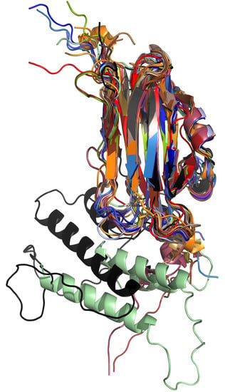
3. Sequences, Structures, and Structural Models of Aegerolysins

We have collected published experimental data on several aegerolysins and their analogs. The sequences of these aegerolysins were taken from published papers or, if not available there, from publicly available genomes; to avoid confusion, they were collected in File S1. The sequences of the aegerolysins ranged from 11 to 92% identical. Only three conserved amino acids, all three glycines, were observed in the alignment between them (Figure S1, star in magenta), and their position in the structure of OlyA6 (PDB ID: 6MYJ) was marked (Figure 2, in magenta). Two of them were located in two different loops on the same side of the molecule; the second loop contains an additional almost conserved glycine (except in Cry34Ab1), followed by a β-sheet containing the third conserved glycine. Due to the smallest side chain, the glycine can increase the local flexibility in the protein structure and the intrinsically destabilize β-sheet [153], possibly helping to accommodate the binding site. In the structure of OlyA6, the position of the fragment of the chemical structure of SM (18:1) is marked (Figure 2, in orange and orange arrow) [73].
Genome sequences from multiple strains are available for some of the species. Although we identified only one gene locus encoding the same aegerolysin sequence in the genome of the two laboratory monokaryotic model strains of P. ostreatus PC9 and PC15, the coding sequences of the aegerolysin protein may vary between other strains [52,53,54,55]. For example, the OlyA6 protein differs in one amino acid (99% identical) from the genomic sequence PlyA(PriA), and PlyA in six (95% identical) [52]. Perhaps it is also plausible to expect P. eryngii, a grassland litter decomposer consisting of at least five varieties such as var. eryngii, ferulae, elaeoselini, thapsiae, and tingitanus, which differ significantly with habitat distribution, host, and morphology, to contain analogs of the aegerolysins PlaA2 or EryA. Similar variations in aegerolysin sequences have already been observed in some Altenaria strains [36]. Of the two aegerolysins described in A. fumigatus, an invariant asp-HS-like sequence was also present in six other strains; variation in one amino acid was observed in asp-HS between the strains, but asp-HS was also absent in one of the strains (sequence of component B from the same locus, asp-HSB, was only partial in the same strain). Variations in the aegerolysin sequences for NigA1 were not observed in any of the nine other A. niger strains examined. A similar observation was made for NigA2, except for a variation of four amino acids in one of the strains. There are some differences in the sequences around the putative intron splice site and at the C terminus among the three A. terreus strains. Four genome sequences of A. oryzae are accessible in public databases; surprisingly, only one aegerolysin with only one amino acid variation is encoded in all four strains. Another strain has an additional gene identical to the strain with three aegerolysin genes. In the last strain, three additional sequences are found, one correspondingly short and two larger, one with a longer C-terminus and the other with a long N-terminus. The sequences of BlyA are identical in all four strains of B. bassiana. In the sequence of Agl1, there is only one amino acid variation among the four T. atroviride strains. One of the phylogenetically more distant strains, P. aeruginosa PA7 (according to whole genome comparison) revealed that RahU is only 77% identical to the PAO strain. Sequencing and annotation errors cannot be excluded.
For some species, all aegerolyins have been reasonably described, while others may have additional genes. One aegerolysin is encoded by the genome of P. ostreatus, A. terreus, B. bassiana, T. atroviride, A. geisen, A. faecalis, T. ni ascovirus 2c; two aegerolyins are encoded by P. eryngii, A. niger, A. fumigatus (except for one strain that has one aegerolysin), P. aeruginosa, and C. bifermentans; A. oryzae encodes one to four and P. includes four; A. aegerita and M. perniciosa encode six. No data for additional aegerolysin were found for L. rhinocerotis.
Five structures of aegerolysins have been solved: P. ostreatus PlyA (PDB ID: 4OEBA) and OlyA6 (PDB ID: 6MYJ and some analogs), B. thuringiensis Cry34Ab1 (Gpp34Ab1) (PDB ID: 4JOX), A. faecalis AfIP-1A (PDB ID: 5V3S), and P. aeruginosa RahU (PDB ID: 6ZC1). In addition, two structures of the two-component partner proteins P. ostreatus PlyB (PDB ID: 4OEJ) and B. thuringiensis Cry35Ab1 (Tpp35Ab1) (PDB ID: 4JP0) have been solved, and furthermore, the membrane embedded pleurotolysin PlyA/Plyb pore with 13-fold symmetry (PDB ID: 4V2T) from P. ostreatus (Figure 1). Structures for other proteins were modeled (Figure 2) using the recently developed and highly accurate protein structure prediction program AlphaFold2 [14], providing a new dimension in the overview of this protein family.
Despite the low identity of the protein sequences (Figure S1), it is noteworthy that all aegerolysin models exhibit a tight β-sandwich fold similar to the actinoporin family of pore-forming proteins as identified by crystal structures (Figure 2, Graphical abstract) [60]. The presence of a short N-terminal helix was confirmed in the AfIP-1A structure (PDB ID: 5V3S). However, for some aegerolysins, such as Aa-Pri1 and Cbm17.1, the presence of a shorter N-terminal helix could also be uncertain. Since some aegerolysins were isolated from fungi or bacteria, some of them were produced recombinantly from a DNA sequence, and for the rest only expression profiles were identified, there is already some uncertainty the exact protein sequences, especially at the ends. In addition, the determination of the N-terminal structure is sometimes less efficient due to flexibility, resulting in less accurate fold predictions. These short helices were also not supported by protein secondary structure predictions (Figure S2). However, two large additional α-helices were modeled for viral and insect aegerolysins (Figure 2 and Figure S2).

4. Aegerolysin Binary Partner Proteins

Binary and quaternary cytolytic complexes of bacterial origin in which aegerolysin-like proteins are combined with larger, non-aegerolysin-like protein partner(s), described to date, include: Cry16Aa/Cry17Aa/Cbm17.1/Cbm17.2 from C. bifermentas subsp. malaysia [40]; Cry34Ab1/Cry35Ab1 from B. thuringiensis [130,134]; and AflP-1A/AflP-1B from A. faecalis [143]. In fungus P. ostreatus, PlyA forms a pore embedded in the membrane together with PlyB (Figure 1) [60]. Similar cytolytic effects were observed when PlyB was combined with other Pleurotus-derived aegerolysins, e.g., OlyA6, PlyA2 and EryA [78]. These heteromeric aegerolysin-based cytolytic complexes have been exploited as potent biopesticides for specific pests, with Cry16Aa/Cry17Aa/Cbm17.1/Cbm17.2 acting against Aedes mosquitoes, and Cry34Ab1/Cry35Ab1, AflP-1A/AflP-1B, OlyA6/PlyB, PlyA/PlyB, PlyA2/PlyB, or EryA/PlyB acting against Coleoptera species, especially the western corn rootworm.
Unexpectedly, partner proteins can be classified into five groups (Table 2, Figure 2): (1) PlyB and EryB have a similar MACPF fold; (2) the remaining models, including BlyB, showed a reasonably good superposition; BlyB has been shown to best align the structure of bacterial GNIP1Aa, another MACPF domain-containing protein [52,121]; (3) the Cry35Ab1 structure and (4) the AfIP-1B model do not superimpose with the PlyB structure or with each other; for AfIP-1B, the MACPF domain was found to be insignificant [52]; (5) Cry16Aa and Cry17Aa only superimpose with each other.
Proteins containing a membrane-attack complex/perforin (MACPF) domain are transmembrane pore-forming proteins important for both human immunity and pathogen virulence. Little is known about the function of MACPF-like domain proteins in filamentous fungi and their taxonomic distribution. The number of putative MACPF proteins in a single fungal species ranges from zero to ten or more [155]. The identification and annotation of putative MACPF-like proteins is generally more error-prone because these genes have a higher number of introns [155]. The sequences can be divided into two groups. The proteins in the first group, such as PlyB or EryB, were assigned to the MACPF domain PF01823, which was confirmed by the 13-amino acid signature Y/F-G-X2-F/Y-X6-G-G typical of this domain, and they are grouped with human perforin. The second group of sequences is not recognized by the Pfam tool but still contains the typical MACPF/CDC signature, such as BlyB [155]. Fungal MACPF proteins probably contribute to various specific processes. While some of them were found in secretomes (without a typical signal sequence being recognized), others are intracellular, and some of them might be involved in pathogenesis, although probably not all [155]. The large number of introns, the sporadic taxonomic distribution, and the different number of MACPF proteins per fungal species might indicate the involvement of horizontal gene transfer mechanisms in specific ecological niches [155]. Putative MACPF-like domains were identified in the genomes of fungal species with different lifestyles; some of them are pathogenic, such as plant pathogen M. perniciosa, the caterpillar fungus C. militaris, or the nematode trap fungus Arthrobotrys oligospora. Some of the species are also saprophytic, such as the white rot fungus P. ostreatus, grassland-litter decomposers P. eryngii, saprophytic and food-producing A. oryzae, or saprophytic soil fungus A. nidulans [155].
However, the binary protein partner Cry35Ab1 does not have a MACPF domain associated with it. Instead, it contains the C-terminal domain toxin 10 (PF05431), which is typical of a family of insecticidal crystal toxins of Bacillus, named after the insecticidal crystal toxin P42. Cry35Ab1 also has an additional N-terminal ricin-type β-trefoil lectin domain (PF00652) (classified as insignificant only) [52,130].
Cry16Aa and Cry17Aa are delta endotoxins composed of three distinct structural domains, endotoxin N, M, and C, respectively. An N-terminal helical bundle domain is involved in membrane insertion and pore formation (PF03945), a central β-sheet domain is involved in receptor binding (PF00555), and the C-terminal β-sandwich domain interacts with the N-terminal domain to form a channel (PF03944). During sporulation, the bacteria produce crystals of delta endotoxins. When an insect ingests these proteins, they are activated by proteolytic cleavage. For all such proteins, the N-terminus is cleaved, and for some members, a C-terminal extension is also cleaved. After activation, the endotoxin binds to the intestinal epithelium and, by the forming cation-selective channels, causes cell lysis, which leads to death [137,156].
Surprisingly, it was observed in the genome of P. ostreatus that the two genes encoding the pair of pore-forming proteins also form a bidirectional pair with 5′–5′ orientation of plyA (priA) and plyB [91,92]. Similar gene pairs encoding putative bicomponent toxins have been observed previously, such as Asp-HS with Asp-HSB, NigA2 with NigB1, and BlyA with BlyB from A. fumigatus, A. niger, and B. bassiana, respectively [52,92]. Moreover, such gene pairs have been also identified in other entomopathogenic fungi besides in B. bassiana, in the genomes of Cordiceps militaris, Metarizium acridum, M. anisopliae, M. robertsii, and Ophiocordiceps sinensis, but the involvement of these protein pairs in pore formation remains to be confirmed [52]. Here, we identified two additional putative pairs: PlyA2 with EryB from P. eryngii and L152 with L152B from A. geisen (Figure 3). The gene locations of the aegerolysin and putative MACPF-like genes in the genomes of some fungi and bacteria are schematically shown in Figure 3. A total of six fungal (putative) bicomponent toxins form a bidirectional gene pair with 5′–5′ orientation of the adjacent gene, and distances between the two genes vary (Figure 3). Clusters of genes encoding fungal secondary metabolites may also contain some reverse-oriented genes that may nevertheless show coordinate expression. In contrast to the fungal gene pairs, the AfIP-1A/AfIP-1B gene pair encoded on the bacterial chromosome has the same sense orientation (Figure 3). The bicomponent toxin gene pair Cry34Ab1/Cry34Ab1 is known to be plasmid-encoded, but the variation of the plasmid in the bacterium B. thuringiensis is exceptionally high to see if these two genes are adjacent. The Cry operon in plasmid pCryO of C. bifermentans subsp. malaysia contains four genes downstream of the promoter pCyt: Cry16Aa is located 91 base pairs (bp) downstream, followed by Cry17Aa, which is located 426 bp downstream, followed by Cbm17.1, and 1022 bp downstream, followed by Cbm17.2 [40]. Their placement at the same locus or under the control of the same promoter does not necessarily lead to their joint action, but rather indicates coordinated expression.
Some of the aegerolysins included in this review are also encoded in genomes that do not have an adjacent (MACPF domain-containing) partner protein; such aegerolysins are the fungal EryA, Aa-Pri1, Asp-HS-like, Ter, AoHlyA, and NigA1, bacterial RahU, insects P23 and the ascoviral TnAV2c gp029 (Table 2). For some of them, insufficient data are available, such as for the MpPRIA1, MpPRIA2, and MpPRIB, because the sequenced countings are too short, or no genomic data were found for GME7309 (Table 2). However, a combined action with MACPF domain-containing proteins encoded elsewhere in the genome cannot be excluded, as some aegerolysins, such as PlyA2 and EryA from P. eryngii, have been shown to exhibit cytolytic activity when combined with their non-native partner with component B, PlyB, from P. osteratus [78].

5. Lifestyle of Organisms Encoding for Aegerolysins and Putative Function of Aegerolysins

The aegerolysins listed in this review belong to several taxonomically diverse organisms; in fungi, they belong to four species of mushrooms and seven filamentous fungi, and they were also identified in four bacterial species, moth, and ascovirus (Table 1). Most of these species, which have very different lifestyles, combine several life forms or hosts; there are many transitions between these lifestyles or switches to other lifestyles. Each species belongs to a particular niche, some are very specific, and others are very ubiquitous and cosmopolitan. Some of these species could or would share the same niche, others would not. A wide range of lifestyles has been identified for organisms containing aegerolysins (Table 1): saprotrophic (white rot, grassland-litter decomposer, plant debris in compost piles, hay and straw stacks), entomopathogenic, mosquito larvicidal, endophytic, mycoparasitic (including oomycetes), nematocidal, plant pathogenic, facultative biotrophic, defoliating (feeding on a wide variety of plants), obligate pathogenic to moth larvae, ubiquitous opportunistic pathogenic to a wide range of hosts: humans, vertebrates, plants, insects, nematodes, mollusks, protozoans, or animal and human parasites.
In the majority of the species listed, saprotrophic lifestyle predominates, as in the white rot fungi P. ostreatus, L. rhinocerotis, and A. aegerita, or in decomposers of grassland litter such as P. eryngii or Aspergilli living on plant debris in compost piles and stacks of highly moist hay and straw. Just because saprotrophic organisms feed primarily on dead or decaying matter, it is easy to confuse the saprotrophic niche with an environment where there is no competition. However, niche, such as wood decay, is one of the most competitive environments. Wood decay fungi have historically been characterized as either white rot fungi, which degrade all components of plant cell walls, including lignin, leaving a white color and rotting texture of the remaining crystalline cellulose, or brown rot fungi, which leave the lignin largely intact. However, genomic analyzes have shown that the prevailing paradigm of white rot and brown rot cannot to capture the diversity of wood decomposition mechanisms by fungi [15]. There is usually one fungal species that dominates a rotting trunk. The ability of fungi to colonize and decompose wood may be strongly influenced by wood-inhabiting bacteria that grow on readily usable compounds released by fungal enzymes. However, it is not known how white rot fungi (or other saprotrophic fungi) cope with the presence of potentially competing bacteria [160]. According to Gause’s principle of competitive exclusion, two species with identical niches (competing for a single resource) cannot coexist indefinitely [161]. In primary resource acquisition, fungi compete for influence over a resource but do not directly challenge others, whereas secondary resource acquisition involves combative interactions [162].
Although wood decomposition is largely driven by microbial activities, invertebrates, such as insect larvae and nematodes, also play important roles in both temperate and tropical environments. Key mechanisms include enzymatic digestion by endogenous enzymes and those produced by their symbionts, substrate modification by tunneling or fragmentation, biotic interactions, and nitrogen fertilization through nitrogen fixation by endosymbionts and free-living bacteria [163]. The effects of individual invertebrate species can be accelerating or inhibiting, but the cumulative effect of the entire community generally accelerates wood decomposition, at least in the early stages of the process [163]. Some taxa appear to be particularly influential in promoting wood decomposition, such as large wood-boring beetles (Coleoptera) and termites (Termitoidae), especially those cultivating fungal gardens [163].
In certain situations, saprophytic fungi can even become pathogenic; it seems they have tools at the ready just in case. For example, because Aspergilli are able to live in compost or haystacks at elevated temperatures, they can also become (opportunistic) pathogens for mammals. Mushrooms can be poisonous to organisms (such as rodents, humans, or snails) that choose to consume them in excessive quantities (P. ostreatus [164]), facultative pathogens on some other tree species (A. aegerita), or facultative biotrophs on roots of herbaceous plants (P. eryngii).
Fungal, but also bacterial entomopathogens, are largely facultative parasites and play an important role in controlling the density of insect populations in nature; some of these organisms have been used for biological control of insect pests in agriculture. The entomopathogenicity of fungi has evolved several times; convergent evolution led to the formation of similar protein families in different species to adapt to insect hosts [165]. The host specificity of insect pathogens is related to the characteristics of the fungal genome and lifestyle. Species with a narrow host range usually have reduced protein family sizes, but retain the ability to reproduce sexually compared to generalists [165]. Similar to plant pathogens, entomopathogenic fungi encode a number of effector-like proteins in their genomes, suggesting the presence of analogous gene-for-gene relationships in fungus-insect interactions [165]. Insect pathogens employ different strategies to invade the host and evade its immune response [165]. The pattern of competition among entomopathogens for insect individuals remains unclear. For example, empirical competition for hosts or niches between the species of B. bassiana and M. robertsii was studied; insects were largely killed and mycotized by M. robertsii, regardless of the initial co-inoculation dose and infection order [166]. Thus, these results support the pattern of competitive exclusion between insect pathogenic fungi that occurs from outside to inside of insect hosts. However, parasexual recombination between the compatible strains apparently occurs after coinfection [166]. The sexual cycle of the heterothallic Metarhizium and Beauveria species rarely occurs in the field. Instead, parasexual recombination takes place between compatible strains through the process of hyphal fusion, heterokaryosis, and mitotic crossing-over for limited genetic recombination [167,168]. Thus, it seems that exclusive competition may occur between incompatible strains, but recombination arises from the compatible strains of fungal insect parasites [166]. Some entomopathogenic fungi, such as B. bassiana and T. atroviride, are also considered as endophytes, protecting plant roots by being mycoparasitic (including oomycetes) [169,170].
It has been shown that P. ostreatus paralyzes nematodes from the genera Caenorhabditis, Diploscapter, Oscheius, Rhabditis, Pristionchus, Panagrellus, Acrobeloides, Cephalobus, Mesorhabditis, and Pelodera, and consumes prey [15]. The unclear nematocidal mechanism of P. ostreatus triggers massive calcium influx and rapid cell necrosis in the neuromuscular system of C. elegans via the sensory cilia of this nematode, a mechanism of action is common against all nematodes [15]. Pleurotus eryngii also elicits a nematocidal action. Several bacterial pathogens, including P. luminescens [171] and B. thuringiensis [172], are also known to induce necrotic responses in C. elegans. Nematopathogenic fungi have been identified as a basis for biological control of root-knot nematodes [173].
Membrane lipids or their specific combinations have been often associated with the binding of aegerolysins. The most common was the combination of CPE/Chol or SM/Chol. Aegerolysins sometimes interact with both of these lipid combinations, sometimes excluding one another, or bind to CPE alone, but never to free SM. They can also bind a combination of other lipids like CAEP/POPC/Chol or CL/DPPC/Chol (Table 2). The molar ratio of lipids may be important. For some of aegerolysins an unknown protein receptor in the lipid membrane has been proposed, or for others binding was not studied. Different lipids or their combination may define the binding target of aegerolysins because different organisms have different composition of major membrane lipids, particularly the sphingolipids.
Sphingolipids are a numerous and versatile group of lipids in eukaryote and in some bacterial taxa. These amphipathic molecules can be abundant components of cell membranes, and their sphingosine-based degradation products are involved in many physiological processes, including signaling [77]. One of the major sphingolipids in mammalian cell membranes that has been extensively studied is SM, which plays a central role in the formation of lipid rafts and ordered membrane domains, as well as cell signaling processes and regulation of plasma membrane and cholesterol homeostasis [77]. SM is also the major sphingolipid of nematode C. elegans. CPE is the major sphingolipid of Oomycetes, such as Pythium ultimum, Phytophthora infestans, and P. capsici, as well as of some flagellated protozoa, and the major sphingolipid in the lipid membranes of various invertebrates, including insects. In some invertebrates, CPE is combined with SM, such as mosquitoes Aedes aegypti, Culex quinquefasciatus, or moths as Manduca sexta. In some Bacteroidetes, CPE is combined with ceramide phosphoglycerol, such as in Flectobacillus major and Bacteroides fragilis. CAEP is a CPE analog of aquatic invertebrates, such as mollusks and cnidarians [77].
The aegerolysins appear to have a (different?) function(s) associated with the specific stage of their development; it seems possible that they serve the interaction between organisms in the environment. Based on their sequence and binding diversity, it appears that these proteins might serve to scan the membranes of potential competitors or hosts in the niche. After binding to a specific lipid or lipid combination, or to yet unknown proteins receptors in lipid membranes, some of them may serve to dock various partner proteins from the organism of origin or perhaps from the target organism after the proteolytic cleavage. It appears that aegerolysins represent a part of adaptive response to the environment, by sensing lipid components of organisms in their environment and excluding competitors by forming two-component pores as a kind of primitive “immune interaction.” Some partner proteins have a similar (MACPF) fold to some molecules, such as MPEG-1, or proteins C8 and C9, which are involved in the formation of a heterooligomeric protein-membrane attack complex (MAC), and have a function in mammalian immune defense [126,127]. It is possible that organisms such as hemibiotrophic plant pathogen Moniliophthora perniciosa can increase their flexibility in lipid sensing with additional copies (six) of aegerolysin genes (if differently expressed).
Horizontal gene transfer often plays a role in shaping the mechanisms by which a pathogen evades, counteracts, mimics, subverts, and manipulates the host [174]. Plasmid-encoded genes for aegerolyins and partner proteins of the ubiquitous, aerobic, and spore-forming organism, such as B. thuringiensis, which is an opportunistic pathogen for very diverse hosts, including vertebrates, plants, insects, nematodes, mollusks, protozoa, animal and human parasites, appear to be particularly susceptible to their dissemination. Especially in combination with pore formation, which may facilitate the presence or uptake of heterologous DNA molecules or their parts present in the environment. It is also known that B. thuringiensis harbors a variety of transposable elements, including insertion sequences and transposons [139,175]. In addition, it has been already shown that partial genes can serve as a source of novel Cry toxins [176]. Other possible vectors are (DNA) viruses. Whether Trichoplusia ni and Heliothis virescens ascoviruses acquired aegerolysin-like genes from their insect hosts or vice versa is unclear, but their presence in both, which may indicate horizontal gene transfer, suggests that this family of structurally similar proteins is more widespread and functionally diverse than previously thought [48].

6. Conclusions and Future Research

It seems possible that aegerolysins enable an adaptive response to the environment by sensing (lipid) components of the environment and excluding competitors by forming two-component pores. However, many questions remain.
(1) Given the low identity of the protein sequences, the conserved structure of the aegerolysin molecules seems to be most important for their function. It remains to be clarified how the (lipid) binding interaction is specified and what contributes to the differences in specificity.
(2) Some organisms encode multiple aegerolysin copies. Some of the aegerolysins recruit partner proteins to function in pore formation. It is interesting to show whether other aegerolysin copies from the same organism also recruit the same component B protein. It has been shown in the laboratory that component B exchange is possible between closely related species. It is a challenge to show whether such acquisition of component B also occurs in nature, and how homologous the component B must be.
(3) If other copies of aegerolysin do not acquire component B, the question arises as to what their function is; perhaps they serve as decoys.
(4) Some of the partner proteins are MACPF domain-containing proteins. Some of the microbial or mammalian MACPF/CDC proteins form pores without binding to aegerolyins because they have their own C-terminal binding domain. It has been shown that some of them can be cleaved by unknown proteases [177]. It would be very interesting to show that the C-terminal binding domain of a MACPF domain-containing protein from the target organism can be replaced by aegerolysin from the competitor, which would then lead to a counteraction.

7. Materials and Methods

7.1. Literature Search

Search for publications on aegerolyins was performed by Scopus title-abstract-keywords search (TITLE-ABS-KEY (aegerolysin*) [178]; 43 documents were initially identified: [1,2,3,4,5,6,7,8,9,10,11,28,36,48,52,63,64,65,66,67,72,75,77,78,79,80,81,82,84,87,91,92,93,101,104,105,114,128,130,144,145,147,148]; Clostridium bifermentas aergerolysins were added to the collection.

7.2. Comparative Genomics

Mining for the number aegerolysins from fungi was performed on the web portal MycoCosm, mining for the number aegerolysins from (bacteria and archaea) on the web portal Ensembl Bacteria, databases were searched for domain PF06355 named aegerolysin [12,13,179,180]. The genomes of were screened for differences in aegerolysin sequences and adjacent MACPF-like genes (PF01823) [12,13,179,180,181]. We found a genome of a single strain available for: Agrocybe aegerita AAE-3 [12,17], Alternaria geisen BMP2338 [12,159], Trichoderma atroviride [12,182], Lignosus rhinocerotis [12,106], Moniliophthora perniciosa FA553 [12,183], Pleurotus eryngii ATCC 90,797 [12,157], and Pseudoplusia includens chromosome 6 PRJEB38103 (GCA_941860345.1). Multiple strains were available for: Alcaligenes faecalis: GCA_003521065, GCA_005311025, GCA_003813085, AN70 (GCA_004319585), and YBY (GCA_003122065) [13]; Aspergilus fumigatus: Af293 [12,22,184]; A1163 [12,22,185,186,187]; GCA_002234955.1, GCA_002234985.1, Z5, and var. RP-2014 [181]; Aspergillus niger: CBS 513.88 [12,27,184], (lacticoffeatus) CBS 101883, (phoenicis Corda) Thom ATCC 13,157 [12,188], ATCC 1015 [12,189], NRRL3 [12,188,190], ATCC 13496, ATCC 64,974 N402, GCA_001515345.1, GCA_002211485.2, and GCA_000002855.2 [181]; Aspergilus terreus: NIH 2624 [12,184], and IFO6365 [181]; Aspergilus oryzae: RIB40 [12,191], 100-8, 3.042, and BCC7051 [181]; Beauveria bassiana: ARSEF 2860 [12,158]; D15, GCA_001682635.1, and JEF-007 [181]; Pleurotus ostreatus PC9 v1.0 and PC15 v2.0 [12,53,54,55], and Pseudomonas aeruginosa PAO1 and PA7 [13,192]. The aegerolysin protein sequences of different strains were aligned using the ClustalW web tool [193].

7.3. Phylogenetic Analysis

Phylogenetic analysis of ClustalW aligned aegerolysins and inferred by the Maximum Likelihood method was performed using Molecular Evolutionary Genetics Analysis, version 11 (MEGA11) [154,194].

7.4. Protein Structure Prediction

The deep learning algorithm AlphaFold2 was used for protein structure modeling [14]. UCSF ChimeraX version 1.3 (8 December 2021) and UCSF ChimeraX version 1.4 (3 June 2022) were used to run AlphaFold version 2 in conjunction with a free and accessible platform for protein folding ColabFold [195,196,197,198,199].

7.5. Protein Structure Presentation

Cartoon representation of the structures was done using PyMOL, version 2.2.0 [62]. Amino acid at N- and C-terminus of the model are marked.

7.6. Alignment of Proteins and Prediction of Secondary Structure

A multiple sequence alignment was performed with the workbench editor Jalview version 2.11.2.4 using the Clustal W version 2.1 /Clustal X algorithm [200,201]. The protein secondary structure prediction server JPred4, accessible via Jalview, was used to predict the secondary structure of proteins [202].

Supplementary Materials

The following supporting information can be downloaded at: https://www.mdpi.com/article/10.3390/toxins14090629/s1, Supplementary File. Sequences of aegerolysins and partner proteins; Figure S1. Alignments of amino acid sequences of aegerolysins; Figure S2. Prediction of secondary structures of aegerolysins.

Author Contributions

Conceptualization, N.K.; methodology, N.K.; software, N.K.; validation, N.K.; formal analysis, N.K.; investigation, N.K.; resources, N.K.; data curation, N.K.; writing—original draft preparation, N.K., M.S.; writing—review and editing, N.K., M.S.; visualization, N.K.; supervision, N.K.; project administration, N.K.; funding acquisition, N.K. All authors have read and agreed to the published version of the manuscript.

Funding

This research was funded by the Slovenian Research Agency, grant number P1-0391 and P1-0207.

Institutional Review Board Statement

Not applicable.

Informed Consent Statement

Not applicable.

Data Availability Statement

No new data were created or analyzed in this study. Data sharing is not applicable to this article.

Acknowledgments

The authors would like to thank for Gregor Anderluh, Anastasija Panevska, and especially Kristina Sepčić for critical opinion and valuable discussion on aegerolysins, Rudi Kraševec for some ecological insights, to Gasper Šolinc and Matic Kisovec for sharing some bioinformatic tips.

Conflicts of Interest

The authors declare that there are no conflict of interest.

References

  1. Berne, S.; Lah, L.; Sepčić, K. Aegerolysins: Structure, function, and putative biological role. Protein Sci. 2009, 18, 694–706. [Google Scholar] [CrossRef] [PubMed]
  2. Butala, M.; Novak, M.; Kraševec, N.; Skočaj, M.; Veranič, P.; Maček, P.; Sepčić, K. Aegerolysins: Lipid-binding proteins with versatile functions. Semin. Cell Dev. Biol. 2017, 72, 142–151. [Google Scholar] [CrossRef] [PubMed]
  3. Nayak, A.P.; Green, B.J.; Beezhold, D.H. Fungal hemolysins. Med. Mycol. 2013, 51, 1–16. [Google Scholar] [CrossRef] [PubMed]
  4. Novak, M.; Kraševec, N.; Skočaj, M.; Maček, P.; Anderluh, G.; Sepčić, K. Fungal aegerolysin-like proteins: Distribution, activities, and applications. Appl. Microbiol. Biotechnol. 2015, 99, 601–610. [Google Scholar] [CrossRef] [PubMed]
  5. Ota, K.; Butala, M.; Viero, G.; Dalla Serra, M.; Sepčić, K.; Maček, P. Fungal MACPF-like proteins and aegerolysins: Bi-component pore-forming proteins? In Sub-Cellular Biochemistry; Anderluh, G., Gilbert, R., Eds.; Subcellular Biochemistry; Springer: Dordrecht, The Netherlands, 2014; Volume 80, pp. 271–291. ISBN 978-94-017-8880-9. [Google Scholar]
  6. Yamaji-Hasegawa, A.; Hullin-Matsuda, F.; Greimel, P.; Kobayashi, T. Pore-forming toxins: Properties, diversity, and uses as tools to image sphingomyelin and ceramide phosphoethanolamine. Biochim. Biophys. Acta Biomembr. 2016, 1858, 576–592. [Google Scholar] [CrossRef]
  7. Kishimoto, T.; Ishitsuka, R.; Kobayashi, T. Detectors for evaluating the cellular landscape of sphingomyelin- and cholesterol-rich membrane domains. Biochim. Biophys. Acta Mol. Cell Biol. Lipids 2016, 1861, 812–829. [Google Scholar] [CrossRef]
  8. Hullin-Matsuda, F.; Makino, A.; Murate, M.; Kobayashi, T. Probing phosphoethanolamine-containing lipids in membranes with duramycin/cinnamycin and aegerolysin proteins. Biochimie 2016, 130, 81–90. [Google Scholar] [CrossRef]
  9. Hullin-Matsuda, F.; Murate, M.; Kobayashi, T. Protein probes to visualize sphingomyelin and ceramide phosphoethanolamine. Chem. Phys. Lipids 2018, 216, 132–141. [Google Scholar] [CrossRef]
  10. Panevska, A.; Skočaj, M.; Modic, Š.; Razinger, J.; Sepčić, K. Aegerolysins from the fungal genus Pleurotus—Bioinsecticidal proteins with multiple potential applications. J. Invertebr. Pathol. 2021, 186, 107474. [Google Scholar] [CrossRef]
  11. Grundner, M.; Panevska, A.; Sepčić, K.; Skočaj, M. What can mushroom proteins teach us about lipid rafts? Membranes 2021, 11, 264. [Google Scholar] [CrossRef]
  12. MycoCosm—The Fungal Genomics Resouce (DOE Joint Genome Institute). Available online: https://mycocosm.jgi.doe.gov/mycocosm/home (accessed on 14 July 2022).
  13. Ensembl Bacteria Release 54—Jul 2022 © EMBL-EBI. Available online: http://bacteria.ensembl.org/index.html (accessed on 2 August 2022).
  14. Jumper, J.; Evans, R.; Pritzel, A.; Green, T.; Figurnov, M.; Ronneberger, O.; Tunyasuvunakool, K.; Bates, R.; Žídek, A.; Potapenko, A.; et al. Highly accurate protein structure prediction with AlphaFold. Nature 2021, 596, 583–589. [Google Scholar] [CrossRef]
  15. Lee, C.-H.; Chang, H.-W.; Yang, C.-T.; Wali, N.; Shie, J.-J.; Hsueh, Y.-P. Sensory cilia as the Achilles heel of nematodes when attacked by carnivorous mushrooms. Proc. Natl. Acad. Sci. USA 2020, 117, 6014–6022. [Google Scholar] [CrossRef] [PubMed]
  16. Zervakis, G.I.; Ntougias, S.; Gargano, M.L.; Besi, M.I.; Polemis, E.; Typas, M.A.; Venturella, G. A reappraisal of the Pleurotus eryngii complex—New species and taxonomic combinations based on the application of a polyphasic approach, and an identification key to Pleurotus taxa associated with Apiaceae plants. Fungal Biol. 2014, 118, 814–834. [Google Scholar] [CrossRef] [PubMed]
  17. Gupta, D.K.; Rühl, M.; Mishra, B.; Kleofas, V.; Hofrichter, M.; Herzog, R.; Pecyna, M.J.; Sharma, R.; Kellner, H.; Hennicke, F.; et al. The genome sequence of the commercially cultivated mushroom Agrocybe aegerita reveals a conserved repertoire of fruiting-related genes and a versatile suite of biopolymer-degrading enzymes. BMC Genom. 2018, 19, 48. [Google Scholar] [CrossRef] [PubMed]
  18. Meinhardt, L.W.; Rincones, J.; Bailey, B.A.; Aime, M.C.; Griffith, G.W.; Zhang, D.; Pereira, G.A.G. Moniliophthora perniciosa, the causal agent of witches’ broom disease of cacao: What’s new from this old foe? Mol. Plant Pathol. 2008, 9, 577–588. [Google Scholar] [CrossRef] [PubMed]
  19. Fung, S.-Y.; Tan, C.-S. Tiger milk mushroom (the Lignosus trinity) in Malaysia: A medicinal treasure trove. In Medicinal Mushrooms; Springer: Singapore, 2019; pp. 349–369. [Google Scholar]
  20. de Vries, R.P.; Riley, R.; Wiebenga, A.; Aguilar-Osorio, G.; Amillis, S.; Uchima, C.A.; Anderluh, G.; Asadollahi, M.; Askin, M.; Barry, K.; et al. Comparative genomics reveals high biological diversity and specific adaptations in the industrially and medically important fungal genus Aspergillus. Genome Biol. 2017, 18, 28. [Google Scholar] [CrossRef]
  21. Abdel-Azeem, A.M.; Salem, F.M.; Abdel-Azeem, M.A.; Nafady, N.A.; Mohesien, M.T.; Soliman, E.A. Biodiversity of the genus Aspergillus in different habitats. In New and Future Developments in Microbial Biotechnology and Bioengineering; Elsevier: Amsterdam, The Netherlands, 2016; pp. 3–28. [Google Scholar]
  22. Nierman, W.C.; Pain, A.; Anderson, M.J.; Wortman, J.R.; Kim, H.S.; Arroyo, J.; Berriman, M.; Abe, K.; Archer, D.B.; Bermejo, C.; et al. Genomic sequence of the pathogenic and allergenic filamentous fungus Aspergillus fumigatus. Nature 2005, 438, 1151–1156. [Google Scholar] [CrossRef]
  23. Mullins, J.; Harvey, R.; Seaton, A. Sources and incidence of airborne Aspergillus fumigatus (Fres). Clin. Exp. Allergy 1976, 6, 209–217. [Google Scholar] [CrossRef]
  24. Schuster, E.; Dunn-Coleman, N.; Frisvad, J.C.; Van Dijjck, P. On the safety of Aspergillus niger—A review. Appl. Microbiol. Biotechnol. 2002, 59, 426–435. [Google Scholar] [CrossRef]
  25. Perrone, G.; Susca, A.; Cozzi, G.; Ehrlich, K.; Varga, J.; Frisvad, J.C.; Meijer, M.; Noonim, P.; Mahakarnchanakul, W.; Samson, R.A. Biodiversity of Aspergillus species in some important agricultural products. Stud. Mycol. 2007, 59, 53–66. [Google Scholar] [CrossRef]
  26. Joint FAO/WHO Expert Committee on Food Additives. Safety Evaluation of Certain Mycotoxins in Food/Prepared by the Fifty-Sixth Meeting of the Joint FAO/WHO Expert Committee on Food Additives (JECFA); Joint FAO/WHO Expert Committee on Food Additives: Rome, Italy, 2001. [Google Scholar]
  27. Pel, H.J.; de Winde, J.H.; Archer, D.B.; Dyer, P.S.; Hofmann, G.; Schaap, P.J.; Turner, G.; de Vries, R.P.; Albang, R.; Albermann, K.; et al. Genome sequencing and analysis of the versatile cell factory Aspergillus niger CBS 513.88. Nat. Biotechnol. 2007, 25, 221–231. [Google Scholar] [CrossRef] [PubMed]
  28. Nayak, A.P.; Blachere, F.M.; Hettick, J.M.; Lukomski, S.; Schmechel, D.; Beezhold, D.H. Characterization of recombinant terrelysin, a hemolysin of Aspergillus terreus. Mycopathologia 2011, 171, 23–34. [Google Scholar] [CrossRef] [PubMed]
  29. Machida, M.; Yamada, O.; Gomi, K. Genomics of Aspergillus oryzae: Learning from the history of koji mold and exploration of its future. DNA Res. 2008, 15, 173–183. [Google Scholar] [CrossRef] [PubMed]
  30. Vega, F.E.; Goettel, M.S.; Blackwell, M.; Chandler, D.; Jackson, M.A.; Keller, S.; Koike, M.; Maniania, N.K.; Monzón, A.; Ownley, B.H.; et al. Fungal entomopathogens: New insights on their ecology. Fungal Ecol. 2009, 2, 149–159. [Google Scholar] [CrossRef]
  31. Rasool, S.; Vidkjær, N.H.; Hooshmand, K.; Jensen, B.; Fomsgaard, I.S.; Meyling, N.V. Seed inoculations with entomopathogenic fungi affect aphid populations coinciding with modulation of plant secondary metabolite profiles across plant families. New Phytol. 2021, 229, 1715–1727. [Google Scholar] [CrossRef] [PubMed]
  32. Druzhinina, I.S.; Kubicek, C.P. Ecological Genomics of Trichoderma. In The Ecological Genomics of Fungi; John Wiley & Sons, Inc.: Hoboken, NJ, USA, 2013; pp. 89–116. [Google Scholar]
  33. Druzhinina, I.S.; Seidl-Seiboth, V.; Herrera-Estrella, A.; Horwitz, B.A.; Kenerley, C.M.; Monte, E.; Mukherjee, P.K.; Zeilinger, S.; Grigoriev, I.V.; Kubicek, C.P. Trichoderma: The genomics of opportunistic success. Nat. Rev. Microbiol. 2011, 9, 749–759. [Google Scholar] [CrossRef]
  34. Atanasova, L.; Le Crom, S.L.; Gruber, S.; Coulpier, F.; Seidl-Seiboth, V.; Kubicek, C.P.; Druzhinina, I.S. Comparative transcriptomics reveals different strategies of Trichodermamycoparasitism. BMC Genom. 2013, 14, 121. [Google Scholar] [CrossRef]
  35. Atanasova, L. Ecophysiology of Trichoderma in Genomic Perspective. In Biotechnology and Biology of Trichoderma; Elsevier: Amsterdam, The Netherlands, 2014; pp. 25–40. [Google Scholar]
  36. Roberts, R.G.; Bischoff, J.F.; Reymond, S.T. Differential gene expression in Alternaria gaisen exposed to dark and light. Mycol. Prog. 2011, 11, 373–382. [Google Scholar] [CrossRef]
  37. Simmons, E.G.; Roberts, R.G. Alternaria themes and variations (73). Mycotaxon 1993, 48, 109–140. [Google Scholar]
  38. Argôlo-Filho, R.; Loguercio, L. Bacillus thuringiensis is an environmental pathogen and host-specificity has developed as an adaptation to human-generated ecological niches. Insects 2013, 5, 62–91. [Google Scholar] [CrossRef]
  39. Palma, L.; Muñoz, D.; Berry, C.; Murillo, J.; Caballero, P. Bacillus thuringiensis toxins: An overview of their biocidal activity. Toxins 2014, 6, 3296–3325. [Google Scholar] [CrossRef] [PubMed] [Green Version]
  40. Qureshi, N.; Chawla, S.; Likitvivatanavong, S.; Lee, H.L.; Gill, S.S. The Cry Toxin operon of Clostridium bifermentans subsp. malaysia is highly toxic to Aedes larval mosquitoes. Appl. Environ. Microbiol. 2014, 80, 5689–5697. [Google Scholar] [CrossRef] [PubMed]
  41. Ju, S.; Lin, J.; Zheng, J.; Wang, S.; Zhou, H.; Sun, M. Alcaligenes faecalis ZD02, a Novel Nematicidal Bacterium with an Extracellular Serine Protease Virulence Factor. Appl. Environ. Microbiol. 2016, 82, 2112–2120. [Google Scholar] [CrossRef]
  42. Finlay, B.B. Bacterial Disease in Diverse Hosts. Cell 1999, 96, 315–318. [Google Scholar] [CrossRef]
  43. Chieda, Y.; Iiyama, K.; Yasunaga-Aoki, C.; Lee, J.M.; Kusakabe, T.; Shimizu, S. Pathogenicity of gacA mutant of Pseudomonas aeruginosa PA01 in the silkworm, Bombyx mori. FEMS Microbiol. Lett. 2005, 244, 181–186. [Google Scholar] [CrossRef]
  44. Sousa, A.; Pereira, M. Pseudomonas aeruginosa diversification during infection development in cystic fibrosis lungs—A review. Pathogens 2014, 3, 680–703. [Google Scholar] [CrossRef]
  45. Starkey, M.; Rahme, L.G. Modeling Pseudomonas aeruginosa pathogenesis in plant hosts. Nat. Protoc. 2009, 4, 117–124. [Google Scholar] [CrossRef]
  46. Bano, N.; Musarrat, J. Characterization of a new Pseudomonas aeruginosa strain NJ-15 as a potential biocontrol agent. Curr. Microbiol. 2003, 46, 324–328. [Google Scholar] [CrossRef]
  47. Specht, A.; Sosa-Gómez, D.R.; Roque-Specht, V.F.; Valduga, E.; Gonzatti, F.; Schuh, S.M.; Carneiro, E. Biotic potential and life tables of Chrysodeixis includens (Lepidoptera: Noctuidae), Rachiplusia nu, and Trichoplusia ni on soybean and forage turnip. J. Insect Sci. 2019, 19, 8. [Google Scholar] [CrossRef]
  48. Zhang, S.; Clark, K.D.; Strand, M.R. The protein P23 identifies capsule-forming plasmatocytes in the moth Pseudoplusia includens. Dev. Comp. Immunol. 2011, 35, 501–510. [Google Scholar] [CrossRef]
  49. Cheng, X.-W.; Wang, L.; Carner, G.R.; Arif, B.M. Characterization of three ascovirus isolates from cotton insects. J. Invertebr. Pathol. 2005, 89, 193–202. [Google Scholar] [CrossRef] [PubMed]
  50. Wang, L.; Xue, J.; Seaborn, C.P.; Arif, B.M.; Cheng, X.-W. Sequence and organization of the Trichoplusia ni ascovirus 2c (Ascoviridae) genome. Virology 2006, 354, 167–177. [Google Scholar] [CrossRef] [PubMed]
  51. Stasiak, K.; Renault, S.; Federici, B.A.; Bigot, Y. Characteristics of pathogenic and mutualistic relationships of ascoviruses in field populations of parasitoid wasps. J. Insect Physiol. 2005, 51, 103–115. [Google Scholar] [CrossRef] [PubMed]
  52. Kraševec, N.; Panevska, A.; Lemež, Š.; Razinger, J.; Sepčić, K.; Anderluh, G.; Podobnik, M. Lipid-Binding aegerolysin from biocontrol fungus Beauveria bassiana. Toxins 2021, 13, 820. [Google Scholar] [CrossRef] [PubMed]
  53. Alfaro, M.; Castanera, R.; Lavín, J.L.; Grigoriev, I.V.; Oguiza, J.A.; Ramírez, L.; Pisabarro, A.G. Comparative and transcriptional analysis of the predicted secretome in the lignocellulose-degrading basidiomycete fungus Pleurotus ostreatus. Environ. Microbiol. 2016, 18, 4710–4726. [Google Scholar] [CrossRef] [PubMed]
  54. Riley, R.; Salamov, A.A.; Brown, D.W.; Nagy, L.G.; Floudas, D.; Held, B.W.; Levasseur, A.; Lombard, V.; Morin, E.; Otillar, R.; et al. Extensive sampling of basidiomycete genomes demonstrates inadequacy of the white-rot/brown-rot paradigm for wood decay fungi. Proc. Natl. Acad. Sci. USA 2014, 111, 9923–9928. [Google Scholar] [CrossRef]
  55. Castanera, R.; López-Varas, L.; Borgognone, A.; LaButti, K.; Lapidus, A.; Schmutz, J.; Grimwood, J.; Pérez, G.; Pisabarro, A.G.; Grigoriev, I.V.; et al. Transposable elements versus the fungal genome: Impact on whole-genome architecture and transcriptional profiles. PLoS Genet. 2016, 12, e1006108. [Google Scholar] [CrossRef]
  56. Lakkireddy, K.K.R.; Navarro-González, M.; Velagapudi, R.; Kües, U. Proteins expressed during hyphal aggregation for fruting body formation in basidiomycetes. In Proceedings of the 7th International Conference on Mushroom Biology and Mushroom Products, Arcachon, France, 4–7 October 2011; Savoie, J.-M., Foulongne-Oriol, M., Largeteau, M., Barroso, G., Eds.; Institut National de la Recherche Agronomique (INRA): Paris, France, 2011; pp. 82–94. [Google Scholar]
  57. Bernheimer, A.W.; Avigad, L.S. A cytolytic protein from the edible mushroom, Pleurotus ostreatus. Biochim. Biophys. Acta Gen. Subj. 1979, 585, 451–461. [Google Scholar] [CrossRef]
  58. Lee, S.-H.; Kim, B.-G.; Kim, K.-J.; Lee, J.-S.; Yun, D.-W.; Hahn, J.-H.; Kim, G.-H.; Lee, K.-H.; Suh, D.-S.; Kwon, S.-T.; et al. Comparative analysis of sequences expressed during the liquid-cultured mycelia and fruit body stages of Pleurotus ostreatus. Fungal Genet. Biol. 2002, 35, 115–134. [Google Scholar] [CrossRef]
  59. Tomita, T.; Noguchi, K.; Mimuro, H.; Ukaji, F.; Ito, K.; Sugawara-Tomita, N.; Hashimoto, Y. Pleurotolysin, a novel sphingomyelin-specific two-component cytolysin from the edible mushroom Pleurotus ostreatus, assembles into a transmembrane pore complex. J. Biol. Chem. 2004, 279, 26975–26982. [Google Scholar] [CrossRef]
  60. Lukoyanova, N.; Kondos, S.C.; Farabella, I.; Law, R.H.P.; Reboul, C.F.; Caradoc-Davies, T.T.; Spicer, B.A.; Kleifeld, O.; Traore, D.A.K.; Ekkel, S.M.; et al. Conformational changes during pore formation by the perforin-related protein pleurotolysin. PLoS Biol. 2015, 13, e1002049. [Google Scholar] [CrossRef] [PubMed] [Green Version]
  61. Huang, G.; Voorspoels, A.; Versloot, R.C.A.; Van Der Heide, N.J.; Carlon, E.; Willems, K.; Maglia, G. PlyAB nanopores detect single amino acid differences in folded haemoglobin from blood. Angew. Chem. Int. Ed. 2022, 61, e202206227. [Google Scholar] [CrossRef] [PubMed]
  62. The Molecular Graphics System PyMOL (Schrödinger, LLC). Available online: https://pymol.org/2/ (accessed on 4 October 2021).
  63. Berne, S.; Križaj, I.; Pohleven, F.; Turk, T.; Maček, P.; Sepčić, K. Pleurotus and Agrocybe hemolysins, new proteins hypothetically involved in fungal fruiting. Biochim. Biophys. Acta 2002, 1570, 153–159. [Google Scholar] [CrossRef]
  64. Maličev, E.; Chowdhury, H.H.; Maček, P.; Sepčić, K. Effect of ostreolysin, an Asp-hemolysin isoform, on human chondrocytes and osteoblasts, and possible role of Asp-hemolysin in pathogenesis. Med. Mycol. 2007, 45, 123–130. [Google Scholar] [CrossRef]
  65. Sepčić, K.; Berne, S.; Rebolj, K.; Batista, U.; Plemenitaš, A.; Šentjurc, M.; Maček, P. Ostreolysin, a pore-forming protein from the oyster mushroom, interacts specifically with membrane cholesterol-rich lipid domains. FEBS Lett. 2004, 575, 81–85. [Google Scholar] [CrossRef] [PubMed]
  66. Vidic, I.; Berne, S.; Drobne, D.; Maček, P.; Frangež, R.; Turk, T.; Štrus, J.; Sepčić, K. Temporal and spatial expression of ostreolysin during development of the oyster mushroom (Pleurotus ostreatus). Mycol. Res. 2005, 109, 377–382. [Google Scholar] [CrossRef] [PubMed]
  67. Rebolj, K.; Sepčić, K. Ostreolysin, a cytolytic protein from culinary-medicinal oyster mushroom Pleurotus ostreatus (Jacq.: Fr.) P. Kumm. (Agaricomycetideae), and its potential use in medicine and biotechnology. Int. J. Med. Mushrooms 2008, 10, 293–302. [Google Scholar] [CrossRef]
  68. Rebolj, K.; Bakrač, B.; Garvas, M.; Ota, K.; Šentjurc, M.; Potrich, C.; Coraiola, M.; Tomazzolli, R.; Serra, M.D.; Maček, P.; et al. EPR and FTIR studies reveal the importance of highly ordered sterol-enriched membrane domains for ostreolysin activity. Biochim. Biophys. Acta Biomembr. 2010, 1798, 891–902. [Google Scholar] [CrossRef] [PubMed]
  69. Rebolj, K.; Ulrih, N.P.; Maček, P.; Sepčić, K. Steroid structural requirements for interaction of ostreolysin, a lipid-raft binding cytolysin, with lipid monolayers and bilayers. Biochim. Biophys. Acta Biomembr. 2006, 1758, 1662–1670. [Google Scholar] [CrossRef]
  70. Chowdhury, H.H.; Rebolj, K.; Kreft, M.; Zorec, R.; Maček, P.; Sepčić, K. Lysophospholipids prevent binding of a cytolytic protein ostreolysin to cholesterol-enriched membrane domains. Toxicon 2008, 51, 1345–1356. [Google Scholar] [CrossRef]
  71. Sepčić, K.; Berne, S.; Potrich, C.; Turk, T.; Maček, P.; Menestrina, G. Interaction of ostreolysin, a cytolytic protein from the edible mushroom Pleurotus ostreatus, with lipid membranes and modulation by lysophospholipids. Eur. J. Biochem. 2003, 270, 1199–1210. [Google Scholar] [CrossRef] [PubMed] [Green Version]
  72. Ota, K.; Leonardi, A.; Mikelj, M.; Skočaj, M.; Wohlschlager, T.; Künzler, M.; Aebi, M.; Narat, M.; Križaj, I.; Anderluh, G.; et al. Membrane cholesterol and sphingomyelin, and ostreolysin A are obligatory for pore-formation by a MACPF/CDC-like pore-forming protein, pleurotolysin B. Biochimie 2013, 95, 1855–1864. [Google Scholar] [CrossRef] [PubMed]
  73. Endapally, S.; Frias, D.; Grzemska, M.; Gay, A.; Tomchick, D.R.; Radhakrishnan, A. Molecular discrimination between two conformations of sphingomyelin in plasma membranes. Cell 2019, 176, 1040–1053.e17. [Google Scholar] [CrossRef] [PubMed]
  74. Skočaj, M.; Resnik, N.; Grundner, M.; Ota, K.; Rojko, N.; Hodnik, V.; Anderluh, G.; Sobota, A.A.; Maček, P.; Veranič, P.; et al. Tracking cholesterol/sphingomyelin-rich membrane domains with the ostreolysin A-mCherry protein. PLoS ONE 2014, 9, e92783. [Google Scholar] [CrossRef]
  75. Skočaj, M.; Yu, Y.; Grundner, M.; Resnik, N.; Bedina Zavec, A.; Leonardi, A.; Križaj, I.; Guella, G.; Maček, P.; Kreft Erdani, M.; et al. Characterisation of plasmalemmal shedding of vesicles induced by the cholesterol/sphingomyelin binding protein, ostreolysin A-mCherry. Biochim. Biophys. Acta Biomembr. 2016, 1858, 2882–2893. [Google Scholar] [CrossRef] [PubMed]
  76. Resnik, N.; Repnik, U.; Kreft, M.E.; Sepčić, K.; Maček, P.; Turk, B.; Veranič, P. Highly selective anti-cancer activity of cholesterol-interacting agents methyl-β-cyclodextrin and ostreolysin A/pleurotolysin B protein complex on urothelial cancer cells. PLoS ONE 2015, 10, e0137878. [Google Scholar] [CrossRef] [PubMed]
  77. Panevska, A.; Skočaj, M.; Križaj, I.; Maček, P.; Sepčić, K. Ceramide phosphoethanolamine, an enigmatic cellular membrane sphingolipid. Biochim. Biophys. Acta Biomembr. 2019, 1861, 1284–1292. [Google Scholar] [CrossRef] [PubMed]
  78. Milijaš Jotić, M.; Panevska, A.; Iacovache, I.; Kostanjšek, R.; Mravinec, M.; Skočaj, M.; Zuber, B.; Pavšič, A.; Razinger, J.; Modic, Š.; et al. Dissecting out the molecular mechanism of insecticidal activity of ostreolysin A6/pleurotolysin B complexes on western corn rootworm. Toxins 2021, 13, 455. [Google Scholar] [CrossRef]
  79. Panevska, A.; Hodnik, V.; Skočaj, M.; Novak, M.; Modic, Š.; Pavlic, I.; Podržaj, S.; Zarić, M.; Resnik, N.; Maček, P.; et al. Pore-forming protein complexes from Pleurotus mushrooms kill western corn rootworm and Colorado potato beetle through targeting membrane ceramide phosphoethanolamine. Sci. Rep. 2019, 9, 5073. [Google Scholar] [CrossRef]
  80. Bhat, H.B.; Ishitsuka, R.; Inaba, T.; Murate, M.; Abe, M.; Makino, A.; Kohyama-Koganeya, A.; Nagao, K.; Kurahashi, A.; Kishimoto, T.; et al. Evaluation of aegerolysins as novel tools to detect and visualize ceramide phosphoethanolamine, a major sphingolipid in invertebrates. FASEB J. 2015, 29, 3920–3934. [Google Scholar] [CrossRef]
  81. Novak, M.; Krpan, T.; Panevska, A.; Shewell, L.K.; Day, C.J.; Jennings, M.P.; Guella, G.; Sepčić, K. Binding specificity of ostreolysin A6 towards Sf9 insect cell lipids. Biochim. Biophys. Acta Biomembr. 2020, 1862, 183307. [Google Scholar] [CrossRef]
  82. Panevska, A.; Glavan, G.; Jemec Kokalj, A.; Kukuljan, V.; Trobec, T.; Žužek, M.C.M.C.; Vrecl, M.; Drobne, D.; Frangež, R.; Sepčić, K.; et al. Effects of bioinsecticidal aegerolysin-based cytolytic complexes on non-target organisms. Toxins 2021, 13, 457. [Google Scholar] [CrossRef]
  83. Landi, N.; Grundner, M.; Ragucci, S.; Pavšič, M.; Mravinec, M.; Pedone, P.V.; Sepčić, K.; Di Maro, A. Characterization and cytotoxic activity of ribotoxin-like proteins from the edible mushroom Pleurotus eryngii. Food Chem. 2022, 396, 133655. [Google Scholar] [CrossRef]
  84. Juntes, P.; Rebolj, K.; Sepčić, K.; Maček, P.; Cecilija Žužek, M.; Cestnik, V.; Frangež, R. Ostreolysin induces sustained contraction of porcine coronary arteries and endothelial dysfunction in middle- and large-sized vessels. Toxicon 2009, 54, 784–792. [Google Scholar] [CrossRef]
  85. Rebolj, K.; Batista, U.; Sepčić, K.; Cestnik, V.; Maček, P.; Frangež, R. Ostreolysin affects rat aorta ring tension and endothelial cell viability in vitro. Toxicon 2007, 49, 1211–1213. [Google Scholar] [CrossRef]
  86. Žužek, M.C.; Maček, P.; Sepčić, K.; Cestnik, V.; Frangež, R. Toxic and lethal effects of ostreolysin, a cytolytic protein from edible oyster mushroom (Pleurotus ostreatus), in rodents. Toxicon 2006, 48, 264–271. [Google Scholar] [CrossRef]
  87. Balbi, T.; Trenti, F.; Panevska, A.; Bajc, G.; Guella, G.; Ciacci, C.; Canonico, B.; Canesi, L.; Sepčić, K. Ceramide aminoethylphosphonate as a new molecular target for pore-forming aegerolysin-based protein complexes. Front. Mol. Biosci. 2022, 9, 902706. [Google Scholar] [CrossRef]
  88. Nimri, L.; Spivak, O.; Tal, D.; Schälling, D.; Peri, I.; Graeve, L.; Salame, T.M.; Yarden, O.; Hadar, Y.; Schwartz, B. A recombinant fungal compound induces anti-proliferative and pro-apoptotic effects on colon cancer cells. Oncotarget 2017, 8, 28854–28864. [Google Scholar] [CrossRef]
  89. Oren, T.; Nimri, L.; Yehuda-Shnaidman, E.; Staikin, K.; Hadar, Y.; Friedler, A.; Amartely, H.; Slutzki, M.; Di Pizio, A.; Niv, M.Y.; et al. Recombinant ostreolysin induces brown fat-like phenotype in HIB-1B cells. Mol. Nutr. Food Res. 2017, 61, 1700057. [Google Scholar] [CrossRef]
  90. Nimri, L.; Staikin, K.; Peri, I.; Yehuda-Shnaidman, E.; Schwartz, B. Ostreolysin induces browning of adipocytes and ameliorates hepatic steatosis. J. Gastroenterol. Hepatol. 2018, 33, 1990–2000. [Google Scholar] [CrossRef] [PubMed]
  91. Novak, M.; Čepin, U.; Hodnik, V.; Narat, M.; Jamnik, M.; Kraševec, N.; Sepčić, K.; Anderluh, G. Functional studies of aegerolysin and MACPF-like proteins in Aspergillus niger. Mol. Microbiol. 2019, 112, 1253–1269. [Google Scholar] [CrossRef] [PubMed]
  92. Kraševec, N.; Novak, M.; Barat, S.; Skočaj, M.; Sepčić, K.; Anderluh, G. Unconventional secretion of nigerolysins A from Aspergillus species. Microorganisms 2020, 8, 1973. [Google Scholar] [CrossRef]
  93. Ngai, P.H.K.; Ng, T.B.B. A hemolysin from the mushroom Pleurotus eryngii. Appl. Microbiol. Biotechnol. 2006, 72, 1185–1191. [Google Scholar] [CrossRef] [PubMed]
  94. Bhat, H.B.; Kishimoto, T.; Abe, M.; Makino, A.; Inaba, T.; Murate, M.; Dohmae, N.; Kurahashi, A.; Nishibori, K.; Fujimori, F.; et al. Binding of a pleurotolysin ortholog from Pleurotus eryngii to sphingomyelin and cholesterol-rich membrane domains. J. Lipid Res. 2013, 54, 2933–2943. [Google Scholar] [CrossRef] [PubMed]
  95. Kurahashi, A.; Sato, M.; Kobayashi, T.; Nishibori, K.; Fujimori, F. Homologous genes, Pe.pleurotolysin A and Pe.ostreolysin, are both specifically and highly expressed in primordia and young fruiting bodies of Pleurotus eryngii. Mycoscience 2014, 55, 113–117. [Google Scholar] [CrossRef]
  96. Shibata, T.; Kudou, M.; Hoshi, Y.; Kudo, A.; Nanashima, N.; Miyairi, K. Isolation and characterization of a novel two-component hemolysin, erylysin A and B, from an edible mushroom, Pleurotus eryngii. Toxicon 2010, 56, 1436–1442. [Google Scholar] [CrossRef] [PubMed]
  97. Resnik, N.; Sepčić, K.; Plemenitaš, A.; Windoffer, R.; Leube, R.; Veranič, P. Desmosome assembly and cell-cell adhesion are membrane raft-dependent processes. J. Biol. Chem. 2011, 286, 1499–1507. [Google Scholar] [CrossRef]
  98. Grundner, M.; Munjaković, H.; Tori, T.; Sepčić, K.; Gašperšič, R.; Oblak, Č.; Seme, K.; Guella, G.; Trenti, F.; Skočaj, M. Ceramide phosphoethanolamine as a possible marker of periodontal disease. Membranes 2022, 12, 655. [Google Scholar] [CrossRef]
  99. Sakihara, T.; Takiguchi, N.; Uzawa, H.; Serizawa, R.; Kobayashi, T. Erylysin A inhibits cytokinesis in Escherichia coli by binding with cardiolipin. J. Biochem. 2021, 170, 369–377. [Google Scholar] [CrossRef]
  100. Fernandez Espinar, M.T.; Labarere, J.; Labarère, J. Cloning and sequencing of the Aa-Pri1 gene specifically expressed during fruiting initiation in the edible mushroom Agrocybe aegerita, and analysis of the predicted amino-acid sequence. Curr. Genet. 1997, 32, 420–424. [Google Scholar] [CrossRef]
  101. Pires, A.B.L.; Gramacho, K.P.; Silva, D.C.; Góes-Neto, A.; Silva, M.M.; Muniz-Sobrinho, J.S.; Porto, R.F.; Villela-Dias, C.; Brendel, M.; Cascardo, J.C.M.; et al. Early development of Moniliophthora perniciosa basidiomata and developmentally regulated genes. BMC Microbiol. 2009, 9, 158. [Google Scholar] [CrossRef] [Green Version]
  102. Berne, S.; Pohleven, J.; Vidic, I.; Rebolj, K.; Pohleven, F.; Turk, T.; Maček, P.; Sonnenberg, A.; Sepčić, K. Ostreolysin enhances fruiting initiation in the oyster mushroom (Pleurotus ostreatus). Mycol. Res. 2007, 111, 1431–1436. [Google Scholar] [CrossRef]
  103. Kües, U.; Liu, Y. Fruiting body production in basidiomycetes. Appl. Microbiol. Biotechnol. 2000, 54, 141–152. [Google Scholar] [CrossRef]
  104. Berne, S.; Sepčić, K.; Anderluh, G.; Turk, T.; Maček, P.; Poklar Ulrih, N. Effect of pH on the pore forming activity and conformational stability of ostreolysin, a lipid raft-binding protein from the edible mushroom Pleurotus ostreatus. Biochemistry 2005, 44, 11137–11147. [Google Scholar] [CrossRef]
  105. Yap, H.-Y.Y.; Fung, S.-Y.; Ng, S.-T.; Tan, C.-S.; Tan, N.-H. Genome-based proteomic analysis of Lignosus rhinocerotis (Cooke) Ryvarden sclerotium. Int. J. Med. Sci. 2015, 12, 23–31. [Google Scholar] [CrossRef]
  106. Yap, H.-Y.Y.; Chooi, Y.-H.; Firdaus-Raih, M.; Fung, S.-Y.; Ng, S.-T.; Tan, C.-S.; Tan, N.-H. The genome of the tiger milk mushroom, Lignosus rhinocerotis, provides insights into the genetic basis of its medicinal properties. BMC Genom. 2014, 15, 635. [Google Scholar] [CrossRef]
  107. Iwata, K.; Matsuda, A.; Wakabayashi, K.; Fununaga, N.; Fukunaga, N. Endotoxin-like substance from Aspergillus fumigatus. Jpn. J. Med. Mycol. 1962, 3, 66–73. [Google Scholar] [CrossRef]
  108. Sakaguchi, O.; Shimada, H.; Yokota, K. Proceedings: Purification and characteristics of hemolytic toxin from Aspergillus fumigatus. Jpn. J. Med. Sci. Biol. 1975, 28, 328–331. [Google Scholar]
  109. Ebina, K.; Yokota, K.; Sakaguchi, O. Studies on toxin of Aspergillus fumigatus. XIV. Relationship between Asp-hemolysin and experimental infection for mice. Jpn. J. Med. Mycol. 1982, 23, 246–252. [Google Scholar] [CrossRef]
  110. Ebina, K.; Sakagami, H.; Yokota, K.; Kondo, H. Cloning and nucleotide sequence of cDNA encoding Asp-hemolysin from Aspergillus fumigatus. Biochim. Biophys. Acta 1994, 1219, 148–150. [Google Scholar] [CrossRef]
  111. Kudo, Y.; Fukuchi, Y.; Kumagai, T.; Ebina, K.; Yokota, K. Oxidized low-density lipoprotein-binding specificity of Asp-hemolysin from Aspergillus fumigatus. Biochim. Biophys. Acta Gen. Subj. 2001, 1568, 183–188. [Google Scholar] [CrossRef]
  112. Kumagai, T.; Nagata, T.; Kudo, Y.; Fukuchi, Y.; Ebina, K.; Yokota, K. Cytotoxic activity and cytokine gene induction of Asp-hemolysin to murine macrophages. Jpn. J. Med. Mycol. 1999, 40, 217–222. [Google Scholar] [CrossRef] [Green Version]
  113. Kumagai, T.; Nagata, T.; Kudo, Y.; Fukuchi, Y.; Ebina, K.; Yokota, K. Cytotoxic activity and cytokine gene induction of Asp-hemolysin to vascular endothelial cells. J. Pharm. Soc. Jpn. 2001, 121, 271–275. [Google Scholar] [CrossRef]
  114. Wartenberg, D.; Lapp, K.; Jacobsen, I.D.; Dahse, H.-M.; Kniemeyer, O.; Heinekamp, T.; Brakhage, A.A. Secretome analysis of Aspergillus fumigatus reveals Asp-hemolysin as a major secreted protein. Int. J. Med. Microbiol. 2011, 301, 602–611. [Google Scholar] [CrossRef]
  115. Rementeria, A.; López-Molina, N.; Ludwig, A.; Vivanco, A.B.; Bikandi, J.; Pontón, J.; Garaizar, J. Genes y moléculas implicados en la virulencia de Aspergillus fumigatus. Rev. Iberoam. Micol. 2005, 22, 1–23. [Google Scholar] [CrossRef]
  116. Nayak, A.P.; Green, B.J.; Janotka, E.; Hettick, J.M.; Friend, S.; Vesper, S.J.; Schmechel, D.; Beezhold, D.H. Monoclonal antibodies to hyphal exoantigens derived from the opportunistic pathogen Aspergillus terreus. Clin. Vaccine Immunol. 2011, 18, 1568–1576. [Google Scholar] [CrossRef]
  117. Nayak, A.P.; Green, B.J.; Friend, S.; Beezhold, D.H. Development of monoclonal antibodies to recombinant terrelysin and characterization of expression in Aspergillus terreus. J. Med. Microbiol. 2012, 61, 489–499. [Google Scholar] [CrossRef]
  118. Bando, H.; Hisada, H.; Ishida, H.; Hata, Y.; Katakura, Y.; Kondo, A. Isolation of a novel promoter for efficient protein expression by Aspergillus oryzae in solid-state culture. Appl. Microbiol. Biotechnol. 2011, 92, 561–569. [Google Scholar] [CrossRef]
  119. Hisada, H.; Tsutsumi, H.; Ishida, H.; Hata, Y. High production of llama variable heavy-chain antibody fragment (VHH) fused to various reader proteins by Aspergillus oryzae. Appl. Microbiol. Biotechnol. 2013, 97, 761–766. [Google Scholar] [CrossRef]
  120. Yamada, R.; Yoshie, T.; Wakai, S.; Asai-Nakashima, N.; Okazaki, F.; Ogino, C.; Hisada, H.; Tsutsumi, H.; Hata, Y.; Kondo, A. Aspergillus oryzae-based cell factory for direct kojic acid production from cellulose. Microb. Cell Fact. 2014, 13, 71. [Google Scholar] [CrossRef]
  121. Zaitseva, J.; Vaknin, D.; Krebs, C.; Doroghazi, J.; Milam, S.L.; Balasubramanian, D.; Duck, N.B.; Freigang, J. Structure–function characterization of an insecticidal protein GNIP1Aa, a member of an MACPF and β-tripod families. Proc. Natl. Acad. Sci. USA 2019, 116, 2897–2906. [Google Scholar] [CrossRef]
  122. Rosado, C.J.; Buckle, A.M.; Law, R.H.P.; Butcher, R.E.; Kan, W.-T.; Bird, C.H.; Ung, K.; Browne, K.A.; Baran, K.; Bashtannyk-Puhalovich, T.A.; et al. A common fold mediates vertebrate defense and bacterial attack. Science 2007, 317, 1548–1551. [Google Scholar] [CrossRef] [Green Version]
  123. Pang, S.S.; Bayly-Jones, C.; Radjainia, M.; Spicer, B.A.; Law, R.H.P.; Hodel, A.W.; Parsons, E.S.; Ekkel, S.M.; Conroy, P.J.; Ramm, G.; et al. The cryo-EM structure of the acid activatable pore-forming immune effector Macrophage-expressed gene 1. Nat. Commun. 2019, 10, 4288. [Google Scholar] [CrossRef]
  124. Ni, T.; Jiao, F.; Yu, X.; Aden, S.; Ginger, L.; Williams, S.I.; Bai, F.; Pražák, V.; Karia, D.; Stansfeld, P.; et al. Structure and mechanism of bactericidal mammalian perforin-2, an ancient agent of innate immunity. Sci. Adv. 2020, 6, eaax8286. [Google Scholar] [CrossRef]
  125. Jiao, F.; Dehez, F.; Ni, T.; Yu, X.; Dittman, J.S.; Gilbert, R.; Chipot, C.; Scheuring, S. Perforin-2 clockwise hand-over-hand pre-pore to pore transition mechanism. Nat. Commun. 2022, 13, 5039. [Google Scholar] [CrossRef]
  126. Spicer, B.A.; Law, R.H.P.; Caradoc-Davies, T.T.; Ekkel, S.M.; Bayly-Jones, C.; Pang, S.-S.; Conroy, P.J.; Ramm, G.; Radjainia, M.; Venugopal, H.; et al. The first transmembrane region of complement component-9 acts as a brake on its self-assembly. Nat. Commun. 2018, 9, 3266. [Google Scholar] [CrossRef]
  127. Lovelace, L.L.; Cooper, C.L.; Sodetz, J.M.; Lebioda, L. Structure of human C8 protein provides mechanistic insight into membrane pore formation by complement. J. Biol. Chem. 2011, 286, 17585–17592. [Google Scholar] [CrossRef]
  128. Dubey, M.; Jensen, D.F.; Karlsson, M. Functional characterization of the AGL1 aegerolysin in the mycoparasitic fungus Trichoderma atroviride reveals a role in conidiation and antagonism. Mol. Genet. Genom. 2021, 296, 131–140. [Google Scholar] [CrossRef]
  129. Pigott, C.R.; Ellar, D.J. Role of receptors in Bacillus thuringiensis crystal toxin activity. Microbiol. Mol. Biol. Rev. 2007, 71, 255–281. [Google Scholar] [CrossRef]
  130. Kelker, M.S.; Berry, C.; Evans, S.L.; Pai, R.; McCaskill, D.G.; Wang, N.X.; Russell, J.C.; Baker, M.D.; Yang, C.; Pflugrath, J.W.; et al. Structural and biophysical characterization of Bacillus thuringiensis insecticidal proteins Cry34Ab1 and Cry35Ab1. PLoS ONE 2014, 9, e112555. [Google Scholar] [CrossRef]
  131. Humphreys, M.J.; Berry, C. Variants of theBacillus sphaericus binary toxins: Implications for differential toxicity of strains. J. Invertebr. Pathol. 1998, 71, 184–185. [Google Scholar] [CrossRef]
  132. Narva, K.E.; Wang, N.X.; Herman, R. Safety considerations derived from Cry34Ab1/Cry35Ab1 structure and function. J. Invertebr. Pathol. 2017, 142, 27–33. [Google Scholar] [CrossRef]
  133. Masson, L.; Schwab, G.; Mazza, A.; Brousseau, R.; Potvin, L.; Schwartz, J.-L. A novel Bacillus thuringiensis (PS149B1) containing a Cry34Ab1/Cry35Ab1 binary toxin specific for the eestern corn rootworm Diabrotica virgifera virgifera LeConte forms ion channels in lipid membranes. Biochemistry 2004, 43, 12349–12357. [Google Scholar] [CrossRef]
  134. Moellenbeck, D.J.; Peters, M.L.; Bing, J.W.; Rouse, J.R.; Higgins, L.S.; Sims, L.; Nevshemal, T.; Marshall, L.; Ellis, R.T.; Bystrak, P.G.; et al. Insecticidal proteins from Bacillus thuringiensis protect corn from corn rootworms. Nat. Biotechnol. 2001, 19, 668–672. [Google Scholar] [CrossRef]
  135. Li, H.; Olson, M.; Lin, G.; Hey, T.; Tan, S.Y.; Narva, K.E. Bacillus thuringiensis Cry34Ab1/Cry35Ab1 interactions with western corn rootworm midgut membrane binding sites. PLoS ONE 2013, 8, e53079. [Google Scholar] [CrossRef]
  136. Wang, H.; Eyun, S.; Arora, K.; Tan, S.; Gandra, P.; Moriyama, E.; Khajuria, C.; Jurzenski, J.; Li, H.; Donahue, M.; et al. Patterns of gene expression in western corn rootworm (Diabrotica virgifera virgifera) neonates, challenged with Cry34Ab1, Cry35Ab1 and Cry34/35Ab1, based on next-generation sequencing. Toxins 2017, 9, 124. [Google Scholar] [CrossRef]
  137. Schnepf, H.E.; Lee, S.; Dojillo, J.A.; Burmeister, P.; Fencil, K.; Morera, L.; Nygaard, L.; Narva, K.E.; Wolt, J.D. Characterization of Cry34/Cry35 binary insecticidal proteins from diverse Bacillus thuringiensis strain collections. Appl. Environ. Microbiol. 2005, 71, 1765–1774. [Google Scholar] [CrossRef]
  138. Berry, C.; O’Neil, S.; Ben-Dov, E.; Jones, A.F.; Murphy, L.; Quail, M.A.; Holden, M.T.G.; Harris, D.; Zaritsky, A.; Parkhill, J. Complete sequence and organization of pBtoxis, the toxin-coding plasmid of Bacillus thuringiensis subsp. israelensis. Appl. Environ. Microbiol. 2002, 68, 5082–5095. [Google Scholar] [CrossRef]
  139. Gillis, A.; Fayad, N.; Makart, L.; Bolotin, A.; Sorokin, A.; Kallassy, M.; Mahillon, J. Role of plasmid plasticity and mobile genetic elements in the entomopathogen Bacillus thuringiensis serovar israelensis. FEMS Microbiol. Rev. 2018, 42, 829–856. [Google Scholar] [CrossRef]
  140. Barloy, F.; Lecadet, M.M.; Delécluse, A. Cloning and sequencing of three new putative toxin genes from Clostridium bifermentans CH18. Gene 1998, 211, 293–299. [Google Scholar] [CrossRef]
  141. Juárez-Pérez, V.; Delécluse, A. The Cry toxins and the putative hemolysins of Clostridium bifermentans ser. malaysia are not involved in mosquitocidal activity. J. Invertebr. Pathol. 2001, 78, 57–58. [Google Scholar] [CrossRef] [PubMed]
  142. Barloy, F.; Delécluse, A.; Nicolas, L.; Lecadet, M.M. Cloning and expression of the first anaerobic toxin gene from Clostridium bifermentans subsp. malaysia, encoding a new mosquitocidal protein with homologies to Bacillus thuringiensis delta-endotoxins. J. Bacteriol. 1996, 178, 3099–3105. [Google Scholar] [CrossRef] [PubMed] [Green Version]
  143. Yalpani, N.; Altier, D.; Barry, J.; Kassa, A.; Nowatzki, T.M.; Sethi, A.; Zhao, J.-Z.; Diehn, S.; Crane, V.; Sandahl, G.; et al. An Alcaligenes strain emulates Bacillus thuringiensis producing a binary protein that kills corn rootworm through a mechanism similar to Cry34Ab1/Cry35Ab1. Sci. Rep. 2017, 7, 3063. [Google Scholar] [CrossRef] [PubMed]
  144. Pérez Ortega, C.; Leininger, C.; Barry, J.; Poland, B.; Yalpani, N.; Altier, D.; Nelson, M.E.; Lu, A.L. Coordinated binding of a two-component insecticidal protein from Alcaligenes faecalis to western corn rootworm midgut tissue. J. Invertebr. Pathol. 2021, 183, 107597. [Google Scholar] [CrossRef]
  145. Miklavič, Š.; Kogovšek, P.; Hodnik, V.; Korošec, J.; Kladnik, A.; Anderluh, G.; Gutierrez-Aguirre, I.; Maček, P.; Butala, M.; Miklavič, S.; et al. The Pseudomonas aeruginosa RhlR-controlled aegerolysin RahU is a low-affinity rhamnolipid-binding protein. FEMS Microbiol. Lett. 2015, 362, fnv069. [Google Scholar] [CrossRef]
  146. Rao, J.; Elliott, M.R.; Leitinger, N.; Jensen, R.V.; Goldberg, J.B.; Amin, A.R. RahU: An inducible and functionally pleiotropic protein in Pseudomonas aeruginosa modulates innate immunity and inflammation in host cells. Cell. Immunol. 2011, 270, 103–113. [Google Scholar] [CrossRef]
  147. Rao, J.; DiGiandomenico, A.; Unger, J.; Bao, Y.; Polanowska-Grabowska, R.K.R.K.; Goldberg, J.B.J.B. A novel oxidized low-density lipoprotein-binding protein from Pseudomonas aeruginosa. Microbiology 2008, 154, 654–665. [Google Scholar] [CrossRef]
  148. Kočar, E.; Lenarčič, T.; Hodnik, V.; Panevska, A.; Huang, Y.; Bajc, G.; Kostanjšek, R.; Naren, A.P.A.P.; Maček, P.; Anderluh, G.; et al. Crystal structure of RahU, an aegerolysin protein from the human pathogen Pseudomonas aeruginosa, and its interaction with membrane ceramide phosphorylethanolamine. Sci. Rep. 2021, 11, 6572. [Google Scholar] [CrossRef]
  149. Cui, L.; Cheng, X.; Li, L.; Li, J. Identification of Trichoplusia ni ascovirus 2c virion structural proteins. J. Gen. Virol. 2007, 88, 2194–2197. [Google Scholar] [CrossRef]
  150. Liu, Y.-Y.; Xian, W.-F.; Xue, J.; Wei, Y.-L.; Cheng, X.-W.; Wang, X. Complete genome sequence of a renamed isolate, Trichoplusia ni Ascovirus 6b, from the United States. Genome Announc. 2018, 6, e00148-18. [Google Scholar] [CrossRef]
  151. Huang, G.-H.; Hou, D.-H.; Wang, M.; Cheng, X.-W.; Hu, Z. Genome analysis of Heliothis virescens ascovirus 3h isolated from China. Virol. Sin. 2017, 32, 147–154. [Google Scholar] [CrossRef] [PubMed]
  152. Zaghloul, H.A.H.; Hice, R.; Arensburger, P.; Federici, B.A. Early in vivo transcriptome of Trichoplusia ni ascovirus core genes. J. Gen. Virol. 2022, 103, 001737. [Google Scholar] [CrossRef] [PubMed]
  153. Merkel, J.S.; Regan, L. Aromatic rescue of glycine in β sheets. Fold. Des. 1998, 3, 449–456. [Google Scholar] [CrossRef]
  154. Tamura, K.; Stecher, G.; Kumar, S. MEGA11: Molecular evolutionary genetics analysis version 11. Mol. Biol. Evol. 2021, 38, 3022–3027. [Google Scholar] [CrossRef]
  155. Anderluh, G.; Kisovec, M.; Kraševec, N.; Gilbert, R.J.C. Distribution of MACPF/CDC Proteins. Subcell. Biochem. 2014, 80, 7–30. [Google Scholar] [CrossRef]
  156. Bravo, A.; Gill, S.S.; Soberón, M. Mode of action of Bacillus thuringiensis Cry and Cyt toxins and their potential for insect control. Toxicon 2007, 49, 423–435. [Google Scholar] [CrossRef] [PubMed]
  157. Ruiz-Dueñas, F.J.; Barrasa, J.M.; Sánchez-García, M.; Camarero, S.; Miyauchi, S.; Serrano, A.; Linde, D.; Babiker, R.; Drula, E.; Ayuso-Fernández, I.; et al. Genomic analysis enlightens Agaricales lifestyle evolution and increasing peroxidase diversity. Mol. Biol. Evol. 2021, 38, 1428–1446. [Google Scholar] [CrossRef]
  158. Xiao, G.; Ying, S.-H.; Zheng, P.; Wang, Z.-L.; Zhang, S.; Xie, X.-Q.; Shang, Y.; St. Leger, R.J.; Zhao, G.-P.; Wang, C.; et al. Genomic perspectives on the evolution of fungal entomopathogenicity in Beauveria bassiana. Sci. Rep. 2012, 2, 483. [Google Scholar] [CrossRef]
  159. Dang, H.X.; Pryor, B.; Peever, T.; Lawrence, C.B. The Alternaria genomes database: A comprehensive resource for a fungal genus comprised of saprophytes, plant pathogens, and allergenic species. BMC Genom. 2015, 16, 239. [Google Scholar] [CrossRef]
  160. Folman, L.B.; Klein Gunnewiek, P.J.A.; Boddy, L.; De Boer, W. Impact of white-rot fungi on numbers and community composition of bacteria colonizing beech wood from forest soil. FEMS Microbiol. Ecol. 2008, 63, 181–191. [Google Scholar] [CrossRef]
  161. Kneitel, J. Gause’s Competitive Exclusion Principle. In Encyclopedia of Ecology; Elsevier: Amsterdam, The Netherlands, 2008; pp. 1731–1734. [Google Scholar]
  162. Klepzig, K.D. Competition between a biological control fungus, Ophiostoma piliferum, and symbionts of the southern pine beetle. Mycologia 1998, 90, 69–75. [Google Scholar] [CrossRef]
  163. Ulyshen, M.D. Wood decomposition as influenced by invertebrates. Biol. Rev. 2016, 91, 70–85. [Google Scholar] [CrossRef] [PubMed]
  164. Al-Deen, I.H.S.; Twaij, H.A.A.; Al-Badr, A.A.; Istarabadi, T.A.W. Toxicologic and histopathologic studies of Pleurotus ostreatus mushroom in mice. J. Ethnopharmacol. 1987, 21, 297–305. [Google Scholar] [CrossRef]
  165. Wang, C.; Wang, S. Insect pathogenic fungi: Genomics, molecular interactions, and genetic improvements. Annu. Rev. Entomol. 2017, 62, 73–90. [Google Scholar] [CrossRef] [PubMed]
  166. Li, S.; Yi, W.; Chen, S.; Wang, C. Empirical support for the pattern of competitive exclusion between insect parasitic fungi. J. Fungi 2021, 7, 385. [Google Scholar] [CrossRef] [PubMed]
  167. Zheng, P.; Xia, Y.; Zhang, S.; Wang, C. Genetics of Cordyceps and related fungi. Appl. Microbiol. Biotechnol. 2013, 97, 2797–2804. [Google Scholar] [CrossRef] [PubMed]
  168. Mei, L.; Chen, M.; Shang, Y.; Tang, G.; Tao, Y.; Zeng, L.; Huang, B.; Li, Z.; Zhan, S.; Wang, C. Population genomics and evolution of a fungal pathogen after releasing exotic strains to control insect pests for 20 years. ISME J. 2020, 14, 1422–1434. [Google Scholar] [CrossRef] [PubMed]
  169. Parsa, S.; Ortiz, V.; Vega, F.E. Establishing fungal entomopathogens as endophytes: Towards endophytic biological control. J. Vis. Exp. 2013, 74, e50360. [Google Scholar] [CrossRef]
  170. Ownley, B.H.; Gwinn, K.D.; Vega, F.E. Endophytic fungal entomopathogens with activity against plant pathogens: Ecology and evolution. In The Ecology of Fungal Entomopathogens; Springer: Dordrecht, The Netherlands, 2009; pp. 113–128. [Google Scholar]
  171. Wong, D.; Bazopoulou, D.; Pujol, N.; Tavernarakis, N.; Ewbank, J.J. Genome-wide investigation reveals pathogen-specific and shared signatures in the response of Caenorhabditis elegans to infection. Genome Biol. 2007, 8, R194. [Google Scholar] [CrossRef]
  172. Zhang, F.; Peng, D.; Cheng, C.; Zhou, W.; Ju, S.; Wan, D.; Yu, Z.; Shi, J.; Deng, Y.; Wang, F.; et al. Bacillus thuringiensis crystal protein Cry6Aa triggers Caenorhabditis elegans necrosis pathway mediated by aspartic protease (ASP-1). PLoS Pathog. 2016, 12, e1005389. [Google Scholar] [CrossRef]
  173. Pestsov, G.V.; Lushnikov, O.V.; Glazunova, A.V. Nematopathogenic fungi as the basis of the biological control of root-knot nematodes. Agrar. Sci. 2019, 326, 122–125. [Google Scholar] [CrossRef]
  174. Soanes, D.; Richards, T.A. Horizontal gene iransfer in eukaryotic plant pathogens. Annu. Rev. Phytopathol. 2014, 52, 583–614. [Google Scholar] [CrossRef] [PubMed]
  175. Schnepf, E.; Crickmore, N.; Van Rie, J.; Lereclus, D.; Baum, J.; Feitelson, J.; Zeigler, D.R.; Dean, D.H. Bacillus thuringiensis and its pesticidal crystal proteins. Microbiol. Mol. Biol. Rev. 1998, 62, 775–806. [Google Scholar] [CrossRef] [PubMed]
  176. Sajid, M.; Geng, C.; Li, M.; Wang, Y.; Liu, H.; Zheng, J.; Peng, D.; Sun, M. Whole-genome analysis of Bacillus thuringiensis revealing partial genes as a source of novel Cry toxins. Appl. Environ. Microbiol. 2018, 84, e00277-18. [Google Scholar] [CrossRef] [PubMed]
  177. Liu, X.; Lieberman, J. Knocking ’em dead: Pore-forming proteins in immune defense. Annu. Rev. Immunol. 2020, 38, 455–485. [Google Scholar] [CrossRef] [PubMed]
  178. Scopus Title-Abstract-Keywords Search. Available online: https://www.scopus.com/search/form.uri?display=basic#basic (accessed on 12 July 2022).
  179. Grigoriev, I.V.; Nikitin, R.; Haridas, S.; Kuo, A.; Ohm, R.; Otillar, R.; Riley, R.; Salamov, A.; Zhao, X.; Korzeniewski, F.; et al. MycoCosm portal: Gearing up for 1000 fungal genomes. Nucleic Acids Res. 2014, 42, D699–D704. [Google Scholar] [CrossRef] [PubMed]
  180. Howe, K.L.; Contreras-Moreira, B.; De Silva, N.; Maslen, G.; Akanni, W.; Allen, J.; Alvarez-Jarreta, J.; Barba, M.; Bolser, D.M.; Cambell, L.; et al. Ensembl Genomes 2020—enabling non-vertebrate genomic research. Nucleic Acids Res. 2020, 48, D689–D695. [Google Scholar] [CrossRef]
  181. Ensembl Fungi Release 54—Jul 2022 © EMBL-EBI. Available online: https://fungi.ensembl.org/index.html (accessed on 2 August 2022).
  182. Kubicek, C.P.; Herrera-Estrella, A.; Seidl-Seiboth, V.; Martinez, D.A.; Druzhinina, I.S.; Thon, M.; Zeilinger, S.; Casas-Flores, S.; Horwitz, B.A.; Mukherjee, P.K.; et al. Comparative genome sequence analysis underscores mycoparasitism as the ancestral life style of Trichoderma. Genome Biol. 2011, 12, R40. [Google Scholar] [CrossRef]
  183. Mondego, J.M.C.; Carazzolle, M.F.; Costa, G.G.L.; Formighieri, E.F.; Parizzi, L.P.; Rincones, J.; Cotomacci, C.; Carraro, D.M.; Cunha, A.F.; Carrer, H.; et al. A genome survey of Moniliophthora perniciosa gives new insights into Witches’ Broom Disease of cacao. BMC Genom. 2008, 9, 548. [Google Scholar] [CrossRef]
  184. Arnaud, M.B.; Cerqueira, G.C.; Inglis, D.O.; Skrzypek, M.S.; Binkley, J.; Chibucos, M.C.; Crabtree, J.; Howarth, C.; Orvis, J.; Shah, P.; et al. The Aspergillus Genome Database (AspGD): Recent developments in comprehensive multispecies curation, comparative genomics and community resources. Nucleic Acids Res. 2012, 40, D653–D659. [Google Scholar] [CrossRef]
  185. Fedorova, N.D.; Khaldi, N.; Joardar, V.S.; Maiti, R.; Amedeo, P.; Anderson, M.J.; Crabtree, J.; Silva, J.C.; Badger, J.H.; Albarraq, A.; et al. Genomic islands in the pathogenic filamentous fungus Aspergillus fumigatus. PLoS Genet. 2008, 4, e1000046. [Google Scholar] [CrossRef] [PubMed]
  186. Ronning, C.M.; Fedorova, N.D.; Bowyer, P.; Coulson, R.; Goldman, G.; Stanley Kim, H.; Turner, G.; Wortman, J.R.; Yu, J.; Anderson, M.J.; et al. Genomics of Aspergillus fumigatus. Rev. Iberoam. Micol. 2005, 22, 223–228. [Google Scholar] [CrossRef]
  187. Joardar, V.; Abrams, N.F.; Hostetler, J.; Paukstelis, P.J.; Pakala, S.; Pakala, S.B.; Zafar, N.; Abolude, O.O.; Payne, G.; Andrianopoulos, A.; et al. Sequencing of mitochondrial genomes of nine Aspergillus and Penicillium species identifies mobile introns and accessory genes as main sources of genome size variability. BMC Genom. 2012, 13, 698. [Google Scholar] [CrossRef] [PubMed]
  188. Vesth, T.C.; Nybo, J.L.; Theobald, S.; Frisvad, J.C.; Larsen, T.O.; Nielsen, K.F.; Hoof, J.B.; Brandl, J.; Salamov, A.; Riley, R.; et al. Investigation of inter- and intraspecies variation through genome sequencing of Aspergillus section Nigri. Nat. Genet. 2018, 50, 1688–1695. [Google Scholar] [CrossRef]
  189. Andersen, M.R.; Salazar, M.P.; Schaap, P.J.; Van De Vondervoort, P.J.I.; Culley, D.; Thykaer, J.; Frisvad, J.C.; Nielsen, K.F.; Albang, R.; Albermann, K.; et al. Comparative genomics of citric-acid-producing Aspergillus niger ATCC 1015 versus enzyme-producing CBS 513.88. Genome Res. 2011, 21, 885–897. [Google Scholar] [CrossRef] [PubMed]
  190. Aguilar-Pontes, M.V.; Brandl, J.; McDonnell, E.; Strasser, K.; Nguyen, T.T.M.; Riley, R.; Mondo, S.; Salamov, A.; Nybo, J.L.; Vesth, T.C.; et al. The gold-standard genome of Aspergillus niger NRRL 3 enables a detailed view of the diversity of sugar catabolism in fungi. Stud. Mycol. 2018, 91, 61–78. [Google Scholar] [CrossRef] [PubMed]
  191. Machida, M.; Asai, K.; Sano, M.; Tanaka, T.; Kumagai, T.; Terai, G.; Kusumoto, K.-I.; Arima, T.; Akita, O.; Kashiwagi, Y.; et al. Genome sequencing and analysis of Aspergillus oryzae. Nature 2005, 438, 1157–1161. [Google Scholar] [CrossRef]
  192. Stover, C.K.; Pham, X.Q.; Erwin, A.L.; Mizoguchi, S.D.; Warrener, P.; Hickey, M.J.; Brinkman, F.S.L.; Hufnagle, W.O.; Kowalik, D.J.; Lagrou, M.; et al. Complete genome sequence of Pseudomonas aeruginosa PAO1, an opportunistic pathogen. Nature 2000, 406, 959–964. [Google Scholar] [CrossRef]
  193. ClustalW 2.1 (Kyoto University Bioinformatics Centre). Available online: https://www.genome.jp/tools-bin/clustalw (accessed on 25 July 2022).
  194. Jones, D.T.; Taylor, W.R.; Thornton, J.M. The rapid generation of mutation data matrices from protein sequences. Bioinformatics 1992, 8, 275–282. [Google Scholar] [CrossRef]
  195. Pettersen, E.F.; Goddard, T.D.; Huang, C.C.; Couch, G.S.; Greenblatt, D.M.; Meng, E.C.; Ferrin, T.E. UCSF Chimera—A visualization system for exploratory research and analysis. J. Comput. Chem. 2004, 25, 1605–1612. [Google Scholar] [CrossRef]
  196. Pettersen, E.F.; Goddard, T.D.; Huang, C.C.; Meng, E.C.; Couch, G.S.; Croll, T.I.; Morris, J.H.; Ferrin, T.E. UCSF ChimeraX: Structure visualization for researchers, educators, and developers. Protein Sci. 2021, 30, 70–82. [Google Scholar] [CrossRef] [PubMed]
  197. UCSF ChimeraX. Available online: http://www.cgl.ucsf.edu/chimera/ (accessed on 25 July 2022).
  198. Goddard, T.D.; Huang, C.C.; Meng, E.C.; Pettersen, E.F.; Couch, G.S.; Morris, J.H.; Ferrin, T.E. UCSF ChimeraX: Meeting modern challenges in visualization and analysis. Protein Sci. 2018, 27, 14–25. [Google Scholar] [CrossRef] [PubMed] [Green Version]
  199. Mirdita, M.; Schütze, K.; Moriwaki, Y.; Heo, L.; Ovchinnikov, S.; Steinegger, M. ColabFold: Making protein folding accessible to all. Nat. Methods 2022, 19, 679–682. [Google Scholar] [CrossRef] [PubMed]
  200. Waterhouse, A.M.; Procter, J.B.; Martin, D.M.A.; Clamp, M.; Barton, G.J. Jalview Version 2—A multiple sequence alignment editor and analysis workbench. Bioinformatics 2009, 25, 1189–1191. [Google Scholar] [CrossRef]
  201. Larkin, M.A.; Blackshields, G.; Brown, N.P.; Chenna, R.; McGettigan, P.A.; McWilliam, H.; Valentin, F.; Wallace, I.M.; Wilm, A.; Lopez, R.; et al. Clustal W and Clustal X version 2.0. Bioinformatics 2007, 23, 2947–2948. [Google Scholar] [CrossRef] [PubMed]
  202. Drozdetskiy, A.; Cole, C.; Procter, J.; Barton, G.J. JPred4: A protein secondary structure prediction server. Nucleic Acids Res. 2015, 43, W389–W394. [Google Scholar] [CrossRef]
Figure 1. Pore-forming pleurotolysin complex of Pleurotus ostreatus. The hetero 39-mer structure of the pore was solved (PDB ID: 4V2T) [60]; it consists of 26 aegerolysin molecules PlyA and 13 MACPF protein molecules PlyB; the size of the pore is 8 × 10 nm. (a) Bottom view; (b) Top view; PlyA, pleurotolysin A—in grey, magenta, and orange; PlyB, pleurotolysin B—in cian. Presentation of the structure by PyMOL [62].
Toxins 14 00629 g001
Figure 2. Phylogenetic tree of aegerolysins with some published information combined with their structures and models. Circles, proteins from fungi; A color legend is supplied for individual taxa: proteins from Agaricomycetes (OlyA6 and PlyB in grey and others in brown), Eurotiomycetes (in blue), Sordariomycetes (in green), and Dothideomycetes (in violet). Squares, bacteria; Firmicutes (in red), and Proteobacteria (in orange). Triangle, insects (in light green); diamond, virus (in black). Aegerolysin phylogeny (calculated by Mega [154]) is not following taxon phylogeny. Structures and AlphaFold2 calculated models of aegerolysins and protein partners [14]; presentation by PyMOL [62]. Aegerolysins: OlyA6/PlyA, ostreolysin A6/pleurotolysin A from Pleurotus ostreatus, PDB ID: 6MYJ; EryA, erylysin A, and PlyA2, pleurotolysin A2 from Pleurotus eryngii; Aa-Pri1, aegerolysin Aa-Pri1 from Cyclocybe aegerita (Agrocybe aegerita); MpPRIA1 and MpPRIA2, putative aegerolysin genes from Moniliophthora perniciosa; GME7309, Lignosus rhinocerotis aegerolsin-domain-containing protein; Asp-HS, Asp-hemolysin, and Asp-HS-like, Asp hemolysin-like from Neosartorya fumigata (Aspergillus fumigatus); Ter, terrelysin from A. terreus; AoHlyA, A. oryzae hemolysin; NigA1 and NigA2, nigerolysin A1 and A2 from A. niger; BlyA, beauveriolysin A from Beauveria bassiana; Agl1, Hypocrea atroviridis (Trichoderma atroviride) aegerolysin; L152, Alternaria geisen aegerolysin; Cry34Ab1 (Gpp34Ab1) 13.6 kDa insecticidal crystal protein PDB ID: 4JOX from Bacillus thuringiensis; Cbm17.1 and Cbm17.2, hemolysin-like protein Cbm17.1 and Cbm17.2 from Paraclostridium bifermentans (Clostridium bifermentans); AfIP-1A, two-component insecticidal protein 16 kDa unit, PDB ID: 5V3S, from Alcaligenes faecalis; RahU, RahU protein, PDB ID: 6ZC1, from Pseudomonas aeruginosa; P23, protein 23 from Chrysodeixis includes (Pseudoplusia includes); and TnAV6a1 gp029; ORF029 from Trichoplusia ni ascovirus 6a1 (2c). Partner proteins: PlyB, pleurotolysin B, PDB ID: 4OEJ; EryB, erylysin B; NigB1, nigerolysin B1, BlyB, beauveriolysin B; Cry35Ab1 (Tpp35Ab1), 43.8 kDa insecticidal crystal protein, PDB ID: 4JP0; Cry16Aa and Cry17Aa, pesticidal crystal-like protein Cry16Aa and Cry17Aa; and AfIP-1B, two-component insecticidal protein 77 kDa unit. The position of the only three conserved glycines is marked in the structure of OlyA6 (in magenta) (Figure S1), the position of the fragment of chemical structure of SM (18:1) (in orange and marked with arrow). SM, sphingomyelin; Typical signature Y/F-G-X2-F/Y-X6-G-G for fungal MACPF domain is marked in PlyB (magenta); x, Cry16Aa/Cry17Aa/Cbm17.1/Cbm17.2 act together; ?, no info about component B; +, having component B.
Toxins 14 00629 g002
Figure 3. Aegerolysin gene loci in fungal and bacterial genomes. Grey arrows, aegerolysin genes; Cian arrows, membrane-attack-complex/perforin (MACPF)-like genes; plyA (priA) aegerolysin with plyB, pleurotolysin B gene from Pleurotus ostreatus PC9 whole genome sequence; [53]; PlyA2 with EryB from P. eryngii ATCC 907,970 [157]; Asp-HS with Asp-HSB from Neosartorya fumigata (Aspergilus fumigatus) Af293 [22]; nigA2 with nigB1, nigerolysin A2, and B1 gene A. niger CBS 513.88 [27]; blyA and blyB, beauveriolysin A and B gene from Beauveria bassiana ARSEF 2860 [158], L152 with L152B from Alternaria geisen BMP2338 [159], and AfIP-1A with AfIP-1B from Alcaligenes faecalis GCA_003521065; numbers, size of genes, and distance among genes in base pairs. Gene sizes and distances are scaled.
Toxins 14 00629 g003
Table 1. Organisms that contain aegerolysins according to taxonomy and lifestyles.
Organism NameOther NamesTaxonomyLifestyle/NicheReference
Fungi
Pleurotus ostreatusOyster mushroom
Hiratake
Agaricomycotina
Agaricales
Saprotroph
White rot
Nematocidal
[15]
Pleurotus eryngiiKing oyster or trumpet or brown mushroom
Boletus of the steppes
French horn mushroom
Aliʻi oyster
Agaricomycotina
Agaricales
Saprotroph
Grassland-litter decomposer
Facultatively biotrophic
Nematocidal
[16]
Cyclocybe aegeritaAgrocybe aegerita
Poplar mushroom
Tea tree mushroom
Cha shu gu
Yanagi-matsutake
Sword-belt mushroom
Velvet pioppini
Agaricomycotina
Agaricales
Saprotroph
Weak white rot on hardwoods
Facultatively pathogenic
[17]
Moniliophthora perniciosaCrinipellis perniciosa
Witches’ broom disease
Agaricomycotina
Agaricales
Hemibiotrophic plant pathogen
Broad range of host
[18]
Lignosus rhinocerotisTiger milk mushroomAgaricomycotina
Polyporales
Saprotroph
White rot
[19]
Neosartorya fumigataAspergillus fumigatusEurotimycetes
Eurotiales
Saprotroph
Ubiquitous in soil and compost
Human (opportunistic) pathogen
[20,21,22,23]
Aspergillus niger Eurotimycetes
Eurotiales
Saprotroph
Ubiquitous in soil and compost
Human opportunistic pathogen
[20,21,24,25,26,27]
Aspergillus terreus Eurotimycetes
Eurotiales
Saprotroph
Human opportunistic pathogen
[20,21,28]
Aspergillus oryzae Eurotimycetes
Eurotiales
Saprotroph[20,21,29]
Beauveria bassiana Sordariomycetes
Hypocreales
Entomopathogen
Endophyte
Soil and insects
[30,31]
Hypocrea atroviridisTrichoderma atrovirideSordariomycetes
Hypocreales
Mycoparasitic (including oomycetes)
Cosmopolitan, soil
[32,33,34,35]
Alternaria geisenBlack spot of Japanese pearDothideomycetes
Pleosporales
Plant pathogen[36,37]
Bacteria
Bacillus thuringiensis FirmicutesUbiquitous opportunistic pathogen on vertebrates, plants, insects, nematodes, mollusks, protozoan, animal, and human parasites.
Soils, grain dusts, dead insects, water
Aerobic and spore-forming
[38,39]
Paraclostridium bifermentans
subsp. malaysia
Clostridium bifermentans
subsp. malaysia
FirmicutesAnaerobic, forming endospores
Mosquito larvicidal
[40]
Alcaligenes faecalis ProteobacteriaSoil, water, environments associated with humans
Human opportunistic pathogen
Nematocidal
[41]
Pseudomonas aeruginosa ProteobacteriaUbiquitous opportunistic pathogen on:
humans, vertebrates, plants, and insects
[42,43,44,45,46]
Insecta
Chrysodeixis includensPseudoplusia includesLepidoptera
Noctuidae
Plant pest (defoliator)
Larvae feed on a wide range of plants
[47,48]
Viria
Trichoplusia ni ascovirus 6a1Trichoplusia ni ascovirus 2cVaridnaviria
Ascoviridae
Obligate pathogen
P. includens moth larvae
[47,49,50,51]
Table 2. List of published aegerolysins.
Short NameAegerolysin/
Structure
Membrane
Receptor
FunctionPartner Protein Short NamePartner Protein/
Structure
OrganismReference
PlyAPleurotolysin A
PDB ID: 4OEBA
SM/Choln.d.PlyBPleurotolysin B
PDB ID: 4OEJ
Membrane embedded PlyA/PlyB pore
PDB ID: 4V2T
Pleurotus ostreatus[59,61]
OlyAOstreolysin ASM/Chol
CPE/Chol
Lipid rafts
Involvement in mushroom fruiting
Anticancer (+PlyB)
PlyBPleurotolysin B
PDB ID:
4OEJ
Pleurotus ostreatus[63,64,65,66,67,68,69,70,71,76,80,84,85,86,102,104]
OlyA6Ostreolysin A6
PDB ID: 6MYJ
SM/Chol
CPE/Chol
CAEP/POPC/Chol
Lipid rafts
Insecticidal (+PlyB)PlyBPleurotolysin B
PDB ID:
4OEJ
Pleurotus ostreatus[72,73,74,75,78,79,80,81,82,83,87]
rOlyRecombinant ostreolysinLipid rafts?Antiproliferative
Pro-apoptotic
n.d.n.d.Pleurotu ostreatus[88,89,90]
PlyA2Pe.PlyA/Pleurotolysin A2SM/Chol
CPE/Chol
CPE
Lipid rafts
Insecticidal
(+PlyB)
EryBErylysin BPleurotus eryngii[15,79,80,93,94,95,96]
EryAErylysin ACPE/Chol
CL/DPPC/Chol
Insecticidal (+PlyB)
Inhibition of cytokinesis
NoNoPleurotus eryngii[78,80,96,98,99]
Aa-Pri1Aegerolysin Aa-Pri1n.d. NoNoAgrocybe aegerita[63,100]
MpPRIA1Putative aegerolysinn.d. MpPLYB?n.d.Moniliophthora perniciosa[101]
MpPRIA2Putative aegerolysinn.d. MpPLYB?n.d.Moniliophthora perniciosa[101]
GME7309GME7309_g aegerolysin-domain-containing proteinn.d. n.d.n.d.Lignosus rhinocerotis[105]
Asp-HSAsp-hemolysinOxidized low-density lipoproteinsCytotoxic effects on murine macrophages and vascular endothelial cells
Induce cytokine genes
Asp-HSBn.d.Aspergillus fumigatus[107,108,109,110,111,112,113,114,115]
Asp-HS-likeAsp hemolysin-liken.d.n.d.NoNoAspergillus
fumigatus
[114]
NigA1Nigerolysin A1n.d.n.d.NoNoAspergillus niger[91,92]
NigA2Nigerolysin A2CPE/Choln.d.NigB1Nigerolysin B1Aspergillus niger[91,92]
TerTerrelysinn.d.n.d.NoNoAspergillus terreus[28,116,117]
AoHlyAAspergillus oryzae hemolysinn.d.n.d.NoNoAspergillus oryzae[118,119,120]
BlyABeauveriolysin ASM/Chol
CPE/Chol
n.d.BlyBBeauveriolysin BBeauveria bassiana[52]
Agl1Trichoderma atroviride aegerolysinConidiation
Antagonism
n.d.NoNoTrichoderma atroviride[128]
L152Alternaria geisen aegerolysinn.d.n.d.L152Bn.d.Alternaria geisen[36]
Cry34Ab1
(Gpp34Ab1)
13.6 kDa Insecticidal crystal protein
PDB ID: 4JOX
Unknown protein receptorInsecticidal
(+Cry34Ab1)
Cry35Ab1
(Tpp35Ab1)
43.8 kDa insecticidal crystal protein
PDB ID: 4JP0
Bacillus thuringiensis[39,129,130,131,132,133,134,135,136]
Cbm17.1Hemolysin-like protein Cbm17.1n.d.Insecticidal
(+Cry16Aa/Cry17Aa/
Cbm17.2)
Cry16Aa, Cry17Aa, Cbm17.2Pesticidal crystal-like protein Cry16Aa and Cry17Aa, Hemolysin-like protein Cbm17.2Clostridium bifermentans[40,140,142]
Cbm17.2Hemolysin-like protein Cbm17.2n.d.Insecticidal
(+Cry16Aa/Cry17Aa/
Cbm17.1)
Cry16Aa, Cry17Aa, Cbm17.2Pesticidal crystal-like protein Cry16Aa and Cry17Aa, Hemolysin-like protein Cbm17.1Clostridium bifermentans[40,140,142]
AfIP-1ATwo-component insecticidal protein 16 kDa unit
PDB ID: 5V3S
Unknown protein receptor
AfIP-1A/AfIP-1B membrane pore
n.d.AfIP-1BTwo-component insecticidal protein
77 kDa unit
Alcaligenes faecalis[143,144]
RahURahU protein
PDB ID: 6ZC1
CPE/Choln.d.NoNoPseudomonas aeruginosa[145,148]
P23n.d.n.d.n.d.NoNoPseudoplusia includes[48]
TnAV2cgp029n.d.n.d.n.d.NoNoTrichoplusia ni ascovirus 2c[49,50,149,150,152]
Chol, cholesterol; CPE, Ceramide phosphoethanolamine; SM, sphingomyelin; CAEP, ceramide aminoethylphosphonate; DPPC, dipalmitoylphosphatidylcholine; CL, cardiolipin; POPC, phosphatidylcholine; DPPC, dipalmitoylphosphatidylcholine; n.d., no data; ?, not enough data.
Publisher’s Note: MDPI stays neutral with regard to jurisdictional claims in published maps and institutional affiliations.

