Article

Biodegradation of Free Gossypol by Helicoverpa armigera Carboxylesterase Expressed in Pichia pastoris

College of Animal Science and Technology, Shihezi University, Shihezi 832000, China
*
Authors to whom correspondence should be addressed.
Toxins 2022, 14(12), 816; https://doi.org/10.3390/toxins14120816
Submission received: 20 September 2022 / Revised: 17 November 2022 / Accepted: 18 November 2022 / Published: 22 November 2022
(This article belongs to the Special Issue Animal Poisoning: Toxins from Plants or Feed)

Abstract

:
Gossypol is a polyphenolic toxic secondary metabolite derived from cotton. Free gossypol in cotton meal is remarkably harmful to animals. Furthermore, microbial degradation of gossypol produces metabolites that reduce feed quality. We adopted an enzymatic method to degrade free gossypol safely and effectively. We cloned the gene cce001a encoding carboxylesterase (CarE) into pPICZαA and transformed it into Pichia pastoris GS115. The target protein was successfully obtained, and CarE CCE001a could effectively degrade free gossypol with a degradation rate of 89%. When esterase was added, the exposed toxic groups of gossypol reacted with different amino acids and amines to form bound gossypol, generating substances with (M + H) m/z ratios of 560.15, 600.25, and 713.46. The molecular formula was C27H28O13, C34H36N2O6, and C47H59N3O3. The observed instability of the hydroxyl groups caused the substitution and shedding of the group, forming a substance with m/z of 488.26 and molecular formula C31H36O5. These properties render the CarE CCE001a a valid candidate for the detoxification of cotton meal. Furthermore, the findings help elucidate the degradation process of gossypol in vitro.
Key Contribution: Helicoverpa armigera carboxylesterase can effectively degrade free gossypol in small molecular substances with a degradation rate of 89%.

1. Introduction

Gossypol is a toxic phenolic compound that occurs naturally in cottonseeds [1]. Cottonseed meal is a major protein-rich byproduct of cotton processing and contains 38–65% crude protein [2]. It is considered an alternative protein source in animal feed due to its low cost and high content of protein, carbohydrates, and minerals [3,4,5]. The presence of gossypol restricts the use of cottonseed and its derivatives in animal feed [6]. The negative effects of gossypol on animal health have long been recognized. Furthermore, owing to the presence of a large number of microorganisms and soluble proteins in the rumen, free gossypol (FG) binds to proteins to form non-toxic bound gossypol, which has a remarkably more potent toxic effect on non-ruminants than on ruminants [7,8,9,10]. Animals may develop anorexia upon consumption of FG, which reduces productivity [11] and shows [12] hepatotoxicity [13] in the animals. This, in turn, leads to economic losses. The European Union Food Safety Authority (ESFA) recommends that the maximum concentration of FG in cottonseed meal administered as full-price feed be 1200 mg/kg. However, commercial cottonseed meal contains up to 7000 mg/kg of FG, which is far beyond the concentration range that exerts toxic effects animals can tolerate [14]. Therefore, the detoxification of FG is needed. Furthermore, these findings point to the necessity of eliminating FG in feeds.
Traditional methods have been used to reduce the toxicity of gossypol in cottonseed byproducts, including mechanical processing and chemical treatments, such as n-hexane extraction and Fe2+ methods [15]. Although the gossypol concentration is decreased by those methods, the quality of cottonseed protein, vitamin content, and feed palatability are also decreased [16]. Currently, the most popular method for detoxifying cottonseed meal is microbial solid fermentation by Candida sp. [17,18], Aspergillus sp. [19,20], Rhizopus oryzae, and Mucor rouxii [20]. Fungal metabolites, such as fungi and mycotoxins, produced during fermentation can be toxic to animals [21]. Hence, the safety of fermentation products used as animal feeds must be assessed [10]. The breakdown of FG in feed by enzymes is garnering significant interest [22]. Essentially, detoxification-related genes convert exogenous toxins through their encoded enzymes [23]. Enzymatic biocatalysts not only retain the ability to degrade gossypol but also do not introduce unwanted cells or their metabolites [24]. Therefore, the use of detoxifying enzymes with the potential to ensure feed safety and quality is a promising strategy. Surprisingly, to overcome the toxicity of gossypol, Helicoverpa armigera has developed a powerful detoxification system comprising detoxification enzymes such as CarE [25,26]. CarEs are α/β hydrolase proteins [27,28]. These are important detoxifying enzymes in insects and play an important role in the metabolism of toxic plant chemosensitive substances [29,30]. These enzymes have been associated with a series of reactions, demonstrate a wide range of substrate specificity, and play a role in the detoxification of exogenous substances, such as drugs and pesticides [31]. These detoxifying enzymes are primarily involved in the hydrolysis, sequestration, and binding of various plant chemosensitive substances [32]. An increase in CarE activity in H. armigera is observed with an increased dose of gossypol [33,34]. In contrast, when cotton leaves treated with high doses of the plant-protectant jasmonic acid (JA) are fed to H. armigera, the activity of CarE in H. armigera is decreased [35]. Transcriptomic data from Jin Minghui derived under conditions involving gossypol feeding showed that the expression of H. armigera cce001a (CarE gene) was significantly upregulated and silenced by RNAi technology, and the weight gain of H. armigera was significantly suppressed [36]. These findings suggest that CarEs play an important role in the metabolism of gossypol. However, in vitro degradation of gossypol by CarE has not been examined. We hypothesized that this gene may also be involved in the degradation of gossypol.
This study explored the role of the cce001a-encoded CarE derived from H. armigera in gossypol detoxification. The Pichia pastoris expression system has the advantage of protein folding and post-translational modifications [37]. To this end, we established an efficient and safe expression system (Figure 1) to produce gossypol degrading enzyme (CCE001a), and then examined the ability of this protein to degrade gossypol and the putative intermediate metabolic pathway for gossypol degradation, thereby reducing the risk of toxic effects of gossypol on animals.

2. Results and Discussion

2.1. Source of CarE and Construction of Expression Vector

The amino acid sequence of the protein of CarE CCE001a (GenBank accession number: HM191471.1) isolated from the intestine of H. armigera was selected. Bioinformatics analysis showed that the gene comprised an open reading frame (ORF) of 1668 nucleotides encoding 555 amino acid residues. The gene product showed a molecular weight of 62.81 kDa, an isoelectric point of 5.32, and an instability coefficient of 35.22. Based on these findings, it was considered a stable protein. The average value of hydrophilicity (GRAVY) was found to be −0.262, indicating an overall hydrophilic protein. The presence of a signal peptide at amino acid position 17 indicated that the protein may be secreted (Figure S1). Signal peptide sequences are important for the identification and characterization of new candidates [38], which serve to secrete proteins. These proteins are then guided by signal peptides across the cell where they are synthesized in other tissue cells [39]. The target protein is more readily secreted and expressed in the supernatant in the presence of the signal peptide, which would reduce the time consumption [40,41]. PCR products were amplified and cloned into the EcoRI and XbaI sites of pPICZαA (Supplementary Material File S1), which was verified by double digestion, and two bands were visible with bands of 1668 bp and 3500 bp (Figure 2), and a recombinant plasmid pPICZαA-cce001a was constructed. Sequencing results verified the presence and correct orientation of the cce001a ORF, laying the foundation for subsequent expression.

2.2. Expression of Recombinant CarE CCE001a

The most convenient method to characterize the ability of CarEs to degrade FG involves the use of recombinant enzymes [42]. H. armigera cce001g [43], cce001d, cce001h, cce016a, and cce001j [44] have been expressed using prokaryotic expression and Baculovirus systems. However, since the prokaryotic system expression is prone to protein folding errors [45], cce001a was not expressed successfully in previous studies.
The recombinant yeast genome was extracted as a template using 5′ AOX and 3′ AOX P. pastoris-specific primers (Figure 3a) to obtain a 1668 bp band. A 500 bp band was visible when a segment of the exogenous gene was used as a primer (Figure 3b). The exogenous gene was successfully integrated into the P. pastoris genome. To confirm the expression and secretion of recombinant enzymes, the expression supernatant was examined by SDS-PAGE after methanol stimulation, and a distinct band with a molecular weight of 76 kDa was observed at 72 h and 96 h (Figure 4a). No target protein was detected in the empty pPICZαA-GS115 plasmid. The expressed target protein was larger in size than the predicted value of the target protein due to the presence of one potential n-glycosylation site (NetNGlyc) [46,47] in CCE001a (Figure S2). Western blotting revealed a specific His-tag band corresponding to CCE001a (Figure 4b), whereas no His-tag band was detected in the GS115 strain transformed with empty pPICZαA vector. This finding confirmed that the target protein was successfully expressed in P. pastoris.

2.3. Exploration of CCE001a’s Activity on Model Substrates

To evaluate the functional characteristics of the recombinant protein, it was necessary to validate the activity. The effects of CarEs, such as H. armigera CarE expressed in E. coli [48,49,50,51] and 14 types of H. armigera CarEs obtained from baculovirus sf9 on the model substrate 1-naphthol, have been evaluated [44]. We also evaluated the enzyme activity of the recombinant CCE001a protein with the model substrate alpha-naphthyl acetate, and the protein concentration was determined using Bradford’s method [52]. At a wavelength of 450 nm, the absorbance of CarE increased with time within a certain time range. The absorbance was the highest at 150 s (Figure 5), The enzyme activity of the recombinant protein was shown to be 145.05 nmol/min/mg.prot of protein, which was measured at 450 nm.

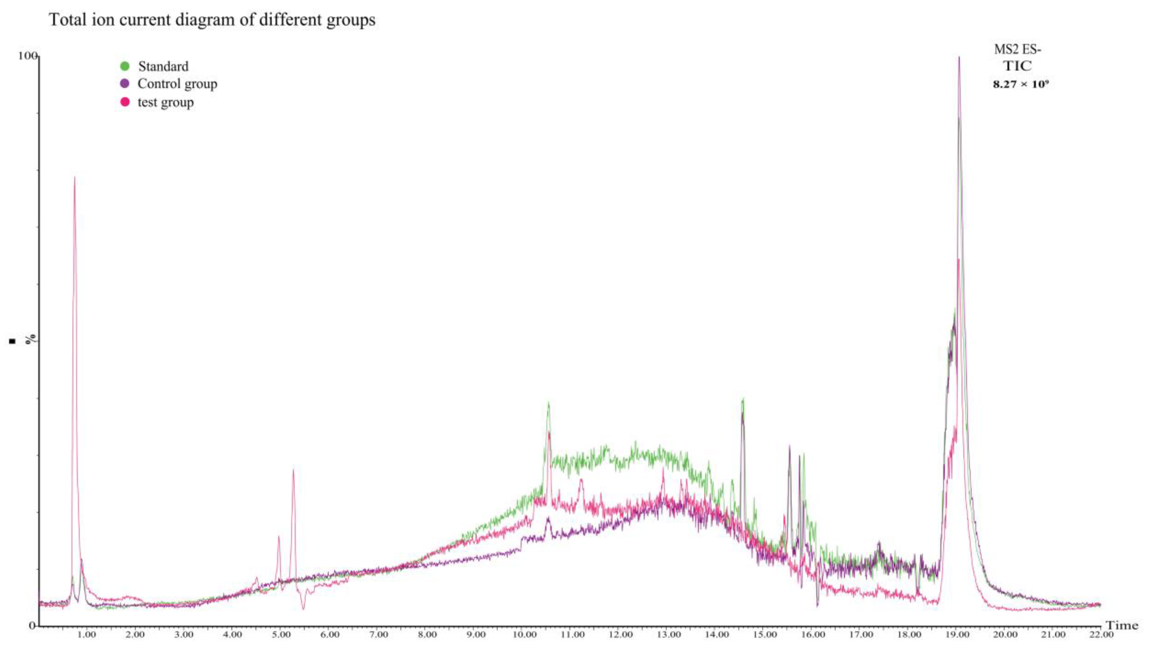
2.4. Gossypol Analysis

The degradation rate of FG is the most prominent indicator of changes in gossypol content. During the whole reaction, it is most important to detect the change in FG, and the total amount of gossypol is represented by the sum of free and bound gossypol [53]. Many studies tend to ignore the changes in total gossypol (TG) levels. In this study, in order to rule out the possibility that changes in gossypol levels can be attributed to physical action rather than biodegradation, we detected TG levels in the system. The traditional aniline method for the determination of gossypol was not used due to the low recovery of gossypol and a complex peak formed by the combination of gossypol and N, N-dimethylformamide (DMF) [54]. FG remaining in the reaction mixture was measured using the HPLC method with the aforementioned modifications [55,56]. In this study, the content of FG and TG were determined systematically. Since gossypol is unstable and easily oxidized, the addition of NADPH-Na4 initiates the reaction and stabilizes gossypol [55]. The results are shown in Table 1. The results of the blank group showed that the levels of total and FG decreased from 450 µg to 406.62 µg due to spontaneous oxidation over time even without the action of the enzyme (Table 1). This finding can be attributed to the inherent instability of gossypol. Although no plasmid was inserted in the control group, P. pastoris GS115 itself expressed certain endogenous enzymes. Hence, TG and FG levels decreased, which was consistent with the results of previous studies [57]. Under the influence of recombinant CarE, TG and FG levels decreased rapidly in the experimental group. The degradation rate of FG was 89%, and the difference between the experimental group and the control group were extremely significant (p < 0.01). Previously, a Lactobacillus strain screened from the rumen of dairy cows demonstrated the best degradation effect on gossypol. With an increase in the time of microbial action on gossypol, the degradation rate reaches 83% [9]. The enzymatic degradation effect of exogenous substances is better than that of microorganisms, and the time is shorter. Overall, finding an efficient and safe enzyme provides a theoretical basis for the degradation of gossypol. The biodegradation effect of recombinant CarE on gossypol was evaluated. Furthermore, FG was converted into degradation products, which laid the foundation for the subsequent LC-MS identification of its properties.

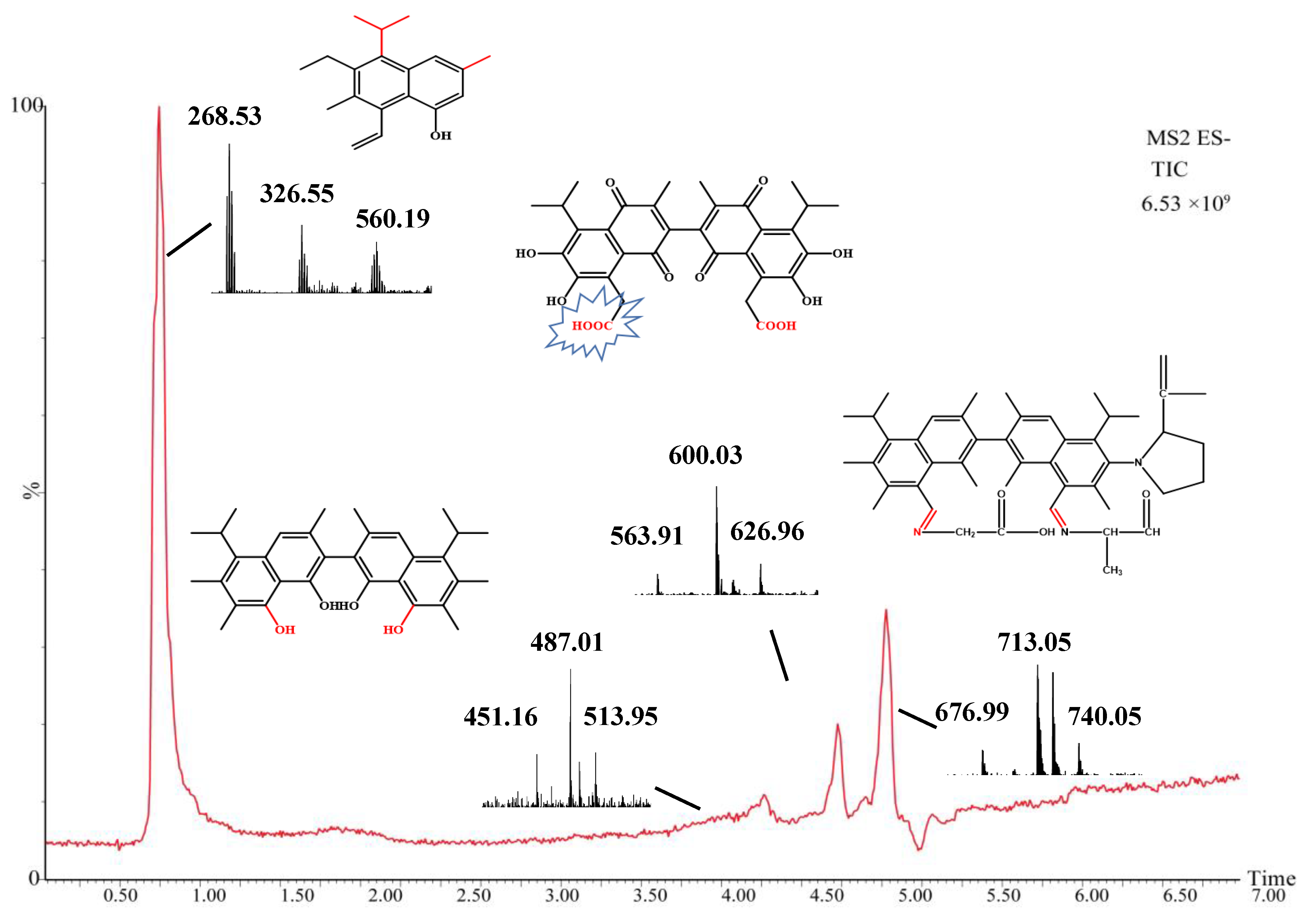
2.5. Mass Spectrometry Analysis of Intermediate Products

CarE can degrade gossypol, Hence, an evaluation of its structure is necessary. Degrading gossypol is a process which aims to mitigate the toxicity of FG by converting it into less toxic or non-toxic substances. One way to reduce the toxicity of gossypol and enhance its biological properties is to convert it into azepine derivatives, such as Schiff bases or hydrazones. The Schiff base or hydrazone gossypol can undergo Schiff base formation [58,59], ozonation [15], oxidation [60], and methylation [61] to form gossypol derivatives.
CarEs is a serine hydrolase that reacts with many compounds with different structures that contain ester bonds [62,63,64,65,66]. As mentioned above, the toxicity of gossypol can be attributed to six phenolic hydroxyl groups and two aldehyde groups, and CarEs can catalyze the hydrolysis of esters or amide compounds into corresponding alcohols and carboxylic acids [67]. CarEs are widely considered attractive and advantageous biocatalysts due to their characteristics, such as their ability to accept a wide range of substrates and their high stereospecificity, high tolerance to organic solvents, and lack of cofactors required for the reaction [68]. Conserved region prediction analysis was performed with the H. armigera CarE CCE001a protein (https://www.genome.jp/tools/motif//, 18 April 2022). The protein showed a co-esterase family (PF00135) domain and a dehydrogenase family (PF07859) domain (Figure S3). The enzymatic reaction itself is a complex process involving graded metabolites. Hence, we initially hypothesized that the action of CarEs on gossypol may involve the hydrolase activity of the enzyme and the reaction of unknown intermediate metabolites. Gossypol is metabolized in different pathways in animals such as pigs [69] and hens [70]; however, these metabolites have not been completely elucidated as gossypol and its derivatives are excreted in low concentrations in animal feces [71,72]. Gossypol is bioconverted to gossypol ketone, gossypol acid, and demethylated gossypol acid [71]. The main scavenging mechanisms of gossypol include glucoaldehyde acidification and bile excretion [72]. In our reaction, CarE showed the characteristics of a hydrolase and some unknown enzymatic functions. The phenolic hydroxyl and aldehyde groups of gossypol were exposed, and the stability was relatively poor. Therefore, we explored the possibility of another degradation mechanism of gossypol. Metabolites were identified from the reaction system using UPLC-QTOF/MS and Masslynx 4.1 software (Waters Corporation, Milford, MA, USA) analysis and divided into standard, control, and test group. Gossypol formed a deprotonated molecule [M-H] at m/z 518.1857 with a theoretical mass of 517.1910 in the negative ion scan mode. A comparison of the total ion current of the three groups (Figure 6) shows that the peak observed in the test group (which included the recombinant CarE in the reaction) at a retention interval of 19 min corresponds to gossypol, and its lower signal intensity quantitatively indicates that the gossypol content decreased under the influence of the recombinant CarE. In the endogenous enzyme group, the gossypol level was lower than that of the standard product. Although no recombinant CarE was added, the endogenous enzymes of P. pastoris may bind with gossypol to form bound gossypol, which reduced the TG content. After recombinant CarE was added to the test group (Figure 7), a new peak was observed that was not seen in the results from the standard and control groups. The peak had a retention time of 0.75 min and was measured at m/z 268.53. The chemical formula was C19H24O. We designated it as compound M0. The theoretical mass was 269.19, and the product was confirmed to be hemigossypol [73]. Gossypol itself is a polyphenol binaphthyl, which endows the binaphthyl bond with instability due to photosensitivity [74]. Another peak in the test group was observed at a retention interval of 4.51 min with m/z as 487.01 This compound, designated M1, had a chemical formula of C31H36O5 and a theoretical mass of 489.26. It was also the product of recombinant CarE activity, during which the exposed groups of gossypol were removed to detoxify gossypol. The degradation of gossypol by enzymes is less frequently examined. Previous studies have focused on the oxidation of gossypol by H. armigera P450 enzymes [57]. Laccase cyclization-hydroxyl aldehyde condensation [61] is a reaction of gossypol that renders it less toxic. This finding also coincides with the fact that enzymes have a wide range of substrates. Another detoxification product, M2, appeared at a retention time of 4.97 min, m/z of 600.03. This substance, with a chemical formula of C34H36N2O8 and theoretical mass of 601.25, has not been observed in other studies. Compound M2 was formed owing to the effect of CarE on the toxic aldehyde group of gossypol. This reaction formed an intermediate carboxylic acid in place of the aldehyde. Further, hydrolysis of the carboxylic acid derivative realized the complete elimination of the aldehyde group. Another detoxification product corresponding to the peak at 5.28 min was designated M3 with an m/z of 713.05. The structural formula of M2 was C47H59N3O3, with a theoretical mass of 714.46, and it is considered to be an Aza derivative of gossypol. We analyzed the recombinant CarE and found that the alanine and leucine levels were the highest. Furthermore, the aldehyde group on gossypol could react with the α-NH2 of amino acids to form Schiff bases, thereby forming azides. This finding is consistent with those of previous reports, which showed that binding or removing toxic aldehyde groups from gossypol can effectively reduce its toxicity. The metabolic pathway for the in vitro degradation of gossypol is shown in Figure 8. The exact mass, elemental composition, and molecular formula of the gossypol degradation products are listed in Table 2. In general, the metabolism of gossypol by the enzymatic reaction of H. armigera CCE001a is a complex process involving hydrolysis, dehydrogenation, and covalent binding to amine products. Hence, defining the metabolism of gossypol at all levels is challenging.

3. Conclusions

In this study, a CarE from H. armigera, CCE001a, was successfully expressed in P. pastoris GS115, and the activity of recombinant CarE on FG was examined. After treatment with recombinant CarE, the degradation of TG was 90%, and the degradation of FG was up to 89%. Detoxification is realized via radical and hydroxyl group attacks or reduction in gossypol levels mediated via binding with amino acids of the CarE to form azide compounds. This study confirms that recombinant CarE isolated from H. armigera can be utilized as an effective gossypol-degrading enzyme for cottonseed meal, a high-protein animal raw material. This enzyme would aid in detoxifying the cottonseed meal, allowing it to be used as animal feed.

4. Materials and Methods

4.1. Source of Target Gene Sequence

H. armigera was collected from cotton-growing sites in Shihezi, Xinjiang Autonomous Region, China. H. armigera was reared on a gossypol diet under laboratory conditions. Total RNA was extracted from the gossypol-treated fifth-generation larvae of H. armigera using TRIzol reagent (Sigma-Aldrich, St. Louis, MO, USA). The extracted RNA was transcribed into cDNA following the manufacturer’s protocol (Promega, Madison, WI, USA).

4.2. cce001a Cloning and Expression Vector Construction

The cce001a gene (GenBank accession number: HM191471.1) was amplified, using TaKaRa Ex Taq® (TaKaRa, Dalian, China) with degenerate primers 001a F (ATGTCAGACAGCGCACAGGACG) and 001a R (ACTCCATACATCTGCTGAATAT). Restriction sites were introduced using cDNA extracted from the midgut of H. armigera as the template. Polymerase chain reaction (PCR) cycling conditions are listed as follows: one cycle at 94 °C for 3 min, followed by 35 cycles at 94 °C for 30 s, 60 °C for 30 s, and then 72 °C for 2 min. The PCR product was purified using an EZNA Gel Extraction Kit (Omega BioTek, Norcross, GA, USA), cloned into the pGEM-T vector, and transformed into E. coli Stbl3-competent cells for two-way sequencing (Shanghai Jierui, Shanghai, China). After the expression vector was digested with EcoRI and XbaI, the PCR product and expression vector were ligated using a Gibson assembly. The plasmid was transformed into E. coli DH5α-competent cells, extracted, and sent to Shanghai Jierui for sequencing.

4.3. Electrotransformation, Screening, and Identification of Transformants with High Expression of CCE001a in P. pastoris

A total of 5 μg of the pPICZαA-CCE001a plasmid was linearized by the SacI rapid digestion method and electroporated into P. pastoris GS115-competent cells (80 μL) under the following conditions: 25 μF, 200 Ω, and 1.5 kV using a micropulser electroporator (Bio-Rad, Hercules, CA, USA). Following this, 1 mL of pre-cooled sorbitol (1 M) was added to the electro-rotor cup and the cells were incubated at 28 °C for 2 h. The cells were then spread on YPDS (Yeast Extract Peptone Dextrose Medium Sorbitol)-Zeo(Bleomycin) plates containing 100 mg/mL Zeo and cultured at 28 °C for 48–72 h. To screen for high-copy recombinant clones, the cells were incubated in the presence of 0.5–3.0 mg/mL Zeocin® at 28 °C until colonies were visible. Resistant clones were selected and the yeast genome was extracted. Using the genome as a template and the universal primers 5′AOX (GACTGGTTCCAATTGACAAGC) and 3′AOX (GCAAATGGCATTCTGACATCC), positive recombinants were screened by PCR. Positive clones were selected and inoculated into a medium containing 100 mM potassium phosphate (pH 6.0), 0.34% yeast nitrogen base, 0.00004% biotin, 1% yeast extract, 2% peptone, and 1% glycerol. The mixture was incubated at 28 °C with shaking (250 rpm) for 24 h. Cell pellets were collected, resuspended in buffered methanol-complex medium (BMMY), and incubated again at 28 °C with shaking (250 rpm), with the addition of methanol every 24 h to maintain its content at 1% of the total volume. The total time required for continuous fermentation in the shake flask was 120 h. Samples were derived every 12 h and stored at −80 °C for subsequent use.

4.4. Sodium Dodecyl Sulfate-Polyacrylamide Gel Electrophoresis (SDS-PAGE) and Western Blotting

Cells stimulated with methanol were collected at various time points during their incubation, concentrated by centrifugation at 12,000× g at 4 °C, and then disrupted by ultrasonication (Thermo Fisher Scientific, Waltham, MA, USA). Proteins in the supernatant were precipitated with trichloroacetic acid, boiled for 10 min and mixed with an equal volume of 1× loading buffer. SDS-PAGE was performed on a 10% polyacrylamide gel and Coomassie Brilliant Blue R250 was used to detect the protein bands. After transfer onto a polyvinylidene fluoride membrane, CarE was detected with an anti-His mouse polyclonal antibody (Cell Signalling Technology, Danvers, UT, USA) and diaminobenzidine substrate (Shenggong, Shanghai, China).

4.5. CarE Activity Measurements

CarE activity was determined by measuring the conversion of α-naphthyl acetate to 1-naphthyl ester. Assay reagents were mixed as specified in Table 1 in a 96-well plate and CarE activity was calculated by measuring the absorbance at 450 nm. Two reactions were set up, namely, one containing the enzyme solution and one blank with distilled water. Absorbance (A1) was measured every 10 s after mixing the reagents for 5 min at 37 °C, and absorbance (A2) was measured after 310 s. ΔA was calculated by measuring the difference between A1 and A2. The enzyme activity unit was defined as an increase of 1 unit in the catalytic light absorption value per min per mg of protein and per mL of the reaction system at 37 °C. The following formula was used to determine the U/mg:
CarE enzyme activity (U/mg protein) = 8 × (△A measuring tube − △A blank tube) × Cpr × F.

4.6. Ultra-High Performance Liquid Chromatography (UHPLC) Analysis of Gossypol

Active recombinant CarE was expressed in P. pastoris GS115 and UHPLC was used to analyze free gossypol after treatment with this enzyme. Free gossypol (20 μL at 300 μg/mL) was added to 200 μL of phosphate-buffered saline (PBS). Approximately 100 μg of protein was added to the mixture, which was then transferred to 1.5 mL tubes. To start the reaction, 4 μL NADPH-Na4 (25 mM) was added, and the mixture was incubated in the dark at 37 °C for 1.5 h. To stop the reaction, 400 μL of 70% acetone aqueous solution was added. An ultrasonic cleaner was used for sample extraction over a period of one hour with intermittent turning to maximize the extraction. Samples were centrifuged at 12,000× g for 1 min at 4 °C and the supernatant was collected. The supernatant was analyzed by HPLC [42], with total gossypol determined as described previously. The following groups were analyzed: blank group (BL-1), control group (Co-1), test group (MY-1).
The gossypol standard stock solution was diluted with 70% acetone aqueous solution to prepare gradient concentrations of 1–50 μg/mL. A total of 4 μL NADPH-Na4 was added to each reaction to evaluate the best chromatographic conditions and compare the peak area Y with concentration X. Linear regression was performed to obtain the standard reaction equation. A SUPELCOSILTM C18 column (4.6 mm × 250 mm, 5 μm; Sigma-Aldrich) and a 20-μL loop were used for chromatographic separation at 25 °C. Acetonitrile and 0.3% formic acid were used as mobile phases A and B, respectively. The gradient was linearly increased to 75% A and 25% B within 10 min, maintained at 85% A and 15% B for 10 min, and finally returned to 60% A and 40% B. The injection volume was 20 μL, the column temperature was maintained at 25 °C, and the detection wavelength was set at 380 nm. No less than three replicates per sample were evaluated.

4.7. UPLC-Quadrupole Time of Flight Mass Spectrometry (UPLC-QTOF/MS) Analysis

UPLC-QTOF/MS was used to determine whether recombinant CarE degraded gossypol and its resulting metabolites. The samples subjected to treatments included the control group (no enzymes added), endogenous enzyme group, and test group (esterase protein). The reaction mixture was incubated with gossypol in PBS (pH 7.0) at 30 °C for 30 min in a water bath. Next, 450 μL of methanol (−20 °C) was added, and the solution was vortexed for 30 s and centrifuged at 12,000 rpm and 4 °C for 10 min. The supernatant was transferred to a new 1.5 mL centrifuge tube, to which 300 μL acetonitrile was added. The tube was centrifuged at 12,000 rpm and 4 °C for 10 min, and 200 μL of the supernatant was passed through a 0.22 μm filter. The resulting sample was subjected to UPLC-QTOF/MS.

4.7.1. UPLC Conditions

An ACQUITY UPLC HSS T3 column (100 mm × 2.1 mm, 1.8 μm; Waters, Milford, MA, USA) was used with a flow rate of 0.3 mL/min, injection volume of 5 μL, and column temperature of 35 °C. Methanol (chromatographic grade) and acetonitrile (chromatographic grade) solutions were used as mobile phases A and B, respectively. Gradient elution was maintained as follows: 0 to 2.00 min with 20% B, 2.00 to 5.00 min with 40% B, 5.00 to 8.0 min with 60% B, 8.00 to 12 min with 80% B, and 12.00 to 22.00 min with 100% B.

4.7.2. MS Conditions

An electrospray negative ion mode was used as the ion source and the ion transfer tube temperature was 320 °C. The quantitative detection method was set to full-scan mode. The resolution was set to full scan (full mass) and secondary mass spectrometry scanning (MS/MS) was performed at 17,500. The isolation window (n window) was 1.0 m/z, the electrospray voltage was set to 3500 V, and the sheath gas pressure was set to 275.8 kPa. The auxiliary gas rate, backflush gas pressure, auxiliary gas heating temperature, and RF prism voltage were 180 L/h, 13.8 kPa, 300 °C, and 50%, respectively.

4.8. Data Analysis

One-way ANOVA was performed using SPSS 11.5 (SPSS Inc., Chicago, IL, USA), and Duncan’s method was used for significant difference analysis (p < 0.05). All data were expressed as mean  ±  SEMs. Mass spectral data were analyzed by Masslynx 4.1 (Waters) based on full scan analysis and extracted ion chromatograms. ChemDraw software (CambridgeSoft, Cambridge, MA, USA) was used to draw chemical structural formulas.

Supplementary Materials

The following supporting information can be downloaded at: https://www.mdpi.com/article/10.3390/toxins14120816/s1, Figure S1: CCE001a has a signal peptide at the 17 amino acid position; Figure S2: CCE001a protein contains an N-glycosylation site. The protein has an N-glycosylation site; Figure S3. CCE001a protein showed a co-esterase family (PF00135) domain and a dehydrogenase family (PF07859) domain; File S1: cce001a DNA sequence and pPICZαa-CCE001a sequence.

Author Contributions

Conceptualization, L.Z. and W.Z.; methodology, L.Z. and C.C.; software, L.Z. and X.Y.; validation, L.Z., C.C. and W.Z.; formal analysis, L.Z. and J.N.; data curation, L.Z.; writing—original draft preparation, L.Z., R.H. and C.N.; writing—review and editing, L.Z., R.H. and W.Z.; visualization, L.Z.; supervision, W.Z. and C.C.; project administration, W.Z. and C.C.; funding acquisition, W.Z. All authors have read and agreed to the published version of the manuscript.

Funding

This work was supported by the National Natural Science Foundation of China (Grant No. 31860660, 32060770) and the Shihezi university research startup project (Grant No. RCZK201901, KX01500301).

Institutional Review Board Statement

The animal research protocol was approved by the Experimental Animal Ethics Committee of the First Hospital of Shihezi University School of Medicine. Approval number: A2016-072-03. Date: 5 March 2016.

Informed Consent Statement

Not applicable.

Data Availability Statement

The data that support the findings of this study are available from the corresponding author upon reasonable request.

Conflicts of Interest

The authors declare no conflict of interest.

References

  1. Zhao, T.; Xie, Q.; Li, C.; Li, C.; Mei, L.; Yu, J.Z.; Chen, J.; Zhu, S. Cotton roots are the major source of gossypol biosynthesis and accumulation. BMC Plant Biol. 2020, 20, 88. [Google Scholar] [CrossRef] [PubMed]
  2. Martins, L.S.; Paulino, M.F.; Marcondes, M.I.; Rennó, L.N.; Mageste de Almeida, D.; Lopes, S.A.; Marquez, D.E.C.; Manso, M.R.; Gomes da Silva, A.; Valente, É.E.L. Cottonseed meal is a suitable replacement for soybean meal in supplements fed to Nellore heifers grazing Brachiaria decumbens. Anim. Prod. Sci. 2016, 57, 1893–1898. [Google Scholar] [CrossRef]
  3. Gadelha, I.C.; Fonseca, N.B.; Oloris, S.C.; Melo, M.M.; Soto-Blanco, B. Gossypol toxicity from cottonseed products. Sci. World J. 2014, 2014, 231635. [Google Scholar] [CrossRef] [PubMed] [Green Version]
  4. Li, M.H.; Robinson, E.H. Use of Cottonseed meal in aquatic animal diets: A review. N. Am. J. Aquac. 2006, 68, 14–22. [Google Scholar] [CrossRef]
  5. Yang, A.; Zhang, C.; Zhang, B.; Wang, Z.; Zhu, L.; Mu, Y.; Wang, S.; Qi, D. Effects of dietary cottonseed oil and cottonseed meal supplementation on liver lipid content, fatty acid profile and hepatic function in laying hens. Animals 2021, 11, 78. [Google Scholar] [CrossRef]
  6. Zhou, M.; Zhang, C.; Wu, Y.; Tang, Y. Metabolic engineering of gossypol in cotton. Appl. Microbiol. Biotechnol. 2013, 97, 6159–6165. [Google Scholar] [CrossRef]
  7. Willard, S.T.; Neuendorff, D.A.; Lewis, A.W.; Randel, R.D. Effects of free gossypol in the diet of pregnant and postpartum Brahman cows on calf development and cow performance. J. Anim. Sci. 1995, 73, 496–507. [Google Scholar] [CrossRef]
  8. Reiser, R.; Fu, H.C. The mechanism of gossypol detoxification by ruminant animals. J. Nutr. 1962, 76, 215–218. [Google Scholar] [CrossRef]
  9. Wang, W.K.; Li, W.J.; Wu, Q.C.; Wang, Y.L.; Li, S.L.; Yang, H.J. Isolation and identification of a rumen Lactobacillus bacteria and its degradation potential of gossypol in cottonseed meal during solid-state fermentation. Microorganisms 2021, 9, 2200. [Google Scholar] [CrossRef]
  10. Zhang, Y.; Zhang, Z.; Dai, L.; Liu, Y.; Cheng, M.; Chen, L. Isolation and characterization of a novel gossypol-degrading bacteria Bacillus subtilis strain Rumen Bacillus Subtilis. Asian-Australas. J. Anim. Sci. 2018, 31, 63–70. [Google Scholar] [CrossRef]
  11. Alexander, J.; Benford, D.; Cockburn, A. Nitrite as undesirable substances in animal feed-Scientific opinion of the panel on contaminants in the food chain. EFSA J. 2009, 7, 1017. [Google Scholar] [CrossRef]
  12. Lim, W.; Ham, J.; Park, S.; Bae, H.; You, S.; Song, G. Gossypol induces disruption of spermatogenesis and steroidogenesis in male mice. J. Agric. Food Chem. 2019, 67, 2075–2085. [Google Scholar] [CrossRef] [PubMed]
  13. Fonseca, N.B.d.S.; Gadelha, I.C.N.; Oloris, S.C.S.; Soto-Blanco, B. Effectiveness of albumin-conjugated gossypol as an immunogen to prevent gossypol-associated acute hepatotoxicity in rats. Food Chem. Toxicol. 2013, 56, 149–153. [Google Scholar] [CrossRef] [Green Version]
  14. EFSA Panel on Contaminants in the Food Chain (CONTAM); Knutsen, H.K.; Barregård, L.; Bignami, M.; Brüschweiler, B.; Ceccatelli, S.; Dinovi, M.; Edler, L.; Grasl-Kraupp, B.; Hogstrand, C.; et al. Presence of free gossypol in whole cottonseed. EFSA J. 2017, 15, e04850. [Google Scholar] [CrossRef] [PubMed] [Green Version]
  15. Romero, A.C.; Calori-Domingues, M.A.; Abdalla, A.L.; Augusto, P.E.D. Evaluation of ozone technology as an alternative for degradation of free gossypol in cottonseed meal: A prospective study. Food Addit. Contam. Part A Chem. Anal. Control Expo. Risk Assess. 2021, 38, 659–669. [Google Scholar] [CrossRef] [PubMed]
  16. Wang, X.; Howell, C.P.; Chen, F.; Yin, J.; Jiang, Y. Chapter 6 Gossypol-A polyphenolic compound from cotton plant. Adv. Food Nutr Res. 2009, 58, 215–263. [Google Scholar] [CrossRef] [PubMed]
  17. Zhang, W.; Xu, Z.; Sun, J.; Yang, X. A Study on the reduction of gossypol levels by mixed culture solid substrate fermentation of cottonseed meal. Asian-Australas. J. Anim. Sci. 2006, 19, 1314–1321. [Google Scholar] [CrossRef]
  18. Khalaf, M.A.; Meleigy, S.A. Reduction of free gossypol levels in cottonseed meal by microbial treatment. Int. J. Agric. Biol. 2008, 10, 185–190. [Google Scholar]
  19. Mellon, J.E.; Zelaya, C.A.; Dowd, M.K. Inhibitory effects of gossypol-related compounds on growth of Aspergillus flavus. Lett. Appl. Microbiol. 2011, 52, 406–412. [Google Scholar] [CrossRef]
  20. Yang, X.; Sun, J.Y.; Guo, J.L.; Weng, X.Y. Identification and proteomic analysis of a novel gossypol-degrading fungal strain. J. Sci. Food Agric. 2012, 92, 943–951. [Google Scholar] [CrossRef]
  21. Yang, C.; Song, G.; Lim, W. Effects of mycotoxin-contaminated feed on farm animals. J. Hazard. Mater. 2020, 389, 122087. [Google Scholar] [CrossRef]
  22. Rajarathnam, S.; Shashirekha, M.N.; Bano, Z. Biodegradation of gossypol by the white oyster mushroom, Pleurotus florida, during culturing on rice straw growth substrate, supplemented with cottonseed powder. World J. Microbiol. Biotechnol. 2001, 17, 221–227. [Google Scholar] [CrossRef]
  23. Banerjee, S.; Maiti, T.K.; Roy, R.N. Enzyme producing insect gut microbes: An unexplored biotechnological aspect. Crit. Rev. Biotechnol. 2022, 42, 384–402. [Google Scholar] [CrossRef] [PubMed]
  24. Wang, S.; Wang, X.; Penttinen, L.; Luo, H.; Zhang, Y.; Liu, B.; Yao, B.; Hakulinen, N.; Zhang, W.; Su, X. Patulin detoxification by recombinant manganese peroxidase from Moniliophthora roreri expressed by Pichia pastoris. Toxins 2022, 14, 440. [Google Scholar] [CrossRef] [PubMed]
  25. Jin, M.; Cheng, Y.; Guo, X.; Li, M.; Chakrabarty, S.; Liu, K.; Wu, K.; Xiao, Y. Down-regulation of lysosomal protein ABCB6 increases gossypol susceptibility in Helicoverpa armigera. Insect Biochem. Mol. Biol. 2020, 122, 103387. [Google Scholar] [CrossRef] [PubMed]
  26. Tian, X.; Ruan, J.; Huang, J.; Fang, X.; Mao, Y.; Wang, L.; Chen, X.; Yang, C. Gossypol: Phytoalexin of cotton. Sci. China Life Sci. 2016, 59, 122–129. [Google Scholar] [CrossRef] [Green Version]
  27. Kulieva, A.M.; Dalimov, D.N.; Dorenskaya, G.M.; Charieva, O.V.; Rozengart, V.I.; Kugusheva, L.I.; Moralev, S.N.; Babaev, B.N.; Abduvakhabov, A.A. Biochemical investigation of cholinesterases and carboxylesterases from the cotton bollwormHeliothis armigera. Chem. Nat. Compd. 1994, 30, 116–120. [Google Scholar] [CrossRef]
  28. Yang, S.; Wu, H.; Xie, J.; Rantala, M.J. Depressed performance and detoxification enzyme activities of Helicoverpa armigera fed with conventional cotton foliage subjected to methyl jasmonate exposure. Entomol. Exp. Appl. 2013, 147, 186–195. [Google Scholar] [CrossRef]
  29. Mulrooney, J.E.; Parrott, W.L.; Jenkins, J.N. Tolerance of pyrethroid-resistant tobacco budworm (Lepidoptera: Noctuidae) larvae to gossypol and piperonyl butoxide. J. Econ. Entomol. 1993, 86, 1014–1018. [Google Scholar] [CrossRef]
  30. Despres, L.; David, J.P.; Gallet, C. The evolutionary ecology of insect resistance to plant chemicals. Trends Ecol. Evol. 2007, 22, 298–307. [Google Scholar] [CrossRef]
  31. Kim, K.K.; Song, H.K.; Shin, D.H.; Hwang, K.Y.; Choe, S.; Yoo, J.O.; Suh, S.W. Crystal structure of carboxylesterase from Pseudomonas fluorescens, an a/b hydrolase with broad substrate specificity. Structure 1997, 5, 1571–1584. [Google Scholar] [CrossRef]
  32. Heidel-Fischer, H.M.; Vogel, H. Molecular mechanisms of insect adaptation to plant secondary compounds. Curr. Opin. Insect Sci. 2015, 8, 8–14. [Google Scholar] [CrossRef] [PubMed]
  33. Kong, G.; Daud, M.K.; Zhu, S. Effects of pigment glands and gossypol on growth, development and insecticide-resistance of cotton bollworm (Heliothis armigera (Hübner)). Crop Prot. 2010, 29, 813–819. [Google Scholar] [CrossRef]
  34. Wu, G.; Harris, M.K.; Guo, J.; Wan, F. Temporal allocation of metabolic tolerance in the body of beet armyworm in response to three gossypol-cotton cultivars. Sci. China C Life Sci. 2009, 52, 1140–1147. [Google Scholar] [CrossRef]
  35. Yang, S.; Cao, Q.; Peng, K.; Xie, J. Jasmonic acid-treated cotton plant leaves impair larvae growth performance, activities of detoxification enzymes, and insect humoral immunity of cotton bollworm. Neotrop. Entomol. 2022, 51, 570–582. [Google Scholar] [CrossRef] [PubMed]
  36. Jin, M.; Liao, C.; Fu, X.; Holdbrook, R.; Wu, K.; Xiao, Y. Adaptive regulation of detoxification enzymes in Helicoverpa armigera to different host plants. Insect Mol. Biol. 2019, 28, 628–636. [Google Scholar] [CrossRef] [PubMed]
  37. Zhao, J.; Liu, N.; Ma, J.; Huang, L.; Liu, X. Effect of silencing CYP6B6 of Helicoverpa armigera (Lepidoptera: Noctuidae) on its growth, development, and insecticide tolerance. J. Econ. Entomol. 2016, 109, 2506–2516. [Google Scholar] [CrossRef]
  38. Kang, Z.; Huang, H.; Zhang, Y.; Du, G.; Chen, J. Recent advances of molecular toolbox construction expand Pichia pastoris in synthetic biology applications. World J. Microbiol. Biotechnol. 2017, 33, 19. [Google Scholar] [CrossRef]
  39. Duan, G.; Ding, L.; Wei, D.; Zhou, H.; Chu, J.; Zhang, S.; Qian, J. Screening endogenous signal peptides and protein folding factors to promote the secretory expression of heterologous proteins in Pichia pastoris. J. Biotechnol. 2019, 306, 193–202. [Google Scholar] [CrossRef]
  40. Nguyen, H.Q.; Vu, V.H.; Le, P.D.; Chu, H.M. High-level expression, purification and properties of an Endochitinase gene without signal peptide from Lecanicillium lecanii 43H in Pichia pastoris. Mol. Biol. Rep. 2018, 45, 1067–1075. [Google Scholar] [CrossRef]
  41. Fonseca, M.I.; Molina, M.A.; Winnik, D.L.; Busi, M.V.; Farina, J.I.; Villalba, L.L.; Zapata, P.D. Isolation of a laccase-coding gene from the lignin-degrading fungus Phlebia brevispora BAFC 633 and heterologous expression in Pichia pastoris. J. Appl. Microbiol. 2018, 124, 1454–1468. [Google Scholar] [CrossRef] [PubMed]
  42. Baghban, R.; Farajnia, S.; Ghasemi, Y.; Mortazavi, M.; Zarghami, N.; Samadi, N. New developments in Pichia pastoris expression system, review and update. Curr. Pharm. Biotechnol. 2018, 19, 451–467. [Google Scholar] [CrossRef] [PubMed]
  43. Bai, L.S.; Zhao, C.X.; Xu, J.J.; Feng, C.; Li, Y.Q.; Dong, Y.L.; Ma, Z.Q. Identification and biochemical characterization of carboxylesterase 001G associated with insecticide detoxification in Helicoverpa armigera. Pestic. Biochem. Physiol. 2019, 157, 69–79. [Google Scholar] [CrossRef] [PubMed]
  44. Teese, M.G.; Farnsworth, C.A.; Li, Y.; Coppin, C.W.; Devonshire, A.L.; Scott, C.; East, P.; Russell, R.J.; Oakeshott, J.G. Heterologous expression and biochemical characterisation of fourteen esterases from Helicoverpa armigera. PLoS ONE 2013, 8, e65951. [Google Scholar] [CrossRef]
  45. Xu, Z.; Kong, J.; Zhang, S.; Wang, T.; Liu, X. Comparison of Enzyme Secretion and Ferulic Acid Production by Escherichia coli Expressing Different Lactobacillus Feruloyl Esterases. Front. Microbiol. 2020, 1, 1. [Google Scholar] [CrossRef]
  46. Wang, Z.; Guo, C.; Liu, L.; Huang, H. Effects of N-glycosylation on the biochemical properties of recombinant bEKL expressed in Pichia pastoris. Enzym. Microb. Technol. 2018, 114, 40–47. [Google Scholar] [CrossRef]
  47. Han, M.; Wang, X.; Ding, H.; Jin, M.; Yu, L.; Wang, J.; Yu, X. The role of N-glycosylation sites in the activity, stability, and expression of the recombinant elastase expressed by Pichia pastoris. Enzym. Microb. Technol. 2014, 54, 32–37. [Google Scholar] [CrossRef]
  48. Li, Y.; Liu, J.; Lu, M.; Ma, Z.; Cai, C.; Wang, Y.; Zhang, X. Bacterial expression and kinetic analysis of carboxylesterase 001D from Helicoverpa armigera. Int. J. Mol. Sci. 2016, 17, 493. [Google Scholar] [CrossRef] [Green Version]
  49. Luo, S.; Shu, C.; Xu, C.; Wang, R. Molecular cloning and expression in vitro of a carboxylesterase gene from the Glanville fritillary butterfly (Melitaea cinxia). Gene 2013, 524, 275–281. [Google Scholar] [CrossRef]
  50. Wang, Z.G.; Jiang, S.S.; Mota-Sanchez, D.; Wang, W.; Li, X.R.; Gao, Y.L.; Lu, X.P.; Yang, X.Q. Cytochrome P450-mediated lambda-cyhalothrin-resistance in a field strain of Helicoverpa armigera from northeast China. J. Agric. Food Chem. 2019, 67, 3546–3553. [Google Scholar] [CrossRef]
  51. Wu, S.; Yang, Y.; Yuan, G.; Campbell, P.M.; Teese, M.G.; Russell, R.J.; Oakeshott, J.G.; Wu, Y. Overexpressed esterases in a fenvalerate resistant strain of the cotton bollworm, Helicoverpa armigera. Insect Biochem. Mol. Biol. 2011, 41, 14–21. [Google Scholar] [CrossRef] [PubMed]
  52. Bradford, M.M. A rapid and sensitive method for the quantitation of microgram quantities of protein utilizing the principle of protein-dye binding. Anal. Biochem. 1976, 72, 248–254. [Google Scholar] [CrossRef]
  53. Huang, J.Q.; Fang, X.; Tian, X.; Chen, P.; Lin, J.L.; Guo, X.X.; Li, J.X.; Fan, Z.; Song, W.M.; Chen, F.Y.; et al. Aromatization of natural products by a specialized detoxification enzyme. Nat. Chem. Biol. 2020, 16, 250–256. [Google Scholar] [CrossRef] [PubMed]
  54. Hron, R.J.; Kuk, M.S.; Abraham, G. Determination of free and total gossypol by high performance liquid chromatography. J. Am. Oil Chem. Soc. 1990, 67, 182–187. [Google Scholar] [CrossRef]
  55. Cheng, C.; Cun-Xi, N.; Jing, L.; Yong-Qiang, W.; Yan-Feng, L.; Wen-Xia, G.; Wen-Ju, Z. Validated method to determine (±)-gossypol inCandida tropicalisculture by high-performance liquid chromatography. Acta Chromatogr. 2018, 30, 269–273. [Google Scholar] [CrossRef]
  56. Dowd, M.K. Stability of the gossypol-amine adducts used for chromatographic measurement of total and isomeric gossypol. J. Am. Oil Chem. Soc. 2020, 97, 671–675. [Google Scholar] [CrossRef]
  57. Chen, C.; Pi, W.; Zhang, Y.; Nie, C.X.; Liang, J.; Ma, X.; Wang, Y.; Ge, W.; Zhang, W.J. Effect of a functional recombinant cytochrome P450 enzyme of Helicoverpa armigera on gossypol metabolism co-expressed with NADPH-cytochrome P450 reductase in Pichia pastoris. Pestic. Biochem. Physiol. 2019, 155, 15–25. [Google Scholar] [CrossRef]
  58. Beyazit, N.; Cakran, H.S.; Cabir, A.; Akiscan, Y.; Demetgul, C. Synthesis, characterization and antioxidant activity of chitosan Schiff base derivatives bearing (−)-gossypol. Carbohydr. Polym. 2020, 240, 116333. [Google Scholar] [CrossRef]
  59. Vu, V.V.; Nhung, T.T.; Thanh, N.T.; Chinh, L.V.; Tien, V.D.; Thuy, V.T.; Thi Thao, D.; Nam, N.H.; Koeckritz, A.; Vu, T.K. Synthesis and biological evaluation of new (−)-gossypol-derived Schiff bases and hydrazones. J. Chem. 2017, 2017, 1–8. [Google Scholar] [CrossRef] [Green Version]
  60. Zhang, B.; Li, L.; Liu, Y.; Wang, Q. Antiviral mechanism study of gossypol and its Schiff base derivatives based on reactive oxygen species (ROS). RSC Adv. 2016, 6, 87637–87648. [Google Scholar] [CrossRef]
  61. Wang, L.; Chen, M.; Luo, X.; Fan, Y.; Zheng, Z.; He, Z.; Yin, R.; Meng, T.; Xu, S.; Pan, Y.; et al. Intramolecular annulation of gossypol by laccase to produce safe cottonseed protein. Front. Chem. 2020, 8, 583176. [Google Scholar] [CrossRef] [PubMed]
  62. Merali, Z.; Ross, S.; Pare, G. The pharmacogenetics of carboxylesterases: CES1 and CES2 genetic variants and their clinical effect. Drug Metabol. Drug Interact. 2014, 29, 143–151. [Google Scholar] [CrossRef] [PubMed]
  63. Satoh, T.; Hosokawa, M. Structure, function and regulation of carboxylesterases. Chem. Biol. Interact. 2006, 162, 195–211. [Google Scholar] [CrossRef] [PubMed]
  64. Zou, L.W.; Jin, Q.; Wang, D.D.; Qian, Q.K.; Hao, D.C.; Ge, G.B.; Yang, L. Carboxylesterase inhibitors: An update. Curr. Med. Chem. 2018, 25, 1627–1649. [Google Scholar] [CrossRef] [PubMed]
  65. Marchler-Bauer, A.; Bo, Y.; Han, L.; He, J.; Lanczycki, C.J.; Lu, S.; Chitsaz, F.; Derbyshire, M.K.; Geer, R.C.; Gonzales, N.R.; et al. CDD/SPARCLE: Functional classification of proteins via subfamily domain architectures. Nucleic Acids Res. 2017, 45, D200–D203. [Google Scholar] [CrossRef] [Green Version]
  66. Marchler-Bauer, A.; Derbyshire, M.K.; Gonzales, N.R.; Lu, S.; Chitsaz, F.; Geer, L.Y.; Geer, R.C.; He, J.; Gwadz, M.; Hurwitz, D.I.; et al. CDD: NCBI’s conserved domain database. Nucleic Acids Res. 2015, 43, D222–D226. [Google Scholar] [CrossRef] [Green Version]
  67. Wang, D.D.; Zou, L.W.; Jin, Q.; Hou, J.; Ge, G.B.; Yang, L. Recent progress in the discovery of natural inhibitors against human carboxylesterases. Fitoterapia 2017, 117, 84–95. [Google Scholar] [CrossRef]
  68. Bornscheuer, U.T. Microbial carboxyl esterases: Classification, properties and application in biocatalysis. FEMS Microbiol. Rev. 2002, 26, 73–81. [Google Scholar] [CrossRef]
  69. Katoh, N.; Kimura, K. Inhibition by gossypol of cyclic nucleotide-independent phosvitin kinase from pig testis. Nihon Juigaku Zasshi 1989, 51, 105–109. [Google Scholar] [CrossRef]
  70. Gamboa, D.A.; Calhoun, M.C.; Kuhlmann, S.W.; Haq, A.U.; Bailey, C.A. Tissue distribution of gossypol enantiomers in broilers fed various cottonseed meals. Poult. Sci. 2001, 80, 920–925. [Google Scholar] [CrossRef]
  71. Abou-Donia, M.B. Physiological effects and metabolism of gossypol. Am. J. Physiol. Endocrinol. Metab. 1976, 61, 6. [Google Scholar] [CrossRef]
  72. Krempl, C.; Sporer, T.; Reichelt, M.; Ahn, S.J.; Heidel-Fischer, H.; Vogel, H.; Heckel, D.G.; Joussen, N. Potential detoxification of gossypol by UDP-glycosyltransferases in the two Heliothine moth species Helicoverpa armigera and Heliothis virescens. Insect Biochem. Mol. Biol. 2016, 71, 49–57. [Google Scholar] [CrossRef] [PubMed]
  73. Bell, A.A.; Stipanovic, R.D.; Howell, C.R.; Fryxell, P.A. Antimicrobial terpenoids of Gossypium: Hemigossypol, 6-methoxyhemigossypol and 6-deoxyhemigossypol. Phytochemistry 1975, 14, 225–231. [Google Scholar] [CrossRef]
  74. Heywood, R. The toxicology of gossypol acetic acid and (−)-gossypol. Contraception 1988, 37, 185–190. [Google Scholar] [CrossRef]
  75. Abou-Donia, M.B.; Dieckert, J.W. Metabolic fate of gossypol: The metabolism of [14C]gossypol in swine. Toxicol. Appl. Pharmacol. 1975, 31, 32–46. [Google Scholar] [CrossRef]
Figure 1. Plasmid maps of pPICZαA and pPICZαA-cce001a. Aox1P blue arrow promoter is the direction of methanol-induced protein expression; Ori yellow arrow refers to the replication initiation site; gray box is the polyclonal site; black arrow is the transcription terminator; green arrow is the antibiotic marker; red arrow is the cce001a gene. Cloning of the cce001a fragment into the polyclonal site (MCS) of the linearized expression vector pPICZαA by Gibson assembly. Assembly with pPICZαA to form a complete construct.
Toxins 14 00816 g001
Figure 2. DNA marker (lane M); pPICZαA-cce001a verified by EcoRI and XbaI double digestion (lane 1).
Toxins 14 00816 g002
Figure 3. (a) DNA marker (lane M),Empty pPICZαA (lane 1), pPICZαA-cce001a (lane 2, 3, 4). The PCR templates of empty pPICZαA and pPICZαA-cce001a involved recombinant GS115 genome fragments. The following primers were used: 5′AOX and 3′AOX primers (lanes 2, 3, and 4). (b) Empty pPICZαA (lane 1), pPICZαA-cce001a (lane 2, 3, and 4). PCR templates of empty pPICZαA plasmid and pPICZαA-cce001a comprised recombinant GS115 genome fragments, and cce001a specific primers were used (lanes 2, 3, and 4).
Toxins 14 00816 g003
Figure 4. Secretion-induced protein expression and Western blot analysis of pPICZαA-CCE001a recombinant strain. (a) The sample was separated on a 10% SDS-PAGE gel; lane M represents the protein molecular marker, Lane 1 specifies the pPICZαA-GS115 control, lanes 2–7 correspond to the expression supernatant of GS115-pPICZαA-CCE001a in the time range of 12 h to 96 h; lane 2 = 12 h, lane 3 = 24 h, lane 4 = 36 h, lane 5 = 48 h, lane 6 = 72 h, and lane 7 = 96 h. (b) Western blot analysis of recombinant CarE using anti-His tag antibody. M represents the protein molecular marker, lane 1 is the Western blot analysis of control, lane 2 and lane 3 are the Western blot analysis of recombinant CarE.
Toxins 14 00816 g004
Figure 5. Absorbance of CarE at OD450 nm over time.
Toxins 14 00816 g005
Figure 6. Total ion current diagram demonstrating various experimental groups. Green represents the gossypol standard, purple represents the control group, and red represents the test group.
Toxins 14 00816 g006
Figure 7. TIC and mass spectrum of gossypol metabolites detected by UPLC-QTOF/MS under negative ion scanning. The total ion current diagrams of the three groups were compared, and the substances appearing in the test group, but not in the other groups, were analyzed further. Due to the instability of gossypol, it was degraded to semi-gossypol and its derivatives. GS115-pPICZαA-CCE001a esterase group used gossypol as a substrate. Furthermore, nitrogenous substances in degrading enzymes can interact with gossypol and metabolize it. The products of interest formed in the reaction showed m/z ratios of 268.18, 600.25, 713.46, 560.15, and 488.26.
Toxins 14 00816 g007
Figure 8. Schematic diagram of the degradation pathway of gossypol mediated by the CCE001a enzyme of H. armigera. Note: The dotted arrow indicates the intermediate product of gossypol obtained after the action of esterase and gossypol, “*” indicates different types of functional groups and the solid arrow indicates the metabolic pathway of gossypol [75] reported in a previous study.
Toxins 14 00816 g008
Table 1. Measurement of gossypol levels and degradation rate after the addition of recombinant CarE.
GroupingGossypol Content (μg/mL)Detoxification Rate (%)
TGFGTGFG
Bl-1406.62 ± 0.09 a258.63 ± 0.01 a1042
Co-185.76 ± 0.07 b48.15 ± 0.07 b8081
MY-142.34 ± 0.05 c27.43 ± 0.03 c9089
Note: In Table 1 BL-1, Co-1, and MY-1 represent the blank group, control group, and test group, respectively. Values are presented as mean ± SEMs. Different lowercase letters in the same column indicate significant differences (p < 0.05). TG, total gossypol; FG, free gossypol.
Table 2. The mass spectra data for gossypol and its intermediates.
CompoundExperimental Mass (m/z)Theoretical Mass (m/z)Retention Time (min)Molecular Formula
Gossypol517.18518.1919.7C30H30O8
M0268.18269.190.75C19H24O
M1488.22489.264.97C31H36O5
M2600.25601.254.97C32H30O12
M3713.46714.465.28C47H59N3O3
Publisher’s Note: MDPI stays neutral with regard to jurisdictional claims in published maps and institutional affiliations.

