Article

Structural Modeling of Cell Wall Peptidase CwpFM (EntFM) Reveals Distinct Intrinsically Disordered Extensions Specific to Pathogenic Bacillus cereus Strains

by
Seav-Ly Tran
1,
Delphine Cormontagne
1,
Jasmina Vidic
1,
Gwenaëlle André-Leroux
2,* and
Nalini Ramarao
1,*
1
Micalis Institute, INRAE, AgroParisTech, Université Paris-Saclay, 78350 Jouy-en-Josas, France
2
MaIAGE, INRAE, AgroParisTech, Université Paris-Saclay, 78350 Jouy-en-Josas, France
*
Authors to whom correspondence should be addressed.
Toxins 2020, 12(9), 593; https://doi.org/10.3390/toxins12090593
Submission received: 10 August 2020 / Revised: 4 September 2020 / Accepted: 9 September 2020 / Published: 14 September 2020
(This article belongs to the Special Issue Bacillus cereus Toxins)

Abstract

:
The emergence of B. cereus as an opportunistic food-borne pathogen has intensified the need to distinguish strains of public health concern. The heterogeneity of the diseases associated with B. cereus infections emphasizes the versatility of these bacteria strains to colonize their host. Nevertheless, the molecular basis of these differences remains unclear. Several toxins are involved in virulence, particularly in gastrointestinal disorders, but there are currently no biological markers able to differentiate pathogenic from harmless strains. We have previously shown that CwpFM is a cell wall peptidase involved in B. cereus virulence. Here, we report a sequence/structure/function characterization of 39 CwpFM sequences, chosen from a collection of B. cereus with diverse virulence phenotypes, from harmless to highly pathogenic strains. CwpFM is homology-modeled in silico as an exported papain-like endopeptidase, with an N-terminal end composed of three successive bacterial Src Homology 3 domains (SH3b1–3) likely to control protein–protein interactions in signaling pathways, and a C-terminal end that contains a catalytic NLPC_P60 domain primed to form a competent active site. We confirmed in vitro that CwpFM is an endopeptidase with a moderate peptidoglycan hydrolase activity. Remarkably, CwpFMs from pathogenic strains harbor a specific stretch of twenty residues intrinsically disordered, inserted between the SH3b3 and the catalytic NLPC_P60 domain. This strongly suggests this linker as a marker of differentiation between B. cereus strains. We believe that our findings improve our understanding of the pathogenicity of B. cereus while advancing both clinical diagnosis and food safety.
Key Contribution: CwpFM as an exported papain-like endopeptidase with PG hydrolase activity; CwpFMs from pathogenic B. cereus strains harbor a specific disordered linker; the CwpFM linker is a new marker of pathogenic B. cereus strains.

Graphical Abstract

1. Introduction

The cell wall (CW) of bacteria is an intricate mesh of macromolecules composed of peptidoglycan (PG), a complex polymer formed by linear glycan chains cross-linked by peptide stems. The glycan chains are usually long and alternate N-acetylglucosamine (NAG) with N-acetylmuramic acid (NAM) residues through β-(1, 4) bonds. In most Gram-positive bacteria like Bacillus cereus, the core structure of a non-cross-linked stem attached is L-Ala-Ɣ-D-Glu-L-Lys-D-Ala-D-Ala. Due to environmental adaptation, the PG core can undergo chemical modification like O-acetylation, N-deacetylation amidation or can incorporate Gly and non-canonical D-amino acids [1]. Additionally, the CW accommodates macromolecular components such as teichoic acid, lipoteichoic acids, polysaccharides and complex proteins like export secretion systems. CW integrity and plasticity need to be tuned to adapt readily to the bacterial cell cycle or to the changing environment, and its biosynthesis must be also highly regulated as CW disruption leads to bacterial cell death [2]. Accordingly, PG metabolism requires an exquisite and timely remodeling, organized by bacterial cell wall hydrolases (CWHs) [3]. CWHs play an essential physiological role in cell wall integrity. Additionally, they are implicated in bacterial adhesion and invasiveness [4], persistence in the host [5] and in the initiation step of biofilm formation [6], therefore contributing to bacterial pathogenicity. Moreover, the resulting cell wall fragments are recycled as signaling molecules to trigger bacterial communication, immune response or antibiotics resistance [5,7]. Finally, they also prime the insertion of supramolecular structures like secretion, flagella or pili systems [8]. Three types of CWH exist, each displaying a specificity towards PG: (i) cell wall amidase (CWA) catalyzes the hydrolysis of the amide bond between NAM and L-Ala at the N-terminal of the stem peptide; (ii) cell wall glycosidase (CWG) cleaves the glycosidic linkages; and (iii) cell wall peptidase (CWP) hydrolyses the amide bonds with the PG chains [8]. Recent structural data evidence that CWHs show modularity, with a catalytic domain combined to one or several CW binding domains (CBDs) located at the N- or C-terminal ends. This modular organization associated with CBD repeats is highly suspected to engage these hydrolases as a platform responsible for CW integrity. Interestingly, due to their inherent flexibility and lack of structural characterization, the linker regions that connect the binding domains to the catalytic one have received little attention until now. Nevertheless, interest is increasing as they are shown to play a role in domains orientation or swapping, and dynamics that result in substrate specificity and affinity [1,2,3].
The Bacillus cereus group is composed of rod-shaped, spore-forming, aerobic or facultative anaerobic species [9], among which the most noticeable members are species from B. cereus sensu stricto (usually referred to as B. cereus), B. mycoides, B. pseudomycoides, B. weihenstephanensis, B. anthracis, B. thuringiensis, B. cytotoxicus and B. toyonensis. The genetic proximity of the species within the B. cereus group has not permitted their distinction from a phylogenetic point of view; nevertheless, species can be differentiated through phenotypic characteristics, host species predilection and clinical manifestation [10,11]. Four members are pathogens, namely B. thuringiensis, B. anthracis, B. cytotoxicus and B. cereus. Despite being long considered as nonpathogenic to humans, B. thuringiensis has been occasionally identified as responsible for human infections including food poisoning-associated diarrheas, ocular infections, periodontitis, burn and wound infections or bacteremia [12]. In addition, B. thuringiensis can produce potent insecticidal crystal proteins and has been used since a decade ago as a bio-insecticide [13]. B. anthracis is the agent responsible for anthrax disease in animals and humans, and is an important biological warfare agent [14]. B. cytotoxicus is a human pathogen associated with occasional fatal food poisoning [15]. B. cereus is a major food-borne human pathogen (FBO), responsible for two types of food poisoning, the emetic and gastrointestinal syndromes [16]. The emetic syndrome is caused by an emetic toxin called cereulide, and results in vomiting [17,18,19,20]. The diarrheal syndrome is associated with three pore-forming enterotoxins, called Cytotoxin K (CytK1 and CytK2), Enterotoxin Nhe (non-hemolytic enterotoxin) and Enterotoxin Hbl (Hemolysin BL) [20,21]. Recently, the non-redundant Nhe and Hbl toxins were shown to share the same mode of action and to operate synergistically to activate the NLRP3 inflammasome, which is a critical component of host innate immune defense [22]. Other factors have also been implicated in B. cereus virulence. In particular hemolysins, proteases and repair factors have been shown to be involved in the resistance of B. cereus to the host immune system [23,24,25,26,27]. Furthermore, we have shown that EntFM, formerly isolated after purification from B. cereus strain FM-1 and identified as an enterotoxin [28], is in fact a cell wall peptidase. Accordingly, EntFM was renamed CwpFM [29]. CwpFM is a 45-kDa protein, responsible for characteristic enterotoxin symptoms in animal models, increases in vascular permeability in rabbits, infiltration into the ligated loops of rabbits and toxicity towards Vero epithelial cells, whereas it did not show either hemolytic or lecithinase activities [30,31]. Consistently, we have evidenced that CwpFM of B. cereus is involved in bacterial motility and morphology, adhesion to epithelial cells and biofilm formation and is essential to promote B. cereus virulence in an insect model of infection [27]. Prevalence studies have shown that the cwpFM gene is present on the chromosome as a single copy and restricted to almost all members of the B. cereus group [32,33,34].
Here, we report a sequence/structure/function characterization, following the subsequent bioinformatic analysis of 39 CwpFM sequences, chosen from a collection of B. cereus with a diverse virulence phenotype, from harmless to highly pathogenic strains. We also homology-modeled the 3D structure of the CwpFM protein from B. cereus. We show that CwpFM is an exported papain-like endopeptidase with, at the N-terminal end, three successive bacterial Src Homology 3 domains (SH3b1-3), and at the C-terminal end, a catalytic NLPC_P60 domain (New lipoprotein C/Protein of 60 KDa), that involves a competent active site. We confirmed in vitro that CwpFM is an endopeptidase despite a moderate peptidoglycan hydrolase activity. Additionally, we predicted that each of the three linkers connecting SH3b1-2, SH3b2-3 or SH3b3 to NLPC_P60 modules associates low-complexity with an intrinsically disordered pattern. Remarkably, CwpFMs from pathogenic strains harbor a specific stretch of intrinsically disordered linkers, particularly long, located between SH3b3 and catalytic NLPC_P60 domains. This pattern exquisitely discriminates pathogenic from non-pathogenic strains. Thus, we propose that it could define a marker of differentiation between the B. cereus strains.

2. Results

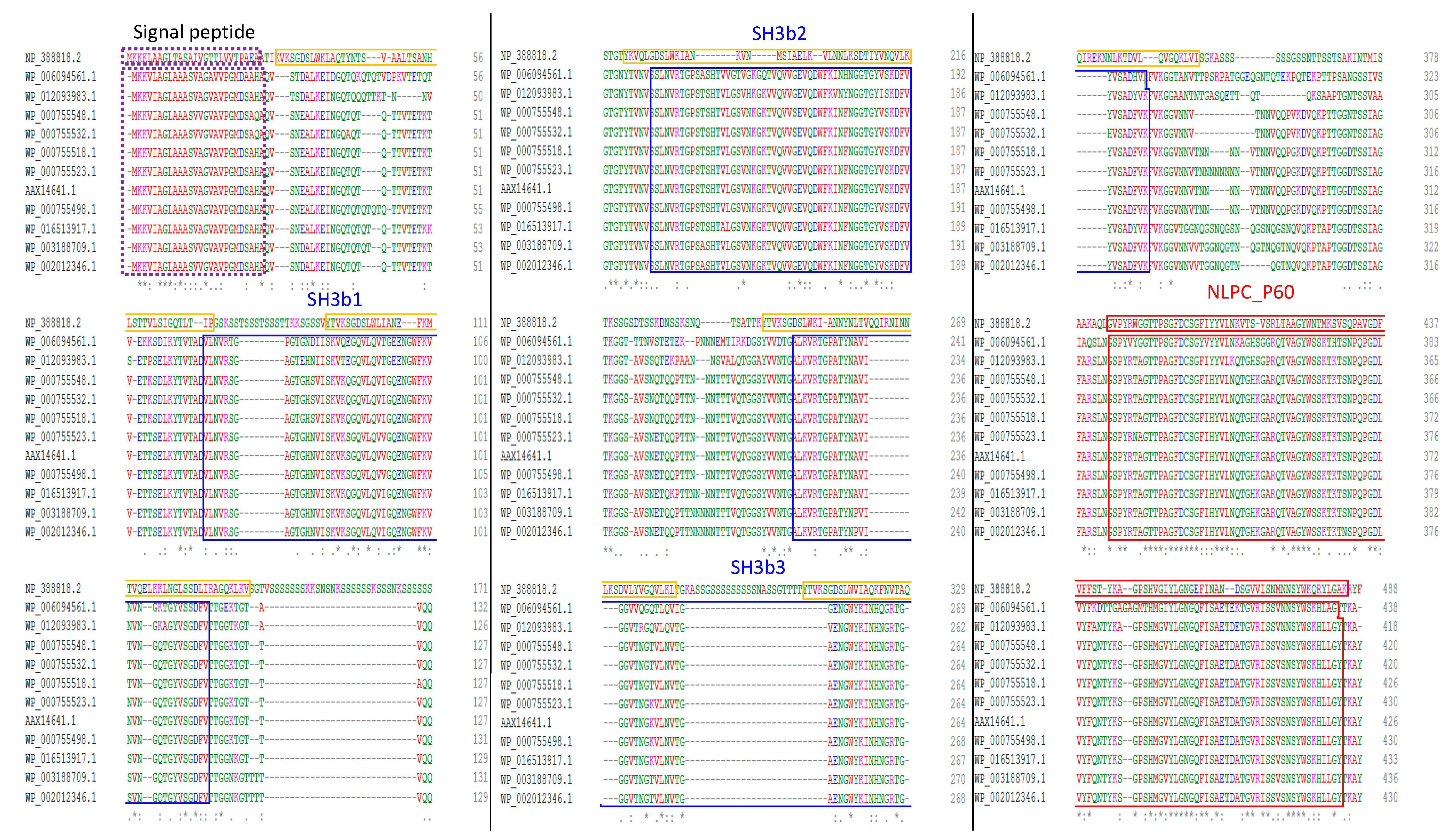
2.1. CwpFM is Present and Highly Conserved within the Members of the B. cereus Group

We have previously shown that CwpFM is only detected within the members of the B. cereus group [26]. Here, we did a comparative analysis of CwpFM from 10 strains representative of the B. cereus group (Table 1). A Blastn search with the complete published sequence of cwpFM from B. cereus isolate A6 (AY789084.1) was performed to identify CwpFM homologues in the selected genome (Table 2).
Only one copy of the gene was detected in each Bacillus genome. All the proteins encoded by the identified CwpFM homologues belong to the C40 family peptidase. Our bioinformatic analysis revealed that CwpFM is highly conserved among the B. cereus group. Sequence identities, when compared to CwpFM of the A6 strain, range from 72% for B. pseudomycoides DSM 12442 to 99% for B. cereus ATCC 14579. The most closely related sequences are between B. cereus and B. thuringiensis species with identities above 95.5%.

2.2. Domain Organization of the CwpFM Homologues from the B. cereus Group Indicates the Presence of Cell Wall Degradation Domains

All the CwpFM amino acid sequences from strains representative of the B. cereus group were aligned and domains were identified using both Clustal Omega (clustalo 1.2.4) and InterPro online tools. A conserved signal peptide composed of 26 amino acid residues was predicted to translocate using the Sec translocon, and to be cleaved by Signal peptidase I at the cleavage site AH/QA_QV, located between positions 26 and 27. The prediction of secondary domains for CwpFMs indicated a modular topology combining three protein–protein interaction SH3b domains and one catalytic NLPC_P60 family for all the strains (Figure 1). This topology is predicted to be strictly conserved for all the strains, even for CwpFM from B. pseudomycoides that shows a sensitively lower identity with the other members of the B. cereus group. More largely, the topology is fully consistent with the modular architecture highlighted for CWPs in bacterial CWHs because it associates several cell wall binding domains to a catalytic domain [8].
Sequence alignment of CwpFM homologues from B. cereus isolate A6 (AAX14641.1), B. cereus ATCC 14579 (WP_000755498.1), B. cereus ATCC 10987 (WP_000755518.1), B. thuringiensis 407 cry- (WP_000755523.1), B. thuringiensis serovar konkukian str. 97–27 (WP_000755548.1), B. mycoides ATCC 6462 (WP_003188709.1), B. pseudomycoides DSM 12442 (WP_006094561.1), B. weihenstephanensis KBAB4 (WP_002012346.1), B. anthracis str Ames (WP_000755532.1), B. cytotoxicus NVH 391–98 (WP_012093983.1) and B. toyonensis BCT-7112 (WP_016513917.1) show a similar domain organization (Figure 1). LytF of B. subtilis (NP_388818.2) was also aligned. Sequences were aligned using Clustal Omega (1.2.4) and domains were predicted using Interproscan (InterPro 78.1). The black box indicates the signal peptide, the blue boxes highlight SH3b domains and the red box shows the NLPC_P60 domain characteristic of the cell wall peptidases.
To gain further insights into the potential functions of CwpFM, the domain organization of CwpFM was compared with known NLPC_P60 proteins of B. cereus and B. subtilis (Figure 2). In all cases, a single NLPC_P60 domain is located at the C-terminus of the proteins. According to our domain prediction analysis, CwpFM and YkfC from B. cereus contain three and one SH3b domains, respectively, in addition to their catalytic domain NLPC_P60. However, the crystal structure of YkfC from B. cereus revealed that it actually contains two N-terminal SH3b domains [35]. By contrast, YkfC of B. subtilis has an NLPC_P60 domain, but neither any defined SH3b nor LysM Cell wall binding domain. Despite the strong biochemical characterization of YkfC in B. cereus and B. subtilis, there is no description of their physiological role in the bacteria. B. subtilis LytF shares 25% identity and 36% similarity with CwpFM of the ATCC 14579 B. cereus strain. When compared to CwpFM of the B. cereus group members, the LytF sequence differs in domain organization, evidencing a conserved NLPC_P60 domain at the C-terminus as in CwpFM, but no SH3b domain (Figure 1 and Figure 2). By contrast, LytF possesses five LysM domains. LytE, CwlO and CwlS of B. subtilis contain one NLPC_P60 domain and three, zero and four LysM domains, respectively. In any case, no SH3b domain was defined. This is consistent with the fact that the majority of cell wall peptidases display, in addition to their catalytic site, at least one domain necessary for the interaction with peptides, carbohydrates and lipids of the cell wall, such as SH3b or LysM domains. The endolysins BlyA (YomC) and LytD (CwlG) of B. subtilis also possess SH3b domains, but no NLPC_P60 domains. BlyA carries two SH3b domains linked to an N-acetylmuramoyl-L-alanine amidase catalytic domain. For the sake of clarity, the domain organization for known NLPC_P60 proteins of B. cereus and B. subtilis is resumed in Figure 2 below. Markedly, these differences in domain organization may reflect substrate specificities of the proteins, and functional and synergistic differences between B. cereus and B. subtilis CWHs.

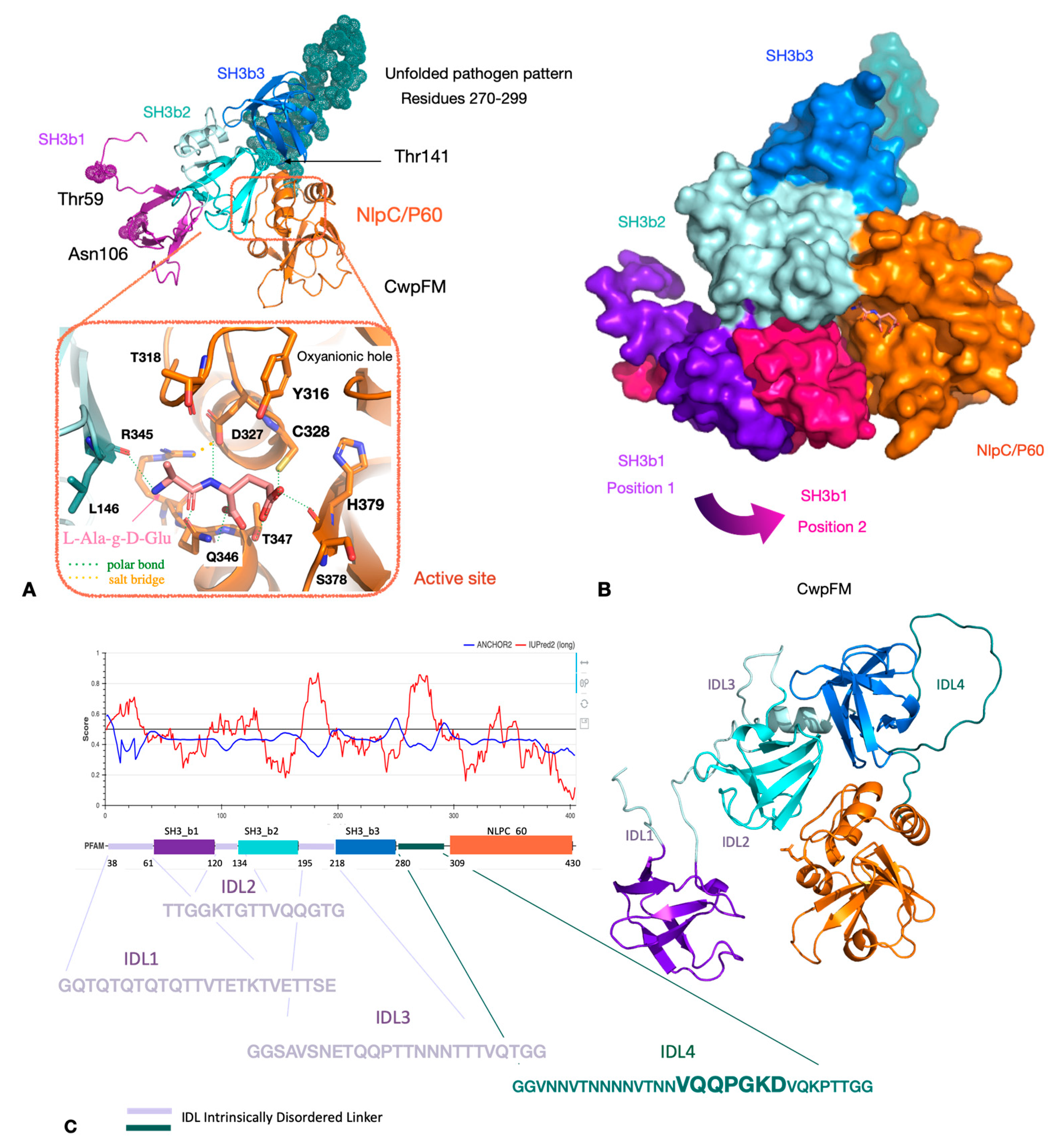
2.3. Homology Modeling of CwpFM 3D Structure Highlights a Conserved Modular Topology Composed of Three SH3b Domains Connected to an NLPC_P60 Cysteine Peptidase Domain, and Emphasizes the Presence of Disordered Connecting Domains that Could Play a Role in Specificity and Affinity towards PG and Proteins

To highlight the main sequence/structure/function features of CwpFM within the B. cereus group, we focused on CwpFM full-length from B. cereus ATCC 14579 that contains 420 residues, the first 26 of which were confirmed to be a signal peptide by the Phobius server [35]. The mature sequence, i.e., the endopeptidase with the signal peptide excised, was predicted to be 37% similar to the putative dipeptidyl-peptidase VI from Bacteroides ovatus by HHpred (pdb id 3NPF). In addition, the N-terminal segment of CwpFM from B. cereus (amino acid residues 14–73) was predicted by HHpred to be 37% sequence-similar to the SH3b part of the putative endopeptidase from Anabena variabilis ATCC29413 (pdb id 2HBW) [36]. Since the similarity scores were high enough, we homology-modeled CwpFM from B. cereus with the above-cited templates. Expectedly, the homology model evidences a classical endopeptidase topology with three SH3b domains, named SH3b1, SH3b2 and SH3b3, located at the N-terminal-end, and the NLPC_P60 endopeptidase domain, located at the C-terminal end (Figure 3A,B). Both SH3b2 and NLPC_P60 contribute clearly to the formation of the active site, and possibly SH3b1, of which orientation varies most, up to 13 Å, according to the 100 models computed by the homology modeling. Interestingly, this could give a hint on the flexibility amplitude of this domain, with respect to the other domains (Figure 3B). However, modeling cannot infer on the possible swapping of SH3b domains, known to exist in CWPs, but hard to predict in silico. Thus, at this stage, one cannot thus exclude or certify any close interaction between SH3b1, SH3b2 and NLPC_P60. The NLPC_P60 domain harbors a typical papain catalytic dyad, composed of strictly conserved Cys328 and His379 residues (Figure 3A). Additionally, it displays the strictly conserved Tyr316, known to act as the oxyanion hole, mandatory for endopeptidases. Further, sequence analysis evidences that both Tyr316 and Cys328 belong to the conserved catalytic motif YX10DCS. Thus, according to papain-like endopeptidases, the catalytic site of CwpFM is complete and prone to be active. Markedly, the sequence reveals large insertions from 17 to 34 residues, located at the N-terminus of each SH3b and between SH3b3 and NLPC_P60 domains (Figure 3C). Those insertions display low complexity with a large prevalence of polar Gln, Thr and Asn residues, and they are also highly susceptible to post-translational modifications and likely to be partially or completely unfolded. Accordingly, they have been named intrinsically disordered linkers and numbered from IDL1 to IDL4 (Figure 3C). Their role could be to adjust the positioning of each SH3b, either close to the active site for a functional role in specificity or remotely positioned to bind to the PG matrix or other cognate partners.

2.4. CwpFM Structure Is Able to Accommodate a PG Fragment

To ascertain, more precisely, the function of CwpFM, a L-Ala-Ɣ-D-Glu PG fragment, which corresponds to the reaction product performed by CWPs, was docked into the binding site formed at the interface between SH3b2 and NLPC_P60 domains (Figure 3B). The redocking of the L-Ala- Ɣ-D-Glu moiety inside YkfC shows a strictly similar accommodation, with respect to its crystal position, and a binding energy of −2.8 kcal/mol. The structure of B. cereus YkfC in complex with L-Ala-Ɣ-D-Glu was the first structural representative of an NLPC_P60 enzyme with a bound ligand. The enzyme releases L-Ala-Ɣ-D-Glu dipeptides from cell wall peptides via cleavage of an L-Ala- Ɣ-D-Glu-|-L-Lys bond.
The docking of L-Ala- Ɣ -D-Glu inside CwpFM gives an interaction energy of −3.7 kcal/mol, which is in the same range as in YkfC, thus suggesting that a short substrate with a free alanine residue could be accommodated in the active site of CwpFM. Additionally, the substrate accommodation highlights Tyr316, Asp327, Cys328, Ser329, Arg345, Gln346 and His379 (CwpFM numbering), as binding residues (Figure 3A). Interestingly, they are conserved in dipeptidyl-peptidase from B. ovatus and dipeptidyl-peptidase from Anabaea variables ATCC 29413. Particularly, Asp327 and Arg345 form a salt bridge strictly conserved in the three enzymes and involved in the hydrogen bonds network that connects many residues of the active site. Nevertheless, the binding shows a moderate affinity and the IDL extensions, which are difficult to model, could participate in reshaping the binding groove while enhancing the affinity. In short, CwpFM is able to accommodate the L-Ala- Ɣ-D-Glu product fragment of the peptidase reaction, but it is likely that a longer substrate with the L-Ala extremity, free or not, could also bind.

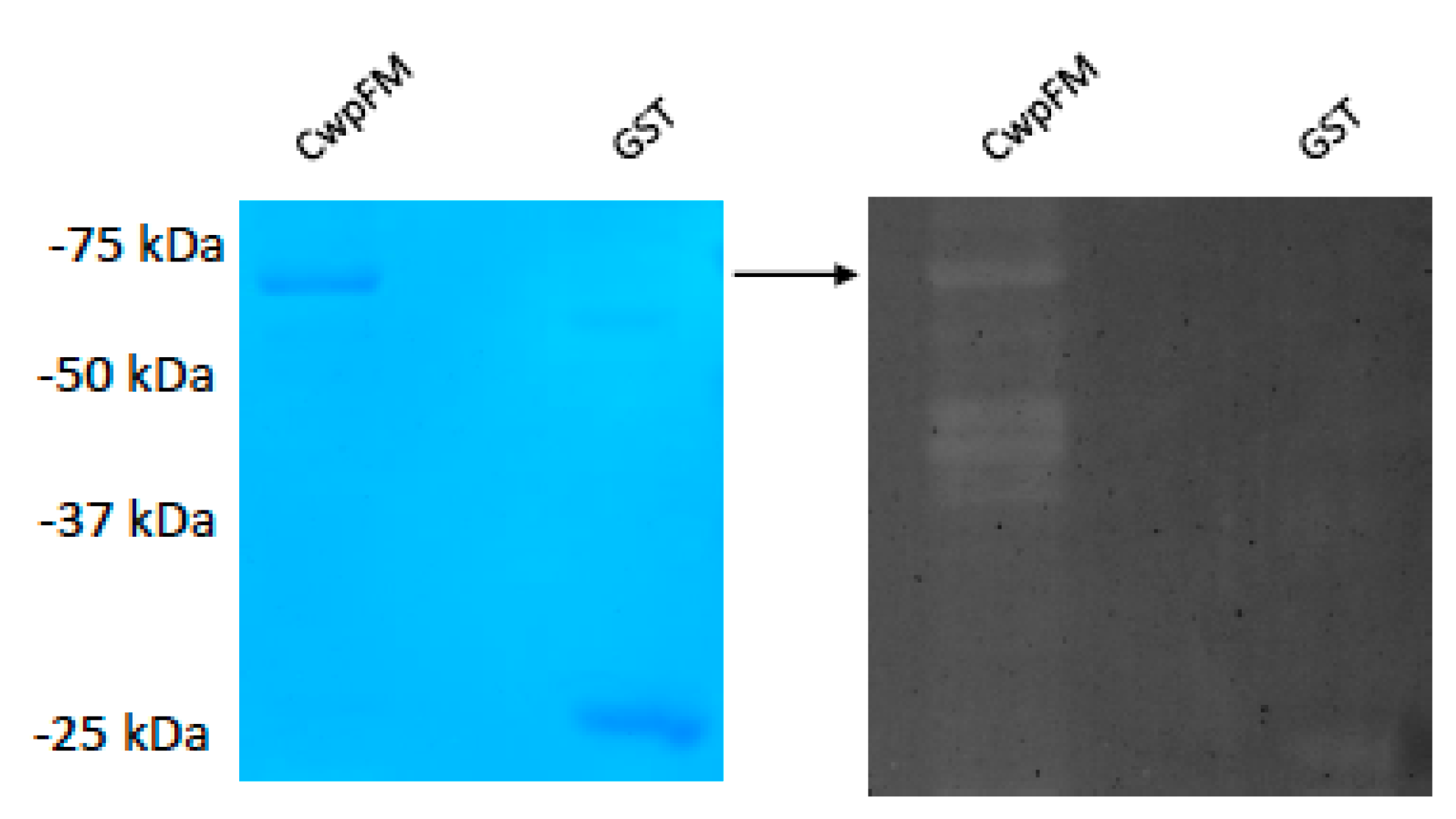
2.5. B. cereus CwpFM Shows a Weak PG Hydrolase Activity

Our modeling of CwpFM identified a catalytic domain typical of cysteine papain peptidase and computed a reasonable affinity towards a PG moiety, so we aimed to characterize CwpFM PG hydrolase activity. We expressed and purified B. cereus CwpFM with a GST tag on the N-terminal end of the protein. Next, purified CwpFM enzyme activity was examined by zymography analysis using the Micrococcus cell wall as a substrate (Figure 4). The results showed a small clearance band in the zymogram. No clearing zones were observed when the same amount of purified GST was subjected to zymography. These results demonstrate that the CwpFM protein exhibits a weak CW degrading activity, which is consistent with our 3D model that illustrated a low binding affinity with PG.

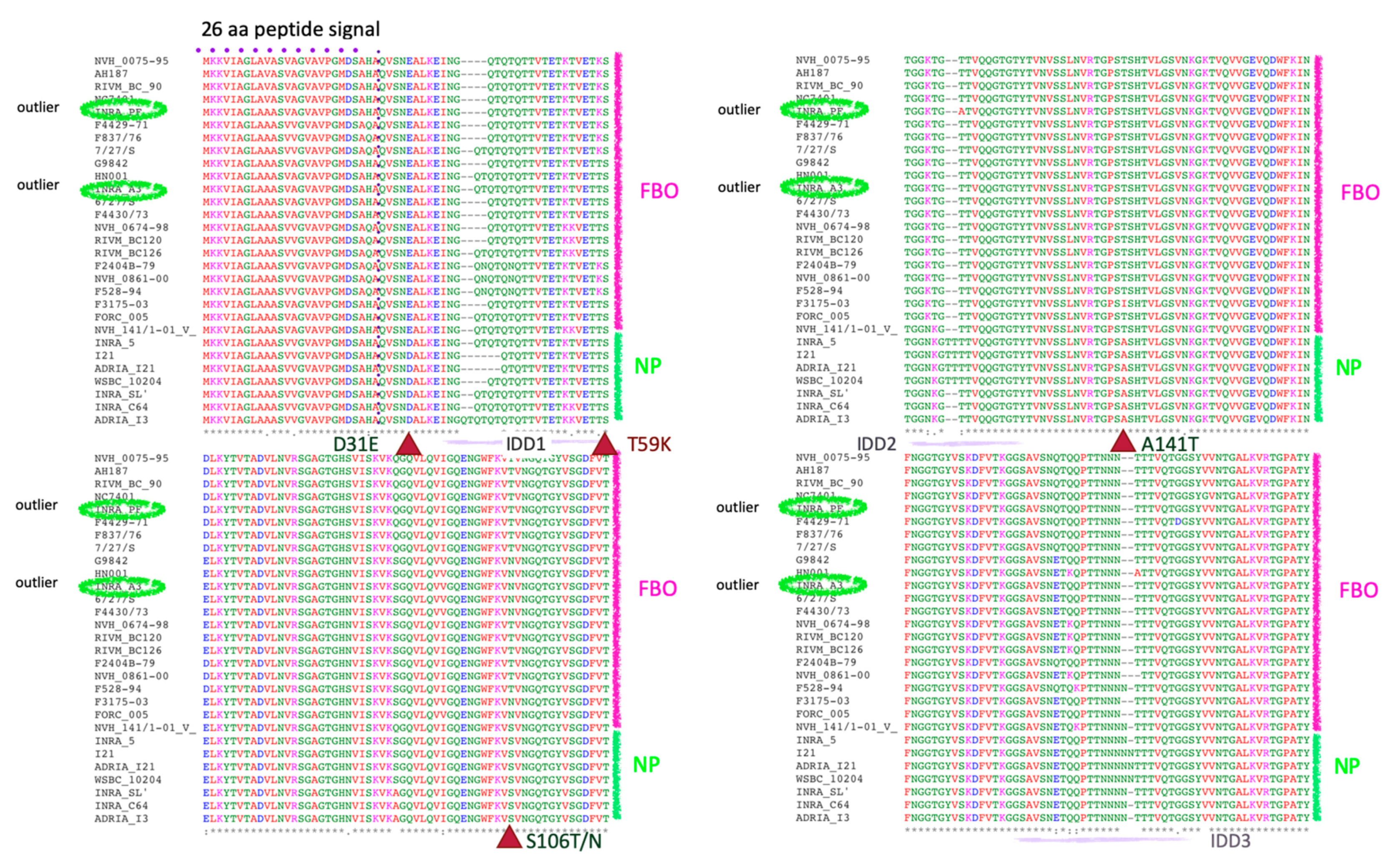
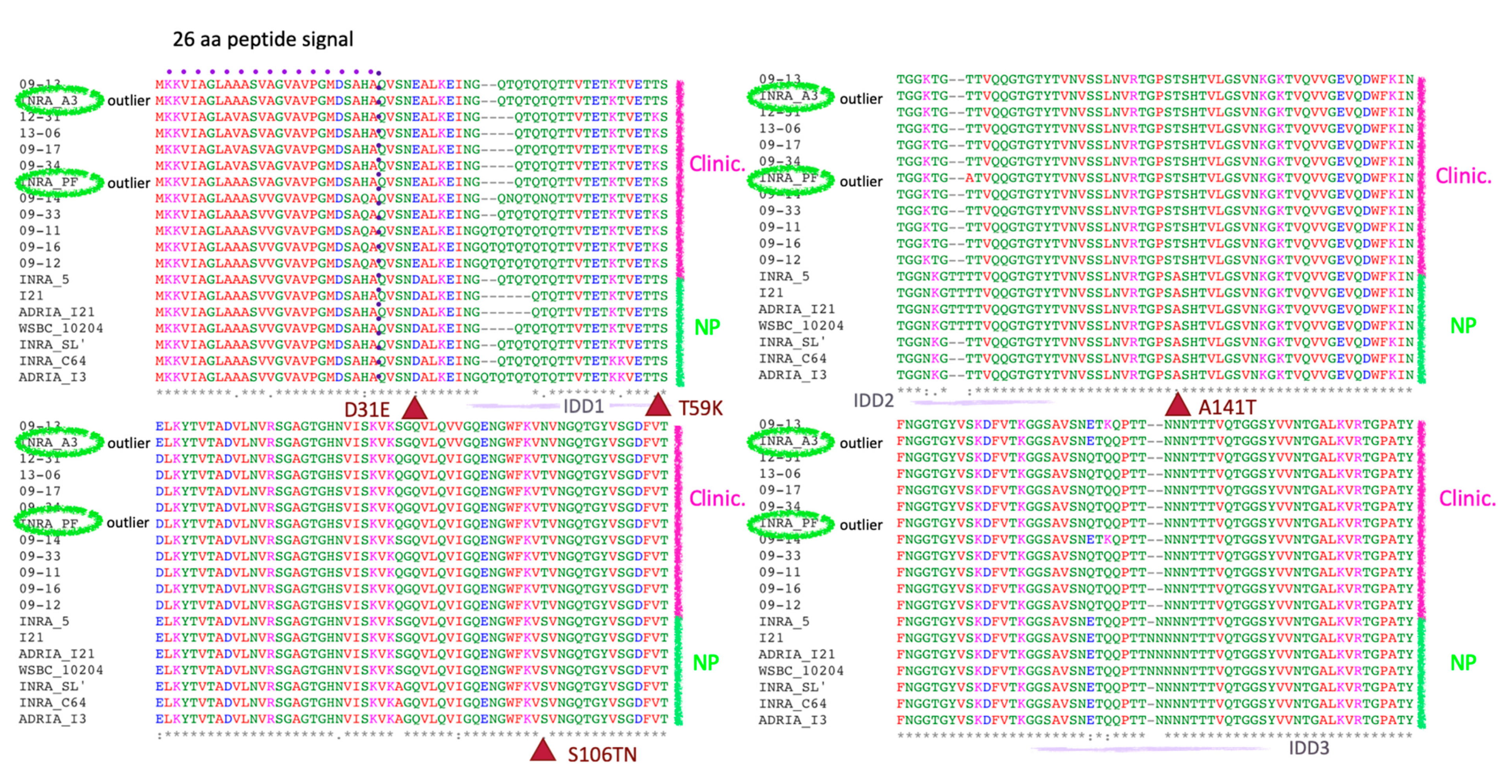
2.6. CwpFM Distribution in B. cereus Strains of Various Pathogenic Potentials

We have shown that CwpFM is present and conserved amongst the B. cereus group members. However, within B. cereus sensu stricto strains, the pathologies vary from harmless to highly toxic strains. We thus studied CwpFM from B. cereus strains causing different pathologies: FBO, clinical non-gastrointestinal infections and non-pathogenic strains. We performed a homology search using the nucleotide sequence of cwpFM from B. cereus isolate A6 (AY789084.1) and we identified homologues of cwpFM as a single copy in every genome (Table 3), the result of which correlates with the high prevalence of the gene described in the literature. Despite being prevalent, these ORFs are variously annotated Peptidase P60, C40 family peptidase, putative endopeptidase LytE or Enterotoxin, in the databases. On average, the cwpFM genes showed between 89% and 99% identity. For the FBO and clinical strains, the cwpFM genes showed above 94% identity with the reference strain A6. The identity was, on average, lower for the non-pathogenic strains ranging from 89% to 92% identity, with two exceptions. These exceptions belong to the strains INRA PF (97% identity) and INRA A3 (99% identity).

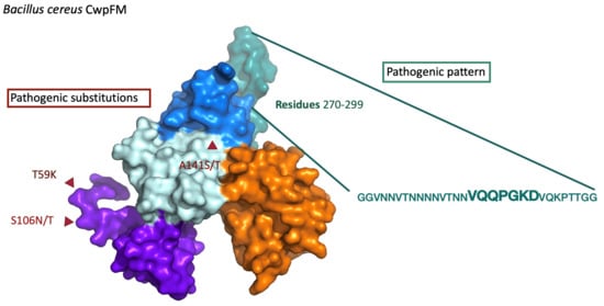
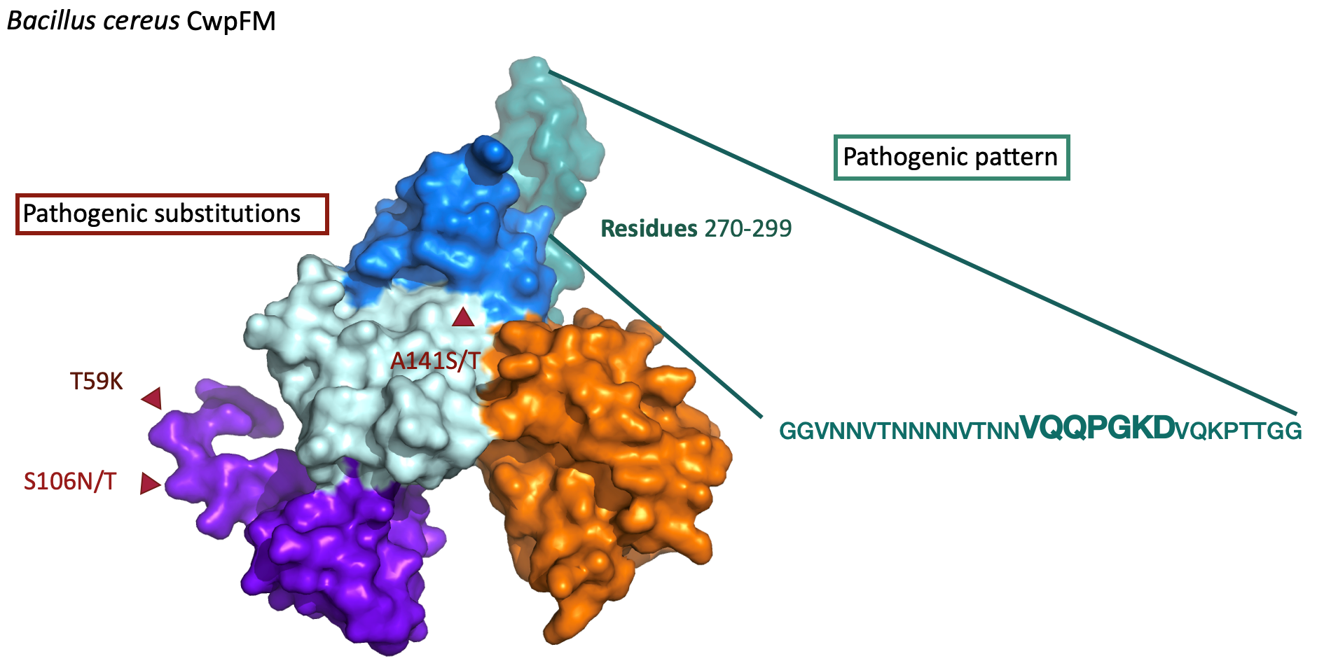
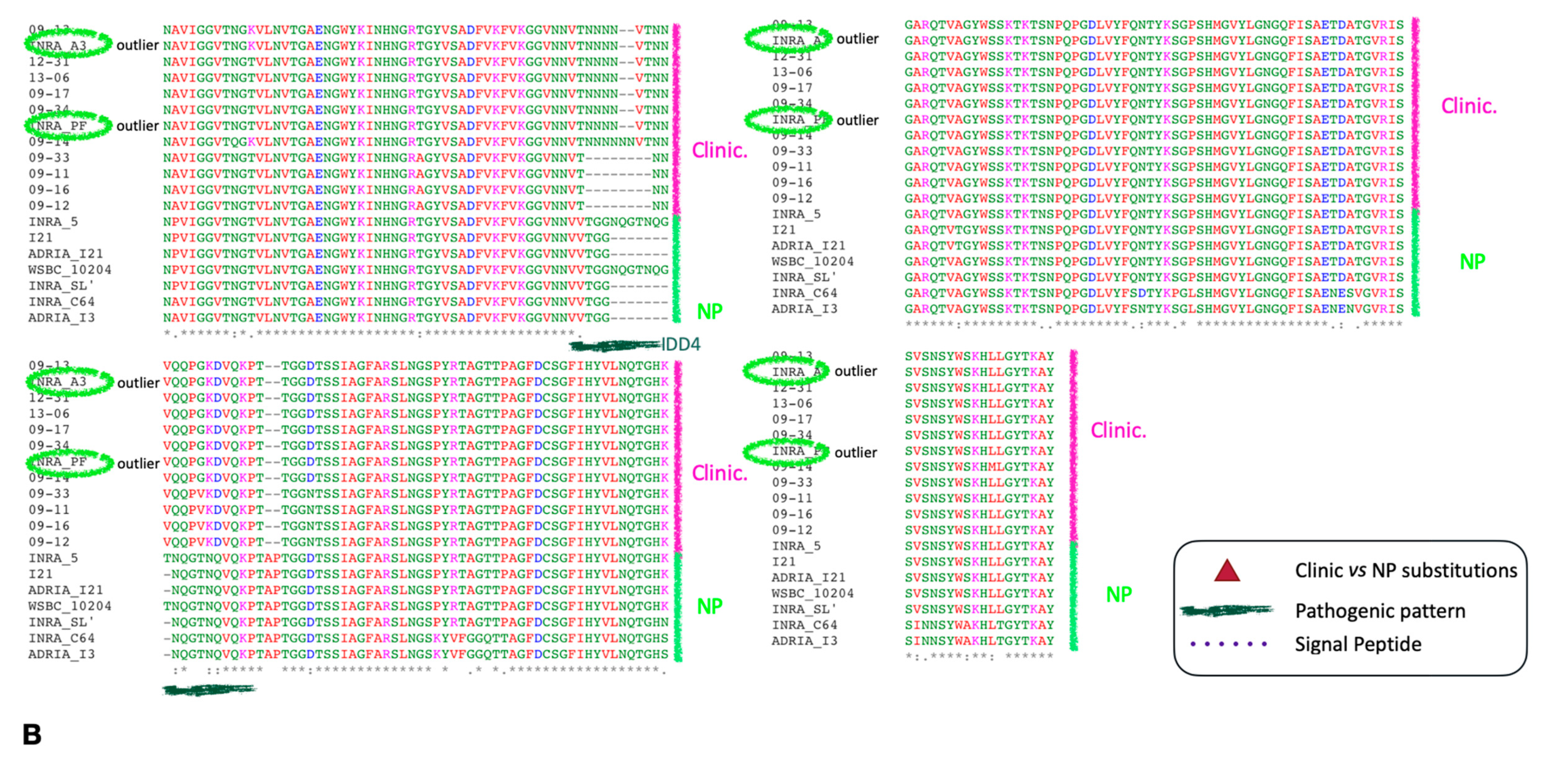
2.7. Analysis of the Differences at the 2D and 3D Levels

Since we aimed to extract significant differences that could discriminate pathogenic from non-pathogenic strains, we align series of strain sequences using MAFFT. The first alignment is between FBO and non-pathogenic strains (Figure 5A), whilst the second is between clinical and non-pathogenic ones (Figure 5B).
The sequences of CwpFM cluster according to the origin of their B. cereus strain, with only two exceptions. Indeed, the CwpFM from the pathogenic strains, either FBO or clinical, are clearly separated from the non-pathogenic strains. The outsider strains INRA-PF and INRA-A3 clustered within the pathogenic strains group. Three point-mutations, Asp/Glu31, Thr/Asn106 and Thr/Ile141, and, more importantly, a 20-residues segment (276–296) located between SH3b3 and NLPC_P60 could be noted as significantly different between non-pathogenic strains and pathogenic strains. Glu31 is positioned at the N-terminal end, close to the excised peptide. Glu31 is substituted by an aspartic acid in non-pathogenic strains. Its substitution could not be mapped onto the CwpFM 3D structure because the protein was homology-modeled starting from residue 53. Thr/Asn106 in pathogenic strains is replaced by a serine, whilst Thr/Ile141 is substituted by an alanine, in non-pathogenic strains. In our homology model, Thr/Asn106 in SH3b1 and Thr/Ile151 in SH3b2 are positioned too far from each other to be in contact together (Figure 3A). Nevertheless, one can hypothesize that domain swapping or a close interaction between the two SH3b domains could favor the polar interaction between the two residues, the possibility of which cannot be excluded as domain swapping of SH3bs has been already mentioned for CWPs. Correspondingly, this feature could be claimed for the covariation of residues. Reversely, if we consider those residues as not engaged in mutual interaction, they could be largely accessible to any binding or post-translational modification. Markedly, the most significant difference is a segment, called IDL4, an intrinsically disordered linker, located between residues 280 and 309, that is clearly distinct in sequence when compared between non-pathogenic and pathogenic strains. Clearly, this segment could not be modeled (i) because it is partly an insertion as compared to our 3D templates, and (ii) because it is very low in complexity, and thus largely unstructured. Accordingly, in both non-pathogenic and pathogenic strains, we expect these segments of CwpFM to be natively unfolded. Notably, IDL4 displays a significant difference in sequences between non-pathogenic and pathogenic strains as VTGG(X)NQGTNQ (X being-, T or NQGTNQGT) is replaced by T(N)0–6VTNNVQQPGKD (Figure 3C and Figure 5).
The pathogenic strains harbor a significant amount of Asn residues, while the non-pathogenic ones display more Gly and Gln residues. Asn residues are highly susceptible to hydroxylation which is a post-translational modification shown recently as reversible [37]. Therefore, one cannot exclude the hydroxylation of Asn residues, which could contribute to protein modification, flexibility and anchoring at the PG. This is fully consistent with the linker role of IDL4 that connects SH3b3 to the catalytic NLPC_P60. Such modifications could tune the adequate positioning of SH3b3 towards protein partners, including SH3b1 and SH3b2, and adapt the exquisite mobility of the catalytic domain towards the PG. Due to the length and prevalence of Asn in all pathogenic strains, this segment could be considered as a pattern signature for B. cereus virulent strains.

3. Discussion

B. cereus is a serious cause of food poisoning. It is largely known that the emetic toxin and the enterotoxins Nhe, Hbl and CytK are responsible for vomiting and diarrhea syndromes, respectively [21,38]. Many other putative virulence factors have been described in B. cereus. However, their precise role in bacterial pathogenesis is still uncertain, although their involvement in virulence has been suggested due to their toxic effects on cellular models, insects or mammals. Unlike B. anthracis, B. thuringiensis and the specific B. cereus emetic strains, whose toxin genes are carried by plasmids, B. cereus virulence factors are specified by genes located on the chromosome and the virulence is probably multifactorial [20,38,39]. Among them, CwpFM, formerly identified as an enterotoxin, is in fact a cell wall peptidase of the NLPC_P60 family of peptidases, which is one of the most abundant secreted cell wall peptidase CWP families. Nevertheless, the CwpFM family lacks distribution, prevalence, sequence characterization and a molecular description of its mode of action.
Here, we report for the first time a distribution analysis of CwpFM within the Bacillus group. To infer the sequence/structure/function of CwpFM within the B. cereus group and gain molecular consistency, the features observed from bioinformatics analysis were mapped onto a 3D structure of CwpFM from B. cereus ATCC1479 modeled in silico. We highlight that CwpFM from B. cereus is a papain-like cysteine endopeptidase that displays the emblematic catalytic motif Y316X10DCS329 associated with the strictly conserved histidine residue H379 of the NLPC_P60 family. Cys328 of this motif and His379 residues form the catalytic dyad, while Y316 of the motif frames the oxyanionic hole, expected to occur in every peptidase protein. Thus, CwpFM displays a competent active site. Additionally, docking computation evidences that a PG moiety is able to bind to the active site. In line with that, structural bioinformatics analysis highlights that the binding site is composed of residues strictly conserved and located in both SH3b2 and NLPC_P60 domains. Only Gly172 in CwpFM replaces the bulky Tyr118 or Tyr80, and Gln356 replaces Asp256 or Asp221, in the YkfC of B. cereus and in the putative dipeptidyl-peptidase VI from Bacteroides ovatus, respectively. Since tyrosine and aspartate residues are known to interact together to fix the free amine group of the Alanine peptido-glycan moiety, the absence of a side chain in Gly172 could preclude the close interaction with Gln346 and result in an enlargement of the binding site while allowing the accommodation of substrates with an attached fragment at the Alanine extremity. Those structural features, key in the specific recognition of murein peptides by the subfamily of the NLPC_P60 protein, interrogate the substrate specificity and affinity. Accordingly, we could only detect in vitro a weak peptidoglycan hydrolase activity from purified CwpFM. One could argue that the PG from Micrococcus lysodeikticus ATCC M3770 may not be the cognate substrate, neither in PG length nor in molecular characteristics. One can also explain that the post-translational modification profile of CwpFM has not reached its optimum activity, due to its production in E. coli, that is unable to perform the hydroxylation of asparagine or phosphorylation of serine and threonine residues [40]. Still, the optimum substrate(s), the activation and the toxin target mode of action of CwpFM are open questions. We can also speculate that CwpFM activity is dependent of another enzyme/activator. As an example, it has been demonstrated that the amidase LytH of Staphylococcus aureus is only active in the presence of its membrane partner ActH [41]. With respect to the modular topology of CwpFM, the SH3b and catalytic domains could synergize to attain full endopeptidase activity. Further work is needed to identify the other substrates and/or activator allowing B. cereus CwpFM to be fully active, and to decipher the toxin mode of action.
B. cereus CwpFM contains three SH3b domains. Thus, despite its homology with B. subtilis LytF, the role of CwpFM probably differs from LytF. Indeed, the binding domains ensure the localization and the proper function of the CWH, particularly in CWPs [8]. Nine SH3b domains have been described (SH3b-1 to SH3b-9) so far and domains can be found at the two terminal ends of CWPs. Of note, cell wall binding domains that can be found along the mono-polypeptide chain generally improve the efficiency of the enzymatic activity, either by increasing the concentration of the ligand at the active site or by anchoring properly onto the enzyme, or even by reshaping the active site [1,8]. In line with that, the SH3b domains demonstrate a selective affinity for pentaglycine cross-bridges [42]. Additionally, it has been shown that loops from the SH3b domain can dock into the ends of the catalytic groove, remodel the substrate binding site and thus modulate substrate specificity [43]. This specificity can be driven by a single mutation. In line with that, two out of three mutations, that have been identified between non-pathogenic and pathogenic strains, are positioned on loops that could participate in substrate affinity or binding site reshaping. Further, it has recently been demonstrated that recognition is shared by two independent SH3bs, tightly engaged to each other, allowing protein clusterization [44,45]. Their interaction potentiates and compensates for the weak affinity of individual SH3b towards pentaglycine [42,46].
In B. subtilis, numerous CWPs such as LytF (CwlE), LytE (CwlF) and CwlS are known to work in synergy to ensure cell separation during vegetative growth, and deletion of these genes results in a long filament-like bacteria phenotype [47,48]. They all display an NLPC_P60 domain and a variable number of LysM (Lysin M) domains comprised between zero and five. LysM is amongst the most frequent CW binding motifs and has been shown to recognize the N-acetylglucosamine (GlcNac) polymers (NAG-X-NAG motif) of PG [49,50]. Particularly, LysM is involved in the specific localization of LytF at the separation sites and poles of B. subtilis [51]. LytE, in combination with CwlO, has also been associated with cell growth and morphogenesis as they both participate in breaking the PG cross-links along the lateral side of bacteria to support the process of elongation. Although domain prediction analysis reveals no presence of CW-binding domains within CwlO, it is most likely that a domain recognizing a specific fragment of the PG (probably LysM) is present to dictate the enzyme specificity of action. B. subtilis BlyA has three SH3b domains. To date, no physiological function has been linked to B. subtilis BlyA, however, BlyA from Borrelia Burgdorferi is a bacteriophage-encoded holin which, if expressed in E. coli, can induce damage to the E. coli cell envelope and allows the release of intracellular cytotoxin ClyA, inducing hemolysis on blood agar [52]. LytD (CwlG) was predicted to harbor one SH3b domain and a sporulation-like domain that may indicate a role during sporulation. Finally, LytD (CwlG) is an N-acetylglucosaminidase that has been implicated but is not essential in cell separation and motility [53]. Therefore, the exact role of LytD (CwlG) remains unclear.
CwpFM is present in all strains of our collection of B. cereus, gathering strains of various origins and causing different pathologies. CwpFM is a major B. cereus toxin that is involved in virulence. We have previously shown that CwpFM is involved in the bacterial shape and division, in adhesion to eukaryotic cells and in promoting virulence. Presence of the gene is now routinely assessed in combination with other diarrheal toxins-encoded genes such as ces, nhe and hbl to determine the potential pathogenicity of a strain. Data show that cwpFM is widely distributed (detection rate of 68–98%) in B. cereus isolated from diverse food matrices [53,54,55,56,57]. cwpFM is also detected in strains associated with food-borne illnesses [33,58] and is even present in emetic strains [59]. Due to the high distribution of cwpFM in pathogenic but also non-pathogenic strains, it is hard to use the detection of the cwpFM gene as a biomarker of pathogenicity. However, an accurate bioinformatics comparison between the sequences of our strain collection was performed, and then the residues distinct between non-pathogenic, FBO and clinical strains were mapped onto the homology model of CwpFM to check if the sequence and 3D structure could correlate with the pathogenicity of a strain [60]. All CwpFMs from B. cereus display four intrinsically disordered linkers (IDL) as connectors between SH3b and/or NLPC_P60 domains. Particularly, the IDL4 that connects SH3b3 to the catalytic domain is particularly long and displays a significant difference that both aggregates pathogenic FBO and clinical strains, while it segregates non-pathogenic strains. The stretch is NQGTNQVQ in non-pathogenic sequences that is replaced by the VQQPGKD patch in pathogenic ones. Additionally, one can observe an extra stretch of up to 10 additional asparagine residues found to be inserted in all pathogenic strains (FBO and clinical) and strictly absent in non-pathogenic ones. Such differences observed not only in length but also in the low complexity with a high prevalence of Asn (for pathogenic strains) vs Gly/Gln (for non-pathogenic strains) can have conformational and functional consequences. Interestingly, as this specific pattern defines the IDL4 junction domain between SH3b and NLPC_P60 domains, we suspect that this domain, natively unfolded, could play a role in the recruitment of binding partners, putatively through glycosylated Asn residues. The disordered part of CwpFM may possibly be involved in bacterial pathogenicity. Indeed, although deficient in stable secondary and tertiary structures under physiological conditions of pH and salinity, disordered parts in proteins may function as dynamic ensembles of interconverting conformers. Unstructured parts of CwpFM are depleted in hydrophobic amino acid residues, but enriched in polar and charged residues. It was shown that disordered proteins, enriched in polar/charged residues, are highly hydrated compared to ordered ones and as such, they behave distinctly in bulk and air/water or lipid/water interfaces [61]. For instance, α-synuclein, amyloid-β peptide and PB1-F2 influenza protein are disordered monomeric peptides in aqueous solution, but may adopt a β-sheet conformation that further aggregates into toxic amyloid fibrils in contact with negatively charged phospholipids and induces membrane morphological changes and disruption [62,63,64]. We previously reported the morphological changes of a mammalian cell membrane exposed to recombinant CwpFM [31], which may be linked to this region.
The precise role of CwpFM in pathogenic and non-pathogenic strains remains to be studied. The differences in sequences may have a direct or indirect role during virulence, possibly on the protein partners or post-translational modifications. This is a challenging question to address because many hydrolases, produced by the bacteria, may have a redundant function and take over in the case of a mutation. In addition, some have different functions and can even have more than one function [65]. For instance, in Enterococcus faecalis, the SagA protein is a secreted endopeptidase, which has an antibiotic role against enteric pathogens such as Clostridium difficle [66]. Nocardia seriolae protein NLPC_P60 is a cell wall peptidase also identified as a virulence factor [67]. In Mycobacterium tuberculosis, the protein Rv0024 has been shown to be involved in biofilm formation. Those biofilms have been found to be resistant to cell wall-acting anti-TB drugs [68]. Still, M. tuberculosis, a mutant lacking the NLPC_P60 protein, is more sensitive to antibiotics and lysozymes, leading to a decrease in the survival in macrophages [69], and the Rv2190c protein is required not only for cell wall maintenance but also for virulence since a mutant is less virulent in a mice model of infection in vivo [70]. Furthermore, NLPC_P60 was described as a virulence factor in Bacillus anthracis as it is part of its secretome and can be found in the blood of infected animals [71]. The function of CWP can also indirectly contribute to bacterial virulence. A recent work has demonstrated that the PG hydrolase Cwp19 contributes to Clostridium difficile autolysis, which induces the release of bacterial toxins [72]. Markedly, PG fragments released by CWPs can also act as signaling molecules to promote the presence of antimicrobial agents or to interact with the component of the host [73,74].
The versatility of these endopeptidases in the virulence of multiple bacteria towards the host could be explained by their modular architecture that also integrates intrinsically disordered segments and point mutations, both possibly subjected to post-translational modification. These data pave the way for further in silico and in vitro studies because they explore beyond the chromosomal gene presence and ground the first description of a pattern within B. cereus CwpFM sequences capable of discriminating pathogenic from non-pathogenic strains.

4. Materials and Methods

4.1. Bacterial Strain Sequences

The genome and cwpFM sequences of 10 strains belonging to the B. cereus group were retrieved from NCBI (Table 1). The genome and cwpFM sequences of 20 B. cereus strains isolated from food or human samples and associated with food poisoning were also retrieved from NCBI (Table 2). In addition, this study includes ten strains isolated from human samples following systemic or local infections [75] and nine non-pathogenic strains, isolated from food that did not cause infection in humans or animals [76,77]. The corresponding genome sequences were retrieved from the European Nucleotide Archive (ENA) or obtained in this study (Table 2).

4.2. Sequence Alignment, Conserved Motifs and Domain Analysis of the Proteins

Multiple nucleotide sequence alignments were conducted using the BLASTn interface. Multiple amino acid sequence alignments of the CwpFM proteins were performed using Clustal Omega v1.2.4 (https://www.ebi.ac.uk/Tools/msa/clustalo/). Signal peptide sequences were predicted using Phobius [36] and SignalP 5.0 [78] servers. The potential protein domains were identified using InterProScan protein domain prediction analysis (www.ebi.ac.uk/Tools/pfa/iprscan/).

4.3. D Alignments and Clustering of the Strains

Alignments were performed with MAFFT—Multiple Alignment using Fast Fourier Transform—v6.861b, with the default options [79]. It is a high-speed multiple alignment program which implements fast Fourier transform (FFT) to optimize protein alignments based on the physical properties of the amino acids. The program uses progressive and iterative alignment and is implemented at the ebi (https://www.ebi.ac.uk/Tools/msa/mafft/).

4.4. Molecular Modeling

CwpFM from the B. cereus ATCC 14579 strain (CwpFM_BC) was homology-modeled using the model-building software Modeller (mod9v18) [80]. The crystal structures of the apo putative dipeptidyl-peptidase VI from Bacteroides ovatus (pdb id 3NPF), and the SH3b domain of the putative endopeptidase from Anabena variabilis ATCC 29413 (pdb id 2HBW amino acid residues 14–73) served as 3D templates (10.2210/pdb3NPF/pdb; 10.2210/pdb2HBW/pdb) [35]. The 3D templates were previously chosen from the HHpred webserver, dedicated to structural homology detection. They were sorted as the two first hits. One hundred homology models of CwpFM were generated, and one was eventually chosen from a selection of the five best models with respect to the lowest values of the Modeller score function, best stereochemistry, as checked by Molprobity (http://molprobity.biochem.duke.edu/), and visual inspection, using PyMOL 2.0.7 (Schrödinger, LLC, New York, NY, USA). The selected model was then minimized using the Biologic suite of Schrödinger, LLC, New York, NY, USA. The sequences were also analyzed using IUPred to characterize disordered segments and identify molecular recognition features (https://iupred2a.elte.hu/) [81].

4.5. Docking

As a prerequisite before performing our docking of the L-Ala- Ɣ-D-Glu ligand in our model of CwpFM, we validated our protocol by redocking the ligand in the active site of YkfC (pdb id 3H41) because it is a structural and functional homologue in a holo configuration. Formerly, the ligand was discarded from YkfC to get the apo form and the tri-oxidized Cys238 of the crystal structure was reduced into a Cys residue to mimic the active site, with respect to the papain family of cysteine peptidases (pdb 3H41) [35]. Docking was performed using Autodock4 with the following parameters: a grid box centered on L-Ala- Ɣ-D-Glu bound to the catalytic site, encompassing all the residues involved in the interaction, a genetic algorithm of Lamarck, ten runs of computation with a final ranking and clustering of the docked peptide. The best pose computed for L-Ala- Ɣ-D-Glu superimposes very well to the L-Ala- Ɣ-D-Glu crystal position, so the protocol was validated and used for subsequent docking of L-Ala- Ɣ-D-Glu, in the apo form of a 3D template of dipeptidyl-peptidase VI from Bacteroides ovatus (pdb id 3NPF) and the homology-modeled CpwFM, after superimposition of their NLPC_P60 domains. The analysis of the complexes and figures were generated with PyMOL 2.0.7. (Schrödinger, LLC., New York, NY, USA).

4.6. Expression and Purification of the CwpFM-GST-Tagged Protein

The plasmid pGEX6P1-GST-CwpFM was constructed as follows. The cwpFM gene was amplified from the B. thuringiensis 407 cry- chromosome by PCR using the primer pairs CwpFM-GST-1 (5′-CGGGATCCCAAGTTTCAAATGAAGCGCTAA-3′) and CwpFM-GST-2 (5′-CCGCTCGAGTCCCAAGTTTCCTTGGAAAGCC-3′). The DNA fragment was inserted between the BamHI and XhoI sites of plasmid pGEX6P1 (GE Healthcare), and the resulting plasmid was introduced into E. coli M15 [pREP4] (Qiagen). Additionally, E. coli BL21 bacteria were transformed with pGEX6P1–GST plasmid. Strains were grown at 37 °C for 8 h in 100 mL of LB containing ampicillin (100 μg/mL) for the strain BL21 or ampicillin and kanamycin (40 µg/mL) for the strain M15. Protein expression was induced by adding 100 μg/mL isopropyl-β-d-thio-galactoside for 4h at 30 °C. For the purification of the recombinant GST and CwpFM-GST fusion, bacterial pellets were re-suspended in lysis buffer (50 mm Tris-HCl, pH 7.8, 60 mm NaCl, 1 mm EDTA, 2 mm DTT, 0.2% Triton X-100) supplemented with complete Protease Inhibitor mixture (Roche), and incubated for 1 h on ice. Cells were then disrupted using a BAZIC Z cell disruptor (Constant Systems Ltd., Daventry, UK) at a pressure of 1600 bars, and centrifuged at 4 °C for 30 min at 10,000× g. GSH-Sepharose 4B beads (GE Healthcare) were added to clarified supernatants and incubated at 4 °C for 3 h. Beads were then washed two times in lysis buffer and three times in 20 mM Tris buffer, pH 8. Purity was assessed by 12% (v/v) SDS-PAGE with Coomassie blue staining. Protein concentration was measured using a Bradford assay (Sigma France, Lezennes, France).

4.7. Zymogram

CwpFM cell wall hydrolyzing activity was assessed using zymogram analysis. An amount of 3.5 µg of purified GST and CwpFM-GST was loaded onto SDS-PAGE using 12% (v/v) polyacrylamide separating gels containing 0·2% (v/v) Micrococcus lysodeikticus ATCC M3770 (Sigma) as the enzyme substrate. Micrococci were suspended in water and heat-inactivated at 120 °C for 10 min before they were mixed into the resolving gel. After sample migration, gels were washed with deionized water for 1 h at room temperature and incubated in 50 mM Tris-HCl pH 8 containing 1% Triton X100 (v/v) for 24 h at 37 °C. The total amount of proteins was detected by staining of the SDS-PAGE gel with Coomassie blue staining. The CwpFM hydrolyzing activity was characterized by a lysis plague visible as a halo on the gel at the level of the protein.

Author Contributions

Conceptualization: J.V., S.-L.T., N.R., G.A.-L.; validation: S.-L.T., N.R., G.A.-L.; formal analysis: S.-L.T., G.A.-L.; methodology/investigation: J.V., S.-L.T., D.C., G.A.-L.; writing—original draft preparation: J.V., S.-L.T., N.R., G.A.-L., D.C.; writing—review and editing: J.V., S.-L.T., N.R., G.A.-L., funding acquisition: N.R. All authors have read and agreed to the published version of the manuscript.

Funding

This research received no particular funding.

Acknowledgments

We thank Simon Palussière and Marie-Pierre Chapot-Chartier for their helpful assistance with zymogram assays.

Conflicts of Interest

The authors declare no conflict of interest.

References

  1. Alcorlo, M.; Martinez-Caballero, S.; Molina, R.; Hermoso, J.A. Carbohydrate recognition and lysis by bacterial peptidoglycan hydrolases. Curr. Opin. Struct. Biol. 2017, 44, 87–100. [Google Scholar] [CrossRef]
  2. Do, T.; Page, J.E.; Walker, S. Uncovering the activities, biological roles, and regulation of bacterial cell wall hydrolases and tailoring enzymes. J. Biol. Chem. 2020, 295, 3347–3361. [Google Scholar] [CrossRef] [Green Version]
  3. Dorr, T.; Moynihan, P.J.; Mayer, C. Editorial: Bacterial Cell Wall Structure and Dynamics. Front. Microbiol. 2019, 10, 2051. [Google Scholar] [CrossRef] [Green Version]
  4. He, J.; Fu, W.; Zhao, S.; Zhang, C.; Sun, T.; Jiang, T. Lack of MSMEG_6281, a peptidoglycan amidase, affects cell wall integrity and virulence of Mycobacterium smegmatis. Microb. Pathog. 2019, 128, 405–413. [Google Scholar] [CrossRef]
  5. Healy, C.; Gouzy, A.; Ehrt, S. Peptidoglycan Hydrolases RipA and Ami1 Are Critical for Replication and Persistence of Mycobacterium tuberculosis in the Host. MBio 2020, 11, e03315-19. [Google Scholar] [CrossRef] [Green Version]
  6. Mahasenan, K.V.; Batuecas, M.T.; De Benedetti, S.; Kim, C.; Rana, N.; Lee, M.; Hesek, D.; Fisher, J.F.; Sanz-Aparicio, J.; Hermoso, J.A.; et al. Catalytic Cycle of Glycoside Hydrolase BglX from Pseudomonas aeruginosa and Its Implications for Biofilm Formation. ACS Chem. Biol. 2020, 15, 189–196. [Google Scholar] [CrossRef]
  7. Lonergan, Z.R.; Nairn, B.L.; Wang, J.; Hsu, Y.P.; Hesse, L.E.; Beavers, W.N.; Chazin, W.J.; Trinidad, J.C.; VanNieuwenhze, M.S.; Giedroc, D.P.; et al. An Acinetobacter baumannii, Zinc-Regulated Peptidase Maintains Cell Wall Integrity during Immune-Mediated Nutrient Sequestration. Cell Rep. 2019, 26, 2009–2018.e6. [Google Scholar] [CrossRef] [Green Version]
  8. Vermassen, A.; Leroy, S.; Talon, R.; Provot, C.; Popowska, M.; Desvaux, M. Cell Wall Hydrolases in Bacteria: Insight on the Diversity of Cell Wall Amidases, Glycosidases and Peptidases Toward Peptidoglycan. Front. Microbiol. 2019, 10, 331. [Google Scholar] [CrossRef]
  9. Ramarao, N.; Lereclus, D.; Sorokin, A. The Bacillus cereus group. In Molecular Medical Microbiology, 2nd ed.; Academic Press: Cambrige, MA, USA, 2015; Volume III, pp. 1041–1078. [Google Scholar]
  10. Kolsto, A.B.; Tourasse, N.J.; Okstad, O.A. What sets Bacillus anthracis apart from other Bacillus species? Annu. Rev. Microbiol. 2009, 63, 451–476. [Google Scholar] [CrossRef]
  11. Jessberger, N.; Krey, V.M.; Rademacher, C.; Bohm, M.E.; Mohr, A.K.; Ehling-Schulz, M.; Scherer, S.; Martlbauer, E. From genome to toxicity: A combinatory approach highlights the complexity of enterotoxin production in Bacillus cereus. Front. Microbiol. 2015, 6, 560. [Google Scholar]
  12. Celandroni, F.; Salveti, S.; Senesi, S.; Ghelardi, E. Bacillus thuringiensis membrane-damaging toxins acting on mammalian cells. FEMS 2014, 361, 95–103. [Google Scholar]
  13. Carlson, C.R.; Caugant, D.A.; Kolstø, A.-B. Genotypic diversity among Bacillus cereus and Bacillus thuringiensis strains. Appl. Environ. Microbiol. 1994, 60, 1719–1725. [Google Scholar] [CrossRef] [PubMed] [Green Version]
  14. Mock, M.; Fouet, A. Anthrax. Annu. Rev. Microbiol. 2001, 55, 647–671. [Google Scholar] [CrossRef]
  15. Stevens, M.J.A.; Tasara, T.; Klumpp, J.; Stephan, R.; Ehling-Schulz, M.; Johler, S. Whole-genome-based phylogeny of Bacillus cytotoxicus reveals different clades within the species and provides clues on ecology and evolution. Sci. Rep. 2019, 9, 1984. [Google Scholar] [CrossRef] [Green Version]
  16. Glasset, B.; Herbin, S.; Guillier, L.; Cadel-Six, S.; Vignaud, M.L.; Grout, J.; Pairaud, S.; Michel, V.; Hennekinne, J.A.; Ramarao, N.; et al. Bacillus cereus-induced food-borne outbreaks in France, 2007 to 2014: Epidemiology and genetic characterisation. Eurosurveillance 2016, 21, 30413. [Google Scholar] [CrossRef]
  17. Agata, N.; Ohta, M.; Mori, M.; Isobe, M. A novel dodecadepsipeptide, cereulide, is an emetic toxin of Bacillus cereus. FEMS Microbiol. Lett. 1995, 129, 17–20. [Google Scholar]
  18. Mahler, H.; Pasa, A.; Kramer, J.; Schulte, P.; Scoging, A.; Bar, W.; Krahenbuhl, S. Fulminant liver failure in association with the emetic toxin of Bacillus cereus. N. Engl. J. Med. 1997, 336, 1142–1148. [Google Scholar] [CrossRef]
  19. Naranjo, M.; Denayer, S.; Botteldoorn, N.; Delbrassinne, L.; Veys, J.; Waegenaere, J.; Sirtaine, N.; Driesen, R.B.; Sipido, K.R.; Mahillon, J.; et al. Sudden death of a young adult associated with Bacillus cereus food poisoning. J. Clin. Microbiol. 2011, 49, 4379–4381. [Google Scholar] [CrossRef] [Green Version]
  20. Ramarao, N.; Sanchis, V. The pore-forming hemolysins of Bacillus cereus: A review. Toxins 2013, 5, 1119–1139. [Google Scholar] [CrossRef] [Green Version]
  21. Granum, E.; Lund, T. Bacillus cereus and its food poisoning toxins. FEMS Microbiol. Lett. 2006, 157, 223–228. [Google Scholar] [CrossRef]
  22. Fox, D.; Mathur, A.; Xue, Y.; Liu, Y.; Tan, W.H.; Feng, S.; Pandey, A.; Ngo, C.; Hayward, J.A.; Atmosukarto, I.I.; et al. Bacillus cereus non-haemolityc enterotoxin activates the NLRP3 inflammasome. Nat. Commun. 2020, 11, 760–775. [Google Scholar] [CrossRef] [PubMed]
  23. Haydar, A.; Tran, S.L.; Guillemet, E.; Darrigo, C.; Perchat, S.; Lereclus, D.; Coquet, L.; Jouenne, T.; Ramarao, N. InhA1-Mediated Cleavage of the Metalloprotease NprA Allows Bacillus cereus to Escape From Macrophages. Front. Microbiol. 2018, 9, 1063. [Google Scholar] [CrossRef] [PubMed] [Green Version]
  24. Tran, S.L.; Guillemet, E.; Ngo-Camus, M.; Clybouw, C.; Puhar, A.; Moris, A.; Gohar, M.; Lereclus, D.; Ramarao, N. Hemolysin II is a Bacillus cereus virulence factor that induces apoptosis of macrophages. Cell Microbiol. 2011, 13, 92–108. [Google Scholar] [CrossRef]
  25. Guillemet, E.; Lereec, A.; Tran, S.L.; Royer, C.; Barbosa, I.; Sansonetti, P.; Lereclus, D.; Ramarao, N. The bacterial DNA repair protein Mfd confers resistance to the host nitrogen immune response. Sci. Rep. 2016, 6, 29349. [Google Scholar] [CrossRef] [Green Version]
  26. Guillemet, E.; Cadot, C.; Tran, S.L.; Guinebretiere, M.H.; Lereclus, D.; Ramarao, N. The InhA metalloproteases of Bacillus cereus contribute concomitantly to virulence. J. Bacteriol. 2010, 192, 286–294. [Google Scholar] [CrossRef] [Green Version]
  27. Darrigo, C.; Guillemet, E.; Dervyn, R.; Ramarao, N. The Bacterial Mfd Protein Prevents DNA Damage Induced by the Host Nitrogen Immune Response in a NER-Independent but RecBC-Dependent Pathway. PLoS ONE 2016, 11, e0163321. [Google Scholar] [CrossRef]
  28. Asano, S.; Nukumizu, Y.; Bando, H.; Iizuka, T.; Yamamoto, T. Cloning of novel enterotoxin genes from Bacillus cereus and Bacillus thuringiensis. Appl. Environ. Microbiol. 1997, 63, 1054–1057. [Google Scholar] [CrossRef] [Green Version]
  29. Tran, S.L.; Guillemet, E.; Gohar, M.; Lereclus, D.; Ramarao, N. CwpFM (EntFM) is a Bacillus cereus potential cell wall peptidase implicated in adhesion, biofilm formation and virulence. J. Bacteriol. 2010, 192, 2638–2642. [Google Scholar] [CrossRef] [Green Version]
  30. Shinagawa, K.; Sugiyama, J.; Terada, T.; Matsusaka, N.; Sugii, S. Improved methods for purification of an enterotoxin produced by Bacillus cereus. FEMS Microbiol. Lett. 1991, 80, 1–6. [Google Scholar] [CrossRef]
  31. Boonchai, N.; Asano, S.; Bando, H.; Wiwat, C. Study on cytotoxicity and nucleotide sequences of Enterotoxin FM of Bacillus cereus isolated from various food sources. J. Med. Assoc. Thail. 2008, 91, 1425–1432. [Google Scholar]
  32. Chon, J.W.; Yim, J.H.; Kim, H.S.; Kim, D.H.; Kim, H.; Oh, D.H.; Kim, S.K.; Seo, K.H. Quantitative Prevalence and Toxin Gene Profile of Bacillus cereus from Ready-to-Eat Vegetables in South Korea. Foodborne Pathog. Dis. 2015, 12, 795–799. [Google Scholar] [CrossRef] [PubMed]
  33. Kim, J.B.; Kim, J.M.; Cho, S.H.; Oh, H.S.; Choi, N.J.; Oh, D.H. Toxin genes profiles and toxin production ability of Bacillus cereus isolated from clinical and food samples. J. Food Sci. 2011, 76, T25–T29. [Google Scholar] [CrossRef] [PubMed]
  34. Ngamwongsatit, P.; Buasri, W.; Puianariyanon, P.; Pulsrikarn, C.; Ohba, M.; Assavanig, A.; Panbangreb, W. Broad distribution of enterotoxin genes (hblCDA, nheABC, cytK, and entFM) among Bacillus thuringiensis and Bacillus cereus as shown by novel primers. Int. J. Food Microbiol. 2008, 121, 352–356. [Google Scholar] [CrossRef]
  35. Kall, L.; Krogh, A.; Sonnhammer, E.L. Advantages of combined transmembrane topology and signal peptide prediction--the Phobius web server. Nucleic Acids Res. 2007, 35, W429–W432. [Google Scholar] [CrossRef] [PubMed] [Green Version]
  36. Xu, Q.; Abdubek, P.; Astakhova, T.; Axelrod, H.L.; Bakolitsa, C.; Cai, X.; Carlton, D.; Chen, C.; Chiu, H.J.; Chiu, M.; et al. Structure of the gamma-D-glutamyl-L-diamino acid endopeptidase YkfC from Bacillus cereus in complex with L-Ala-gamma-D-Glu: Insights into substrate recognition by NlpC/P60 cysteine peptidases. Acta Crystallogr. Sect. F Struct. Biol. Cryst. Commun. 2010, 66 Pt 10, 1354–1364. [Google Scholar] [CrossRef] [Green Version]
  37. Rodriguez, J.; Haydinger, C.D.; Peet, D.J.; NGuyen, L.K.; von Kriegsheim, A. Asparagine hydroxylation is likely to be a reversible post-translational modification. BioRxiv 2020. [Google Scholar] [CrossRef]
  38. Ehling-SChulz, M.; Lereclus, D.; Koehler, T. The Bacillus cereus group-Bacillus species with pathogenic potential. Microbiol. Spect. 2019, 7, 10. [Google Scholar]
  39. Ramarao, N.; Tran, S.L.; Marin, M.; Vidic, J. Advanced method for detection of Bacillus cereus and its pathogenic factors. Sensors 2020, 20, 2667. [Google Scholar] [CrossRef]
  40. Martinez, M.; Alzari, P.M.; André-Leroux, G. Signalling mechanism in Procaryotes. In Bacterial Membranes: Structural and Molecular Biology; Remaut, H., Fronzes, R., Eds.; Honrizon Scientific Press: Poole, UK, 2014. [Google Scholar]
  41. Do, T.; Schaefer, K.; Santiago, A.G.; Coe, K.A.; Fernandes, P.B.; Kahne, D.; Pinho, M.G.; Walker, S. Staphylococcus aureus cell growth and division are regulated by an amidase that trims peptides from uncrosslinked peptidoglycan. Nat. Microbiol. 2020, 5, 291–303. [Google Scholar] [CrossRef]
  42. Mitkowski, P.; Jagielska, E.; Nowak, E.; Bujnicki, J.M.; Stefaniak, F.; Niedzialek, D.; Bochtler, M.; Sabala, I. Structural bases of peptidoglycan recognition by lysostaphin SH3b domain. Sci. Rep. 2019, 9, 5965. [Google Scholar] [CrossRef] [Green Version]
  43. Xu, Q.; Mengin-Lecreulx, D.; Liu, X.W.; Patin, D.; Farr, C.L.; Grant, J.C.; Chiu, H.J.; Jaroszewski, L.; Knuth, M.W.; Godzik, A.; et al. Insights into Substrate Specificity of NlpC/P60 Cell Wall Hydrolases Containing Bacterial SH3 Domains. MBio 2015, 6, e02327-14. [Google Scholar] [CrossRef] [PubMed] [Green Version]
  44. Gonzalez-Delgado, L.S.; Walters-Morgan, H.; Salamaga, B.; Robertson, A.J.; Hounslow, A.M.; Jagielska, E.; Sabala, I.; Williamson, M.P.; Lovering, A.L.; Mesnage, S. Two-site recognition of Staphylococcus aureus peptidoglycan by lysostaphin SH3b. Nat. Chem. Biol. 2020, 16, 24–30. [Google Scholar] [CrossRef] [PubMed]
  45. Lee, K.O.; Kong, M.; Kim, I.; Bai, J.; Cha, S.; Kim, B.; Ryu, K.S.; Ryu, S.; Suh, J.Y. Structural Basis for Cell-Wall Recognition by Bacteriophage PBC5 Endolysin. Structure 2019, 27, 1355–1365.e4. [Google Scholar] [CrossRef] [PubMed]
  46. Benesik, M.; Novacek, J.; Janda, L.; Dopitova, R.; Pernisova, M.; Melkova, K.; Tisakova, L.; Doskar, J.; Zidek, L.; Hejatko, J.; et al. Role of SH3b binding domain in a natural deletion mutant of Kayvirus endolysin LysF1 with a broad range of lytic activity. Virus Genes 2018, 54, 130–139. [Google Scholar] [CrossRef] [PubMed]
  47. Margot, P.; Pagni, M.; Karamata, D. Bacillus subtilis 168 gene lytF encodes a gamma-D-glutamate-meso-diaminopimelate muropeptidase expressed by the alternative vegetative sigma factor, sigmaD. Microbiology 1999, 145 Pt 1, 57–65. [Google Scholar] [CrossRef] [Green Version]
  48. Fukushima, T.; Afkham, A.; Kurosawa, S.; Tanabe, T.; Yamamoto, H.; Sekiguchi, J. A new D,L-endopeptidase gene product, YojL (renamed CwlS), plays a role in cell separation with LytE and LytF in Bacillus subtilis. J. Bacteriol. 2006, 188, 5541–5550. [Google Scholar] [CrossRef] [Green Version]
  49. Mesnage, S.; Dellarole, M.; Baxter, N.J.; Rouget, J.B.; Dimitrov, J.D.; Wang, N.; Fujimoto, Y.; Hounslow, A.M.; Lacroix-Desmazes, S.; Fukase, K.; et al. Molecular basis for bacterial peptidoglycan recognition by LysM domains. Nat. Commun. 2014, 5, 4269. [Google Scholar] [CrossRef]
  50. Wong, J.E.; Alsarraf, H.M.; Kaspersen, J.D.; Pedersen, J.S.; Stougaard, J.; Thirup, S.; Blaise, M. Cooperative binding of LysM domains determines the carbohydrate affinity of a bacterial endopeptidase protein. FEBS J. 2014, 281, 1196–1208. [Google Scholar] [CrossRef] [Green Version]
  51. Carballido-Lopez, R.; Formstone, A.; Li, Y.; Ehrlich, S.D.; Noirot, P.; Errington, J. Actin homolog MreBH governs cell morphogenesis by localization of the cell wall hydrolase LytE. Dev. Cell 2006, 11, 399–409. [Google Scholar] [CrossRef] [Green Version]
  52. Damman, C.J.; Eggers, C.H.; Samuels, D.S.; Oliver, D.B. Characterization of Borrelia burgdorferi BlyA and BlyB proteins: A prophage-encoded holin-like system. J. Bacteriol. 2000, 182, 6791–6797. [Google Scholar] [CrossRef] [Green Version]
  53. Blackman, S.A.; Smith, T.J.; Foster, S.J. The role of autolysins during vegetative growth of Bacillus subtilis 168. Microbiology 1998, 144 Pt 1, 73–82. [Google Scholar] [CrossRef] [PubMed] [Green Version]
  54. Sanchez Chica, J.; Correa, M.M.; Aceves-Diez, A.E.; Rasschaert, G.; Heyndrickx, M.; Castaneda-Sandoval, L.M. Genomic and Toxigenic Heterogeneity of Bacillus cereus sensu lato Isolated from Ready-to-Eat Foods and Powdered Milk in Day Care Centers in Colombia. Foodborne Pathog. Dis. 2020, 17, 340–347. [Google Scholar] [CrossRef] [PubMed] [Green Version]
  55. Zhao, S.; Chen, J.; Fei, P.; Feng, H.; Wang, Y.; Ali, M.A.; Li, S.; Jing, H.; Yang, W. Prevalence, molecular characterization, and antibiotic susceptibility of Bacillus cereus isolated from dairy products in China. J. Dairy Sci. 2020, 103, 3994–4001. [Google Scholar] [CrossRef] [PubMed]
  56. Park, K.M.; Kim, H.J.; Jeong, M.; Koo, M. Enterotoxin Genes, Antibiotic Susceptibility, and Biofilm Formation of Low-Temperature-Tolerant Bacillus cereus Isolated from Green Leaf Lettuce in the Cold Chain. Foods 2020, 9, 249. [Google Scholar] [CrossRef] [Green Version]
  57. Gao, T.; Ding, Y.; Wu, Q.; Wang, J.; Zhang, J.; Yu, S.; Yu, P.; Liu, C.; Kong, L.; Feng, Z.; et al. Prevalence, Virulence Genes, Antimicrobial Susceptibility, and Genetic Diversity of Bacillus cereus Isolated From Pasteurized Milk in China. Front. Microbiol. 2018, 9, 533. [Google Scholar] [CrossRef] [Green Version]
  58. Chon, J.W.; Kim, J.H.; Lee, S.J.; Hyeon, J.Y.; Song, K.Y.; Park, C.; Seo, K.H. Prevalence, phenotypic traits and molecular characterization of emetic toxin-producing Bacillus cereus strains isolated from human stools in Korea. J Appl Microbiol 2012, 112, 1042–1049. [Google Scholar] [CrossRef]
  59. Yang, Y.; Gu, H.; Yu, X.; Zhan, L.; Chen, J.; Luo, Y.; Zhang, Y.; Lu, Y.; Jiang, J.; Mei, L. Genotypic heterogeneity of emetic toxin producing Bacillus cereus isolates from China. FEMS Microbiol. Lett. 2017, 364, fnw237. [Google Scholar] [CrossRef] [Green Version]
  60. Appadurai, R.; Uversky, V.N.; Srivastava, A. The Structural and Functional Diversity of Intrinsically Disordered Regions in Transmembrane Proteins. J. Membr. Biol. 2019, 252, 273–292. [Google Scholar] [CrossRef]
  61. Li, Q.; Chevalier, C.; Henry, C.; Richard, C.-A.; Moudjou, M.; Vidic, J. Shadoo binds lipid membranes and undergoes aggregation and fibrillization. Biochem. Biophys. Res. Commun. 2013, 438, 519–525. [Google Scholar] [CrossRef]
  62. Chevalier, C.; Al Bazzal, A.; Vidic, J.; Février, V.; Bourdieu, C.; Bouguyon, E.; Le Goffic, R.; Vautherot, J.-F.; Bernard, J.; Moudjou, M. PB1-F2 influenza A virus protein adopts a β-sheet conformation and forms amyloid fibers in membrane environments. J. Biol. Chem. 2010, 285, 13233–13243. [Google Scholar] [CrossRef] [Green Version]
  63. Vidic, J.; Richard, C.-A.; Péchoux, C.; Da Costa, B.; Bertho, N.; Mazerat, S.; Delmas, B.; Chevalier, C. Amyloid assemblies of influenza A virus PB1-F2 protein damage membrane and induce cytotoxicity. J. Biol. Chem. 2016, 291, 739–751. [Google Scholar] [CrossRef] [PubMed] [Green Version]
  64. Galvagnion, C.; Brown, J.W.; Ouberai, M.M.; Flagmeier, P.; Vendruscolo, M.; Buell, A.K.; Sparr, E.; Dobson, C.M. Chemical properties of lipids strongly affect the kinetics of the membrane-induced aggregation of α-synuclein. Proc. Natl. Acad. Sci. USA 2016, 113, 7065–7070. [Google Scholar] [CrossRef] [PubMed] [Green Version]
  65. Vollmer, W.; Joris, B.; Charlier, P.; Foster, S. Bacterial peptidoglycan (murein) hydrolases. FEMS Microbiol. Rev. 2008, 32, 259–286. [Google Scholar] [CrossRef] [PubMed] [Green Version]
  66. Mohamed, J.A.; Teng, F.; Nallapareddy, S.R.; Murray, B.E. Pleiotrophic effects of 2 Enterococcus faecalis sagA-like genes, salA and salB, which encode proteins that are antigenic during human infection, on biofilm formation and binding to collagen type i and fibronectin. J. Infect. Dis. 2006, 193, 231–240. [Google Scholar] [CrossRef] [PubMed] [Green Version]
  67. Hou, S.; Chen, G.; Wang, W.; Xia, L.; Wang, Z.; Lu, Y. Identification of a cell-wall peptidase (NlpC/P60) from Nocardia seriolae which induces apoptosis in fathead minnow cells. J. Fish Dis. 2020, 43, 571–581. [Google Scholar] [CrossRef]
  68. Padhi, A.; Naik, S.K.; Sengupta, S.; Ganguli, G.; Sonawane, A. Expression of Mycobacterium tuberculosis NLPC/p60 family protein Rv0024 induce biofilm formation and resistance against cell wall acting anti-tuberculosis drugs in Mycobacterium smegmatis. Microbes Infect. 2016, 18, 224–236. [Google Scholar] [CrossRef]
  69. Gao, L.Y.; Pak, M.; Kish, R.; Kajihara, K.; Brown, E.J. A mycobacterial operon essential for virulence in vivo and invasion and intracellular persistence in macrophages. Infect. Immun. 2006, 74, 1757–1767. [Google Scholar] [CrossRef] [Green Version]
  70. Parthasarathy, G.; Lun, S.; Guo, H.; Ammerman, N.C.; Geiman, D.E.; Bishai, W.R. Rv2190c, an NlpC/P60 family protein, is required for full virulence of Mycobacterium tuberculosis. PLoS ONE 2012, 7, e43429. [Google Scholar] [CrossRef]
  71. Sela-Abramovich, S.; Chitlaru, T.; Gat, O.; Grosfeld, H.; Cohen, O.; Shafferman, A. Novel and unique diagnostic biomarkers for Bacillus anthracis infection. Appl. Environ. Microbiol. 2009, 75, 6157–6167. [Google Scholar] [CrossRef] [Green Version]
  72. Wydau-Dematteis, S.; El Meouche, I.; Courtin, P.; Hamiot, A.; Lai-Kuen, R.; Saubamea, B.; Fenaille, F.; Butel, M.J.; Pons, J.L.; Dupuy, B.; et al. Cwp19 Is a Novel Lytic Transglycosylase Involved in Stationary-Phase Autolysis Resulting in Toxin Release in Clostridium difficile. MBio 2018, 9, e00648-18. [Google Scholar] [CrossRef] [Green Version]
  73. Boudreau, M.A.; Fisher, J.F.; Mobashery, S. Messenger functions of the bacterial cell wall-derived muropeptides. Biochemistry 2012, 51, 2974–2990. [Google Scholar] [CrossRef] [PubMed] [Green Version]
  74. Schaub, R.E.; Perez-Medina, K.M.; Hackett, K.T.; Garcia, D.L.; Dillard, J.P. Neisseria gonorrhoeae PBP3 and PBP4 Facilitate NOD1 Agonist Peptidoglycan Fragment Release and Survival in Stationary Phase. Infect. Immun. 2019, 87, e00833-18. [Google Scholar] [CrossRef] [PubMed] [Green Version]
  75. Glasset, B.; Herbin, S.; Granier, S.; Cavalié, L.; Lafeuille, E.; Guérin, C.; Ruimy, R.; Casagrande, F.; Levast, M.; Chautemps, N.; et al. Bacillus cereus, a serious cause of nosocomial infections: Epidemiologic and genetic survey. PLoS ONE 2018, 13, e0194346. [Google Scholar] [CrossRef] [PubMed]
  76. Guinebretière, M.H.; Broussolle, V.; Nguyen-The, C. Enterotoxigenic profiles of food-poisoning and food-borne Bacillus cereus strains. J. Clin. Microbiol. 2002, 40, 3053–3056. [Google Scholar] [CrossRef] [PubMed] [Green Version]
  77. Kamar, R.; Gohar, M.; Jehanno, I.; Rejasse, A.; Kallassy, M.; Lereclus, D.; Sanchis, V.; Ramarao, N. Pathogenic potential of Bacillus cereus strains as revealed by phenotypic analysis. J. Clin. Microbiol. 2013, 51, 320–323. [Google Scholar] [CrossRef] [Green Version]
  78. Armenteros, J.J.A.; Tsirigos, K.D.; SØnderby, C.K.; Petersen, T.N.; Winther, O.; Brunak, S.; von Heijne, G.; Nielsen, H. SignalP5.0 improves signal peptide predictions using deep neural networks. Nat. Biotechnol. 2019, 37, 420–423. [Google Scholar] [CrossRef] [PubMed]
  79. Katoh, K.; Standley, D.M. MAFFT multiple sequence alignment software version 7: Improvements in performance and usability. Mol. Biol. Evol. 2013, 30, 772–780. [Google Scholar] [CrossRef] [Green Version]
  80. Webb, B.; Salí, A. Protein Structure Modeling with MODELLER. Methods Mol. Biol. 2017, 1654, 39–54. [Google Scholar]
  81. Mézáros, B.; Erdös, G.; Dostányi, Z. IUPred2A: Context-dependent prediction of protein disorder as a function of redox state and protein binding. Nucleic Acid Res. 2018, 46, 329–337. [Google Scholar] [CrossRef]
Figure 1. Multiple sequence alignment and domain organization of CwpFM homologues.
Toxins 12 00593 g001
Figure 2. Schematic representation of cell wall hydrolases of B. cereus and B. subtilis. Domain organization of seven cell wall hydrolases from B. subtilis and two from B. cereus. InterProScan protein domain prediction analysis indicates the presence of a short signal peptide domain (black ellipse) at the N-terminus of every enzyme, except for YkfC (B. cereus) and BlyA (B. subtilis). Catalytic domains of three types can be distinguished: NLPC_P60 domain (red rectangle), N-acetylmuramoyl-L-alanine amidase, catalytic domain (orange rectangle) and Mannosyl-glycoprotein endo-beta-N-acetylglucosamidase-like domain (purple rectangle). The two cell-wall binding domains SH3b and LysM are shown in green and yellow squares, respectively. LytD has three sporulation-like domains specifically found in bacterial proteins involved in sporulation and cell division.
Toxins 12 00593 g002
Figure 3. 3D model of CwpFM from B. cereus ATCC 14579. Panel A upper view: the homology model of mature CwpFM highlights its modular topology with SH3b1 in purple (N-terminal end), SH3b2 in light blue, SH3b3 in marine blue and the catalytic NLPC_P60 domain in orange (C-terminal end). Panel A lower view: close view of the active site. The L-Ala-Ɣ-D-Glu ligand, accommodated in the active site, is shown as salmon sticks, and the residues of the active site are shown in light blue (SH3b2) and orange (NLPC_P60). Panel B: surface of CwpFM with the same color code as in A. The catalytic pocket is highlighted and the flexibility of SH3b1 is emphasized with two extreme positions, one in purple and the other in hot magenta, coming from two distinct homology models, and were superimposed on the catalytic domain. Panel C: details on the position, sequence and conformation of the insertions numbered IDL1, IDL2, IDL3 and IDL4. Figures were made by PyMOL, LLC, Schrödinger.
Toxins 12 00593 g003
Figure 4. SDS-12%PAGE and zymography of CwpFM-GST. CwpFM-GST was overexpressed in the E. coli M15 strain. Lane M, protein standard; left panel: SDS-PAGE; right panel: zymography. The arrow indicates the position of the purified CwpFM-GST.
Toxins 12 00593 g004
Figure 5. Alignment of CwpFM sequences of (A) NP vs. FBO strains and (B) NP vs. clinical strains. Panel A: CwpFM protein sequences of 9 non-pathogenic strains (NP) were aligned with those of 20 FBO strains and, in panel B: with 10 clinical strains. Alignments were performed using MAFFT. Amino acid substitutions strictly distinct between FBO or clinical and NP are indicated as red arrows.
Toxins 12 00593 g005aToxins 12 00593 g005bToxins 12 00593 g005cToxins 12 00593 g005d
Table 1. Distribution of CwpFM within the Bacillus cereus group. Characteristics of representative strains of the Bacillus cereus group.
SpeciesStrainOrigin/DescriptionPanC GroupRef Genome
B. cereusATCC 14579Isolated from a farmhouse in the United States, 1916IVNC_004722.1
B. cereusATCC 10987Isolated from dairy cheese in Canada, 1930IIINC_003909.8
B. thuringiensisBt407 cry-Soil isolate that has been cured of the plasmid that encodes the intesticidal crystalline toxinIVNC_018877.1
B. thuringiensisserovar konkukian str. 97–27Soil organism isolated from a severe tissue necrosis of a soldier severely wounded by a land mine explosion in former Yugoslavia, 1995IIINC_005957.1
B. mycoidesATCC 6462Also known as DSM 2048; isolated from soilVINZ_CP009692.1
B. pseudomycoidesDSM 12442Also known as NRRL B617; isolated from soil in Ghana, 1998INZ_CM000745.1
B. weihenstephanensisKBAB4Psychrotolerant soil isolate isolated in forest soil in France, 2000VINC_010184.1
B. anthracisAmesIsolated from a dead cow in Texas, 1981IIINC_007530.2
B. cytotoxicusNVH 391-98Isolated from a food poisoning outbreak (vegetable puree) in a nursing home for elderly people in France, 1998VIINC_009674.1
B. toyonensisBCT-7112Isolated for use as probiotics in animal nutrition in Japan, 1966VNC_022781.1
Table 2. Blastn of B. cereus isolate A6 cwpFM nucleotide sequence (AY789084.1) against the genome of representative strains of the Bacillus cereus group.
SpeciesStrainIdentitiesGapsStrandGene IdentificationProtein NameProtein ReferenceMax ScoreTotal ScorerQuery CoverE ValuePer. Ident.
B. cereusATCC 145791277/1293 (99%)12/1293 (0%)Plus/PlusBC_RS09755C40 family peptidaseWP_000755498.18388381.000.099.07%
B. cereusATCC 109871238/1281 (97%)0/1281 (0%)Plus/PlusBCE_RS10070C40 family peptidaseWP_000755518.18288281.000.097.89%
B. thuringiensisBt407 cry-1268/1293 (98%)12/1293 (0%)Plus/PlusBTB_RS09950C40 family peptidaseWP_000755523.18368361.000.098.84%
B. thuringiensisserovar konkukian str. 97-271215/1281 (95%)18/1281 (1%)Plus/PlusBT9727_RS09585C40 family peptidaseWP_000755548.18008001.000.095.54%
B. mycoidesATCC 64621178/1312 (90%)32/1312 (2%)Plus/MinusBG05_RS21755C40 family peptidaseWP_003188709.16676671.000.088.99%
B. pseudomycoidesDSM 12442997/1324 (75%)47/1324 (3%)Plus/PlusBPMYX0001_RS08705C40 family peptidaseWP_006094561.15825820.990.072.27%
B. weihenstephanensisKBAB41168/1296 (90%)18/1296 (1%)Plus/PlusBCERKBAB4_RS09410C40 family peptidaseWP_002012346.16726721.000.091.44%
B. anthracisAmes1211/1281 (95%)18/1281 (1%)Plus/PlusGBAA_RS09755C40 family peptidaseWP_000755532.17967961.000.095.07%
B. cytotoxicusNVH 391-981050/1290 (81%)39/1290 (3%)Plus/PlusBCER98_RS07835C40 family peptidaseWP_012093983.16446440.990.081.65%
B. toyonensisBCT-71121200/1309 (92%)35/1309 (2%)Plus/PlusBTOYO_RS22750C40 family peptidaseWP_016513917.17117111.000.091.45%
Table 3. Characterization of B. cereus strains used in this study and their corresponding CwpFM proteins.
StrainCollectionOrigin/DescriptionRef GenomeIdentitiesGapsStrandGene IdentificationProtein NameProtein Reference
NVH 0075/95FBOStew with vegetables, food poisoning outbreak of diarrheal syndrome in Norway, 1995LABM00000000.11236/1281(96%)0/1281(0%)Plus/PlusTU63_19225peptidase P60KMP84856.1
G9842FBOHuman stool, outbreak that involved three individuals in the USA, 1996NC_011772.11273/1299(98%)18/1299(1%)Plus/PlusBCG9842_RS09225C40 family peptidaseWP_000755522.1
AH187FBOHuman vomit of a person having previously eaten cooked rice in London, emetic outbreak in UK, 1972NC_011658.11236/1281(96%)0/1281(0%)Plus/PlusBCAH187_RS10030C40 family peptidaseWP_000755553.1
NC7401FBOFeces/vomit, food poisoning in Japan, 1994NC_016771.11234/1281(96%)0/1281(0%)Plus/PlusBCN_RS09705C40 family peptidaseWP_014297774.1
NVH_141/1-01_V_C53FBOVegetarian pasta, diarrheal food poisoning outbreak in Norway, 2001FMJK00000000.11191/1299(92%)27/1299(2%)Plus/PlusBC141101_01248EnterotoxinSCN16078.1
NVH 0674-98FBOMashed swedes/scrambled eggs, diarrheal food poisoning in Norway, 1998FMJM00000000.11231/1281(96%)6/1281(0%)Plus/PlusBC067498_01849EnterotoxinSCN44411.1
HN001FBOHuman vomit, food poisoning in China, 2000NZ_CP011155.11272/1287(99%)6/1287(0%)Plus/PlusWR52_RS09190C40 family peptidaseWP_063536128.1
NVH 0861-00FBOIce scream, diarrheal food poisoning in Norway, 2000FMBJ00000000.11221/1305(94%)27/1305(2%)Plus/PlusBC0861_01953EnterotoxinSCC09072.1
RIVM_BC120FBOHuman feces, diarrheal food poisoning in NetherlandsFMIJ00000000.11237/1281(97%)0/1281(0%)Plus/PlusBCRIVMBC120_02096Enterotoxin FMSCL92408.1
RIVM_BC126FBOHuman feces, diarrheal food poisoning in NetherlandsFMJJ00000000.11238/1287(96%)6/1287(0%)Plus/PlusBCRIVMBC126_01928EnterotoxinSCN07222.1
F2404B-79FBODiarrheal food poisoning outbreak in the UKFMJG00000000.11231/1287(96%)6/1287(0%)Plus/PlusBCF24048_01956EnterotoxinSCM94734.1
6/27/SFBOHuman feces, diarrhealNZ_LABV00000000.11278/1281(99%)0/1281(0%)Plus/PlusTU48_RS34500C40 family peptidaseWP_000755525.1
F3175/03(D7)FBOHuman feces, diarrhealNZ_JYPI00000000.11214/1289(94%)13/1289(1%)Plus/PlusTU54_28630peptidase P60KMP31545.1
F528/94 FBOPoisoning outbreak from beef and chow rice in the UK, 1994NZ_JYPH00000000.11207/1290(94%)27/1290(2%)Plus/PlusTU52_12565peptidase P60KMP35524.1
F4429/71FBOVanilla pudding, diarrhealNZ_JYPJ00000000.11214/1281(95%)18/1281(1%)Plus/MinusTU55_10935peptidase P60KMP45116.1
RIVM BC 90FBOHuman feces, diarrheal, 1999LABN00000000.11236/1281(96%)0/1281(0%)Plus/PlusTU64_27355peptidase P60KMP79201.1
7/27/SFBOHuman feces, diarrhealNZ_LABW00000000.11232/1287(96%)6/1287(0%)Plus/PlusTU49_15830peptidase P60KMP18977.1
FORC_005FBOKorean side dish, food-borne illness in South KoreaNZ_CP009686.11215/1301(93%)25/1301(1%)Plus/PlusFORC5_RS09925C40 family peptidaseWP_044307235.1
F4430/73FBOPeas soup, diarrheal syndrome in Belgium, 1973JYPK00000000.11278/1281(99%)0/1281(0%)Plus/MinusTU56_09675peptidase P60KMP71144.1
F837/76FBOFood-borne outbreak in the UK, 1976NC_016779.11214/1281(95%)18/1281(1%)Plus/PlusBCF_RS09455C40 family peptidaseWP_000755546.1
09–13ClinicalPremature newborn, blood culture, 2009this study1277/1287 (99%)6/1287 (0%)Plus/Plus putative peptidoglycan endopeptidase LytE
09–14ClinicalPremature newborn, blood culture, 2009this study1235/1293 (96%)12/1293 (0%)Plus/Plus putative peptidoglycan endopeptidase LytE
09–33ClinicalNew born, axilla, 2009ERS15072181215/1287 (94%)24/1287 (1%)Plus/Plus putative peptidoglycan endopeptidase LytE
12–31ClinicalPremature newborn, blood culture, 2011this study1236/1281 (96%)0/1281 (0%)Plus/Plus putative peptidoglycan endopeptidase LytE
13–06ClinicalIntensive care unit, blood culture from catheter, 2011this study1236/1281 (96%)0/1281 (0%)Plus/Plus putative peptidoglycan endopeptidase LytE
09–11ClinicalPremature newborn, blood culture, 2009ERS14933021215/1293 (94%)30/1293 (2%)Plus/Plus putative peptidoglycan endopeptidase LytE
09–16ClinicalNew born, Umbilicus, 2009ERS14940271215/1293 (94%)30/1293 (2%)Plus/Plus putative peptidoglycan endopeptidase LytE
09–12ClinicalPremature newborn, cerebrospinal fluid, 2009ERS14940261215/1293 (94%)30/1293 (2%)Plus/Plus putative peptidoglycan endopeptidase LytE
09–17ClinicalSurface of neonatology ward (window sill), 2009ERS14940281236/1281 (96%)0/1281 (0%)Plus/Plus putative peptidoglycan endopeptidase LytE
09–34ClinicalPremature newborn, stomach tube feeding, 2009ERS14940311236/1281 (96%)0/1281 (0%)Plus/Plus putative peptidoglycan endopeptidase LytE
INRA PFNon-pathogenicMilk proteinthis study1237/1281 (97%)0/1281 (0%)Plus/Plus putative peptidoglycan endopeptidase LytE
INRA 5Non-pathogenicPasteurized zucchini pureeFLZU010000001187/1309 (91%)29/1309 (2%)Plus/Plus putative peptidoglycan endopeptidase LytE
INRA C64Non-pathogenicPasteurized vegetablesthis study1157/1294 (89%)29/1294 (2%)Plus/Plus putative peptidoglycan endopeptidase LytE
ADRIA I3Non-pathogenicCooked foodsthis study1156/1300 (89%)35/1300 (2%)Plus/Plus putative peptidoglycan endopeptidase LytE
INRA A3Non-pathogenicStarchLABH01000000 1277/1287 (99%)6/1287 (0%)Plus/Plus putative peptidoglycan endopeptidase LytE
INRA SL’Non-pathogenicSoilthis study1184/1290 (92%)21/1290 (1%)Plus/Plus putative peptidoglycan endopeptidase LytE
I21Non-pathogenicUnknownthis study1170/1293 (90%)30/1293 (2%)Plus/Plus putative peptidoglycan endopeptidase LytE
ADRIA I21Non-pathogenicCooked foodsFMJF01000000 1170/1293 (90%)30/1293 (2%)Plus/Plus putative peptidoglycan endopeptidase LytE
WSBC 10204Non-pathogenicPasteurized milkPRJNA258373 1187/1306 (91%)26/1306 (1%)Plus/Plus putative peptidoglycan endopeptidase LytE

