Abstract
Envenomation by Loxosceles spiders (Sicariidae family) has been thoroughly documented. However, little is known about the potential toxicity of members from the Sicarius genus. Only the venom of the Brazilian Sicarius ornatus spider has been toxicologically characterized. In Chile, the Sicarius thomisoides species is widely distributed in desert and semidesert environments, and it is not considered a dangerous spider for humans. This study aimed to characterize the potential toxicity of the Chilean S. thomisoides spider. To do so, specimens of S. thomisoides were captured in the Atacama Desert, the venom was extracted, and the protein concentration was determined. Additionally, the venoms were analyzed by electrophoresis and Western blotting using anti-recombinant L. laeta PLD1 serum. Phospholipase D enzymatic activity was assessed, and the hemolytic and cytotoxic effects were evaluated and compared with those of the L. laeta venom. The S. thomisoides venom was able to hydrolyze sphingomyelin as well as induce complement-dependent hemolysis and the loss of viability of skin fibroblasts with a dermonecrotic effect of the venom in rabbits. The venom of S. thomisoides showed intraspecific variations, with a similar protein pattern as that of L. laeta venom at 32–35 kDa, recognized by serum anti-LlPLD1. In this context, we can conclude that the venom of Sicarius thomisoides is similar to Loxosceles laeta in many aspects, and the dermonecrotic toxin present in their venom could cause severe harm to humans; thus, precautions are necessary to avoid exposure to their bite.
Key Contribution:
We describe the potential toxicity for humans of the Chilean S. thomisoides spider venom and how its venom resembles the toxic effect produced by the L. laeta venom.
1. Introduction
The spider family called Sicariidae includes the genera Loxosceles, Sicarius, and, recently, Hexophthalma [1]. All the spider species in this family are considered potentially harmful to humans [2]. However, while the Loxosceles spiders are thoroughly documented as a cause of envenomation in humans, called Loxoscelism [3,4], little information is available about the toxic potential of Sicarius spiders.
The distinctive morphology of Loxosceles includes a brown-colored body with a dark, violin-shaped mark on the cephalothorax. It also has three pairs of eyes arranged in a U-shaped pattern, with sexual dimorphism; females have larger abdomens, and males have palps with a specialized structure called the spermophore [3]. Envenomation by Loxosceles is characterized by local lesions, with erythema, edema, inflammation, and dermonecrosis as well as systemic reactions such as hemolysis, renal failure, and hematologic alterations that include thrombocytopenia, disseminated intravascular coagulation, and hemolytic anemia [4,5,6].
The genus Sicarius [7] is larger and more aggressive than Loxosceles. These spiders inhabit desert and semidesert climates and are known as six-eyed sand spiders regarding their habit of covering up and burying themselves with sand [8,9]. Several species of Sicarius have been reported in Chile. Among them, the species Sicarius thomisoides (Walckenaer, 1847) [7] has a wide distribution, from the desert biomes of the Atacama Desert to central zones [8]. These spiders have nocturnal habits, so they shelter under large and warm rocks during the day [9]. This species is larger than others of the same genus in Chile, measuring between 12 and 20 mm in body length (not including the legs). Currently, it is the only known species that can bite and attack vertebrates [10].
Sicarius and Loxosceles share the presence of phospholipase D (formerly sphingomyelinase D) in their venoms, in contrast to other Haplogyne spiders [11,12]. The phospholipase D (PLD) family of toxins is the most studied and well-known component of Loxosceles venom, and both native and recombinant PLDs alone can reproduce all the clinical symptoms of Loxoscelism [13]. Although the presence and activity of PLDs have been demonstrated in some species of Sicarius from Africa (now considered as Hexophthalma genus), less has been studied about the toxic potential in South American Sicarius venom to cause a pathology resembling that caused by Loxosceles venom. Only one previous study on the Brazilian spider Sicarius ornatus demonstrated the active presence of sphingomyelinase D, as well as hemolytic and cytotoxic activities of the venom [14]. Here, we characterized the venom of the Chilean spider Sicarius thomisoides, demonstrating the presence of active phospholipase D, by comparing the hemolytic and cytotoxic activities with those of L. laeta and reporting the venom capacity to produce a dermonecrotic lesion.
2. Results
2.1. Description of the Captured S. thomisoides and Determination of the Venom Protein Concentration
Sicarius thomisoides spiders were captured in semidesert areas, where they live under rocks (Figure 1). Sixteen S. thomisoides specimens were captured, three of which were identified as females, while no males were captured. The rest of the thirteen specimens captured were identified in the nymph stage (immature or juveniles). S. thomisoides spiders are morphologically characterized by body pigmentation between bone-white to a light brown or a type of “earthy brown” and the presence of mace-shaped body macrosetes with combs to which soil particles can adhere, covering the dorsal surface of their carapace, opistosome, and legs. Their body size ranged from 15 to 50 mm long, including the legs [7]. Thus, all the captured S. thomisoides showed the characteristic light brown pigmentation including the soil particles adhered on their body (Figure 1). Because spiders of the Sicarius genus belong to the Sicariidae family, where the Loxosceles genus is also included [1], we first compared the body length and weight of the captured S. thomisoides spiders with the L. laeta. The captured females of S. thomisoides were three times larger than those of L. laeta. In S. thomisoides females, the body length ranged from 2 to 3 cm up to 5 to 8 cm, including their legs, and they weighed between 0.5 and 1.0 g. However, L. laeta females showed a size ranging from 1 to 1.5 cm in body length up to 4–5 cm in body length, including the legs, and they weighed between 0.03 and 0.4 g (Table S1). Additionally, length and weight differences were observed between nymphs of S. thomisoides; thus, they were classified as small nymphs or early nymphs (first to sixth nymphal stage; size= 0.5–0.7 cm in body length, and 3 to 3.5 cm in body length including the legs; the weight ranged between 0.06 and 0.19 g) and larger nymphs or those in the later nymphal stage (sixth to ninth nymphal stage; size= 1.5–2 cm in body length and 3.5–4 cm in body length including the legs; the weight ranged between 0.2 and 0.4 g). Additionally, the larger nymph stage specimens of S. thomisoides showed a size similar to that of the L. laeta adults (Table S1).
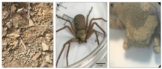
Figure 1.
Sicarius thomisoides spider. (a) S. thomisoides in its natural habitat in La Chimba National Park, Antofagasta, Chile. (b) Female S. thomisoides. (c) Magnification (2×) of S. thomisoides cephalothorax. Scale bar: 1 cm.
The amount of spider venom (µg/spider) for the different specimens of S. thomisoides increased according to the size, weight, and development stage (from nymphs to adults) (Figure 2a), with a mean value ± standard deviation (SD) of 370.3 ± 37.49 µg/spider and a median of 368.3 µg/spider for females, a mean value ± SD of 240.4 ± 105.5 µg/spider and median of 235.4 µg/spider for large nymphs, and mean value ± SD of 50.66 ± 58.23 µg/spider and median of 41.18 µg/spider for small nymphs. However, the venom protein concentrations (µg/spider) of L. laeta adult specimens (female and male) and nymphs were similar (Figure 2b). The mean and median values were as follows: 48.8 ± 46.3 µg/spider and 34.7 µg/spider, respectively, for females; 12.4 ± 9.9 µg/spider and 10.1 µg/spider, respectively, for males; 48.2 ± 22.1 µg/spider and 50.7 µg/spider, respectively, for nymphs. Additionally, the protein yield in the venom of S. thomisoides females was 7.7 times higher than that in female L. laeta venom (Figure 2c). Additionally, the venom protein yield of S. thomisoides large nymphs was 5 times higher than that of L. laeta females (Figure 2d). However, the venom protein yield of S. thomisoides small nymphs showed no differences with those of the venom proteins of L. laeta females.
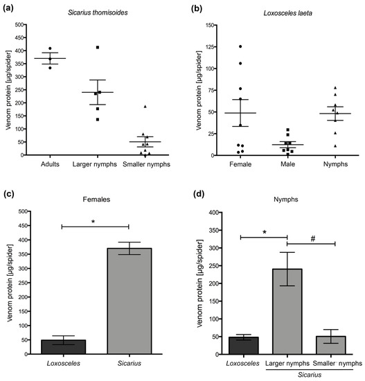
Figure 2.
S. thomisoides venom protein quantitation. (a) Venom protein (μg/spider) distribution of S. thomisoides adults (n = 3), larger nymphs (n = 5), and smaller nymphs (n = 9). (b) Venom protein (μg/spider) distribution of Loxosceles laeta in adults: female (n = 9) and male (n = 8) and nymphs (n = 8). (c) Comparison of the venom protein (μg/spider) between S. thomisoides and L. laeta female specimens. (d) Comparison of the venom protein (μg/spider) between S. thomisoides and L. laeta nymph specimens. The results were expressed as the means ± SD. (*) Statistical significance (p < 0.05) between venom protein means of females from S. thomisoides and L. laeta. (#) Statistical significance (p < 0.05) between the venom protein means of L. laeta nymphs and S. thomisoides small nymphs.
2.2. Electrophoretic Characterization of Venom from the S. thomisoides Spider and Evaluation of Phospholipase D activity
The spider venom was analyzed by SDS-PAGE, showing differences in the number of protein bands of venoms from females and nymphs of S. thomisoides and in comparison with females, males, and nymphs from L. laeta (Figure 3a). However, a similar protein band pattern was detected between 32 and 35 kDa. To assess whether the venom from the S. thomisoides spider has phospholipase D, as well as the one found in the band range of 32–35 kDa in L. laeta, the venoms were analyzed by Western blotting using polyclonal serum raised against a recombinant isoform phospholipase D1 from L. laeta (rLlPLD1). As shown in Figure 3b, the polyclonal antibodies recognized a single band of the venom from L. laeta and S. thomisoides. However, the detection of S. thomisoides PLDs was lower in larger nymphs or females than in the small nymphs. To corroborate whether the bands recognized by pAb-anti-rLlPLD1 in the venom from S. thomisoides were phospholipase D, we measured the ability of phospholipase D in the venom of females and nymphs of S. thomisoides and L. laeta to hydrolyze sphingomyelin. As shown in Figure 4, the venom from females and nymphs of S. thomisoides showed significant sphingomyelinase D activity compared with the negative control (assay buffer). However, these activities were significantly lower than sphingomyelinase D activity in females and nymphs of L. laeta. Thus, the presence and activity of phospholipase D in the venom of S. thomisoides spiders were demonstrated.
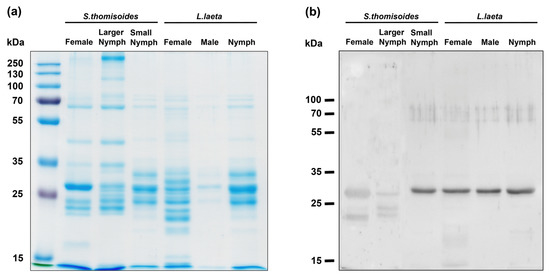
Figure 3.
Electrophoretic pattern of S. thomisoides venom and phospholipase D detection by Western blotting. (a) Venoms (10 µg) in the females and nymphs of S. thomisoides were subjected to 12% SDS-PAGE and compared with 10 µg of venom from females, males, and nymphs of L. laeta. (b) The venoms were detected by Western blotting using mouse polyclonal serum against rLlPLD1 from L. laeta diluted 1:1000, followed by anti-mouse IgG (H + L)-HRP conjugate diluted 1:40,000 and developed using electrochemiluminescence (ECL).
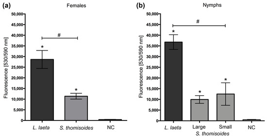
Figure 4.
Comparison of phospholipase D activity between the S. thomisoides and L. laeta venoms. (a) Female venoms. (b) Nymphs venoms. The results were expressed as the means ± SEM. (*) Statistical significance (p < 0.05) between the means of L. laeta or S. thomisoides venoms compared with the negative control (NC). (#) Statistical significance (p < 0.05) between the means of L. laeta and S. thomisoides venoms.
2.3. The Venom of S. thomisoides Spiders Induces Hemolysis, Cytotoxicity, and Dermonecrosis
To assess whether the venom of S. thomisoides has toxic activity similar to that of the L. laeta spider, we compared the hemolytic and cytotoxic activities of both venoms. As shown in Figure 5a, the venom of S. thomisoides produced complement-dependent hemolysis in a concentration-dependent way, similar to L. laeta venom at 10 µg/mL. Additionally, the venom from S. thomisoides showed significant cytotoxic activity against HFF-1 human skin fibroblasts after 24 h of treatment with 5, 10, and 20 µg of venom. No significant differences were found with the cytotoxic activity of the venom from L. laeta (Figure 5b). Finally, the ability of the S. thomisoides venom to produce dermonecrosis in the skin of rabbits was assessed. As shown in Figure 6, the venom of S. thomisoides (50 µg) produced dermonecrosis progressively over time (2 to 24 h), forming a necrotic lesion surrounded by erythema and marked edema. However, when 10 µg of StV was used, only erythema and edema were observed at 24 h (Figure S1). Therefore, the venom of the S. thomisoides spider fulfills the toxic parameters to cause harm to humans.
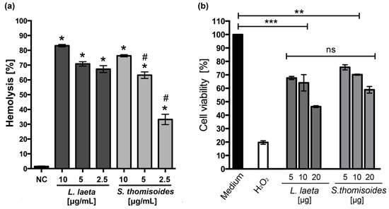
Figure 5.
Comparison of complement-dependent hemolytic and cytotoxic activities between the venoms of S. thomisoides and L. laeta. (a) Complement-dependent hemolysis of human erythrocytes treated with 10, 5, and 2.5 µg/mL of pooled venoms of females from S. thomisoides and L. laeta. The hemolysis absorbance was read at 414 nm and expressed as the hemolysis percentage of three different experiments in triplicate (mean ± SEM). NC: negative control. (*) Statistical significance (p < 0.05) between the means of S. thomisoides or L. laeta venoms from the negative control. (#) Statistical significance (p < 0.05) between the means of S. thomisoides and L. laeta venoms. (b) Viability assay of HFF-1 skin fibroblasts to test the cytotoxic activity of pooled venom of females from S. thomisoides and L. laeta venoms. The cells were treated for 24 h at 37 °C with 5, 10, and 20 µg of venoms from S. thomisoides and L. laeta. The control of 100% viability was treated only with the medium. The results were expressed as the viability percentage against the control of 100% (medium) in two experiments in triplicate (mean ± SEM). ns: no statistical significance. (*) Statistical significance (p < 0.05) between the means of S. thomisoides or L. laeta venoms compared with the control (100% viability). **, p < 0.01, ***, p < 0.001.
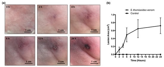
Figure 6.
Dermonecrosis assay of S. thomisoides venom. (a) The pooled venom of females of S. thomisoides (50 µg) was inoculated into the rabbit skin, and a lesion was recorded between 2 and 24 h post inoculation. Scale bar = 1 cm. (b) The lesion areas (cm2) from three treated rabbits were measured and graphed. Control: PBS-treated rabbits.
3. Discussion
A spider is considered potentially dangerous for humans when the spider has venom with documented pathogenic activity for humans. Additionally, it must live where humans live or carry out their activities (synanthropism), as well as possess chelicerae capable of perforating the skin and injecting its venom [15]. In Chile, synanthropic spiders that are considered as dangerous include those from the Sicariidae and Theridiidae families, which are represented by the Loxosceles laeta, Latrodectus, and Steatoda species [16]. However, little attention has been given to nonsynanthropic spiders, such as those of the Sicarius species. Therefore, we studied the venom toxicity of S. thomisoides, which has been documented as one of the largest and most aggressive spiders in the genus Sicarius in Chile [8,9]. These spiders are poisonous and prey on insects and occasionally vertebrates [10]. This background makes them an ideal model to study the potential toxicity of this spider’s venom. Thus, we were able to capture some individuals of the Chilean spider S. thomisoides from desert and coastal locations of the Atacama Desert, in the north of Chile, which were quite nearby to human urban settlements. Unfortunately, only few adults were captured, and all of them were females (n = 3). In this regard, the presence of adults in the populations of this species is quite rare, since they seem to be long-lived spiders that take a long time to reach adulthood [8], as was shown by the number of larger nymphs (sixth to ninth nymph stages) captured, which could easily be confounded with adults if properly sexual characterization is not performed. In addition, due to the time of year when spiders were captured (late summer to late fall), we were unable to find adult males since, as ectothermic organisms, the microhabitat selection is limited by the thermal conditions of their environment [9]. Furthermore, the frequency of males from S. thomisoides spiders has been reported to be less than 3% of the spider population in different coastal and inland locations in the north of Chile [9]. However, a comparative sex-linked study with more adult specimens seems to be necessary to determine the toxic potential of venoms from males and females of S. thomisoides spiders, based on the results shown here, and also to accurately correlate the morphology and size differences with the venom protein yield.
Thus, our data demonstrated that S. thomisoides spiders possess a higher venom protein yield (µg/spider) and venom volume than L. laeta. These features are particularly interesting because L. laeta have been considered as the one of the Loxosceles species, with the highest protein concentration and most dangerous venom [17,18]. Thus, we focused on comparing the biochemical and biological properties of venoms between S. thomisoides and L. laeta. The differences in the venom volume and concentration between the spiders could be explained by size differences between the spiders and those of their venom glands (since S. thomisoides is bigger than L. laeta) [10]. Thereby, we found that S. thomisoides adults were three times larger in size than L. laeta adults. Similar differences in the venom concentration between Sicarius and Loxosceles were also reported for females and males in the Sicarius ornatus species compared with the females of L. laeta. However, no comparison was performed between the nymph stage of both spider genera [14]. Here, we showed marked differences in the venom concentration for the later stages of development of S. thomisoides compared with any nymph stage of L. laeta. The cause may be related to the spider’s body size because small individuals of S. thomisoides with a similar size to that of any nymph stage of L. laeta had the same concentration of venom as that of L. laeta. Moreover, prey selection could also influence the differences in venom volume. Regarding this, spiders with a generalist prey diet have been reported to have large-sized venom glands, a large venom volume, and more complex venom than spiders with specialized prey selection that have smaller glands and less complex venom [19]. Thus, both genera, Sicarius and Loxosceles, which belong to the Sicariidae family, can be considered spiders with a generalized prey selection [19]. Therefore, in addition to the spider size differences, the significant contrast in the venom volume and protein concentration observed could be related to the adaptation of prey present in their habitat. Loxosceles inhabits intradomiciliary environments, whereas Sicarius inhabits extradomiciliary environments where prey diversity is higher; thus, Sicarius can prey on many epigeal insects [8] as well on smaller vertebrates such as the gecko Phyllodactylus gerrhopygus [10].
The variations in the electrophoretic pattern of the venom from S. thomisoides shown between adult and nymph venom were consistent with intraspecific variations. These intraspecific variations were also reported for the venoms of female and male S. ornatus [14], as well as for the venoms of Loxosceles species, including L. laeta [17,18]. Additionally, regardless of the similarities between the protein profiles of venom from S. thomisoides and L. laeta, we observed differences between the electrophoretic pattern of venoms, indicating interspecific variations. However, both spider venoms showed similar protein bands in the range of 25 to 35 kDa. In Loxosceles venom, the protein components between 25 and 35 kDa have been characterized as members of the phospholipase D family, and they are present in different Loxosceles species [11]. Thus, the protein bands of S. thomisoides venom in this range could be considered as paralog PLD enzymes of those present in Loxosceles venom [2,20]. Therefore, we used polyclonal anti-recombinant PLD from L. laeta serum to detect potential PLDs in S. thomisoides venom; a single band was recognized, confirming the presence of PLDs in the venom. This detection was higher in the nymph venom than in the adult venom, likely because of the monospecific characteristics of the serum we used and the differences between the development stages of the spider, where nymphs could be considered more active than adults, consequently expressing more PLD. Additionally, we cannot rule out that Western blotting differences were the consequence of antigenic differences between PLDs from Sicarius and Loxosceles. However, recently, we reported that natural-produced and specific-produced antibodies against L. laeta venom could cross-detect the venom from Sicarius spiders [21], corroborating the similarities between the protein components of venoms from S. thomisoides and L. laeta. Additionally, polyclonal serum against L. intermedia SMase D has been reported to detect the S. ornatus protein band at 32–35 kDa, confirming that venom proteins in this protein range were PLDs [14].
The presence of PLD activity in the venom of S. thomisoides is critical to consider this venom toxic to humans, as all recombinant PLDs from Loxosceles that possess in vitro sphingomyelinase activity can trigger the local and systemic effects observed for the venom [13]. Loxosceles PLD activity has been reported to be associated with the hydrolysis of sphingomyelin to form ceramide 1-phosphate (C1P) and choline, as well as the hydrolysis of lysophospholipids such as lysophosphatidylcholine to form lysophosphatidic acid (LPA) and choline [22,23,24,25]. This sphingomyelinase D activity has been demonstrated in different African and American Sicarius species, with higher activity in African than in American species [2]. Due to the above findings, we evaluated the phospholipase D activity in S. thomisoides venom against the sphingomyelin substrate, observing that the venom possesses phospholipase D activity, but with differences in the activity at the intraspecies level (at the stages of development, from nymphs to adults). This finding was also confirmed by Western blot analysis. However, the phospholipase D activity of S. thomisoides venom was significantly lower than that of L. laeta venom. These differences were also shown in the adults of S. ornatus compared with the activity in the venoms of L. laeta and L. gaucho [14]. The cause may be the differences in the number of isoforms present in the venom of Sicarius compared with that in the venom of Loxosceles. Indeed, the differences in the number of isoforms between the species of Loxosceles spiders have been documented [13,26,27].
Furthermore, differences in the tertiary structure of Sicarius PLD compared with that of PLDs of Loxosceles could affect the hydrolysis capacity of the enzyme. In this regard, two classes of phospholipase D (class I and class II) have been documented in the venom of Loxosceles, with differences in their tertiary structure and the presence of a single disulfide bridge and extended hydrophobic loop (class I), or an additional intrachain disulfide bridge linked to a flexible loop and catalytic loop (class II), which are further subdivided into more active isoforms (classes IIa) and less active or inactive isoforms (classes IIb) [28,29]. Subsequent studies at the proteomic level are required to respond to the above, either through purification of the enzyme, followed by three-dimensional structure identification by crystallography, or by the cloning and expression of recombinant isoforms of PLDs from Sicarius, followed by three-dimensional structure prediction using 3D modeling.
The pathophysiological effects of Loxosceles venom associated with the activity of PLDs include complement-dependent hemolysis, cytotoxicity, and dermonecrosis [23,30]. To assess whether the S. thomisoides venom could produce the three key aspects shown for Loxosceles venom, human erythrocytes were incubated with S. thomisoides or L. laeta venom and compared. The hemolytic effect of S. thomisoides venom was similar to that of L. laeta venom, and only less hemolysis was shown when a lower concentration was assayed. The venom of the S. ornatus species also showed complement-dependent hemolytic activity, which was induced by glycophorin C cleavage after the binding of SMase-D to the erythrocyte membrane [14]. Whether S. thomisoides complement-dependent hemolytic activity was a consequence of the same mechanism as that for the venom of S. ornatus remains to be elucidated. Nevertheless, it is highly possible that both species of South American Sicarius—S. ornatus and the S. thomisoides—share the same hemolytic mechanism due to the results shown here and shown for Loxosceles species [31]. Moreover, S. thomisoides venom showed cytotoxicity to human skin HFF-1 fibroblasts, without significant differences compared with L. laeta venom. However, the effect of S. thomisoides venom on the viability of dermal human skin fibroblasts was early (24 h) and higher than that shown by S. ornatus venom on HaCaT human epidermal keratinocytes, which showed reduced viability only after 48 to 72 h of treatment [14]. Although the differences could be explained by the interspecific variations of the venoms, we cannot rule out that the Sicarius venom has a different effect depending on the skin cell type (dermal over epidermal) and availability of sphingomyelin in cell membranes because of the differences in the regulation of lipids, as was shown for cholesterol synthesis between human skin fibroblasts and keratinocytes [32]. Finally, we verified that one of the largest species of Sicarius present in Chile was able to produce dermonecrosis on rabbit skin. Few reports have shown dermonecrotic lesions caused by Sicarius spiders. Most have been associated with the African species Sicarius testaceus [33]; however, this specie is now considered as member of the Hexophthalma genus, as well as others African Sicarius [34]. Thus, comparative dermonecrosis studies between genera are required to determine possible differences at the genera level. However, the ability of South American Sicarius to produce dermonecrosis has been demonstrated in a single report documenting a human bitten by the South American Sicarius tropicus who developed a necrotic lesion [35]. In our dermonecrosis assay we demonstrated that 50 µg of S. thomisoides venom was able to produce dermonecrosis in rabbits. However, a lower dose (10 µg) failed to produce dermonecrosis. The venom concentration of 50 µg of S. thomisoides venom seems to be enough to produce dermonecrosis. Thus, higher doses of venom inoculated by adults of S. thomisoides could cause more serious lesions, since adults posses up to eight times the concentration assayed. These concentrations correlate with the range of venom able to produce dermonecrosis in rabbits by L. intermedia venom, where 20 µg of venom was demonstrated as the lower dose able to produce dermonecrosis, with an average of 40 µg of venom [36,37].
In conclusion, because the venom of S. thomisoides has phospholipase D activity and can cause hemolysis, cell death of skin fibroblasts, and dermonecrosis, it fulfills the requirements to be considered a spider able to produce harmful effects to humans. Therefore, S. thomisoides must be included and considered a dangerous spider in Chile, and precautions must be taken to avoid human exposure to this spider.
4. Materials and Methods
4.1. Spiders and Venoms
Sixteen Sicarius thomisoides spiders were captured in desert and semidesert areas such as La Chimba National Park and Juan Lopez Bay, in the Antofagasta Region, as well as in La Huayca, in the Tarapacá Region; both regions are located in northern Chile and close to urban settlements where humans live. Additionally, twenty-five Loxosceles laeta spiders were captured from domiciliary habitats in the city of Antofagasta. Among them, nine specimens were classified as females, eight as males, and eight as nymphs or immatures. All the spiders were maintained at the Molecular Parasitology Research Laboratory at the University of Antofagasta, Chile. However, the spider identification was performed at the Environmental Research Center (CENIMA) at the Arturo Prat University in Iquique based on the morphological characteristics of the spiders. The L. laeta spider was identified using the morphological characteristics reported by Gertsch (1967) [38]; the specimens were then classified according to stages in adults (males and females) and nymphs. S. thomisoides spiders were identified using morphological characteristics reported by Magalhaes et al. (2017) [8]; the specimens were classified according to stages in adults or nymphs. Nymphs were subclassified as small nymphs (from first to sixth nymph stages) and large nymphs (from sixth to ninth nymph stages) based on their body size and weight differences.
S. thomisoides (Stv) and L. laeta venom (Llv) were extracted by electrostimulation from the spiders after a week of captivity and with no previous feeding. Then, the venom droplets were collected with a micropipette in 30 μL of PBS and stored at −80 °C until use, as previously reported [39]. The protein concentration of venom samples was evaluated by the Bradford dye-binding method [40] using the Bio-Rad Protein Assay kit (Bio-Rad Laboratories, Inc., Hercules, CA, USA), and the amount of protein in μg by spider was calculated based on the volume of venom collected.
All the protocols for the biological research in invertebrate and/or biotechnological species, including the procedures for spider capture, spider maintenance, and venom extraction, were approved by the Ethics Committee in Scientific Research of the University of Antofagasta (CEIC-UA) (CEIC-REV No. 06/2019).
4.2. Electrophoresis and Immunoblotting
Ten micrograms of venom samples from females, large and small nymphs of S. thomisoides, and both adults (male and female) and nymphs of L. laeta spiders was electrophoretically separated on 12% SDS-PAGE gels under nonreducing conditions and stained with Coomassie blue G-250. Next, the gels were transferred to nitrocellulose membranes. After transfer, the membranes were blocked for 2 h with 5% nonfat milk in TBS/0.1% Tween20 (TBS-T) and incubated for 1 h at room temperature with polyclonal mouse anti-rLlPLD1 serum (1:1000 dilution) produced previously [41]. The membranes were washed six times for 10 min each with TBS-T and incubated with anti-mouse IgG (H + L)-HRP conjugate (1:40,000 dilution) in TBS-T for 1 h at room temperature. After another six washes with TBS-T, the membranes were developed using the ECLTM Western blotting detection reagent kit (GE Healthcare, Piscataway, NJ, USA). Mouse polyclonal anti-L. laeta recombinant phospholipase D1 (rLlPLD1) was prepared as previously documented [41].
4.3. Phospholipase D Activity
The phospholipase D activities of S. thomisoides or L. laeta venom toward the sphingomyelin substrate were measured using the Amplex Red Sphingomyelinase D Assay kit (Molecular Probes, Eugene, OR) following the manufacturer’s instructions [41]. In summary, 2.5 μg/mL of venom from the S. thomisoides and L. laeta females and nymphs was incubated with sphingomyelin in Amplex Red reaction buffer at 37 °C for 1 h, and the fluorescence was measured using an Infinite M200 PRO microplate reader (Tecan) spectrofluorometer and excitation/emission filters of 530 and 590 nm, respectively. The assays were performed in duplicate for three separate experiments. The fluorescence was compared against the negative control (NC) using only the Amplex Red reaction buffer.
4.4. Complement-Dependent Hemolysis Assay
The human erythrocyte complement-dependent hemolysis assay was performed as previously described [41]. Briefly, human erythrocytes were washed three times with Veronal Buffered Saline (VBS; pH 7.4, 10 mM sodium barbitone, 0.15 mM CaCl2, 0.5 mM MgCl2, and 145 mM NaCl) and resuspended at 2% in VBS. The cells were sensitized for 30 min at 37 °C with 10, 5, or 2.5 μg/mL of pooled venom from the females of S. thomisoides or L. laeta in 100 μL of VBS. After incubation, the sensitized erythrocytes were washed three times with VBS and analyzed by the complement-dependent hemolysis assay. Next, 100 μL of sensitized erythrocytes was mixed with 100 μL of normal human serum (NHS; 1:2 in VBS). The negative control was evaluated by incubating the erythrocytes with VBS, while the total hemolysis control was incubated with H2O. After incubation for 1 h at 37 °C, the nonlysed cells were centrifuged at 440× g for 5 min, the supernatant was collected, and absorbance was measured at 414 nm. The results were expressed as a percentage of hemolysis and calculated based on the absorbance of the 100% hemolysis control. The assays were performed in duplicate for two independent experiments. The erythrocytes and normal serum were obtained from the same donor.
4.5. Viability Assay
The viability assay was performed as previously described [42]. The cultures of HFF-1 human skin fibroblasts with more than 95% viability were used for the viability assay. Briefly, a cell suspension of 2 × 10⁴ cells/wells in DMEM serum-free medium was placed in each well of a 96-well culture plate. Subsequently, the cells were incubated for 4 h at 37 °C in a humidified atmosphere containing 5% CO2, allowing cell adherence. Next, 40 µg of pooled venom from the adults of S. thomisoides or L. laeta was two-fold diluted in serum-free DMEM medium and was added to the cells, followed by incubation at 37 °C for 24 h under an atmosphere containing 5% CO2. Untreated cells were used as a 100% viability control (negative control), while cells treated with 0.3% hydrogen peroxide (H2O2) were used as a positive mortality control. Next, the cell viability for each treatment was determined using Cell Titer 96 Aqueous One Solution (Promega) according to the manufacturer’s instructions, in which 20 µL of reagent was dispensed to each well and incubated at 37 °C for 1 h. Subsequently, viable cells were measured at 490 nm in an Infinite M200 PRO microplate reader (Tecan Group Ltd., Männedorf, Switzerland). The percentage of viability was calculated as follows: % viability = (Abs490 nm sample—Abs490 nm blank control)/(Abs490 nm control 100% viability—Abs490 nm blank control) × 100. Additionally, the inhibitory concentration 50 (IC50) for each treatment was calculated using nonlinear regression on a sigmoidal curve. Assays were performed in triplicate for two independent experiments.
4.6. Dermonecrosis Assay
Groups of three New Zealand male rabbits were inoculated intradermally, into a previously shaved right leg, with 10 µg or 50 µg of pooled venom from the females of S. thomisoides. Next, the presence of erythema, edema, or necrosis was documented by photography of the inoculated area at 2, 4, 6, 12, and 24 h after inoculation. Sterile PBS inoculated into the left leg of rabbits was used as negative control. The lesion area (cm2) of the region of interest (ROI) for each dermonecrotic lesion was analyzed using ImageJ software (National Institutes of Health, Bethesda, MD, USA) and graphed [43].
4.7. Statistical Analysis
One-way ANOVA followed by Tukey’s multiple comparisons test was performed using GraphPad Prism software v.6.0. A significant criterion of p-value < 0.05 was used.
Supplementary Materials
The following are available online at https://www.mdpi.com/2072-6651/12/11/702/s1: Table S1: Characteristic of captured Sicarius thomisoides specimens (development stage, sex, weight, venom volume extracted, and venom protein yield by spider); Figure S1: Dermonecrosis assay of S. thomisoides venom using 10 µg of venom inoculated into rabbit skin.
Author Contributions
Conceptualization and design of the experiments, A.C. and A.T.-R.; recollection of spider specimens, I.P.-S., V.V. and A.T.-R.; performed the experiments, T.A.-S., J.M.R., I.P.-S., V.V., N.O., and J.E.A.; analysis of data, T.A.-S., I.P.-S., V.V., and A.C.; writing, T.A.-S., and A.C.; Review and editing, A.T.-R., J.E.A., and A.C. All authors have read and agreed to the published version of the manuscript.
Funding
This research was supported by Fondo para el Desarrollo en Investigación Científica y/o Tecnológica para actividades de titulación de pregrado 2019 (ATI19-1-07), and partially by Semillero-UA #5301 from Dirección Gestión en Investigación, Vicerrectoria de Investigación, Innovación y Postgrado, Universidad de Antofagasta.
Conflicts of Interest
The authors declare no conflict of interest. The founding sponsors had no role in the design of the study; in the collection, analyses, or interpretation of data; in the writing of the manuscript, and in the decision to publish the results.
References
- WSC. World spider catalog. Version 21.0. Natural History Museum Bern. 2020. Available online: http://wsc.nmbe.ch (accessed on 7 July 2020).
- Binford, G.J.; Bodner, M.R.; Cordes, M.H.; Baldwin, K.L.; Rynerson, M.R.; Burns, S.N.; Zobel-Thropp, P.A. Molecular evolution, functional variation, and proposed nomenclature of the gene family that includes sphingomyelinase D in Sicariid spider venoms. Mol. Biol. Evol. 2009, 26, 547–566. [Google Scholar] [CrossRef] [PubMed]
- Gremski, L.H.; Trevisan-Silva, D.; Ferrer, V.P.; Matsubara, F.H.; Meissner, G.O.; Wille, A.C.; Vuitika, L.; Dias-Lopes, C.; Ullah, A.; de Moraes, F.R.; et al. Recent advances in the understanding of brown spider venoms: From the biology of spiders to the molecular mechanisms of toxins. Toxicon 2014, 83, 91–120. [Google Scholar] [CrossRef] [PubMed]
- Da Silva, P.H.; da Silveira, R.B.; Appel, M.H.; Mangili, O.C.; Gremski, W.; Veiga, S.S. Brown spiders and loxoscelism. Toxicon 2004, 44, 693–709. [Google Scholar] [CrossRef]
- Swanson, D.L.; Vetter, R.S. Loxoscelism. Clin. Dermatol. 2006, 24, 213–221. [Google Scholar] [CrossRef]
- Chaim, O.M.; Trevisan-Silva, D.; Chaves-Moreira, D.; Wille, A.C.; Ferrer, V.P.; Matsubara, F.H.; Mangili, O.C.; da Silveira, R.B.; Gremski, L.H.; Gremski, W.; et al. Brown spider (Loxosceles genus) venom toxins: Tools for biological purposes. Toxins 2011, 3, 309–344. [Google Scholar] [CrossRef] [PubMed]
- Walckenaer, C.A. Dernier supplément. In Histoire Naturelle des Insectes: Aptères; Walckenaer, C.A., Gervais, P., Eds.; Librairie Encyclopédique de Roret: Paris, France, 1847; pp. 365–564. [Google Scholar]
- Magalhaes, I.L.F.; Brescovit, A.D.; Santos, A.J. Phylogeny of Sicariidae spiders (Araneae: Haplogynae), with a monograph on neotropical Sicarius. Zool. J. Linn. Soc. 2017, 179, 767–864. [Google Scholar]
- Taucare-Rios, A.; Veloso, C.; Bustamante, R.O. Microhabitat selection in the sand recluse spider (Sicarius thomisoides): The effect of rock size and temperature. J. Nat. Hist. 2017, 51, 2199–2210. [Google Scholar] [CrossRef]
- Taucare-Rios, A.; Piel, W.H. Predation on the gecko Phyllodactylus gerrhopygus (Wiegmann) (Squamata: Gekkonidae) by the six-eyed sand spider Sicarius thomisoides (Walckenaer) (Araneae: Sicariidae). Rev. Soc. Entomol. Argent. 2020, 79, 48–51. [Google Scholar] [CrossRef]
- Binford, G.J.; Wells, M.A. The phylogenetic distribution of sphingomyelinase D activity in venoms of Haplogyne spiders. Comp. Biochem. Physiol. B Biochem. Mol. Biol. 2003, 135, 25–33. [Google Scholar] [CrossRef]
- Zobel-Thropp, P.A.; Bodner, M.R.; Binford, G.J. Comparative analyses of venoms from American and African Sicarius spiders that differ in sphingomyelinase D activity. Toxicon 2010, 55, 1274–1282. [Google Scholar] [CrossRef]
- Gremski, L.H.; da Justa, H.C.; da Silva, T.P.; Polli, N.L.C.; Antunes, B.C.; Minozzo, J.C.; Wille, A.C.M.; Senff-Ribeiro, A.; Arni, R.K.; Veiga, S.S. Forty years of the description of brown spider venom phospholipases-D. Toxins 2020, 12, 164. [Google Scholar] [CrossRef] [PubMed]
- Lopes, P.H.; Bertani, R.; Goncalves-de-Andrade, R.M.; Nagahama, R.H.; van den Berg, C.W.; Tambourgi, D.V. Venom of the brazilian spider Sicarius ornatus (Araneae, Sicariidae) contains active sphingomyelinase D: Potential for toxicity after envenomation. PLoS Negl. Trop. Dis. 2013, 7, e2394. [Google Scholar] [CrossRef] [PubMed]
- Schenone, H. Toxic pictures produced spiders bites in Chile: Latrodectism and loxoscelism. Rev. Med. Chil. 2003, 131, 437–444. [Google Scholar] [PubMed]
- Taucare-Rios, A. Synantropic dangerous spiders from Chile. Rev. Med. Chil. 2012, 140, 1228–1229. [Google Scholar] [PubMed]
- De Oliveira, C.K.; Goncalves de Andrade, R.M.; Giusti, A.L.; Dias da Silva, W.; Tambourgi, D.V. Sex-linked variation of Loxosceles intermedia spider venoms. Toxicon 1999, 37, 217–221. [Google Scholar] [CrossRef]
- De Oliveira, K.C.; Goncalves de Andrade, R.M.; Piazza, R.M.; Ferreira, J.M., Jr.; van den Berg, C.W.; Tambourgi, D.V. Variations in Loxosceles spider venom composition and toxicity contribute to the severity of envenomation. Toxicon 2005, 45, 421–429. [Google Scholar] [CrossRef]
- Pekar, S.; Bocanek, O.; Michalek, O.; Petrakova, L.; Haddad, C.R.; Sedo, O.; Zdrahal, Z. Venom gland size and venom complexity-essential trophic adaptations of venomous predators: A case study using spiders. Mol. Ecol. 2018, 27, 4257–4269. [Google Scholar] [CrossRef]
- Binford, G.J.; Callahan, M.S.; Bodner, M.R.; Rynerson, M.R.; Nunez, P.B.; Ellison, C.E.; Duncan, R.P. Phylogenetic relationships of Loxosceles and Sicarius spiders are consistent with Western Gondwanan vicariance. Mol. Phylogenet. Evol. 2008, 49, 538–553. [Google Scholar] [CrossRef]
- Aran-Sekul, T.; Rojas, J.M.; Subiabre, M.; Cruz, V.; Cortes, W.; Osorio, L.; Gonzalez, J.; Araya, J.E.; Catalan, A. Heterophilic antibodies in sera from individuals without loxoscelism cross-react with phospholipase D from the venom of Loxosceles and Sicarius spiders. J. Venom Anim. Toxins Incl. Trop. Dis. 2018, 24, 18. [Google Scholar] [CrossRef]
- Kurpiewski, G.; Forrester, L.J.; Barrett, J.T.; Campbell, B.J. Platelet aggregation and sphingomyelinase D activity of a purified toxin from the venom of Loxosceles reclusa. Biochim. Biophys. Acta 1981, 678, 467–476. [Google Scholar] [CrossRef]
- Tambourgi, D.V.; Magnoli, F.C.; van den Berg, C.W.; Morgan, B.P.; de Araujo, P.S.; Alves, E.W.; Da Silva, W.D. Sphingomyelinases in the venom of the spider Loxosceles intermedia are responsible for both dermonecrosis and complement-dependent hemolysis. Biochem. Biophys. Res. Commun. 1998, 251, 366–373. [Google Scholar] [CrossRef] [PubMed]
- Van Meeteren, L.A.; Frederiks, F.; Giepmans, B.N.; Pedrosa, M.F.; Billington, S.J.; Jost, B.H.; Tambourgi, D.V.; Moolenaar, W.H. Spider and bacterial sphingomyelinases D target cellular lysophosphatidic acid receptors by hydrolyzing lysophosphatidylcholine. J. Biol. Chem. 2004, 279, 10833–10836. [Google Scholar] [CrossRef] [PubMed]
- Lee, S.; Lynch, K.R. Brown recluse spider (Loxosceles reclusa) venom phospholipase D (PLD) generates lysophosphatidic acid (LPA). Biochem. J. 2005, 391, 317–323. [Google Scholar] [CrossRef] [PubMed]
- Machado, L.F.; Laugesen, S.; Botelho, E.D.; Ricart, C.A.; Fontes, W.; Barbaro, K.C.; Roepstorff, P.; Sousa, M.V. Proteome analysis of brown spider venom: Identification of loxnecrogin isoforms in Loxosceles gaucho venom. Proteomics 2005, 5, 2167–2176. [Google Scholar] [CrossRef]
- Kalapothakis, E.; Chatzaki, M.; Goncalves-Dornelas, H.; de Castro, C.S.; Silvestre, F.G.; Laborne, F.V.; de Moura, J.F.; Veiga, S.S.; Chavez-Olortegui, C.; Granier, C.; et al. The loxtox protein family in Loxosceles intermedia (Mello-Leitao) venom. Toxicon 2007, 50, 938–946. [Google Scholar] [CrossRef]
- Murakami, M.T.; Fernandes-Pedrosa, M.F.; de Andrade, S.A.; Gabdoulkhakov, A.; Betzel, C.; Tambourgi, D.V.; Arni, R.K. Structural insights into the catalytic mechanism of sphingomyelinases D and evolutionary relationship to glycerophosphodiester phosphodiesterases. Biochem. Biophys. Res. Commun. 2006, 342, 323–329. [Google Scholar] [CrossRef]
- De Giuseppe, P.O.; Ullah, A.; Silva, D.T.; Gremski, L.H.; Wille, A.C.; Chaves Moreira, D.; Ribeiro, A.S.; Chaim, O.M.; Murakami, M.T.; Veiga, S.S.; et al. Structure of a novel class II phospholipase D: Catalytic cleft is modified by a disulphide bridge. Biochem. Biophys. Res. Commun. 2011, 409, 622–627. [Google Scholar] [CrossRef]
- Paixao-Cavalcante, D.; van den Berg, C.W.; de Freitas Fernandes-Pedrosa, M.; Goncalves de Andrade, R.M.; Tambourgi, D.V. Role of matrix metalloproteinases in HaCaT keratinocytes apoptosis induced by Loxosceles venom sphingomyelinase D. J. Invest. Dermatol. 2006, 126, 61–68. [Google Scholar] [CrossRef]
- Tambourgi, D.V.; Morgan, B.P.; de Andrade, R.M.; Magnoli, F.C.; van Den Berg, C.W. Loxosceles intermedia spider envenomation induces activation of an endogenous metalloproteinase, resulting in cleavage of glycophorins from the erythrocyte surface and facilitating complement-mediated lysis. Blood 2000, 95, 683–691. [Google Scholar] [CrossRef]
- Ponec, M.; Havekes, L.; Kempenaar, J.; Vermeer, B.J. Cultured human skin fibroblasts and keratinocytes: Differences in the regulation of cholesterol synthesis. J. Invest. Dermatol. 1983, 81, 125–130. [Google Scholar] [CrossRef]
- Van Aswegen, G.; Van Rooyen, J.M.; Van der Nest, D.G.; Veldman, F.J.; De Villiers, T.H.; Oberholzer, G. Venom of a six-eyed crab spider, Sicarius testaceus (Purcell, 1908), causes necrotic and haemorrhagic lesions in the rabbit. Toxicon 1997, 35, 1149–1152. [Google Scholar] [CrossRef]
- Lotz, L. An update on the spider genus Hexophthalma (Araneae: Sicariidae) in the afrotropical region, with descriptions of new species. Eur. J. Taxon. 2018, 424, 1–18. [Google Scholar] [CrossRef]
- Dos-Santos, M.C.; Cardoso, J.L.C. Lesao dermonecrótica por Sicarius tropicus, simulando loxoscelismo cutaneo. Rev. Soc. Bras. Med. Trop. 1992, 25, 115–123. [Google Scholar]
- Monteiro, C.L.; Rubel, R.; Cogo, L.L.; Mangili, O.C.; Gremski, W.; Veiga, S.S. Isolation and identification of Clostridium perfringens in the venom and fangs of Loxosceles intermedia (brown spider): Enhancement of the dermonecrotic lesion in loxoscelism. Toxicon 2002, 40, 409–418. [Google Scholar] [CrossRef]
- Ospedal, K.Z.; Appel, M.H.; Fillus Neto, J.; Mangili, O.C.; Sanches Veiga, S.; Gremski, W. Histopathological findings in rabbits after experimental acute exposure to the Loxosceles intermedia (brown spider) venom. Int. J. Exp. Pathol. 2002, 83, 287–294. [Google Scholar] [CrossRef] [PubMed]
- Gertsch, W.J. The spider genus Loxosceles in South America (Araneae Scytodidae). Bull. Am. Mus. Nat. Hist. 1967, 136, 117–174. [Google Scholar]
- Catalan, A.; Espoz, M.C.; Cortes, W.; Sagua, H.; Gonzalez, J.; Araya, J.E. Tetracycline and penicillin resistant Clostridium perfringens isolated from the fangs and venom glands of Loxosceles laeta: Its implications in loxoscelism treatment. Toxicon 2010, 56, 890–896. [Google Scholar] [CrossRef]
- Bradford, M.M. A rapid and sensitive method for the quantitation of microgram quantities of protein utilizing the principle of protein-dye binding. Anal. Biochem. 1976, 72, 248–254. [Google Scholar] [CrossRef]
- Catalan, A.; Cortes, W.; Sagua, H.; Gonzalez, J.; Araya, J.E. Two new phospholipase D isoforms of Loxosceles laeta: Cloning, heterologous expression, functional characterization, and potential biotechnological application. J. Biochem. Mol. Toxicol. 2011, 25, 393–403. [Google Scholar] [CrossRef]
- Rojas, J.M.; Aran-Sekul, T.; Cortes, E.; Jaldin, R.; Ordenes, K.; Orrego, P.R.; Gonzalez, J.; Araya, J.E.; Catalan, A. Phospholipase D from Loxosceles laeta spider venom induces IL-6, IL-8, CXCL1/GRO-alpha, and CCL2/MCP-1 production in human skin fibroblasts and stimulates monocytes migration. Toxins 2017, 9, 125. [Google Scholar] [CrossRef]
- Schneider, C.A.; Rasband, W.S.; Eliceiri, K.W. Nih image to imagej: 25 years of image analysis. Nat. Methods 2012, 9, 671–675. [Google Scholar] [CrossRef] [PubMed]
Publisher’s Note: MDPI stays neutral with regard to jurisdictional claims in published maps and institutional affiliations. |
© 2020 by the authors. Licensee MDPI, Basel, Switzerland. This article is an open access article distributed under the terms and conditions of the Creative Commons Attribution (CC BY) license (http://creativecommons.org/licenses/by/4.0/).