Article

Virulence Characteristics and Antimicrobial Resistance Profiles of Shiga Toxin-Producing Escherichia coli Isolates from Humans in South Africa: 2006–2013

by
Musafiri Karama
1,*,
Beniamino T. Cenci-Goga
1,2,
Mogaugedi Malahlela
1,
Anthony M. Smith
3,
Karen H. Keddy
3,
Saeed El-Ashram
4,5,
Lawan M. Kabiru
6 and
Alan Kalake
7
1
Department of Paraclinical Sciences, Faculty of Veterinary Science, University of Pretoria, Onderstepoort 0110, South Africa
2
Dipartimento di Medicina Veterinaria, Laboratorio di Ispezione degli Alimenti di Origine Animale, University of Perugia, 06126 Perugia, Italy
3
Centre for Enteric Diseases, National Institute for Communicable Diseases, Johannesburg 2131, South Africa
4
College of Life Science and Engineering, University Foshan, Foshan 528231, China
5
Faculty of Science, Kafrelsheikh University, Kafrelsheikh 33516, Egypt
6
Department of Veterinary Public Health and Preventive Medicine, Ahmadu Bello University, Zaria 2222, Nigeria
7
Gauteng Department of Agriculture and Rural Development, Johannesburg 2001, South Africa
*
Author to whom correspondence should be addressed.
Toxins 2019, 11(7), 424; https://doi.org/10.3390/toxins11070424
Submission received: 9 June 2019 / Revised: 1 July 2019 / Accepted: 3 July 2019 / Published: 19 July 2019

Abstract

:
Shiga toxin-producing Escherichia coli (STEC) isolates (N = 38) that were incriminated in human disease from 2006 to 2013 in South Africa were characterized by serotype, virulence-associated genes, antimicrobial resistance and pulsed-field gel electrophoresis (PFGE). The isolates belonged to 11 O:H serotypes. STEC O26:H11 (24%) was the most frequent serotype associated with human disease, followed by O111:H8 (16%), O157:H7 (13%) and O117:H7 (13%). The majority of isolates were positive for key virulence-associated genes including stx1 (84%), eaeA (61%), ehxA (68.4%) and espP (55%), but lacked stx2 (29%), katP (42%), etpD (16%), saa (16%) and subA (3%). stx2 positive isolates carried stx2c (26%) and/or stx2d (26%) subtypes. All pathogenicity island encoded virulence marker genes were detected in all (100%) isolates except nleA (47%), nleC (84%) and nleD (76%). Multidrug resistance was observed in 89% of isolates. PFGE revealed 34 profiles with eight distinct clusters that shared ≥80% intra-serotype similarity, regardless of the year of isolation. In conclusion, STEC isolates that were implicated in human disease between 2006 and 2013 in South Africa were mainly non-O157 strains which possessed virulence genes and markers commonly associated with STEC strains that have been incriminated in mild to severe human disease worldwide. Improved STEC monitoring and surveillance programs are needed in South Africa to control and prevent STEC disease in humans.
Key Contribution: This study is a key contribution towards STEC surveillance and improves our understanding on emerging serotypes and virulence factors of STEC isolates that affect humans in South Africa.

1. Introduction

Shiga toxin-producing Escherichia coli (STEC) is a zoonotic foodborne and waterborne pathogen associated with enteric disease in humans characterized by abdominal cramps, mild to severe diarrhea, hemorrhagic colitis (HC) and hemolytic uremic syndrome (HUS) [1]. Majowicz et al. [2] have estimated that STEC is responsible for 2.8 million cases per year of enteric disease in humans, globally. Humans acquire STEC through ingestion of contaminated foods of animal origin, water and raw vegetables or contact with infected animals [3,4,5].
More than 600 STEC serotypes have been recovered from animals, humans, the environment and various foods around the world [6,7,8]. STEC O157:H7 is the most common serotype in human infections, but non-O157 STEC have also emerged as an important cause of enteric disease [6,7,8,9] and may account for up to 80% of human STEC infections [2,10]. Furthermore, most STEC infections in humans have been ascribed to STEC strains belonging to seven major serogroups, including STEC O157, O26, O111, O103, O145, O121 and O45 [7,11].
Shiga toxins (stx1 and stx2) are the main virulence factors of STEC. At least 15 Shiga toxin subtypes have been identified (http://old.iss.it/binary/vtec/cont/STEC_2018_Wrap_up.pdf), including stx1a, stx1c, stx1d, stx1e, stx2c, stx2d, stx2e, stx2f, stx2g, stx2h, stx2i, stx2j, stx2k and stxl [12,13,14]. Another key virulence factor of STEC is the E. coli attachment and effacement gene (eae) which encodes intimin (eaeA) [15,16,17]. Intimin (eaeA) is encoded on the locus of the enterocyte effacement (LEE) pathogenicity island [16] and is responsible for attaching and effacing (AE) lesions which are observed in the intestinal epithelium of human and animal hosts affected with eaeA positive E. coli, including STEC [16,17].
STEC produce plasmid-encoded virulence-associated proteins, including hemolysin (hlyA or ehxA) [18], catalase peroxidase (katP) [19], extracellular serine protease (espP) [20] and a type II secretion pathway (etpD) [21]. STEC autoagglutinating adhesin (saa) and subtilase cytotoxin (subA) are also recognized as key plasmid-encoded virulence factors in eaeA negative STEC strains [22,23]. Plasmid-encoded virulence genes have been associated with increased adherence and colonization (saa) of enterocytes, cytotoxicity (subA), reduction of oxidative stress (katP), exoprotein secretion (etpD), suppression of the host’s immune system and cleavage of coagulation factor V (espP) and lysis of human erythrocytes with subsequent mucosal hemorrhage in the host (hlyA) [18,20,21,22,23,24,25].
Several genes that are encoded on pathogenicity islands (O-islands-OIs) other than the LEE are also considered STEC virulence markers [26]. Particularly, genes located on OI-122 [11,27], including pagC, which encodes a protein sharing 46% homology with the phoP-activated gene C product in Salmonella enterica serovar Typhimurium. PagC enables survival in macrophages. Another OI-122–encoded gene is sen, which encodes a protein that is 38.2% homologous to Shigella flexneri enterotoxin 2 [11,27], as well as efa1 and efa2 (enterohemorrhagic E. coli factor for adherence), which have been associated with epithelial cell adhesion and inhibition of bovine peripheral blood lymphocyte proliferation [11,28]. OI-122 marker genes have been used to characterize and classify STEC serotypes into seropathotypes based on their association with outbreaks and severe disease complications, such as HC and HUS in humans [11]. In addition, genes that are carried on OI-43/48 are also considered key virulence markers: iha (iron-regulated gene A), which encodes an adhesin [29] and ureC [30,31] and terC [31], which encode urease and tellurite resistance, respectively. Furthermore, numerous non-LEE encoded effector (nles) genes that are carried on various pathogenicity islands are considered virulence markers: nleA (OI-71), nleB/Z4328 (OI-122), nleB2, nleC (OI-36), nleD (OI-57), nleE/Z4329 (OI-122), nleG (OI-71), nleG2-1 (OI-71), nleG2-3 (OI-57), nleG6-2 (OI-57), nleG9 (OI-71), nleH1-2 (OI-171) [32] and ent/espL2 (OI-122). These genes are mainly found in high risk (HUS) STEC strains and are associated with colonization and survival in the host, and may interfere with signaling pathways during inflammation [33]. Non-LEE encoded effector genes have been used in molecular epidemiology to differentiate highly virulent from less virulent STEC strains (molecular risk assessment) [32,34,35].
The emergence of antimicrobial resistant bacteria as a consequence of the indiscriminate use and abuse of antimicrobials in animals and humans has become a public health concern [36,37,38,39]. Currently, the use of antimicrobials for treatment of STEC infections in humans is controversial and not recommended in many countries, because a number of studies have shown that antimicrobials, such as ciprofloxacin and trimethoprim-sulphamethoxazole, can induce STEC lysogenic bacteriophages to enter the lytic cycle, which exacerbates STEC infections by increasing Shiga toxin production with resultant severe STEC disease manifestations in humans [39,40]. In addition, STEC bacteriophages may carry genes encoding antimicrobial resistance which can be transferred to naïve E. coli, which are then converted into antimicrobial resistant strains. Therefore, E. coli, including STEC, as commensal bacteria in the intestinal tracts of animals and humans are considered reliable indicators of antimicrobial resistance, and have been used for monitoring and surveillance of antimicrobial resistance in livestock, humans and along the food chain.
Although the first case and largest outbreak of human STEC disease in South Africa occurred in 1990 and 1992, respectively [41,42], ten years after the first report of a human STEC disease outbreak in the world in the USA [43], data on the epidemiology, virulence characteristics, antimicrobial resistance patterns and genotypes of human STEC isolates from South Africa remain scanty. In this study, STEC isolates that were incriminated in human disease outbreaks in South Africa from 2006–2013 were fully serotyped, screened for virulence-associated genes and antimicrobial resistance patterns and subtyped by pulsed-field gel electrophoresis (PFGE). The overall aim of this study was to contribute to STEC surveillance and gain insight into the molecular epidemiology of STEC isolates that have been implicated in human disease in South Africa.

2. Results

2.1. STEC Serotypes and Their Frequency in Human Outbreaks

Serotyping (O:H) assigned the 38 STEC isolates to 11 O:H serotypes, including 11 O serogroups and 6 H types. The isolates belonged to the following serotypes: O5:HNT (2), O8:H19 (1), O22:H16 (1), O26:H11 (9), O107:H7 (3), O111:H8 (6), O113:H21 (1), O117:H7 (5), O118:H16 (1), O156:H7 (2), O157:H7 (5), ONT:H8 (1) and ONT:H21 (1) (Table 1). Overall, STEC O157:H7 accounted for 13.1% (5/38) and non-O157 serotypes were associated with 87% (33/38) of STEC isolates that caused human disease in South Africa from 2006 to 2013. STEC O26:H11 was the most frequent serotype, responsible for 24% (9/38) of human disease cases which occurred over five years (2007, 2008, 2009, 2010 and 2013), followed by STEC O111:H8, which was incriminated in 16% (6/38) of cases which occurred over four years (2006, 2008, 2009 and 2010). STEC O157:H7 and O117:H7 were associated with 13% (5/38) of human disease cases for each individual serotype, which occurred over four (2009, 2010, 2011 and 2012) and three years (2006, 2010, and 2012), respectively, while STEC O107:H7 was implicated in 8% (3/38) of cases which all occurred in 2007. The remaining serotypes (O5:HNT, O8:H19, O22:H16, ONT:H8, O118:H16, ONT:H21 and O156:H7) were implicated once or twice in human disease in the period spanning from 2006–2013.

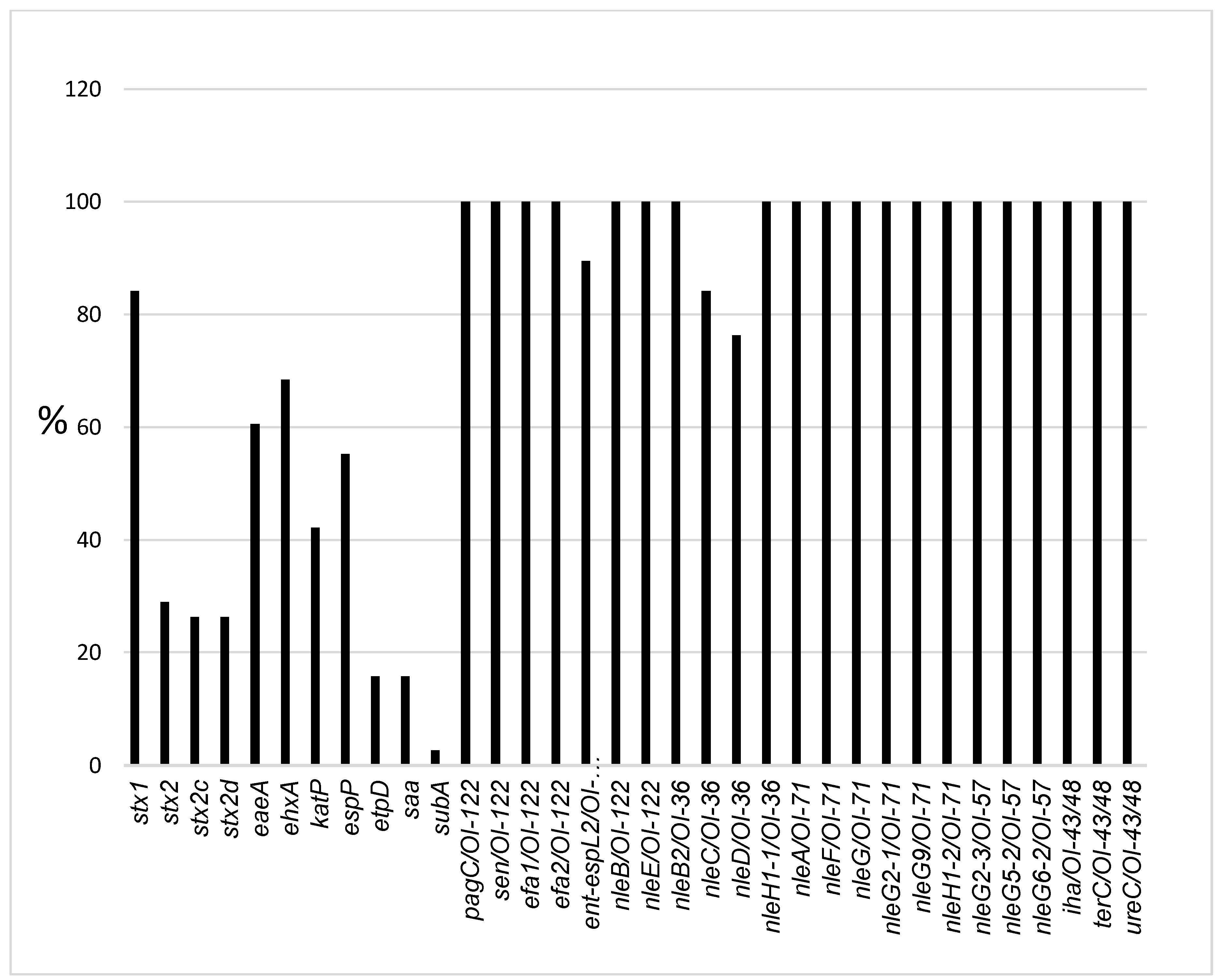
2.2. Virulence-Associated Gene Distribution

The frequencies of virulence genes are depicted in Figure 1. PCR revealed that overall, 84% (32/38) of isolates carried stx1, 29% (11/38) carried stx2, 26% (10/38) carried stx2c and 26% (10/38) carried stx2d (Table 1 and Figure 1). Both stx1 and stx2 were detected concurrently in 13% (5/38) of isolates while both stx1 and stx2c were detected in 0.5% (2/38) of isolates. stx1 alone or stx2 alone were detected in 66% (25/38) and 16% (6/38) of isolates, respectively. The following stx subtypes were not detected: stx1c and stx1d, stx2e, stx2f and stx2g. Additional genotypes were also observed: stx1 + stx2c in 8% (3/38), stx1 + stx2 + stx2d in 8% (3/38), stx2 + stx2c + stx2d in 13% (5/38) and stx1 + stx2 + stx2c + stx2d in 5% (2/38) of isolates. Both stx2c + stx2d were detected concurrently in 18% (7/38) of isolates which belonged to the following serotypes: STEC O157:H7 (5), STEC O8:H19 (1) and STEC O113:H2 (1). The eaeA gene was detected in 60.5% (23/38) of isolates which belonged to the following serotypes: STEC O5:HNT, O26:H11, O111:H8, O118:H16, O157:H7 and ONT:H8.
Plasmid-encoded genes were distributed as follows: ehxA, 68.4% (21/38); katP, 42% (16/38); espP, 55% (21/38); etpD, 16% (6/38) and saa, 16% (6/38) (Table 1 and Figure 1). Only one isolate carried subA. A complete plasmid (ehxA, katP, espP and etpD) was detected in STEC O157:H7 isolates only while O26:H11 isolates possessed all plasmid marker genes except etpD. The following OI marker genes were present in 100% of isolates: OI-122 markers: pagC, sen, efa1 and efa2 except ent/espL2, which was detected in 89.4% of isolates; OI-43/48 markers: terC, ureC and iha were present in 100% of isolates. Most nle effector genes, including nleB, nleB2, nleF, nleE, nleG, nleG2-1, nleG2-3, nleG5-2, nleG6-2, nleG9, nleH1-1 and nleH1-2, were present in 100% of isolates, while nleA was found in 47% (18/38), nleC in 84% (32/38), and nleD in 76% (29/38).
Overall, the STEC isolates carried 50–74% (20–28 out of 34) genes. STEC O157:H7 carried 28/34 genes, STEC O118:H16 had 27/34 genes, STEC O26:H11 had 26–27/34 genes, STEC O5:HNT had 25–26/34 genes, STEC O8:H19 had 25/34 genes and two STEC O111:H8 isolates had 25/34 genes. The remaining serotypes, including STEC O111:H8, O113:H21, O107:H7, O117:H7, O22:H16, O156:H7, ONT:H21 and ONT:H8, carried 21–24/34 genes.

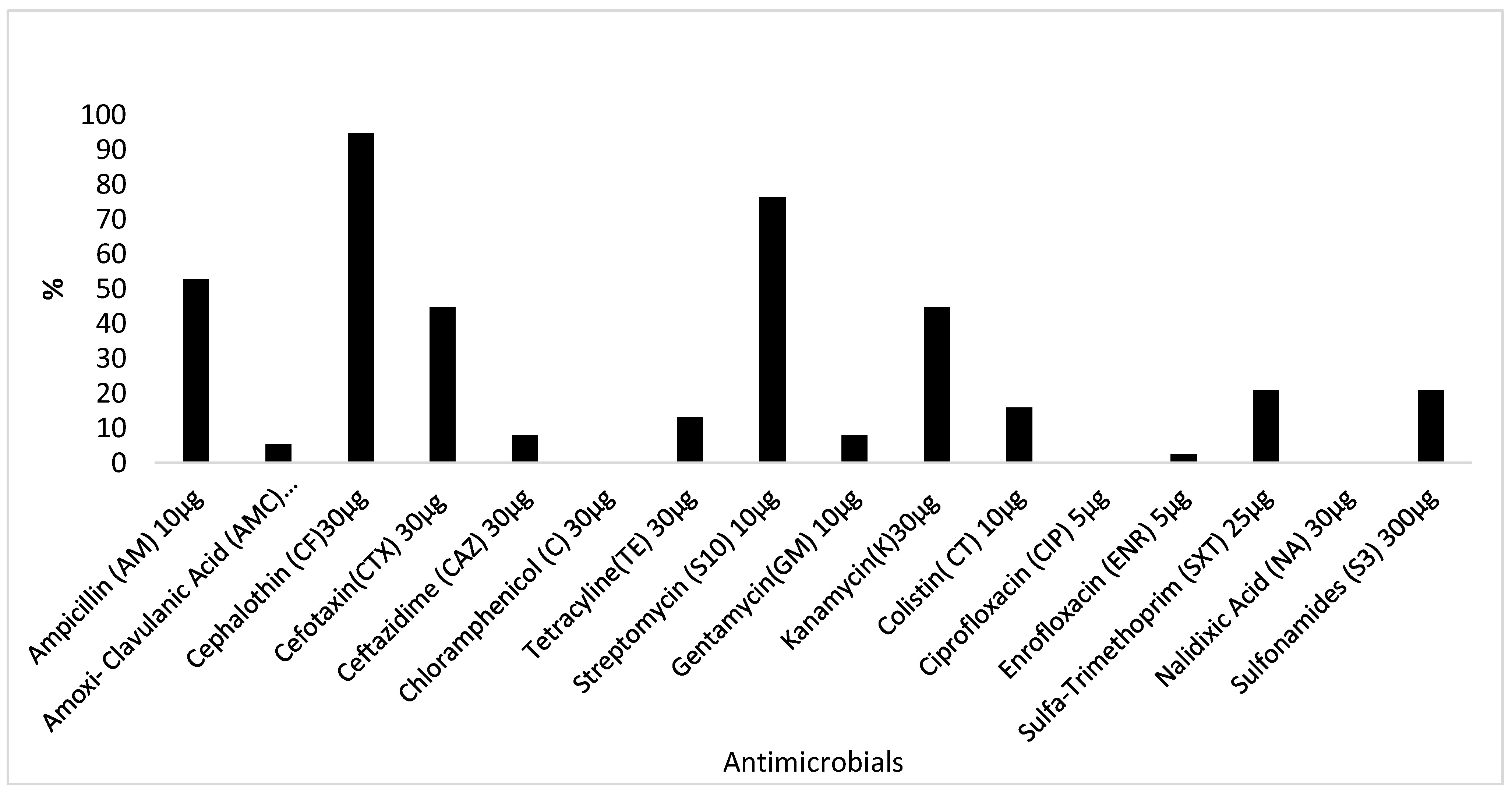
2.3. Antimicrobial Resistance Profiling

The distribution of antimicrobial resistance patterns is depicted in Figure 2. All the 38 STEC isolates were resistant to one or more antimicrobial. The following resistance rates were recorded per antimicrobial: cephalotin, 95% (36/38); streptomycin, 76% (29/38); ampicillin, 53% (20/38); amoxicillin-clavulanic acid, 5% (2/38); cefotaxin and kanamycin, 45% (17/38); sulfonamides and sulfa-trimethoprim, 21% (8/38); colistin, 16% (6/38); tetracycline, 13% (5/38) and ceftazidime and gentamycin, 8% (3/38). None of the isolates were resistant to chloramphenicol, ciprofloxacin, enrofloxacin or nalidixic acid. Multi-resistance (resistance to two or more classes of antimicrobials) was observed in 89% (34/38) of isolates. Two main multi-resistance patterns were detected: ampicillin/cephalothin/streptomycin, 42% (16/38) and cephalothin/streptomycin, 32% (12/38). Resistance to five or more antimicrobials was detected in 29% (11/38) of isolates.

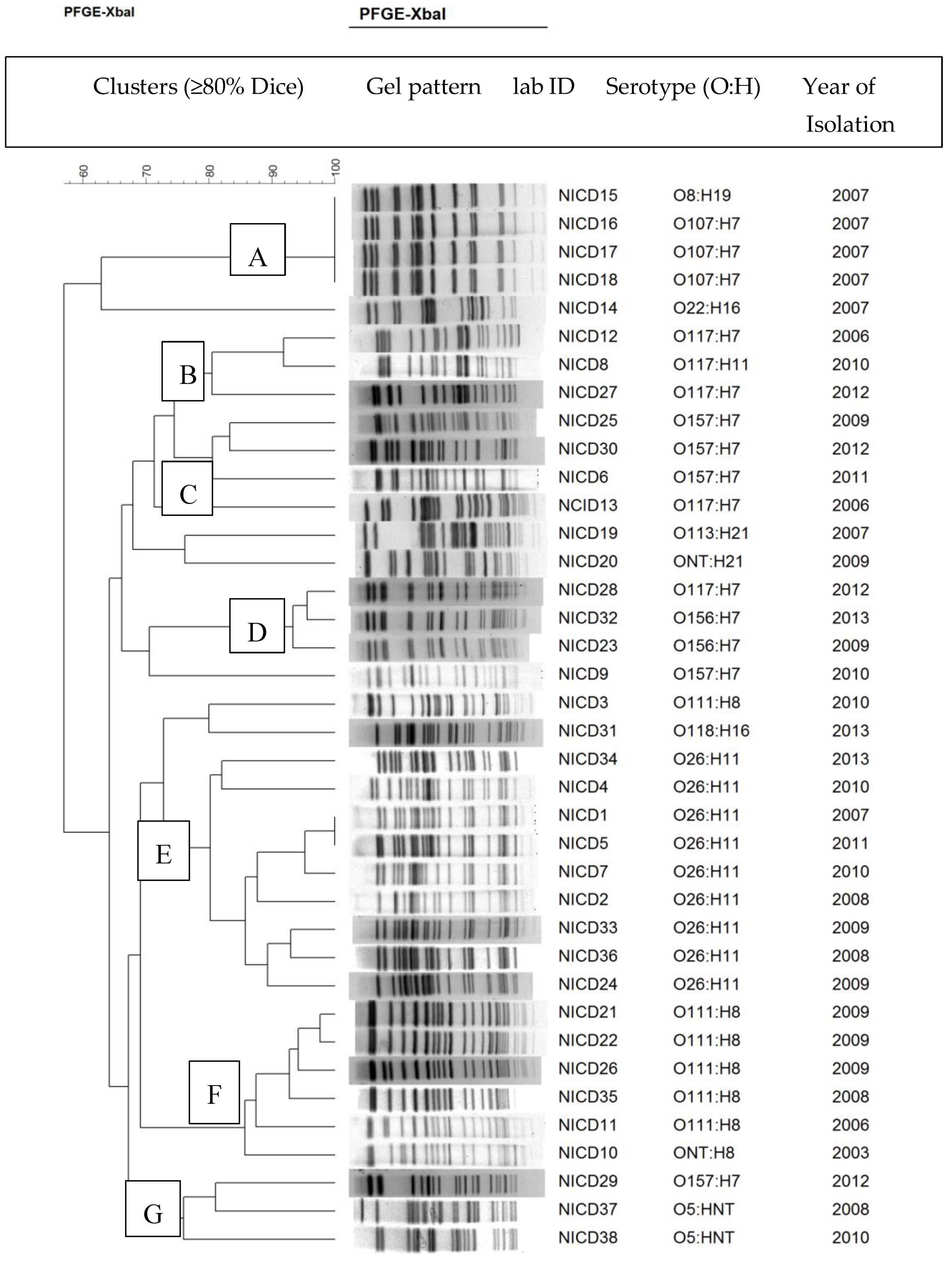
2.4. Pulsed-Field Gel Electrophoresis

PFGE was performed to investigate genetic relationships among isolates. The 38 STEC isolates were all typeable by PFGE. The isolates were classified into 33 PFGE profiles. Based on a Dice similarity index ≥80%, the 33 PFGE profiles were grouped into seven distinct clusters (A, B, C, D, E, F, and G) comprising mainly three to nine isolates/per cluster (Figure 3). STEC that belonged to the same serotype clustered together regardless of the year of isolation. Two major clusters emerged: cluster E, which grouped all the nine STEC O26:H11 isolates which were implicated in human disease in 2007, 2008, 2009, 2010, 2011 and 2013 and cluster F, which included five STEC O111:H8 isolates which were incriminated in human infections in 2006, 2008 and 2009. The remaining five clusters (A, B, C, D and G) were minor clusters of less than five isolates per cluster.

3. Discussion

Serotyping, virulence characterization, antimicrobial resistance and PFGE genotyping of STEC isolates that are recovered from infected humans are used in high income countries with robust STEC monitoring and surveillance programs for risk assessment purposes. While current data on the molecular epidemiology of STEC are readily available in high income countries, information on the occurrence and characteristics of STEC isolates from infected humans in low income countries, such as South Africa and most middle income economies, is scarce. Currently, STEC surveillance in South Africa remains passive and STEC disease in South Africa may be underestimated due to lack of active surveillance and/or underreporting. In addition, with cattle being an important STEC reservoir [44], the apparent sporadic nature of the disease in humans may be an inaccurate reflection of the true prevalence of STEC infection in humans. In this study, STEC isolates that were implicated in sporadic human disease in South Africa between 2006 and 2013 were characterized for virulence-associated genes and antimicrobial resistance profiles. Isolates were also genotyped by PFGE. STEC O26:H11 was the most frequent serotype associated with human disease, followed by O111:H8 and O157:H7 and O117:H7, contrary to world trends which have implicated STEC O157:H7 as the most frequent serotype associated with human infections [2]. Furthermore, the STEC isolates which were implicated in human disease in South Africa from 2006–2013 were mostly non-O157 (86.8%) serotypes, in agreement with global trends which have shown that non-O157 STEC are responsible for the majority of human disease cases worldwide [2,9].
Overall, the STEC isolates belonged to 11 O:H serotypes including O5:HNT, O8:H19, O22:H16, O26:H11, O107:H7, O111:H8, O113:H21, O117:H7, O118:H16, O156:H7, O157:H7, ONT:H8 and ONT:H21. Apart from STEC O107:H7, all the isolates belonged to serotypes which have been previously incriminated in mild to severe human STEC disease in various countries worldwide [6,7,8,9,45]. Altogether, three “big 7” STEC serotypes, including STEC O26:H11, O111:H8 and O157:H7, accounted for 53% of all isolates that were associated with human disease in South Africa from 2006–2013. This is consistent with previous reports which have shown that STEC O26:H11, O111:H8 (16%) and O157:H7 are among the three most frequent serotypes that are commonly associated with human disease globally [6,7,8,9,45].
The vast majority of STEC isolates were stx1 positive (84%), while only a low number (29%) possessed stx2. No stx1 subtypes were detected in this study. The stx1 + eaeA genotype was mostly observed among STEC O26:H11 and O111:H8 strains. STEC that carry stx1 alone or both stx1 + eaeA are considered low risk and are commonly associated with mild diarrhea or non-HUS infections in humans [46,47]. However, in some cases, stx1 + eaeA positive STEC have been involved in severe disease and outbreaks, suggesting that some stx1 + eaeA positive STEC strains may have yet unidentified virulence factors that make them highly virulent [48].
Almost all stx2 positive isolates carried both stx2c and stx2d subtypes concurrently. STEC isolates that possessed stx2 exhibited stx2 + stx2c + stx2d + eaeA or stx2 + stx2c + stx2d gene combinations. stx1 + stx2 + stx2c + stx2d + eaeA or stx2 + stx2c + stx2d + eaeA were observed in STEC O157:H7 isolates only while the stx2 + stx2c + stx2d genotype was detected in STEC O113:H21. The stx1 + stx2 + stx2d genotype was detected in STEC O107:H7 isolates, while the stx1 + stx2 + stxc + stx2d genotype was observed in one STEC O8:H19 isolate. STEC that possess stx2 alone or stx2 + eaeA have been significantly associated with severe disease in humans, including HC and HUS, in comparison to STEC that possess stx1 alone or stx1 + stx2 concurrently [46,47]. The proportion of individual STEC isolates that carried both stx2c and stx2d concurrently was higher compared to the results of two studies, which previously reported single strains that possess both subtypes [12,49]. The presence of both stx2c and/or stx2d subtypes in a STEC isolate has been associated with human disease, varying from mild to bloody diarrhea and HUS [12,49,50,51,52].
The eaeA gene was detected in the majority of isolates (60.5%). The eaeA gene was observed in the following serotypes: STEC O5:HNT, O26:H11, O111:H8, O118:H16, O157:H7 and ONT:H8. The high frequency of eaeA in these human STEC isolates is in sharp contrast with the very low rates of eaeA found in STEC isolates from cattle in South Africa [44], which are considered an important STEC reservoir, suggesting that only a small subset of cattle STEC that are eaeA positive with a high capacity of being easily transmitted to humans may be involved in human disease in South Africa.
The majority of STEC isolates carried plasmid-encoded virulence markers ehxA, katP and espP but lacked etpD, saa and subA. A complete plasmid (pO157) (ehxA, katP, espP and etpD) was observed in O157:H7 isolates only, while O26:H11 isolates possessed ehxA, katP, espP and etpD but lacked etpD, which was only found in all STEC O157:H7 and O5:HNT strains. The plasmid-encoded genes katP, espP and etpD are more frequent in STEC isolates that are recovered from human disease outbreaks and cases of HUS, particularly [11,28]. As previously shown, the saa gene was present in only a small number of eaeA-negative STEC isolates [53,54] which belonged to serotypes O113:H21, O8:19 and O107:H7. STEC O113:H21 has been previously incriminated in cases of severe human STEC infections (HUS) in Australia and Europe, while STEC O8:H19 has been associated with diarrhea in various countries [7,23]. To the best of our knowledge, this is the first report that has incriminated STEC O107:H7 in human disease, suggesting that STEC O107:H7 may be an emerging serotype worth monitoring in South Africa.
Most of the isolates carried the full complement of OI-122 and OI-43/48 marker genes and all non-LEE encoded effector genes, except nleA. OI-122 and OI-43/48 and non-LEE effectors genes encode proteins that play an important role in STEC virulence as substrates that are translocated through a type III secretion system. Overall, significantly more OI-122, OI-43/48 and non-LEE effector genes were observed in STEC isolates that were stx2 and eaeA positive with a complete plasmid (ehxA, katP, espP and etpD), in agreement with Ju et al. [55]. Possession of OI-122, OI-43/48 and the non-LEE effector genes together with stx2, eaeA and a complete plasmid is a hallmark of highly virulent STEC strains that are frequently associated with outbreaks and severe disease such as HC and HUS [11,56,57], whereas serotypes lacking these genes are mostly implicated in mild human disease [11,32,57,58].
Interestingly, STEC O157:H7, O26:H11, O111:H8, O113:H21, O118:H16, O5:HNT and O8:H19 harbored the highest number of virulence genes (24–28 genes). Possession of the highest number of virulence-associated genes significantly correlated with the presence of eaeA. While the presence of a high number of virulence genes in STEC O157:H7, O26:H11, O111:H8 and O113:H21 isolates that are commonly implicated in severe foodborne STEC disease outbreaks around the world may not be surprising [6,7,8,9,11], possession of a high number of virulence-associated genes in STEC O118:H16, O5:HNT and O8:H19 serotypes that are rarely associated with severe disease and outbreaks globally [6,7] suggests that these serotypes should be closely monitored in South Africa as they may present a high risk for humans.
All of the 38 STEC isolates were resistant to one or more antimicrobials. Most (>50%) of the isolates were resistant to cephalothin, streptomycin and ampicillin, while moderate resistance rates (40–50% of isolates) were observed for cefotaxin and kanamycin, and lower resistance levels (< 25%) were observed for sulfonamides, sulfa-trimethoprim and colistin. The levels of resistance recorded in this study were very high compared to rates that have been reported elsewhere [59,60,61]. Isolates were multi-resistant to ampicillin/cephalothin/streptomycin, or cephalothin/streptomycin, mainly. These antimicrobials are commonly used for the treatment of human bacterial diseases in clinical medicine. The high levels of resistance against these antimicrobials suggests that these compounds may be misused or abused in clinical medicine and are exerting selective pressure on the STEC isolates under study.
Pulsed-field gel electrophoresis was used to classify the 38 STEC isolates into 33 profiles which were assigned to eight distinct clusters. Among the eight clusters, four distinct clusters grouped STEC O26:H11, five of the six STEC O111:H8 and all STEC O107:H7 and O157:H7. The four clusters were serotype specific with very high intra-cluster similarity (>80%), regardless of the year of isolation. The high intra-serotype similarity among STEC O26:H11, O111:H8, O107:H7 and O156:H7 strains regardless of their year of isolation suggests that the STEC isolates which were implicated in disease in South Africa from 2006 to 2013 are clonally related STEC strains which may have a high capacity for survival and persistence in their respective reservoir or sources.

4. Conclusions

In summary, the majority of STEC infections which occurred in South Africa from 2006 and 2013 were caused by serotypes O26:H11, O111:H8 and O157:H7 that belong to “big 7” serogroups, in agreement with global trends. Virulotyping revealed that most of the isolates were stx1 + eaeA positive, fewer isolates carried stx2 only or concurrently with stx2c and stx2d and a smaller number was positive for both stx1 and stx2. Most isolates carried all or most pathogenicity island encoded virulence-associated genes. PFGE revealed high intra-serotype similarity among STEC. The majority of isolates were resistant to two or more antimicrobials that are commonly used in clinical medicine for treatment of various bacterial diseases. To the best of our knowledge, this is the first detailed characterization of human STEC isolates from South Africa. Further molecular epidemiology studies that compare STEC isolates of human, animal and food origin are needed to fully understand the epidemiology of STEC and identify reservoirs and sources of human disease in South Africa.

5. Materials and Methods

5.1. STEC Strains and Culture Conditions

A total of 38 STEC isolates which were previously recovered from humans showing foodborne disease symptoms, including mild to severe diarrhea, between 2006 and 2013 were characterized in this study. These isolates were obtained from the culture collection of the Centre for Enteric Diseases, National Institute of Communicable Diseases, South Africa (CED-NICD). Because the isolates were obtained through passive surveillance, detailed clinical information on patients from which the STEC isolates were recovered was unavailable. The 38 STEC isolates belonged to the following serogroups: O5 (2), O8 (1), O107 (3), O22 (1), O26 (9), O111 (6), O113 (1), O117 (5), O118 (1), O156 (2), O157 (5) and ONT (1). This study was approved by the University of Pretoria Research Ethics Committee (REC005-18).

5.2. STEC Cultures

The isolates were resuscitated on Luria Bertani Agar after incubation at 37 °C overnight. DNA was extracted from bacterial cells by the boiling method, as described previously [59]. Briefly, a sterile inoculating loop was used to harvest pure E. coli colony sweeps from Luria Bertani Agar plates. A loop-full of colony sweeps was suspended in a 1.5 mL Eppendorf tube containing 1 mL of FA Buffer (Becton Dickinson and Company, Sparks, MD, USA). Bacterial suspensions were mixed and washed by vortexing, followed by centrifugation (15,000× g) for 5 min. After the first wash and centrifugation cycle, the supernatant was discarded and the bacterial pellet was re-suspended in FA buffer (Becton, Dickinson and Company Sparks, MD, USA). After two additional washes and centrifugation cycles, the pellet was suspended in 500 µL of sterile water and thoroughly vortexed. The homogeneous cell suspension was boiled [62] to 100 °C for 15 min, then stored at −20 °C until further processing.

5.3. STEC PCR Serotyping (O:H)

STEC isolates were further serotyped for flagellar antigens (H antigens) only, as STEC serogrouping (O antigen typing) had been carried out previously by the tube agglutination test (antisera manufactured by the Statens Serum Institut, Copenhagen, Denmark). H typing was carried out by three multiplex PCR assays that targeted 14 genes encoding Escherichia coli flagellar genes (fliC) that are commonly found in STEC [63].

5.4. Virulotyping

Virulotyping was performed using previously described primers and PCR cycling conditions to amplify 34 genes that encode various STEC virulence factors and markers. A multiplex PCR was used for the detection of stx1, stx2, eaeA, and ehxA [64]. Detection of stx2 subtypes (stx1c, stx1d, stx2c, stx2d, stx2e, stx2f and stx2g) was carried out according to Scheutz et al. [12]. Single PCR reactions were conducted to amplify plasmid-encoded virulence markers: katP [19], espP [20], etpD [21], saa [64] and subA [65]. Previous PCR protocols with slight modifications were used to screen for OI-encoded genes including OI-43/48: iha, ureC and terC [29,30,31] and OI-122: pagC (Z4321), sen (Z4326), efa1 (Z4332) and efa2 (Z4333) [11]. The PCR protocols of Coombes et al. [32] were used to search for non-LEE- encoded effector genes: nleA (OI-71), nleB (OI-122), nleC (OI-36), nleD (OI-57), nleE (OI-122), nleG (OI-71), nleG2-1 (OI-71), nleG2-3 (OI-57), nleG6-2 (OI-57), nleG9 (OI-71) and nleH1-2 (OI-171). STEC O157:H7 strain EDL 933 (E. coli O157:H7) was used as a positive control.

5.5. Antimicrobial Susceptibility Testing

Antimicrobial resistance profiles of STEC isolates were determined against a panel of 12 antimicrobials by the disk diffusion method as described by the Clinical and Laboratory Standards Institute (CLSI) [66]. The panel of antimicrobials consisted of ampicillin (10 µg), amoxicillin-clavulanic acid (20 and 10 µg, respectively), cephalothin (30 µg), cefotaxin (30 µg), ceftazid (30 µg), chloramphenicol (30 µg), tetracycline (30 µg), streptomycin (10 µg), gentamicin (10 µg), kanamycin (30 µg), colistin (10 µg), ciprofloxacin (5 µg), enrofloxacin (5 µg), sulfa-trimethoprim (1.25 and 23.75 µg, respectively), nalidixic acid, (30 µg) and sulfonamides (300 µg). Antimicrobial disks (BBL Sensi Disk) were obtained from Becton, Dickinson and Company, Sparks, MD, USA) and Oxoid, Basingstoke, Hampshire, England). E. coli ATCC 25922 was used as the control strain. Isolates were classified as susceptible, intermediate or resistant to each antimicrobial agent and in the final analysis intermediate readings were assigned to the resistant category.

5.6. Pulsed-Field Gel Electrophoresis

PFGE was carried out according to the CDC/PulseNet protocol to determine relationships among STEC, particularly STEC belonging to the same serotype (http://www.cdc.gov/pulsenet/pdf/ecoli-shigella-salmonella-pfge-protocol508.pdf). Salmonella serotype Braenderup (strain H9812; American Type Culture Collection catalog no. BAA-664) was used as the marker for all PFGE gels. Briefly, DNA was extracted and digested with XbaI. XbaI PFGE patterns were analyzed for similarity, and a dendrogram was generated by the Bionumerics software, version 6.5 (Applied Maths, Sint Martens-Latem, Belgium) with Dice similarity indices (complete linkage; optimization, 1.5%; position tolerance, 1.5%) and the unweighted-pair group method with arithmetic means. In this study, a cluster was defined as STEC isolates that grouped together in the dendrogram with a Dice similarity index equal or above 80%.

5.7. Statistical Analysis

Fisher’s exact test was used to determine if there were statistically significant differences between the proportions of STEC genes (SPSS Statistics 19; IBM, Armonk, NY, USA). A p-value <0.05 was considered significant.

Author Contributions

Conceptualization, M.K. and A.K.; data curation, M.M., A.M.S. and K.H.K.; formal analysis, M.K. and M.M.; funding acquisition, M.K., B.T.C-G. and A.K.; investigation, M.K., B.T.C-G. and M.M. methodology, B.T.C-G., M.M., A.M.S. and K.H.K.; resources, B.T.C-G., A.M.S., K.H.K. and A.K.; supervision, M.K.; writing—original draft, M.K.; Writing—review and editing, A.M.S, S.E-A. and L.M.K.

Funding

Funding from the National Research Foundation (NRF) of South Africa: Thuthuka (TTK13062619943), Research Technology Fund (RTF14012762427), the Gauteng Department of Agriculture and Rural Development (GDARD Grants-2013-2015) and the South African Medical Research Council (Self-Initiated Research 2017-2019) is gratefully acknowledged. Furthermore, we sincerely thank Alfredo Caprioli and Rosangela Tozzoli at the European Union Reference Laboratory for Escherichia coli (Istituto Superiore di Sanità, Rome Italy) for kindly providing STEC reference positive control strains.

Conflicts of Interest

The authors declare no conflict of interest.

References

  1. Kaper, J.B.; Karmali, M.A. The continuing evolution of a bacterial pathogen. Proc. Natl. Acad. Sci. USA 2008, 105, 4535–4536. [Google Scholar] [CrossRef] [Green Version]
  2. Majowicz, S.E.; Scallan, E.; Jones-Bitton, A.; Sargeant, J.M.; Stapleton, J.; Angulo, F.J.; Yeung, D.H.; Kirk, M.D. Global incidence of human Shiga toxin–producing Escherichia coli infections and deaths: A systematic review and knowledge synthesis. Foodborne Pathog. Dis. 2014, 11, 447–455. [Google Scholar] [CrossRef]
  3. Hussein, H.S. Prevalence and pathogenicity of Shiga toxin-producing Escherichia coli in beef cattle and their products. J. Anim. Sci. 2007, 85, E63–E72. [Google Scholar] [CrossRef]
  4. Hussein, H.S.; Sakuma, T. Invited review: prevalence of Shiga toxin-producing Escherichia coli in dairy cattle and their products. J. Dairy Sci. 2005, 88, 450–465. [Google Scholar] [CrossRef]
  5. Berger, C.N.; Sodha, S.V.; Shaw, R.K.; Griffin, P.M.; Pink, D.; Hand, P.; Frankel, G. Fresh fruit and vegetables as vehicles for the transmission of human pathogens. Environ. Microbiol. 2010, 12, 2385–2397. [Google Scholar] [CrossRef]
  6. Beutin, L.; Fach, P. Detection of Shiga toxin-producing Escherichia coli from nonhuman sources and strain typing. In Enterohemorrhagic Escherichia coli and Other Shiga Toxin-Producing, E. coli; American Society of Microbiology: Washington, DC, USA, 2015; pp. 299–319. [Google Scholar]
  7. EFSA Panel on Biological Hazards (BIOHAZ). Scientific Opinion on VTEC-Seropathotype and Scientific Criteria Regarding Pathogenicity Assessment. EFSA J. 2013, 11, 3138. [Google Scholar]
  8. Bettelheim, K.A. The non-O157 Shiga-toxigenic (verocytotoxigenic) Escherichia coli; under-rated pathogens. Crit. Rev. Microbiol. 2007, 33, 67–87. [Google Scholar] [CrossRef]
  9. Johnson, K.E.; Thorpe, C.M.; Sears, C.L. The emerging clinical importance of non-O157 Shiga toxin—Producing Escherichia coli. Clin. Infect. Dis. 2006, 43, 1587–1595. [Google Scholar]
  10. Scallan, E.; Griffin, P.M.; Angulo, F.J.; Tauxe, R.V.; Hoekstra, R.M. Foodborne illness acquired in the United States—Unspecified agents. Emerg. Infect. Dis. 2011, 17, 16–22. [Google Scholar] [CrossRef]
  11. Karmali, M.A.; Mascarenhas, M.; Shen, S.; Ziebell, K.; Johnson, S.; Reid-Smith, R.; Isaac-Renton, J.; Clark, C.; Rahn, K.; Kaper, J.B. Association of genomic O island 122 of Escherichia coli EDL 933 with verocytotoxin-producing Escherichia coli seropathotypes that are linked to epidemic and/or serious disease. J. Clin. Microbiol. 2003, 41, 4930–4940. [Google Scholar] [CrossRef]
  12. Scheutz, F.; Teel, L.D.; Beutin, L.; Piérard, D.; Buvens, G.; Karch, H.; Mellmann, A.; Caprioli, A.; Tozzoli, R.; Morabito, S.; et al. Multicenter evaluation of a sequence-based protocol for subtyping Shiga toxins and standardizing Stx nomenclature. J. Clin. Microbiol. 2012, 50, 2951–2963. [Google Scholar] [CrossRef]
  13. Lacher, D.W.; Gangiredla, J.; Jackson, S.A.; Elkins, C.A.; Feng, P.C.H. A novel microarray design for molecular serotyping of Shiga toxin-producing Escherichia coli isolated from fresh produce. Appl. Environ. Microbiol. 2014, 80, 4677–4682. [Google Scholar] [CrossRef]
  14. Bai, X.; Fu, S.; Zhang, J.; Fan, R.; Xu, Y.; Sun, H.; He, X.; Xu, J.; Xiong, Y. Identification and pathogenomic analysis of an Escherichia coli strain producing a novel Shiga toxin 2 subtype. Sci. Rep. 2018. [Google Scholar] [CrossRef]
  15. Tzipori, S.; Gunzer, F.; Donnenberg, M.S.; de Montigny, L.; Kaper, J.B.; Donohue-Rolfe, A. The role of the eaeA gene in diarrhea and neurological complications in a gnotobiotic piglet model of enterohemorrhagic Escherichia coli infection. Infect. Immun. 1995, 63, 3621–3627. [Google Scholar]
  16. McDaniel, T.K.; Jarvis, K.G.; Donnenberg, M.S.; Kaper, J.B. A genetic locus of enterocyte effacement conserved among diverse enterobacterial pathogens. Proc. Natl. Acad. Sci. USA 1995, 92, 1664–1668. [Google Scholar] [CrossRef]
  17. Donnenberg, M.S.; Tacket, C.O.; James, S.P.; Losonsky, G.; Nataro, J.P.; Wasserman, S.S.; Kaper, J.B.; Levine, M.M. Role of the eaeA gene in experimental enteropathogenic Escherichia coli infection. J. Clin. Investig. 1993, 92, 1412–1417. [Google Scholar] [CrossRef]
  18. Schmidt, H.; Beutin, L.; Karch, H. Molecular analysis of the plasmid-encoded hemolysin of Escherichia coli O157:H7 strain EDL 933. Infect. Immun. 1995, 63, 1055–1061. [Google Scholar]
  19. Brunder, W.; Schmidt, H.; Karch, H. KatP, a novel catalase-peroxidase encoded by the large plasmid of enterohaemorrhagic Escherichia coli O157:H7. Microbiology 1996, 142, 3305–3315. [Google Scholar] [CrossRef]
  20. Brunder, W.; Schmidt, H.; Karch, H. EspP, a novel extracellular serine protease of enterohaemorrhagic Escherichia coli O157:H7 cleaves human coagulation factor V. Mol. Microbiol. 1997, 24, 767–778. [Google Scholar] [CrossRef]
  21. Schmidt, H.; Henkel, B.; Karch, H. A gene cluster closely related to type II secretion pathway operons of gram-negative bacteria is located on the large plasmid of enterohemorrhagic Escherichia coli O157 strains. FEMS Microbiol. Lett. 1997, 148, 265–272. [Google Scholar] [CrossRef]
  22. Paton, A.W.; Srimanote, P.; Talbot, U.M.; Wang, H.; Paton, J.C. A new family of potent AB5 cytotoxins produced by Shiga toxigenic Escherichia coli. J. Exp. Med. 2004, 200, 35–46. [Google Scholar] [CrossRef]
  23. Paton, A.W.; Srimanote, P.; Woodrow, M.C.; Paton, J.C. Characterization of Saa, a novel autoagglutinating adhesin produced by locus of enterocyte effacement-negative Shiga-Toxigenic Escherichia coli Strains that are virulent for humans. Infect. Immun. 2001, 69, 6999–7009. [Google Scholar] [CrossRef]
  24. Brunder, W.; Schmidt, H.; Frosch, M.; Karch, H. The large plasmids of Shiga-toxin-producing Escherichia coli (STEC) are highly variable genetic elements. Microbiology 1999, 145, 1005–1014. [Google Scholar] [CrossRef]
  25. Paton, A.W.; Paton, J.C. Escherichia coli subtilase cytotoxin. Toxins 2010, 2, 215–228. [Google Scholar] [CrossRef]
  26. Schmidt, H.; Hensel, M. Pathogenicity islands in bacterial pathogenesis. Clin. Microbiol. Rev. 2004, 17, 14–56. [Google Scholar] [CrossRef]
  27. Konczy, P.; Ziebell, K.; Mascarenhas, M.; Choi, A.; Michaud, C.; Kropinski, A.M.; Whittam, T.S.; Wickham, M.; Finlay, B.; Karmali, M.A. Genomic O island 122, locus for enterocyte effacement, and the evolution of virulent verocytotoxin-producing Escherichia coli. J. Bacteriol. 2008, 190, 5832–5840. [Google Scholar] [CrossRef]
  28. Abu-Median, A.B.; Van Diemen, P.M.; Dziva, F.; Vlisidou, I.; Wallis, T.S.; Stevens, M.P. Functional analysis of lymphostatin homologues in enterohaemorrhagic Escherichia coli. FEMS Microbiol. Lett. 2006, 258, 43–49. [Google Scholar] [CrossRef]
  29. Schmidt, H.; Zhang, W.-L.; Hemmrich, U.; Jelacic, S.; Brunder, W.; Tarr, P.I.; Dobrindt, U.; Hacker, J.; Karch, H. Identification and characterization of a novel genomic island integrated at selC in locus of enterocyte effacement-negative, Shiga toxin-producing Escherichia coli. Infect. Immun. 2001, 69, 6863–6873. [Google Scholar] [CrossRef]
  30. Nakano, M.; Iida, T.; Ohnishi, M.; Kurokawa, K.; Takahashi, A.; Tsukamoto, T.; Yasunaga, T.; Hayashi, T.; Honda, T. Association of the urease gene with enterohemorrhagic Escherichia coli strains irrespective of their serogroups. J. Clin. Microbiol. 2001, 39, 4541–4543. [Google Scholar] [CrossRef]
  31. Taylor, D.E.; Rooker, M.; Keelan, M.; Ng, L.-K.; Martin, I.; Perna, N.T.; Burland, N.T.V.; Blattner, F.R. Genomic variability of O islands encoding tellurite resistance in enterohemorrhagic Escherichia coli O157:H7 isolates. J. Bacteriol. 2002, 184, 4690–4698. [Google Scholar] [CrossRef]
  32. Coombes, B.K.; Wickham, M.E.; Mascarenhas, M.; Gruenheid, S.; Finlay, B.B.; Karmali, M.A. Molecular analysis as an aid to assess the public health risk of non-O157 Shiga toxin-producing Escherichia coli strains. Appl. Environ. Microbiol. 2008, 74, 2153–2160. [Google Scholar] [CrossRef]
  33. Coombes, B.K.; Gilmour, M.W.; Goodman, C.D. The evolution of virulence in non-O157 Shiga toxin-producing Escherichia coli. Front. Microbiol. 2011, 2, 90. [Google Scholar] [CrossRef]
  34. Karmali, M.A.; Gannon, V.; Sargeant, J.M. Verocytotoxin-producing Escherichia coli (VTEC). Vet. Microbiol. 2010, 140, 360–370. [Google Scholar] [CrossRef]
  35. Melton-Celsa, A.; Mohawk, K.; Teel, L.; O’Brien, A. Pathogenesis of Shiga-toxin producing Escherichia coli. In Ricin and Shiga Toxins; Springer: Berlin/Heidelberg, Germany, 2011; pp. 67–103. [Google Scholar]
  36. Schwarz, S.; Kehrenberg, C.; Walsh, T.R. Use of antimicrobial agents in veterinary medicine and food animal production. Int. J. Antimicrob. Agents 2001, 17, 431–437. [Google Scholar] [CrossRef]
  37. Walsh, C.; Fanning, S. Antimicrobial resistance in foodborne pathogens-a cause for concern? Curr. Drug Targets 2008, 9, 808–815. [Google Scholar] [CrossRef]
  38. Dibner, J.J.; and Richards, J.D. Antibiotic growth promoters in agriculture: history and mode of action. Poult. Sci. 2005, 84, 634–643. [Google Scholar] [CrossRef]
  39. Smith, K.E.; Wilker, P.R.; Reiter, P.L.; Hedican, E.B.; Bender, J.B.; Hedberg, C.W. Antibiotic treatment of Escherichia coli O157 infection and the risk of hemolytic uremic syndrome, Minnesota. Pediatr. Infect. Dis. J. 2012, 31, 37–41. [Google Scholar] [CrossRef]
  40. Melton-Celsa, A.R.; O’Brien, A.D. New therapeutic developments against Shiga toxin-producing Escherichia coli. In Enterohemorrhagic Escherichia coli and Other Shiga Toxin-Producing, E. coli; American Society of Microbiology: Washington, DC, USA, 2015; pp. 361–380. [Google Scholar]
  41. Browning, N.G.; Botha, J.R.; Sacho, H.; Moore, P.J. Escherichia coli O157:H7 haemorrhagic colitis. Report of the first South African case. S. Afr. J. Surg. 1990, 28, 28–29. [Google Scholar]
  42. Isaäcson, M.; Canter, P.H.; Effler, P.; Arntzen, L.; Bomans, P.; Heenan, R. Haemorrhagic colitis epidemic in Africa. Lancet 1993, 341, 961. [Google Scholar]
  43. Riley, L.W.; Remis, R.S.; Helgerson, S.D.; McGee, H.B.; Wells, J.G.; Davis, B.R.; Hebert, R.J.; Olcott, E.S.; Johnson, L.M.; Hargrett, N.T.; et al. Hemorrhagic colitis associated with a rare Escherichia coli serotype. N. Engl. J. Med. 1983, 308, 681–685. [Google Scholar] [CrossRef]
  44. Mainga, A.O.; Cenci-Goga, B.T.; Malahlela, M.N.; Tshuma, T.; Kalake, A.; Karama, M. Occurrence and characterization of seven major Shiga toxin-producing Escherichia coli serotypes from healthy cattle on cow–calf operations in South Africa. Zoonoses Public Health. 2018, 65, 777–789. [Google Scholar] [CrossRef]
  45. Rivero, M.A.; Passucci, J.A.; Rodriguez, E.M.; Parma, A.E. Role and clinical course of verotoxigenic Escherichia coli infections in childhood acute diarrhoea in Argentina. J. Med. Microbiol. 2010, 59, 345–352. [Google Scholar] [CrossRef]
  46. Haugum, K.; Brandal, L.T.; Lindstedt, B.-A.; Wester, A.L.; Bergh, K.; Afset, J.E. PCR-based detection and molecular characterization of shiga toxin-producing Escherichia coli strains in a routine microbiology laboratory over 16 years. J. Clin. Microbiol. 2014, 52, 3156–3163. [Google Scholar] [CrossRef]
  47. Friedrich, A.W.; Bielaszewska, M.; Zhang, W.-L.; Pulz, M.; Kuczius, T.; Ammon, A.; Karch, H. Escherichia coli harboring Shiga toxin 2 gene variants: frequency and association with clinical symptoms. J. Infect. Dis. 2002, 185, 74–84. [Google Scholar] [CrossRef]
  48. Boerlin, P.; McEwen, S.A.; Boerlin-Petzold, F.; Wilson, J.B.; Johnson, R.P.; Gyles, C.L. Association between virulence factors of Shiga toxin-producing Escherichia coli and disease in humans. J. Clin. Microbiol. 1999, 37, 497–503. [Google Scholar]
  49. Feng, P.C.; Jinneman, K.; Scheutz, F.; Monday, S.R. Specificity of PCR and serological assays in the detection of Escherichia coli Shiga toxin subtypes. Appl. Environ. Microbiol. 2011, 77, 6699–6702. [Google Scholar] [CrossRef]
  50. Iyoda, S.; Manning, S.D.; Seto, K.; Kimata, K.; Isobe, J.; Etoh, Y.; Ichihara, S.; Migita, Y.; Ogata, K.; Honda, M.; et al. Phylogenetic clades 6 and 8 of enterohemorrhagic Escherichia coli O157:H7 with particular stx subtypes are more frequently found in isolates from hemolytic uremic syndrome patients than from asymptomatic carriers. In Open Forum Infectious Diseases; Oxford University Press: Oxford, UK, 2014. [Google Scholar]
  51. Kawano, K.; Ono, H.; Iwashita, O.; Kurogi, M.; Haga, T.; Maeda, K.; Goto, Y. Relationship between stx genotype and Stx2 expression level in Shiga toxin-producing Escherichia coli O157 strains. Jpn. J. Infect. Dis. 2012, 65, 322–325. [Google Scholar] [CrossRef]
  52. Orth, D.; Grif, K.; Khan, A.B.; Naim, A.; Dierich, M.P.; Würzner, R. The Shiga toxin genotype rather than the amount of Shiga toxin or the cytotoxicity of Shiga toxin in vitro correlates with the appearance of the hemolytic uremic syndrome. Diagn. Microbiol. Infect. Dis. 2007, 59, 235–242. [Google Scholar] [CrossRef]
  53. Jenkins, C.; Perry, N.T.; Cheasty, T.; Shaw, D.J.; Frankel, G.; Dougan, G.; Gunn, G.J.; Smith, H.R.; Paton, A.W.; Paton, J.C. Distribution of the saa gene in strains of Shiga toxin-producing Escherichia coli of human and bovine origins. J. Clin. Microbiol. 2003, 41, 1775–1778. [Google Scholar] [CrossRef]
  54. Aidar-Ugrinovich, L.; Blanco, J.; Blanco, M.; Blanco, J.E.; Leomil, L.; Dahbi, G.; Mora, A.; Onuma, D.L.; Silveira, W.D.; de Castro, A.F.P. Serotypes, virulence genes, and intimin types of Shiga toxin-producing Escherichia coli (STEC) and enteropathogenic E. coli (EPEC) isolated from calves in São Paulo, Brazil. Int. J. Food Microbiol. 2007, 115, 297–306. [Google Scholar] [CrossRef]
  55. Ju, W.; Shen, J.; Toro, M.; Zhao, S.; Meng, J. Distribution of pathogenicity islands OI-122, OI-43/48, OI-57 and high pathogenicity island (HPI) in Shiga Toxin-producing Escherichia coli. Appl. Environ. Microbiol. 2013, 79, 3406–3412. [Google Scholar] [CrossRef] [PubMed]
  56. Buvens, G.; De Gheldre, Y.; Dediste, A.; de Moreau, A.-I.; Mascart, G.; Simon, A.; Allemeersch, D.; Scheutz, F.; Lauwers, S.; Piérard, D. Incidence and virulence determinants of verocytotoxin-producing Escherichia coli infections in the Brussels-Capital Region, Belgium, in 2008–2010. J. Clin. Microbiol. 2012, 50, 1336–1345. [Google Scholar] [CrossRef] [PubMed]
  57. Buvens, G.; Piérard, D. Virulence profiling and disease association of verocytotoxin-producing Escherichia coli O157 and non-O157 isolates in Belgium. Foodborne Pathog. Dis. 2012, 9, 530–535. [Google Scholar] [CrossRef] [PubMed]
  58. Bugarel, M.; Beutin, L.; Martin, A.; Gill, A.; Fach, P. Micro-array for the identification of Shiga toxin-producing Escherichia coli (STEC) seropathotypes associated with hemorrhagic colitis and hemolytic uremic syndrome in humans. Int. J. Food Microbiol. 2010, 142, 318–329. [Google Scholar] [CrossRef] [PubMed]
  59. Cergole-Novella, M.C.; Pignatari, A.C.C.; Castanheira, M.; Guth, B.E.C. Molecular typing of antimicrobial-resistant Shiga-toxin-producing Escherichia coli strains (STEC) in Brazil. Res. Microbiol. 2011, 162, 117–123. [Google Scholar] [CrossRef] [PubMed]
  60. Mukherjee, S.; Mosci, R.E.; Anderson, C.M.; Snyder, B.A.; Collins, J.; Rudrik, J.T.; Manning, S.D. Antimicrobial drug–resistant Shiga toxin–producing Escherichia coli infections, Michigan, USA. Emerg. Infect. Dis. 2017, 23, 1609–1611. [Google Scholar] [CrossRef] [PubMed]
  61. Day, M.; Doumith, M.; Jenkins, C.; Dallman, T.J.; Hopkins, K.L.; Elson, R.; Godbole, G.; Woodford, N. Antimicrobial resistance in Shiga toxin-producing Escherichia coli serogroups O157 and O26 isolated from human cases of diarrhoeal disease in England, 2015. J. Antimicrobial Chemother. 2017, 72, 145–152. [Google Scholar] [CrossRef]
  62. Feng, P.C.H.; Monday, S.R. Multiplex PCR for specific identification of enterohemorrhagic Escherichia coli strains in the O157:H7 complex. In Foodborne Pathogens; Humana Press: Totowa, NJ, USA, 2006; pp. 47–55. [Google Scholar]
  63. Singh, P.; Sha, Q.; Lacher, D.W.; Del Valle, J.; Mosci, R.E.; Moore, J.A.; Scribner, K.T.; Manning, S.D. Characterization of enteropathogenic and Shiga toxin-producing Escherichia coli in cattle and deer in a shared agroecosystem. Front. Cell. Infect. Microbiol. 2015, 5, 29. [Google Scholar] [CrossRef]
  64. Paton, A.W.; Paton, J.C. Direct detection and characterization of Shiga toxigenic Escherichia coli by multiplex PCR for stx1, stx2, eae, ehxA, and saa. J. Clin. Microbiol. 2002, 40, 271–274. [Google Scholar] [CrossRef]
  65. Paton, A.W.; Paton, J.C. Multiplex PCR for direct detection of Shiga toxigenic Escherichia coli strains producing the novel subtilase cytotoxin. J. Clin. Microbiol. 2005, 43, 2944–2947. [Google Scholar] [CrossRef]
  66. Clinical Laboratory Standards. CLSI Performance Standards for Antimicrobial Disk and Dilution Susceptibility Tests for Bacteria Isolated from Animals, Approved Standard, 3rd ed.; CLSI document VET01S; National Committee for Clinical Laboratory Standards: Wayne, PA, USA, 2015. [Google Scholar]
Figure 1. Distribution of virulence-associated genes in STEC isolates from humans in South Africa.
Toxins 11 00424 g001
Figure 2. Antimicrobial resistance profiles of STEC isolates from humans in South Africa.
Toxins 11 00424 g002
Figure 3. Dendrogram showing relationships among STEC isolates that were incriminated in human disease in South Africa from 2006 to 2013.
Toxins 11 00424 g003
Table 1. Proportions of essential chromosomal and plasmid-encoded virulence-associated genes in Shiga toxin-producing Escherichia coli (STEC) isolates from humans in South Africa.
NoYearSerotypestx1stx2stx2cstx2deaeAehxAkatPespPetpDsaasubA
12007O26:H11+++++
22008O26:H11+++++
32010O111:H8++++
42010O26:H11++
52011O26:H11+++++
62011O157:H7++++++++
72010O26:H11+++++
82010O117:H7+
92010O157:H7++++++++
102003ONT:H8++
112006O111:H8+++
122006O117:H7+
132006O117:H7+
142007O22:H16+
152007O8:H19+++++++
162007O107:H7++++++
172007O107:H7++++++
182007O107:H7++++++
192007O113:H21+++++++
202009ONT:H21+++
212009O111:H8++
222009O111:H8+++
232009O156:H7+
242009O26:H11+++++
252009O157:H7+++++++++
262009O111:H8+++
272012O117:H7+
282012O117:H7+
292012O157:H7++++++++
302012O157:H7++++++++
312013O118:H16+++++
322013O156:H7+
332009O26:H11+++++
342013O26:H11+++++
352008O111:H8+++
362008O26:H11+++++
372010O5:HNT+++++
382008O5:HNT++++++
TOTAL3211101023261621661
% Positive842926266168425516163

