Review

Spider Venom: Components, Modes of Action, and Novel Strategies in Transcriptomic and Proteomic Analyses

by
Nicolas Langenegger
*,
Wolfgang Nentwig
and
Lucia Kuhn-Nentwig
*
Institute of Ecology and Evolution, University of Bern, Baltzerstrasse 6, CH-3012 Bern, Switzerland
*
Authors to whom correspondence should be addressed.
Toxins 2019, 11(10), 611; https://doi.org/10.3390/toxins11100611
Submission received: 26 September 2019 / Accepted: 18 October 2019 / Published: 22 October 2019
(This article belongs to the Section Animal Venoms)

Abstract

:
This review gives an overview on the development of research on spider venoms with a focus on structure and function of venom components and techniques of analysis. Major venom component groups are small molecular mass compounds, antimicrobial (also called cytolytic, or cationic) peptides (only in some spider families), cysteine-rich (neurotoxic) peptides, and enzymes and proteins. Cysteine-rich peptides are reviewed with respect to various structural motifs, their targets (ion channels, membrane receptors), nomenclature, and molecular binding. We further describe the latest findings concerning the maturation of antimicrobial, and cysteine-rich peptides that are in most known cases expressed as propeptide-containing precursors. Today, venom research, increasingly employs transcriptomic and mass spectrometric techniques. Pros and cons of venom gland transcriptome analysis with Sanger, 454, and Illumina sequencing are discussed and an overview on so far published transcriptome studies is given. In this respect, we also discuss the only recently described cross contamination arising from multiplexing in Illumina sequencing and its possible impacts on venom studies. High throughput mass spectrometric analysis of venom proteomes (bottom-up, top-down) are reviewed.
Key Contribution: This review combines functional and structural aspects of spider venom components with the analytic possibilities given by rapidly developing DNA sequencing techniques, mass spectrometry, and bioinformatics.

1. Introduction

Many animal lineages independently developed toxic secretions. Such secretions can be either deployed through direct contact or ingestion (poison), or through a wound by stingers, teeth, fangs, harpoons, or other specialized tools (venoms) [1]. While poison is predominantly used as chemical deterrent to predation, venom is used to fight competitors or aggressors, and/or for predation. Possession of venom can thereby provide a possibility to subdue a prey without getting involved in a potentially dangerous fight [1]. Venoms contain dozens of different toxins, many of which acting as neurotoxins by targeting ion channels and receptors at membranes of excitable cells [2]. Some venomous animals pose a serious threat to primates. Among them, snakes have probably been the first predators of higher mammals (Placentalia) and have promoted the development of visual systems and fear in primates [3,4]. Potential threats to primates is also seen as the main reason for the long-lasting fascination of humans towards venomous animals [5].
Already long time ago, humans started to use animal venoms for their benefit. Native Americans, for example, applied rattle-snake venom to the tips of their arrows to increase the damage caused by a strake [6], and in Ayurveda, a historical Indian medicine, cobra venom was used to treat arthritis [7]. Today, more than ever, venoms are recognized as rich source for development of pharmaceuticals and venom research is mainly driven by screenings for possible drug leads [8]. Multiple drugs have been developed from venoms. Among them are an analgesic from a cone snail neurotoxin [9] and an antihypertensive from a snake venom angiotensin-converting-enzyme inhibitor [10]. Venom components are also increasingly used in research of their biological targets (e.g., ion channels) [11], and venoms of imminent danger to humans are studied for development of envenoming therapies [12]. Also, venoms of animals preying on insects are studied for development of insecticides [13].
Besides snakes and some marine animals, commonly known venomous animals especially comprise arthropods, such as scorpions, various insects, and spiders. Spiders are after insects the largest taxonomic group of terrestrial organisms and occupy most ecological niches of our planet [14]. Spider venom has for a long time received only small attention due to its limited impact on human health. Research only gained in focus after realizing the huge pharmaceutical potential of spider venom peptides [15].
After a long period dominated by Edman degradation, new DNA sequencing techniques and soft ionization techniques for mass spectrometry (matrix-assisted laser desorption/ionization (MALDI), and electrospray ionization (ESI)) accelerated also spider venom research. Meanwhile, dozens of venom gland cDNA libraries, sequenced by Sanger, Roche 454 and Illumina techniques [16,17,18,19,20,21,22,23,24] diversified our knowledge on spider venom within less than two decades considerably. Here we review the state of the art of the composition of spider venoms and the currently available methods to analyze it.

2. Spider Venom Composition

2.1. Spider Venom

Venom is produced in specialized venom glands. Such glands are already present in the oldest taxon of spiders, the Mesothelae, which developed about 300 million years ago [25,26]. During the evolution of spiders, the venom glands and venom delivery systems evolved from the very small and hardly functional glands of Mesothelae to well-developed glands located in the basal cheliceral segment of mygalomorph spiders (“tarantulas”). With the separation and evolvement of araneomorph spiders (“true spiders”), the venom glands relocated into the prosoma, where they could occupy more space, and gained in size [14]. Together with the relocation of the venom gland, also the orientation of the chelicerae changed from orthognath (fangs face straight down) in mygalomorph spiders to labidognath (fangs directed towards each other) in araneomorph spiders [27]. This change is reflected in the older name for these spider groups, Orthognatha and Labidognatha, respectively. The relocation of the venom gland and reorientation of the chelicerae enabled a reduction in body size without major impact on a spider’s defensive and attacking power. The reduction in body size was a prerequisite for the use of functional webs, especially aerial webs, which have developed in several lineages. Today over 90% of all known spiders are araneomorph and among them 45–50% build webs [14].
Independently from their ability to build a web or not, the use of venom for hunting and defense seems to be a story of success for spiders. Loss or reduction of venom glands is only described for spiders of the family Uloboridae, which instead intensively wrap their prey in cribellate silk [28]. Not a loss, but a partial refunctionalization of the venom gland is observed for spitting spiders (Scytodidae). In addition to venom, their venom glands produce a protein-based glue that is spit onto a prey from a distance of approx. 2 cm and leads to immediate immobilization [29].
Most spiders are polyphagous and naturally prey on invertebrates, mainly insects [14]. Specialization on one type of prey is rare and restricted to a few spider taxa: specialization on ants has been reported for many Zodarion species (Zodariidae) [30], Ammoxenus species (Ammoxenidae) feed exclusively on termites [31], Ero species and other mimetids (Mimetidae) as well as Portia species (Salticidae) prey only on spiders, spiders of the genera Celaenia and Mastophora (Araneidae) are highly specialized moth predators [32], and Bagheera kiplingi (Salticidae) is the only spider that predominantly feeds on plants [33]. To what extend a specialization on one type of prey affects the composition of the venom is largely uninvestigated in spiders. However, a certain heterogeneity between venoms of individuals of the same species depending on age, gender, or geographic location has been reported and supports a possible adaptation of the venom composition [34,35]. Adaptive evolution of venom components on prey is described for other venomous animals, such as snakes [36,37,38], and cone snails [39,40].
Spider venom is efficient against a broad spectrum of prey groups, its effect on humans, however, is largely overestimated. Bites with serious effects are limited to a few spider taxa only, including the mygalomorph Australian funnel-web spiders (Atrax sp. and Hadronyche sp., Atracidae), and the araneomorph recluse spiders (Loxosceles sp., Sicariidae), widow spiders (Latrodectus sp., Theridiidae), and armed spiders (Phoneutria sp., Ctenidae) [41]. The low impact on humans is also reflected in the number of recorded deaths per year. This is estimated to be less than 5 deaths per year worldwide [42]. Comparatively, 180 fatalities were recorded from scorpion stings [43] and over 125,000 fatalities per year were reported for snake bites [44]. Reasons for the low number of spider related fatalities in humans are that (1) >85% of all spider species are <15 mm body length and produce only a very small quantity of venom (usually <10 µL per pair of venom glands), (2) most taxa are not aggressive, (3) most spiders do not have mouthparts powerful enough or chelicerae long enough to penetrate the human skin [45], and most importantly, (4) humans are no prey for spiders, thus, evolutionary distant from a typical spiders’ prey, which the venom is optimized on.
The toxicity on humans is also directly influencing the research on venom. The knowledge on venom components was for a long time largely restricted to spiders with medical importance to humans or to spiders with relatively large body sizes, making them easy to handle. The ArachnoServer [46,47], a spider specific toxin database, only lists 1561 entries from 100 species (as of 15 May 2019), while 48,389 spider species are described to exist in the World Spider Catalogue [48]. This relatively low rate of knowledge, in combination with the high diversity in knowledge-depth on single venoms or venom component groups, makes it challenging to provide a comprehensive overview on spider venom.
In general, the spider venom of a given species is a mixture of over hundred components acting on different targets including various receptors, mostly located in the muscular or nervous system, cell membranes, and extracellular matrix. Although single components may be toxic, it is the synergistic action between the components, which deploys the full toxicity of the venom. The peptide and protein concentration of spider venom is often relatively high with reports reaching from 65 µg/µL [49] to 150 µg/µL [50]. It is evident, that production of a fluid with these amounts of peptides/proteins comes at high energetic costs for the spider. Hereof some spiders have been shown to economically use their venom by adaptation of the injected amounts depending on prey size and movement [51] or endangerment by the prey [52], a venom usage strategy described by the venom optimization hypothesis [53].
Spiders produce their venom components in specialized secretory cells in the venom gland. The venom gland is surrounded by muscular layers controlling venom release by squeezing the venom gland [54]. Depending on the spider species, venom is released into the glandular lumen by disintegration of entire cells (holocrine secretion) or by pinch off of parts of cells to form extracellular membrane-bound vesicles and release of venom components from these vesicles (apocrine secretion) [54,55,56].
Spider venom components are typically divided into four groups. (1) Small molecular mass compounds (SMMSs), (2) antimicrobial peptides (only a few spider families), (3) peptide neurotoxins, and (4) proteins and enzymes. Details on the different groups of venom components are given in the following sections.

2.2. Small Molecular Mass Compounds

Small molecular mass compounds (SMMCs) are thought to be present in most spider venoms. They include ions, organic acids, nucleotides, nucleosides, amino acids, amines, and polyamines [14]. Many SMMCs are only marginally investigated compared to venom peptides and systematic studies are rare. The here presented data and many generally valid conclusions on spider venom SMMCs are mostly derived from only a few investigations of venoms of selected species.
Venoms of the spider Cupiennius salei (classically listed as Ctenidae, recently moved to Trechaleidae) and Aphonopelma hentzi (published as Eurypelma californicum) (Theraphosidae) are rich in potassium (approx. 200 mM, and 70 mM, respectively) and poor in sodium (approx. 10 mM) [50,57]. These cation concentrations are opposite to the hemolymph concentrations. The high potassium content of venom is described to induce depolarization of excitable cell membranes, leading to paralysis of the prey, and to synergistically enhance the activity of venom peptides [58,59].
Spider venom has an acidic pH with pH values reported between 5.3 and 6.1. Main contributors to this acidic environment are organic acids, primarily citric acid, which is by far the most described organic acid present in spider venom [14]. Citric acid is also present in snake, scorpion, bee, wasp, and ant venoms and was proposed to reversibly inhibit divalent cation dependent enzymes (e.g., phospholipase A2 or metallopeptidases) in the venom gland by complexation of Ca2+ and Zn2+. After venom injection, dilution of citric acid would shift the equilibrium in direction of complexation of the cations by the venom enzymes. The enzymes would thereby get reactivated [60].
Many SMMCs effect neuronal or neuromuscular signal transduction. Nucleosides (some with sulfate-ester) are known from many spider venoms and have been reported to block kininate receptors and L-type Ca2+ channels [61]. Nucleosides are a major component of the venom of Loxosceles sp. [62] and they even make up to 50% of the venom dry weight in Tegenaria sp. [63].
Acylpolyamines are a diverse class of molecules built from an aromatic acyl group and a polyamine backbone (some with amino acids in the backbone). They possess masses ranging from 350 to 1000 Da and are described to plug open ionotropic glutamate receptors and to induce paralysis in a prey item [42,64]. Amino acid-containing acylpolyamines are described for Araneidae, non-amino acid-containing for Agelenidae, Amaurobiidae, and Pisauridae (all araneomorph) and Ctenizidae, Theraphosidae, and Atracidae (mygalomorph). Glutamate receptors are also blocked by polyamines not containing an acyl group (e.g., spermidine, spermine) reported from the venom of some spiders [65]. Other SMMCs reported form spider venom act as neurotransmitter in invertebrates. Among them GABA, glutamate, acetylcholine, adrenaline, and biogenic amines, such as noradrenaline, histamine, octopamine, tyramine, serotonin, and dopamine [14,42,66,67]. Further SMMCs include all proteinogenic amino acids, the non-proteinogenic amino acid taurine, and nucleotides [14].

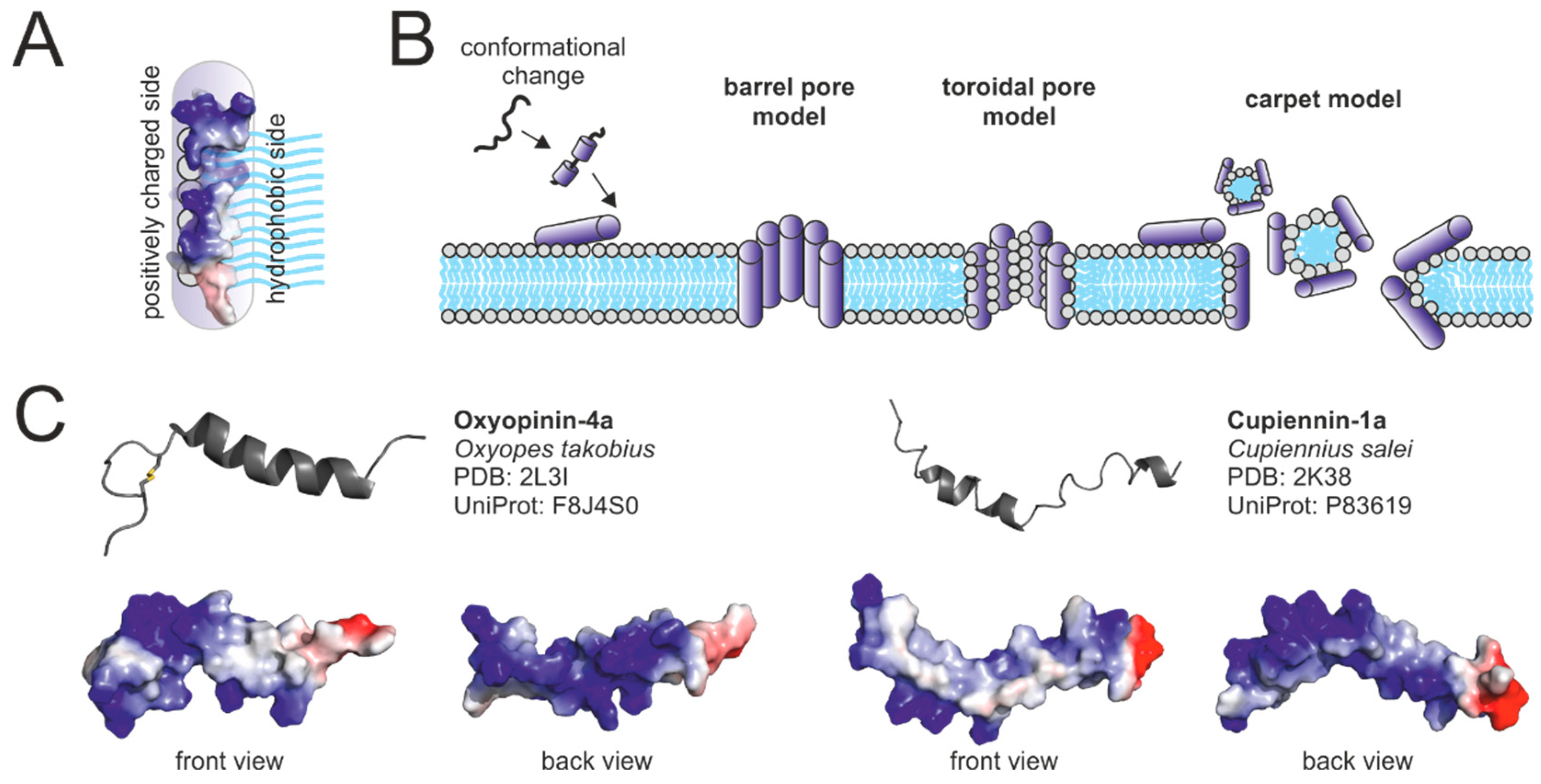
2.3. Antimicrobial Peptides

Antimicrobial peptides (AMPs) are also termed cytolytic or cationic peptides. AMPs often feature high positive net charges and a high number of hydrophobic amino acids. They are widely distributed as major components of animal immune systems and are also present in various arthropod venoms, such as ant, scorpion, bee, and wasp venoms [68,69]. AMPs disrupt the integrity of cellular membranes. They are attracted to the surface of cellular membranes owing to interaction of their positively charged amino acid side chains with negatively charged head groups of phospholipids or other negatively charged surface molecules. In proximity to membranes, AMPs exhibit an amphipathic α-helical structure. This amphipathic structure facilitates the insertion of AMPs into the cell membrane. Positively charged parts of the α-helix interact with negatively charged phospholipid head groups and hydrophobic parts of the α-helix with phospholipid tails (Figure 1A).
Multiple models for the membranolytic activity of AMPs have been proposed (Figure 2B). In the barrel pore model, multiple AMPs oligomerize and insert perpendicularly into the membrane, forming a pore where the hydrophilic region of the α-helices face the center of the pore. The toroidal pore model is similar to the barrel pore model but assumes that pore-forming AMPs always interact with lipid head groups. In the carpet model, AMPs coat parts of membranes leading to peptide-micelle formation and formation of holes in the membrane [71]. The efficiency of the membranolytic effect may be influenced by the lipid composition of cellular membranes and the membrane potential. Cupiennins (AMPs from the venom of the spider Cupiennius salei) have been reported to strongly interact with negatively charged 1-palmitoyl-2-oleoyl phosphatidyl-DL-glycerol vesicles, but only weakly with zwitterionic 1-Palmitoyl-2-oleoyl phosphatidylcholine vesicles [72]. Oxyopinins (from Oxyopes takobius, erroneously identified as O. kitabensis) more efficiently lyse phosphatidylcholine (zwitterionic) vesicles than phosphatidylethanolamine (zwitterionic) or phosphatidic acid (negative) vesicles [73]. Phospholipid headgroup size and related membrane density and fluidity have also been named as possible influencers of the cytolytic efficiency [73]. In addition, latracins (Lachesana tarabaevi) only act on membranes with a negative potential, resembling the potential of living cells (>−100 mV), but not on membranes with a positive potential [74]. The efficiency of the cytolytic activity may also be influenced by molecules exposed on the surface of membranes, such as sialic acid, which was shown to promote binding and hemolytic activity of a cupiennin 1a analogue (Cupiennius salei) [75].
AMPs have so far only been reported to occur in venoms of Cupiennius salei and the spider families Zodariidae, Lycosidae, and Oxyopidae [14]. Spider venom AMPs feature masses between 2–5 kDa and are, in most cases, cysteine free peptides. It is not unusual, that they contain a glycine or proline residue, inducing a hinge in their α-helical structure. Two representatives of spider venom AMPs are shown in Figure 1C.
Some spider venom AMPs are supposed to act according to the toroidal pore model [76,77,78], others according to the carpet model [74,79] (Figure 1B). In addition, Corzo et al. [73] hypothesized, that the mechanism of action may depend on the type (zwitterionic or anionic) of lipids, the target membrane is composed of. Besides the membranolytic activity of AMPs, activities including the inhibition of the neuronal nitric oxide synthase [80] and of the superoxide producing NADPH oxidase [75] have been reported for single peptides. Other hypothesized functions include antimicrobial defense of gland and delivery system [69], or, controversially discussed, the pre-digestion of prey [14,81]. AMPs of the spiders Cupiennius salei and Oxyopes takobius have been shown to synergistically act with neurotoxins [52,58,73]. It has been proposed that AMPs facilitate the movement and access of neurotoxins to their targets [58]. The exact mechanism of the synergistic action, however, remains uninvestigated.

2.4. Cysteine-Rich Peptides

Cysteine-rich peptides are the best investigated venom components and are believed to exist in most spider venoms [14]. Spider venoms typically contain dozens of different cysteine-rich peptides, whereof most are thought to act on channels and receptors on membranes of excitable cells. That is why they are often referred to as neurotoxic peptides or neurotoxin-like peptides. Cysteine-rich peptides typically have molecular masses between 3 and 9 kDa, and feature ≥6 cysteine residues in conserved structural motifs [14].

2.4.1. Structural Motifs

The most prominent structural motif of spider venom cysteine-rich peptides is the inhibitor cystine knot (ICK) motif. A number of other structural motifs are less frequently found in spider venom peptides. These are (1) the disulphide-directed β-hairpin (DDH) fold, (2) the Kunitz motif, (3) the colipase like fold, also known as MIT1-like fold, and (4) the helical arthropod-neuropeptide-derived (HAND) motif.
The ICK motif was initially characterized by a triple stranded antiparallel β-sheet stabilized by six cysteines arranged in the disulphide bonds C1–C4, C2–C5, C3–C6. The cystine C3–C6 thereby penetrates the characteristic ring formed by the two other disulphide bonds and the peptide backbone (Figure 2A). This cystine structure is often referred to as cystine knot, and peptides featuring this knot as knottins [82,83]. Meanwhile, some ICK-like peptides have been found to lack the first β-sheet, but still featuring the β-hairpin formed from the β-sheets 2 and 3 [84]. Many peptides feature the ICK as core motif but contain additional cysteines. A common variation is the inclusion of a fourth disulphide bridge in the loop of the β-hairpin between C5 and C6 of the simple ICK motif (e.g., [85]). Peptides featuring the ICK motif are found in the venom of various organisms besides spiders (e.g., scorpions [86], and cone snails [87]), but also in peptides with different functions, such as protease inhibitors of plants [88], an immunopeptide from horseshoe crabs [89], or an elicitor protein of a fungal tomato pathogen [90].
The DDH fold (Figure 2B) is widely distributed among different organisms [84]. It features two mandatory disulphide bonds (C1–C3, C2–C4) and an antiparallel β-hairpin, comparable with the β-hairpin formed by the beta sheets 2 and 3 of ICK peptides [14]. The loop before the first β-sheet (between Cys 2 and Cys 3) commonly comprises a Gly or Pro residue supporting a tight turn of the peptide backbone [91]. Some scientists considered the DDH as an ancient fold, from which the ICK fold evolved [91], others suggested, that the DDH is a derivative of the ICK [92]. The Kunitz motif (Figure 2C) is the active domain of a class of serine protease inhibitors. It usually features an antiparallel β-sheet and a C-terminal α-helical part constrained by three disulphide bridges (C1–C6, C2–C4, C3–C5). Venom peptides with this motif were originally isolated from snakes (green mamba) [93] and later also identified in the venom of other animals including cone snails [94] and sea anemones [95]. Despite their structural similarity to protease inhibitors, many Kunitz domain containing venom peptides have been described to block potassium ion channels [96,97,98]. Some only retained very weak inhibitory effects on serine proteases [98].
A colipase like fold (Figure 2D) of a venom peptide was first reported for the snake toxin mamba intestinal toxin 1 (MIT-1) [99,100]. Later, homologues were reported from frog skin [101], humans, mice [102], and further animals [103]. These peptides commonly comprise the four amino acids AVIT at their N-terminus. Accordingly, the peptide family is also known as AVIT family [104]. The characteristic disulphide bridge pattern (C1–C4, C2–C5, C3–C7, C6–C9, C8–C10) is similar to the one of colipases and the C-terminal region of Dickkopf proteins [104]. This fold has been discussed to conform two head to tail connected DDH motifs [91]. MIT-1 and its homologue from the skin of the toad Bombina variegata were shown to bind mammal prokinectin receptors and to cause short term gastric smooth muscle contraction and hyperalgesia [101]. Dickkopf-like activities could not be detected [104]. Homologues in spider venom have first been reported from Australian funnel-web spiders. These peptides, however lack the N-terminal AVIT domain and they do not bind prokinectin receptors, nor induce contractions of gastrointestinal smooth muscle, or cause toxic effect on house crickets [105,106]. Related peptides have meanwhile also been reported in other spiders [107], their function, however, remains unknown.
HAND toxins (Figure 2E) are composed of four tightly packed α-helices stabilized by three disulphide bridges (C1–C5, C2–C4, C3–C6). Unlike the vast majority of other spider venom peptides, their precursors do not contain a propeptide. HAND toxins are widespread in araneomorph spiders (e.g., Nephila sp., Loxosceles sp., Sicarius sp., Heteropoda sp., Latrodectus sp., Tegenaria sp.) and are also found in centipedes, wasps, ticks, and scorpions [108,109]. They are thought to have convergently evolved from neuropeptide hormones of the crustacean hyperglycaemic hormone and arthropod ion transport peptide families [109,110]. The first HAND toxins isolated from spider venom, the latrodectins, were co-purified with large pore-forming proteins (alpha-latrotoxins) and were thought to act as co-factors for them [111,112]. Latrodectins were reported to lack toxicity on mice and cockroach [113]. Today, HAND toxins are also known for many spiders, which do not contain alpha-latrotoxin homologues. Toxic effects are not reported with one exemption: U1-agatoxin-Ta1a (O46166) from the spider Eratigena agrestis, shows insecticidal activity probably resulting from directly targeting the insect’s central nervous system (CNS) [114].
Most commonly, spider venom cysteine-rich peptides are monomers. To the best of our knowledge, there are currently only two heterodimeric structures known (U2-ctenitoxin-Cs1a, P83919, Cupiennius salei [59], and omega-agatoxin-1A, P15969, Agelenopsis aperta [115]). These two toxins are further discussed below.

2.4.2. Targets of Neurotoxic Peptides

Neurotoxic peptides modulate a broad range of channels and receptors on membranes of excitable cells (e.g., nerves and muscles). Their target specificity is thereby not reliably predictable given the structural motif or the primary sequence of a peptide. Specificities and potency are experimentally investigated, frequently in patch-clamp experiments, e.g., [116]. Neurotoxic peptides form stable complexes with channels or receptors and are effective already at nanomolar concentrations. Thus, neurotoxic peptides exhibit their toxic effects already at concentrations, which are at least one order of magnitude lower than the one of non-selective toxins, such as AMPs. The toxic effects, however, can be dependent on the type of prey, the specificity of the neurotoxins for the specific channels, and the receptors expressed by that prey [14,81]. When counting today (15 May 2019), 481 out of 1561 entries listed in the ArachnoServer have a described target. Of them 46% act on voltage-activated sodium channels (NaV) of different subtypes by either delaying their inactivation (e.g., delta-ctenitoxin-Pn2c, O76199), inhibiting their opening (e.g., mu-theraphotoxin-Hs2a_1, P83303), or shifting their potential limit for activation (e.g., beta-theraphotoxin-Cm1a, P84507). Approx. 37% inhibit voltage gated calcium channels (CaV, e.g., omega-ctenitoxin-Cs1a, P81694), and 15% inhibit voltage gated potassium channels (KV, e.g., Kunitz-type kappa-PI-theraphotoxin-Hs1a, P68425). Other targets include acid-sensing ion channels (e.g., pi-theraphotoxin-Pc1a, P60514), NMDA-glutamate-receptors (e.g., gamma-ctenitoxin-Pn1a, P59367), calcium-activated potassium channels (e.g., kappa-hexatoxin-Hv1e [sic], S0F1M9), and transient receptor potential (TRP) channel (e.g., tau-theraphotoxin-Gr1a, M5AXK5). The action of a toxin is not necessarily restricted to one type of channel (e.g., beta/kappa-theraphotoxin-Hlv1a, B3EWN3; mu/omega-theraphotoxin-Hs1a(1), B3FIR8). Ion channels on membranes of excitable cells are responsible for proper signal transduction. Calcium channels are involved in neurotransmitter release from presynaptic cells, voltage gated sodium channels enable the transport of the action potential along excitable cells, and voltage gated potassium channels are crucial for restoring a resting state in depolarized cells. Disruption of the channels function (e.g., by inhibition or non-natural activation through toxins), may affects coordination, locomotion, respiration, and cardiac functions leading to various symptoms including convulsions, paralysis, and death. Toxins acting on the capsaicin receptor (transient receptor potential channel) may induce inflammatory pain [117,118].

2.4.3. Nomenclature of Toxins

In 2008, King et al. [119] recognized the need of a universal nomenclature for maintaining an overview on the rising number of known peptides of venomous animals and introduced a rational nomenclature based on their molecular target. This three-part-nomenclature includes a (1) Greek letter describing a toxin’s activity or target (e.g., “ω” for CaV blockers, or “U” for unknown), followed by (2) an indicator of the source organism’s family, genus, and species, (e.g., “ctenitoxin-Cs” for Cupiennius salei (Ctenidae), and (3) a number to distinguish toxins with identical parts 1 and 2. Examples for the nomenclature of peptides acting on the most relevant targets for spider venoms are given in Table 1. The big advantage of this nomenclature is the fast identification of a toxin’s target in its name. This, however, is only possible, if the target is known and the ArachnoServer lists currently (15 May 2019) 61% of toxins with unknown target. Recent developments in omics techniques resulted in a flood of toxin sequences with no associated functional data, deposited in online repositories. For such toxins, the nomenclature is restricted to consecutively numbering the toxins of a spider e.g., U1-ctenitoxin-Cs1a, U2-ctenitoxin-Cs1a, U3-ctenitoxin-Cs1a.
In addition, the linkage of this nomenclature to the spider family, genus, and species names can lead to confusion caused by taxonomical rearrangements of spiders. As example, Atrax and Hadronyche species have recently been moved from Hexathelidae to an own family, Atracidae [48]. Consequently, delta-hexatoxin-Hf1a from Hadronyche formidabilis would require renaming to delta-atracitoxin-Hf1a. Also, U10-theraphotoxin-Hs1a would require renaming to U10-theraphotoxin-Cs1a because the theraphosid Haplopelma schmidti has meanwhile been moved into the genus Cyriopagopus.

2.4.4. Molecular Binding of Neurotoxins

Binding of toxins to their receptors is often studied by combination of patch-clamp electrophysiology with recombinant expression of the toxin and/or receptor and site directed mutagenesis (e.g., alanine screening), [120,121,122,123]. Structural data of toxins (e.g., from NMR spectroscopy) are often included to study structure activity relationships (SAR). Exceptional insight is also gained from the only recently published first crystal structures of spider venom toxins bound to their target channels [124,125,126].
Most, but not all [127] spider venom neurotoxins are today thought to bind their target channels indirectly. They first partition into the cellular membrane and then laterally diffuse to reach the target channel. Hereof, several neurotoxic peptides have been found to bind model membranes and to form trimolecular complexes consisting of the toxin bound to the membrane and the target receptor [128]. Membrane contact is thought to enhance the affinity of inhibition as a large part of the binding energy deviates from membrane binding [129]. Furthermore, membrane binding was also shown to induce structural changes of a toxin [130], and cause ideal positioning of a toxin to bind voltage-gated ion channels [131].
Neurotoxin–membrane interaction was extensively studied with VSTx1 (kappa-theraphotoxin-Gr3a, P60980). This tarantula venom toxin induces a thinning of the phospholipid bilayer and locates superficially at the lipid–water interface inserting several hydrophobic amino acids into the membrane interior, while basic residues mainly retain in the aqueous phase [131]. The toxin region interacting with the membrane thereby resembles a hydrophobic patch surrounded by a ring of positively charged amino acids. This motif is present in many spider toxins and is generally thought to be important for membrane partitioning (Figure 3A) [128,132,133]. Electrostatic interactions between the ring of positively charged amino acids and lipid head groups thereby seem to be important for membrane binding as some toxins were found to have a higher affinity to membranes with high amounts of negatively charged lipids (e.g., phosphatidylglycerols) than to membranes with zwitterionic lipids (e.g., phosphatidylcholines) [127,132,134,135]. The targets of most spider venom neurotoxins are voltage-gated sodium, potassium, and calcium channels. These channels are generally composed of a central pore determining the ion selectivity and four surrounding voltage-sensing domains regulating opening and closing of the central pore. While most KV are tetramers, most NaV and CaV are composed of one polypeptide with four repeated similar domains (1 to 4). Every repeat (or subunit of the KV channel) contains six transmembrane helices (S1–S6) with S1–S4 forming a voltage sensing domain and S5–S6 forming the central pore. Many channels also contain an associated regulatory domain at the cytosolic site [136].
Binding of spider neurotoxins to voltage gated ion channels was predominantly studied for NaV channels. A recent 3D structure of a toxin-receptor complex is shown in Figure 3B. Most toxins were shown to bind to the helices S3 and S4 of the voltage sensing domain, allosterically modulating pore gating [137,138]. This is in contrast to the action of venom acylpolyamines, such as argiotoxin-636 (from Argiope lobata), which inhibits calcium-permeable AMPA receptors by directly inserting into the pore [139]. Toxin-channel interaction may also involve domains in proximity to S3 and S4 (e.g., S1–S2 loop, and S5), as recently shown for the double ICK peptide Dc1a bound to a mammalian NaV channel [124]. Binding is often based on interaction of certain residues of the hydrophobic patch and the surrounding positive charged amino acids with hydrophobic and anionic amino acids of the target channel (compare Figure 3B1) [128]. A similar toxin surface was also shown to be involved in binding of the ASIC channel inhibitor PcTx 1 (pi-theraphotoxin-Pc1a, P60514) [140,141]. In contrast to voltage gated channel modulators, whose binding sites on channels are partially located within the membrane, PcTx 1, binds 45 Å above the lipid bilayer [140]. Intriguingly, PcTx 1 was anyhow shown to superficially interact with membranes. It has been proposed, that this interaction may be advantageous for finding its target channel due to reduction of the dimensions of diffusion [141].

2.4.5. Modular Toxins—Combination of Toxin Structures

Most spider venom toxins can be either classified as “classic” AMP or “classic” neurotoxin-like peptide. Some toxins, however comprise a multi-domain construction with each domain resembling the structure of a “classic” spider toxin [142]. Multiple combinations of domains are thereby possible: neurotoxin-AMP, AMP-neurotoxin, AMP-AMP, and neurotoxin-neurotoxin.
Toxins exhibiting neurotoxic and cytolytic domains and corresponding activities exist in two forms. The cationic anti-microbial domain may locate C-terminal (e.g., CsTx-1, Cupiennius salei [20]; purotoxin-2, Alopecosa marikovskyi [143]) or N-terminal (spiderines, Oxyopes takobius [142,144]) of the neurotoxic domain (Figure 4A,B). Multiple assets from the two-domain construction of these toxins have been suggested. Oparin et al. [143] and Kuhn-Nentwig et al. [145] hypothesized that the α-helical part of these two-domain toxins binds to the cell membrane, thus increasing the efficiency of binding of the neurotoxic part to a receptor. Besides such a broader mode of action also an aggravation of the development of resistance against these two-domain toxins was suggested by Kuhn-Nentwig et al. [20]. The combination of cytolytic and neurotoxic activities of toxins is not restricted to spiders, but was also reported for scorpions [146].
Toxins with two consecutive ICK domains are described from four venom peptides of Hadronyche infensa (Hi1a–Hi1d) (Figure 4C). Each domain is highly similar to a known ASIC1a inhibitor. This is why the tandem structure of these toxins is thought to be a result of a duplication event of a gene encoding a single ICK gene. Hi1a was found to delay the activation of ASIC1a, a channel involved in stroke-induced neuronal damage, making it a promising candidate for development of neuroprotective stroke medication [147]. Toxins with similar double-ICK construction were also reported for the theraphosid Cyriopagopus schmidti (tau-theraphotoxin-Hs1a) [117] and Cheiracanthium punctorium (CpTx1–4) [148,149]. CpTx1–4 comprise at least 1/3 of all venom proteins of Cheiracanthium punctorium. It is likely, that CpTx1–4 additionally contain an α-helical motif C-terminally of the ICKs, making them similar to two joined neurotoxins with an additional cytolytic domain.
Besides toxins with two neurotoxin-like domains, there are also toxins with two joined cytolytic domains (Figure 4D): The zodariid Lachesana tarabaevi expresses cyto-insectotoxins comprising two separated domains with a high propensity to form amphiphilic α-helices in proximity to membranes. These toxins show increased insecticidal activity compared to “simple” cytolytic peptides of the same spider [150].

2.5. Enzymes and Proteins

For a long time, enzymes and proteins were assigned a minor role in many spider venoms. Most publications constrained to enzymes described to target the extracellular matrix or the membrane of cells, facilitating the movement of toxins in the prey as so-called spreading factors. These enzymes include hyaluronidases, collagenases, and phospholipases. Hyaluronidases cleave the 1-4-linkages between N-acetylglucosamine and glucoronate in hyaluronan, a key component of the extracellular matrix. Another key component of the animal extracellular matrix, collagen, is cleaved by matrix-metalloproteases named collagenases. Phospholipases directly target the cells’ lipid bilayer by hydrolysis of phospholipids [14]. While many spider venoms contain phospholipase A [14], sicariid spiders contain extraordinarily high venom concentrations of phospholipase D (also referred to as sphingomyelinase D) [151]. This enzyme is responsible for necrotic effects in humans and efficiently immobilizes insects, also due to an additional neurotoxic effect [152].
In recent past, an increasing number of additional spider venom enzymes have been found and suggest a separation of spider venom proteins into three functional classes. (1) Enzymes acting as spreading factor (see above), (2) proteins with a function in the venom gland of the spider including the maturing of toxins (see next chapter), and (3) proteins directly targeting important systems of the prey organism. Functions of venom proteins, however, are in most cases not experimentally studied, but only hypothesized based on similarity to known enzymes.
Proteins with a potential function in the venom gland include protein disulphide isomerases [107], a peptide isomerase [153], carboxypeptidases [107], and the recently identified 28 kDa serine proteases responsible for propeptide cleavage of immature toxin precursors (see next chapter). Other proteins, such a leucine-rich repeat domain-containing protein or a tachylectin 5A-like protein, showing similarity to immune active proteins of spider-related animals, were hypothesized to possibly be involved in protection of the venom gland against microbial infections [107].
Proteins probably directly targeting important systems in the prey may be large neurotoxic proteins (see below), an acetylcholinesterase responsible for degradation of the neurotransmitter acetylcholine at synapses [21], or an α-amylase, hypothesized to cause hyperglycemia in the prey due to fast release of high amounts of glucose from the prey’s glycogen storages [107].
Proteins with a high similarity to angiotensin-converting enzyme, an exo-metallopeptidase processing peptide hormones involved in blood pressure regulation of vertebrates, were identified in various venoms of spiders [107,154,155] and scorpions [156]. These enzymes could be involved in venom peptide processing or target peptide hormones in the prey [107]. Another group of frequently described spider venom proteins are cysteine-rich secretory proteins (CRISPs) homologues [21,22,107]. CRISPs belong to the CAP (cysteine-rich secretory proteins, antigen 5, and pathogenesis-related 1 proteins) protein superfamily and are widely distributed among animal venoms, e.g., scorpions and snakes [157], but are also found in mammalian epididymis and the immune system [158]. Many different functions are described for CRISPs, including involvement in sperm-egg fusion [159] and immune defense [160] for mammalian CRISPs, or ion channel blocking effects for snake venom CRISPs [161,162,163]. In addition, a CAP superfamily saliva protein of blood-sucking insects was recently described to inhibit platelet aggregation and act as antioxidant enzyme by scavenging of superoxide [164]. The function of CRISPs in spider venom, however, remains elusive.
The majority of the here described venom proteins act as enzymes. A notable exception are latrotoxins, a group of neurotoxic proteins with masses between approx. 110 and 130 kDa initially reported from widow spiders (Latrodectus species) [165]. Similar toxins were later also reported from the venom of Steatoda and from genomic data of Parasteatoda (both Theridiidae), but the presence in the genome does not necessarily imply expression in the venom gland [166,167,168]. Latrotoxins exert different specificities, for insects, vertebrates, or crustaceans, and are accordingly named latrotoxins (against vertebrates), latroinsectotoxins, or latrocrustotoxins. The best studied latrotoxin is alpha-latrotoxin, which can cause fatalities in vertebrates. The main toxic action of alpha-latrotoxin relies on the formation of homotetrameric complexes that insert into presynaptic neuronal membranes, forming cation permeable pores [169,170]. Pore formation leads to massive influx of Ca2+, stimulating the release of all known types of neurotransmitters from presynaptic cells ultimately leading to the blockade of signal transmission inducing clinical symptoms such as muscular paralysis [81]. Binding of alpha-latrotoxin to membranes is thought to be mediated through surface proteins. Three alpha-latrotoxin receptors could so far be identified: Neurexin, latrophilin, and tyrosine phosphatase σ. Alpha-latrotoxin may also induce neurotransmitter release through a pathway, which is independent from influx of Ca2+ into the presynaptic cell (for a review see Yan and Wang [171]). Latrotoxins comprise a common structure featuring an N-terminal part with conserved regions, a central part containing multiple ankyrin repeats, and a C-terminal propeptide [165]. Ankyrin repeats mediate protein-protein interactions and are one of the most frequently observed amino acid motifs [172]. Posttranslational removal of the C-terminal propeptide seems to be required to exceed a toxic action as propeptide containing precursors of δ-latroinsectotoxin from Latrodectus tredecimguttatus have been shown to be inactive [173].

2.6. Spider Venom Peptide Precursors and Their Maturing

Neurotoxic and cytolytic spider venom peptides are expressed as precursors undergoing proteolytic cleavage to yield the mature peptide. Precursors of neurotoxic peptides typically comprise three consecutive parts. (1) A signal peptide, which is removed during translocation through the endoplasmic reticulum, (2) a propeptide, and (3) the mature toxin. Most propeptides feature lengths between approx. 6 and 30 amino acids and commonly contain a high number of negatively charged amino acids often compensating for the overall positive charge of the mature peptide (Figure 5) [107]. It has been proposed, that this charge compensation is beneficial for the storage of the immature toxin, as electrostatic repulsion between the precursors is reduced [107]. Propeptides may also assure correct peptide folding, sorting, and maturation [81] and prosequence elements of AMPs may neutralize their lytic activity before cleavage [174].
In 2005, Kozlov et al. [17] recognized that the four C-terminal amino acids of the propeptide form a conserved motif. This motif comprises an arginine residue at position −1 and at least one glutamic acid residue at positions −2 to −4 before the start of the mature peptide. The motif, named Processing Quadruplet Motif (PQM), is thought to act as protease recognition site for the propeptide cleaving protease. Later, a minority of neurotoxic peptide precursors were found to feature a dibasic cleavage motif (e.g., KR, RR) instead of a PQM, or not to comprise any propeptide [145,175].
Antimicrobial peptides show a precursor organization comparable to neurotoxic peptides comprising a signal peptide followed by an acid propeptide and the mature peptide. Besides these simple precursors, there are also binary or complex precursors, comprising multiple mature AMPs in a row, separated by short spacer sequences (Figure 5E) [176]. The spacer sequences thereby contain protease recognition motifs for posttranslational cleavage, allowing release of the individual AMPs from the long precursor. The recognition motifs are usually a PQM at the C-terminal end of the spacer and a so-called inverted Processing Quadruplet Motif (iPQM) at the N-terminal site of the spacer. An iPQM is basically a mirrored PQM comprising an Arg residue at position 1 and at least one Glu residue at positions 2 to 4. During removal of spacers from the complex precursors, the PQM is cleaved C-terminally of its Arg-residue in analogy to the propeptide cleavage. iPQM cleavage, however is proposed to be a two-step processing process, comprising first cleavage C-terminally of the Arg residue of the iPQM by an endopeptidase, and second, removal of the remaining Arg-residue by a carboxypeptidase (Figure 5A) [177]. The endopeptidase involved in PQM and iPQM cleavage is thereby likely to be the same [178].
Expression of AMP-precursors featuring a propeptide has been proposed to be a way for preventing potential harmful cytolytic actions during the biosynthesis of a toxin. It is believed that the negatively charged propeptides or spacers thereby interact with the positively charged clusters of the mature toxin neutralizing its effect before the toxin is activated by the processing into its mature form [74,174]. At which state of venom peptide production the protease-mediated maturing occurs (e.g., before or after storage in secretory vesicles) remains uninvestigated. However, the existence of heterodimeric toxins with disulphide linked chains as described below implies a precursor cleavage after the disulphide bridge formation.

2.6.1. Comparison of Protease Cleavage Motifs to Motifs of Other Organisms

Distinguishing between cleavage motifs can be challenging, as certain sequences may be following the rule of several described motifs (e.g., EVKR may be classified as dibasic motif and as PQM). Dibasic cleavage motifs and similar motifs comprising a basic residue at the cleavage site and another one 0 to 6 amino acids before, are widespread among neuropeptides, hormones and growth factors of different organisms. This motifs are cleaved by subtilisin-like proprotein convertases (calcium dependent serine proteases) and the resulting C-terminally exposed basic amino acids are subsequently removed by metallo-carboxypeptidases [177,179,180,181]. Dibasic motifs or motifs with an extended distance between the two basic residues have also been found in precursors of peptides from sea anemones, scorpions, snakes and amphibians. Few of these motifs also feature Glu-residues in the range of +4 or −4 amino acids from the cleavage site, making it possible to be additionally classified as PQMs and iPQMs [177]. Unambiguous PQMs are rarely present in precursor sequences (e.g., P86399). Thus spider venom peptide maturing seem to rather represent an exception compared to the maturing of venom peptides of other animals [81].
Toxin precursors of other animals may also feature a construction different from the classic spider venom peptide precursor. Scorpion toxin precursors, for example, may be free of propeptides, contain propeptides N-terminally of the mature peptide [86,182], or contain propeptides C-terminally of their mature toxin [183,184]. We are not aware of C-terminal propeptides in spider venom peptide precursors. Gomesin, however, an AMP isolated from spider hemocytes, contains a C-terminal propeptide separated by a dibasic motif from the mature chain [185].
Production of toxins through complex toxin precursors, giving rise to multiple mature peptides, is not a singularity of spiders, but has also been described for other organisms including the toad Bombina maxima [186], the sea anemone Urticina grebelnyi [187], and some centipedes [188]. Complex structures of AMPs of the immune system are, among others, known from the honey bee [189], and from plants [190].

2.6.2. Cleavage for Heterodimerization of Spider Toxins

Today, we know only a few heterodimeric neurotoxin-like peptides from spider venom. CsTx-13 (U2-ctenitoxin-Cs1a, P83919) from the spider Cupiennius salei is an ICK-motif peptide with two inter-chain disulphide bridges (Figure 5D) acting synergistically with its main monomeric neurotoxin CsTx-1 (omega-ctenitoxin-Cs1a, P81694) and its main cytolytic peptide cupiennin 1a (M-ctenitoxin-Cs1a, P83619) [59]. In CsTx-13, the two chains are of similar length. The only other well characterized heterodimeric spider venom toxin, omega-agatoxin-1A (P15969) of the agelenid Agelenopsis aperta, in contrary, possesses a very short chain consisting of only three amino acids connected with a disulphide bridge to a longer chain (Figure 5C) [191]. A homologue of omega-agatoxin-1A has also been found in the venom gland transcriptome of Cupiennius salei (CsTx-40, MH754614) [107,178]. All these peptides undergo heterodimerization by proteolytic cleavage of a monomeric precursor. The precursor comprises both mature chains separated by short spacers (RSETDR, RSEESER, and RNEEAER, respectively). The evident similarity of the precursor structure to the precursors of binary and complex precursors of AMPs suggests an analogue maturing mechanism by cleavage of the PQM and iPQM followed by removal of the residual Arg at the C-terminal end of the chain 1 [177,178].
Besides protease mediated cleavage of the peptide precursors, spider venom toxins may undergo additional posttranslational modification. Commonly observed modifications include removal of C-terminal Arg-residues by a so far uncharacterized carboxypeptidase, and peptidylglycine alpha-amidating monooxygenase mediated C-terminal amidation of peptides (Figure 5) [107]. In addition, pyroglutamate formation at N-terminal glutamine, formation of O-palmitoyl threonine, and isomerization of an amino acid have been described [176,192,193].

2.7. Concluding Remarks on Venom Components and Their Toxic Action

Here, we reviewed spider venom components in groups (i.e., small molecular mass compounds, AMPs, cysteine-rich peptides, and proteins). Some venom components known today cannot be assigned to any of the above groups. Among them are short peptides with similarity to peptide-hormones of the kinin family, such as tachykinin-like short cationic peptides from the ctenid Phoneutria nigriventer, that provoke smooth muscle contractions in guinea pig ileum [194], or bradykinin-potentiating peptides from Lycosa erythrognatha (mentioned as Scaptocosa raptoria) [195,196] and Latrodectus tredecimguttatus [197]. Some of these peptides have been shown to relax rat duodenum, increase capillary permeability, and to inhibit angiotensin converting enzyme [196].
Despite the here presented description of the toxic action of single venom components or groups of venom components, the importance of synergistic effects for the overall toxicity of venom should not be underestimated. Examples for reported synergistic actions are enhanced toxicity of neurotoxins in presence of potassium ions [58] or synergistic actions between the heterodimeric neurotoxin CsTx-13 (U2-ctenitoxin-Cs1a, P83919, Cupiennius salei) and the main monomeric toxin of the same spider (CsTx-1, omega-ctenitoxin-Cs1a, P81694) [59]. In addition, neurotoxins with different functions may interplay to ensure efficient effects in the prey. For example, it can be assumed, that the δ-toxins, and the κ-toxin co-occurring in the venom of the theraphosid Chilobrachys guangxiensis preserve action potentials (delayed inactivation of NaV channels through δ-toxins) and simultaneously prevent their termination (blocking activation of KV channels by κ-toxin) leading to tetanic paralysis [198]. Synergistic actions in insecticidal activity have also been reported for AMPs and neurotoxins [52,73].
Additionally, it has been shown, that neurotoxins have a stronger affinity for ion channels, if the lipid membrane contains more anionic ceramide 1-phosphate, than zwitterionic sphingomyelin. Sphingomyelin is hydrolyzed to ceramide 1-phosphate by sphingomyelinase D, an enzyme found in the venom of sicariid spiders, what implies a synergistic effect between neurotoxins and sphingomyelinase D [199,200]. Synergistic actions are also assumed for glutamate and acylpolyamines. Acylpolyamines block ionotropic glutamate receptors by inserting into the activated channel. Venom glutamate activates these channels upon venom injection and thus allows efficient blockade [81].
The here mentioned synergistic actions between venom components are only the tip of the iceberg and the interplay between venom components has only been marginally studied today. This especially concerns venom proteins that have only recently gained in focus [107]. Furthermore, it can be assumed that venom components may also have different purposes, sites, and time-courses of action [198]. Some components are likely to induce pain and mainly have a defensive purpose (e.g., ATP, serotonin, histamine), others ensure reversible, but immediate paralysis, or traverse the blood-brain barrier to act directly on the CNS and induce irreversible flaccid paralysis 20 to 30 minutes after injection [198]. All in all, the envenomation process seems to rely on a complex interplay of different venom components, which cannot be fully understood by individual characterization of the components. In the words of the Greek philosopher Aristotle, one may state that venom is more than the sum of its parts.

3. Analysis of Venom Components

Spider venom research started mid of the 20th century [201]. First studies were mainly performed with venom as a whole and especially concerned the effect of spider venom on animals and humans including the development of antivenin [202]. Venom was obtained by electrical milking, similar to the method which is still widely used today [203]. The small quantities of gained venom were for a long time a limiting factor for analysis of single venom components. Only after the advent of modern chromatographic separation techniques (especially high-performance liquid chromatography, HPLC), scientists were equipped with the appropriate tools for efficient isolation of toxins from small quantities of venom [34]. In the following years, multiple single peptide compounds and SMMCs were described or isolated from venoms. Multidimensional liquid chromatography was the method of choice for isolation of peptides [50,204,205,206]. Peptide sequences were often analyzed by Edman degradation and SMMCs were investigated by methods including mass spectrometry [206,207] and thin layer chromatography [50].
More and more emphasis was put on the development of drugs and insecticides from venom components [208,209]. Venom components with activity against a desired target started to get identified by assay guided fractionation using screening assays relying on methods as automated-patch-clamp electrophysiology, or fluorescent-resonance energy transfer [210]. Upcoming DNA sequencing techniques and soft ionization techniques for mass spectrometry (i.e., matrix-assisted laser desorption/ionization (MALDI), and electrospray ionization (ESI)) accelerated venom research and offered insights into venoms, that were only available in very limited quantities.
Researches started to investigate sequences of single transcripts [211,212]. This was followed by generations of whole venom gland cDNA libraries, first sequenced by Sanger sequencing [16,17,18,19], later by Roche 454 sequencing [20,21], and Illumina sequencing [22,24]. Transcript sequences started to get used for cloning, design of mutants and heterologous expression, or chemical toxin synthesis to gain new structural and functional information (for a recent review see [210]).
Also in the last decade, mass spectrometry gained in importance for protein analyses and was increasingly used to study venom peptides and proteins. While analysis were first mainly restricted to purified components, rapid advances in detection speed, sensitivity, and coupling of liquid chromatography to mass spectrometers enabled automated studies of complex mixtures of peptides and proteins. An increasing number of transcriptomic studies were also paired with proteomic studies offering complementary data. Today, integration of proteomic, genomic and transcriptomic approaches in venom studies are termed venomics [213].
Transcriptomics and mass-spectrometry based proteomic studies are common in spider venom research and are further detailed in the following paragraphs. Genomic studies of spiders are rare, with only four genomes published so far (as of 9 July 2019): Stegodyphus mimosarum, Acanthoscurria geniculata [214], Nephila clavipes [215], and Parasteatoda tepidariorum [167,216]. Additionally, three genomes are deposited on the NCBI genome repository without any linked publication: Dysdera sylvatica, Latrodectus hesperus, and Loxosceles reclusa. For spiders, genome sequencing and analysis has been reported to be very challenging due to the high amount of repetitive elements likely to be a result of gene and whole genome duplications [216,217]. These repetitive elements enormously complicate de-novo assembly of reads. In this respect, the inclusion of data from long-read sequencing methods (see below) has been proposed to overcome fragmented genome assemblies [215,217]. More high-quality genomic data could thereby dramatically increase our understanding of the evolution of spider venom components.

3.1. Venom Gland Transcriptome Analysis

Analysis of the venom gland transcriptome offers insight into the composition of protein and peptide venom components and is the method of choice to study venom components if the availability of venom is poor. In the early phase of transcript analysis, cDNA sequences were sequenced in targeted approaches. The probably first transcriptomic analysis of spider venom toxins, performed by the group of the Russian researcher Eugene V. Grishin, investigated the coding sequence of alpha-latrotoxin by targeted sequencing of the respective clone from a cDNA library. The clone was identified by hybridization of a synthetic oligonucleotide based on the sequence of a tryptic peptide of alpha-latrotoxin [212]. Later, multiple studies used rapid amplification of cDNA ends (RACE) [218,219,220], requiring a specific oligonucleotide for amplification of the cDNA of interest (3’ RACE) or for reverse transcription of the transcript of interest (5’ RACE) [221]. These labor-intensive targeted sequencing approaches were soon replaced by random screening of cDNA libraries, allowing parallel analysis of numerous venom peptide and protein coding transcripts.
Sequencing techniques are under fast development and a variety of different techniques have been used for analysis of spider venom gland transcriptomes. As of 2 September 2019, 44 spider venom gland samples are listed in the biological source material database BioSample (search: “Araneae” [Organism] AND (venom [All Fields] AND gland [All Fields]; entries manually reviewed). Thereof, 12 samples have linked entries in the expressed sequencing tag database (dbEST) arising from Sanger sequencing, and 32 in the short read archive (SRA), a repository for next generation sequencing data. Many of the here deposited data, however, are not linked to and analyzed in original research papers. Table 2 gives an overview on published transcriptomic data from spider venom glands.
First venom gland transcriptomes were analyzed by Sanger sequencing [16,17]. mRNA was isolated, reverse transcribed, cloned into plasmids, amplified in bacteria, and finally the inserts of plasmids were sequenced by Sanger sequencing. This method generates sequences of up to 900 nucleotides in length, referred to as expressed sequencing tags (ESTs) [243]. Depending on the length of a transcript, ESTs may not span an entire transcript, but only represent portions of it. An attempt to decrease the data complexity for downstream analysis is to assemble ESTs belonging to the same mRNA to so called contigs. This computational process, however, is not trivial and may produce artefact contigs that do not represent real mRNAs. The availability of reference sequences, such as a reference genome, can therefore significantly improve transcriptomic analysis as ESTs do not need to be de-novo assembled, but can simply be mapped to reference sequences. High-quality spider genomes, however, are extremely rare and sequencing reads are usually de-novo assembled. De-novo assembly quality is significantly influenced by the length of the assembled sequencing reads with longer reads, resulting in better assemblies [210]. Development of next generation sequencing techniques, such as 454 sequencing or Illumina sequencing drastically increased the number of generated sequencing reads, allowing to generate more comprehensive transcriptomic data and to better estimate transcript abundance from the number of respective reads. However, these advances came at the cost of worse accuracy and shorter sequencing reads (e.g., depending on the protocol, mostly 150 base pairs (bp) for Illumina or up to 700 bp for 454 sequencing [243]) complicating de-novo assembly, especially for Illumina data. While Sanger sequencing of venom gland transcriptomes often resulted in the generation of approx. 100 contigs and somewhat more ESTs, that could not be assembled (referred to as singletons), reads from next generation techniques are often assembled to several thousands of contigs (Table 2), requiring bioinformatic tools to identify sequences of toxin-like peptides from the database of contigs.

3.1.1. Identification of Toxin-Like Transcripts

Different strategies have been used to identify toxin-like peptides from transcriptomic data (Table 2). The most widely used approach is Basic Local Alignment Search Tool (BLAST) of contigs against specialized databases containing known toxins, such as ArachnoServer [46,47], the animal toxin database of UniProt (ToxProt), or more comprehensive databases, such as UniProtKB. Many studies also incorporate protein domain prediction and gene ontology annotation of contigs. Hits are typically searched for toxin related terms and are manually reviewed. While many studies focus on neurotoxin-like peptides, structural features of the latter are also commonly used for toxin identification. These include cysteine count, cysteine pattern search, and presence of signal and/or propeptide cleavage sites. Prediction of possible propeptide cleavage sites was significantly improved with the publication of spiderP and its successor spiderProHMM, both automated propeptide prediction software for spider toxins. Many of the above-mentioned approaches assume a certain degree of similarity of newly identified toxin-like peptides with known toxins. This makes it challenging to identify entirely new toxin families. In addition, as toxins are derived from body peptides, simple homology search approaches (e.g., BLAST) involve the danger of identifying body peptides and wrongly annotating them as toxins (i.e., wrong positive) [244]. The number of false positive identifications may be diminished by integration of proteomic data, phylogenetic analysis of putative toxin families, and/or the use of comparative transcriptomics of non-venom producing tissues and venom gland [244,245]. Accordingly, Haney et al. [22] used a multi-tissue sequencing approach to investigate the venom of the spider Latrodectus hesperus and sequenced mRNA isolated from venom gland, silk gland, and the prosoma without the venom gland, allowing to identify transcripts with venom gland biased expression.
Comparison of transcriptome derived toxin-like sequences with transcriptomic data of haemocytes was recently used to reinforce venom gland specific expression of single toxins of Cupiennius salei Kuhn-Nentwig et al. [107], and Huang et al. [224] compared tissue specific expression of toxin-like peptides and proteins identified in the venom gland transcriptome of the lycosid Pardosa pseudoannulata. Putative cysteine-rich venom peptides were found to be uniquely or predominantly expressed in the venom glands while some putative venom protein coding transcripts showed surprisingly high expression levels in brain tissue or fat body. This demonstrates the complexity to accurately predict venom proteins from transcriptomic data.
The identification of toxin-like sequences may be influenced by the protocol of data analysis. Different protocols for data acquisition, however, may also influence toxin recovery and quantification. For example, transcript levels may be influenced by the duration between the last venom gland depletion by milking or feeding, and the dissection of venom glands. Many studies report a timeframe between 48 h and one week from last milking to dissection of the venom glands (Table 2). Venom gland dissection is even performed at different time intervals after milking in some studies to diminish the potential effects of a time-dependent expression of certain venom components. Experimental data on time-dependent expression of venom peptides and proteins, however, is lacking.
Toxin discovery may also substantially depend on the used sequencing method. Comparing 454 with Illumina sequencing, the greater read depth of Illumina has been shown to improve toxin discovery. In a recent studies of the venom of the remipede crustacean Xibalbanus tulumensis, 23 of 32 toxin families were only identified from Illumina data [246], and for Conus tribelei, 10 of 30 conopeptide superfamilies were only identified from Illumina data [247]. Interestingly however, for the spider Phoneutria nigriventer, Diniz et al. [154] identified more toxin-like peptides with traditional Sanger sequencing than with Illumina sequencing.

3.1.2. Recent Developments in Sequencing Techniques

DNA sequencing techniques are currently under constant development. Meanwhile techniques for single molecule sequencing, such as Oxford Nanopore Technologies (Nanopore), and Pacific Bioscience (PacBio), termed third generation sequencing, are increasingly available. To our knowledge, these techniques have so far not yet been applied for spider venom gland studies. With read lengths between 10,000 and 100,000 bases, third generation sequencing techniques produce much longer reads than next generation sequencing [248]. These read lengths enable full-length mRNA sequencing overcoming problems in de-novo assembly often encountered in next generation sequencing protocols. Full-length mRNA sequencing also provides a significant advantage for quantification of transcripts of closely related isoforms [249]. This can be challenging with short read methods as a read may not be unambiguously assigned to a single isoform.
Third generation sequencing methods, however, have two major drawbacks. (1) High error rates of up to 15% (compared to approx. 0.1% for Illumina), and (2) low throughput compared to Illumina sequencing [249]. These drawbacks can be diminished if data from third generation sequencing is jointly assembled with data from a next generation sequencing technique [250,251]. This hybrid assembly approach, has been shown to facilitate de-novo genome assembly, and may also be applicable to study genomic data of spiders, which has up to now been a challenging task due to many repetitive elements, difficult to assemble from short reads. Third generation sequencing techniques may also drastically facilitate analysis of highly repetitive transcripts, such as the ones coding for complex cytolytic precursors (see above). The attractiveness of using PacBio alone for de-novo genome or transcriptome sequencing was very recently enhanced with the publication of a circular consensus sequencing protocol allowing reduction of error rates to <1% for single-molecule reads of up to 15 kb length [252,253].
In spider venomic studies, third generation techniques have not established yet and many recently published data were still acquired with next generation techniques or conventional Sanger sequencing. While 454 sequencing was meanwhile discontinued by Roche, Illumina sequencing stays widely used and gets further developed with the only recently released NovaSeq 6000 sequencing system.

3.1.3. Cross Contamination among Multiplexed Samples in Illumina Sequencing

Illumina is a sequencing by synthesis method relying on detection of fluorescent tagged nucleotides. Synthesis is performed from templates immobilized on the surface of a so-called flow cell. One flow cell thereby features several lanes (e.g., eight for Illumina HiSeq4000), in which the sequencing reaction is performed physically separated. One lane generates up to 280 million reads of 125 bp if sequenced with the meanwhile discontinued Illumina HiSeq2500. The number of reads per lane is even higher in newer Illumina sequencing solutions, such as Illumina HiSeq4000, and Nova Seq 6000, that use a special flow cell surface (patterned flow cells).
This high number of reads is an overkill for many analyses, however, economical use of sequencing capacities is possible using multiplexing, the sequencing of multiple different samples in one sequencing lane. For sample multiplexing, adapters with sample-specific indexes (barcodes) are attached to each sequencing fragment during the preparation of the sequencing library. Subsequently, multiple libraries can be pooled and sequenced in the same sequencing lane. The unique indexes are sequenced in a separate sequencing run and are used to reassign a sequence read to its respective sample in a process called demultiplexing. Despite the wide use of multiplexing, recent studies unveiled massive cross contamination between multiplexed samples caused by misassignment of individual reads to the wrong sample. This “index hopping” or “index swapping” was early described by Kircher et al. [254] who proposed the use of double barcoding (e.g., the use of two separate indexes per sample, introduced in the 3’ and 5’ adapter) to diminish the problem.
It took some years until Illumina itself and further scientists published studies concerning index misassignment. Today, Illumina states that the main underlying process is contamination from free adapters that were not completely removed during library preparation [255]. These adapters can lead to extension of library fragments with the wrong index adapter, an effect which is bigger if cluster generation is achieved via ExAmp chemistry used for patterned flow cells, as used for HiSeqX, HiSeq 4000/3000, and NovaSeq 6000. Illumina itself estimates the misassignment rates up to 2% and ≤1% for patterned and non-patterned flow cells, respectively [255]. Using patterned flow cells and single indexing, Costello et al. [256] reported 0.2% to 6% (average 0.89%) cross contamination by index misassignment, that can however be significantly reduced using non-redundant double-indexing and filtering of unexpected adapter combinations during demultiplexing. Even higher contaminations of 5% to 7% were reported by Sinha et al. [257]. Studies showed, that non-patterned flow cells performed way better with contaminations of 0.09% to 0.6% in MiSeq [256] or even complete absence of misassignment [257]. Interestingly, Griffiths et al. [258] reported up to 2.5% misindexing, however found free DNA barcode concentrations not to affect the rate of cross contamination. Owens et al. [259] could not find index swapping in data neither from HiSeq 2500 nor from HiSeq X. In summary, studies show different cross contamination rates of 0–10% but commonly propose to diminish cross contamination by use of non-redundant double barcoding and/or completely abandoning multiplexing [257,259].
The reported rates of cross contamination may be a big issue in experiments concluding results from very low expressed genes, such as some recent experiments in cancer genomics [256]. Non-realized cross contamination between multiplexed samples may also have a significant impact on the outcome in venomic studies. For example, multiplexing of venom gland transcriptome libraries of different organisms may lead to the wrong conclusion that these organisms share common venom peptides or proteins. Expression level estimates will surprisingly show that the related transcripts are low expressed in all but one organism. While the experienced scientist may recognize that the results as biased, especially if the phylogenetic distance between the multiplexed organisms is high, cross contamination in multiple tissue profiling experiments may be a bigger issue. Venom gland specific transcripts may be found at lower expression levels in other tissues as well.
Interestingly, a similar case has been recently described in a study comparing the transcriptomes of venom glands, silk glands, ovaries, and the prosoma without the venom glands of Parasteatoda tepidariorum ([23] STable6). The authors reported that “in contrast with the selective expression in the venom gland as posited in the traditional model of venom evolution …, many of the individual transcripts that produce venom proteins in this study have some level of expression in other tissues (silk, ovary, or cephalothorax).” [23]. The exact sequencing conditions including multiplexing level and samples multiplexed are not disclosed. Upon request, the corresponding author stated that sequencing was done in a single lane of an Illumina HiSeq4000 instrument. Some libraries were re-sequenced in a different sequencing run under non-disclosed multiplexing conditions (personal communication, Haney 2019). In the light of the latest findings concerning cross-contamination in Illumina sequencing, it cannot be excluded that some these transcripts may rather be a result of cross-contamination than they are truly expressed in other tissues.

3.2. High Throughput Mass Spectrometric Analysis of the Venom Proteoms

For a long time, the method of choice for sequence analysis of venom peptides and proteins was purification of analytes and subsequent Edman degradation. This labor- and cost-intensive approach lost in importance with upcoming high-throughput mass spectrometric techniques [34]. Today, two main workflows are used in mass-spectrometry based comprehensive analyses of proteomes: (1) Tandem mass spectrometric analysis of fragments resulting from enzymatic or chemical digestion of proteins and peptides (bottom-up proteomics), and (2) direct analysis of intact proteins and peptides by tandem mass spectrometry (top-down proteomics).

3.2.1. Bottom-Up Proteomics

Most proteomic studies of venoms use bottom-up approaches. Some studies involve extensive decomplexation of samples down to single peptides/proteins or groups of peptides/proteins prior to proteolytic digestion. A standard protocol developed for the analysis of snake venom starts with decomplexation of venom by fractionation through reverse phase HPLC (RP-HPLC). RP-HPLC fractions are subsequently separated by sodium dodecyl sulphate polyacrylamide gel electrophoresis (SDS-PAGE), single protein bands are excised from the gel, and proteins are in-gel digested with a protease (mostly trypsin). The generated protein fragments are analyzed by tandem mass spectrometry to gain sequence information. Relative abundances of venom peptides and proteins can be estimated from the peak integral of PR-HPLC peaks and/or the staining intensity of bands in SDS-PAGE. Another approach for offline-separation and relative quantification of venom components is 2D electrophoresis (isoelectric focusing and SDS-PAGE) of venom. This 2D gel-based approach, however, bears some constraints in quantification of very low or very highly expressed peptides and proteins due to the limited dynamic range of protein concentration that can be resolved by electrophoresis [260].
Extensive decomplexation of samples before proteolytic digest allows to gain further information of single peptides/proteins or groups of peptides/proteins. For example, gels may be blotted to a membrane for subsequent immunoblotting analysis using antivenoms [261,262]. However, decomplexation of venom components and handling of dozens of single samples for downstream mass spectrometric analysis is cost- and labor-intensive. Direct mass-spectrometric analysis of peptide and protein fragments resulting from proteolytic digestion of crude venom, or venom fractionated in only few fractions is significantly faster and easier to perform. In this approach, also termed “shotgun” proteomics, fragments resulting from proteolytic digestion of peptides and proteins are typically separated by capillary RP-HPLC and sequenced by on-line tandem mass spectrometry. Multidimensional separation of peptide and protein fragments (e.g., by ion-exchange chromatography and RP-HPLC) can thereby increase peptide resolution. Direct shotgun proteomics is highly sensitive and also reveals information on small peptides (<2–3 kDa), which may be lost in gel-based approaches. The major drawback, however, is the lack of easy possibilities for quantification of venom components. Isotope based methods for absolute quantification require sample spiking with one specific isotope labelled standard per quantified proteoform, which is not applicable [263]. Label-free quantification techniques based on fragment ion peak intensities may be used for the relative comparison of identical components among different samples (comparative proteomics), but are not suitable for quantification of different components in one sample [260]. Thus, shotgun-proteomics may be suitable to provide qualitative, but not quantitative information on venom components [230].
In all of the above-mentioned methods, proteolytically digested proteins and peptides are fragmented in a mass spectrometer (usually by collision induced decay, CID) to gain sequence information from fragment spectra. The several thousands of resulting fragment spectra are in-silico matched against a database of possible proteins. In the best case, this database consists of protein sequences deduced from transcriptomic or genomic data of the analyzed species. Lack of this data often limits the identification of single proteins and mostly only allows to assign proteins to a known protein family based on similarity of short sequence stretches. Protein families are thereby either identified by direct search of fragment ion spectra against a more comprehensive database (e.g., containing all spider proteins) or, allowing also identification of less similar proteins, by de-novo sequencing and subsequent search of de-novo sequenced fragments against a comprehensive database.
The majority of recently published proteomic studies on spider venom are not quantitative and use shotgun approaches and species-specific databases for matching of fragment ion spectra (for an overview on selected studies see Table 3). Sequence databases are mostly derived from translated sequences of a transcriptome analysis performed in the same study and proteomic results are used for qualitative validation of peptides inferred from transcriptomic data. For identification of proteins and protein families, few studies used non-species-specific databases or de-novo sequencing. De-novo sequencing was used in combination with database searches by Duan et al. [236] and Liao et al. [264]. In the analyzed studies, components of crude venom were either directly digested or pre-separated (mostly by SDS-PAGE) prior to digestion and mass spectrometric analysis. Multiple studies combined different protocols for maximizing data output. Gel-based and direct crude venom analysis were used by Santana et al. [265], and Duan et al. [236]. Other studies used enzymes with different specificities to improve the protein coverage [107,266].
Incomplete protein coverage and ambiguity in assigning sequenced fragments to closely related protein isoforms is a major difficulty in bottom-up proteomics, especially in shotgun approaches. This not only affects proteoform identification but also MS-based quantification [1,267,268]. Differentiation of isoforms is only possible if a unique peptide for each isoform can be unambiguously sequenced. Incomplete sequence information also significantly complicates the identification of heterodimeric toxins, e.g., CsTx-13 (U2-ctenitoxin-Cs1a, P83919) or omega-agatoxin-1A (P15969), or of C-terminally truncated toxins, e.g., CsTx-2a or CsTx-2b (omega-ctenitoxin-Cs1a, P81694)). These shortcomings may be eliminated by the use of top-down proteomics.

3.2.2. Top-Down Proteomics

In top-down proteomics, intact peptides and proteins are directly analyzed by fragmentation in the mass spectrometer without prior digestion. The analysis of intact peptides and proteins, significantly simplifies the identification of individual proteoforms with single nucleotide polymorphisms (SNP), alternative splicing variants, and post translational modifications [267,271]. A prerequisite for top-down analyses is the availability of a mass spectrometer with fast acquisition and high resolving power for precursor and especially fragment ion spectra. Commonly used are Fourier-transform ion cyclotron resonance and Fourier-transform orbitrap mass spectrometers [267]. Analytes are usually pre-separated by online RP-HPLC. However, this cannot completely isolate all proteoforms. A higher degree of pre-separation (e.g., by multidimensional HPLC) can increase the resolution and isolation of single proteoforms resulting in more protein identifications [271]. Classic CID and HCD (higher-energy collisional dissociation) may be used as fragmentation method, however electron-transfer dissociation (ETD) or combined fragmentation approaches such as electron-transfer/higher-energy collision dissociation (EThcD) are more popular, since they provide better protein coverage [267].
Top-down proteomics yields extremely complex spectra. Data analysis is performed in-silico and includes isotopic peak picking and deconvolution followed by matching of fragment spectra against a user-provided database of intact proteins, and statistical validation of protein identifications [271]. Top-down proteomics is especially suitable for analysis of peptides and small proteins up to approx. 30 kDa in mass [267] and analysis of high mass proteins is difficult [271]. Today, most venomic studies still mainly relay on bottom-up proteomics. Top-down approaches are increasingly used to complement bottom-up data of spider [21,107] and snake [272,273,274,275,276] venoms.
Recently, Calderon-Celis et al. [277,278] showed that snake venom proteins can be absolutely quantified using a top-down proteomic approach. In their study, sulphur containing venom proteins were in parallel (1) quantified via inductive coupled plasma (ICP) mass spectrometry using 32S/34S isotope dilution and (2) identified by ESI QTOF-MS [277]. ICP-MS is a highly sensitive method normally used in inorganic elementary analysis. Single atoms are ionized in a plasma and subsequently analysed in the mass spectrometer. The generated ion-signal is thereby proportional to the ion concentration in a wide dynamic range allowing absolute quantification of the ion concentration if a sample is spiked with an isotopically enriched standard (isotope dilution) [263]. ICP-MS based quantification of venom proteins may provide a promising tool for further venomic studies, but it also reveals a major challenge ahead because the full separation of proteoforms prior to mass spectrometry is a prerequisite for ICP-MS based quantification [277].

3.3. Annotation of Spider Venom Toxins

In the last decade, advances in omics techniques lead to a rapid increase in toxin sequences deposited in online repositories. The main repositories for manually annotated and reviewed spider toxin sequences are ArachnoServer [47] and ToxProt, the animal toxin annotation project of UniProt [279]. Sequences from high-throughput transcriptomics are often deposited in nucleotide sequence databases.
Many venomic studies today group newly identified toxins into families of similar toxins. Family annotation is thereby conducted among toxins of a species and/or toxins are compared with entries of inter-species repositories and annotated according to their similarity to known homologues. Detection of homologue sequences is in many studies achieved by pairwise sequence comparison tools, such as the Basic Local Alignment Search Tool (BLAST). An alternative approach is the use of profile Hidden Markov Models (HMMs) for annotation of toxins to an existing protein or peptide family [107].
Such profile HMMs are statistical models often calculated from multiple sequence alignments of protein domains, families, or structural motifs. For every position of a sequence, HMMs contain probability scores for all possible amino acids, insertions, and deletions to occur at this given position. Thus, in contrary to BLAST, search of sequences against profile HMMs employs position specific scoring matrices. Penalties for mutations in conserved protein regions are higher than for mutations in highly variable regions. This results in higher sensitivities, particularly for sequences with distant similarities [280]. Multiple sequence alignments and related HMMs are deposited in online repositories, such as Pfam [281] and sequences of interests can be scanned against predictive models of multiple databases using InterPro [282]. ToxProt, the animal toxin annotation project of UniProt, involves family classification according to Pfam profile HMMs.
In 2017, Koua and Kuhn-Nentwig found these family annotations not to comprise all known toxin sequences anymore and reported a high hierarchical heterogeneity in family and subfamily organization [283]. They published new spider venom specific HMMs which we recently further refined during our analysis of 50 spider venom gland transcriptomes. These profile HMMs enable comprehensive peptide family classification and comparison of large datasets of toxins of different species, thereby allowing new evolutionary insights into spider venoms. The mentioned toxin family classification system has been proposed to complement the currently valid rational toxin nomenclature by King et al. [119] to easily infer structural similarities of toxins [283].

3.4. Concluding Remarks on the Analysis of Venom Components

In the last years the focus of venomic studies experienced a shift from single protein analysis to comprehensive assessment of the whole transcriptome and proteome, which is today possible with extreme sensitivity [263,284]. While spider venom proteomes have up to date mainly been qualitatively assessed, quantitative studies are more widespread amongst venomic analysis of other animals (e.g., snakes). Quantitative approaches have recently also been applied in top-down proteomics [277], the latest protocols in proteomic venom studies. Today, top-down approaches however seem still in their infancy and are more challenging to perform than bottom-up approaches. This is mainly due to fewer expertise, need for protocols for sample pre-separation and expensive high-resolution mass spectrometers, and less sophisticated data analysis tools [263,284]. Many advantages, such as gaining information about posttranslational modifications and qualitative discrimination between closely related proteoforms, however, predict the use of top-down approaches alongside with transcriptomic or genomic analysis a bright future in venomics [267,271]. In addition, integration of the newest single-molecule long-read sequencing techniques promise great improvements in de-novo read assembly, which is challenging with data generated with today’s widely used Illumina sequencing. Researchers working with Illumina data, should pay great attention to avoid multiplexing-based cross contamination, which could possibly affect the conclusions of their studies.

Author Contributions

N.L. prepared the draft of this article, L.K.-N. and W.N. contributed equally to its further elaboration and discussion.

Funding

This research was funded by the Swiss National Science Foundation, Grant 31003A_162564 to W.N.

Conflicts of Interest

The authors declare no conflict of interest.

References

  1. Calvete, J.J. Venomics: Integrative venom proteomics and beyond. Biochem. J. 2017, 474, 611–634. [Google Scholar] [CrossRef]
  2. Peigneur, S.; Tytgat, J. Toxins in Drug Discovery and Pharmacology. Toxins 2018, 10, 126. [Google Scholar] [CrossRef]
  3. Soares, S.C.; Lindstrom, B.; Esteves, F.; Ohman, A. The Hidden Snake in the Grass: Superior Detection of Snakes in Challenging Attentional Conditions. PLoS ONE 2014, 9, e114724. [Google Scholar] [CrossRef]
  4. Isbell, L.A. Snakes as agents of evolutionary change in primate brains. J. Hum. Evol. 2006, 51, 1–35. [Google Scholar] [CrossRef] [PubMed]
  5. Casewell, N.R.; Wuster, W.; Vonk, F.J.; Harrison, R.A.; Fry, B.G. Complex cocktails: The evolutionary novelty of venoms. Trends Ecol. Evol. 2013, 28, 219–229. [Google Scholar] [CrossRef] [PubMed]
  6. Jones, D.E. Poison Arrows: North American Indian Hunting and Warfare; University of Texas Press: Austin, TX, USA, 2007. [Google Scholar] [CrossRef]
  7. Gomes, A.; Bhattacharya, S.; Chakraborty, M.; Bhattacharjee, P.; Mishra, R.; Gomes, A. Anti-arthritic activity of Indian monocellate cobra (Naja kaouthia) venom on adjuvant induced arthritis. Toxicon 2010, 55, 670–673. [Google Scholar] [CrossRef] [PubMed]
  8. Norton, R.S. Enhancing the therapeutic potential of peptide toxins. Expert Opin. Drug Discov. 2017, 12, 611–623. [Google Scholar] [CrossRef]
  9. Wie, C.S.; Derian, A. Ziconotide. In StatPearls; StatPearls Publishing LLC.: Treasure Island, FL, USA, 2019. [Google Scholar]
  10. Opie, L.H.; Kowolik, H. The discovery of captopril: From large animals to small molecules. Cardiovasc. Res. 1995, 30, 18–25. [Google Scholar] [CrossRef]
  11. Dutertre, S.; Lewis, R.J. Use of venom peptides to probe ion channel structure and function. J. Biol. Chem. 2010, 285, 13315–13320. [Google Scholar] [CrossRef]
  12. Harrison, R.A.; Cook, D.A.; Renjifo, C.; Casewell, N.R.; Currier, R.B.; Wagstaff, S.C. Research strategies to improve snakebite treatment: Challenges and progress. J. Proteom. 2011, 74, 1768–1780. [Google Scholar] [CrossRef]
  13. Smith, J.J.; Herzig, V.; King, G.F.; Alewood, P.F. The insecticidal potential of venom peptides. Cell. Mol. Life Sci. 2013, 70, 3665–3693. [Google Scholar] [CrossRef] [PubMed]
  14. Kuhn-Nentwig, L.; Stöcklin, R.; Nentwig, W. Venom Composition and Strategies in Spiders: Is Everything Possible? Adv. Insect Physiol. 2011, 40, 1–86. [Google Scholar] [CrossRef]
  15. Escoubas, P.; Sollod, B.; King, G.F. Venom landscapes: Mining the complexity of spider venoms via a combined cDNA and mass spectrometric approach. Toxicon 2006, 47, 650–663. [Google Scholar] [CrossRef] [PubMed]
  16. Satake, H.; Villegas, E.; Oshiro, N.; Terada, K.; Shinada, T.; Corzo, G. Rapid and efficient identification of cysteine-rich peptides by random screening of a venom gland cDNA library from the hexathelid spider Macrothele gigas. Toxicon 2004, 44, 149–156. [Google Scholar] [CrossRef] [PubMed]
  17. Kozlov, S.; Malyavka, A.; McCutchen, B.; Lu, A.; Schepers, E.; Herrmann, R.; Grishin, E. A novel strategy for the identification of toxinlike structures in spider venom. Proteins 2005, 59, 131–140. [Google Scholar] [CrossRef]
  18. Fernandes-Pedrosa Mde, F.; Junqueira-de-Azevedo Ide, L.; Goncalves-de-Andrade, R.M.; Kobashi, L.S.; Almeida, D.D.; Ho, P.L.; Tambourgi, D.V. Transcriptome analysis of Loxosceles laeta (Araneae, Sicariidae) spider venomous gland using expressed sequence tags. BMC Genom. 2008, 9, 279. [Google Scholar] [CrossRef]
  19. Chen, J.; Zhao, L.; Jiang, L.; Meng, E.; Zhang, Y.; Xiong, X.; Liang, S. Transcriptome analysis revealed novel possible venom components and cellular processes of the tarantula Chilobrachys jingzhao venom gland. Toxicon 2008, 52, 794–806. [Google Scholar] [CrossRef]
  20. Kuhn-Nentwig, L.; Fedorova, I.M.; Lüscher, B.P.; Kopp, L.S.; Trachsel, C.; Schaller, J.; Vu, X.L.; Seebeck, T.; Streitberger, K.; Nentwig, W.; et al. A venom-derived neurotoxin, CsTx-1, from the spider Cupiennius salei exhibits cytolytic activities. J. Biol. Chem. 2012, 287, 25640–25649. [Google Scholar] [CrossRef]
  21. Undheim, E.A.; Sunagar, K.; Herzig, V.; Kely, L.; Low, D.H.; Jackson, T.N.; Jones, A.; Kurniawan, N.; King, G.F.; Ali, S.A.; et al. A proteomics and transcriptomics investigation of the venom from the barychelid spider Trittame loki (brush-foot trapdoor). Toxins 2013, 5, 2488–2503. [Google Scholar] [CrossRef]
  22. Haney, R.A.; Ayoub, N.A.; Clarke, T.H.; Hayashi, C.Y.; Garb, J.E. Dramatic expansion of the black widow toxin arsenal uncovered by multi-tissue transcriptomics and venom proteomics. BMC Genom. 2014, 15, 366. [Google Scholar] [CrossRef]
  23. Haney, R.A.; Matte, T.; Forsyth, F.S.; Garb, J.E. Alternative Transcription at Venom Genes and Its Role as a Complementary Mechanism for the Generation of Venom Complexity in the Common House Spider. Front. Ecol. Evol. 2019, 7. [Google Scholar] [CrossRef] [PubMed] [Green Version]
  24. He, Q.; Duan, Z.; Yu, Y.; Liu, Z.; Liu, Z.; Liang, S. The venom gland transcriptome of Latrodectus tredecimguttatus revealed by deep sequencing and cDNA library analysis. PLoS ONE 2013, 8, e81357. [Google Scholar] [CrossRef] [PubMed]
  25. Penney, D. Spider Research in the 21st Century: Trends and Perspectives; Siri Scientific Press: Manchester, UK, 2013. [Google Scholar]
  26. Foelix, R.; Erb, B. Mesothelae have venom glands. J. Arachnol. 2010, 38, 596–598. [Google Scholar] [CrossRef]
  27. Foelix, R. Biology of Spiders, 3rd ed.; Oxford University Press: New York, NY, USA, 2011. [Google Scholar]
  28. Weng, J.L.; Barrantes, G.; Eberhard, W.G. Feeding by Philoponella vicina (Araneae, Uloboridae) and how uloborid spiders lost their venom glands. Can. J. Zool. 2006, 84, 1752–1762. [Google Scholar] [CrossRef]
  29. Zobel-Thropp, P.A.; Correa, S.M.; Garb, J.E.; Binford, G.J. Spit and venom from Scytodes spiders: A diverse and distinct cocktail. J. Proteome Res. 2014, 13, 817–835. [Google Scholar] [CrossRef] [PubMed]
  30. Pekar, S.; Toft, S.; Hruskova, M.; Mayntz, D. Dietary and prey-capture adaptations by which Zodarion germanicum, an ant-eating spider (Araneae: Zodariidae), specialises on the Formicinae. Naturwissenschaften 2008, 95, 233–239. [Google Scholar] [CrossRef] [PubMed]
  31. Dippenaar-Schoeman, A.S.; Meyer, M.K.P. On the species of the African genus Ammoxenus (Araneae: Ammoxenidae), with decriptions of two new species. J. Entomol. Soc. South. Afr. 1980, 43, 41–49. [Google Scholar]
  32. Gemeno, C.; Yeargan, K.V.; Haynes, K.F. Aggressive Chemical Mimicry by the Bolas Spider Mastophora hutchinsoni: Identification and Quantification of a Major Prey’s Sex Pheromone Components in the Spider’s Volatile Emissions. J. Chem. Ecol. 2000, 26, 1235–1243. [Google Scholar] [CrossRef]
  33. Meehan, C.J.; Olson, E.J.; Reudink, M.W.; Kyser, T.K.; Curry, R.L. Herbivory in a spider through exploitation of an ant-plant mutualism. Curr. Biol. 2009, 19, R892–R893. [Google Scholar] [CrossRef]
  34. Escoubas, P.; Quinton, L.; Nicholson, G.M. Venomics: Unravelling the complexity of animal venoms with mass spectrometry. J. Mass Spectrom. 2008, 43, 279–295. [Google Scholar] [CrossRef]
  35. Zobel-Thropp, P.A.; Bulger, E.A.; Cordes, M.H.J.; Binford, G.J.; Gillespie, R.G.; Brewer, M.S. Sexually dimorphic venom proteins in long-jawed orb-weaving spiders (Tetragnatha) comprise novel gene families. PeerJ 2018, 6, e4691. [Google Scholar] [CrossRef] [PubMed]
  36. Barlow, A.; Pook, C.E.; Harrison, R.A.; Wuster, W. Coevolution of diet and prey-specific venom activity supports the role of selection in snake venom evolution. Proc. Biol. Sci. 2009, 276, 2443–2449. [Google Scholar] [CrossRef] [PubMed] [Green Version]
  37. Jorge da Silva, N., Jr.; Aird, S.D. Prey specificity, comparative lethality and compositional differences of coral snake venoms. Comp. Biochem. Physiol. Toxicol. Pharmacol. 2001, 128, 425–456. [Google Scholar] [CrossRef]
  38. Gibbs, H.L.; Mackessy, S.P. Functional basis of a molecular adaptation: Prey-specific toxic effects of venom from Sistrurus rattlesnakes. Toxicon 2009, 53, 672–679. [Google Scholar] [CrossRef]
  39. Elliger, C.A.; Richmond, T.A.; Lebaric, Z.N.; Pierce, N.T.; Sweedler, J.V.; Gilly, W.F. Diversity of conotoxin types from Conus californicus reflects a diversity of prey types and a novel evolutionary history. Toxicon 2011, 57, 311–322. [Google Scholar] [CrossRef] [PubMed]
  40. Duda, T.F., Jr.; Chang, D.; Lewis, B.D.; Lee, T. Geographic variation in venom allelic composition and diets of the widespread predatory marine gastropod Conus ebraeus. PLoS ONE 2009, 4, e6245. [Google Scholar] [CrossRef]
  41. Nentwig, W.; Kuhn-Nentwig, L. Spider Venoms Potentially Lethal to Humans. In Spider Ecophysiology; Nentwig, W., Ed.; Springer: Berlin/Heidelberg, Germany, 2013; pp. 253–264. [Google Scholar] [CrossRef]
  42. Nentwig, W.; Kuhn-Nentwig, L. Main Components of Spider Venoms. In Spider Ecophysiology; Nentwig, W., Ed.; Springer: Berlin/Heidelberg, Germany, 2013; pp. 191–202. [Google Scholar]
  43. Chippaux, J.P.; Goyffon, M. Epidemiology of scorpionism: A global appraisal. Acta Trop. 2008, 107, 71–79. [Google Scholar] [CrossRef]
  44. Chippaux, J.P. Snake-bites: Appraisal of the global situation. Bull. World Health Organ. 1998, 76, 515–524. [Google Scholar]
  45. Nentwig, W. (Ed.) Spider Ecophysiology, 1st ed.; Springer: Berlin/Heidelberg, Germany, 2013; pp. 505–508. [Google Scholar]
  46. Herzig, V.; Wood, D.L.; Newell, F.; Chaumeil, P.A.; Kaas, Q.; Binford, G.J.; Nicholson, G.M.; Gorse, D.; King, G.F. ArachnoServer 2.0, an updated online resource for spider toxin sequences and structures. Nucleic Acids Res. 2011, 39, 653–657. [Google Scholar] [CrossRef]
  47. Pineda, S.S.; Chaumeil, P.A.; Kunert, A.; Kaas, Q.; Thang, M.W.C.; Li, L.; Nuhn, M.; Herzig, V.; Saez, N.J.; Cristofori-Armstrong, B.; et al. ArachnoServer 3.0: An online resource for automated discovery, analysis and annotation of spider toxins. Bioinformatics 2017, 34, 1074–1076. [Google Scholar] [CrossRef]
  48. World Spider Catalog Version 20.5. Available online: http://wsc.nmbe.ch (accessed on 27 August 2019).
  49. Savel-Niemann, A. Tarantula (Eurypelma californicum) venom, a multicomponent system. Biol. Chem. Hoppe Seyler 1989, 370, 485–498. [Google Scholar] [CrossRef]
  50. Kuhn-Nentwig, L.; Schaller, J.; Nentwig, W. Purification of toxic peptides and the amino acid sequence of CSTX-1 from the multicomponent venom of Cupiennius salei (Araneae: Ctenidae). Toxicon 1994, 32, 287–302. [Google Scholar] [CrossRef]
  51. Malli, H.; Kuhn-Nentwig, L.; Imboden, H.; Nentwig, W. Effects of size, motility and paralysation time of prey on the quantity of venom injected by the hunting spider Cupiennius salei. J. Exp. Biol. 1999, 202, 2083–2089. [Google Scholar]
  52. Kuhn-Nentwig, L.; Schaller, J.; Nentwig, W. Biochemistry, toxicology and ecology of the venom of the spider Cupiennius salei (Ctenidae). Toxicon 2004, 43, 543–553. [Google Scholar] [CrossRef]
  53. Wigger, E.; Kuhn-Nentwig, L.; Nentwig, W. The venom optimisation hypothesis: A spider injects large venom quantities only into difficult prey types. Toxicon 2002, 40, 749–752. [Google Scholar] [CrossRef]
  54. Malli, H.; Kuhn-Nentwig, L.; Imboden, H.; Moon, M.J.; Wyler, T. Immunocytochemical localization and secretion process of the toxin CSTX-1 in the venom gland of the wandering spider Cupiennius salei (Araneae: Ctenidae). Cell Tissue Res. 2000, 299, 417–426. [Google Scholar] [CrossRef]
  55. Silva, L.M.; Botelho, A.C.; Nacif-Pimenta, R.; Martins, G.F.; Alves, L.C.; Brayner, F.A.; Fortes-Dias, C.L.; Pimenta, P.F. Structural analysis of the venom glands of the armed spider Phoneutria nigriventer (Keyserling, 1891): Microanatomy, fine structure and confocal observations. Toxicon 2008, 51, 693–706. [Google Scholar] [CrossRef]
  56. Yigit, N.; Guven, T. Functional structure of Agelena labyrinthica’s (Araneae:Agelenidae) venom gland and electrophoresis of venom. Toxicon 2006, 47, 58–67. [Google Scholar] [CrossRef]
  57. Savel-Niemann, A.; Roth, D. Biochemical analysis of tarantula venom (Eurypelma californicum). Naturwissenschaften 1989, 76, 212–213. [Google Scholar] [CrossRef]
  58. Wullschleger, B.; Nentwig, W.; Kuhn-Nentwig, L. Spider venom: Enhancement of venom efficacy mediated by different synergistic strategies in Cupiennius salei. J. Exp. Biol. 2005, 208, 2115–2121. [Google Scholar] [CrossRef]
  59. Wullschleger, B.; Kuhn-Nentwig, L.; Tromp, J.; Kämpfer, U.; Schaller, J.; Schürch, S.; Nentwig, W. CSTX-13, a highly synergistically acting two-chain neurotoxic enhancer in the venom of the spider Cupiennius salei (Ctenidae). Proc. Natl. Acad. Sci. USA 2004, 101, 11251–11256. [Google Scholar] [CrossRef]
  60. Odell, G.V.; Fenton, A.W.; Ownby, C.L.; Doss, M.P.; Schmidt, J.O. The role of venom citrate. Toxicon 1999, 37, 407–409. [Google Scholar] [CrossRef]
  61. McCormick, J.; Li, Y.; McCormick, K.; Duynstee, H.I.; van Engen, A.K.; van der Marel, G.A.; Ganem, B.; van Boom, J.H.; Meinwald, J. Structure and Total Synthesis of HF-7, a Neuroactive Glyconucleoside Disulfate from the Funnel-Web Spider Hololena curta. J. Am. Chem. Soc. 1999, 121, 5661–5665. [Google Scholar] [CrossRef]
  62. Schroeder, F.C.; Taggi, A.E.; Gronquist, M.; Malik, R.U.; Grant, J.B.; Eisner, T.; Meinwald, J. NMR-spectroscopic screening of spider venom reveals sulfated nucleosides as major components for the brown recluse and related species. Proc. Natl. Acad. Sci. USA 2008, 105, 14283–14287. [Google Scholar] [CrossRef] [Green Version]
  63. Taggi, A.E.; Meinwald, J.; Schroeder, F.C. A new approach to natural products discovery exemplified by the identification of sulfated nucleosides in spider venom. J. Am. Chem. Soc. 2004, 126, 10364–10369. [Google Scholar] [CrossRef]
  64. Manzoli-Palma, M.d.F.; Gobbi, N.; Palma, M.S. The chelation of metal ions by the acylpolyamine toxins from the web-spider Nephilengys cruentata: Effects in the intoxication/detoxification of preys. Chemoecology 2006, 16, 203–208. [Google Scholar] [CrossRef]
  65. Bowie, D.; Mayer, M.L. Inward rectification of both AMPA and kainate subtype glutamate receptors generated by polyamine-mediated ion channel block. Neuron 1995, 15, 453–462. [Google Scholar] [CrossRef] [Green Version]
  66. Rash, L.D.; Hodgson, W.C. Pharmacology and biochemistry of spider venoms. Toxicon 2002, 40, 225–254. [Google Scholar] [CrossRef]
  67. Blenau, W.; Baumann, A. Molecular and pharmacological properties of insect biogenic amine receptors: Lessons from Drosophila melanogaster and Apis mellifera. Arch. Insect Biochem. Physiol. 2001, 48, 13–38. [Google Scholar] [CrossRef]
  68. Bontems, F.; Roumestand, C.; Gilquin, B.; Menez, A.; Toma, F. Refined structure of charybdotoxin: Common motifs in scorpion toxins and insect defensins. Science 1991, 254, 1521–1523. [Google Scholar] [CrossRef]
  69. Kuhn-Nentwig, L. Antimicrobial and cytolytic peptides of venomous arthropods. Cell. Mol. Life Sci. 2003, 60, 2651–2668. [Google Scholar] [CrossRef] [PubMed]
  70. Dolinsky, T.J.; Nielsen, J.E.; McCammon, J.A.; Baker, N.A. PDB2PQR: An automated pipeline for the setup of Poisson-Boltzmann electrostatics calculations. Nucleic Acids Res. 2004, 32, W665–W667. [Google Scholar] [CrossRef] [PubMed]
  71. Bahar, A.A.; Ren, D. Antimicrobial peptides. Pharmaceuticals 2013, 6, 1543–1575. [Google Scholar] [CrossRef] [PubMed]
  72. Kuhn-Nentwig, L.; Dathe, M.; Walz, A.; Schaller, J.; Nentwig, W. Cupiennin 1d *: The cytolytic activity depends on the hydrophobic N-terminus and is modulated by the polar C-terminus. FEBS Lett. 2002, 527, 193–198. [Google Scholar] [CrossRef]
  73. Corzo, G.; Villegas, E.; Gomez-Lagunas, F.; Possani, L.D.; Belokoneva, O.S.; Nakajima, T. Oxyopinins, large amphipathic peptides isolated from the venom of the wolf spider Oxyopes kitabensis with cytolytic properties and positive insecticidal cooperativity with spider neurotoxins. J. Biol. Chem. 2002, 277, 23627–23637. [Google Scholar] [CrossRef]
  74. Kozlov, S.A.; Vassilevski, A.A.; Feofanov, A.V.; Surovoy, A.Y.; Karpunin, D.V.; Grishin, E.V. Latarcins, antimicrobial and cytolytic peptides from the venom of the spider Lachesana tarabaevi (Zodariidae) that exemplify biomolecular diversity. J. Biol. Chem. 2006, 281, 20983–20992. [Google Scholar] [CrossRef]
  75. Kuhn-Nentwig, L.; Willems, J.; Seebeck, T.; Shalaby, T.; Kaiser, M.; Nentwig, W. Cupiennin 1a exhibits a remarkably broad, non-stereospecific cytolytic activity on bacteria, protozoan parasites, insects, and human cancer cells. Amino Acids 2011, 40, 69–76. [Google Scholar] [CrossRef]
  76. Pukala, T.L.; Boland, M.P.; Gehman, J.D.; Kuhn-Nentwig, L.; Separovic, F.; Bowie, J.H. Solution structure and interaction of cupiennin 1a, a spider venom peptide, with phospholipid bilayers. Biochemistry 2007, 46, 3576–3585. [Google Scholar] [CrossRef]
  77. Belokoneva, O.S.; Satake, H.; Mal’tseva, E.L.; Pal’mina, N.P.; Villegas, E.; Nakajima, T.; Corzo, G. Pore formation of phospholipid membranes by the action of two hemolytic arachnid peptides of different size. Biochim. Biophys. Acta 2004, 1664, 182–188. [Google Scholar] [CrossRef] [Green Version]
  78. Nomura, K.; Corzo, G. The effect of binding of spider-derived antimicrobial peptides, oxyopinins, on lipid membranes. Biochim. Biophys. Acta 2006, 1758, 1475–1482. [Google Scholar] [CrossRef] [Green Version]
  79. Dubovskii, P.V.; Volynsky, P.E.; Polyansky, A.A.; Karpunin, D.V.; Chupin, V.V.; Efremov, R.G.; Arseniev, A.S. Three-dimensional structure/hydrophobicity of latarcins specifies their mode of membrane activity. Biochemistry 2008, 47, 3525–3533. [Google Scholar] [CrossRef]
  80. Pukala, T.L.; Doyle, J.R.; Llewellyn, L.E.; Kuhn-Nentwig, L.; Apponyi, M.A.; Separovic, F.; Bowie, J.H. Cupiennin 1a, an antimicrobial peptide from the venom of the neotropical wandering spider Cupiennius salei, also inhibits the formation of nitric oxide by neuronal nitric oxide synthase. FEBS J. 2007, 274, 1778–1784. [Google Scholar] [CrossRef]
  81. Vassilevski, A.A.; Kozlov, S.A.; Grishin, E.V. Molecular diversity of spider venom. Biochemistry Biokhimiia 2009, 74, 1505–1534. [Google Scholar] [CrossRef]
  82. Pallaghy, P.K.; Nielsen, K.J.; Craik, D.J.; Norton, R.S. A common structural motif incorporating a cystine knot and a triple-stranded beta-sheet in toxic and inhibitory polypeptides. Protein Sci. 1994, 3, 1833–1839. [Google Scholar] [CrossRef]
  83. Norton, R.S.; Pallaghy, P.K. The cystine knot structure of ion channel toxins and related polypeptides. Toxicon 1998, 36, 1573–1583. [Google Scholar] [CrossRef]
  84. Escoubas, P.; Rash, L. Tarantulas: Eight-legged pharmacists and combinatorial chemists. Toxicon 2004, 43, 555–574. [Google Scholar] [CrossRef]
  85. Schaller, J.; Kämpfer, U.; Schürch, S.; Kuhn-Nentwig, L.; Haeberli, S.; Nentwig, W. CSTX-9, a toxic peptide from the spider Cupiennius salei: Amino acid sequence, disulphide bridge pattern and comparison with other spider toxins containing the cystine knot structure. Cell. Mol. Life Sci. 2001, 58, 1538–1545. [Google Scholar] [CrossRef]
  86. Gao, B.; Harvey, P.J.; Craik, D.J.; Ronjat, M.; De Waard, M.; Zhu, S. Functional evolution of scorpion venom peptides with an inhibitor cystine knot fold. Biosci. Rep. 2013, 33. [Google Scholar] [CrossRef]
  87. Kwon, S.; Bosmans, F.; Kaas, Q.; Cheneval, O.; Conibear, A.C.; Rosengren, K.J.; Wang, C.K.; Schroeder, C.I.; Craik, D.J. Efficient enzymatic cyclization of an inhibitory cystine knot-containing peptide. Biotechnol. Bioeng. 2016, 113, 2202–2212. [Google Scholar] [CrossRef]
  88. Le Nguyen, D.; Heitz, A.; Chiche, L.; Castro, B.; Boigegrain, R.A.; Favel, A.; Coletti-Previero, M.A. Molecular recognition between serine proteases and new bioactive microproteins with a knotted structure. Biochimie 1990, 72, 431–435. [Google Scholar] [CrossRef]
  89. Fujitani, N.; Kawabata, S.; Osaki, T.; Kumaki, Y.; Demura, M.; Nitta, K.; Kawano, K. Structure of the antimicrobial peptide tachystatin A. J. Biol. Chem. 2002, 277, 23651–23657. [Google Scholar] [CrossRef]
  90. Vervoort, J.; van den Hooven, H.W.; Berg, A.; Vossen, P.; Vogelsang, R.; Joosten, M.H.; de Wit, P.J. The race-specific elicitor AVR9 of the tomato pathogen Cladosporium fulvum: A cystine knot protein. Sequence-specific 1H NMR assignments, secondary structure and global fold of the protein. FEBS Lett. 1997, 404, 153–158. [Google Scholar] [CrossRef]
  91. Wang, X.; Connor, M.; Smith, R.; Maciejewski, M.W.; Howden, M.E.; Nicholson, G.M.; Christie, M.J.; King, G.F. Discovery and characterization of a family of insecticidal neurotoxins with a rare vicinal disulfide bridge. Nat. Struct. Biol. 2000, 7, 505–513. [Google Scholar] [CrossRef]
  92. Sunagar, K.; Undheim, E.A.; Chan, A.H.; Koludarov, I.; Munoz-Gomez, S.A.; Antunes, A.; Fry, B.G. Evolution stings: The origin and diversification of scorpion toxin peptide scaffolds. Toxins 2013, 5, 2456–2487. [Google Scholar] [CrossRef]
  93. Harvey, A.L.; Karlsson, E. Dendrotoxin from the venom of the green mamba, Dendroaspis angusticeps. A neurotoxin that enhances acetylcholine release at neuromuscular junction. Naunyn-Schmiedeberg’s Arch. Pharmacol. 1980, 312, 1–6. [Google Scholar] [CrossRef]
  94. Bayrhuber, M.; Vijayan, V.; Ferber, M.; Graf, R.; Korukottu, J.; Imperial, J.; Garrett, J.E.; Olivera, B.M.; Terlau, H.; Zweckstetter, M.; et al. Conkunitzin-S1 is the first member of a new Kunitz-type neurotoxin family. Structural and functional characterization. J. Biol. Chem. 2005, 280, 23766–23770. [Google Scholar] [CrossRef]
  95. Schweitz, H.; Bruhn, T.; Guillemare, E.; Moinier, D.; Lancelin, J.M.; Beress, L.; Lazdunski, M. Kalicludines and kaliseptine. Two different classes of sea anemone toxins for voltage sensitive K+ channels. J. Biol. Chem. 1995, 270, 25121–25126. [Google Scholar] [CrossRef]
  96. Peng, K.; Lin, Y.; Liang, S.P. Nuclear magnetic resonance studies on huwentoxin-XI from the Chinese bird spider Ornithoctonus huwena: 15N labeling and sequence-specific 1H, 15N nuclear magnetic resonance assignments. Acta Biochim. Biophys. Sin. 2006, 38, 457–466. [Google Scholar] [CrossRef]
  97. Schweitz, H.; Heurteaux, C.; Bois, P.; Moinier, D.; Romey, G.; Lazdunski, M. Calcicludine, a venom peptide of the Kunitz-type protease inhibitor family, is a potent blocker of high-threshold Ca2+ channels with a high affinity for L-type channels in cerebellar granule neurons. Proc. Natl. Acad. Sci. USA 1994, 91, 878–882. [Google Scholar] [CrossRef]
  98. Harvey, A.L. Twenty years of dendrotoxins. Toxicon 2001, 39, 15–26. [Google Scholar] [CrossRef]
  99. Boisbouvier, J.; Albrand, J.P.; Blackledge, M.; Jaquinod, M.; Schweitz, H.; Lazdunski, M.; Marion, D. A structural homologue of colipase in black mamba venom revealed by NMR floating disulphide bridge analysis. J. Mol. Biol. 1998, 283, 205–219. [Google Scholar] [CrossRef] [PubMed] [Green Version]
  100. Joubert, F.J.; Strydom, D.J. Snake venom. The amino acid sequence of protein A from Dendroaspis polylepis polylepis (black mamba) venom. Hoppe Seyler Z. Phisol. Chem. 1980, 361, 1787–1794. [Google Scholar] [CrossRef] [PubMed]
  101. Mollay, C.; Wechselberger, C.; Mignogna, G.; Negri, L.; Melchiorri, P.; Barra, D.; Kreil, G. Bv8, a small protein from frog skin and its homologue from snake venom induce hyperalgesia in rats. Eur. J. Pharmacol. (Mol. Pharmacol. Sect.) 1999, 374, 189–196. [Google Scholar] [CrossRef]
  102. Wechselberger, C.; Puglisi, R.; Engel, E.; Lepperdinger, G.; Boitani, C.; Kreil, G. The mammalian homologues of frog Bv8 are mainly expressed in spermatocytes. FEBS Lett. 1999, 462, 177–181. [Google Scholar] [CrossRef]
  103. Negri, L.; Lattanzi, R.; Giannini, E.; Melchiorri, P. Bv8/Prokineticin proteins and their receptors. Life Sci. 2007, 81, 1103–1116. [Google Scholar] [CrossRef]
  104. Kaser, A.; Winklmayr, M.; Lepperdinger, G.; Kreil, G. The AVIT protein family. Secreted cysteine-rich vertebrate proteins with diverse functions. EMBO Rep. 2003, 4, 469–473. [Google Scholar] [CrossRef]
  105. Szeto, T.H.; Wang, X.H.; Smith, R.; Connor, M.; Christie, M.J.; Nicholson, G.M.; King, G.F. Isolation of a funnel-web spider polypeptide with homology to mamba intestinal toxin 1 and the embryonic head inducer Dickkopf-1. Toxicon 2000, 38, 429–442. [Google Scholar] [CrossRef]
  106. Wen, S.; Wilson, D.T.; Kuruppu, S.; Korsinczky, M.L.; Hedrick, J.; Pang, L.; Szeto, T.; Hodgson, W.C.; Alewood, P.F.; Nicholson, G.M. Discovery of an MIT-like atracotoxin family: Spider venom peptides that share sequence homology but not pharmacological properties with AVIT family proteins. Peptides 2005, 26, 2412–2426. [Google Scholar] [CrossRef] [Green Version]
  107. Kuhn-Nentwig, L.; Langenegger, N.; Heller, M.; Koua, D.; Nentwig, W. The Dual Prey-Inactivation Strategy of Spiders-In-Depth Venomic Analysis of Cupiennius salei. Toxins 2019, 11, 167. [Google Scholar] [CrossRef]
  108. Undheim, E.A.; Grimm, L.L.; Low, C.F.; Morgenstern, D.; Herzig, V.; Zobel-Thropp, P.; Pineda, S.S.; Habib, R.; Dziemborowicz, S.; Fry, B.G.; et al. Weaponization of a Hormone: Convergent Recruitment of Hyperglycemic Hormone into the Venom of Arthropod Predators. Structure 2015, 23, 1283–1292. [Google Scholar] [CrossRef] [Green Version]
  109. McCowan, C.; Garb, J.E. Recruitment and diversification of an ecdysozoan family of neuropeptide hormones for black widow spider venom expression. Gene 2014, 536, 366–375. [Google Scholar] [CrossRef] [PubMed]
  110. Gasparini, S.; Kiyatkin, N.; Drevet, P.; Boulain, J.C.; Tacnet, F.; Ripoche, P.; Forest, E.; Grishin, E.; Menez, A. The low molecular weight protein which co-purifies with alpha-latrotoxin is structurally related to crustacean hyperglycemic hormones. J. Biol. Chem. 1994, 269, 19803–19809. [Google Scholar] [PubMed]
  111. Grishin, E.V.; Himmelreich, N.H.; Pluzhnikov, K.A.; Pozdnyakova, N.G.; Storchak, L.G.; Volkova, T.M.; Woll, P.G. Modulation of functional activities of the neurotoxin from black widow spider venom. FEBS Lett. 1993, 336, 205–207. [Google Scholar] [CrossRef] [Green Version]
  112. Kiyatkin, N.; Dulubova, I.; Chekhovskaya, I.; Lipkin, A.; Grishin, E. Structure of the low molecular weight protein copurified with alpha-latrotoxin. Toxicon 1992, 30, 771–774. [Google Scholar] [CrossRef]
  113. Volkova, T.M.; Pluzhnikov, K.A.; Woll, P.G.; Grishin, E.V. Low molecular weight components from black widow spider venom. Toxicon 1995, 33, 483–489. [Google Scholar] [CrossRef]
  114. Johnson, J.H.; Bloomquist, J.R.; Krapcho, K.J.; Kral, R.M., Jr.; Trovato, R.; Eppler, K.G.; Morgan, T.K.; DelMar, E.G. Novel insecticidal peptides from Tegenaria agrestis spider venom may have a direct effect on the insect central nervous system. Arch. Insect Biochem. Physiol. 1998, 38, 19–31. [Google Scholar] [CrossRef]
  115. Santos, A.D.; Imperial, J.S.; Chaudhary, T.; Beavis, R.C.; Chait, B.T.; Hunsperger, J.P.; Olivera, B.M.; Adams, M.E.; Hillyard, D.R. Heterodimeric structure of the spider toxin omega-agatoxin IA revealed by precursor analysis and mass spectrometry. J. Biol. Chem. 1992, 267, 20701–20705. [Google Scholar]
  116. Alewood, D.; Birinyi-Strachan, L.C.; Pallaghy, P.K.; Norton, R.S.; Nicholson, G.M.; Alewood, P.F. Synthesis and characterization of delta-atracotoxin-Ar1a, the lethal neurotoxin from venom of the Sydney funnel-web spider (Atrax robustus). Biochemistry 2003, 42, 12933–12940. [Google Scholar] [CrossRef]
  117. Bohlen, C.J.; Priel, A.; Zhou, S.; King, D.; Siemens, J.; Julius, D. A bivalent tarantula toxin activates the capsaicin receptor, TRPV1, by targeting the outer pore domain. Cell 2010, 141, 834–845. [Google Scholar] [CrossRef]
  118. Siemens, J.; Zhou, S.; Piskorowski, R.; Nikai, T.; Lumpkin, E.A.; Basbaum, A.I.; King, D.; Julius, D. Spider toxins activate the capsaicin receptor to produce inflammatory pain. Nature 2006, 444, 208–212. [Google Scholar] [CrossRef]
  119. King, G.F.; Gentz, M.C.; Escoubas, P.; Nicholson, G.M. A rational nomenclature for naming peptide toxins from spiders and other venomous animals. Toxicon 2008, 52, 264–276. [Google Scholar] [CrossRef] [PubMed] [Green Version]
  120. Murray, J.K.; Ligutti, J.; Liu, D.; Zou, A.; Poppe, L.; Li, H.; Andrews, K.L.; Moyer, B.D.; McDonough, S.I.; Favreau, P.; et al. Engineering potent and selective analogues of GpTx-1, a tarantula venom peptide antagonist of the Na(V)1.7 sodium channel. J. Med. Chem. 2015, 58, 2299–2314. [Google Scholar] [CrossRef] [PubMed]
  121. Revell, J.D.; Lund, P.E.; Linley, J.E.; Metcalfe, J.; Burmeister, N.; Sridharan, S.; Jones, C.; Jermutus, L.; Bednarek, M.A. Potency optimization of Huwentoxin-IV on hNav1.7: A neurotoxin TTX-S sodium-channel antagonist from the venom of the Chinese bird-eating spider Selenocosmia huwena. Peptides 2013, 44, 40–46. [Google Scholar] [CrossRef] [PubMed]
  122. Smith, J.J.; Cummins, T.R.; Alphy, S.; Blumenthal, K.M. Molecular interactions of the gating modifier toxin ProTx-II with NaV 1.5: Implied existence of a novel toxin binding site coupled to activation. J. Biol. Chem. 2007, 282, 12687–12697. [Google Scholar] [CrossRef] [PubMed]
  123. Deng, M.; Luo, X.; Jiang, L.; Chen, H.; Wang, J.; He, H.; Liang, S. Synthesis and biological characterization of synthetic analogs of Huwentoxin-IV (Mu-theraphotoxin-Hh2a), a neuronal tetrodotoxin-sensitive sodium channel inhibitor. Toxicon 2013, 71, 57–65. [Google Scholar] [CrossRef] [PubMed]
  124. Shen, H.; Li, Z.; Jiang, Y.; Pan, X.; Wu, J.; Cristofori-Armstrong, B.; Smith, J.J.; Chin, Y.K.Y.; Lei, J.; Zhou, Q.; et al. Structural basis for the modulation of voltage-gated sodium channels by animal toxins. Science 2018, 362, eaau2596. [Google Scholar] [CrossRef] [Green Version]
  125. Xu, H.; Li, T.; Rohou, A.; Arthur, C.P.; Tzakoniati, F.; Wong, E.; Estevez, A.; Kugel, C.; Franke, Y.; Chen, J.; et al. Structural Basis of Nav1.7 Inhibition by a Gating-Modifier Spider Toxin. Cell 2019, 176, 1238–1239. [Google Scholar] [CrossRef] [Green Version]
  126. Baconguis, I.; Gouaux, E. Structural plasticity and dynamic selectivity of acid-sensing ion channel-spider toxin complexes. Nature 2012, 489, 400–405. [Google Scholar] [CrossRef]
  127. Deplazes, E.; Henriques, S.T.; Smith, J.J.; King, G.F.; Craik, D.J.; Mark, A.E.; Schroeder, C.I. Membrane-binding properties of gating modifier and pore-blocking toxins: Membrane interaction is not a prerequisite for modification of channel gating. Biochim. Biophys. Acta 2016, 1858, 872–882. [Google Scholar] [CrossRef]
  128. Agwa, A.J.; Peigneur, S.; Chow, C.Y.; Lawrence, N.; Craik, D.J.; Tytgat, J.; King, G.F.; Henriques, S.T.; Schroeder, C.I. Gating modifier toxins isolated from spider venom: Modulation of voltage-gated sodium channels and the role of lipid membranes. J. Biol. Chem. 2018, 293, 9041–9052. [Google Scholar] [CrossRef] [Green Version]
  129. Lee, S.Y.; MacKinnon, R. A membrane-access mechanism of ion channel inhibition by voltage sensor toxins from spider venom. Nature 2004, 430, 232–235. [Google Scholar] [CrossRef] [PubMed]
  130. Ryu, J.H.; Jung, H.J.; Konishi, S.; Kim, H.H.; Park, Z.Y.; Kim, J.I. Structure-activity relationships of omega-Agatoxin IVA in lipid membranes. Biochem. Biophys. Res. Commun. 2017, 482, 170–175. [Google Scholar] [CrossRef] [PubMed]
  131. Mihailescu, M.; Krepkiy, D.; Milescu, M.; Gawrisch, K.; Swartz, K.J.; White, S. Structural interactions of a voltage sensor toxin with lipid membranes. Proc. Natl. Acad. Sci. USA 2014, 111, E5463–E5470. [Google Scholar] [CrossRef] [PubMed] [Green Version]
  132. Jung, H.J.; Lee, J.Y.; Kim, S.H.; Eu, Y.J.; Shin, S.Y.; Milescu, M.; Swartz, K.J.; Kim, J.I. Solution structure and lipid membrane partitioning of VSTx1, an inhibitor of the KvAP potassium channel. Biochemistry 2005, 44, 6015–6023. [Google Scholar] [CrossRef] [PubMed]
  133. Takahashi, H.; Kim, J.I.; Min, H.J.; Sato, K.; Swartz, K.J.; Shimada, I. Solution structure of hanatoxin1, a gating modifier of voltage-dependent K(+) channels: Common surface features of gating modifier toxins. J. Mol. Biol. 2000, 297, 771–780. [Google Scholar] [CrossRef] [PubMed]
  134. Henriques, S.T.; Deplazes, E.; Lawrence, N.; Cheneval, O.; Chaousis, S.; Inserra, M.; Thongyoo, P.; King, G.F.; Mark, A.E.; Vetter, I.; et al. Interaction of Tarantula Venom Peptide ProTx-II with Lipid Membranes Is a Prerequisite for Its Inhibition of Human Voltage-gated Sodium Channel NaV1.7. J. Biol. Chem. 2016, 291, 17049–17065. [Google Scholar] [CrossRef] [Green Version]
  135. Lau, C.H.Y.; King, G.F.; Mobli, M. Molecular basis of the interaction between gating modifier spider toxins and the voltage sensor of voltage-gated ion channels. Sci. Rep. 2016, 6, 34333. [Google Scholar] [CrossRef]
  136. Darnell, J.E.; Lodish, H.; Berk, A.; Zipursky, L.; Matsudaira, P.; Baltimore, D. Molecular Cell Biology, 4th ed.; W. H. Freeman: New York, NY, USA, 2000. [Google Scholar]
  137. Cardoso, F.C.; Lewis, R.J. Structure-Function and Therapeutic Potential of Spider Venom-Derived Cysteine Knot Peptides Targeting Sodium Channels. Front. Pharmacol. 2019, 10, 366. [Google Scholar] [CrossRef]
  138. Milescu, M.; Lee, H.C.; Bae, C.H.; Kim, J.I.; Swartz, K.J. Opening the shaker K+ channel with hanatoxin. J. Gen. Physiol. 2013, 141, 203–216. [Google Scholar] [CrossRef]
  139. Twomey, E.C.; Yelshanskaya, M.V.; Vassilevski, A.A.; Sobolevsky, A.I. Mechanisms of Channel Block in Calcium-Permeable AMPA Receptors. Neuron 2018, 99, 956–968. [Google Scholar] [CrossRef]
  140. Dawson, R.J.; Benz, J.; Stohler, P.; Tetaz, T.; Joseph, C.; Huber, S.; Schmid, G.; Hugin, D.; Pflimlin, P.; Trube, G.; et al. Structure of the acid-sensing ion channel 1 in complex with the gating modifier Psalmotoxin 1. Nat. Commun. 2012, 3, 936. [Google Scholar] [CrossRef] [PubMed]
  141. Gupta, K.; Zamanian, M.; Bae, C.; Milescu, M.; Krepkiy, D.; Tilley, D.C.; Sack, J.T.; Yarov-Yarovoy, V.; Kim, J.I.; Swartz, K.J. Tarantula toxins use common surfaces for interacting with Kv and ASIC ion channels. Elife 2015, 4, e06774. [Google Scholar] [CrossRef] [PubMed] [Green Version]
  142. Vassilevski, A.A.; Sachkova, M.Y.; Ignatova, A.A.; Kozlov, S.A.; Feofanov, A.V.; Grishin, E.V. Spider toxins comprising disulfide-rich and linear amphipathic domains: A new class of molecules identified in the lynx spider Oxyopes takobius. FEBS J. 2013, 280, 6247–6261. [Google Scholar] [CrossRef] [PubMed]
  143. Oparin, P.B.; Nadezhdin, K.D.; Berkut, A.A.; Arseniev, A.S.; Grishin, E.V.; Vassilevski, A.A. Structure of purotoxin-2 from wolf spider: Modular design and membrane-assisted mode of action in arachnid toxins. Biochem. J. 2016, 473, 3113–3126. [Google Scholar] [CrossRef] [PubMed]
  144. Sachkova, M.Y.; Slavokhotova, A.A.; Grishin, E.V.; Vassilevski, A.A. Genes and evolution of two-domain toxins from lynx spider venom. FEBS Lett. 2014, 588, 740–745. [Google Scholar] [CrossRef] [Green Version]
  145. Kuhn-Nentwig, L.; Nentwig, W. The Cytotoxic Mode of Action of the Venom of Cupiennius salei (Ctenidae). In Spider Ecophysiology; Nentwig, W., Ed.; Springer: Berlin/Heidelberg, Germany, 2013; pp. 217–228. [Google Scholar]
  146. Diego-Garcia, E.; Abdel-Mottaleb, Y.; Schwartz, E.F.; de la Vega, R.C.; Tytgat, J.; Possani, L.D. Cytolytic and K+ channel blocking activities of beta-KTx and scorpine-like peptides purified from scorpion venoms. Cell. Mol. Life Sci. 2008, 65, 187–200. [Google Scholar] [CrossRef]
  147. Chassagnon, I.R.; McCarthy, C.A.; Chin, Y.K.; Pineda, S.S.; Keramidas, A.; Mobli, M.; Pham, V.; De Silva, T.M.; Lynch, J.W.; Widdop, R.E.; et al. Potent neuroprotection after stroke afforded by a double-knot spider-venom peptide that inhibits acid-sensing ion channel 1a. Proc. Natl. Acad. Sci. USA 2017, 114, 3750–3755. [Google Scholar] [CrossRef] [Green Version]
  148. Vassilevski, A.A.; Fedorova, I.M.; Maleeva, E.E.; Korolkova, Y.V.; Efimova, S.S.; Samsonova, O.V.; Schagina, L.V.; Feofanov, A.V.; Magazanik, L.G.; Grishin, E.V. Novel class of spider toxin: Active principle from the yellow sac spider Cheiracanthium punctorium venom is a unique two-domain polypeptide. J. Biol. Chem. 2010, 285, 32293–32302. [Google Scholar] [CrossRef]
  149. Sachkova, M.Y.; Slavokhotova, A.A.; Grishin, E.V.; Vassilevski, A.A. Structure of the yellow sac spider Cheiracanthium punctorium genes provides clues to evolution of insecticidal two-domain knottin toxins. Insect Mol. Biol. 2014, 23, 527–538. [Google Scholar] [CrossRef]
  150. Vassilevski, A.A.; Kozlov, S.A.; Samsonova, O.V.; Egorova, N.S.; Karpunin, D.V.; Pluzhnikov, K.A.; Feofanov, A.V.; Grishin, E.V. Cyto-insectotoxins, a novel class of cytolytic and insecticidal peptides from spider venom. Biochem. J. 2008, 411, 687–696. [Google Scholar] [CrossRef] [Green Version]
  151. Binford, G.J.; Bodner, M.R.; Cordes, M.H.; Baldwin, K.L.; Rynerson, M.R.; Burns, S.N.; Zobel-Thropp, P.A. Molecular evolution, functional variation, and proposed nomenclature of the gene family that includes sphingomyelinase D in sicariid spider venoms. Mol. Biol. Evol. 2009, 26, 547–566. [Google Scholar] [CrossRef] [PubMed]
  152. Zobel-Thropp, P.A.; Kerins, A.E.; Binford, G.J. Sphingomyelinase D in sicariid spider venom is a potent insecticidal toxin. Toxicon 2012, 60, 265–271. [Google Scholar] [CrossRef] [PubMed]
  153. Shikata, Y.; Watanabe, T.; Teramoto, T.; Inoue, A.; Kawakami, Y.; Nishizawa, Y.; Katayama, K.; Kuwada, M. Isolation and characterization of a peptide isomerase from funnel web spider venom. J. Biol. Chem. 1995, 270, 16719–16723. [Google Scholar] [CrossRef] [PubMed]
  154. Diniz, M.R.V.; Paiva, A.L.B.; Guerra-Duarte, C.; Nishiyama, M.Y., Jr.; Mudadu, M.A.; Oliveira, U.; Borges, M.H.; Yates, J.R.; Junqueira-de-Azevedo, I.L. An overview of Phoneutria nigriventer spider venom using combined transcriptomic and proteomic approaches. PLoS ONE 2018, 13, e0200628. [Google Scholar] [CrossRef]
  155. Akhunov, A.A.; Makevnina, L.G.; Golubenko, Z.; Paskhina, T.S. Kininase of the Latrodectus tredecimguttatus venom: A study of its enzyme substrate specificity. Immunopharmacology 1996, 32, 160–162. [Google Scholar] [CrossRef]
  156. Cajado-Carvalho, D.; Kuniyoshi, A.K.; Duzzi, B.; Iwai, L.K.; Oliveira, U.C.; Junqueira de Azevedo, I.L.; Kodama, R.T.; Portaro, F.V. Insights into the Hypertensive Effects of Tityus serrulatus Scorpion Venom: Purification of an Angiotensin-Converting Enzyme-Like Peptidase. Toxins 2016, 8, 348. [Google Scholar] [CrossRef]
  157. Yamazaki, Y.; Hyodo, F.; Morita, T. Wide distribution of cysteine-rich secretory proteins in snake venoms: Isolation and cloning of novel snake venom cysteine-rich secretory proteins. Arch. Biochem. Biophys. 2003, 412, 133–141. [Google Scholar] [CrossRef]
  158. Gibbs, G.M.; O’Bryan, M.K. Cysteine rich secretory proteins in reproduction and venom. Soc. Reprod. Fertil. Suppl. 2007, 65, 261–267. [Google Scholar]
  159. Da Ros, V.; Busso, D.; Cohen, D.J.; Maldera, J.; Goldweic, N.; Cuasnicu, P.S. Molecular mechanisms involved in gamete interaction: Evidence for the participation of cysteine-rich secretory proteins (CRISP) in sperm-egg fusion. Soc. Reprod. Fertil. Suppl. 2007, 65, 353–356. [Google Scholar]
  160. Kjeldsen, L.; Cowland, J.B.; Johnsen, A.H.; Borregaard, N. SGP28, a novel matrix glycoprotein in specific granules of human neutrophils with similarity to a human testis-specific gene product and a rodent sperm-coating glycoprotein. FEBS Lett. 1996, 380, 246–250. [Google Scholar] [CrossRef]
  161. Guo, M.; Teng, M.; Niu, L.; Liu, Q.; Huang, Q.; Hao, Q. Crystal structure of the cysteine-rich secretory protein stecrisp reveals that the cysteine-rich domain has a K+ channel inhibitor-like fold. J. Biol. Chem. 2005, 280, 12405–12412. [Google Scholar] [CrossRef] [PubMed]
  162. Brown, R.L.; Haley, T.L.; West, K.A.; Crabb, J.W. Pseudechetoxin: A peptide blocker of cyclic nucleotide-gated ion channels. Proc. Natl. Acad. Sci. USA 1999, 96, 754–759. [Google Scholar] [CrossRef] [PubMed] [Green Version]
  163. Wang, F.; Li, H.; Liu, M.N.; Song, H.; Han, H.M.; Wang, Q.L.; Yin, C.C.; Zhou, Y.C.; Qi, Z.; Shu, Y.Y.; et al. Structural and functional analysis of natrin, a venom protein that targets various ion channels. Biochem. Biophys. Res. Commun. 2006, 351, 443–448. [Google Scholar] [CrossRef] [PubMed]
  164. Assumpcao, T.C.; Ma, D.; Schwarz, A.; Reiter, K.; Santana, J.M.; Andersen, J.F.; Ribeiro, J.M.; Nardone, G.; Yu, L.L.; Francischetti, I.M. Salivary antigen-5/CAP family members are Cu2+-dependent antioxidant enzymes that scavenge O(2)(-). and inhibit collagen-induced platelet aggregation and neutrophil oxidative burst. J. Biol. Chem. 2013, 288, 14341–14361. [Google Scholar] [CrossRef] [PubMed]
  165. Grishin, E.V. Black widow spider toxins: The present and the future. Toxicon 1998, 36, 1693–1701. [Google Scholar] [CrossRef]
  166. Garb, J.E.; Hayashi, C.Y. Molecular evolution of alpha-latrotoxin, the exceptionally potent vertebrate neurotoxin in black widow spider venom. Mol. Biol. Evol. 2013, 30, 999–1014. [Google Scholar] [CrossRef]
  167. Gendreau, K.L.; Haney, R.A.; Schwager, E.E.; Wierschin, T.; Stanke, M.; Richards, S.; Garb, J.E. House spider genome uncovers evolutionary shifts in the diversity and expression of black widow venom proteins associated with extreme toxicity. BMC Genom. 2017, 18, 178. [Google Scholar] [CrossRef]
  168. Mironov, S.L.; Sokolov Yu, V.; Chanturiya, A.N.; Lishko, V.K. Channels produced by spider venoms in bilayer lipid membrane: Mechanisms of ion transport and toxic action. Biochim. Biophys. Acta 1986, 862, 185–198. [Google Scholar] [CrossRef]
  169. Shatursky, O.; Pashkov, V.N.; Bulgacov, O.V.; Grishin, E.V. Interaction of alpha-latroinsectotoxin from Latrodectus mactans venom with bilayer lipid membranes. Biochim. Biophys. Acta 1995, 1233, 14–20. [Google Scholar] [CrossRef]
  170. Ushkaryov, Y.A.; Volynski, K.E.; Ashton, A.C. The multiple actions of black widow spider toxins and their selective use in neurosecretion studies. Toxicon 2004, 43, 527–542. [Google Scholar] [CrossRef]
  171. Yan, S.; Wang, X. Recent Advances in Research on Widow Spider Venoms and Toxins. Toxins 2015, 7, 5055–5067. [Google Scholar] [CrossRef] [PubMed] [Green Version]
  172. Mosavi, L.K.; Cammett, T.J.; Desrosiers, D.C.; Peng, Z.Y. The ankyrin repeat as molecular architecture for protein recognition. Protein Sci. 2004, 13, 1435–1448. [Google Scholar] [CrossRef] [PubMed]
  173. Dulubova, I.E.; Krasnoperov, V.G.; Khvotchev, M.V.; Pluzhnikov, K.A.; Volkova, T.M.; Grishin, E.V.; Vais, H.; Bell, D.R.; Usherwood, P.N. Cloning and structure of delta-latroinsectotoxin, a novel insect-specific member of the latrotoxin family: Functional expression requires C-terminal truncation. J. Biol. Chem. 1996, 271, 7535–7543. [Google Scholar] [CrossRef] [PubMed]
  174. Vassilevski, A.A.; Kozlov, S.A.; Grishin, E.V. Antimicrobial peptide precursor structures suggest effective production strategies. Recent Pat. Inflamm. Allergy Drug Discov. 2008, 2, 58–63. [Google Scholar] [CrossRef]
  175. Kuhn-Nentwig, L. Cytolytic and antimicrobial peptides in the venom of scorpions and spiders. In Animal Toxins: State of the Art—Perspectives in Health and Biotechnology; Lima, M., Pimenta, A., France Martin-Eauclaire, M., Zingali, R., Rochat, H., Eds.; Universidade Federal de Minas Gerais: Belo Horizonte, Brasil, 2009; Volume 15, pp. 249–256. [Google Scholar]
  176. Dubovskii, P.V.; Vassilevski, A.A.; Kozlov, S.A.; Feofanov, A.V.; Grishin, E.V.; Efremov, R.G. Latarcins: Versatile spider venom peptides. Cell. Mol. Life Sci. 2015, 72, 4501–4522. [Google Scholar] [CrossRef] [PubMed]
  177. Kozlov, S.A.; Grishin, E.V. The universal algorithm of maturation for secretory and excretory protein precursors. Toxicon 2007, 49, 721–726. [Google Scholar] [CrossRef]
  178. Langenegger, N.; Koua, D.; Schürch, S.; Heller, M.; Nentwig, W.; Kuhn-Nentwig, L. Identification of a precursor processing protease from the spider Cupiennius salei essential for venom neurotoxin maturation. J. Biol. Chem. 2018, 293, 2079–2090. [Google Scholar] [CrossRef]
  179. Rholam, M.; Fahy, C. Processing of peptide and hormone precursors at the dibasic cleavage sites. Cell. Mol. Life Sci. 2009, 66, 2075–2091. [Google Scholar] [CrossRef]
  180. Duckert, P.; Brunak, S.; Blom, N. Prediction of proprotein convertase cleavage sites. Protein Eng. Des. Sel. 2004, 17, 107–112. [Google Scholar] [CrossRef] [Green Version]
  181. Rouille, Y.; Duguay, S.J.; Lund, K.; Furuta, M.; Gong, Q.; Lipkind, G.; Oliva, A.A., Jr.; Chan, S.J.; Steiner, D.F. Proteolytic processing mechanisms in the biosynthesis of neuroendocrine peptides: The subtilisin-like proprotein convertases. Front. Neuroendocrinol. 1995, 16, 322–361. [Google Scholar] [CrossRef]
  182. Zhu, S.; Peigneur, S.; Gao, B.; Luo, L.; Jin, D.; Zhao, Y.; Tytgat, J. Molecular diversity and functional evolution of scorpion potassium channel toxins. Mol. Cell. Proteom. 2011, 10, M110.002832. [Google Scholar] [CrossRef] [PubMed]
  183. Luo, F.; Zeng, X.C.; Hahin, R.; Cao, Z.J.; Liu, H.; Li, W.X. Genomic organization of four novel nondisulfide-bridged peptides from scorpion Mesobuthus martensii Karsch: Gaining insight into evolutionary mechanism. Peptides 2005, 26, 2427–2433. [Google Scholar] [CrossRef] [PubMed]
  184. Gao, B.; Sherman, P.; Luo, L.; Bowie, J.; Zhu, S. Structural and functional characterization of two genetically related meucin peptides highlights evolutionary divergence and convergence in antimicrobial peptides. FASEB J. 2009, 23, 1230–1245. [Google Scholar] [CrossRef] [PubMed]
  185. Lorenzini, D.M.; Fukuzawa, A.H.; da Silva, P.I., Jr.; Machado-Santelli, G.; Bijovsky, A.T.; Daffre, S. Molecular cloning, expression analysis and cellular localization of gomesin, an anti-microbial peptide from hemocytes of the spider Acanthoscurria gomesiana. Insect Biochem. Mol. Biol. 2003, 33, 1011–1016. [Google Scholar] [CrossRef]
  186. Wang, T.; Zhang, J.; Shen, J.H.; Jin, Y.; Lee, W.H.; Zhang, Y. Maximins S, a novel group of antimicrobial peptides from toad Bombina maxima. Biochem. Biophys. Res. Commun. 2005, 327, 945–951. [Google Scholar] [CrossRef]
  187. Osmakov, D.I.; Kozlov, S.A.; Andreev, Y.A.; Koshelev, S.G.; Sanamyan, N.P.; Sanamyan, K.E.; Dyachenko, I.A.; Bondarenko, D.A.; Murashev, A.N.; Mineev, K.S.; et al. Sea anemone peptide with uncommon beta-hairpin structure inhibits acid-sensing ion channel 3 (ASIC3) and reveals analgesic activity. J. Biol. Chem. 2013, 288, 23116–23127. [Google Scholar] [CrossRef]
  188. Undheim, E.A.; Sunagar, K.; Hamilton, B.R.; Jones, A.; Venter, D.J.; Fry, B.G.; King, G.F. Multifunctional warheads: Diversification of the toxin arsenal of centipedes via novel multidomain transcripts. J. Proteom. 2014, 102, 1–10. [Google Scholar] [CrossRef]
  189. Casteels-Josson, K.; Capaci, T.; Casteels, P.; Tempst, P. Apidaecin multipeptide precursor structure: A putative mechanism for amplification of the insect antibacterial response. EMBO J. 1993, 12, 1569–1578. [Google Scholar] [CrossRef]
  190. Marcus, J.P.; Green, J.L.; Goulter, K.C.; Manners, J.M. A family of antimicrobial peptides is produced by processing of a 7S globulin protein in Macadamia integrifolia kernels. Plant J. 1999, 19, 699–710. [Google Scholar] [CrossRef]
  191. Adams, M.E.; Bindokas, V.P.; Hasegawa, L.; Venema, V.J. Omega-agatoxins: Novel calcium channel antagonists of two subtypes from funnel web spider (Agelenopsis aperta) venom. J. Biol. Chem. 1990, 265, 861–867. [Google Scholar]
  192. Branton, W.D.; Rudnick, M.S.; Zhou, Y.; Eccleston, E.D.; Fields, G.B.; Bowers, L.D. Fatty acylated toxin structure. Nature 1993, 365, 496–497. [Google Scholar] [CrossRef] [PubMed]
  193. Kuwada, M.; Teramoto, T.; Kumagaye, K.Y.; Nakajima, K.; Watanabe, T.; Kawai, T.; Kawakami, Y.; Niidome, T.; Sawada, K.; Nishizawa, Y.; et al. Omega-agatoxin-TK containing D-serine at position 46, but not synthetic omega-[L-Ser46] agatoxin-TK, exerts blockade of P-type calcium channels in cerebellar Purkinje neurons. Mol. Pharmacol. 1994, 46, 587–593. [Google Scholar] [PubMed]
  194. Pimenta, A.M.; Rates, B.; Bloch, C., Jr.; Gomes, P.C.; Santoro, M.M.; de Lima, M.E.; Richardson, M.; Cordeiro Mdo, N. Electrospray ionization quadrupole time-of-flight and matrix-assisted laser desorption/ionization tandem time-of-flight mass spectrometric analyses to solve micro-heterogeneity in post-translationally modified peptides from Phoneutria nigriventer (Aranea, Ctenidae) venom. Rapid Commun. Mass Spectrom. 2005, 19, 31–37. [Google Scholar] [CrossRef] [PubMed]
  195. Ferreira, L.A.; Alves, W.E.; Lucas, M.S.; Habermehl, G.G. Isolation and characterization of a bradykinin potentiating peptide (BPP-S) isolated from Scaptocosa raptoria venom. Toxicon 1996, 34, 599–603. [Google Scholar] [CrossRef]
  196. Ferreira, L.A.; Lucas, S.M.; Alves, E.W.; Hermann, V.V.; Reichl, A.P.; Habermehl, G.; Zingali, R.B. Isolation, characterization and biological properties of two kinin-like peptides (peptide-S and peptide-r) from Scaptocosa raptoria venom. Toxicon 1998, 36, 31–39. [Google Scholar] [CrossRef]
  197. Akchunov, A.A.; Golubenko, Z.; Sosnina, N. Isolation and characterization of biological properties of inhibitors angiotensin-1-converting enzyme from the spider venom Latrodectus tredecimguttatus. Agents Actions Suppl. 1992, 38 Pt 1, 469–474. [Google Scholar]
  198. King, G.F.; Hardy, M.C. Spider-venom peptides: Structure, pharmacology, and potential for control of insect pests. Annu. Rev. Entomol. 2013, 58, 475–496. [Google Scholar] [CrossRef]
  199. Milescu, M.; Bosmans, F.; Lee, S.; Alabi, A.A.; Kim, J.I.; Swartz, K.J. Interactions between lipids and voltage sensor paddles detected with tarantula toxins. Nat. Struct. Mol. Biol. 2009, 16, 1080–1085. [Google Scholar] [CrossRef]
  200. Ramu, Y.; Xu, Y.; Lu, Z. Enzymatic activation of voltage-gated potassium channels. Nature 2006, 442, 696–699. [Google Scholar] [CrossRef]
  201. McCrone, J.D. Spider venoms: Biochemical aspects. Am. Zool. 1969, 9, 153–156. [Google Scholar] [CrossRef]
  202. Lucas, S.M. The history of venomous spider identification, venom extraction methods and antivenom production: A long journey at the Butantan Institute, Sao Paulo, Brazil. J. Venom. Anim. Tox. Incl. Trop. Dis. 2015, 21, 21. [Google Scholar] [CrossRef] [PubMed]
  203. Barrio, A.; Brazil, O.V. Ein neues Verfahren der Giftentnahme bei Spinnen. Experientia 1950, 6, 112–113. [Google Scholar] [CrossRef]
  204. Bachmann, M. Isolation and partial characterization of a toxin from the venom of the East African orthognath spider Pterinochilus spec. Toxicon 1982, 20, 547–552. [Google Scholar] [CrossRef]
  205. Entwistle, I.D.; Johnstone, R.A.; Medzihradszky, D.; May, T.E. Isolation of a pure toxic polypeptide from the venom of the spider Phoneutria nigriventer and its neurophysiological activity on an insect femur preparation. Toxicon 1982, 20, 1059–1067. [Google Scholar] [CrossRef]
  206. Skinner, W.S.; Adams, M.E.; Quistad, G.B.; Kataoka, H.; Cesarin, B.J.; Enderlin, F.E.; Schooley, D.A. Purification and characterization of two classes of neurotoxins from the funnel web spider, Agelenopsis aperta. J. Biol. Chem. 1989, 264, 2150–2155. [Google Scholar]
  207. Lange, C.; Paris, C.; Celerier, M.L. The components of the venom of a spider Scodra griseipes. 2. Structural information on biogenic amines using tandem mass spectrometry. Rapid Commun. Mass Spectrom. 1992, 6, 517–519. [Google Scholar] [CrossRef]
  208. Saez, N.J.; Senff, S.; Jensen, J.E.; Er, S.Y.; Herzig, V.; Rash, L.D.; King, G.F. Spider-venom peptides as therapeutics. Toxins 2010, 2, 2851–2871. [Google Scholar] [CrossRef]
  209. Windley, M.J.; Herzig, V.; Dziemborowicz, S.A.; Hardy, M.C.; King, G.F.; Nicholson, G.M. Spider-venom peptides as bioinsecticides. Toxins 2012, 4, 191–227. [Google Scholar] [CrossRef]
  210. Pineda, S.S.; Undheim, E.A.; Rupasinghe, D.B.; Ikonomopoulou, M.P.; King, G.F. Spider venomics: Implications for drug discovery. Future Med. Chem. 2014, 6, 1699–1714. [Google Scholar] [CrossRef]
  211. Ostrow, K.L.; Mammoser, A.; Suchyna, T.; Sachs, F.; Oswald, R.; Kubo, S.; Chino, N.; Gottlieb, P.A. cDNA sequence and in vitro folding of GsMTx4, a specific peptide inhibitor of mechanosensitive channels. Toxicon 2003, 42, 263–274. [Google Scholar] [CrossRef]
  212. Kiyatkin, N.I.; Dulubova, I.E.; Chekhovskaya, I.A.; Grishin, E.V. Cloning and structure of cDNA encoding alpha-latrotoxin from black widow spider venom. FEBS Lett. 1990, 270, 127–131. [Google Scholar] [CrossRef]
  213. Wilson, D.; Daly, N.L. Venomics: A Mini-Review. High-Throughput 2018, 7, 19. [Google Scholar] [CrossRef]
  214. Sanggaard, K.W.; Bechsgaard, J.S.; Fang, X.; Duan, J.; Dyrlund, T.F.; Gupta, V.; Jiang, X.; Cheng, L.; Fan, D.; Feng, Y.; et al. Spider genomes provide insight into composition and evolution of venom and silk. Nat. Commun. 2014, 5, 3765. [Google Scholar] [CrossRef] [PubMed]
  215. Babb, P.L.; Lahens, N.F.; Correa-Garhwal, S.M.; Nicholson, D.N.; Kim, E.J.; Hogenesch, J.B.; Kuntner, M.; Higgins, L.; Hayashi, C.Y.; Agnarsson, I.; et al. The Nephila clavipes genome highlights the diversity of spider silk genes and their complex expression. Nat. Genet. 2017, 49, 895–903. [Google Scholar] [CrossRef] [PubMed]
  216. Schwager, E.E.; Sharma, P.P.; Clarke, T.; Leite, D.J.; Wierschin, T.; Pechmann, M.; Akiyama-Oda, Y.; Esposito, L.; Bechsgaard, J.; Bilde, T.; et al. The house spider genome reveals an ancient whole-genome duplication during arachnid evolution. BMC Biol. 2017, 15, 62. [Google Scholar] [CrossRef] [PubMed]
  217. Garb, J.E.; Sharma, P.P.; Ayoub, N.A. Recent progress and prospects for advancing arachnid genomics. Curr. Opin. Insect Sci. 2018, 25, 51–57. [Google Scholar] [CrossRef] [PubMed]
  218. Wang, X.H.; Connor, M.; Wilson, D.; Wilson, H.I.; Nicholson, G.M.; Smith, R.; Shaw, D.; Mackay, J.P.; Alewood, P.F.; Christie, M.J.; et al. Discovery and structure of a potent and highly specific blocker of insect calcium channels. J. Biol. Chem. 2001, 276, 40306–40312. [Google Scholar] [CrossRef]
  219. Diao, J.; Lin, Y.; Tang, J.; Liang, S. cDNA sequence analysis of seven peptide toxins from the spider Selenocosmia huwena. Toxicon 2003, 42, 715–723. [Google Scholar] [CrossRef]
  220. Pescatori, M.; Bradbury, A.; Bouet, F.; Gargano, N.; Mastrogiacomo, A.; Grasso, A. The cloning of a cDNA encoding a protein (latrodectin) which co-purifies with the alpha-latrotoxin from the black widow spider Latrodectus tredecimguttatus (Theridiidae). Eur. J. Biochem. 1995, 230, 322–328. [Google Scholar] [CrossRef]
  221. Frohman, M.A.; Dush, M.K.; Martin, G.R. Rapid production of full-length cDNAs from rare transcripts: Amplification using a single gene-specific oligonucleotide primer. Proc. Natl. Acad. Sci. USA 1988, 85, 8998–9002. [Google Scholar] [CrossRef]
  222. Paiva, A.L.B.; Mudadu, M.A.; Pereira, E.H.T.; Marri, C.A.; Guerra-Duarte, C.; Diniz, M.R.V. Transcriptome analysis of the spider Phoneutria pertyi venom glands reveals novel venom components for the genus Phoneutria. Toxicon 2019, 163, 59–69. [Google Scholar] [CrossRef] [PubMed]
  223. Hu, Z.; Chen, B.; Xiao, Z.; Zhou, X.; Liu, Z. Transcriptomic Analysis of the Spider Venom Gland Reveals Venom Diversity and Species Consanguinity. Toxins 2019, 11, 68. [Google Scholar] [CrossRef] [PubMed]
  224. Huang, L.; Wang, Z.; Yu, N.; Li, J.; Liu, Z. Toxin diversity revealed by the venom gland transcriptome of Pardosa pseudoannulata, a natural enemy of several insect pests. Comp. Biochem. Physiol. D Gen. Prot. 2018, 28, 172–182. [Google Scholar] [CrossRef] [PubMed]
  225. Oldrati, V.; Koua, D.; Allard, P.M.; Hulo, N.; Arrell, M.; Nentwig, W.; Lisacek, F.; Wolfender, J.L.; Kuhn-Nentwig, L.; Stocklin, R. Peptidomic and transcriptomic profiling of four distinct spider venoms. PLoS ONE 2017, 12, e0172966. [Google Scholar] [CrossRef] [PubMed]
  226. Cheng, T.C.; Long, R.W.; Wu, Y.Q.; Guo, Y.B.; Liu, D.L.; Peng, L.; Li, D.Q.; Yang, D.W.; Xu, X.; Liu, F.X.; et al. Identification and characterization of toxins in the venom gland of the Chinese bird spider, Haplopelma hainanum, by transcriptomic analysis. Insect Sci. 2016, 23, 487–499. [Google Scholar] [CrossRef]
  227. Zhang, F.; Liu, C.; Tan, H.; Wang, H.; Jiang, Y.; Liang, S.; Zhang, F.; Liu, Z. A survey of the venom of the spider Lycosa vittata by biochemical, pharmacological and transcriptomic analyses. Toxicon 2015, 107, 335–343. [Google Scholar] [CrossRef] [PubMed]
  228. Zhang, Y.; Huang, Y.; He, Q.Z.; Luo, J.; Zhu, L.; Lu, S.S.; Liu, J.Y.; Huang, P.F.; Zeng, X.Z.; Liang, S.P. Structural and Functional Diversity of Peptide Toxins from Tarantula Haplopelma hainanum (Ornithoctonus hainana) Venom Revealed by Transcriptomic, Peptidomic, and Patch Clamp Approaches. J. Biol. Chem. 2015, 290, 14192–14207. [Google Scholar] [CrossRef]
  229. Xu, X.; Wang, H.; Zhang, F.; Hu, Z.; Liang, S.; Liu, Z. A Comparative Analysis of the Venom Gland Transcriptomes of the Fishing Spiders Dolomedes mizhoanus and Dolomedes sulfurous. PLoS ONE 2015, 10, e0139908. [Google Scholar] [CrossRef]
  230. Zobel-Thropp, P.A.; Thomas, E.Z.; David, C.L.; Breci, L.A.; Binford, G.J. Plectreurys tristis venome: A proteomic and transcriptomic analysis. J. Venom Res. 2014, 5, 33–47. [Google Scholar]
  231. Zhang, Y.; Huang, Y.; He, Q.; Liu, J.; Luo, J.; Zhu, L.; Lu, S.; Huang, P.; Chen, X.; Zeng, X.; et al. Toxin diversity revealed by a transcriptomic study of Ornithoctonus huwena. PLoS ONE 2014, 9, e100682. [Google Scholar] [CrossRef]
  232. Clarke, T.H.; Garb, J.E.; Hayashi, C.Y.; Haney, R.A.; Lancaster, A.K.; Corbett, S.; Ayoub, N.A. Multi-tissue transcriptomics of the black widow spider reveals expansions, co-options, and functional processes of the silk gland gene toolkit. BMC Genom. 2014, 15, 365. [Google Scholar] [CrossRef] [PubMed]
  233. Kozlov, S.A.; Lazarev, V.N.; Kostryukova, E.S.; Selezneva, O.V.; Ospanova, E.A.; Alexeev, D.G.; Govorun, V.M.; Grishin, E.V. Comprehensive analysis of the venom gland transcriptome of the spider Dolomedes fimbriatus. Sci. Data 2014, 1, 140023. [Google Scholar] [CrossRef] [PubMed]
  234. Wong, E.S.; Hardy, M.C.; Wood, D.; Bailey, T.; King, G.F. SVM-based prediction of propeptide cleavage sites in spider toxins identifies toxin innovation in an Australian tarantula. PLoS ONE 2013, 8, e66279. [Google Scholar] [CrossRef] [PubMed]
  235. Jiang, L.; Liu, C.; Duan, Z.; Deng, M.; Tang, X.; Liang, S. Transcriptome analysis of venom glands from a single fishing spider Dolomedes mizhoanus. Toxicon 2013, 73, 23–32. [Google Scholar] [CrossRef] [PubMed]
  236. Duan, Z.; Cao, R.; Jiang, L.; Liang, S. A combined de novo protein sequencing and cDNA library approach to the venomic analysis of Chinese spider Araneus ventricosus. J. Proteom. 2013, 78, 416–427. [Google Scholar] [CrossRef] [PubMed]
  237. Kimura, T.; Ono, S.; Kubo, T. Molecular Cloning and Sequence Analysis of the cDNAs Encoding Toxin-Like Peptides from the Venom Glands of Tarantula Grammostola rosea. Int. J. Pept. 2012, 2012, 731293. [Google Scholar] [CrossRef]
  238. Zhang, Y.; Chen, J.; Tang, X.; Wang, F.; Jiang, L.; Xiong, X.; Wang, M.; Rong, M.; Liu, Z.; Liang, S. Transcriptome analysis of the venom glands of the Chinese wolf spider Lycosa Singoriensis. Zoology 2010, 113, 10–18. [Google Scholar] [CrossRef]
  239. Tang, X.; Zhang, Y.; Hu, W.; Xu, D.; Tao, H.; Yang, X.; Li, Y.; Jiang, L.; Liang, S. Molecular diversification of peptide toxins from the tarantula Haplopelma hainanum (Ornithoctonus hainana) venom based on transcriptomic, peptidomic, and genomic analyses. J. Proteome Res. 2010, 9, 2550–2564. [Google Scholar] [CrossRef]
  240. Gremski, L.H.; da Silveira, R.B.; Chaim, O.M.; Probst, C.M.; Ferrer, V.P.; Nowatzki, J.; Weinschutz, H.C.; Madeira, H.M.; Gremski, W.; Nader, H.B.; et al. A novel expression profile of the Loxosceles intermedia spider venomous gland revealed by transcriptome analysis. Mol. Biosyst. 2010, 6, 2403–2416. [Google Scholar] [CrossRef]
  241. Diego-Garcia, E.; Peigneur, S.; Waelkens, E.; Debaveye, S.; Tytgat, J. Venom components from Citharischius crawshayi spider (Family Theraphosidae): Exploring transcriptome, venomics, and function. Cell. Mol. Life Sci. 2010, 67, 2799–2813. [Google Scholar] [CrossRef]
  242. Jiang, L.; Peng, L.; Chen, J.; Zhang, Y.; Xiong, X.; Liang, S. Molecular diversification based on analysis of expressed sequence tags from the venom glands of the Chinese bird spider Ornithoctonus huwena. Toxicon 2008, 51, 1479–1489. [Google Scholar] [CrossRef] [PubMed]
  243. Liu, L.; Li, Y.; Li, S.; Hu, N.; He, Y.; Pong, R.; Lin, D.; Lu, L.; Law, M. Comparison of next-generation sequencing systems. J. Biomed. Biotechnol. 2012, 2012, 251364. [Google Scholar] [CrossRef] [PubMed]
  244. Smith, J.J.; Undheim, E.A.B. True Lies: Using Proteomics to Assess the Accuracy of Transcriptome-Based Venomics in Centipedes Uncovers False Positives and Reveals Startling Intraspecific Variation in Scolopendra Subspinipes. Toxins 2018, 10, 96. [Google Scholar] [CrossRef] [PubMed]
  245. Rodriguez de la Vega, R.C.; Giraud, T. Intragenome Diversity of Gene Families Encoding Toxin-like Proteins in Venomous Animals. Integr. Comp. Biol. 2016, 56, 938–949. [Google Scholar] [CrossRef] [PubMed]
  246. Von Reumont, B.M.; Undheim, E.A.B.; Jauss, R.T.; Jenner, R.A. Venomics of Remipede Crustaceans Reveals Novel Peptide Diversity and Illuminates the Venom’s Biological Role. Toxins 2017, 9, 234. [Google Scholar] [CrossRef] [PubMed]
  247. Barghi, N.; Concepcion, G.P.; Olivera, B.M.; Lluisma, A.O. High conopeptide diversity in Conus tribblei revealed through analysis of venom duct transcriptome using two high-throughput sequencing platforms. Mar. Biotechnol. 2015, 17, 81–98. [Google Scholar] [CrossRef]
  248. Giordano, F.; Aigrain, L.; Quail, M.A.; Coupland, P.; Bonfield, J.K.; Davies, R.M.; Tischler, G.; Jackson, D.K.; Keane, T.M.; Li, J.; et al. De novo yeast genome assemblies from MinION, PacBio and MiSeq platforms. Sci. Rep. 2017, 7, 3935. [Google Scholar] [CrossRef]
  249. Bayega, A.; Wang, Y.C.; Oikonomopoulos, S.; Djambazian, H.; Fahiminiya, S.; Ragoussis, J. Transcript Profiling Using Long-Read Sequencing Technologies. Methods Mol. Biol. 2018, 1783, 121–147. [Google Scholar] [CrossRef]
  250. Goodwin, S.; Gurtowski, J.; Ethe-Sayers, S.; Deshpande, P.; Schatz, M.C.; McCombie, W.R. Oxford Nanopore sequencing, hybrid error correction, and de novo assembly of a eukaryotic genome. Genome Res. 2015, 25, 1750–1756. [Google Scholar] [CrossRef] [Green Version]
  251. Au, K.F.; Underwood, J.G.; Lee, L.; Wong, W.H. Improving PacBio long read accuracy by short read alignment. PLoS ONE 2012, 7, e46679. [Google Scholar] [CrossRef]
  252. Wenger, A.M.; Peluso, P.; Rowell, W.J.; Chang, P.-C.; Hall, R.J.; Concepcion, G.T.; Ebler, J.; Fungtammasan, A.; Kolesnikov, A.; Olson, N.D.; et al. Highly-accurate long-read sequencing improves variant detection and assembly of a human genome. BioRxiv 2019, 519025. [Google Scholar] [CrossRef] [Green Version]
  253. Pacific Biosciences of California, Inc. Pacific Biosciences Announces a New Paradigm in DNA Sequencing—Highly Accurate Single-Molecule Long Reads; Pacific Biosciences of California, Inc.: Menlo Park, CA, USA, 2018. [Google Scholar]
  254. Kircher, M.; Sawyer, S.; Meyer, M. Double indexing overcomes inaccuracies in multiplex sequencing on the Illumina platform. Nucleic Acids Res. 2012, 40, e3. [Google Scholar] [CrossRef] [PubMed]
  255. Illumina, Inc. Effects of Index Misassignment on Multiplexing and Downstream Analysis; Illumina, Inc.: San Diego, CA, USA, 2018. [Google Scholar]
  256. Costello, M.; Fleharty, M.; Abreu, J.; Farjoun, Y.; Ferriera, S.; Holmes, L.; Granger, B.; Green, L.; Howd, T.; Mason, T.; et al. Characterization and remediation of sample index swaps by non-redundant dual indexing on massively parallel sequencing platforms. BMC Genom. 2018, 19, 332. [Google Scholar] [CrossRef] [PubMed]
  257. Sinha, R.; Stanley, G.; Gulati, G.S.; Ezran, C.; Travaglini, K.J.; Wei, E.; Chan, C.K.F.; Nabhan, A.N.; Su, T.; Morganti, R.M.; et al. Index switching causes “spreading-of-signal” among multiplexed samples in Illumina HiSeq 4000 DNA sequencing. BioRxiv 2017, 125724. [Google Scholar] [CrossRef]
  258. Griffiths, J.A.; Richard, A.C.; Bach, K.; Lun, A.T.L.; Marioni, J.C. Detection and removal of barcode swapping in single-cell RNA-seq data. Nat. Commun. 2018, 9, 2667. [Google Scholar] [CrossRef]
  259. Owens, G.L.; Todesco, M.; Drummond, E.B.M.; Yeaman, S.; Rieseberg, L.H. A novel post hoc method for detecting index switching finds no evidence for increased switching on the Illumina HiSeq X. Mol. Ecol. Resour. 2018, 18, 169–175. [Google Scholar] [CrossRef]
  260. Lomonte, B.; Calvete, J.J. Strategies in ‘snake venomics’ aiming at an integrative view of compositional, functional, and immunological characteristics of venoms. J. Venom. Anim. Tox. Incl. Trop. Dis. 2017, 23, 26. [Google Scholar] [CrossRef]
  261. Petras, D.; Sanz, L.; Segura, A.; Herrera, M.; Villalta, M.; Solano, D.; Vargas, M.; Leon, G.; Warrell, D.A.; Theakston, R.D.; et al. Snake venomics of African spitting cobras: Toxin composition and assessment of congeneric cross-reactivity of the pan-African EchiTAb-Plus-ICP antivenom by antivenomics and neutralization approaches. J. Proteome Res. 2011, 10, 1266–1280. [Google Scholar] [CrossRef]
  262. Correa-Netto, C.; Teixeira-Araujo, R.; Aguiar, A.S.; Melgarejo, A.R.; De-Simone, S.G.; Soares, M.R.; Foguel, D.; Zingali, R.B. Immunome and venome of Bothrops jararacussu: A proteomic approach to study the molecular immunology of snake toxins. Toxicon 2010, 55, 1222–1235. [Google Scholar] [CrossRef]
  263. Calvete, J.J.; Petras, D.; Calderon-Celis, F.; Lomonte, B.; Encinar, J.R.; Sanz-Medel, A. Protein-species quantitative venomics: Looking through a crystal ball. J. Venom. Anim. Tox. Incl. Trop. Dis. 2017, 23, 27. [Google Scholar] [CrossRef]
  264. Liao, Z.; Cao, J.; Li, S.; Yan, X.; Hu, W.; He, Q.; Chen, J.; Tang, J.; Xie, J.; Liang, S. Proteomic and peptidomic analysis of the venom from Chinese tarantula Chilobrachys jingzhao. Proteomics 2007, 7, 1892–1907. [Google Scholar] [CrossRef] [PubMed]
  265. Santana, R.C.; Perez, D.; Dobson, J.; Panagides, N.; Raven, R.J.; Nouwens, A.; Jones, A.; King, G.F.; Fry, B.G. Venom Profiling of a Population of the Theraphosid Spider Phlogius crassipes Reveals Continuous Ontogenetic Changes from Juveniles through Adulthood. Toxins 2017, 9, 116. [Google Scholar] [CrossRef] [PubMed]
  266. Trevisan-Silva, D.; Bednaski, A.V.; Fischer, J.S.G.; Veiga, S.S.; Bandeira, N.; Guthals, A.; Marchini, F.K.; Leprevost, F.V.; Barbosa, V.C.; Senff-Ribeiro, A.; et al. A multi-protease, multi-dissociation, bottom-up-to-top-down proteomic view of the Loxosceles intermedia venom. Sci. Data 2017, 4, 170090. [Google Scholar] [CrossRef] [PubMed]
  267. Melani, R.D.; Nogueira, F.C.S.; Domont, G.B. It is time for top-down venomics. J. Venom. Anim. Tox. Incl. Trop. Dis. 2017, 23, 44. [Google Scholar] [CrossRef] [PubMed]
  268. Nesvizhskii, A.I.; Aebersold, R. Interpretation of shotgun proteomic data: The protein inference problem. Mol. Cell. Proteom. 2005, 4, 1419–1440. [Google Scholar] [CrossRef] [PubMed]
  269. Estrada-Gomez, S.; Vargas-Munoz, L.J.; Saldarriaga-Cordoba, M.; Cifuentes, Y.; Perafan, C. Identifying different transcribed proteins in the newly described Theraphosidae Pamphobeteus verdolaga. Toxicon 2017, 129, 81–88. [Google Scholar] [CrossRef]
  270. Duan, Z.; Yan, X.; Cao, R.; Liu, Z.; Wang, X.; Liang, S. Proteomic analysis of Latrodectus tredecimguttatus venom for uncovering potential latrodectism-related proteins. J. Biochem. Mol. Toxicol. 2008, 22, 328–336. [Google Scholar] [CrossRef]
  271. Ghezellou, P.; Garikapati, V.; Kazemi, S.M.; Strupat, K.; Ghassempour, A.; Spengler, B. A perspective view of top-down proteomics in snake venom research. Rapid Commun. Mass Spectrom. 2018. [Google Scholar] [CrossRef]
  272. Pla, D.; Petras, D.; Saviola, A.J.; Modahl, C.M.; Sanz, L.; Perez, A.; Juarez, E.; Frietze, S.; Dorrestein, P.C.; Mackessy, S.P.; et al. Transcriptomics-guided bottom-up and top-down venomics of neonate and adult specimens of the arboreal rear-fanged Brown Treesnake, Boiga irregularis, from Guam. J. Proteom. 2018, 174, 71–84. [Google Scholar] [CrossRef]
  273. Petras, D.; Heiss, P.; Harrison, R.A.; Sussmuth, R.D.; Calvete, J.J. Top-down venomics of the East African green mamba, Dendroaspis angusticeps, and the black mamba, Dendroaspis polylepis, highlight the complexity of their toxin arsenals. J. Proteom. 2016, 146, 148–164. [Google Scholar] [CrossRef]
  274. Petras, D.; Heiss, P.; Sussmuth, R.D.; Calvete, J.J. Venom Proteomics of Indonesian King Cobra, Ophiophagus hannah: Integrating Top-Down and Bottom-Up Approaches. J. Proteome Res. 2015, 14, 2539–2556. [Google Scholar] [CrossRef] [PubMed]
  275. Melani, R.D.; Skinner, O.S.; Fornelli, L.; Domont, G.B.; Compton, P.D.; Kelleher, N.L. Mapping Proteoforms and Protein Complexes from King Cobra Venom Using Both Denaturing and Native Top-down Proteomics. Mol. Cell. Proteom. 2016, 15, 2423–2434. [Google Scholar] [CrossRef] [PubMed]
  276. Gocmen, B.; Heiss, P.; Petras, D.; Nalbantsoy, A.; Sussmuth, R.D. Mass spectrometry guided venom profiling and bioactivity screening of the Anatolian Meadow Viper, Vipera anatolica. Toxicon 2015, 107, 163–174. [Google Scholar] [CrossRef] [PubMed]
  277. Calderon-Celis, F.; Cid-Barrio, L.; Encinar, J.R.; Sanz-Medel, A.; Calvete, J.J. Absolute venomics: Absolute quantification of intact venom proteins through elemental mass spectrometry. J. Proteom. 2017, 164, 33–42. [Google Scholar] [CrossRef]
  278. Calderon-Celis, F.; Diez-Fernandez, S.; Costa-Fernandez, J.M.; Encinar, J.R.; Calvete, J.J.; Sanz-Medel, A. Elemental Mass Spectrometry for Absolute Intact Protein Quantification without Protein-Specific Standards: Application to Snake Venomics. Anal. Chem. (Wash.) 2016, 88, 9699–9706. [Google Scholar] [CrossRef]
  279. Jungo, F.; Bougueleret, L.; Xenarios, I.; Poux, S. The UniProtKB/Swiss-Prot Tox-Prot program: A central hub of integrated venom protein data. Toxicon 2012, 60, 551–557. [Google Scholar] [CrossRef] [Green Version]
  280. Bateman, A.; Haft, D.H. HMM-based databases in InterPro. Brief. Bioinform. 2002, 3, 236–245. [Google Scholar] [CrossRef] [Green Version]
  281. El-Gebali, S.; Mistry, J.; Bateman, A.; Eddy, S.R.; Luciani, A.; Potter, S.C.; Qureshi, M.; Richardson, L.J.; Salazar, G.A.; Smart, A.; et al. The Pfam protein families database in 2019. Nucleic Acids Res. 2019, 47, D427–D432. [Google Scholar] [CrossRef]
  282. Mitchell, A.L.; Attwood, T.K.; Babbitt, P.C.; Blum, M.; Bork, P.; Bridge, A.; Brown, S.D.; Chang, H.Y.; El-Gebali, S.; Fraser, M.I.; et al. InterPro in 2019: Improving coverage, classification and access to protein sequence annotations. Nucleic Acids Res. 2019, 47, D351–D360. [Google Scholar] [CrossRef]
  283. Koua, D.; Kuhn-Nentwig, L. Spider Neurotoxins, Short Linear Cationic Peptides and Venom Protein Classification Improved by an Automated Competition between Exhaustive Profile HMM Classifiers. Toxins 2017, 9, 245. [Google Scholar] [CrossRef]
  284. Aebersold, R.; Mann, M. Mass-spectrometric exploration of proteome structure and function. Nature 2016, 537, 347–355. [Google Scholar] [CrossRef] [PubMed]
Figure 1. Antimicrobial peptides (AMPs) and their proposed mechanism of action. (A) Model of interaction between an AMP and phospholipids. AMPs assume an amphipathic α-helical structure in proximity to cellular membranes. The hydrophobic side of the helix (white) inserts into the membrane and interacts with the phospholipid side chains. The positively charged side (blue) interacts with negatively charged lipid head groups. (B) Models of membranolytic actions of AMPs. (C) NMR-based 3D structures of two antimicrobial peptides from spider venom. Electrostatics were computed using PDB2PQR [70]. Blue surfaces represent positively charged surfaces; red negative charged; and white neutral.
Toxins 11 00611 g001
Figure 2. Structural motifs of cysteine-rich spider venom peptides. Disulphide connected cysteines are numbered and disulphide bridges are shown in yellow. A linear schematic representation of the disulphide bridge pattern is shown below the corresponding 3D structure. (A) Inhibitor cystine knot motif. The disulphide bridge colored in orange penetrates the ring opened by the peptide backbone and the two other disulphide bridges. (B) Disulphide-directed β-hairpin motif. The disulphide bridge colored in blue is optional for this motif. (C) Kunitz-type motif. (D) Colipase or MIT1-like motif. (E) Helical arthropod-neuropeptide-derived motif.
Toxins 11 00611 g002
Figure 3. Binding of neurotoxins to a voltage gated ion channel. Peptides are shown space filling, ion channels as cartoons with every domain in a different color. (A) Hydrophobic patch with surrounding ring of charged amino acids. Hydrophobic amino acids are shown in green, negatively charged in blue, and positively charged in red. (B) Four ProTx-2 toxins bound to the human NaV1.7 channel in side view. Toxin surface represents surface charge. Eye-icons and arrows indicate the viewing angel in panels B1 and B2. (B1) View on one voltage-sensing domain of the channel bound to the toxin from the inner side of the membrane towards outwards. (B2) View on the toxin-channel complex form the top. Helices of the voltage sensing domain are indicated with S1–S4.
Toxins 11 00611 g003
Figure 4. 3D structures and schematic structures of modular toxins (multi-domain toxins). Antimicrobial domains are highlighted in blue, disulphide bridges in yellow. (A) Neurotoxin-AMP. (B) AMP-neurotoxin. The structure of spiderine-1a is assembled from two experimental 3D structures of the N- and C-terminal toxin part, respectively. (C) Neurotoxin-neurotoxin. (D) AMP-AMP.
Toxins 11 00611 g004
Figure 5. Maturing of venom peptides. (A) Stepwise schematic representation of the general maturing processes on the example of CsTx-13 (see panel D). (1) The Processing Quadruplet Motif (PQM) protease cleaves C-terminal of the Arg residue of the PQM or inverted Processing Quadruplet Motif (iPQM). (2) A so far uncharacterized carboxypeptidase subsequently removes the C-terminally exposed Arg if present, and (3) if a C-terminally exposed Gly is present, this is eliminated under formation of a C-terminal amide. (BE) Schematic representation of chosen precursor sequences (top) and the corresponding mature sequences of neurotoxins. Maturing processes are indicated by arrows and triangles (see legend in the top right corner). (B) Precursor of a monomeric neurotoxin. (C,D) Precursors of heterodimeric neurotoxins. (E) N-terminal segment of a complex AMP precursor. Amino acids with positive side-chain charges at pH 7 are shown in blue, such with negative charges at pH 7 in red. All here shown precursors also comprise signal peptides which are not shown for reasons of space.
Toxins 11 00611 g005
Table 1. Activities and nomenclature [119] of main spider venom toxins.
PrefixTargetActionExample
ω (omega)CaV channelsInhibits CaV channelsomega-agatoxin-1a, P15969
κ (kappa)KV channelsInhibits KV channelskappa-hexatoxin-Hv1c, P82228
β (beta)NaV channelsShifts voltage dependence of NaV channel activationbeta-hexatoxin-Mg1a, P83561
δ (delta)NaV channelsDelays inactivation of NaV channelsdelta-miturgitoxin-Cp1b, C0HKG8
µ (mu)NaV channelsInhibits NaV channelsmu-diguetoxin-Dc1a, P49126
MmembraneMembranolytic activityM-ctenitoxin-Cs1a, P83619
UunknownUnknown activityU2-ctenitoxin-Cs1a, P83919
Table 2. Overview on selected transcriptomic studies of spider venom glands.
Spider SpeciesSex/Number of Specimens/Time Since Last MilkingSequencing MethodTotal EST/ContigsIsolation of Sequences Based onIdentified Toxins/TranscriptsRef.
Parasteatoda tepidariorum */**
(Theridiidae)
Toxins 11 00611 i001/adult/3 d since last meal, no milkingIllumina HiSeq 4000 (2 × 100 bp) 2 replicatesNA, aligned to genome assemblydifferential expression analysis1318 upregulated transcripts[23]
Phoneutria pertyi
(Ctenidae)
Toxins 11 00611 i001/10 ad./48 hSanger (1305 clones)106 contigs, 189 singletonsBasic Local Alignment Search Tool (BLAST) (UniProtKB) non-hits manual63 transcripts of cys-rich peptides[222]
Cupiennius salei *
(Trechaleidae)
Toxins 11 00611 i001/20 ad./24, 48, 62 h; 8, 14 d454 GS-FLX34,107 contigsBLAST (UniProtKB), signal peptide, HMM, cys-pattern81 transcripts of cys-rich peptides, 56 mature peptides[107]
Selenocosmia jiafu
(Theraphosidae)
NA/6/4 dSanger (1299 clones)752 ESTs, 61 contigs, 196 singletonsBLAST (UniProtKB, nrNCBI), signal peptide257 transcripts, 99 mature peptides[223]
Tetragnatha versicolor
(Tetragnathidae)
Toxins 11 00611 i001/10/2, 3 d
Toxins 11 00611 i002/2/2, 3 d
analysed by sex
Illumina (2 × 50 bp) HiSeq 2500 Toxins 11 00611 i001 16,799 contigs, Toxins 11 00611 i002 24,351 contigs Toxins 11 00611 i001/ Toxins 11 00611 i002 Reads remapped against combined assembly, TRINOTATE pipelineNA, 9177 dimorphic, 1404 non-dimorphic[35]
Pardosa pseudoannulata **
(Lycosidae)
Toxins 11 00611 i001/15 ad./NA Toxins 11 00611 i002/15 ad./NA Toxins 11 00611 i001/45 ad./NA (tissue expression profiling)Illumina HiSeq75,980 contigsBLAST (nrNCBI, ntNCBI, SwissProt), domain prediction, GO, cys pattern48 potential peptide toxins[224]
Phoneutria nigriventer *
(Ctenidae)
Toxins 11 00611 i001/20 ad./48 h
Toxins 11 00611 i001/10 ad./48 h
Illumina (2 × 151 bp) HiSeq 1500 Sanger (1476 electrograms)49,992 contigs
1224 ESTs, 132 contigs, 162 singletons
BLAST (UniProtKB, TSA), domain prediction, GO, expression level BLAST (UniProtKB), ORF, signal peptide, propeptide99 or 98 cys-rich peptide toxins[154]
Poecilotheria Formosa *
(Theraphosidae)
Viridasius fasciatus *
(Viridasiidae)
Latrodectus mactans *
(Theridiidae)
Heteropoda davidbowie *
(Sparassidae)
NA/9 ad./
0, 2, 3, 4 d
NA/10 ad./
0, 2, 3, 4 d
NA/39 ad./
0, 2, 3, 4 d
NA/9 ad./
0, 3, 7 d
Ion Torrent94,148
contigs
330,060
contigs
301,423
contigs
239,749
contigs
private HMMs based on ArachnoServer sequences, read count threshold37 toxins
41 toxins
10 toxins29 toxins
[225]
Cyriopagopus hainanus (sub Haplopelma hainana)
(Theraphosidae)
Toxins 11 00611 i001/1 ad./NAIllumina (2 × 101 bp) HiSeq 200057,181 contigsBLAST (Toxprot, UniProtKB), cys count ≥ 5, domain prediction201 potential toxins[226]
Lycosa vittata
(Lycosidae)
Toxins 11 00611 i001/6 ad./NASanger (500 clones)NANA51 toxin-like peptides[227]
Cyriopagopus hainanus (sub Haplopelma hainana)
(Theraphosidae)
NA/NA/2 d454 GS-FLX65,432
contigs
BLAST (EST NCBI, nrNCBI), ORF, cys count ≥ 4, length ≥ 45 amino acids1136
potential precursors
[228]
Dolomedes sulfureus (Pisauridae) Toxins 11 00611 i001/10 ad./4 dSanger (500 clones)267 ESTs,
25 contigs,
58 singletons
BLAST (nrNCBI, UniProtKB)127 putative toxin precursors, 90 mature peptides[229]
Plectreurys tristis * (Plectreuridae) Toxins 11 00611 i001/5/3, 4 dSanger (1717 clones)307 ESTs,
37 contigs,
105 singletons
BLAST (ArachnoServer, NCBI, private dbs)19 putative peptide transcripts[230]
Cyriopagopus schmidti (sub Ornithoctonus huwena)
(Theraphosidae)
NA/3/2 d454 GS FLX Titanium4224 contigsBLAST (UniProtKB, ToxRelDB, Repbase),
cys pattern
626 toxin precursors, 90 mature peptides[231]
Latrodectus Hesperus */**
(Theridiidae)
Toxins 11 00611 i001/7/NAIllumina (2 × 100 bp)85,193 contigsBLAST (UniProtKB), ORF prediction, differential expression analysis695 venom gland specific transcripts[22]
[232]
Dolomedes fimbriatus
(Pisauridae)
NA/several/7 dSanger (5952 clones)451 contigscys pattern search, signal
peptide, propeptide
451 transcripts, 163 mature peptides[233]
Selenotypus plumipes
(Theraphosidae)
NA/2/NA454 GS-FLX136,469 six-frame translated
sequences
ORF prediction, BLAST (ArachnoServer) signal peptide, propeptide, cys count970 mature (likely to be an overestimate)[234]
Trittame lok i *
(Barychelidae)
Toxins 11 00611 i001/9/4 d454 GS FLX Titanium4711 contigsBLAST UniProtKB46 full-length toxin precursors[21]
Dolomedes mizhoanus
(Pisauridae)
Toxins 11 00611 i001/1/4 dSanger356 ESTs,
19 contigs,
26 singletons
BLAST (nrNCBI, UniProtKB) signal peptide, SpiderP53 or 55 cys-knot toxin precursors, 48 mature peptides[235]
Latrodectus tredecimguttatus
(Theridiidae)
NA/3 ad./NA Toxins 11 00611 i001/15/4 dIllumina (2 × 90 bp) HiSeq 2000 Sanger34,334 contigs
1015 unique ESTs
ORF prediction, BLAST (UniProtKB), Cys-pattern, domain prediction (SMART/Pfam)146 toxin-like proteins[24]
Araneus ventricosus *
(Araneidae)
Toxins 11 00611 i001/20/3 dSanger886 ESTs≥4 cys, signal peptide200 toxin-like precursors[236]
Grammostola rosea
(Theraphosidae)
NA/30/NASanger (1500 clones)869 ESTsBLAST48 peptides[237]
Lycosa singoriensis
(Lycosidae)
NA/20/4 dSanger833 ESTsBLAST (nrNCBI, UniProtKB)223 toxin-like transcripts[238]
Cyriopagopus hainanus * (sub Ornithoctonus hainana)
(Theraphosidae)
Toxins 11 00611 i001/20 ad./NASanger (1049 clones)NABLAST, pairing with proteomic data from N-term
sequencing (Edman)
88 peptide toxins[239]
Loxosceles intermedia
(Sicariidae)
Toxins 11 00611 i001 Toxins 11 00611 i002/350/5 dSanger (2400 clones)1843 ESTs,
257 contigs,
281 singletons
BLAST(GenBank)88 contigs, 80 singletons (toxin sequences)[240]
Pelinobius muticus * (sub Citharischius crawshayi)
(Theraphosidae)
NA/1/2 dSanger (282 clones)236 ESTs,
14 contigs,
30 singletons
BLAST (GenBank, ArachnoServer)11 toxin-like, 3 putative toxin transcripts[241]
Cyriopagopus schmidti (sub Ornithoctonus huwena)
(Theraphosidae)
NA/20/4 dSanger468 ESTs,
24 contigs,
65 singletons
BLAST (nrNCBI, UniProtKB)31 mature peptides[242]
Loxosceles laeta
(Sicariidae)
Toxins 11 00611 i001/100/5 dSanger3008 ESTs,
326 contigs,
1031 singletons
BLAST, domain prediction (SMART/Pfam), signal peptide93 clusters of known toxins, 117 clusters of possible toxins[18]
Agelena orientalis
(Agelenidae)
NA/NA/NASanger (2166 clones)37 contigs,
332 singletons
BLAST (GenBank, peptide sequence databases)48 toxin-like structures[17]
Macrothele gigas *
(Dipluridae)
NA/2 glands/NASanger (300 clones)NANA10 multi-cys peptides[16]
* Studies include proteomic experiments; ** Studies include further tissues besides venom glands. The row “Isolation of sequences based on” describes the methods applied to retrieve toxin sequences from the contig database. Databases used for BLAST are given in brackets. Data not available from the cited reference is designated as NA (not available). The following abbreviations are used: TSA = transcriptome shotgun assembly protein database. GO = gene ontology annotation, ORF = open reading frame prediction, HMMs = Hidden Markov Models, ad. = adult, d = days, h = hour.
Table 3. Overview on selected high-throughput mass spectrometric studies of spider venom.
Spider SpeciesTop-DownBottom-UpVenom Pre-FractionationDigestPost-FractionationData Analysis TypeDatabaseIdentified ToxinsReference
Parasteatoda tepidariorum * (Theridiidae) XSDS-PAGE
(8 fractions)
in-gel trypsinonline RP-HPLCdb-dependendpredicted transcripts from genome assembly≥99 distinct proteins[23]
Cupiennius salei *
(Trechaleidae)
XSEC, desaltingtrypsin, chymotrypsinonline RP-HPLCdb-dependendUniProtKB Araneae sequences + identified transcripts49 peptides,
≥23 proteins
[107]
X SEC, desalting, online RP-HPLC--db-dependend
Tetragnatha versicolor *
(Tetragnathidae)
XSDS-PAGE
(3 fractions)
in-gel trypsinNA, (probably online RP-HPLC)db-dependendtranslated transcriptome sequences + NCBI chelicerate sequences62 distinct proteins in 31 clusters[35]
Phoneutria nigriventer *
(Ctenidae)
X-trypsinMudPITdb-dependendpredicted proteins from transcriptome29 cys-rich peptide toxins[154]
Selenocosmia crassipes
(sub Phlogius crassipes)
(Theraphosidae)
X-trypsinonline RP-HPLCdb-dependendUniProtKB Arthropoda sequencesOntogenetic analysis, depending on group: 12, 11, 8, or 8[265]
XSDS-PAGEin-gel trypsinonline RP-HPLC
Pamphobeteus verdolaga *
(Theraphosidae)
XRP-HPLCLys-C/trypsinonline RP-HPLCdb-dependendsome with sequences from UniProt, NCBI or the ArachnoServer16 peptides[269]
Loxosceles intermedia
(Sicariidae)
X10 kDa cutoff spin-columntrypsin, pepsin, chymotrypsinonline RP-HPLC/
MudPIT
de-novo, spectral-network algorithmesde-novo sequences against Loxosceles UniProt sequences + L. intermedia vg transcriptome190 proteins[266]
Poecilotheria Formosa *
(Theraphosidae)
Viridasius fasciatus *
(Viridasiidae)
Latrodectus mactans *
(Theridiidae)
Heteropoda davidbowie *
(Sparassidae)
Xsolid phase extractionNAonline RP-HPLCdb-dependendassembled contigs from transcriptome sequencing103
9
4
68
[225]
Plectreurys tristis *
(Plectreuridae)
XSDS-PAGEin-gel trypsin-db-dependendplectreurid transcriptome, private haplogyne vg cDNA libraries, chelicerate seq. in NCBI7 astacins-like groups,
7 peptide groups,
1 unknown
[230]
Latrodectus Hesperus *
(Theridiidae)
XNAtrypsinMudPITdb-dependendLatrodectus hesperus seq. on NCBI, ESTs (vg specific) + assembled transcriptome61[22]
Trittame loki *
(Barychelidae)
XNAtrypsinonline RP-HPLCdb-dependendtranslated cDNA library45[21]
X online RP-HPLC--
Araneus ventricosus *
(Araneidae)
X2D gel electrophoresis (106 spots)in-gel trypsinonline RP-HPLCdb-dependend, de-novoNCBInr with animal species restriction, de-novo sequences matched against transcriptomedb-dependend (functional analysis): 65 de-novo: 130[236]
X-trypsinonline RP-HPLC
Latrodectus tredecimguttatus (Theridiidae) XSDS-PAGEin-gel trypsinonline RP-HPLCdb-dependend,
de-novo
NCBInr with animal species restriction75 venom proteins[270]
X-trypsinonline RP-HPLC
Chilobrachys guangxiensis (sub Chilobrachys jingzhao)
(Theraphosidae)
XSEC, > 10 kDa: 2D gel electrophoreses,in-gel trypsinonline RP-HPLC, -db-dependend manually de-novoraw genome data of the arthropod; de-novo:
MS-BLAST
47 from in-gel digestion[264]
Data not available from the cited reference is designated as NA (not available). To following abbreviations are used: SDS-PAGE = sodium dodecyl sulphate polyacrylamide gel electrophoresis, RP-HPLC = reverse phase high performance liquid chromatography, SEC = size exclusion chromatography, db = database, MudPIT = multi-dimensional protein identification technology, vg = venom gland. * Studies include transcriptomics experiments.

