Review

Allelopathic and Bloom-Forming Picocyanobacteria in a Changing World

by
Sylwia Śliwińska-Wilczewska
1,*,
Jakub Maculewicz
1,
Aldo Barreiro Felpeto
2 and
Adam Latała
1
1
Institute of Oceanography, Faculty of Oceanography and Geography, University of Gdansk, Av. Pilsudskiego 46, 81-378 Gdynia, Poland
2
Interdisciplinary Center of Marine and Environmental Research–CIMAR/CIIMAR, University of Porto, Av. General Norton de Matos s/n, 4450-208 Matosinhos, Portugal
*
Author to whom correspondence should be addressed.
Toxins 2018, 10(1), 48; https://doi.org/10.3390/toxins10010048
Submission received: 31 December 2017 / Revised: 17 January 2018 / Accepted: 17 January 2018 / Published: 20 January 2018

Abstract

:
Picocyanobacteria are extremely important organisms in the world’s oceans and freshwater ecosystems. They play an essential role in primary production and their domination in phytoplankton biomass is common in both oligotrophic and eutrophic waters. Their role is expected to become even more relevant with the effect of climate change. However, this group of photoautotrophic organisms still remains insufficiently recognized. Only a few works have focused in detail on the occurrence of massive blooms of picocyanobacteria, their toxicity and allelopathic activity. Filling the gap in our knowledge about the mechanisms involved in the proliferation of these organisms could provide a better understanding of aquatic environments. In this review, we gathered and described recent information about allelopathic activity of picocyanobacteria and occurrence of their massive blooms in many aquatic ecosystems. We also examined the relationships between climate change and representative picocyanobacterial genera from freshwater, brackish and marine ecosystems. This work emphasizes the importance of studying the smallest picoplanktonic fractions of cyanobacteria.
Key Contribution:
We have updated a review of the literature dealing with allelopathic activity of picocyanobacteria; their toxicity; occurrence of their massive blooms and the relationships between climate change and representative picocyanobacterial genera from aquatic ecosystems.

1. Introduction

Picocyanobacteria (with cell size in the range of 0.2–2.0 μm) have been recognized in the last few years as important components of the phytoplankton not only in freshwaters and brackish ecosystems but also in the world ocean [1,2,3], constituting an important link in the food web and the basis of primary production [4]. Despite its small size, picocyanobacteria may account for up to 50% of cyanobacterial biomass in the world ocean [4]. They can be also responsible for up to 80–90% of the total carbon production in aquatic habitats [5].
Picoplanktonic cyanobacteria have developed many adaptations which enable them to spread in aquatic environments. What is more, picocyanobacteria often dominate and occupy the niches which are inaccessible for other photoautotrophs. Picocyanobacteria can float effectively in the water despite the absence of gas vacuoles which results directly from their extremely small size. Due to its small size, they also have a high surface to volume relationship. This enables a faster rate of nutrient uptake and hence growth rate, compared to larger phytoplankton cells [6]. Moreover, there are reports of unicellular picocyanobacterium Synechococcus sp. being able to fix atmospheric nitrogen [7]. Therefore, in oligotrophic regions of seas and oceans [3] as well as in the eutrophic basins and freshwater reservoirs [1,2] picocyanobacteria strong competitors in the phytoplankton community and this allows them to constitute the major fraction of primary production in worlds aquatic ecosystems [8].
Picoplanktonic cyanobacteria are also characterized by high consumer pressure [9,10]. Due to the small size, picocyanobacteria are a major food source for nanoplanktonic protozoa and larger zooplanktonic organisms [11]. Moreover, Motwani and Gorokhova [12] noted that copepods, cladocerans and rotifers were found to consume picocyanobacteria in substantial quantities and confirmed that copepod Acartia tonsa ingested Synechococcus bacillaris, even when the alternative food was plentiful. According to reports by Sorokin et al. [13], in some situations picocyanobacteria can provide up to 98% of phytoplankton production. By making such a large proportion of phytoplankton, picoplanktonic organisms affect not only the composition and quantity of matter but also the flow of energy to higher trophic levels [14]. Furthermore, Motwani and Gorokhova [12] suggested that, picoplankton are important components of mesozooplankton diet, which needs to be taken into account in food web models and productivity assessments. In surface layers, the number of picocyanobacteria varies from a few hundred to several thousand and sometimes even a few million cells per mL of water [14,15]. The large number of autotrophic picoplankton makes these organisms crucial for the ecological stability of aquatic ecosystems [16].

2. The Significance of Picocyanobacteria in Response to Global Change

Global changes in the ocean in the twenty-first century include warming waters, increased water stratification, altered light environments, increased CO2 and lower pH [17]. Additionally, due to other human activities such as agriculture, there is an increased eutrophication, particularly in coastal areas. Then, phytoplankton organisms, including picocyanobacteria, are currently facing multiple environmental changes [17]. Under certain environmental conditions, populations of some species of picocyanobacteria may achieve high abundances and, in consequence, adversely affect other organisms by producing harmful secondary metabolites or contributing to the development of anaerobic conditions in the environment. This phenomenon, called massive blooms, can be caused by both marine and freshwater species of picocyanobacteria [18].
In recent decades, the incidence and intensity of cyanobacterial harmful blooms has increased in aquatic ecosystems [19]. In addition, Brutemark et al. [20] and Paerl and Huisman [21] noted that some bloom-forming cyanobacteria will probably get more common in the future, due to their ability to produce and release allelopathic compounds and because of climate changes. Currently it is believed that cyanobacterial blooms are complex events caused by multiple factors occurring simultaneously [19]. Therefore, detailed studies are needed to fully clarify which environmental factors may influence massive picocyanobacterial occurrence.
Irradiance is one of the major factors controlling growth, photosynthetic activity and distribution of picocyanobacteria [10,22]. Cyanobacteria are generally recognized to prefer low light intensity for growth [23]. Some literature data indicated that under culturing conditions, some strains of picoplanktonic photoautotrophs showed survival and resurgence after 24 weeks of total darkness [24]. Such a pronounced capacity for survival in the dark would enable these organisms to outlive the seasonal rhythm of winter darkness and sinking into the aphotic zone [25]. On the other hand, Kana and Glibert [26] showed that marine Synechococcus sp. could grow at irradiance as high as 2000 μmol photons m−2 s−1. The experiments on three Baltic Synechococcus strains demonstrated their tolerance to elevated light levels and their high capacity to acclimate to irradiance [22]. These strains were able to change the composition of photosynthetic pigments to use light quanta better and to protect themselves from unfavourable effect of excessive light. The ability of Synechococcus to sustain their growth rate in low light conditions and their potentially low photoinhibition in exposure to high light intensities could give picocyanobacteria an advantage in changeable light-limited waters. This also explains why Baltic Synechococcus sp. grow successfully in both well-illuminated surface waters and deeper waters [27]. Irradiance could also play an important role in the regulation of allelochemical production in some picocyanobacteria species [28,29], thus this factor in response to global change, should be considered a significant driving force in sustaining picocyanobacterial blooms.
Temperature is also a very important driver of picocyanobacteria growth and abundance. Significant relationships between picocyanobacteriagrowth rates and biomass accrual have been reported by a number of authors working on a variety of systems (e.g., [30,31]). It has been also shown that an increase in surface water temperatures due to changing global climate could play a role in the proliferation of cyanobacterial blooms [32,33]. In the current century, global air temperatures are expected to increase by additional 1.5–5 °C [34]. Paerl and Huisman [33] noted that the global temperature rise would stabilize or inhibit the eukaryotic phytoplankton, while the number of cyanobacteria would increase. Regarding climate change, picocyanobacteria achieves maximal growth rates at higher temperatures than other cyanobacteria [35] and thus will potentially be promoted by future climatic warming. In laboratory studies, Jodłowska and Śliwińska [22] also found that increasing temperatures from 15 °C to 30 °C increased picocyanobacterial abundances. In addition, Śliwińska-Wilczewska et al. [28] examined whether the production of allelopathic substances by picocyanobacteria is regulated by temperature. The sum of research conducted regarding the ecophysiology and in situ dynamics of picocyanobacteria suggests that they will thrive under the conditions predicted for global climate change [32,33]. The details of how specific genera of picocyanobacteria may respond to climate change are less clear and require further detailed research.
Ocean acidification is another impact of climate change that was suggested to result in a relative increase of picocyanobacteria in ocean phytoplankton communities [17] but so far, there is no supporting evidence from field mesocosms experiments [36].

3. Morphological and Physiological Characteristics of Picoplanktonic Cyanobacteria

Picocyanobacteria are ecologically and genetically diverse and include many clads and species [37]. Picoplanktonic cyanobacteria are usually single-celled forms but may also appear in microcolonies [1]. In marine water, unicellular picoplankton is most often represented by organisms of the genus Prochlorococcus and Synechococcus. In freshwaters ecosystems, the diversity of single-celled picocyanobacteria is greater and includes such genera as Synechococcus, Cyanobium, Synechocystis, Cyanothece and Cyanobacterium [1]. Moreover, colonial picocyanobacteria in freshwater habitats are species of Aphanocapsa, Aphanothece, Chroococcus, Coelosphaerium, Cyanobium, Cyanodictyon, Merismopedia, Romeria, Snowella and Tetracercus [10].
Picocyanobacteria of the Synechococcus group span a range of different colours, depending on their pigment composition [38,39]. Synechococcus sp. consists of strains rich in the pigment phycoerythrin (PE), rendering its representatives a variety of orange, brown, reddish, pink and purple colours and strains rich in phycocyanin (PC), colouring the organism in various shades of blue-green [40]. PE-rich strains of picocyanobacteria are dominant components in open ocean waters, where green and particularly blue light penetrate deeply into the water column. Moreover, red picocyanobacteria can have two different bilin pigments known as phycoerythrobilin (PEB) and phycourobilin (PUB), which both bind to the apoprotein phycoerythrin. PE-rich strains containing relatively high contents of the PUB occur in the clearest ocean waters in which blue light prevails whereas strains rich in PEB occur in more mesotrophic marine waters characterized by blue-green light environments [38,41]. Conversely, PC-rich strains of picocyanobacteria dominate in turbid inland waters in which orange and red light prevail [38,42]. On the other hand, coexistence of PE- and PC-rich picocyanobacteria can be found in waters of intermediate turbidity, such as many freshwater lakes and coastal seas including Baltic Sea [2,38,39,42].
Because of the very small size, there are still great difficulties with the identification of picoplanktonic organisms and the number of well-described taxa is still small. Originally, the classification was based primarily on physiological observations [43]. However, recently used modern techniques, such as epifluorescence microscopy, electron microscopy, flow cytometry and other methods of molecular biology have significantly broadened the criteria for their classification [22,40,44] (Figure 1).
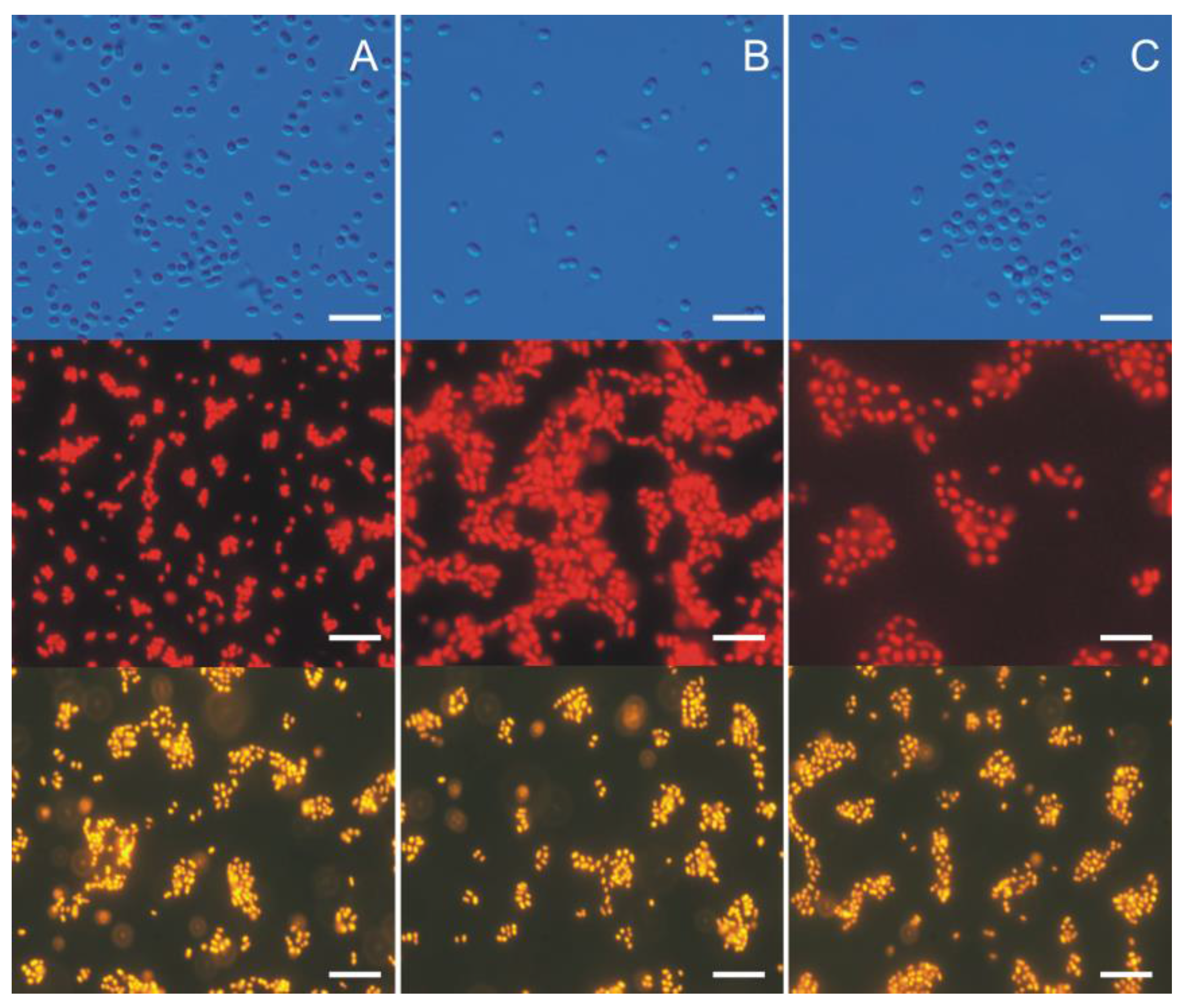
Unlike heterotrophic bacteria, picocyanobacteria, due to the presence of chlorophyll a, have the capacity for red autofluorescence. Picocyanobacteria are also well distinguishable from picoeukaryote green algae and diatoms because, when used with special filters, their fluorescence is yellow-orange (Figure 2). Therefore, research in fluorescence microscopy enable the initial distinction of eukaryotic and prokaryotic organisms, although the precise taxonomic analysis of picoplanktonic cyanobacteria requires a different approach.
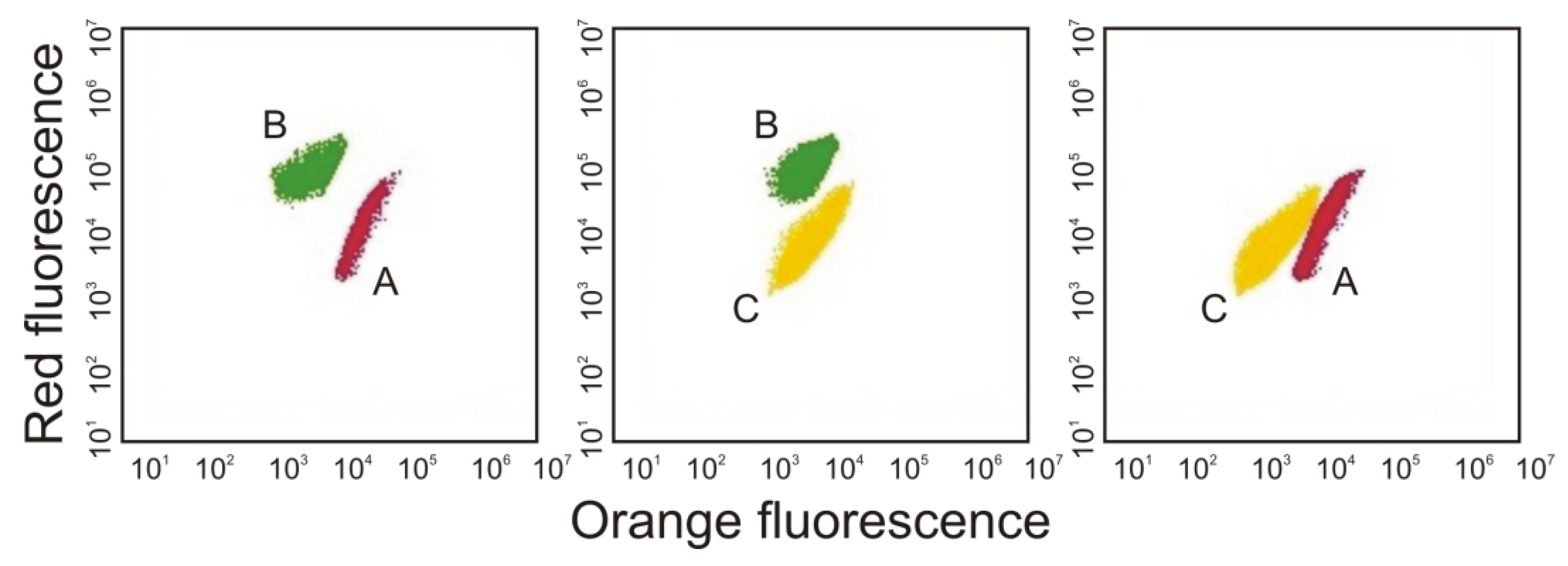
Paz-Yepes et al. [45] also noted that it is not possible to distinguish picocyanobacterial strains from one another by light or epifluorescence microscopy techniques. Therefore, they characterized different Synechococcus sp. strains in co-cultures using the relative abundance of the rpoC1 marker gene and specific primers and conditions for each strain. However, it is also possible to discriminated different picocyanobacterial strains using a flow cytometer on the basis of their pigment fluorescence [38,46] (Figure 3). Cells rich in PE emitted orange light (550–620 nm) when excited by the green laser, whereas cells rich in PC emitted far red light (>670 nm) when excited by the red laser [38]. Moreover, the flow cytometer distinguished between picocyanobacteria and larger phytoplankton by their size (using side scattering).

4. Blooms of Picocyanobacteria

Over the past few decades, the world’s coastal waters have experienced an increase in the number of harmful algal bloom events. Anderson et al. [47] described that blooms are occurring in more areas than ever before and new massive blooms are reported regularly. Thus, the issues of picocyanobacterial blooms require more attention and interest from researchers.
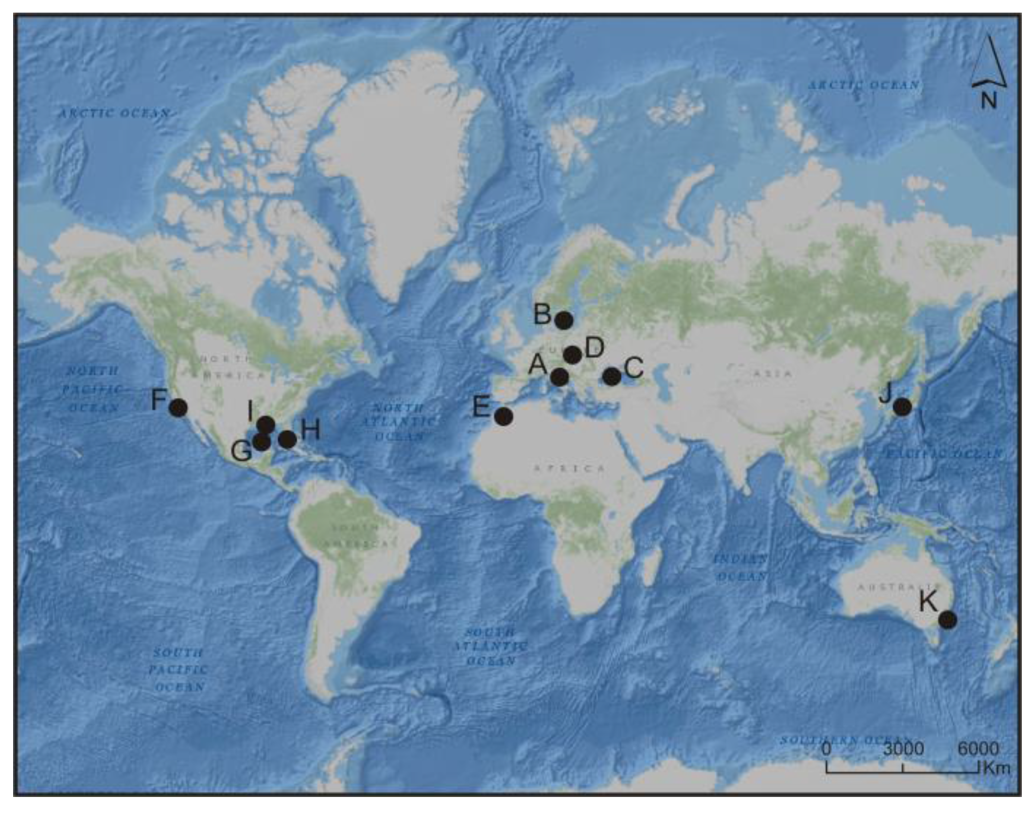
Picocyanobacteria are often described as a non-blooming group (e.g., [48]). However, it is becoming increasingly evident that picocyanobacteria are significant contributors to massive blooms in tropical and subtropical coastal areas and even appear in temperate waters. Significant blooms of picocyanobacteria have been recorded in the Mediterranean Sea [13,15,49], the Baltic Sea [50], the Black Sea [51], Hungarian lakes [52], ponds of Morocco [53,54], San Francisco Bay [55], Gulf of Mexico [56], Florida Bay and Pensacola Bay [30,57,58], the Seto Inland Sea [59] and Gippsland Lakes (Australia) [18] (Figure 4).
The most sufficiently analysed and described bloom of picocyanobacteria occurred in the northern part of the Mediterranean Sea [13,15,49]. In a few research papers, authors described local ecosystem disturbances caused by the mass appearance of picoplankton. Sorokin and Zakuskina [49] showed that the Adriatic coast has been experiencing a super-dense and long-term bloom of picocyanobacteria. The density of the picocyanobacterial bloom varied from 8 to 35 × 106 cells mL−1 and picocyanobacterial fraction of the total phytoplankton biomass was 98% from spring till early summer and 92% in autumn. The eutrophication phenomenon in the coastal ecosystem of Comacchio (Mediterranean Sea) for the first time resulted in the bloom of picocyanobacteria in 1985. During this period, an extremely dense biomass of picoplanktonic cyanobacteria and its negative effects on the environment was a new, unprecedented phenomenon in Europe. Persistent blooming of picocyanobacteria resulted in typical hypereutrophication effects such as: drastic increase in turbidity resulting in death of the benthic flora due to light deficiency; accumulation of organic matter and total phosphorus in the water column and in the sediment; anaerobic conditions in the bottom layer as well as sulphide accumulation in the sediment. Previously, the bottom of the lagoon was covered with numerous species of macrophytes from the genera Valonia, Lamprothamnium, Chaetomorpha and Ruppia. During blooms, benthic vegetation almost completely disappeared and was replaced by microbial mats. The negative impact on the ecosystem has been also enhanced by the toxic effects on animals, which are important links in the pelagic trophic chain. The bloom was accompanied by drastic changes in the benthic communities and the share of filtering fauna in the whole ecosystem metabolism was less than 2%. Moreover, the bloom in the Comacchio lagoons was formed mainly by picocyanobacteria loosely suspended in the mucus, which made the filter clams and the copepods unable to feed on them. The colonies of cyanobacteria protect themselves against their own ingestion [60]. Furthermore, Cyanocystis sp. and Coelosphaerium sp., observed in the Comacchio lagoons, belong to toxic organisms [61]. Therefore, the significant reduction in the number of organisms that typically feed on picoplankton that was observed might be caused by their toxins. However, the toxicity of the bloom-forming picocyanobacteria from the Comacchio lagoons is unknown and this is a significant issue with regard to management and public health problems. In addition, these extremely dense blooms of picoplanktonic cyanobacteria dominated the Comacchio ecosystem for many years without showing any seasonal or long-term changes [49]. Furthermore, Sorokin et al. [13] and Sorokin and Dallocchio [15] recorded picocyanobacterial bloom (5–20 × 106 cells mL−1) in the Venice lagoon (Mediterranean Sea) in the summer period. Sorokin et al. [13] examined that the share of picocyanobacteria of the total phytoplankton varied in the Venice lagoon in July–September from 60 to 98%. Authors noted that the populations of micro- and mesozooplankton were found to be inhibited in areas of intensive picocyanobacterial bloom. Additionally, a significant mortality of key species for the local fishery was recorded. In this study authors clearly demonstrated the harmful effect of the bloom of picoplanktonic organisms that has appeared in several Adriatic lagoons. It was also suggested that the blooms of picocyanobacteria in lagoons appear to be far more disastrous for their resources and environmental state than even toxic dinoflagellate blooms. Moreover, the bloom displays contagious features and shows a tendency to become persistent.
Previous investigations also confirmed that in the temperate zone, picocyanobacterial blooms were observed in summer while picoeukaryotes blooms occurred in winter. Kuosa [50] noted that picoeukaryotes in the Baltic Sea were abundant during the whole year (103–104 cells mL−1), while picocyanobacterial blooms occurred only during summer period (105–106 cells mL−1). In the Black Sea, surface blooms of Synechococcus (105 cells mL−1) were also reported [51]. Water temperatures of 5–10 °C, resulted in large winter picoplankton blooms also in the Büdös-szék (108 × 106 cells mL−1), Kelemen-szék (50 × 106 cells mL−1) and Zab-szék (47 × 106 cells mL−1) lakes (Hungary) [52]. The authors explained that the low temperature and low light intensity in winter provide a competitive advantage to picoeukaryotes, while higher temperatures and higher light intensity are more favourable for picocyanobacteria.
Furthermore, picoplanktonic organisms are able to create harmful blooms in tropical and sub-tropical waters. The occurrence of a summer Synechocystis sp. bloom in the wastewater treatment plants of Marrakech was studied by Mezrioui and Oudra [53] and Oudra et al. [54]. It was suggested that the organisms responsible for this phenomenon could be transferred by migratory birds from North Africa. A similar phenomenon has been observed in marine ecosystems. Ning et al. [55] showed that picocyanobacterium Synechococcus sp. was a persistent component of phytoplankton in all the estuarine habitats in the San Francisco Bay with peak abundance during the spring bloom (5.2 × 10⁵ cells mL−1). This result indicated increasing importance of picocyanobacteria along the gradient of decreasing nutrient concentrations from estuaries to the open ocean. In addition, Wawrik and Paul [56] have clearly demonstrated the importance of nutrient inputs from the Mississippi River in stimulating Synechococcus blooms in the Gulf of Mexico. The massive blooms of Synechococcus sp. (10 × 106 cells mL−1) were also observed by Philips et al. [57] and Gardner and McCarthy [58] in the north-central region of Florida Bay. Murrell and Lores [30] noted that the Synechococcus sp. bloom occurred in the Pensacola Bay during summer, exceeding 3 × 10⁶ cells mL−1, and was strongly correlated with temperature. Moreover, the authors noted that the increase of copepods Oithona sp. coincided with an increase of picophytoplankton. Similarly, Nakamura et al. [59] noted that following the picoplankton bloom (3–6 × 10⁵ cells mL−1), the abundance and biomass of appendicularia Oikupleuradioica in the Seto Inland Sea increased rapidly by ingesting picoplankton populations. Thus, the trophic implications of picocyanobacterial dominance in tropical and sub-tropical waters need to be more precisely assessed. Moreover, a massive and persistent bloom of picocyanobacterium Synechococcus sp., appeared in Gippsland Lakes (Australia) [18]. Author described that it is likely that high temperature and relatively high salinity provide ideal conditions for initiation of the picocyanobacterial bloom.
Many of these observations confirmed the persistence of picocyanobacterial blooms. Sorokin and Zakuskina [49] suggested that the main reason for this phenomenon was the lack of control of the populations of picoplankton organisms by consumers, due to the collapse of filter fauna and the potential mixotrophy of picoplanktonic cyanobacteria. Furthermore, the dominance of picocyanobacteria may be attributable to several of the unique physicochemical characteristics of these organisms, including its small size, cyanobacterial metabolism, euryhaline character, buoyancy and tolerance to high light intensity [57]. It is also possible that picocyanobacteria are achieving a competitive advantage over other organisms due to their wide potential to adapt to changing environmental conditions (e.g., [3,18,22]) and their allelopathic activity (e.g., [28,29,62,63]). The results of previous study confirmed the dangerous character of the picocyanobacterial bloom in aquatic environment. Moreover, described blooms tend to spread and remain for a long time and cause drastic, adverse changes in aquatic ecosystems. Therefore, more efforts have to be done to investigate in depth the autecology and taxonomy of bloom-forming picocyanobacteria and their effect on surrounding ecosystems.

5. Picocyanobacterial Secondary Metabolites

Picocyanobacteria have been rarely studied with respect to their potential as producers of bioactive compounds because most of previous works have assumed a lack of toxin production by these cyanobacteria. However, picoplanktonic cyanobacteria belong to the organisms that produce wide range of secondary metabolites (Table 1). These organisms are also capable of secreting compounds that may be bioaccumulated or biomagnified (e.g., [64,65]). Consequently, their harmful effects on the environment may be related to further links in the trophic chain and also affect humans [66,67,68].
The microcystins (MCs) are cyclic heptapeptides and are by far the most studied of the cyanobacterial toxins [67]. The toxicity of picoplanktonic cyanobacteria Synechocystis sp. and Synechococcus sp. was described for the first time by Lincoln and Carmichael [69] and Mitsui et al. [70]. Almost a decade later, picocyanobacterial toxicity was also confirmed by Domingos et al. [71] and Bláha and Maršálek [72]. Domingos et al. [71] noted that six strains of picocyanobacteria from northern Brazil are capable of producing toxic MCs. Four of these strains have been identified as colonial Aphanocapsa cumulus. The other two strains formed loosely distributed cells making their classification impossible. Domingos et al. [71] examined that concentrations of picocyanobacterial MCs were very low ranging from 0.08 to 3.7 µg g−1 dry weight. Additionally, HPLC analysis of three picocyanobacteria—Synechococcus nidulans, Cyanobium rubescens and Cyanobacterium cedrorum—indicated that they produce MCs [72]. Later studies have also confirmed the release of MC-LR and MC-YR (variants in which the two variable amino acids are leucine-L and argenine-R and tyrosine-Y and argenine-R, respectively) by the Synechococcus sp. strain (SS-1) isolated from the Salton Sea, the largest inland body of water in California [73]. Other studies also have shown that two strains of Synechococcus sp. (63a-1 and 63a-3) isolated from Florida Keys (Atlantic Ocean) are capable of producing small amounts of MC-LR (0.27 μg g−1 and 0.08 μg g−1, respectively) [74]. In turn, Martins et al. [75] screened the picocyanobacterial strains for microcystins by ELISA and examined that Synechococcus sp. and Synechocystis sp. strains produced MCs in small quantities. Subsequent results showed that ELISA tests specific for hepatotoxic MCs gave positive results for two strains of picocyanobacteria, Synechococcus sp. (CENA108) and Merismopedia sp. (CENA106) [76]. Detection of MCs from two Synechocystis sp. strains (Syn-WTP93 and Syn-WTP97) isolated from freshwater reservoirs in Morocco was also described by Oudra et al. [77]. The concentrations of MCs for these strains determined by ELISA varied from 15 to 56 μg g−1 dry weights, respectively. Vareli et al. [78] indicated that picocyanobacteria Synechococcus sp. and Synechocystis sp. were responsible for the presence of MCs in the Amvrakikos Gulf (Mediterranean Sea). Authors noted that the cyanobacterial community was found to be dominated almost exclusively by the picoplanktonic cyanobacteria Synechococcus sp. and Synechocystis sp. Marine Synechococcus sp. and Synechocystis sp. accounted for more than 50% of the total cyanobacterial biomass in the World Ocean. Thus, picocyanobacterial species should be considered also as significant MCs producers [65]. In the initial phase of blooms, hepatotoxins (MCs and nodularin, NOD) occur inside the cyanobacteria cells however, after cell lysis, the release of toxic metabolites causes the increase of their concentration in the water [79]. Therefore, Carmichael and Li [73] and Vareli et al. [65] suggested that the production of MCs by picoplanktonic cyanobacteria indicates that these toxins may be a more common occurrence in aquatic environments than previously thought. On the other hand, the secretion of NOD by picoplanktonic organisms remains still unconfirmed and the research on this subject is insufficient [10,79]. In spite of all, the possibility of producing toxins by picoplanktonic cyanobacteria is especially relevant if we take into consideration that these organisms are one of the most ubiquitous components in freshwater, brackish and marine ecosystems.
It was also demonstrated that the neurotoxic nonprotein amino acid, β-N-methylamino-l-alanine (BMAA), may be produced by all known groups of cyanobacteria [80]. Literature reports have demonstrated the synthesis and secretion of BMAA by the freshwater strain of Synechococcus sp. (PCC 6301) and the marine strain of Prochlorococcus marinus (CCMP1377). The authors examined that the content of free BMAA for Synechococcus sp. and P. marinus was 25 μg g−1 and 32 μg g−1, respectively [80]. Cianca et al. [81] also reported the detection of BMAA in five picocyanobacterial strains of Cyanobium sp. (LEGE 06068), Synechocystis salina (LEGE 06079), Synechocystis cf. salina (LEGE 06083), S. cf. salina (LEGE 07073) and Synechococcus sp. (LEGE 07074), isolated from the Portuguese estuaries of Minho, Douro and Vouga Rivers. It was demonstrated that the content of picocyanobacterial BMAA depended on the picocyanobacterial strain and extraction technique (Methanol/Acetone extraction, followed by HCl extraction, TCA extraction and HCl extraction). Authors examined that the values obtained varied from 0.04 μg g−1 for Cyanobium sp. sequential extraction with Methanol/Acetone plus HCl to 63 μg g−1 for S. salina extraction with HCl. Cox et al. [82] suggested that alternative ecological pathways for the bioaccumulation of cyanobacterial BMAA in aquatic or terrestrial ecosystems may cause increasing concentrations of toxic compounds up the food chain. Cyanobacterial BMAA has been also associated with certain forms of progressive neurodegenerative human diseases [81]. Therefore, Cox et al. [80] suggested that because of the global importance of picocyanobacterial blooms, a broader analysis of the production of BMAA in aquatic ecosystems is strongly needed.
Recent investigations suggest that picocyanobacterium Synechococcus sp. synthesize 2-methylisoborneol (MIB) and geosmin (1,2,7,7-tetramethyl-2-norbomeol) (GSM) [79,83,84]. It was also demonstrated that MIB is produced and secreted during cyanobacterial cell cycle, while GSM is released only after cells’ death. The toxic effects of these compounds to other organisms have not yet been demonstrated but they influence the quality of water and organisms living there [10]. Moreover, it has been suggested that picocyanobacteria might not only produce these compounds but also take part in their transfer to higher trophic levels [84].
For many organisms, lipopolysaccharides (LPS) are the first defence against unfavourable factors [85]. Schmidt et al. [86,87], examined that picocyanobacteria Synechococcus sp. and Synechocystis sp. contained LPS in their cell wall. Authors found that the LPS of eight strains of Synechococcus sp. as well as four strains of Synechocystis sp. contained fucose, mannose, galactose, glucose and glucosamine. Additionally, LPS obtained from Synechococcus sp. exhibited little reactivity in antisera raised in rabbits against homologous cells while the LPS from Synechocystis sp. showed no specific activity. Moreover, Snyder et al. [85] demonstrated that two strains of marine Synechococcus sp. (WH8102 and CC9311) had a very simplified structure of LPS which showed lack of limulus amoebocyte lysate gelation activity. Authors suggested that the highly simplified nature of picocyanobacterial LPS may cause their adaptation to the relatively higher salt levels in marine environments. On the other hand, liposacharids have strongly allergic and irritating effects and also cause decreased activity of glutathione S-transferases, which participate in the detoxification of xenobiotics [88]. However, a harmful or allelopathic effect of picocyanobacterial LPS has not yet been investigated.
Among unique picocyanobacterial bioactive compounds that may influence various organisms are a thioic O-acid ester-containing sulfolipid (thionsulfolipid). Thionsulfolipid was isolated from cells of picocyanobacterium Synechococcus sp. [89]. The authors clearly demonstrated that this thionsulfolipid was toxic to fish (Tanichthys albonubes) and caused growth inhibition of human lymphoma cells at a concentration of 200 μg mL−1. Kunimitsu et al. [89] noted that picocyanobacterial thionsulfolipid has not been found in any other photosynthetic organisms. Additionally, Liu et al. [90] discovered a new stereoisomer of an araguspongine/xestospongin alkaloid—named araguspongine M—which has been isolated from the marine sponge Neopetrosia exigua (formerly Xestospongia exigua), collected in Palau. Authors noted that this compound may be produced by an endosymbiotic Synechococcus-like cyanobacterium. Moreover, structurally related to schizokinen, a citrate-derived hydroxamate siderophore, the synechobactins A–C were isolated from the marine cyanobacterium Synechococcus sp. strain (PCC 7002), grown under iron-limiting conditions [91]. Synechobactins A–C differ among themselves in the identity of the fatty acid residue as dodecanoic acid, decanoic acid, or octanoic acid, respectively. Another approach is genetic modification of Synechocystis sp. strain PCC6803 to produce and secrete fatty acids [92]. However, no harmful effect of these picocyanobacterial compounds has been examined. Therefore, further research should be done to define the distribution of secondary metabolites produced and released by picoplanktonic cyanobacteria and to determine the possible harmful effects of these compounds on other organisms.

6. Allelopathic Activity of Picocyanobacteria and Their Impact on Aquatic Environment

Allelopathy is considered as one of the factors promoting and maintaining the massive cyanobacterial and algal blooms in freshwater, brackish and marine ecosystems around the world [93,94,95,96]. Therefore, the number of reports about the allelopathic effects of different species of cyanobacteria and microalgae has been steadily increasing (e.g., [95,96,97,98,99]). However, only little information on picocyanobacterial allelopathy or their harmful effects on other organisms has been described (Table 2).
The first observation of allelopathic interactions between picocyanobacteria Synechococcus sp. was noted by Paz-Yepes et al. [45]. In this work authors used a liquid and plate assays to determine whether these interactions occur between mentioned marine picocyanobacterial strains: CC9311, WH8102 and CC9605. It was found that Synechococcus sp. CC9605 always dominated when co-cultured with CC9311 or WH8102 in liquid medium. These effects were also seen in solid medium. When a spot of CC9605 was plated on an existing lawn of CC9311 or WH8102, a zone of clearing developed, which was 5-fold larger in the case of WH8102. Surprisingly, no evidences were found of allelopathic effects of these two strains against Synechococcus sp. CC9605. Additionally, Synechococcus sp. CC9311 dominated the co-culture when growing with Synechococcus sp. WH8102, while no reciprocal effect was observed. Moreover, Paz-Yepes et al. [45] suggested that a Microcin C-like molecule is involved in the allelopathic interactions with Synechococcus strain CC9605. On the other hand, it was found that Synechococcus sp. CC9311, which does not encode an McC-like gene cluster, noticeably inhibits the growth of Synechococcus sp. WH8102, presumably by the production of a different allelochemical.
On the other hand, Śliwińska-Wilczewska et al. [28] described for the first time that the Baltic strain of Synechococcus sp. (CCBA-124)affects coexisting diatom N. perminuta negatively. These studies indicated that high light and temperature and low salinity affected the tested picocyanobacteria by increasing its allelopathic activity. Authors noted that the highest decline in diatom growth, chlorophyll a fluorescence and photosynthesis was observed after the addition of cell-free filtrate obtained from culturesgrown at 190 μmol photons m−2 s−1, 25 °C and 8 PSU which coincided with optimal Synechococcus sp. growth conditions. Moreover, these results demonstrated that variation in light intensity, water temperature and salinity should be considered when estimating the potential effects of picocyanobacterial allelopathy in aquatic environments. Additionally, Śliwińska-Wilczewska et al. [63] demonstrated that the picocyanobacterium Synechococcus sp. (CCBA-124) is capable of secreting unidentified allelopathic compounds that have a negative impact on the whole phytoplankton assemblages. Authors examined the influence of allelopathic compounds on the growth, total abundance and structure of phytoplankton community by single and multiple addition of cell-free filtrate of picocyanobacterium Synechococcus sp. In this work, it was demonstrated that the growth of the plankton community was inhibited after exposure to the compounds released by Synechococcus sp. After one week of exposure, the chlorophyll a and chlorophyll c concentration was lower in the treatment with picocyanobacterial filtrate than in the control. Moreover, this study indicated that diatoms of the genera Navicula, Chaetoceros, Amphora, Coscinodiscus, Grammatophora and Nitzschia are the most affected organisms. It was suggested that the allelopathic activity exhibited by the picocyanobacterium Synechococcus sp. is probably one of the major competitive strategies affecting some of the coexisting phytoplankton species in aquatic ecosystems.
An interesting concept in the evolutionary context is the allelopathic interaction between coexisting picoplanktonic and filamentous cyanobacteria. Śliwińska-Wilczewska et al. [29,62] showed that Baltic picocyanobacterium of the genus Synechococcus are able to produce and release unidentified compounds that have allelopathic effect on the selected filamentous cyanobacteria. In these two papers authors described the negative effects of Synechococcus sp. filtrate against Nodularia spumigena [29], Nostoc sp. and Phormidium sp. [62]. It was examined that the negative effects against cyanobacteria were amplified by repeated filtrate additions compared with single filtrate addition. Moreover, the authors showed that the addition of picocyanobacterial filtrate stimulated the growth of Aphanizomenon flos-aquae and had no allelopathic effects on Rivularia sp. [62]. These results demonstrated for the first time that picocyanobacterium Synechococcus sp. negatively and positively affected coexisting filamentous cyanobacteria however, the identification of the allelopathic compounds is necessary to better understand the molecular targets in the affected species.
In the research conducted by Martins et al. [100] aqueous extracts and organic solvent extracts of isolated marine picocyanobacteria strains Synechococcus sp. and Synechocystis sp. were tested for antimicrobial activity against a fungus, Gram-positive and Gram-negative bacteria. In addition, cytotoxic assays have been also performed using primary rat hepatocytes and HL-60 human monocytic leukaemia cells. The work showed that the picocyanobacterial strains were found to have antibiotic activity against two Gram-positive bacteria, Clavibacter michiganensis subsp. insidiosum and Cellulomonas uda, while no inhibitory effects were found against the fungus Candida albicans and Gram-negative bacteria. The results obtained in this study also showed intense effect of the extracts on monocytes and a slight apoptotic effect in the case of hepatocytes, which may indicate that the tested cyanobacteria produce unrecognized toxic substances in small amounts, or the effect of these substances is not very strong. The authors have suggested that the effects on hepatocytes may be delayed and changes in these cells may intensify only after prolonged exposure. In turn, Costa et al. [101] assessed the anticancer potential of extracts from fifteen marine picocyanobacteria strains, belonging to the picoplanktonic genera, Cyanobium, Synechocystis and Synechococcus. In this work, picocyanobacteria crude extract and fractions obtained by chromatography were tested in eight cancer cell lines, which were selected as being representative of several human tumours. Authors found that eight strains of Cyanobium sp., one strain of Synechococcus nidulans, three strains of Synechococcus sp. and three strains of Synechocystis salina were able to induce cytotoxic effects in human cancer cell lines. It was noted that the remaining 59% of strains, considered as having no cytotoxic effect however, the majority of the tested picocyanobacterial strains were capable of inducing cytotoxicity in at least one of the cell lines. Moreover, strain Synechocystis salina LEGE 06155 was one of the most cytotoxic strains with above the 90th percentile of the standardized effect on cancer cell viability, classified as a strong effect. Furthermore, Liu et al. [90] described that araguspongine M, produced by an endosymbiotic Synechococcus-like cyanobacterium, showed cytotoxicity against the human leukaemia cell line HL-60 but did not inhibit the growth of bacteria Escherichia coli, Staphylococcus aureus, Saccharomyces cerevisiae, Mucor hiemalis and Ruegeria atlantica. On the other hand, Noaman et al. [102] showed that an antimicrobial agent produced by the cyanobacterium Synechococcus leopoliensis was active against the Gram-positive bacterium Staphylococcus aureus. Authors examined that temperature 35 °C, pH 8 and 15 days of incubation in the medium BG-11 were the best for antimicrobial agent production. Leucine was the best nitrogen source for antimicrobial activity. It was also found that glucose at a concentration of 0.6 mg L−1 was the most optimal, while mannitol and xylose were not suitable carbon sources for antimicrobial agent production of S. leopoliensis. Authors suggested that the high levels of antimicrobial activity of S. leopoliensis could be attributed to the chemical nature of the supplements.
In another work, Martins et al. [103] described the effects of picocyanobacterial strains Synechococcus sp. and Synechocystis sp. isolated from the marine coast of Portugal on selected marine invertebrates. In this work, crude and partially purified extracts of the picocyanobacterial strains were tested for acute toxicity in nauplii of the brine shrimp Artemia salina, in the rotifer Brachionus plicatillis and in embryos of the sea urchin Paracentrotus lividus and the mussel Mytilus galloprovincialis. According to the results obtained in the assay using A. salina, both Synechococcus sp. and Synechocystis sp. strains displayed a negative effect on the survival of the nauplii, while the crude extracts of selected strains revealed an evident toxic effect by causing 100% mortality after 24 h of exposure. On the other hand, no significant toxic effects were registered against the rotifer B. plicatillis. Moreover, the toxic effect of the different extracts of the picocyanobacterial strains was evident for the embryos of the sea urchin P. lividus and the mussel M. galloprovincialis, with toxic effects resulting in an inhibition of embryogenesis or development of smaller larvae. To the mussel embryos, the effects of picocyanobacterial extracts resulted in a complete inhibition of embryogenesis. The results revealed the ability of marine Synechococcus sp. and Synechocystis sp. crude and partially purified extracts to be toxigenic to early life stages of marine invertebrates. However, the compounds responsible for this action have not been identified [103]. Other studies have also shown that picocyanobacteria Cyanobium sp. (LEGE 06008, 06011 and 06015) and Synechococcus sp. (LEGE 06005) isolated from rocky beaches along the Atlantic Portuguese central coast caused acute toxicity in nauplii of the brine shrimp Artemia salina [104]. In this work, results concerning the methanolic and the aqueous extract were more pronounced for most of the picocyanobacterial strains, with Synechococcus sp. LEGE 06005 and Cyanobium sp. LEGE 06015 reaching 100% nauplii mortality with the aqueous extract. Authors emphasized that the strongest toxic effect was noted with aqueous solutions, suggesting that the toxins produced by the studied organisms dissolve in water [104]. Moreover, Costa et al. [105] examined the toxicological potential of five marine picocyanobacterial Cyanobium sp. strains (LEGE 06098, 06134, 06139, 07175, 07186) isolated from the Portuguese coast using different biological models. A crude extract and three fractions reflecting a preliminary segregation of lipophilic metabolites were tested for picocyanobacterial toxicity with the microalga Nannochloropsis sp., the bacteria Pseudomonas sp., the brine shrimp Artemia salina and fertilized eggs of the sea urchin Paracentrotus lividus. Authors described that Cyanobium sp. strains inhibited Nannochloropsis sp. and Pseudomonas sp. growth and induced a decrease in P. lividus larvae length. On the other hand, no significant apparent adverse effects were noted against Artemia salina. The results obtained indicated that Cyanobium genus may serve as a potential source of interesting bioactive compounds and emphasize the importance of studying the smallest fractions of marine cyanobacteria.
Recent work suggests that picocyanobacteria of the genus Synechococcus may also contribute to behavioural changes and locomotor disorders in vertebrates. In a paper published by Hamilton et al. [106], the effect of two strains of Synechococcus sp. (CC9311 and CC9902) on the fish of the species Embiotoca jacksoni was analysed. Laboratory experiments have shown that organisms exposed to picocyanobacteria are characterized by reduced mobility, moving much slower and spending more time motionless. In addition, the examined fish preferred dark zones of experimental tanks, which may be caused by stress and result from seeking refuge from the negative impact of Synechococcus sp. It was suggested that such changes may be the result of secondary metabolites that are absorbed through the gills and then cross the blood-brain barrier, disturbing the functioning of the neurological system. The experiments also showed that the return to clean water tanks resulted in gradual disappearance of negative effects of exposure to the direct contact with picocyanobacterium Synechococcus sp. This may mean that the compounds produced by Synechococcus sp. reduce the condition of vertebrates and cause behavioural disorders but do not lead to permanent changes in organisms. As a result, it can be assumed that the large quantities of Synechococcus sp. may be associated with negative, although relatively brief, transient effects on the aquatic environment. It was also found that picocyanobacterial strains of the genus Cyanobacterium, Synechococcus and Synechocystis isolated from rocky beaches along the Portuguese coast are able to produce unrecognized, toxic substances, as confirmed by studies in mice [75]. Authors demonstrated that the toxicity to mice was observed after injection of centrifuged and non-centrifuged picocyanobacterial extracts. Experiments reported a reduced rate of respiration and imbalance in those animals affected by the picocyanobacterial compounds. Furthermore, neurotoxic symptoms and effects in liver, kidney, small intestine and lungs was also examined [75]. All the results obtained indicated that the studied picocyanobacteria are a promising source of novel compounds with allelopathic activity.
Despite the ecological importance of picoplanktonic cyanobacteria, very little is known about their allelopathic effects on other organisms. Picocyanobacteria are known to produce a variety of bioactive compounds, whose harmfulness to animals and humans has been clearly demonstrated. At the same time the functional role of these compounds, particularly in terms of the physiology and ecology of the cyanobacteria that produce them, remains largely unknown. Production and release of allelopathic compounds with different properties can give producing species a competitive advantage and build their effective strategy. Furthermore, secreted allelopathic compounds by picocyanobacteria may be responsible for the natural selection of organisms and ecological succession. The results obtained indicated that picocyanobacteria may serve as a potential source of interesting bioactive compounds, which mode of action on target organisms required detailed investigation. Furthermore, the increasing occurrence of picocyanobacteria in large densities at aquatic habitats resulting from eutrophication and global warming poses a serious threat to humans and ecosystems. Clearly there is a need to increase our knowledge about allelochemicals production by picocyanobacteria strains, in order to prevent possible adverse effects of its occurrence.

7. Conclusions

Picocyanobacteria are common in freshwater, brackish and marine ecosystems throughout the world and play an important role in the functioning of aquatic ecosystems. However, despite their ubiquity, picocyanobacteria are still a group of poorly understood organisms. So far, only a few reports have been published discussing the secretion of toxins and other allelopathic compounds as well as creating harmful blooms by picoplanktonic cyanobacteria. Cyanobacterial blooms are a major and growing problem for freshwater and marine ecosystems worldwide that increasingly concerns public health. These toxins are collectively responsible for human fatalities, as well as continued and widespread poisoning of wild and domestic animals. Picocyanobacterial blooms may also cause the death of a large part of both fauna and benthic flora, as well as economic losses. In addition, some picocyanobacteria were capable of secondary metabolites production which may also be a source of harmful bioactive compounds. A dense bloom of picocyanobacteria is a new phenomenon, which still needs further investigation. While the interactive effects of climate change on harmful picocyanobacterial blooms are complex, much of the current knowledge suggests that these processes are likely to enhance the magnitude and frequency of these events. Therefore, it is essential to carefully examine the role of these small organisms in aquatic ecosystems.

Acknowledgments

We thank Sabina Jodłowska for execution photographic documentations of Synechococcus sp. strains on electron microscope. This study was supported by BMN grants, Poland, No. 538-G245-B568-17.

Author Contributions

Sylwia Śliwińska-Wilczewska was involved in all the aspects of this study and wrote this manuscript. Jakub Maculewicz conceived and designed the study, collected the literature and analysed the data. Aldo Barreiro Felpeto and Adam Latała made an important contribution to the revision of the manuscript. All the authors were involved in the preparation of this manuscript.

Conflicts of Interest

The authors declare no conflict of interest.

References

  1. Callieri, C. Single cells and microcolonies of freshwater picocyanobacteria: A common ecology. J. Limnol. 2010, 69, 257–277. [Google Scholar] [CrossRef]
  2. Mazur-Marzec, H.; Sutryk, K.; Kobos, J.; Hebel, A.; Hohlfeld, N.; Błaszczyk, A.; Jasser, I. Occurrence of cyanobacteria and cyanotoxin in the Southern Baltic Proper. Filamentous cyanobacteria versus single-celled picocyanobacteria. Hydrobiologia 2013, 701, 235–252. [Google Scholar] [CrossRef]
  3. Worden, A.Z.; Wilken, S. A plankton bloom shifts as the ocean warms. Science 2016, 354, 287–288. [Google Scholar] [CrossRef] [PubMed]
  4. Flombaum, P.; Gallegos, J.L.; Gordillo, R.A.; Rincon, J.; Zabala, L.L.; Jiao, N.; Karl, D.M.; Li, W.K.; Lomas, M.W.; Veneziano, D. Present and future global distributions of the marine Cyanobacteria Prochlorococcus and Synechococcus. Proc. Natl. Acad. Sci. USA 2013, 110, 9824–9829. [Google Scholar] [CrossRef] [PubMed]
  5. Stockner, J.G. Phototrophic picoplankton: An overview from marine and freshwater ecosystems. Limnol. Oceanogr. 1988, 33, 765–775. [Google Scholar] [CrossRef]
  6. Suttle, C.A.; Harrison, P.J. Phosphate uptake rates of phytoplankton assemblages grown at different dilution rates in semi-continuous culture. Can. J. Fish Aquat. Sci. 1986, 43, 1474–1481. [Google Scholar] [CrossRef]
  7. Chow, T.J.; Tabita, F.R. Reciprocal light-dark transcriptional control of nif and rbc expression and light-dependent posttranslational control of nitrogenase activity in Synechococcus sp. strain RF-1. J. Bacteriol. 1994, 176, 6281–6285. [Google Scholar] [CrossRef] [PubMed]
  8. Richardson, T.L.; Jackson, G.A. Small phytoplankton and carbon export from the surface ocean. Science 2007, 315, 838–840. [Google Scholar] [CrossRef] [PubMed]
  9. Callieri, C.; Stockner, J.G. Freshwater autotrophic picoplankton: A review. J. Limnol. 2002, 61, 1–14. [Google Scholar] [CrossRef]
  10. Jasser, I.; Callieri, C. Picocyanobacteria. In Handbook on Cyanobacterial Monitoring and Cyanotoxin Analysis; Meriluoto, J., Spoof, L., Codd, G.A., Eds.; John Wiley & Sons, Ltd.: Hoboken, NJ, USA, 2017; pp. 19–27. [Google Scholar]
  11. Jyothibabu, R.; Mohan, A.P.; Jagadeeesan, L.; Anjusha, A.; Muraleedharan, K.R.; Lallu, K.R.; Kiran, K.; Ullas, N. Ecology and trophic preference of picoplankton and nanoplankton in the Gulf of Mannar and the Palk Bay, southeast coast of India. J. Mar. Syst. 2013, 111–112, 29–44. [Google Scholar] [CrossRef]
  12. Motwani, N.H.; Gorokhova, E. Mesozooplankton grazing on picocyanobacteria in the Baltic Sea as inferred from molecular diet analysis. PLoS ONE 2013, 8, e79230. [Google Scholar] [CrossRef] [PubMed]
  13. Sorokin, P.Y.; Sorokin, Y.I.; Boscolo, R.; Giovanardi, O. Bloom of picocyanobacteria in the Venice lagoon during summer-autumn 2001: Ecological sequences. Hydrobiologia 2004, 523, 71–85. [Google Scholar] [CrossRef]
  14. Parvathi, A.; Zhong, X.; Ram, A.P.; Jacquet, S. Dynamics of auto- and heterotrophic picoplankton and associated viruses in Lake Geneva. Hydrol. Earth. Syst. Sci. 2014, 18, 1073–1087. [Google Scholar] [CrossRef] [Green Version]
  15. Sorokin, Y.I.; Dallocchio, F. Dynamics of phosphorus in the Venice lagoon during a picocyanobacteria bloom. J. Plankton Res. 2008, 30, 1019–1026. [Google Scholar] [CrossRef]
  16. Sánchez-Baracaldo, P.; Handley, B.A.; Hayes, P.K. Picocyanobacterial community structure of freshwater lakes and the Baltic Sea revealed by phylogenetic analyses and clade-specificquantitative PCR. Microbiology 2008, 154, 3347–3357. [Google Scholar] [CrossRef] [PubMed]
  17. Dutkiewicz, S.; Morris, J.J.; Follows, M.J.; Scott, J.; Levitan, O.; Dyhrman, S.T.; Berman-Frank, I. Impact of ocean acidification on the structure of future phytoplankton communities. Nat. Clim. Chang. 2015, 5, 1002–1006. [Google Scholar] [CrossRef]
  18. Beardall, J. Blooms of Synechococcus: An Analysis of the Problem Worldwide and Possible Causative Factors in Relation to Nuisance Blooms in the Gippsland Lakes; Monash University: Clayton, VIC, Australia, 2008; pp. 1–8. [Google Scholar]
  19. O’Neil, J.M.; Davis, T.W.; Burford, M.A.; Gobler, C.J. The rise of harmful cyanobacteria blooms: The potential roles of eutrophication and climate change. Harmful Algae 2012, 14, 313–334. [Google Scholar] [CrossRef]
  20. Brutemark, A.; Vandelannoote, A.; Engström-Öst, J.; Suikkanen, S. A less saline Baltic Sea promotes cyanobacterial growth, hampers intracellular microcystin production and leads to strain-specific differences in allelopathy. PLoS ONE 2015, 10, e0128904. [Google Scholar] [CrossRef] [PubMed]
  21. Paerl, H.W.; Huisman, J. Blooms like it hot. Science 2008, 320, 57–58. [Google Scholar] [CrossRef] [PubMed]
  22. Jodłowska, S.; Śliwińska, S. Effects of light intensity and temperature on the photosynthetic irradiance response curves and chlorophyll fluorescence in three picocyanobacterial strains of Synechococcus. Photosynthetica 2014, 52, 223–232. [Google Scholar] [CrossRef]
  23. Ibelings, B.W. Changes in photosynthesis in response to combined irradiance and temperature stress in cyanobacterial surface waterblooms. J. Phycol. 1996, 32, 549–557. [Google Scholar] [CrossRef]
  24. Antia, N.J.; Cheng, J.Y. The survival of axenic cultures of marine planktonic algae from prolonged exposure to darkness at 20 °C. Phycologia 1970, 9, 179–183. [Google Scholar] [CrossRef]
  25. Antia, N.J. Effects of temperature on the darkness survival of marine microplanktonic algae. Microb. Ecol. 1976, 3, 41–54. [Google Scholar] [CrossRef] [PubMed]
  26. Kana, T.M.; Glibert, P.M. Effect of irradiances up to 2000 μE m−2 s−1 on marine Synechococcus WH7803-I. Growth, pigmentation and cell composition. Deep Sea Res. Part I 1987, 34, 479–495. [Google Scholar] [CrossRef]
  27. Stal, L.J.; Albertan, P.; Bergman, B.; Von Bröckel, K.; Gallon, J.R.; Hayes, P.K.; Walsby, A.E. BASIC: Baltic Sea cyanobacteria. An investigation of the structure and dynamics of water blooms of cyanobacteria in the Baltic Sea—Responses to a changing environment. Cont. Shelf Res. 2003, 23, 1695–1714. [Google Scholar] [CrossRef]
  28. Śliwińska-Wilczewska, S.; Pniewski, F.; Latała, A. Allelopathic activity of the picocyanobacterium Synechococcus sp. under varied light, temperature and salinity conditions. Int. Rev. Hydrobiol. 2016, 101, 69–77. [Google Scholar] [CrossRef]
  29. Śliwińska-Wilczewska, S.; Pniewski, F.; Latała, A. Allelopathic interactions between Synechococcus sp. and Nodularia spumigena under different light conditions. Allelopath. J. 2016, 37, 241–252. [Google Scholar]
  30. Murrell, M.C.; Lores, E.M. Phytoplankton and zooplankton seasonal dynamics in a subtropical estuary: Importance of cyanobacteria. J. Plankton Res. 2004, 26, 371–382. [Google Scholar] [CrossRef]
  31. Bec, B.; Husseini-Ratrema, J.; Collos, Y.; Souchu, P.; Vaquer, A. Phytoplankton seasonal dynamics in a Mediterranean coastal lagoon: Emphasis on the picoeukaryote community. J. Plankton Res. 2005, 27, 881–894. [Google Scholar] [CrossRef]
  32. Paul, V.J. Global warming and cyanobacterial harmful algal booms. In Cyanobacterial Harmful Algal Blooms: State of the Science and Research Needs; Advances in Experimental Medicine and Biology; Hudnell, K.H., Ed.; Springer: New York, NY, USA, 2008; pp. 239–257. [Google Scholar]
  33. Paerl, H.W.; Huisman, J. Climate change: A catalyst for global expansion of harmful cyanobacterial blooms. Eviron. Microb. Rep. 2009, 1, 27–37. [Google Scholar] [CrossRef] [PubMed]
  34. Houghton, J.T.; Ding, Y.; Griggs, D.J.; Noguer, M.; Van der Lin-den, P.J.; Dai, X.; Maskell, K.; Johnson, C.A. Climate Change 2001: The Scientific Basis; Cambridge University Press: Cambridge, UK, 2001. [Google Scholar]
  35. Moore, L. Comparative physiology of Synechococcus and Prochlorococcus: Influence of light and temperature on growth, pigments, fluorescence and absorptive properties. Mar. Ecol. Prog. Ser. 1995, 116, 259–275. [Google Scholar] [CrossRef]
  36. Gazeau, F.; Sallon, A.; Maugendre, L.; Louis, J.; Dellisanti, W.; Gaubert, M.; Lejeune, P.; Gobert, S.; Borges, A.V.; Harlay, J.; et al. First mesocosm experiments to study the impacts of ocean acidification on plankton communities in the NW Mediterranean Sea (MedSeA project). Estuar. Coast. Shelf Sci. 2017, 186, 11–29. [Google Scholar] [CrossRef]
  37. Ahlgren, N.A.; Rocap, G. Diversity and distribution of marine Synechococcus: Multiple gene phylogenies for consensus classification and development of qPCR assays for sensitive measurement of clades in the ocean. Front. Microbiol. 2012, 3, 213. [Google Scholar] [CrossRef] [PubMed]
  38. Stomp, M.; Huisman, J.; Vörös, L.; Pick, F.R.; Laamanen, M.; Haverkamp, T.; Stal, L.J. Colourful coexistence of red and green picocyanobacteria in lakes and seas. Ecol. Lett. 2007, 10, 290–298. [Google Scholar] [CrossRef] [PubMed]
  39. Haverkamp, T.; Acinas, S.G.; Doeleman, M.; Stomp, M.; Huisman, J.; Stal, L.J. Diversity and phylogeny of Baltic Sea picocyanobacteria inferred from their ITS and phycobiliprotein operons. Environ. Microbiol. 2008, 10, 174–188. [Google Scholar] [CrossRef] [PubMed]
  40. Haverkamp, T.H.; Schouten, D.; Doeleman, M.; Wollenzien, U.; Huisman, J.; Stal, L.J. Colorful microdiversity of Synechococcus strains (picocyanobacteria) isolated from the Baltic Sea. ISME J. 2009, 3, 397–408. [Google Scholar] [CrossRef] [PubMed]
  41. Everroad, R.C.; Wood, A.M. Comparative molecular evolution of newly discovered picocyanobacterial strains reveals a phylogenetically informative variable region of beta-phycoerythrin. J. Phycol. 2006, 42, 1300–1311. [Google Scholar] [CrossRef]
  42. Vörös, L.; Callieri, C.; Katalin, V.; Bertoni, R. Freshwater picocyanobacteria along a trophic gradient and light quality range. In Phytoplankton and Trophic Gradients; Springer: Dordrecht, The Netherlands, 1998; pp. 117–125. [Google Scholar]
  43. Rippka, R. Isolation and purification of cyanobacteria. Methods Enzymol. 1988, 167, 3–27. [Google Scholar] [PubMed]
  44. Larsson, J.; Celepli, N.; Ininbergs, K.; Dupont, C.L.; Yooseph, S.; Bergman, B.; Ekman, M. Picocyanobacteria containing a novel pigment gene cluster dominate the brackish water Baltic Sea. ISME J. 2014, 8, 1892–1903. [Google Scholar] [CrossRef] [PubMed]
  45. Paz-Yepes, J.; Brahamsha, B.; Palenik, B. Role of a Microcin-C-like biosynthetic gene cluster in allelopathic interactions in marine Synechococcus. Proc. Natl. Acad. Sci. USA 2013, 110, 12030–12035. [Google Scholar] [CrossRef] [PubMed]
  46. Stomp, M.; Huisman, J.; de Jongh, F.; Veraart, A.J.; Gerla, D.; Rijkeboer, M.; Ibelings, B.W.; Wollenzien, U.I.A.; Stal, L.J. Adaptive divergence in pigment composition promotes phytoplankton biodiversity. Nature 2004, 432, 104–107. [Google Scholar] [CrossRef] [PubMed]
  47. Anderson, D.M.; Cembella, A.D.; Hallegraeff, G.M. Progress in understanding harmful algal blooms: Paradigm shifts and new technologies for research, monitoring and management. Annu. Rev. Mar. Sci. 2012, 4, 143–176. [Google Scholar] [CrossRef] [PubMed] [Green Version]
  48. Stockner, J.; Callieri, C.; Cronberg, G. Picoplankton and other non-bloom forming cyanobacteria in lakes. In The Ecology of Cyanobacteria: Their Diversity in Time and Space; Whitton, B., Potts, M., Eds.; Kluwer Academic Publishers: Dordrecht, The Netherlands, 2000; pp. 195–238. [Google Scholar]
  49. Sorokin, Y.I.; Zakuskina, O.Y. Features of the Comacchio ecosystem transformed during persistent bloom of picocyanobacteria. J. Oceanogr. 2010, 66, 373–387. [Google Scholar] [CrossRef]
  50. Kuosa, H. Picoplanktonic algae in the northern Baltic Sea: Seasonal dynamics and flagellate grazing. Mar. Ecol. Prog. Ser. 1991, 73, 269–276. [Google Scholar] [CrossRef]
  51. Uysal, Z. Pigments, size and distribution of Synechococcus spp. in the Black Sea. J. Mar. Syst. 2000, 24, 313–326. [Google Scholar] [CrossRef]
  52. Somogyi, B.; Felföldi, T.; Vanyovszki, J.; Ágyi, Á.; Márialigeti, K.; Vörös, L. Winter bloom of picoeukaryotes in Hungarian shallow turbid soda pans and the role of light and temperature. Aquat. Ecol. 2009, 43, 735–744. [Google Scholar] [CrossRef]
  53. Mezrioui, N.; Oudra, B. Dynamics of picoplankton and microplankton flora in the experimental wastewater stabilisation ponds in the arid region of Marrakech, Morocco and Cyanobacteria effect on Escherichia coli and Vibrio cholerae survival. In Wastewater Treatment with Algae; Wong, Y.S., Tam, N.F.Y., Eds.; Springer-Verlag/Landes Bioscience: New York, NY, USA, 1998; pp. 165–188. [Google Scholar]
  54. Oudra, B.; El-Andaloussi-Dadi, M.; Franca, S.; Barros, P.; Martins, R.; Oufdou, K.; Sbiyyaa, B.; Loudiki, M.; Mezrioui, N.; Vasconcelos, V. Harmful cyanobacterial toxic blooms in waste stabilisation ponds. Water Sci. Technol. 2000, 42, 179–186. [Google Scholar]
  55. Ning, X.; Cloern, J.E.; Cole, B.E. Spatial and temporal variability of picocyanobacteria Synechococcus sp. in San Francisco Bay. Limnol. Oceanogr. 2000, 45, 695–702. [Google Scholar] [CrossRef]
  56. Wawrik, B.; Paul, J.H. Phytoplankton community structure and productivity along the axis of the Mississippi River plume in oligotrophic Gulf of Mexico waters. Aquat. Microb. Ecol. 2004, 35, 185–196. [Google Scholar] [CrossRef]
  57. Phlips, E.J.; Badylak, S.; Lynch, T.C. Blooms of the picoplanktonic cyanobacterium Synechococcus in Florida Bay, a subtropical inner-shelf lagoon. Limnol. Oceanogr. 1999, 14, 1166–1175. [Google Scholar] [CrossRef]
  58. Gardner, W.S.; McCarthy, M.J. Nitrogen dynamics at the sediment–water interface in shallow, sub-tropical Florida Bay: Why denitrification efficiency may decrease with increased eutrophication. Biogeochemistry 2009, 95, 185–198. [Google Scholar] [CrossRef]
  59. Nakamura, Y.; Suzuki, K.; Suzuki, S.Y.; Hiromi, J. Production of Oikopleura dioica (Appendicularia) following a picoplankton ‘bloom’ in a eutrophic coastal area. J. Plankton Res. 1997, 19, 113–124. [Google Scholar] [CrossRef]
  60. Passoni, S.; Callieri, C. Picocyanobacteria single forms aggregated and microcolonies. Verh. Internat. Verein Limnol. 2000, 27, 1879–1883. [Google Scholar]
  61. Komárek, J. Taxonomic and species delineation of picoplanktonic cyanoprocaryotes. Algol. Stud. 1996, 83, 119–179. [Google Scholar]
  62. Śliwińska-Wilczewska, S.; Maculewicz, J.; Barreiro Felpeto, A.; Vasconcelos, V.; Latała, A. Allelopathic activity of the picocyanobacterium Synechococcus sp. on filamentous cyanobacteria. J. Exp. Mar. Biol. Ecol. 2017, 496, 16–21. [Google Scholar] [CrossRef]
  63. Śliwińska-Wilczewska, S.; Maculewicz, J.; Tuszer, J.; Dobosz, K.; Kalusa, D.; Latała, A. First record of allelopathic activity of the picocyanobacterium Synechococcus sp. on a natural plankton community. Ecohydrol. Hydrobiol. 2017, 17, 227–234. [Google Scholar] [CrossRef]
  64. Mazur-Marzec, H. Characterization of phycotoxins produced by cyanobacteria. Oceanol. Hydrobiol. Stud. 2006, 35, 85–109. [Google Scholar]
  65. Vareli, K.; Jaeger, W.; Touka, A.; Frillingos, S.; Briasoulis, E.; Sainis, I. Hepatotoxic seafood poisoning (HSP) due to microcystins: A threat from the ocean? Mar. Drugs 2013, 11, 2751–2768. [Google Scholar] [CrossRef] [PubMed]
  66. Pearson, L.; Mihali, T.; Moffitt, M.; Kellmann, R.; Neilan, B. On the chemistry, toxicology and genetics of the cyanobacterial toxins, microcystin, nodularin, saxitoxin and cylindrospermopsin. Mar. Drugs 2010, 8, 1650–1680. [Google Scholar] [CrossRef] [PubMed]
  67. Leão, P.N.; Engene, N.; Antunes, A.; Gerwick, W.H.; Vasconcelos, V. The chemical ecology of cyanobacteria. Nat. Prod. Rep. 2012, 29, 372–391. [Google Scholar] [CrossRef] [PubMed]
  68. Testai, E.; Scardala, S.; Vichi, S.; Buratti, F.M.; Funari, E. Risk to human health associated with the environmental occurrence of cyanobacterial neurotoxic alkaloids anatoxins and saxitoxins. Crit. Rev. Toxicol. 2016, 46, 1–35. [Google Scholar]
  69. Lincoln, E.P.; Carmichael, W.W. Preliminary tests of toxicity of Synechocystis sp. growth on wastewater medium. In The Water Environment: Algal Toxins and Health; Carmichael, W.W., Ed.; Plenum Press: New York, NY, USA, 1981; pp. 223–230. [Google Scholar]
  70. Mitsui, A.; Rosner, D.; Goodman, A.; Reyes-Vasquez, G.; Kusumi, T.; Kodama, T.; Nomoto, K. Hemolytic toxins in a marine cyanobacterium Synechococcus sp. In Proceedings of the International Red Tide Symposium, Takamatsu, Japan, 10–14 November 1987. [Google Scholar]
  71. Domingos, P.; Rubim, T.K.; Molica, R.J.R.; Azevedo, S.M.F.O.; Carmichael, W.W. First report of microcystin production by picoplanktonic cyanobacteria isolated from a northeast Brazilian drinking water supply. Environ. Toxicol. 1999, 14, 31–35. [Google Scholar] [CrossRef]
  72. Bláha, L.; Maršálek, B. Microcystin production and toxicity of picocyanobacteria as a risk factor for drinking water treatment plants. Algol. Stud. 1999, 92, 95–108. [Google Scholar]
  73. Carmichael, W.W.; Li, R. Cyanobacteria toxins in the Salton Sea. Saline Syst. 2006, 2, 5. [Google Scholar] [CrossRef] [PubMed]
  74. Gantar, M.; Sekar, R.; Richardson, L.L. Cyanotoxins from black band disease of corals and from other coral reef environments. Microb. Ecol. 2009, 58, 856–864. [Google Scholar] [PubMed]
  75. Martins, R.; Pereira, P.; Welker, M.; Fastner, J.; Vasconcelos, V.M. Toxicity of culturable cyanobacteria strains isolated from the Portuguese coast. Toxicon 2005, 46, 454–464. [Google Scholar] [CrossRef] [PubMed]
  76. Furtado, A.L.F.F.; do Carmo Calijuri, M.; Lorenzi, A.S.; Honda, R.Y.; Genuário, D.B.; Fiore, M.F. Morphological and molecular characterization of cyanobacteria from a Brazilian facultative wastewater stabilization pond and evaluation of microcystin production. Hydrobiologia 2009, 627, 195–209. [Google Scholar] [CrossRef]
  77. Oudra, B.; Loudiki, M.; Vasconcelos, V.; Sabour, B.; Sbiyyaa, B.; Oufdou, K.; Mezrioui, N. Detection and quantification of microcystins from cyanobacteria strains isolated from reservoirs and ponds in Morocco. Environ. Toxicol. 2002, 17, 32–39. [Google Scholar] [CrossRef] [PubMed]
  78. Vareli, K.; Zarali, E.; Zacharioudakis, G.S.A.; Vagenas, G.; Varelis, V.; Pilidis, G.; Briasoulis, E.; Sainis, I. Microcystin producing cyanobacterial communities in Amvrakikos Gulf (Mediterranean Sea, NW Greece) and toxin accumulation in mussels (Mytilus galloprovincialis). Harmful Algae 2012, 15, 109–118. [Google Scholar] [CrossRef]
  79. Jakubowska, N.; Szeląg-Wasielewska, E. Toxic Picoplanktonic Cyanobacteria-Review. Mar. Drugs 2015, 13, 1497–1518. [Google Scholar] [CrossRef] [PubMed]
  80. Cox, P.A.; Banack, S.A.; Murch, S.J.; Rasmussen, U.; Tien, G.; Bidigare, R.R.; Metcalf, J.S.; Morrison, L.F.; Codd, G.A.; Bergman, B. Diverse taxa of cyanobacteria produce β-N-methylamino-l-alanine, a neurotoxic amino acid. Proc. Natl. Acad. Sci. USA 2005, 102, 5074–5078. [Google Scholar] [PubMed]
  81. Cianca, R.C.C.; Baptista, M.S.; Lopes, V.R.; Vasconcelos, V.M. The non-protein amino acid β-N-methylamino-l-alanine in Portuguese cyanobacterial isolates. Amino Acids 2012, 42, 2473–2479. [Google Scholar]
  82. Cox, P.A.; Banack, S.A.; Murch, S.J. Biomagnification of cyanobacterial neurotoxins and neurodegenerative disease among the Chamorro people of Guam. Proc. Natl. Acad. Sci. USA 2003, 100, 13380–13383. [Google Scholar] [PubMed]
  83. Graham, J.L.; Loftin, K.A.; Ziegler, A.C.; Meyer, M.T. Guidelines for Design and Sampling for Cyanobacterial Toxin and Taste-and-Odor Studies in Lakes and Reservoirs; U.S. Geological Survey: Reston, VA, USA, 2008.
  84. Journey, C.A.; Beaulieu, K.M.; Bradley, P.M. Environmental factors that influence cyanobacteria and geosmin occurrence in reservoirs. In Current Perspectives in Contaminant Hydrology and Water Resources Sustainability; Bradley, P.M., Ed.; Tech Online: Rijeka, Croatia, 2013. [Google Scholar]
  85. Snyder, D.S.; Brahamsha, B.; Azadi, P.; Palenik, B. Structure of compositionally simple lipopolysaccharide from marine Synechococcus. J. Bacteriol. 2009, 191, 5499–5509. [Google Scholar] [PubMed]
  86. Schmidt, W.; Drews, G.; Weckesser, J.; Fromme, I. Characterization of the lipopolysaccharides from eight strains of the cyanobacterium Synechococcus. Arch. Microbiol. 1980, 127, 209–215. [Google Scholar]
  87. Schmidt, W.; Drews, G.; Weckesser, J.; Mayer, H. Lipopolysaccharides in four strains of the unicellular cyanobacterium Synechocystis. Arch. Microbiol. 1980, 127, 217–222. [Google Scholar]
  88. Best, J.H.; Pflugmacher, S.; Wiegand, C.; Eddy, F.B.; Metcalf, J.S.; Codd, G.A. Effects of enteric bacterial and cyanobacterial lipopolysaccharides and of microcystin-LR, on glutathione S-transferase activities in zebra fish (Danio rerio). Aquat. Toxicol. 2002, 60, 223–231. [Google Scholar] [PubMed]
  89. Kunimitsu, K.; Tomoharu, S.; Fujio, S.; Hiroyasu, I. Thioic O-acid ester in sulfolipid isolated from freshwater picoplankton cyanobacterium, Synechococcus sp. Biochim. Biophys. Acta 1993, 1169, 39–45. [Google Scholar]
  90. Liu, H.; Mishima, Y.; Fujiwara, T.; Nagai, H.; Kitazawa, A.; Mine, Y.; Kobayashi, H.; Yao, X.; Yamada, J.; Oda, T.; et al. Isolation of araguspongine M, a new stereoisomer of an araguspongine/xestospongin alkaloid and dopamine from the marine sponge Neopetrosia exigua collected in Palau. Mar. Drugs 2004, 2, 154–163. [Google Scholar]
  91. Ito, Y.; Butler, A. Structure of synechobactins, new siderophores of the marine cyanobacterium Synechococcus sp. PCC 7002. Limnol. Oceanogr. 2005, 50, 1918–1923. [Google Scholar]
  92. Liu, X.; Sheng, J.; Curtiss, R., III. Fatty acid production in genetically modified cyanobacteria. Proc. Natl. Acad. Sci. USA 2011, 108, 6899–6904. [Google Scholar] [CrossRef] [PubMed]
  93. Figueredo, C.C.; Giani, A.; Bird, D.F. Does allelopathy contribute to Cylindrospermopsis raciborskii (cyanobacteria) bloom occurrence and geographic expansion? J. Phycol. 2007, 43, 256–265. [Google Scholar] [CrossRef]
  94. Żak, A.; Kosakowska, A. The influence of extracellular compounds produced by selected Baltic cyanobacteria, diatoms and dinoflagellates on growth of green algae Chlorella vulgaris. Estuar. Coast. Shelf. Sci. 2015, 167, 113–118. [Google Scholar] [CrossRef]
  95. Allen, J.L.; Ten-Hage, L.; Leflaive, J. Regulation of Fatty Acid Production and Release in Benthic Algae: Could Parallel Allelopathy Be Explained with Plant Defence Theories? Microb. Ecol. 2017. [Google Scholar] [CrossRef] [PubMed]
  96. Barreiro, A.; Roy, S.; Vasconcelos, V.M. Allelopathy prevents competitive exclusion and promotes phytoplankton biodiversity. Oikos 2017. [Google Scholar] [CrossRef]
  97. Dias, F.; Antunes, J.T.; Ribeiro, T.; Azevedo, J.; Vasconcelos, V.; Leão, P.N. Cyanobacterial Allelochemicals But Not Cyanobacterial Cells Markedly Reduce Microbial Community Diversity. Front. Microbiol. 2017, 8, 1495. [Google Scholar] [CrossRef] [PubMed]
  98. Pichierri, S.; Accoroni, S.; Pezzolesi, L.; Guerrini, F.; Romagnoli, T.; Pistocchi, R.; Totti, C. Allelopathic effects of diatom filtrates on the toxic benthic dinoflagellate Ostreopsis cf. ovata. Mar. Environ. Res. 2017, 131, 116–122. [Google Scholar] [CrossRef] [PubMed]
  99. Wang, L.; Zi, J.; Xu, R.; Hilt, S.; Hou, X.; Chang, X. Allelopathic effects of Microcystis aeruginosa on green algae and a diatom: Evidence from exudates addition and co-culturing. Harmful Algae 2017, 61, 56–62. [Google Scholar] [CrossRef]
  100. Martins, R.F.; Ramos, M.F.; Herfindal, L.; Sousa, J.A.; Skærven, K.; Vasconcelos, V.M. Antimicrobial and cytotoxic assessment of marine cyanobacteria—Synechocystis and Synechococcus. Mar. Drugs 2008, 6, 1–11. [Google Scholar] [CrossRef] [PubMed]
  101. Costa, M.; Garcia, M.; Costa-Rodrigues, J.; Costa, M.S.; Ribeiro, M.J.; Fernandes, M.H.; Barros, P.; Barreiro, A.; Vasconcelos, V.; Martins, R. Exploring bioactive properties of marine cyanobacteria isolated from the Portuguese coast: High potential as a source of anticancer compounds. Mar. Drugs 2014, 12, 98–114. [Google Scholar] [CrossRef] [PubMed]
  102. Noaman, N.H.; Fattah, A.; Khaleafa, M.; Zaky, S.H. Factors affecting antimicrobial activity of Synechococcus leopoliensis. Microbiol. Res. 2004, 159, 395–402. [Google Scholar] [CrossRef] [PubMed]
  103. Martins, R.F.; Fernandez, N.; Beiras, R.; Vasconcelos, V.M. Toxicity assess­ment of crude and partially purified extracts of marine Synechocystis and Synechococcus cyanobacterial strains in marine invertebrates. Toxicon 2007, 50, 791–799. [Google Scholar] [CrossRef] [PubMed]
  104. Frazão, B.; Martins, R.; Vasconcelos, V. Are known cyanotoxins involved in the toxicity of picoplanktonic and filamentous north atlantic marine cyanobacteria? Mar. Drugs 2010, 8, 1908–1919. [Google Scholar] [CrossRef] [PubMed]
  105. Costa, M.S.; Costa, M.; Ramos, V.; Leão, P.N.; Barreiro, A.; Vasconcelos, V.; Martins, R. Picocyanobacteria from a clade of marine cyanobium revealed bioactive potential against microalgae, bacteria and marine invertebrates. Toxicol. Environ. Health Part A 2015, 78, 432–442. [Google Scholar] [CrossRef] [PubMed]
  106. Hamilton, T.J.; Paz-Yepes, J.; Morrison, R.A.; Palenik, B.; Tresguerres, M. Exposure to bloom-like concentrations of two marine Synechococcus cyanobacteria (strains CC9311 and CC9902) differentially alters fish behaviour. Conserv. Physiol. 2014, 2, 1–9. [Google Scholar] [CrossRef] [PubMed]
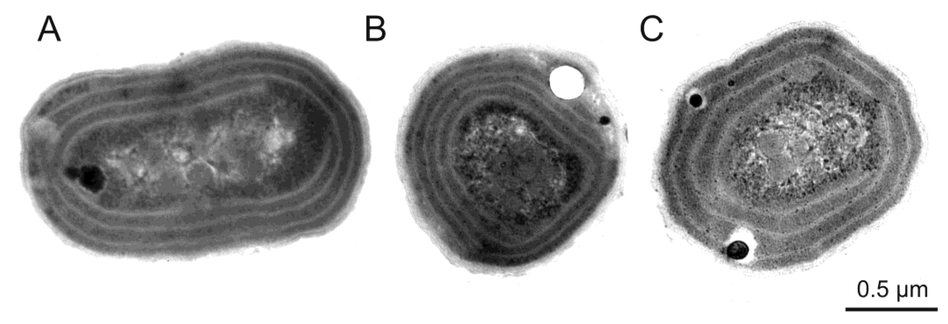
Figure 1. Ultrastructure of three Baltic Synechococcus sp. strains: rich in PE (A); rich in PC (B) and rich in PE containing high contents of the PEB (C) analysed using an electron microscope. Photographs by Śliwińska-Wilczewska.
Toxins 10 00048 g001
Figure 2. Three Baltic Synechococcus sp. strains: rich in PE (A); rich in PC (B) and rich in PE containing high contents of the PEB (C) under a light and epifluorescence microscope. Top panel depicts picocyanobacterial cells from light microscope, whilst middle and bottom panel illustrates target species under epifluorescence microscope using B-2A and G-2A (which excitation are: 450–490 nm and 510–560 nm, respectively) block filters, respectively. Bar denotes 10 μm. Photographs by Śliwińska-Wilczewska.
Toxins 10 00048 g002
Figure 3. Cytograms obtained with co-cultures with three Baltic Synechococcus sp. strains: rich in PE (A), rich in PC (B) and rich in PE containing high contents of the PEB (C)analysed using a Becton Dickinson (BD Biosciences) Accuri™ C6 Plus flow cytometer. Cytograms by Śliwińska-Wilczewska.
Toxins 10 00048 g003
Figure 4. Water reservoirs in which mass occurrence of picoplanktonic cyanobacteria was recorded: Mediterranean Sea (A), Baltic Sea (B), Black Sea (C), Hungarian lakes (D), ponds of Morocco (E), San Francisco Bay (F), Gulf of Mexico (G), Florida Bay (H), Pensacola Bay (I), Seto Inland Sea (J) and Gippsland Lakes (K).
Toxins 10 00048 g004
Table 1. Secondary metabolites produced by picocyanobacteria and their source of origin.
Species (Strain)Location/HabitatSecondary MetabolitesSource
Aphanocapsa cumulusCaruaru reservoirs (Brazil)/freshwaterMC[71]
Synechococcus nidulansUnknown/freshwaterMC[72]
Cyanobium rubescensUnknown/freshwaterMC[72]
Cyanobacterium cedrorumUnknown/freshwaterMC[72]
Synechococcus sp. (SS-1)Salton Sea (California)/marineMC[73]
Synechococcus sp. (63a-1 and 63a-3)Florida Keys (Atlantic Ocean)/marine MC[74]
Synechococcus sp. Portuguese coast (Atlantic Ocean)/marine MC[75]
Synechocystis sp.Portuguese coast (Atlantic Ocean)/marineMC[75]
Synechococcus sp. (CENA108)Cajati (Brazil)/freshwater MC[76]
Merismopedia sp. (CENA106)Cajati (Brazil)/freshwater MC[76]
Synechocystis sp. (Syn-WTP93 and Syn-WTP97)Biological wastewater treatment plant (Morocco)/freshwater MC[77]
Synechococcus sp. Amvrakikos Gulf (Mediterranean Sea)/marine MC[78]
Synechocystis sp.Amvrakikos Gulf (Mediterranean Sea)/marine MC[78]
Synechococcus sp. (PCC 6301)USA/freshwaterBMAA[80]
Prochlorococcus marinus (CCMP1377)Sargasso Sea (Atlantic Ocean)/marine BMAA[80]
Cyanobium sp. (LEGE 06068)Douro estuary/brackishBMAA[81]
Synechocystis salina (LEGE 06079)Douro estuary/brackishBMAA[81]
Synechocystis cf. salina (LEGE 06083)Douro estuary/brackishBMAA[81]
Synechocystis cf. salina (LEGE 07073)Vouga estuary/brackishBMAA[81]
Synechococcus sp. (LEGE 07074)Douro estuary/brackishBMAA[81]
Synechococcus sp.Lake Bowen and Municipal Reservoir #1 (USA)/freshwaterMIB[83,84]
Synechococcus sp.Lake Bowen and Municipal Reservoir #1 (USA)/freshwaterGSM[83,84]
Synechococcus sp. (PCC 6907, 6307, 6911, 6603, 6908, 6311, 6312, 6910)France/freshwaterLPS[86]
Synechocystis sp. (PCC 6714, 6803, 6807, 6308)France/freshwaterLPS[87]
Synechococcus sp. (WH8102 and CC9311)Carribean Sea (Atlantic Ocean) and Pacific Ocean/marineLPS[85]
Synechococcus sp. Unknownthionsulfolipid[89]
endosymbiotic Synechococcus-like cyanobacteriummarine sponge Neopetrosia exigua, Palau (Pacific Ocean)/marinearaguspongine M[90]
Synechococcus sp. (PCC 7002)USA/marinesynechobactins A–C[91]
Synechocystis sp. (PCC6803/SD100)France/freshwaterfatty acids[92]
BMAA, β-N-methylamino-l-alanine. GSM (geosmin), 1,2,7,7-tetramethyl-2-norbomeol. LPS, lipopolysaccharides. MC, microcystin. MIB, 2-methylisoborneol.
Table 2. Allelopathic activity of picocyanobacteria and their effect on target organisms. − indicates inhibiting effects, + indicates stimulating effect, 0—indicates lack of effect.
Donor Species (Strain)Target SpeciesEffect Source
Synechococcus sp. (CC9311)Synechococcus sp. (WH8102)[45]
Synechococcus sp. (WH8102)Synechococcus sp. (CC9311)0[45]
Synechococcus sp. (CC9605)Synechococcus sp. (CC9311), Synechococcus sp. (WH8102)[45]
Synechococcus sp. (CC9311), Synechococcus sp. (WH8102)Synechococcus sp. (CC9605)0[45]
Synechococcus sp. (CCBA-124)Navicula perminuta[28]
Synechococcus sp. (CCBA-124)Nodularia spumigena[29]
Synechococcus sp. (CCBA-124)Nostoc sp., Phormidium sp.[62]
Synechococcus sp. (CCBA-124)Aphanizomenon flos-aquae+[62]
Synechococcus sp. (CCBA-124)Rivularia sp.0[62]
Synechococcus sp. (CCBA-124)Navicula sp., Chaetoceros sp., Amphora sp., Coscinodiscus sp., Grammatophora sp., Nitzschia sp.[63]
Synechocystis sp. (LEANCYA 1, 5, 13, 15, 17, 20, 21) and Synechococcus sp. (LEANCYA 7, 10, 11, 16, 18, 19, 22)Candida albicans0[100]
Synechocystis sp. (LEANCYA 1, 5, 13, 15, 17, 20, 21) and Synechococcus sp. (LEANCYA 7, 10, 11, 16, 18, 19, 22)Cellulomonas uda, Clavibacter michiganensis subsp. insidiosum[100]
Synechocystis sp. (LEANCYA 1, 5, 13, 15, 17, 20, 21) and Synechococcus sp. (LEANCYA 7, 10, 11, 16, 18, 19, 22)Aeromonas hydrophila, A. salmonicida subsp. salmonicida, Bacillus cereus, B. megaterium, Enterobacter cloacae, Escherichia coli, Halomonas aquamarina, H. pacifica, Micrococcus luteus, Photobacterium damselae subsp. piscicida, Proteus vulgaris, Pseudomonas doudoroff, Staphylococcus epidermidis, S. aureus, S. parauberis, Thiobacillus thioparus, Vibrio compbelli, V. harveyi, V. natriegens, V. parahemolyticus, V. fluvialis, V. tubiashii, V. vulnificus, Yersinia ruckeri0[100]
Synechocystis sp. (LEANCYA 5, 13, 17, 20, 21) and Synechococcus sp. (LEANCYA 11, 16, 18, 19)primary rat hepatocytes and HL-60 cells[100]
Cyanobium sp. (LEGE 06098, 06134, 07175, 07186, 06113, 06137, 006097, 06139)human cancer cell lines[101]
Synechococcus nidulans (LEGE 07171)human cancer cell lines[101]
Synechococcus sp. (LEGE 07172, 06005, 06026), human cancer cell lines[101]
Synechocystis salina (LEGE 06099, 06155, 07173)human cancer cell lines[101]
endosymbiotic Synechococcus-like cyanobacteriumhuman leukemia cell line HL-60 [90]
endosymbiotic Synechococcus-like cyanobacteriumEscherichia coli, Staphylococcus aureus, Saccharomyces cerevisiae, Mucor hiemalis, Ruegeria atlantica0[90]
Synechococcus leopoliensis (Utex 625)Staphylococcus aureus[102]
Synechocystis sp. (LEANCYA 1, 5, 13, 17, 20, 21) and Synechococcus sp. (LEANCYA 7, 10, 11, 16, 18, 19)Artemia salina[103]
Synechocystis sp. (LEANCYA 1, 5, 13, 17, 20, 21) and Synechococcus sp. (LEANCYA 7, 10, 11, 16, 18, 19)Brachionus plicatillis0[103]
Synechocystis sp. (LEANCYA 1, 5, 17, 20, 21) and Synechococcus sp. (LEANCYA 7, 11, 16, 18, 19)Paracentrotus lividus[103]
Synechocystis sp. (LEANCYA 21) and Synechococcus sp. (LEANCYA 16)Mytilus galloprovincialis[103]
Cyanobium sp. (LEGE 06008, LEGE 06011 and LEGE 06015) Artemia salina[104]
Synechococcus sp. (LEGE 06005)Artemia salina[104]
Cyanobium sp. (LEGE 06098, 06134, 06139, 07175, 07186)Artemia salina0[105]
Cyanobium sp. (LEGE 06098, 06134, 06139, 07175, 07186)eggs of the sea urchin Paracentrotus lividus[105]
Cyanobium sp. (LEGE 06098, 06134, 06139, 07175, 07186)Pseudomonas sp.[105]
Cyanobium sp. (LEGE 06098, 06134, 06139, 07175, 07186)Nannochloropsis sp.[105]
Synechococcus sp. (CC9311)Embiotoca jacksoni[106]
Synechococcus sp. (CC9902)Embiotoca jacksoni0[106]
Cyanobacterium sp., Synechococcus sp., Synechocystis sp.liver, kidney, small intestine and lungs of mice[75]