Communication

Anti-Osteoporotic Effects of Angelica sinensis (Oliv.) Diels Extract on Ovariectomized Rats and Its Oral Toxicity in Rats

1
Food Resource Research Center, Korea Food Research Institute, Seongnam 463-746, Korea
2
Research Group of Food Functionality, Korea Food Research Institute, Seongnam 463-746, Korea
3
Division of Food Biotechnology, Korea University of Science and Technology, Daejeon 305-350, Korea
*
Author to whom correspondence should be addressed.
Nutrients 2014, 6(10), 4362-4372; https://doi.org/10.3390/nu6104362
Submission received: 23 July 2014 / Revised: 6 October 2014 / Accepted: 10 October 2014 / Published: 16 October 2014

Abstract

:
Angelica sinensis root is one of the herbs most commonly used in China; it is also often included in dietary supplements for menopause in Europe and North America. In the present study, we examined the anti-osteoporotic effects of A. sinensis extract in an ovariectomized (OVX) rat model of osteoporosis as well as toxicity of the extract after repeated oral administration. The OVX rats were treated with 17β-estradiol (10 μg/kg i.p. once daily) or A. sinensis extract (30, 100, and 300 mg/kg, p.o. once daily) for four weeks. The bone (femur) mineral density (BMD) of rats treated with the extract (300 mg/kg) was significantly higher than that of the OVX-control, reaching BMD of the estradiol group. Markers of bone turnover in osteoporosis, serum alkaline phosphatase, collagen type I C-telopeptide and osteocalcin, were significantly decreased in the extract group. The body and uterus weight and serum estradiol concentration were not affected, and no treatment-related toxicity was observed during extract administration in rats. The results obtained indicate that A. sinensis extract can prevent the OVX-induced bone loss in rats via estrogen-independent mechanism.

1. Introduction

Angelica sinensis (Oliv.) Diels. (Chinese Angelica root; Danggui) has been used as a traditional Chinese Medicine (TCM) with a long history of use in China. It is still one of the herbs most commonly used in China, and as a dietary supplement in Europe and North America [1]. A. sinensis extract have also been reported to possess hepatoprotective [2], neuroprotective [3], anti-oxidant [4], anti-osteoarthritis [5], and anti-cancer [6] effects. The major active compounds of A. sinensis responsible for its diverse biological activities are phthalides, organic acids, polysaccharides, and flavones [7].
Pro-inflammatory cytokines such as interleukin-1 (IL-1), IL-6, and tumor necrosis factor-α (TNF-α) are well known regulators of bone metabolism. These cytokines are known as highly potent bone resorption cytokines [8,9,10,11] which can mediate increased bone turnover markers [12]. From the above reports it is hypothesized that the anti-inflammatory action of A. sinensis extract [13,14,15] might have potential anti-osteoporotic effects in an animal model via inhibition of bone turnover markers.
In the present study we examined the anti-osteoporotic effects of A. sinensis extract in an OVX-induced osteoporosis rat model and the toxicity of A. sinensis extract after repeated oral administration. Bone mineral density (BMD) of the femur was determined weekly using dual energy X-ray absorptiometry (DXA). Collagen type I C-telopeptide (CTx) and osteocalcin (OC) concentrations were assayed using ELISA. Serum estradiol concentration was determined by a radioimmunoassay (RIA). Automatic analyzer was used for serum biochemical determination of following parameters: aspartate aminotransferase (AST), alanine transaminase (ALT), gamma-glutamyltransferase (GGT), glucose (GLU), blood urea nitrogen (BUN), alkaline phosphatase (ALP), creatinine (CRE), and total protein (TP).

2. Experimental Section

2.1. Sample Preparation

The dried root of Angelica sinensis (Oliv.) Diels was purchased from Kapdang Co. (Seoul, Korea). The voucher specimen (#NP-1072) was deposited in the Functionality Evaluation Research Group, Korea Food Research Institute, Seongnam, Korea. A. sinensis (300 g) was extracts with 70% ethanol (3000 mL) for 3 h at 80 °C in a reflux apparatus. The process was repeated once, and the extract were combined and filtered through a membrane filter (0.45 µm; Millipore, Billerica, MA, USA). After removing the solvents via rotary evaporation, the remaining extract were vacuum dried (yield value 13.5% w/w). Z-ligustilide, as a major active ingredient of the extract, was purchased from ChromaDex (St. Santa Ana, CA, USA). The compositional analysis of Z-ligustilide from A. sinensis extract was performed using a high performance liquid chromatography (HPLC) system equipped with a Waters 1525 pump, a 2707 auto sampler and a 2998 PDA detector. The chromatographic separation was achieved at 30 °C on Waters Sunfire™ C18 (250 mm × 4 mm i.d., 5 μm particle size) column. The run time was set at 35 min; the flow rate was 1.0 mL/min; and the sample injection volume was 10 μL. The mobile phase was 0.1% (v/v) phosphoric acid (A)–100% acetonitrile (B) filtered through a 0.45 μm filter and degassed prior to use. Separation was achieved with gradient elution using 0.1% phosphoric acid as a solvent. The gradient was reduced by 90% from 0 to 10 min, 75% from 10 to 20 min and 50% from 20 to 30 min, and was increased by 90% from 30 to 35 min to equilibrate the column. The flow rate was set at 1.0 mL/min and the samples were detected at 307 nm.

2.2. Animal and Treatments

Female Sprague-Dawley (SD) rats, 12-weeks old, were purchased from Samtako, Gyeonggi-do, Korea. Animals were housed at two rats per cage in an air-conditioned room at 23 ± 1 °C, 55%–60% relative humidity, a 12 h light/dark cycle (07:00 lights on, 19:00 lights off), and were given a laboratory rodent diet 5010 (PMI Nutrition International, St. Louis, MO, USA). After acclimatization for one week, 13-week-old female SD rats were anesthetized with 2% of isoflurane and ovaries were removed bilaterally. A sham operation, during which the ovaries were just touched with forceps, was performed on the sham group. One week after surgery, rats were divided into five treatment groups: (1) sham + vehicle; (2) OVX + vehicle; (3) OVX + 17β-estradiol (E2, 10 μg/kg once daily, i.p); (4) OVX + A. sinensis extract 30 mg/kg; (5) OVX + A. sinensis extract 100 mg/kg; and (6) OVX + A. sinensis extract 300 mg/kg. A. sinensis extract was dissolved in distilled water administered orally in a volume of 5 mL/kg once daily. E2 was dissolved in distilled water with 1% dimethyl sulfoxide (DMSO) and 0.1% Tween 20. All groups were treated for four weeks. During the experimental period, body weight and femur bone mineral density (BMD) were determined weekly. At the end of the treatment period, the rats were fasted for 12 h, and blood was collected via the abdominal aorta. Uterus and other organs (heart, liver, spleen, and kidney) were dissected, washed with saline solution, and weighed for analysis. Uterus index (mg/g) was calculated by dividing the uterus by the body weight. All animal experiments were carried out according to the guidelines of the Korea Food Research Institutional Animal Care and Use Committee (KFRI-M-12024).

2.3. Rat Toxicity Studies

Repeat-dose oral toxicity study was carried out according to the Organization for Economic Cooperation and Development guideline 407. The 28 days oral dose study in SD rats was performed to assess the general toxicity of A. sinensis extract in rats (n = 5/sex/dose group) at doses of 1000 and 2000 mg/kg following daily oral administration. Groups 1 received 5 mL/kg body weight of distilled water and served as normal control. Groups 2 and 3 received A. sinensis extract at doses of 1000 and 2000 mg/kg body wt, respectively. A. sinensis extract was administered daily for 28 days the same time and observed at least twice daily for morbidity and mortality. Body weights of the animals were evaluated weekly. At the end of the treatment period, the rats were fasted for 12 h, and blood was collected via the abdominal aorta for biochemical analysis.

2.4. Bone Mineral Density Measurements

The BMD of femur was measured by a PIXImus (software version 1.42, GE Lunar PIXImus, GE Healthcare, WI, USA), dual energy X-ray absorptiometer (DXA) for bone density assessment in small laboratory animals. Calibration of the instrument was conducted as recommended by the manufacturer. Quality control with BMD (0.0553 g/cm2) and percentage fat composition (16.7%) of the phantom were also performed each time the instrument was switched on. The percent coefficient of variation for rat BMD at the femur was 1.5%–2.0%. All rats were placed in the same direction.

2.5. Serum Estradiol and Bone Marker Analysis

The serum samples were prepared by centrifugation of the collected blood samples (1000× g for 15 min at 4 °C) and stored at −80 °C for biochemical analysis. The serum concentrations of aspartate aminotransferase (AST), alanine transaminase (ALT), gamma-glutamyltransferase (GGT), glucose (GLU), blood urea nitrogen (BUN), alkaline phosphatase (ALP), creatinine (CRE), and total protein (TP) were determined using an automatic analyzer (ADVIA 1650, Bayer, Tokyo, Japan). Serum hormone level was determined by radioimmunoassay (RIA). The estradiol RIA was performed according to the instructions accompanying a Coat-a-Count kit (Diagnostic Products, Los Angeles, CA, USA). Serum concentrations of the bone formation marker, osteocalcin (OC) [16] were assayed using a rat ELISA kit (Metra OC, Quidel Corporation, San Diego, CA, USA). Serum levels of telopeptides of collagen type I (CTx), which correlate with bone resorption with high levels indicating increased osteoclastic activity [17], were analyzed using ELISA kits (Serum CrossLaps, Nordic Bioscience, Herlev, Denmark; Metra Serum Pyd, Quidel Corporation, San Diego, CA, USA).

2.6. Statistical Analysis

All data were presented as the mean ± standard deviation (SD). Statistical analysis was performed by one-way analysis of variance (ANOVA) with Tukey test to evaluate significant differences between groups using GraphPad Prism 5 (GraphPad Software Inc., La Jolla, CA, USA). p < 0.05 was considered statistically significant.

3. Results

3.1. Compositional Analysis of Z-Ligustilide from A. sinensis Extract
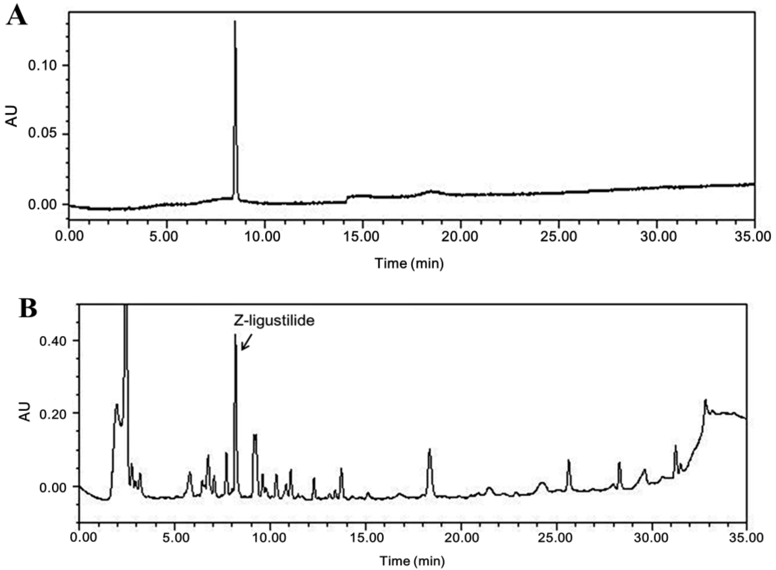
A. sinensis extract was monitored at 307 nm for Z-ligustilide (Figure 1). A. sinensis extract was standardized to contain 1.9% ± 0.21% Z-ligustilide.
Figure 1. High performance liquid chromatography (HPLC) chromatograms of Z-ligustilide (A) and A. sinensis extract (B). Detection was performed using a photodiode array detector. X-axis is retention time (min); Y-axis is absorbance unit (AU).
Nutrients 06 04362 g001

3.2. Body Weight Gain, Uterus Index, and Bone Mineral Density in Rats Treated with A. sinensis Extract

After four weeks of treatments, the body weight gain of the E2 group was significantly lower than that of the OVXcontrol group. However, there was no significant difference in the body weight gain of A. sinensis extract-treated groups (Figure 2A). OVX caused atrophy of uterine tissue, indicating the success of the surgical procedure, and in the E2 group the uterus index increased significantly compared to the OVXcontrol group. However, A. sinensis extract-treated groups did not show an effect on the uterus index following OVX (Figure 2B). After four weeks of treatments, the final femur bone mineral density (BMD) of the A. sinensis extract 300 mg/kg-treated group was significantly higher than that of the OVXcontrol group (Figure 2C).
Figure 2. Effects of A. sinensis extract on body weight gain (A); uterus index (B); and bone mineral density (BMD) (C) in ovariectomized (OVX) rats. The body weight gain was calculated by the equation: final body weight—initial body weight. Uterus was dissected, washed with saline, and weighted. The BMD values were determined by dual energy X-ray absorptiometry. Here and in Figure 3, data are mean ± SD values (n = 10 per group). *** p < 0.001, ** p < 0.01, and * p < 0.05, as compared with the OVX control group.
Nutrients 06 04362 g002

3.3. Serum Bone Marker in Rats Treated with A. sinensis Extract
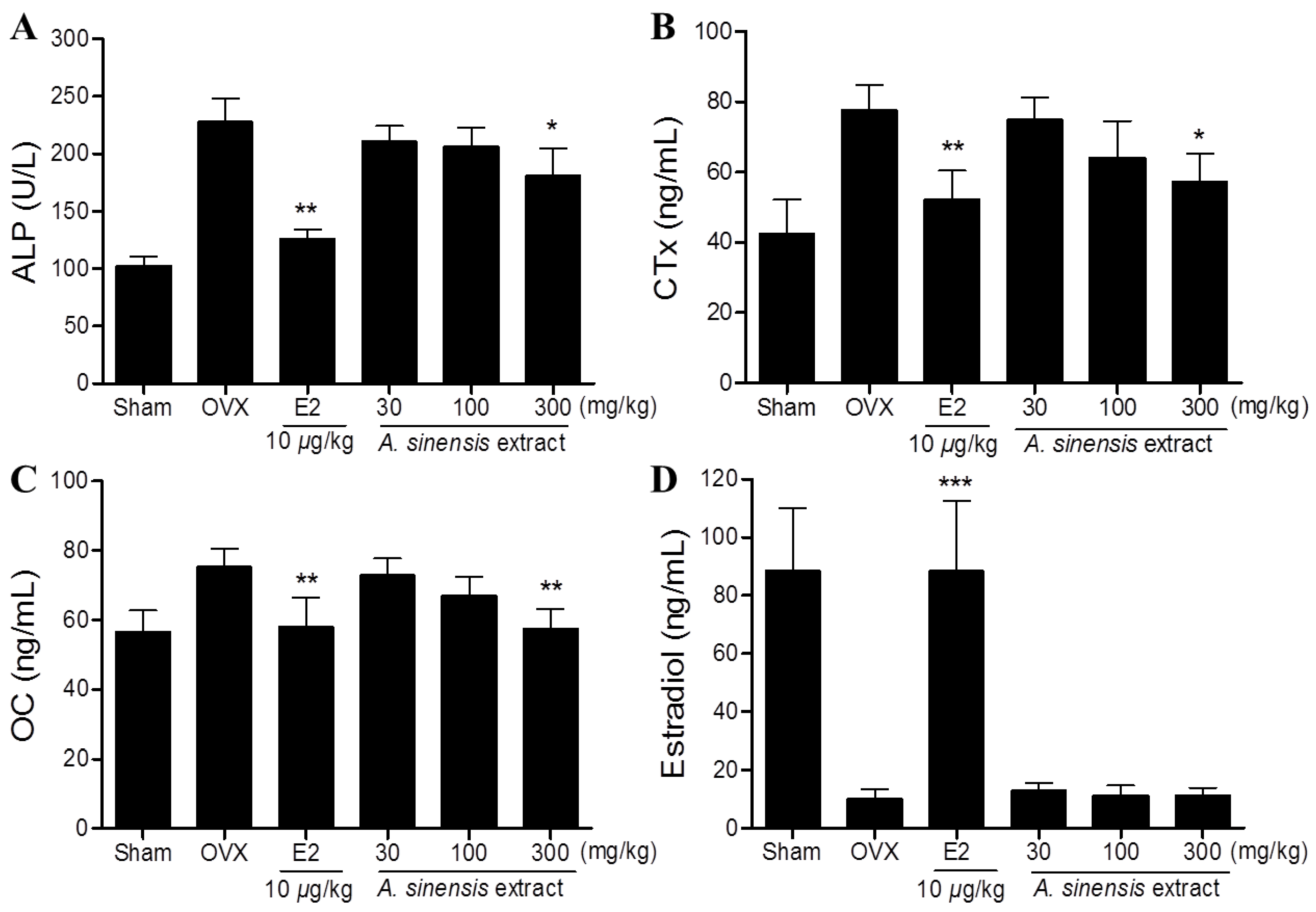
Serum ALP, CTx, and OC concentrations in the OVX-control group were significantly higher compared to the sham group. After four weeks treatments, the A. sinensis extract 300 mg/kg-treated group showed significantly lower serum ALP, CTx, and OC concentrations compared to the OVX-control group (Figure 3). In case of serum estradiol, the A. sinensis extract treated groups were not significantly different from the OVX-control group (Figure 3D).
Figure 3. Effects of A. sinensis extract on serum alkaline phosphatase (ALP) (A); collagen type I C-telopeptide (CTx) (B); osteocalcin (OC) (C); and estradiol (D) concentrations in OVX rats. *** p < 0.001, ** p < 0.01, and * p < 0.05, as compared with the OVX control group.
Nutrients 06 04362 g003

3.4. Effects of A. sinensis Extract on Sub-Chronic Toxicity

No significant differences in body weight were observed between normal and treated groups during the administration period (Table 1). Following sub-chronic toxicity test (28 days), no changes of organs weight and serum biochemical parameters were observed between the control and treatment groups (Table 2 and Table 3).
Table 1. Mean body weight of rats after 28 days treatment with A. sinensis extract.
WeekMean Body Weight (g ± SD)
MaleFemale
NormalExtract (mg/kg)NormalExtract (mg/kg)
1000200010002000
0234.3 ± 9.1227.6 ± 10.3230.0 ± 8.5187.6 ± 3.8185.7 ± 4.9182.8 ± 5.0
1281.7 ± 13.5273.8 ± 12.9279.5 ± 10.4199.0 ± 10.4210.8 ± 7.1212.4 ± 10.8
2321.3 ± 19.2312.9 ± 16.4318.5 ± 12.6219.0 ± 12.2229.0 ± 9.8230.1 ± 7.8
3343.1 ± 22.8338.9 ± 16.6341.2 ± 15.5225.6 ± 8.5230.9 ± 9.1228.4 ± 10.0
4370.1 ± 20.9366.2 ± 19.3368.5 ± 15.8236.4 ± 8.6243.7 ± 10.5240.6 ± 11.4
Table 2. Mean organ weight of rats after 28 days treatment with A. sinensis extract.
OrganMean Weight (g ± SD)
MaleFemale
NormalExtract (mg/kg)NormalExtract (mg/kg)
1000200010002000
Heart0.9 ± 0.00.9 ± 0.00.8 ± 0.10.7 ± 0.00.7 ± 0.00.6 ± 0.1
Liver10.1 ± 1.09.9 ± 1.59.9 ± 1.06.6 ± 0.56.5 ± 0.76.6 ± 0.0
Spleen0.7 ± 0.10.7 ± 0.10.6 ± 0.00.5 ± 0.10.5 ± 0.10.6 ± 0.0
Kidney1.2 ± 0.11.2 ±0.11.2 ± 0.00.8 ± 0.00.7 ± 0.10.7 ± 0.0
Testis1.9 ± 0.22.0 ± 0.12.0 ± 0.2---
Uterus---0.8 ± 0.10.7 ± 0.00. 8 ± 0.0
Table 3. Biochemical parameters of rats after 28 days treatment with A. sinensis extract.
Serum ParameterMean Weight (g ± SD)
MaleFemale
NormalExtract (mg/kg)NormalExtract (mg/kg)
1000200010002000
AST (U/L)66.3 ± 12.276.0 ± 8.572.2 ± 10.5 64.3 ± 10.860.5 ± 6.462.8 ± 5.5
ALT (U/L)29.8 ± 3.4 25.0 ± 1.4126.0 ± 4.2 24.7 ± 4.726.0 ± 2.824.0 ± 2.5
GGT (U/L)5.2 ± 3.74.0 ± 1.44.5 ± 2.4 4.2 ± 3.13.0 ± 0.03.8 ± 0.0
GLU (mmol/L)6.3 ± 0.856.1.0 ± 0.505.9 ± 2.01 5.8 ± 1.086.0.0 ± 0.755.9 ± 0.67
BUN (mg/dL)24.1 ± 2.525.7 ± 1.924.2 ± 3.4 19.6 ± 7.522.5 ± 3.722.4 ± 3.0
ALP (U/L)582.0 ± 20.1652.0 ± 9.9630.0 ± 4.8293.2 ± 52.5312.5 ± 54.5318.5 ± 50.2
CRE (mg/dL)0.21 ± 0.030.15 ± 0.070.18 ± 0.01 0.21 ± 0.030.20 ± 0.000.20 ± 0.01
TP (g/dL)6.0 ± 0.236.2 ± 0.646.2 ± 0.52 6.0 ± 0.255.9 ± 0.076.0 ± 0.05
AST, aspartate aminotransferase; ALT, alanine transaminase; GGT, gamma-glutamyltransferase; GLU, glucose; BUN, blood urea nitrogen; ALP, alkaline phosphatase; CRE, creatinine; TP, total protein.

4. Discussion

Our finding demonstrated that four weeks of treatment with A. sinensis extract significantly decreased the BMD loss in the femur and inhibited the bone turnover markers—serum ALP, OC, and CTx levels compared to the OVXcontrol group without influencing estrogen level.
Bone loss caused by estrogen deficiency in both experimental animals and humans is generally due to an increase in osteoclastic bone resorption [18]. OVX rats, which exhibit most of the characteristics of human postmenopausal osteoporosis [19], are widely used as a model for the evaluation of potential osteoporosis treatments [20].
In our experiments, OVX resulted in significant decrease in femur BMD after four weeks. The BMD loss was accompanied by a significant increase in bone remodeling, as evidenced by the increased biochemical bone turnover markers, such as serum ALP [21,22,23], CTx, and OC levels [24]. In the present study, oral administration of A. sinensis extract at the dose 300 mg/kg significantly decreased BMD loss, which was accompanied by the decrease in serum ALP, CTx, and OC levels compared to a OVXcontrol group. These results suggest that A. sinensis extract decreases bone loss by inhibiting bone turnover induced by OVX.
OVX dramatically increases body weight, while E2 treatment prevents body weight gain [25]. Estrogen deficiency induced body fat accumulation and subsequently caused an increase in body weight [26]. Heine et al. demonstrated that estrogen receptor (ER) knockout mice have higher fat mass and lower energy expenditure than wild-type mice [27]. Estrogen may be involved directly in energy metabolism by binding to ER within the abdominal and subcutaneous fat tissue [28]. Estrogen expresses its activities by binding to different ERs, ERα and ERβ. ERβ is more abundant than ERα in bone tissue, while ERα it is mainly distributed in reproductive cells and is the dominant receptor mediating the effects of E2 in breast and uterus [29]. In our experiments, oral administration of A. sinensis extract did not affect serum estradiol concentration, body weight gain, and uterotrophic activity in OVX rats. These results suggest that, the A. sinensis extract might have anti-osteoporotic effects in OVX rats, without the influence of hormones such as estrogen.
It is important to note that A. sinensis extract contains ferrulic acid, which is a potent antioxidant and a free radical scavenger [30]. It has been demonstrated that oxidation-derived free radicals increase bone resorption by promoting osteoclastic differentiation [31]. Ma et al. demonstrated that Z-ligustilide from the essential oil of A. sinensis inhibits the OVX-induced serum interleukin-1β (IL-1β) and tumor necrosis factor-α (TNF-α) levels [32]. IL-1β is known as a highly potent bone-resorptive cytokine [9] and TNF-α appears to synergize with IL-1 to increase bone resorption [11]. The effects of A. sinensis on bone thus appear to be related to its high contents of the ferulic acid or Z-ligustilide.
The toxicity of A. sinensis extract must be evaluated before it can be used in the drug or supplement development. In the rat toxicity studies, A. sinensis extract was administered orally at doses 1000 and 2000 mg/kg/day. A. sinensis extract caused no changes that could be considered toxicologically significant. Repeated oral administration of A. sinensis extract to rats for 4 weeks resulted in no toxicological changes in any of the clinical signs, body weight changes, serum biochemistry, necropsy findings, and relative organ weights. Thus, under the present experimental conditions, the No Observable Adverse Effect Level (NOAEL) of A. sinensis extract was assumed to be 2000 mg/kg/day for both male and female rats.

5. Conclusions

In conclusion, A. sinensis extract can prevent OVX-induced bone loss with efficacy comparable to that of estrogen. The findings obtained suggest that A. sinensis extract could be an effective natural alternative for the prevention of postmenopausal osteoporosis. Moreover, in the safety evaluation studies, A. sinensis extract was shown to be safe up to 2000 mg/kg/day for 4 weeks of administration in rats. The results presented here provide a foundation for clinical evaluation and demonstrate the potential of A. sinensis extract as an herbal drug.

Acknowledgments

This study was supported by a research grant from the Korea Food Research Institute (E0143023927).

Author Contributions

D.W. Lim and Y.T. Kim designed and performed the study and wrote the paper. All authors read and approved the final manuscript.

Conflicts of Interest

The authors declare no conflict of interest.

References

  1. Hook, I.L. Danggui to angelica sinensis root: Are potential benefits to european women lost in translation? A review. J. Ethnopharmacol. 2014, 152, 1–13. [Google Scholar] [CrossRef] [PubMed]
  2. Ye, Y.N.; Liu, E.S.; Li, Y.; So, H.L.; Cho, C.C.; Sheng, H.P.; Lee, S.S.; Cho, C.H. Protective effect of polysaccharides-enriched fraction from angelica sinensis on hepatic injury. Life Sci. 2001, 69, 637–646. [Google Scholar] [CrossRef] [PubMed]
  3. Huang, S.H.; Lin, C.M.; Chiang, B.H. Protective effects of angelica sinensis extract on amyloid beta-peptide-induced neurotoxicity. Phytomed. Int. J. Phytother. Phytopharmacol. 2008, 15, 710–721. [Google Scholar] [CrossRef]
  4. Wu, S.J.; Ng, L.T.; Lin, C.C. Antioxidant activities of some common ingredients of traditional chinese medicine, angelica sinensis, lycium barbarum and poria cocos. Phytother. Res. PTR 2004, 18, 1008–1012. [Google Scholar] [CrossRef]
  5. Qin, J.; Liu, Y.S.; Liu, J.; Li, J.; Tan, Y.; Li, X.J.; Magdalou, J.; Mei, Q.B.; Wang, H.; Chen, L.B. Effect of angelica sinensis polysaccharides on osteoarthritis in vivo and in vitro: A possible mechanism to promote proteoglycans synthesis. Evid. Based Complement. Altern. Med. 2013, 2013. [Google Scholar] [CrossRef]
  6. Lai, J.N.; Wu, C.T.; Wang, J.D. Prescription pattern of chinese herbal products for breast cancer in taiwan: A population-based study. Evid. Based Complement. Altern. Med. 2012, 2012. [Google Scholar] [CrossRef]
  7. Chen, X.P.; Li, W.; Xiao, X.F.; Zhang, L.L.; Liu, C.X. Phytochemical and pharmacological studies on radix angelica sinensis. Chin. J. Nat. Med. 2013, 11, 577–587. [Google Scholar] [CrossRef] [PubMed]
  8. Manolagas, S.C. Birth and death of bone cells: Basic regulatory mechanisms and implications for the pathogenesis and treatment of osteoporosis. Endocr. Rev. 2000, 21, 115–137. [Google Scholar] [PubMed]
  9. Evans, D.B.; Bunning, R.A.; Russell, R.G. The effects of recombinant human interleukin-1 beta on cellular proliferation and the production of prostaglandin e2, plasminogen activator, osteocalcin and alkaline phosphatase by osteoblast-like cells derived from human bone. Biochem. Biophys. Res. Commun. 1990, 166, 208–216. [Google Scholar] [CrossRef] [PubMed]
  10. Stashenko, P.; Dewhirst, F.E.; Peros, W.J.; Kent, R.L.; Ago, J.M. Synergistic interactions between interleukin 1, tumor necrosis factor, and lymphotoxin in bone resorption. J. Immunol. 1987, 138, 1464–1468. [Google Scholar] [PubMed]
  11. McLean, R.R. Proinflammatory cytokines and osteoporosis. Curr. Osteoporos. Rep. 2009, 7, 134–139. [Google Scholar] [CrossRef] [PubMed]
  12. Wilkinson, J.M.; Hamer, A.J.; Rogers, A.; Stockley, I.; Eastell, R. Bone mineral density and biochemical markers of bone turnover in aseptic loosening after total hip arthroplasty. J. Orthop. Res. Off. Publ. Orthop. Res. Soc. 2003, 21, 691–696. [Google Scholar] [CrossRef]
  13. Ma, Z.; Bai, L. Anti-inflammatory effects of Z-ligustilide nanoemulsion. Inflammation 2013, 36, 294–299. [Google Scholar] [CrossRef] [PubMed]
  14. Saw, C.L.; Wu, Q.; Su, Z.Y.; Wang, H.; Yang, Y.; Xu, X.; Huang, Y.; Khor, T.O.; Kong, A.N. Effects of natural phytochemicals in angelica sinensis (danggui) on nrf2-mediated gene expression of phase ii drug metabolizing enzymes and anti-inflammation. Biopharm. Drug Dispos. 2013, 34, 303–311. [Google Scholar] [CrossRef] [PubMed]
  15. Zhang, C.; Kong, X.; Zhou, H.; Liu, C.; Zhao, X.; Zhou, X.; Su, Y.; Sharma, H.S.; Feng, S. An experimental novel study: Angelica sinensis prevents epidural fibrosis in laminectomy rats via downregulation of hydroxyproline, IL-6, and TGF-β1. Evid. Based Complement. Altern. Med. 2013, 2013. [Google Scholar] [CrossRef]
  16. Thiede, M.A.; Smock, S.L.; Petersen, D.N.; Grasser, W.A.; Thompson, D.D.; Nishimoto, S.K. Presence of messenger ribonucleic acid encoding osteocalcin, a marker of bone turnover, in bone marrow megakaryocytes and peripheral blood platelets. Endocrinology 1994, 135, 929–937. [Google Scholar] [PubMed]
  17. Coleman, R.E. The clinical use of bone resorption markers in patients with malignant bone disease. Cancer 2002, 94, 2521–2533. [Google Scholar] [CrossRef] [PubMed]
  18. Hoegh-Andersen, P.; Tanko, L.B.; Andersen, T.L.; Lundberg, C.V.; Mo, J.A.; Heegaard, A.M.; Delaisse, J.M.; Christgau, S. Ovariectomized rats as a model of postmenopausal osteoarthritis: Validation and application. Arthritis Res. Ther. 2004, 6, R169–R180. [Google Scholar] [CrossRef] [PubMed] [Green Version]
  19. Jee, W.S.; Yao, W. Overview: Animal models of osteopenia and osteoporosis. J. Musculoskelet. Neuronal Interact. 2001, 1, 193–207. [Google Scholar] [PubMed]
  20. Lelovas, P.P.; Xanthos, T.T.; Thoma, S.E.; Lyritis, G.P.; Dontas, I.A. The laboratory rat as an animal model for osteoporosis research. Comp. Med. 2008, 58, 424–430. [Google Scholar] [PubMed]
  21. Nishizawa, Y.; Nakatsuka, K. Gguideline for adequate use of metabolic bone markers in osteoporosis. Nihon Rinsho. Jpn. J. Clin. Med. 2004, 62 (Suppl. 2), S325–S332. [Google Scholar]
  22. Yogesh, H.S.; Chandrashekhar, V.M.; Katti, H.R.; Ganapaty, S.; Raghavendra, H.L.; Gowda, G.K.; Goplakhrishna, B. Anti-osteoporotic activity of aqueous-methanol extract of berberis aristata in ovariectomized rats. J. Ethnopharmacol. 2011, 134, 334–338. [Google Scholar] [CrossRef] [PubMed]
  23. Lim, D.W.; Kim, Y.T. Dried root of rehmannia glutinosa prevents bone loss in ovariectomized rats. Molecules 2013, 18, 5804–5813. [Google Scholar] [CrossRef] [PubMed]
  24. Hertrampf, T.; Schleipen, B.; Offermanns, C.; Velders, M.; Laudenbach, U.; Diel, P. Comparison of the bone protective effects of an isoflavone-rich diet with dietary and subcutaneous administrations of genistein in ovariectomized rats. Toxicol. Lett. 2009, 184, 198–203. [Google Scholar] [CrossRef] [PubMed]
  25. Devareddy, L.; Khalil, D.A.; Smith, B.J.; Lucas, E.A.; Soung, D.Y.; Marlow, D.D.; Arjmandi, B.H. Soy moderately improves microstructural properties without affecting bone mass in an ovariectomized rat model of osteoporosis. Bone 2006, 38, 686–693. [Google Scholar] [CrossRef] [PubMed]
  26. Dang, Z.C.; van Bezooijen, R.L.; Karperien, M.; Papapoulos, S.E.; Lowik, C.W.G.M. Exposure of ks483 cells to estrogen enhances osteogenesis and inhibits adipogenesis. J. Bone Miner. Res. 2002, 17, 394–405. [Google Scholar] [CrossRef] [PubMed]
  27. Heine, P.A.; Taylor, J.A.; Iwamoto, G.A.; Lubahn, D.B.; Cooke, P.S. Increased adipose tissue in male and female estrogen receptor-alpha knockout mice. Proc. Natl. Acad. Sci. USA 2000, 97, 12729–12734. [Google Scholar] [CrossRef] [PubMed]
  28. Joyner, J.M.; Hutley, L.J.; Cameron, D.P. Estrogen receptors in human preadipocytes. Endocrine 2001, 15, 225–230. [Google Scholar] [CrossRef] [PubMed]
  29. Hewitt, S.C.; Korach, K.S. Oestrogen receptor knockout mice: Roles for oestrogen receptors alpha and beta in reproductive tissues. Reproduction 2003, 125, 143–149. [Google Scholar] [CrossRef] [PubMed]
  30. Srinivasan, M.; Sudheer, A.R.; Menon, V.P. Ferulic acid: Therapeutic potential through its antioxidant property. J. Clini. Biochem. Nutr. 2007, 40, 92–100. [Google Scholar] [CrossRef]
  31. Mody, N.; Parhami, F.; Sarafian, T.A.; Demer, L.L. Oxidative stress modulates osteoblastic differentiation of vascular and bone cells. Free Radic. Biol. Med. 2001, 31, 509–519. [Google Scholar] [CrossRef] [PubMed]
  32. Ma, Z.; Bai, L. The anti-inflammatory effect of Z-ligustilide in experimental ovariectomized osteopenic rats. Inflammation 2012, 35, 1793–1797. [Google Scholar] [CrossRef] [PubMed]