Adipocyte Size, Overweight, and Insulin Resistance in Type 2 Diabetes Mellitus and the Impact of Weight Loss: A Systematic Review

 ,
,  , , , , , , , , and
Abstract
1. Introduction
2. Methods
2.1. Design and Registration
2.2. Review Questions and Search Strategy
2.3. Inclusion and Exclusion Criteria
2.4. Quality Assessment of the Included Studies
2.5. Risk of Bias
3. Results
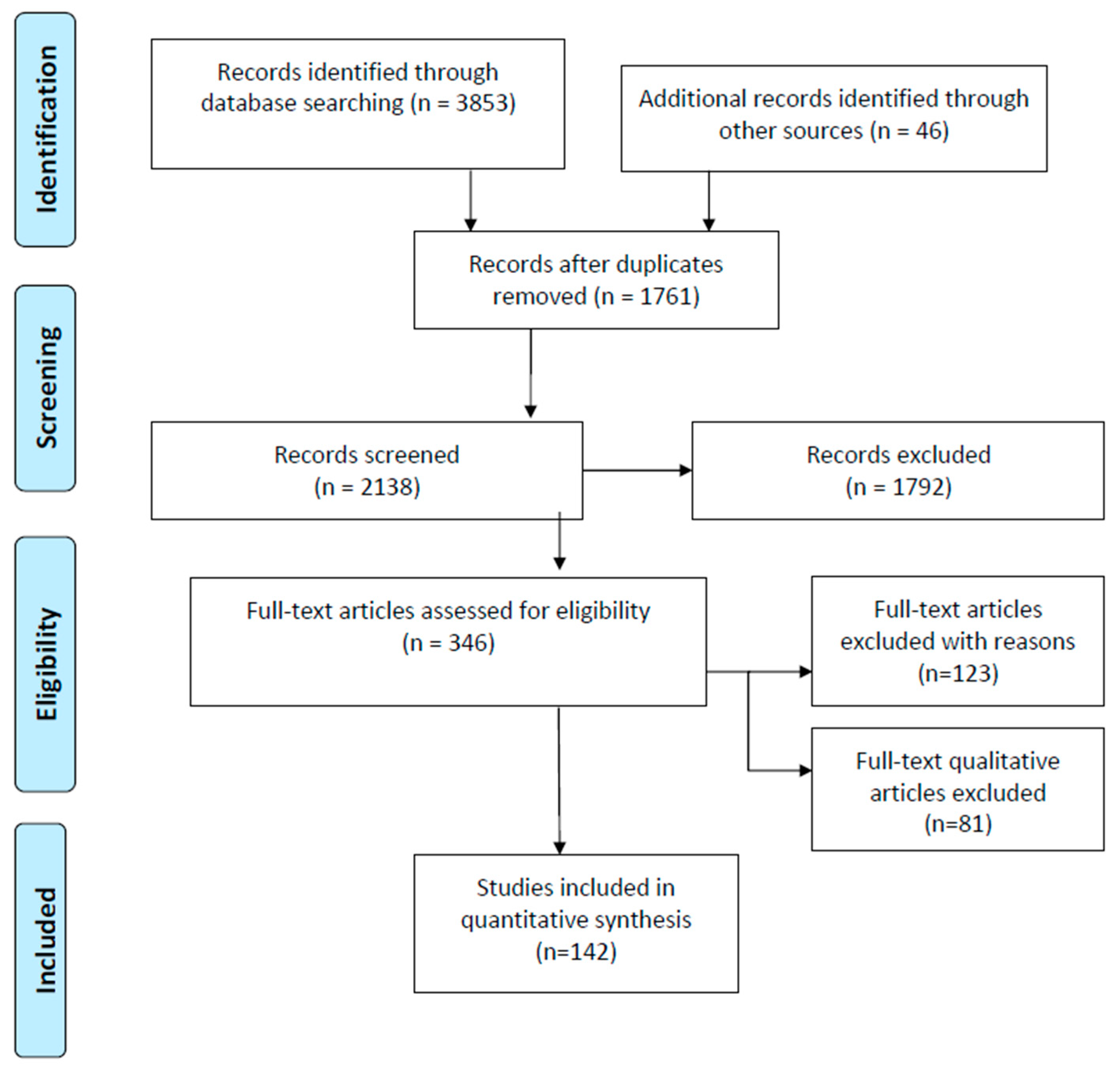
3.1. Search Results
3.2. Characteristics of Included Studies

| Authors | Protocol | Study Design | Overweight/Obesity, Increased Adipocytes | HbA1c, HI, IR, T2DM |
|---|---|---|---|---|
| Human Studies | ||||
| Dundar, 2022 [2] | 860 subjects | Cross-sectional study | Overweight/obesity | Elevated HOMA-IR, developed T2DM |
| Sarkar, 2019 [57] | 650 subjects | Cross-sectional study | Overweight/weight gain | Developed IR, β-cell deficiency, developed T2DM |
| Berglund, 2016 [58] | 331 subjects | Cohort study | Overweight/obesity | Elevated HbA1c, developed HI |
| Cotillard, 2014 [36] | 295 subjects | Cohort study | Overweight/increased adipocytes | Elevated HbA1c, developed HI, T2DM |
| Vertemati, 2008 [59] | 56 subjects | Clinical controlled study | Overweight/increased adipocytes | Developed IR, T2DM |
| Fang, 2015 [28] | 30 patients | Clinical controlled study | Overweight, increased adipocytes | Elevated HbA1c, HOMA-IR, developed T2DM |
| McLaughli, 2014 [60] | 148 subjects | Clinical controlled study | Overweight/increased adipocytes | Elevated HOMA-IR |
| Pasarica, 2009 [34] | 260 patients | Clinical controlled study | Overweight/increased adipocytes | Elevated HOMA-IR, developed T2DM |
| Ricci, 2015 [9] | 22 studies, 4160 subjects | Meta-analysis | Overweight/obesity | Elevated HbA1c, HOMA-IR |
| Musilanga, 2024 [3] | 30 studies | Systematic review, meta-analysis | Overweight/obesity | Elevated HbA1c, HOMA-IR, developed T2DM |
| Tahrani, 2022 [10] | 55 studies | Systematic review | Overweight/obesity | Elevated HbA1c, HOMA-IR |
| Zhao, 2023 [15] | 12 studies | Systematic review | Overweight/obesity/increased adipocytes | Elevated HbA1c, HOMA-IR, lipotoxicity |
| Villagrán-Silva, 2025 [27] | 24 studies | Systematic review | Overweight/obesity | Elevated HbA1c, HOMA-IR, and miRNA |
| Ye, 2022 [61] | 62 studies | Systematic review | Overweight/increased adipocytes/ectopic fat accumulation | Developed IR, T2DM |
| Papaetis, 2025 [29] | 14 studies | Review | Overweight/increased adipocytes | Elevated HOMA-IR, lipotoxicity, developed T2DM |
| Nakamura, 2020 [62] | 32 studies | Review | Overweight/increased adipocytes | Elevated HbA1c, HOMA-IR, lipotoxicity, developed T2DM, cardiomyopathy |
| Nakamura, 2024 [63] | 47 studies | Review | Overweight/increased adipocytes | Elevated HbA1c, HOMA-IR, lipotoxicity, developed T2DM |
| Szablewski, 2024 [64] | 14 studies | Review | Overweight/increased adipocytes | Elevated HOMA-IR, lipotoxicity, developed T2DM |
| Ferrannini, 2004 [65] | 14 studies | Review | Overweight/obesity | Increased β-cell mass, elevated IR, developed T2DM |
| Szukiewicz, 2023 [66] | 27 studies | Review | Overweight/obesity | Developed IR, T2DM, chronic diseases |
| Castillo, 2025 [67] | 38 studies | Review | Overweight/intracellular lipid accumulation | Developed IR |
| Guria, 2023 [68] | 53 studies | Review | Overweight/macrophage lipid infiltration | Developed IR, T2DM |
| van Vliet, 2020 [69] | 24 subjects | Review | Overweight/increased adipocytes/ectopic fat accumulation | Developed IR, HI, developed T2DM |
| Lipke, 2022 [70] | 35 studies | Review | Overweight/increased adipocytes | Elevated HbA1c, HOMA-IR, lipotoxicity |
| Mota, 2016 [71] | 22 studies | Review | Overweight/increased adipocytes | Developed IR, lipotoxicity |
| Longo, 2019 [54] | 19 studies | Review | Overweight/increased adipocytes | Lipotoxicity, developed T2DM |
| Ahmed, 2021 [72] | 26 studies | Review | Overweight/increased adipocytes | Elevated HbA1c, HOMA-IR, lipotoxicity |
| Dahik, 2020 [73] | 37 studies | Review | Overweight/increased adipocytes | Elevated HbA1c, HOMA-IR, lipotoxicity |
| Armato, 2025 [74] | 1860 subjects | Review | Personal overweight | Elevated HbA1c, developed IR |
| Animal Studies | ||||
| Setayesh, 2019 [75] | (36 mice) | Animal controlled study | Overweight/obesity | Developed HI, DNA damage |
| Peyot, 2010 [76] | (Mice) | Animal controlled study | Obesity/lipid deposition | Developed IR, beta-cell failure |
| Bozec, 2016 [77] | (Mice) | Animal controlled study | Overweight/increased adipocytes | Developed IR, hypoxia, adipocyte apoptosis |
| Sakaguchi, 2017 [78] | (Mice) | Animal controlled study | Overweight/increased adipocytes | Developed IR, T2DM, metabolic syndrome |
| Ozcan, 2014 [79] | (90 mice) | Animal study | Overweight/obesity | Developed IR, T2DM |
| Verkest, 2011 [30] | (106 dogs) | Animal study | Overweight/obesity | HI, elevated HOMA-IR, changed adiponectin, leptin, beta-cell function |
| Authors | Protocol | Study Design | Weight Loss | Parameters of T2DM (HbA1c, IR) |
|---|---|---|---|---|
| Human Studies | ||||
| Franz, 2015 [5] | (6754 patients) Diet therapy | RCT | Weight loss ≥ 5% | Decreased HbA1c |
| Tahrani, 2022 [10] | (11 studies) Lifestyle | RCT | Weight loss ≥ 7.8% | Decreased HbA1c, HOMA-IR, T2DM remission |
| Tahrani, 2022 [10] | (12 studies) Pharmacotherapy | RCT | Weight loss ≥ 10% | Decreased HbA1c, HOMA-IR, T2DM remission |
| Lean, 2019 [20] | (149 patients) Lifestyle | RCT | Weight loss ≥ 10% | Decreased HbA1c, HOMA-IR, T2DM remission |
| Fonseca, 2019 [7] | (2432 patients). Semaglutide | RCT | Weight loss ≥ 7% | Decreased HbA1c, HOMA-IR |
| Zhao, 2023 [15] | (26 studies) Diet therapy vs. pharmacotherapy | RCT | Weight loss ≥ 5–10% | Decreased HbA1c, HOMA-IR |
| Buse, 2020 [45] | (American Diabetes Association и European Association for the Study of Diabetes) Pharmacotherapy | RCT | Weight loss ≥ 5% | Decreased HbA1c, HOMA-IR |
| Oshakbayev, 2017 [47] | (272 patients) VLCD | RCT | Weight loss ≥ 10% | Decreased HbA1c, HOMA-IR |
| Daniele [25] | (15 studies) Restriction diet | RCT | Weight loss ≥ 10% | Decreased HbA1c, HOMA-IR, SBP/DBP |
| Oshakbayev, 2019 [48] | (80 patients) VLCD | RCT | Weight loss ≥ 10% | Decreased HbA1c, HOMA-IR, NASH |
| Goni, 2017 [21] | (757 subjects) Low-fat diet vs. high-fat diet | RCT | Weight loss ≥ 5% | Decreased HOMA-IR |
| Banji, 2025 [88] | (56 studies) Pharmacotherapy | RCT | Weight loss ≥ 10–20% | Decreased HbA1c, HOMA-IR, T2DM |
| Albai, 2025 [87] | (256 patients) Pharmacotherapy | RCT | Weight loss ≥ 10% | Decreased HbA1c, HOMA-IR, T2DM, MASLD |
| Lingvay, 2020 [17] | (995 patients) Pharmacotherapy | RCT | Weight loss ≥ 5–10% | Decreased HbA1c, lipids, SBP/DBP, T2DM |
| Davies, 2022 [89] | (57 studies) Drug therapy, VLCD | RCT | Weight loss ≥ 5–20% | Decreased HbA1c, HOMA-IR, lipids, cardiorenal health, T2DM |
| Kashyap, 2022 [11] | (16 studies, 834 patients). VLCD | RCT and non-RCT, prospective controlled | Weight loss ≥ 5% | Decreased HbA1c |
| Horn, 2022 [12] | (45 studies) Lifestyle, pharmacotherapy, bariatric surgery | RCT and non-RCT, prospective controlled | Weight loss ≥ 5–15% | Decreased HbA1c, HOMA-IR |
| Ferrannini, 2004 [65] | (17 studies) Pharmacotherapy vs. bariatric surgery | RCT and non-RCT, prospective controlled | Weight loss ≥ 10% | Decreased HbA1c, HOMA-IR |
| Murphy, 2017 [90] | (33 studies) Bariatric vs. lifestyle | RCT and non-RCT, prospective controlled | Weight loss ≥ 7–10% | Decreased IR, adipocyte size, cardiometabolic diseases |
| Jooste, 2023 [91] | (11 studies and 1519 patients) Restriction diets | RCT and non-RCT, prospective controlled | Weight loss ≥ 5–10% | Decreased HbA1c, HOMA-IR, lipids, T2DM |
| Van den Burg, 2023 [92] | (9 studies) Different diets | RCT and non-RCT, prospective controlled | Weight loss ≥ 5–10% | Decreased HbA1c, BMR, T2DM |
| Schauer, 2012 [93] | (150 patients) Pharmacotherapy and bariatric surgery | RCT and non-RCT, prospective controlled | Weight loss ≥ 10–25% | Decreased HbA1c, HOMA-IR, lipids, SBP/DBP, T2DM |
| Ricci, 2015 [9] | (22 studies, 4160 patients) Bariatric surgery | Non-RCT prospective controlled | Weight loss ≥ 10% | Decreased HbA1c, HOMA-IR, T2DM remission |
| Tahrani, 2022 [10] | (12 studies). Bariatric surgery | Non-RCT prospective controlled | Weight loss ≥ 10–30% | Decreased HbA1c, HOMA-IR, T2DM remission |
| Cotillard, 2014 [36] | (74 patients) Bariatric surgery | Non-RCT, prospective controlled | Weight loss≥ 10% | Decreased HbA1c, HOMA-IR |
| Reinehr, 2004 [94] | (232 patients) Restriction diet | Non-RCT, prospective controlled | Weight loss ≥ 5% | Decreased HOMA-IR, cortisol |
| van Vliet, 2020 [69] | (24 studies) Restriction diet | Non-RCT, prospective controlled | Weight loss ≥ 20% | Decreased HOMA-IR, basal/ postprandial insulin secretion |
| Zhang, 2015 [95] | 16 studies | RCT and non-RCT, prospective controlled | Weight loss/autophagy | Decreased HbA1c, HOMA-IR |
| Oshakbayev, 2026 [96] | (130 patients) Pharmacotherapy vs. bariatric vs. VLCD | Non-RCT, prospective controlled | Weight loss ≥ 10–20% | Decreased HbA1c, HOMA-IR cardiometabolic diseases, T2DM |
| Delrue, 2025 [97] | (25 studies) Restriction diet | Non-RCT, prospective controlled | Weight loss ≥ 10% | Decreased HbA1c, HOMA-IR, T2DM |
| Wei., 2025 [98] | (39 studies) Bariatric surgery | Non-RCT, prospective controlled | Weight loss ≥ 10–20% | Decreased HbA1c, HOMA-IR, T2DM |
| Animal Studies | ||||
| Di Daniele, 2021 [25] | (7 studies) Caloric restriction diet | Controlled studies | Weight loss ≥ 10% | Decreased HbA1c, HOMA-IR, SBP/DBP |
| Zhang, 2015 [95] | 12 studies | Controlled studies | Weight loss/autophagy | Decreased HbA1c, HOMA-IR |
| Setayesh, 2019 [75] | (36 mice) Caloric restriction diet | Controlled studies | Weight loss | Decreased IR, inflammation, and DNA damage in internal organs |
4. Discussion
Strengths and Limitations
5. Conclusions
Author Contributions
Funding
Institutional Review Board Statement
Informed Consent Statement
Data Availability Statement
Acknowledgments
Conflicts of Interest
Abbreviations
| BMI | Body mass index |
| BMR | Basal metabolic rate |
| GLP-1RA | Glucagon-like peptide-1 receptor agonist |
| HbA1c | Glycated hemoglobin |
| HI | Endogenous hyperinsulinism |
| IR | Insulin resistance |
| RCT | Randomized clinical trial |
| SGLT-2i | Sodium–glucose transport protein 2 inhibitor |
| T2DM | Type 2 diabetes mellitus |
References
- Ekanayake, P.; Edelman, S. Identifying patients with type 2 diabetes who might benefit from insulin pump therapy: Literature review, clinical opportunities, potential benefits and challenges. Diabetes Obes. Metab. 2023, 25, 3–20. [Google Scholar] [CrossRef]
- Dündar, I.; Akinci, A. Prevalence of type 2 diabetes mellitus, metabolic syndrome, and related morbidities in overweight and obese children. J. Pediatr. Endocrinol. Metab. 2022, 35, 435–441. [Google Scholar] [CrossRef]
- Musilanga, N.; Nasib, H.; Jackson, G.; Shayo, F.; Nhanga, C.; Girukwigomba, S.; Mwakibolwa, A.; Henry, S.; Kijusya, K.; Msonge, E. Exploring the Prevalence and Components of Metabolic Syndrome in Sub-Saharan African Type 2 Diabetes Mellitus Patients: A Systematic Review and Meta-Analysis. J. Obes. 2024, 2024, 1240457. [Google Scholar] [CrossRef]
- Davies, M.; Lim, S.; Slater, T.; Goldney, J.; Philis-Tsimikas, A.; Franco, D.; Lamptey, R.; Yates, T.; Tankova, T.; Lingvay, I. Type 2 diabetes mellitus. Nat. Rev. Dis. Primers 2026, 12, 13. [Google Scholar] [CrossRef]
- Franz, M.J.; Boucher, J.L.; Rutten-Ramos, S.; VanWormer, J.J. Lifestyle Weight-Loss Intervention Outcomes in Overweight and Obese Adults with Type 2 Diabetes: A Systematic Review and Meta-Analysis of Randomized Clinical Trials. J. Acad. Nutr. Diet. 2015, 115, 1447–1463. [Google Scholar] [CrossRef]
- Wolos-Klosowicz, K.; Bandurska-Stankiewicz, E. Effects of common weight loss plans on diabetes mellitus and cardiovascular risk factors. Prim. Care Diabetes 2022, 16, 252–256. [Google Scholar] [CrossRef]
- Fonseca, V.; Capehorn, M.; Garg, S.; Gimeno, E.; Hansen, O.; Holst, A.; Nayak, G.; Seufert, J. Reductions in Insulin Resistance are Mediated Primarily via Weight Loss in Subjects with Type 2 Diabetes on Semaglutide. J. Clin. Endocrinol. Metab. 2019, 104, 4078–4086. [Google Scholar] [CrossRef] [PubMed]
- Reppo, I.; Jakobson, M.; Volke, V. Effects of Semaglutide and Empagliflozin on Inflammatory Markers in Patients with Type 2 Diabetes. Int. J. Mol. Sci. 2023, 24, 5714. [Google Scholar] [CrossRef] [PubMed]
- Ricci, C.; Gaeta, M.; Rausa, E.; Asti, E.; Bandera, F.; Bonavina, L. Long-Term Effects of Bariatric Surgery on Type II Diabetes, Hypertension and Hyperlipidemia: A Meta-Analysis and Meta-Regression Study with 5-Year Follow-Up. Obes. Surg. 2015, 25, 397–405. [Google Scholar] [CrossRef] [PubMed]
- Tahrani, A.; Morton, J. Benefits of weight loss of 10% or more in patients with overweight or obesity: A review. Obesity 2022, 30, 802–840. [Google Scholar] [CrossRef]
- Kashyap, A.; Mackay, A.; Carter, B.; Fyfe, C.; Johnstone, A.; Myint, P. Investigating the Effectiveness of Very Low-Calorie Diets and Low-Fat Vegan Diets on Weight and Glycemic Markers in Type 2 Diabetes Mellitus: A Systematic Review and Meta-Analysis. Nutrients 2022, 14, 4870. [Google Scholar] [CrossRef] [PubMed]
- Horn, D.B.; Almandoz, J.P.; Look, M. What is clinically relevant weight loss for your patients and how can it be achieved? A narrative review. Postgrad. Med. 2022, 134, 359–375. [Google Scholar] [CrossRef]
- Babak, A.; Rouzbahani, S.; Safaeian, A.; Poonaki, F. Diabetes Mellitus Type 2 and Popular Misconceptions: A Cross-Sectional Study. Health Sci. Rep. 2025, 8, e71308. [Google Scholar] [CrossRef]
- Gieroba, B.; Kryska, A.; Sroka-Bartnicka, A. Type 2 diabetes mellitus—Conventional therapies and future perspectives in innovative treatment. Biochem. Biophys. Rep. 2025, 42, 102037. [Google Scholar] [CrossRef]
- Zhao, X.; An, X.; Yang, C.; Sun, W.; Ji, H.; Lian, F. The crucial role and mechanism of insulin resistance in metabolic disease. Front. Endocrinol. 2023, 14, 1149239. [Google Scholar] [CrossRef]
- American Diabetes Association. 6. Glycemic Goals and Hypoglycemia: Standards of Care in Diabetes-2024. Diabetes Care 2024, 47, S111–S125. [Google Scholar] [CrossRef]
- Lingvay, I.; Capehorn, M.S.; Catarig, A.M.; Johansen, P.; Lawson, J.; Sandberg, A.; Shaw, R.; Paine, A. Efficacy of Once-Weekly Semaglutide vs Empagliflozin Added to Metformin in Type 2 Diabetes: Patient-Level Meta-analysis. J. Clin. Endocrinol. Metab. 2020, 105, e4593–e4604. [Google Scholar] [CrossRef] [PubMed]
- Greenway, F.; Loveridge, B.; Grimes, R.; Tucker, T.; Alexander, M.; Hepford, S.; Fontenot, J.; Nobles-James, C.; Wilson, C.; Starr, A.; et al. Physiologic Insulin Resensitization as a Treatment Modality for Insulin Resistance Pathophysiology. Int. J. Mol. Sci. 2022, 23, 1884. [Google Scholar] [CrossRef]
- DeFronzo, R.A. From the Triumvirate to the Ominous Octet: A New Paradigm for the Treatment of Type 2 Diabetes Mellitus. Diabetes 2009, 58, 773–795. [Google Scholar] [CrossRef] [PubMed]
- Lean, M.; Leslie, W.; Barnes, A.; Brosnahan, N.; Thom, G.; McCombie, L.; Peters, C.; Zhyzhneuskaya, S.; Al-Mrabeh, A.; Hollingsworth, K.; et al. Durability of a primary care-led weight-management intervention for remission of type 2 diabetes: 2-year results of the DiRECT open-label, cluster-randomised trial. Lancet Diabetes Endocrinol. 2019, 7, 344–355. [Google Scholar] [CrossRef]
- Goni, L.; Qi, L.; Cuervo, M.; Milagro, F.; Saris, W.; MacDonald, I.; Langin, D.; Astrup, A.; Arner, P.; Oppert, J.; et al. Effect of the interaction between diet composition and the PPM1K genetic variant on insulin resistance and β cell function markers during weight loss: Results from the Nutrient Gene Interactions in Human Obesity: Implications for dietary guidelines (NUGENOB) randomized trial. Am. J. Clin. Nutr. 2017, 106, 902–908. [Google Scholar] [CrossRef] [PubMed]
- Ludwig, B.; Barthel, A.; Reichel, A.; Block, N.; Ludwig, S.; Schally, A.; Bornstein, S. Modulation of the Pancreatic Islet-Stress Axis as a Novel Potential Therapeutic Target in Diabetes Mellitus. Vitam. Horm. 2014, 95, 195–222. [Google Scholar] [PubMed]
- Tuomilehto, J.; Lindström, J.; Eriksson, J.; Valle, T.; Hamalainen, H.; Ilanne-Parikka, P.; Keinanen-Kiukaanniemi, S.; Laakso, M.; Louheranta, A.; Rastas, M.; et al. Prevention of type 2 diabetes mellitus by changes in lifestyle among subjects with impaired glucose tolerance. N. Engl. J. Med. 2001, 344, 1343–1350. [Google Scholar] [CrossRef]
- Ohishi, M. Hypertension with diabetes mellitus: Physiology and pathology. Hypertens. Res. 2018, 41, 389–393. [Google Scholar] [CrossRef]
- Di Daniele, N.; Marrone, G.; Di Lauro, M.; Di Daniele, F.; Palazzetti, D.; Guerriero, C.; Noce, A. Effects of Caloric Restriction Diet on Arterial Hypertension and Endothelial Dysfunction. Nutrients 2021, 13, 274. [Google Scholar] [CrossRef]
- Soumaya, K. Molecular Mechanisms of Insulin Resistance in Diabetes. In Diabetes: An Old Disease, A New Insight; Springer: New York, NY, USA, 2013; Volume 771, pp. 240–251. [Google Scholar]
- Villagrán-Silva, F.; Loren, P.; Sandoval, C.; Lanas, F.; Salazar, L. Circulating microRNAs as Potential Biomarkers of Overweight and Obesity in Adults: A Narrative Review. Genes 2025, 16, 349. [Google Scholar] [CrossRef]
- Fang, L.; Guo, F.; Zhou, L.; Stahl, R.; Grams, J. The cell size and distribution of adipocytes from subcutaneous and visceral fat is associated with type 2 diabetes mellitus in humans. Adipocyte 2015, 4, 273–279. [Google Scholar] [CrossRef]
- Papaetis, G.; Sacharidou, A.; Michaelides, I.; Mikellidis, K.; Karvounaris, S. Insulin Resistance, Hyperinsulinemia and Atherosclerosis: Insights into Pathophysiological Aspects and Future Therapeutic Prospects. Curr. Cardiol. Rev. 2025, 21, e241006185109. [Google Scholar] [CrossRef]
- Verkest, K.; Fleeman, L.; Morton, J.; Ishioka, K.; Rand, J. Compensation for obesity-induced insulin resistance in dogs: Assessment of the effects of leptin, adiponectin, and glucagon-like peptide-1 using path analysis. Domest. Anim. Endocrinol. 2011, 41, 24–34. [Google Scholar] [CrossRef] [PubMed]
- Hu, F.; Chen, C.; Wang, B.; Stampfer, M.; Xu, X. Leptin concentrations in relation to overall adiposity, fat distribution, and blood pressure in a rural Chinese population. Int. J. Obes. 2001, 25, 121–125. [Google Scholar] [CrossRef]
- Díaz-Soto, G.; de Luis, D.; Conde-Vicente, R.; Izaola-Jauregui, O.; Ramos, C.; Romero, E. Beneficial effects of liraglutide on adipocytokines, insulin sensitivity parameters and cardiovascular risk biomarkers in patients with Type 2 diabetes: A prospective study. Diabetes Res. Clin. Pract. 2014, 104, 92–96. [Google Scholar] [CrossRef] [PubMed]
- Klöting, N.; Blüher, M. Adipocyte dysfunction, inflammation and metabolic syndrome. Rev. Endocr. Metab. Disord. 2014, 15, 277–287. [Google Scholar] [CrossRef]
- Pasarica, M.; Xie, H.; Hymel, D.; Bray, G.; Greenway, F.; Ravussin, E.; Smith, S. Lower Total Adipocyte Number but No Evidence for Small Adipocyte Depletion in Patients with Type 2 Diabetes. Diabetes Care 2009, 32, 900–902. [Google Scholar] [CrossRef]
- Hagberg, C.; Spalding, K. White adipocyte dysfunction and obesity-associated pathologies in humans. Nat. Rev. Mol. Cell Biol. 2024, 25, 270–289. [Google Scholar] [CrossRef]
- Cotillard, A.; Poitou, C.; Torcivia, A.; Bouillot, J.; Dietrich, A.; Klöting, N.; Grégoire, C.; Lolmede, K.; Blüher, M.; Clément, K. Adipocyte Size Threshold Matters: Link with Risk of Type 2 Diabetes and Improved Insulin Resistance After Gastric Bypass. J. Clin. Endocrinol. Metab. 2014, 99, E1466–E1470. [Google Scholar] [CrossRef]
- Monickaraj, F.; Gokulakrishnan, K.; Prabu, P.; Sathishkumar, C.; Anjana, R.; Rajkumar, J.; Mohan, V.; Balasubramanyam, M. Convergence of adipocyte hypertrophy, telomere shortening and hypoadiponectinemia in obese subjects and in patients with type 2 diabetes. Clin. Biochem. 2012, 45, 1432–1438. [Google Scholar] [CrossRef]
- Sandireddy, R.; Sakthivel, S.; Gupta, P.; Behari, J.; Tripathi, M.; Singh, B. Systemic impacts of metabolic dysfunction-associated steatotic liver disease (MASLD) and metabolic dysfunction-associated steatohepatitis (MASH) on heart, muscle, and kidney related diseases. Front. Cell. Dev. Biol. 2024, 12, 1433857. [Google Scholar] [CrossRef]
- Theodorakis, N.; Nikolaou, M. From Cardiovascular-Kidney-Metabolic Syndrome to Cardiovascular-Renal-Hepatic-Metabolic Syndrome: Proposing an Expanded Framework. Biomolecules 2025, 15, 213. [Google Scholar] [CrossRef] [PubMed]
- Lonardo, A.; Mantovani, A.; Targher, G.; Baffy, G. Nonalcoholic Fatty Liver Disease and Chronic Kidney Disease: Epidemiology, Pathogenesis, and Clinical and Research Implications. Int. J. Mol. Sci. 2022, 23, 13320. [Google Scholar] [CrossRef] [PubMed]
- Demirbuga, A.; Poyrazoglu, S.; Bundak, R. Metabolic Syndrome and Type 2 Diabetes Mellitus in Overweight and Obese Children: A Single Centre Experience. J. Child 2024, 24, 154–161. [Google Scholar] [CrossRef]
- Lechi, A. The obesity paradox: Is it really a paradox? Hypertension. Eat. Weight Disord. 2017, 22, 43–48. [Google Scholar] [CrossRef]
- Carbone, S.; Canada, J.M.; Billingsley, H.E.; Siddiqui, M.S.; Elagizi, A.; Lavie, C.J. Obesity paradox in cardiovascular disease: Where do we stand? Vasc. Health Risk Manag. 2019, 15, 89–100. [Google Scholar] [CrossRef]
- Salvestrini, V.; Sell, C.; Lorenzini, A. Obesity May Accelerate the Aging Process. Front. Endocrinol. 2019, 10, 266. [Google Scholar] [CrossRef] [PubMed]
- Buse, J.B.; Wexler, D.J.; Tsapas, A.; Rossing, P.; Mingrone, G.; Mathieu, C.; D’Alessio, D.A.; Davies, M.J. 2019 Update to: Management of Hyperglycemia in Type 2 Diabetes, 2018. A Consensus Report by the American Diabetes Association (ADA) and the European Association for the Study of Diabetes (EASD). Diabetes Care 2020, 43, 487–493. [Google Scholar] [CrossRef]
- Oshakbayev, K.; Dukenbayeva, B.; Otarbayev, N.; Togizbayeva, G.; Tabynbayev, N.; Gazaliyeva, M.; Idrisov, A.; Oshakbayev, P. Weight loss therapy for clinical management of patients with some atherosclerotic diseases: A randomized clinical trial. Nutr. J. 2015, 14, 9. [Google Scholar] [CrossRef] [PubMed]
- Oshakbayev, K.; Dukenbayeva, B.; Togizbayeva, G.; Durmanova, A.; Gazaliyeva, M.; Sabir, A.; Issa, A.; Idrisov, A. Weight loss technology for people with treated type 2 diabetes: A randomized controlled trial. Nutr. Metab. 2017, 14, 11. [Google Scholar] [CrossRef]
- Oshakbayev, K.; Bimbetov, B.; Manekenova, K.; Bedelbayeva, G.; Mustafin, K.; Dukenbayeva, B. Severe nonalcoholic steatohepatitis and type 2 diabetes: Liver histology after weight loss therapy in a randomized clinical trial. Curr. Med. Res. Opin. 2019, 35, 157–165. [Google Scholar] [CrossRef]
- Page, M.; McKenzie, J.; Bossuyt, P.; Boutron, I.; Hoffmann, T.; Mulrow, C.; Shamseer, L.; Tetzlaff, J.; Akl, E.; Brennan, S.; et al. The PRISMA 2020 statement: An updated guideline for reporting systematic reviews. J. Clin. Epidemiol. 2021, 134, 178–189. [Google Scholar] [CrossRef]
- Christ, T.W. Scientific-Based Research and Randomized Controlled Trials, the “Gold” Standard? Alternative Paradigms and Mixed Methodologies. Qual. Inq. 2014, 20, 72–80. [Google Scholar] [CrossRef]
- Grossman, J.; Mackenzie, F.J. The randomized controlled trial—Gold standard, or merely standard? Perspect. Biol. Med. 2005, 48, 516–534. [Google Scholar] [CrossRef]
- Han, Y.; Liu, J.; Li, W.; Zhang, F.; Mao, Y. Association Between Percent Body Fat Reduction and Changes of the Metabolic Score for Insulin Resistance in Overweight/Obese People with Metabolic Dysfunction-Associated Steatotic Liver Disease. Diabetes Metab. Syndr. Obes. 2024, 17, 4735–4747. [Google Scholar] [CrossRef]
- Pujia, A.; Gazzaruso, C.; Ferro, Y.; Mazza, E.; Maurotti, S.; Russo, C.; Lazzaro, V.; Romeo, S.; Montalcini, T. Individuals with Metabolically Healthy Overweight/Obesity Have Higher Fat Utilization than Metabolically Unhealthy Individuals. Nutrients 2016, 8, 2. [Google Scholar] [CrossRef] [PubMed]
- Longo, M.; Zatterale, F.; Naderi, J.; Parrillo, L.; Formisano, P.; Raciti, G.; Beguinot, F.; Miele, C. Adipose Tissue Dysfunction as Determinant of Obesity-Associated Metabolic Complications. Int. J. Mol. Sci. 2019, 20, 2358. [Google Scholar] [CrossRef] [PubMed]
- Åkra, S.; Aksnes, T.; Laa, A.; Eggesbo, H.; Pstad, T.; Jerve, I.; Seljeflot, I. Markers of remodeling in subcutaneous adipose tissue are strongly associated with overweight and insulin sensitivity in healthy non-obese men. Sci. Rep. 2020, 10, 8023. [Google Scholar] [CrossRef]
- Fernandez-Verdejo, R.; Malo-Vintimilla, L.; Gutierrez-Pino, J.; Lopez-Fuenzalida, A.; Olmos, P.; Irarrazaval, P.; Galgani, J. Similar Metabolic Health in Overweight/Obese Individuals with Contrasting Metabolic Flexibility to an Oral Glucose Tolerance Test. Front. Nutr. 2021, 8, 745907. [Google Scholar] [CrossRef]
- Sarkar, J.; Maity, S.; Sen, A.; Nargis, T.; Ray, D.; Chakrabarti, P. Impaired compensatory hyperinsulinemia among nonobese type 2 diabetes patients: A cross-sectional study. Ther. Adv. Endocrinol. Metab. 2019, 10, 2042018819889024. [Google Scholar] [CrossRef]
- Berglund, S.; García-Valdés, L.; Torres-Espinola, F.; Segura, M.; Martínez-Zaldívar, C.; Aguilar, M.; Agil, A.; Lorente, J.; Florido, J.; Padilla, C.; et al. Maternal, fetal and perinatal alterations associated with obesity, overweight and gestational diabetes: An observational cohort study (PREOBE). BMC Public Health 2016, 16, 207. [Google Scholar] [CrossRef]
- Vertemati, M.; Goffredi, M.; Moscheni, C.; Callegari, S.; Vizzotto, L. Human visceral fat in different anthropometric patterns and in diabetes—A morphometric study. Anal. Quant. Cytol. Histol. 2008, 30, 39–46. [Google Scholar]
- McLaughlin, T.; Lamendola, C.; Coghlan, N.; Liu, T.; Lerner, K.; Sherman, A.; Cushman, S. Subcutaneous Adipose Cell Size and Distribution: Relationship to Insulin Resistance and Body Fat. Obesity 2014, 22, 673–680. [Google Scholar] [CrossRef]
- Ye, R.; Richard, G.; Gévry, N.; Tchernof, A.; Carpentier, A. Fat Cell Size: Measurement Methods, Pathophysiological Origins, and Relationships with Metabolic Dysregulations. Endocr. Rev. 2022, 43, 35–60. [Google Scholar] [CrossRef] [PubMed]
- Nakamura, M.; Sadoshima, J. Cardiomyopathy in obesity, insulin resistance and diabetes. J. Physiol. 2020, 598, 2977–2993. [Google Scholar] [CrossRef]
- Nakamura, M. Lipotoxicity as a therapeutic target in obesity and diabetic cardiomyopathy. J. Pharm. Pharm. Sci. 2024, 27, 12568. [Google Scholar] [CrossRef]
- Szablewski, L. Changes in Cells Associated with Insulin Resistance. Int. J. Mol. Sci. 2024, 25, 2397. [Google Scholar] [CrossRef]
- Ferrannini, E.; Camastra, S.; Gastaldelli, A.; Sironi, A.; Natali, A.; Muscelli, E.; Mingrone, G.; Mari, A. β-cell function in obesity: Effects of weight loss. Diabetes 2004, 53, S26–S33. [Google Scholar] [CrossRef] [PubMed]
- Szukiewicz, D. Molecular Mechanisms for the Vicious Cycle between Insulin Resistance and the Inflammatory Response in Obesity. Int. J. Mol. Sci. 2023, 24, 9818. [Google Scholar] [CrossRef]
- Castillo, I.; Argilés, J.; Rueda, R.; Ramírez, M.; Pedrosa, J. Skeletal muscle atrophy and dysfunction in obesity and type-2 diabetes mellitus: Myocellular mechanisms involved. Rev. Endocr. Metab. Disord. 2025, 26, 815–836. [Google Scholar] [CrossRef]
- Guria, S.; Hoory, A.; Das, S.; Chattopadhyay, D.; Mukherjee, S. Adipose tissue macrophages and their role in obesity-associated insulin resistance: An overview of the complex dynamics at play. Biosci. Rep. 2023, 43, BSR20220200. [Google Scholar] [CrossRef]
- van Vliet, S.; Koh, H.; Patterson, B.; Yoshino, M.; LaForest, R.; Gropler, R.; Klein, S.; Mittendorfer, B. Obesity Is Associated with Increased Basal and Postprandial β-Cell Insulin Secretion Even in the Absence of Insulin Resistance. Diabetes 2020, 69, 2112–2119. [Google Scholar] [CrossRef]
- Lipke, K.; Kubis-Kubiak, A.; Piwowar, A. Molecular Mechanism of Lipotoxicity as an Interesting Aspect in the Development of Pathological States-Current View of Knowledge. Cells 2022, 11, 844. [Google Scholar] [CrossRef] [PubMed]
- Mota, M.; Banini, B.; Cazanave, S.; Sanyal, A. Molecular mechanisms of lipotoxicity and glucotoxicity in nonalcoholic fatty liver disease. Metab. Clin. Exp. 2016, 65, 1049–1061. [Google Scholar] [CrossRef] [PubMed]
- Ahmed, B.; Sultana, R.; Greene, M. Adipose tissue and insulin resistance in obese. Biomed. Pharmacother. 2021, 137, 111315. [Google Scholar] [CrossRef]
- Dahik, V.; Frisdal, E.; Le Goff, W. Rewiring of Lipid Metabolism in Adipose Tissue Macrophages in Obesity: Impact on Insulin Resistance and Type 2 Diabetes. Int. J. Mol. Sci. 2020, 21, 5505. [Google Scholar] [CrossRef]
- Armato, J.; Defronzo, R.; Abdul-Ghani, M.; Ruby, R. Pre-Prediabetes: Insulin Resistance Is Associated with Cardiometabolic Risk in Nonobese Patients (STOP DIABETES). J. Clin. Endocrinol. Metab. 2025, 110, e1481–e1487. [Google Scholar] [CrossRef]
- Setayesh, T.; Misík, M.; Langie, S.; Godschalk, R.; Waldherr, M.; Bauer, T.; Leitner, S.; Bichler, C.; Prager, G.; Krupitza, G.; et al. Impact of Weight Loss Strategies on Obesity-Induced DNA Damage. Mol. Nutr. Food Res. 2019, 63, e1900045. [Google Scholar] [CrossRef]
- Peyot, M.; Pepin, E.; Lamontagne, J.; Latour, M.; Zarrouki, B.; Lussier, R.; Pineda, M.; Jetton, T.; Madiraju, S.; Joly, E.; et al. β-Cell Failure in Diet-Induced Obese Mice Stratified According to Body Weight Gain: Secretory Dysfunction and Altered Islet Lipid Metabolism Without Steatosis or Reduced β-Cell Mass. Diabetes 2010, 59, 2178–2187. [Google Scholar] [CrossRef] [PubMed]
- Bozec, A.; Hannemann, N. Mechanism of Regulation of Adipocyte Numbers in Adult Organisms Through Differentiation and Apoptosis Homeostasis. J. Vis. Exp. 2016, 112, e53822. [Google Scholar] [CrossRef]
- Sakaguchi, M.; Fujisaka, S.; Cai, W.; Winnay, J.; Konishi, M.; O’Neill, B.; Li, M.; García-Martín, R.; Takahashi, H.; Hu, J.; et al. Adipocyte Dynamics and Reversible Metabolic Syndrome in Mice with an Inducible Adipocyte-Specific Deletion of the Insulin Receptor. Cell Metab. 2017, 25, 448–462. [Google Scholar] [CrossRef]
- Özcan, U.; Cao, Q.; Yilmaz, E.; Lee, A.; Iwakoshi, N.; Özdelen, E.; Tuncman, G.; Görgün, C.; Glimcher, L.; Hotamisligil, G. Endoplasmic reticulum stress links obesity, insulin action, and type 2 diabetes. Science 2004, 306, 457–461. [Google Scholar] [CrossRef] [PubMed]
- Lee, H.; Wei, Y. Mitochondrial role in life and death of the cell. J. Biomed. Sci. 2000, 7, 2–15. [Google Scholar] [CrossRef] [PubMed]
- Mendes, A.D.; Freitas, A.R.; Vicente, R.; Vitorino, M.; Batista, M.V.; Silva, M.; Braga, S. Adipocyte Microenvironment in Ovarian Cancer: A Critical Contributor? Int. J. Mol. Sci. 2023, 24, 16589. [Google Scholar] [CrossRef]
- Taniguchi, C.; Emanuelli, B.; Kahn, C. Critical nodes in signalling pathways: Insights into insulin action. Nat. Rev. Mol. Cell Biol. 2006, 7, 85–96. [Google Scholar] [CrossRef] [PubMed]
- Kilanowska, A.; Ziólkowska, A. Apoptosis in Type 2 Diabetes: Can It Be Prevented? Hippo Pathway Prospects. Int. J. Mol. Sci. 2022, 23, 636. [Google Scholar] [CrossRef]
- Yang, J.; Wang, Y.; Xu, Y.; Jia, X.; Lu, F. Mechanisms of skeletal muscle atrophy in type 2 diabetes mellitus. Front. Physiol. 2025, 16, 1607873. [Google Scholar] [CrossRef]
- Blondel, O.; Simon, J.; Chevalier, B.; Portha, B. Impaired Insulin Action But Normal Insulin-Receptor Activity In Diabetic Rat-Liver—Effect of Vanadate. Am. J. Physiol. 1990, 258, E459–E467. [Google Scholar] [CrossRef]
- Oshakbayev, K.; Durmanova, A.; Nabiyev, A.; Sarria-Santamera, A.; Idrissov, A.; Bedelbayeva, G.; Gaipov, A.; Mitra, A.; Gazaliyeva, M.; Dukenbayeva, B.; et al. Overweight effects on metabolic rate, time perception, diseases, aging, and lifespan: A systematic review with meta-regression analysis. Transl. Med. Aging 2025, 9, 15–24. [Google Scholar] [CrossRef]
- Albai, O.; Braha, A.; Timar, R.; Lazar, S.; Popescu, S.; Timar, B. The Impact of Antidiabetic Therapy on Liver Injury, Steatosis, and Fibrosis in Patients with Type 2 Diabetes and Metabolic Dysfunction-Associated Steatotic Liver Disease. Medicina 2025, 61, 1850. [Google Scholar] [CrossRef]
- Banji, D.; Alshahrani, S.; Alfarhan, M.; Banji, O. Revolutionizing type 2 diabetes management: The role of the pharmacist in unlocking the potential of tirzepatide. Saudi Pharm. J. 2025, 33, 102345. [Google Scholar] [CrossRef]
- Davies, M.J.; Aroda, V.R.; Collins, B.S.; Gabbay, R.A.; Green, J.; Maruthur, N.M.; Rosas, S.E.; Del Prato, S.; Mathieu, C.; Mingrone, G.; et al. Management of Hyperglycemia in Type 2 Diabetes, 2022. A Consensus Report by the American Diabetes Association (ADA) and the European Association for the Study of Diabetes (EASD). Diabetes Care 2022, 45, 2753–2786. [Google Scholar] [CrossRef]
- Murphy, J.; Moullec, G.; Santosa, S. Factors associated with adipocyte size reduction after weight loss interventions for overweight and obesity: A systematic review and meta-regression. Metab. Clin. Exp. 2017, 67, 31–40. [Google Scholar] [CrossRef] [PubMed]
- Jooste, B.; Kolivas, D.; Brukner, P.; Moschonis, G. Effectiveness of Technology-Enabled, Low Carbohydrate Dietary Interventions, in the Prevention or Treatment of Type 2 Diabetes Mellitus in Adults: A Systematic Literature Review of Randomised Controlled and Non-Randomised Trials. Nutrients 2023, 15, 4362. [Google Scholar] [CrossRef] [PubMed]
- van den Burg, E.; van Peet, P.; Schoonakker, M.; van de Haar, D.; Numans, M.; Pijl, H. Metabolic impact of intermittent energy restriction and periodic fasting in patients with type 2 diabetes: A systematic review. Nutr. Rev. 2023, 81, 1329–1350. [Google Scholar] [CrossRef]
- Schauer, P.; Kashyap, S.; Wolski, K.; Brethauer, S.; Kirwan, J.; Pothier, C.; Thomas, S.; Abood, B.; Nissen, S.; Bhatt, D. Bariatric Surgery versus Intensive Medical Therapy in Obese Patients with Diabetes. N. Engl. J. Med. 2012, 366, 1567–1576. [Google Scholar] [CrossRef]
- Reinehr, T.; Andler, W. Cortisol and its relation to insulin resistance before and after weight loss in obese children. Horm. Res. 2004, 62, 107–112. [Google Scholar] [CrossRef] [PubMed]
- Zhang, N.; Cao, M.; Liu, H.; Xie, G.; Li, Y. Autophagy regulates insulin resistance following endoplasmic reticulum stress in diabetes. J. Physiol. Biochem. 2015, 71, 319–327. [Google Scholar] [CrossRef]
- Oshakbayev, K.; Durmanova, A.; Kuttymuratov, G.; Bikhanov, N.; Nabiyev, A.; Suleimenov, T.; Idrissov, A.; Shakhmarova, T.; Mirmanova, Z.; Rakhimova, S.; et al. Changes in Insulin Resistance with Different Weight Loss Methods in Patients with Type Two Diabetes Mellitus and Hypertension: A Comparative Clinical Trial. J. Clin. Med. 2026, 15, 546. [Google Scholar] [CrossRef]
- Delrue, C.; Speeckaert, R.; Speeckaert, M. The role of intermittent fasting and ketogenic diet in metabolic syndrome and type 2 diabetes. Acta Clin. Belg. 2025, 80, 100–114. [Google Scholar] [CrossRef]
- Wei, X.; Yuan, H.; Wang, D.; Zhao, J.; Fang, F. Effect of bariatric surgery on glycemic and metabolic outcomes in people with obesity and type 2 diabetes mellitus: A systematic review, meta-analysis, and meta-evidence of 39 studies. Front. Nutr. 2025, 12, 1603670. [Google Scholar] [CrossRef]
- Murray, R.K.; Granner, D.K.; Mayes, P.A.; Rodwell, V.W. Illustrated Biochemistry; McGraw Hill: New York, NY, USA, 2003. [Google Scholar]
- Kim, D.; Do, M. BAFF knockout improves systemic inflammation via regulating adipose tissue distribution in high-fat diet-induced obesity. Exp. Mol. Med. 2015, 47, e129. [Google Scholar] [CrossRef] [PubMed]
- Ozkaya, I.; Gurbuz, M. Malnourishment in the overweight and obese elderly. Nutr. Hosp. 2019, 36, 39–42. [Google Scholar] [CrossRef]
- Piaggi, P.; Thearle, M.S.; Krakoff, J.; Votruba, S.B. Higher Daily Energy Expenditure and Respiratory Quotient, Rather Than Fat-Free Mass, Independently Determine Greater ad Libitum Overeating. J. Clin. Endocrinol. Metab. 2015, 100, 3011–3020. [Google Scholar] [CrossRef] [PubMed]
- Ye, Z.; Xu, Y.; Liu, Y. Influences of dietary oils and fats, and the accompanied minor content of components on the gut microbiota and gut inflammation: A review. Trends Food Sci. Technol. 2021, 113, 255–276. [Google Scholar] [CrossRef]
- Kulkarni, V.; Shaw, C. Surfactants, Lipids, and Surface Chemistry. In Essential Chemistry for Formulators of Semisolid and Liquid Dosages; Elsevier: Amsterdam, The Netherlands, 2016; pp. 5–19. [Google Scholar]
- Shibib, L.; Al-Qaisi, M.; Guess, N.; Miras, A.; Greenwald, S.; Pelling, M.; Ahmed, A. Manipulation of Post-Prandial Hyperglycaemia in Type 2 Diabetes: An Update for Practitioners. Diabetes Metab. Syndr. Obes. 2024, 17, 3111–3130. [Google Scholar] [CrossRef] [PubMed]
- Obeagu, E. Red blood cells as biomarkers and mediators in complications of diabetes mellitus: A review. Medicine 2024, 103, e37265. [Google Scholar] [CrossRef] [PubMed]
- Tong, M.; Saito, T.; Zhai, P.; Oka, S.; Mizushima, W.; Nakamura, M.; Ikeda, S.; Shirakabe, A.; Sadoshima, J. Mitophagy Is Essential for Maintaining Cardiac Function During High Fat Diet-Induced Diabetic Cardiomyopathy. Circ. Res. 2019, 124, 1360–1371. [Google Scholar] [CrossRef]
- Ye, J. Mechanism of insulin resistance in obesity: A role of ATP. Front. Med. 2021, 15, 372–382. [Google Scholar] [CrossRef]
- Saroussi, S.; Sanz-Luque, E.; Kim, R.; Grossman, A. Nutrient scavenging and energy management: Acclimation responses in nitrogen and sulfur deprived Chlamydomonas. Curr. Opin. Plant Biol. 2017, 39, 114–122. [Google Scholar] [CrossRef]
- Bhutada, G.; Kavscek, M.; Ledesma-Amaro, R.; Thomas, S.; Rechberger, G.; Nicaud, J.; Natter, K. Sugar versus fat: Elimination of glycogen storage improves lipid accumulation in Yarrowia lipolytica. FEMS Yeast Res. 2017, 17, fox020. [Google Scholar] [CrossRef]
- Darvey, I. How does the ratio of ATP yield from the complete oxidation of palmitic acid to that of glucose compare with the relative energy contents of fat and carbohydrate? Biochem. Educ. 1998, 26, 22–23. [Google Scholar] [CrossRef]
- Su, C.; Lu, Y.; Wang, Z.; Guo, J.; Hou, Y.; Wang, X.; Qin, Z.; Gao, J.; Sun, Z.; Dai, Y.; et al. Atherosclerosis: The Involvement of Immunity, Cytokines and Cells in Pathogenesis, and Potential Novel Therapeutics. Aging Dis. 2023, 14, 1214–1242. [Google Scholar] [CrossRef] [PubMed]
- Morais, J.; Cruz, K.; de Oliveira, A.; Cardoso, B.; Dias, T.; Melo, S.; dos Santos, L.; Severo, J.; de Freitas, S.; Henriques, G.; et al. Association Between Parameters of Cortisol Metabolism, Biomarkers of Minerals (Zinc, Selenium, and Magnesium), and Insulin Resistance and Oxidative Stress in Women with Obesity. Biol. Trace Elem. Res. 2023, 201, 5677–5691. [Google Scholar] [CrossRef]
- Yan, K. Recent advances in the effect of adipose tissue inflammation on insulin resistance. Cell. Signal. 2024, 120, 111229. [Google Scholar] [CrossRef] [PubMed]
- Blagov, A.V.; Churov, A.V.; Golovyuk, A.L.; Lee, A.A.; Kashtalap, V.V.; Sukhorukov, V.N.; Orekhov, A.N. The role of metabolic disorders in the development of atherosclerosis. Cell. Mol. Biol. 2024, 70, 148–155. [Google Scholar] [CrossRef]
- Yu, X.; Fu, Y.; Zhang, D.; Yin, K.; Tang, C. Foam cells in atherosclerosis. Clin. Chim. Acta 2013, 424, 245–252. [Google Scholar] [CrossRef]
- Warkentin, L.; Majumdar, S.; Johnson, J.; Agborsangaya, C.; Rueda-Clausen, C.; Sharma, A.; Klarenbach, S.; Karmali, S.; Birch, D.; Padwal, R. Weight loss required by the severely obese to achieve clinically important differences in health-related quality of life: Two-year prospective cohort study. BMC Med. 2014, 12, 175. [Google Scholar] [CrossRef]
- Stunkard, A. Nutrition, aging and obesity—A critical-review of a complex relationship. Int. J. Obes. 1983, 7, 201–220. [Google Scholar]
- Wajchenberg, B. Subcutaneous and visceral adipose tissue: Their relation to the metabolic syndrome. Endocr. Rev. 2000, 21, 697–738. [Google Scholar] [CrossRef] [PubMed]
- Williams, K.; Wu, X. Imbalanced insulin action in chronic over nutrition: Clinical harm, molecular mechanisms, and a way forward. Atherosclerosis 2016, 247, 225–282. [Google Scholar] [CrossRef]
- Gómez-Hernández, A.; Perdomo, L.; de las Heras, N.; Beneit, N.; Escribano, O.; Otero, Y.; Guillén, C.; Díaz-Castroverde, S.; Gozalbo-López, B.; Cachofeiro, V.; et al. Antagonistic effect of TNF-alpha and insulin on uncoupling protein 2 (UCP-2) expression and vascular damage. Cardiovasc. Diabetol. 2014, 13, 108. [Google Scholar] [CrossRef] [PubMed]
- Szosland, K.; Lewinski, A. Insulin resistance—“The good or the bad and ugly”. Neuroendocrinol. Lett. 2018, 39, 355–362. [Google Scholar]
- Kalra, S.; Vermaz, S.; Kapoor, N. Obesity and the Spleen. J. Pak. Med. Assoc. 2025, 75, 1298–1300. [Google Scholar] [CrossRef]
- Heymsfield, S.B.; Thomas, D.M.; Bosy-Westphal, A.; Mueller, M.J. The anatomy of resting energy expenditure: Body composition mechanisms. Eur. J. Clin. Nutr. 2019, 73, 166–171. [Google Scholar] [CrossRef] [PubMed]
- Atawia, R.; Bunch, K.; Toque, H.; Caldwell, R.; Caldwell, R. Mechanisms of obesity-induced metabolic and vascular dysfunctions. Front. Biosci.-Landmark 2019, 24, 890–934. [Google Scholar] [CrossRef]
- Kraus, W.; McCrory, M.; Bhapkar, M.; Weiss, E.; Martin, C.; DeLany, J.; Roberts, S.; Das, S.; Racette, S. Free-living Total Energy Expenditure Assessed using Three Accelerometer Models Validated against Doubly-Labelled Water. Med. Sci. Sports Exerc. 2017, 49, 529. [Google Scholar] [CrossRef]
- Broskey, N.T.; Marlatt, K.L.; Most, J.; Erickson, M.L.; Irving, B.A.; Redman, L.M. The Panacea of Human Aging: Calorie Restriction Versus Exercise. Exerc. Sport Sci. Rev. 2019, 47, 169–175. [Google Scholar] [CrossRef]
- Sabounchi, N.S.; Rahmandad, H.; Ammerman, A. Best-fitting prediction equations for basal metabolic rate: Informing obesity interventions in diverse populations. Int. J. Obes. 2013, 37, 1364–1370. [Google Scholar] [CrossRef]
- Ravussin, E.; Redman, L.M.; Rochon, J.; Das, S.K.; Fontana, L.; Kraus, W.E.; Romashkan, S.; Williamson, D.A.; Meydani, S.N.; Villareal, D.T.; et al. A 2-Year Randomized Controlled Trial of Human Caloric Restriction: Feasibility and Effects on Predictors of Health Span and Longevity. J. Gerontol. Ser. A-Biol. Sci. Med. Sci. 2015, 70, 1097–1104. [Google Scholar] [CrossRef]
- Martin, A.; Fox, D.; Murphy, C.A.; Hofmann, H.; Koehler, K. Tissue losses and metabolic adaptations both contribute to the reduction in resting metabolic rate following weight loss. Int. J. Obes. 2022, 46, 1168–1175. [Google Scholar] [CrossRef]
- Jesus, P.; Achamrah, N.; Grigioni, S.; Charles, J.; Rimbert, A.; Folope, V.; Petit, A.; Dechelotte, P.; Coeffier, M. Validity of predictive equations for resting energy expenditure according to the body mass index in a population of 1726 patients followed in a Nutrition Unit. Clin. Nutr. 2015, 34, 529–535. [Google Scholar] [CrossRef]
- Popp, C.J.; Zhou, B.Y.; Manigrasso, M.B.; Li, H.L.; Curran, M.; Hu, L.; St-Jules, D.E.; Aleman, J.O.; Vanegas, S.M.; Jay, M.; et al. Soluble Receptor for Advanced Glycation End Products (sRAGE) Isoforms Predict Changes in Resting Energy Expenditure in Adults with Obesity during Weight Loss. Curr. Dev. Nutr. 2022, 6, 1234. [Google Scholar] [CrossRef] [PubMed]
- Müller, M.J.; Enderle, J.; Bosy-Westphal, A. Changes in Energy Expenditure with Weight Gain and Weight Loss in Humans. Curr. Obes. Rep. 2016, 5, 413–423. [Google Scholar] [CrossRef] [PubMed]
- Welle, S.; Forbes, G.B.; Statt, M.; Barnard, R.R.; Amatruda, J.M. Energy-expenditure under free-living conditions in normal-weight and overweight women. Am. J. Clin. Nutr. 1992, 55, 14–21. [Google Scholar] [CrossRef]
- Hintze, L.J.; Goldfield, G.; Seguin, R.; Damphousse, A.; Riopel, A.; Doucet, E. The rate of weight loss does not affect resting energy expenditure and appetite sensations differently in women living with overweight and obesity. Physiol. Behav. 2019, 199, 314–321. [Google Scholar] [CrossRef]
- DeLany, J. Energy Requirement Methodology; Academic Press: Cambridge, MA, USA, 2017; pp. 85–102. [Google Scholar]
- Munro, D.; Pamenter, M.E. Comparative studies of mitochondrial reactive oxygen species in animal longevity: Technical pitfalls and possibilities. Aging Cell 2019, 18, e13009. [Google Scholar] [CrossRef] [PubMed]
- Zampino, M.; Semba, R.D.; Adelnia, F.; Spencer, R.G.; Fishbein, K.W.; Schrack, J.A.; Simonsick, E.M.; Ferrucci, L. Greater skeletal muscle oxidative capacity is associated with higher resting metabolic rate: Results from the Baltimore Longitudinal Study of Aging. J. Gerontol. Ser. A-Biol. Sci. Med. Sci. 2020, 75, 2262–2268. [Google Scholar] [CrossRef]
- Liu, J.; Wang, Y.; Lin, L. Small molecules for fat combustion: Targeting obesity. Acta Pharm. Sin. B 2019, 9, 220–236. [Google Scholar] [CrossRef] [PubMed]
- Piaggi, P.; Thearle, M.S.; Bogardus, C.; Krakoff, J. Fasting Hyperglycemia Predicts Lower Rates of Weight Gain by Increased Energy Expenditure and Fat Oxidation Rate. J. Clin. Endocrinol. Metab. 2015, 100, 1078–1087. [Google Scholar] [CrossRef] [PubMed]
- Bordone, L.; Guarente, L. Calorie restriction, SIRT1 and metabolism: Understanding longevity. Nat. Rev. Mol. Cell Biol. 2005, 6, 298–305. [Google Scholar] [CrossRef]
- Mitchell, S.E.; Tang, Z.; Kerbois, C.; Delville, C.; Derous, D.; Green, C.L.; Wang, Y.; Han, J.J.D.; Chen, L.; Douglas, A.; et al. The effects of graded levels of calorie restriction: VIII. Impact of short term calorie and protein restriction on basal metabolic rate in the C57BL/6 mouse. Oncotarget 2017, 8, 17453–17474. [Google Scholar] [CrossRef][Green Version]
- Lopes, L.L.; Bressan, J.; Peluzio, M.d.C.G.; Hermsdorff, H.H.M. LINE-1 in Obesity and Cardiometabolic Diseases: A Systematic Review. J. Am. Coll. Nutr. 2019, 38, 478–484. [Google Scholar] [CrossRef]
- NCD Risk Factor Collaboration (NCD-RisC). Worldwide trends in diabetes since 1980: A pooled analysis of 751 population-based studies with 4.4 million participants. Lancet 2016, 387, 1513–1530, Erratum in Lancet 2017, 389, e2. https://doi.org/10.1016/s0140-6736(16)32060-8.. [Google Scholar] [CrossRef]
- Gribsholt, S.B.; Cronin-Fenton, D.; Veres, K.; Thomsen, R.W.; Ording, A.G.; Richelsen, B.; Sorensen, H.T. Hospital-diagnosed overweight and obesity related to cancer risk: A 40-year Danish cohort study. J. Intern. Med. 2020, 287, 435–447. [Google Scholar] [CrossRef] [PubMed]
- Gielen, M.; Hageman, G.J.; Antoniou, E.E.; Nordfjall, K.; Mangino, M.; Balasubramanyam, M.; de Meyer, T.; Hendricks, A.E.; Giltay, E.J.; Hunt, S.C.; et al. Body mass index is negatively associated with telomere length: A collaborative cross-sectional meta-analysis of 87 observational studies. Am. J. Clin. Nutr. 2018, 108, 453–475. [Google Scholar] [CrossRef]
- Redman, L.M.; Ravussin, E. Caloric Restriction in Humans: Impact on Physiological, Psychological, and Behavioral Outcomes. Antioxid. Redox Signal. 2011, 14, 275–287. [Google Scholar] [CrossRef] [PubMed]
- Mueller, M.J.; Geisler, C. From the past to future: From energy expenditure to energy intake to energy expenditure. Eur. J. Clin. Nutr. 2017, 71, 358–364. [Google Scholar] [CrossRef] [PubMed]
- Haywood, C.J.; Prendergast, L.A.; Lim, R.; Lappas, M.; Lim, W.K.; Proietto, J. Obesity in older adults: Effect of degree of weight loss on cardiovascular markers and medications. Clin. Obes. 2019, 9, e12316. [Google Scholar] [CrossRef] [PubMed]
- Oshakbayev, K.P.; Alibek, K.; Ponomarev, I.O.; Uderbayev, N.N.; Dukenbayeva, B.A. Weight change therapy as a potential treatment for end-stage ovarian carcinoma. Am. J. Case Rep. 2014, 15, 203–211. [Google Scholar] [CrossRef]
- Most, J.; Redman, L.M. Impact of calorie restriction on energy metabolism in humans. Exp. Gerontol. 2020, 133, 110875. [Google Scholar] [CrossRef]
- Koster-Rasmussen, R.; Simonsen, M.; Siersma, V.; Henriksen, J.; Heitmann, B.; Olivarius, N. Intentional Weight Loss and Longevity in Overweight Patients with Type 2 Diabetes: A Population-Based Cohort Study. PLoS ONE 2016, 11, e0146889. [Google Scholar] [CrossRef]
- ElSayed, N.A.; Aleppo, G.; Aroda, V.R.; Bannuru, R.R.; Brown, F.M.; Bruemmer, D.; Collins, B.S.; Hilliard, M.E.; Isaacs, D.; Johnson, E.L.; et al. Diabetes Technology: Standards of Care in Diabetes-2023. Diabetes Care 2023, 46, S111–S127. [Google Scholar] [CrossRef]
- Faris, M.; Abdelrahim, D.; Herrag, S.; Khaled, M.; Abu Shihab, K.; Alkurd, R.; Madkour, M. Cardiometabolic and obesity risk outcomes of dawn-to-dusk, dry intermittent fasting: Insights from an umbrella review. Clin. Nutr. ESPEN 2025, 67, 127–145. [Google Scholar] [CrossRef]
- Clayton, D.; Varley, I.; Papageorgiou, M. Intermittent fasting and bone health: A bone of contention? Br. J. Nutr. 2023, 130, 1487–1499. [Google Scholar] [CrossRef]
- Dutzmann, J.; Kefalianakis, Z.; Kahles, F.; Daniel, J.; Gufler, H.; Wohlgemuth, W.; Knöpp, K.; Sedding, D. Intermittent Fasting After ST-Segment-Elevation Myocardial Infarction Improves Left Ventricular Function: The Randomized Controlled INTERFAST-MI Trial. Circ.-Heart Fail. 2024, 17, e010936. [Google Scholar] [CrossRef] [PubMed]
- Rodopaios, N.; Manolarakis, G.; Koulouri, A.; Vasara, E.; Papadopoulou, S.; Skepastianos, P.; Dermitzakis, E.; Hassapidou, M.; Linardakis, M.; Kafatos, A. The significant effect on musculoskeletal metabolism and bone density of the Eastern Mediterranean Christian Orthodox Church fasting. Eur. J. Clin. Nutr. 2020, 74, 1736–1742. [Google Scholar] [CrossRef] [PubMed]
- Peng, Y.; Zhong, Z.; Huang, C.; Wang, W. The effects of popular diets on bone health in the past decade: A narrative review. Front. Endocrinol. 2024, 14, 1287140. [Google Scholar] [CrossRef]
- Lorenzo, P.M.; Sajoux, I.; Izquierdo, A.G.; Gomez-Arbelaez, D.; Zulet, M.A.; Abete, I.; Castro, A.I.; Baltar, J.; Portillo, M.P.; Tinahones, F.J.; et al. Immunomodulatory effect of a very-low-calorie ketogenic diet compared with bariatric surgery and a low-calorie diet in patients with excessive body weight. Clin. Nutr. 2022, 41, 1566–1577. [Google Scholar] [CrossRef]
- Schiavon, C.; Cavalcanti, A.; Oliveira, J.; Machado, R.; Santucci, E.; Santos, R.; Oliveira, J.; Damiani, L.; Junqueira, D.; Halpern, H.; et al. Randomized Trial of Effect of Bariatric Surgery on Blood Pressure After 5 Years. J. Am. Coll. Cardiol. 2023, 83, 637–648. [Google Scholar] [CrossRef]
- Artham, S.M.; Lavie, C.J.; Milani, R.V.; Ventura, H.O. The obesity paradox: Impact of obesity on the prevalence and prognosis of cardiovascular diseases. Postgrad. Med. 2008, 120, 34–41. [Google Scholar] [CrossRef]
- Gaddam, K.K.; Ventura, H.O.; Lavie, C.J. Metabolic Syndrome and Heart Failure-The Risk, Paradox, and Treatment. Curr. Hypertens. Rep. 2011, 13, 142–148. [Google Scholar] [CrossRef] [PubMed]
- Sonnefeld, L.; Rohmann, N.; Geisler, C.; Laudes, M. Is human obesity an inflammatory disease of the hypothalamus? Eur. J. Endocrinol. 2023, 188, R37–R45. [Google Scholar] [CrossRef]
- Pineda, R.; Torres, E.; Tena-Sempere, M. Extrahypothalamic Control of Energy Balance and Its Connection with Reproduction: Roles of the Amygdala. Metabolites 2021, 11, 837. [Google Scholar] [CrossRef]
- Redman, L.M.; Smith, S.R.; Burton, J.H.; Martin, C.K.; Il’yasova, D.; Ravussin, E. Metabolic Slowing and Reduced Oxidative Damage with Sustained Caloric Restriction Support the Rate of Living and Oxidative Damage Theories of Aging. Cell Metab. 2018, 27, 805–815. [Google Scholar] [CrossRef]
- Kelemen, E.P.; Cao, N.; Cao, T.; Davidowitz, G.; Dornhaus, A. Metabolic rate predicts the lifespan of workers in the bumble bee Bombus impatiens. Apidologie 2019, 50, 195–203. [Google Scholar] [CrossRef]
- Chetboun, M.; Raverdy, V.; Labreuche, J.; Simonnet, A.; Wallet, F.; Caussy, C.; Antonelli, M.; Artigas, A.; Goma, G.; Meziani, F.; et al. BMI and pneumonia outcomes in critically ill COVID-19 patients: An international multicenter study. Obesity 2021, 29, 1477–1486. [Google Scholar] [CrossRef] [PubMed]
- Soeters, M.; Soeters, P.; Schooneman, M.; Houten, S.; Romijn, J. Adaptive reciprocity of lipid and glucose metabolism in human short-term starvation. Am. J. Physiol.-Endocrinol. Metab. 2012, 303, E1397–E1407. [Google Scholar] [CrossRef] [PubMed]
- Matias-Perez, D.; Hernandez-Bautista, E.; Garcia-Montalvo, I.A. Intermittent fasting may optimize intestinal microbiota, adipocyte status and metabolic health. Asia Pac. J. Clin. Nutr. 2022, 31, 16–23. [Google Scholar] [CrossRef]
- Caron, N.; Peyrot, N.; Caderby, T.; Verkindt, C.; Dalleau, G. Energy Expenditure in People with Diabetes Mellitus: A Review. Front. Nutr. 2016, 3, 56. [Google Scholar] [CrossRef]
- Purdel, C.; Margina, D.; Adam-Dima, I.; Ungurianu, A. The Beneficial Effects of Dietary Interventions on Gut Microbiota-An Up-to-Date Critical Review and Future Perspectives. Nutrients 2023, 15, 5005. [Google Scholar] [CrossRef]
- Seyfried, F.; Phetcharaburanin, J.; Glymenaki, M.; Nordbeck, A.; Hankir, M.; Nicholson, J.K.; Holmes, E.; Marchesi, J.R.; Li, J.V. Roux-en-Y gastric bypass surgery in Zucker rats induces bacterial and systemic metabolic changes independent of caloric restriction-induced weight loss. Gut Microbes 2021, 13, 1875108. [Google Scholar] [CrossRef] [PubMed]
- de Menezes, M.C.; Duarte, C.K.; Costa, D.V.D.; Lopes, M.S.; de Freitas, P.P.; Campos, S.F.; Lopes, A.C.S. A systematic review of effects, potentialities, and limitations of nutritional interventions aimed at managing obesity in primary and secondary health care. Nutrition 2020, 75–76, 110784. [Google Scholar] [CrossRef]


Disclaimer/Publisher’s Note: The statements, opinions and data contained in all publications are solely those of the individual author(s) and contributor(s) and not of MDPI and/or the editor(s). MDPI and/or the editor(s) disclaim responsibility for any injury to people or property resulting from any ideas, methods, instructions or products referred to in the content. |
© 2026 by the authors. Licensee MDPI, Basel, Switzerland. This article is an open access article distributed under the terms and conditions of the Creative Commons Attribution (CC BY) license.
Share and Cite
Oshakbayev, K.P.; Nabiyev, A.N.; Durmanova, A.K.; Kuttymuratov, G.M.; Suleimenov, T.S.; Bikhanov, N.A.; Idrissov, A.S.; Bazheneyeva, G.Z.; Manekenova, K.; Akilzhanova, A.R.; et al. Adipocyte Size, Overweight, and Insulin Resistance in Type 2 Diabetes Mellitus and the Impact of Weight Loss: A Systematic Review. Nutrients 2026, 18, 1382. https://doi.org/10.3390/nu18091382
Oshakbayev KP, Nabiyev AN, Durmanova AK, Kuttymuratov GM, Suleimenov TS, Bikhanov NA, Idrissov AS, Bazheneyeva GZ, Manekenova K, Akilzhanova AR, et al. Adipocyte Size, Overweight, and Insulin Resistance in Type 2 Diabetes Mellitus and the Impact of Weight Loss: A Systematic Review. Nutrients. 2026; 18(9):1382. https://doi.org/10.3390/nu18091382
Chicago/Turabian StyleOshakbayev, Kuat P., Altay N. Nabiyev, Aigul K. Durmanova, Gani M. Kuttymuratov, Timur S. Suleimenov, Nurzhan A. Bikhanov, Alisher S. Idrissov, Guldana Zh. Bazheneyeva, Kenzhekyz Manekenova, Ainur R. Akilzhanova, and et al. 2026. "Adipocyte Size, Overweight, and Insulin Resistance in Type 2 Diabetes Mellitus and the Impact of Weight Loss: A Systematic Review" Nutrients 18, no. 9: 1382. https://doi.org/10.3390/nu18091382
APA StyleOshakbayev, K. P., Nabiyev, A. N., Durmanova, A. K., Kuttymuratov, G. M., Suleimenov, T. S., Bikhanov, N. A., Idrissov, A. S., Bazheneyeva, G. Z., Manekenova, K., Akilzhanova, A. R., & Dukenbayeva, B. A. (2026). Adipocyte Size, Overweight, and Insulin Resistance in Type 2 Diabetes Mellitus and the Impact of Weight Loss: A Systematic Review. Nutrients, 18(9), 1382. https://doi.org/10.3390/nu18091382