How Nutrient Deficiencies Impact the Oral Mucosa and How to Manage Them: A Narrative Review

Abstract
1. Introduction
2. Methodology
2.1. Searching Strategy
2.2. Inclusion and Exclusion Criteria
3. Pathophysiology and Selected Conditions
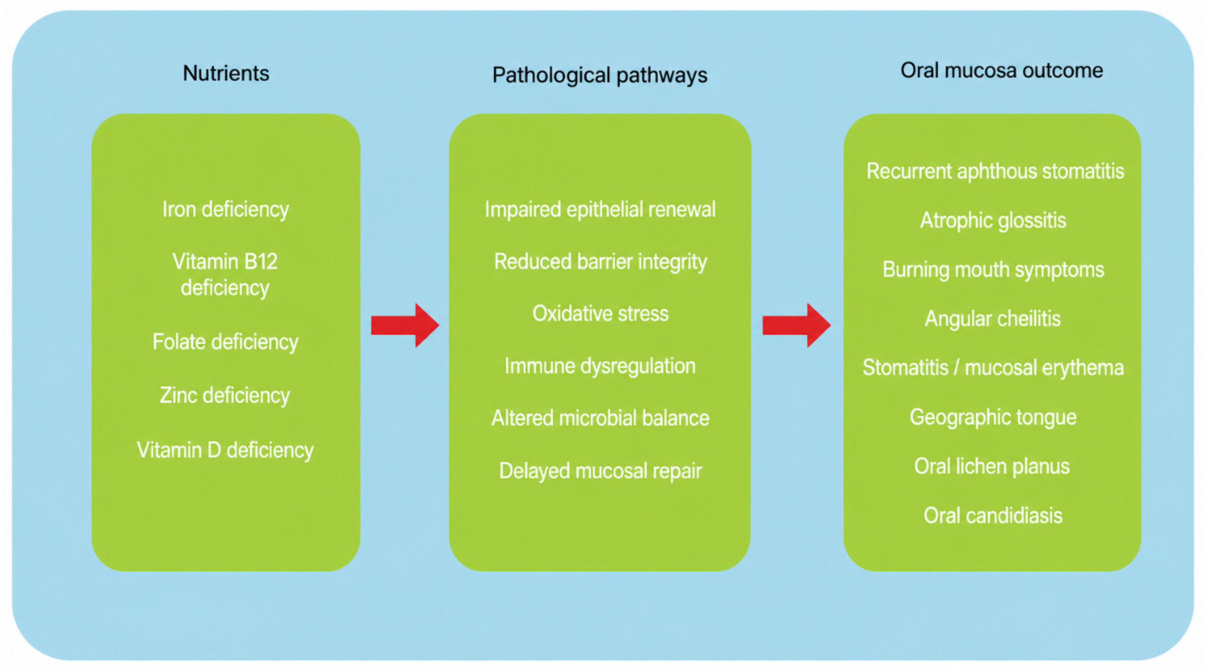
3.1. Pathophysiology
3.2. Recurrent Aphthous Stomatitis (RAS)
3.3. Atrophic Glossitis
3.4. Burning Mouth Symptoms (BMS)/Glossodynia
3.5. Angular Cheilitis and Mucosal Erythema
3.6. Oral Lichen Planus and Related Inflammatory Mucosal Disease
3.7. Geographic Tongue and Lingual Morphologic Change
3.8. Oral Candidiasis in Malnutrition
3.9. Differential Diagnosis and Clinical Interpretation
3.10. Nutrition in Periodontal Disease
4. Diagnostic Work-Up
5. Management Implications
6. Limitations of the Evidence
7. Conclusions
Author Contributions
Funding
Data Availability Statement
Conflicts of Interest
References
- Walker, J.E.G. Aphthous Ulceration and Vitamin B12 Deficiency. Br. J. Oral Surg. 1973, 11, 165–170. [Google Scholar] [CrossRef] [Scilit] [PubMed]
- Wray, D.; Ferguson, M.M.; Mason, D.K.; Hutcheon, A.W.; Dagg, J.H. Recurrent Aphthae: Treatment with Vitamin B12, Folic Acid, and Iron. BMJ 1975, 2, 490–493. [Google Scholar] [CrossRef] [Scilit] [PubMed]
- Tyldesley, W.R. Stomatitis and Recurrent Oral Ulceration: Is a Full Blood Screen Necessary? Br. J. Oral Surg. 1983, 21, 27–30. [Google Scholar] [CrossRef] [Scilit] [PubMed]
- Avery, K.T.; Shapiro, S.; Hamby, C.L. Oral Manifestations of Vitamin Deficiencies. J. Colo. Dent. Assoc. 1985, 63, 4–7. [Google Scholar] [PubMed]
- Wang, Z.; Cao, H.; Xiong, J.; Lu, Y.; Deng, Y.; Nan, H.; Zheng, S.; Ye, H.; Cao, Z. Recent Advances in the Aetiology of Recurrent Aphthous Stomatitis (RAS). Postgrad. Med. J. 2022, 98, 57–66. [Google Scholar] [CrossRef] [Scilit]
- Lin, D.; Yang, L.; Wen, L.; Lu, H.; Chen, Q.; Wang, Z. Crosstalk between the Oral Microbiota, Mucosal Immunity, and the Epithelial Barrier Regulates Oral Mucosal Disease Pathogenesis. Mucosal Immunol. 2021, 14, 1247–1258. [Google Scholar] [CrossRef] [Scilit] [PubMed]
- Strączek, A.; Szałkowska, J.; Sutkowska, P.; Srebrna, A.; Puzio, N.; Piasecka, A.; Piskorz, N.; Błaszczyk, J.; Thum-Tyzo, K. Impact of Nutrition on the Condition of the Oral Mucosa and Periodontium: A Narrative Review. Dent. Med. Probl. 2023, 60, 697–707. [Google Scholar] [CrossRef] [Scilit] [PubMed]
- Zinder, R.; Cooley, R.; Vlad, L.G.; Molnar, J.A. Vitamin A and Wound Healing. Nutr. Clin. Pract. 2019, 34, 839–849. [Google Scholar] [CrossRef] [Scilit]
- Lin, J.A.; Woods, E.R.; Bern, E.M. Common and Emergent Oral and Gastrointestinal Manifestations of Eating Disorders. Gastroenterol. Hepatol. 2021, 17, 157–167. [Google Scholar]
- Boukssim, S.; Chbicheb, S. Oral Manifestations of Vitamin B12 Deficiency Associated with Pernicious Anemia: A Case Report. Int. J. Surg. Case Rep. 2024, 121, 109931. [Google Scholar] [CrossRef] [Scilit] [PubMed]
- Radochová, V.; Slezák, R.; Radocha, J. Oral Manifestations of Nutritional Deficiencies: Single Centre Analysis. Acta Medica 2020, 63, 95–100. [Google Scholar] [CrossRef] [Scilit] [PubMed]
- Tolkachjov, S.N.; Bruce, A.J. Oral Manifestations of Nutritional Disorders. Clin. Dermatol. 2017, 35, 441–452. [Google Scholar] [CrossRef] [Scilit] [PubMed]
- Chi, A.C.; Neville, B.W.; Krayer, J.W.; Gonsalves, W.C. Oral Manifestations of Systemic Disease. Am. Fam. Physician 2010, 82, 1381–1388. [Google Scholar] [PubMed]
- Alotaibi, T. Malnutrition and Diet Role in Prevention of Oral Disease. EC Dent. Sci. 2019, 18, 2206–2213. [Google Scholar]
- Chamut, S.; Alhassan, M.; Hameedaldeen, A.; Kaplish, S.; Yang, A.H.; Wade, C.G.; Alghamdi, S.; Chamut, D.; Novy, B.B.; Chandel, T. Every Bite Counts to Achieve Oral Health: A Scoping Review on Diet and Oral Health Preventive Practices. Int. J. Equity Health 2024, 23, 261. [Google Scholar] [CrossRef] [Scilit] [PubMed]
- Bhateja, S.; Kaushik, A.; Tripathi, R.; Asija, S.; Tanwar, R.; Meraj, A. Nutritional Aspects of Oral Disorders—A Review. IP J. Nutr. Metab. Health Sci. 2022, 5, 1–6. [Google Scholar] [CrossRef] [Scilit]
- Ghasemi, S.; Farokhpour, F.; Mortezagholi, B.; Movahed, E.; Ghaedi, A.; Gargari, M.K.; Khanzadeh, M.; Bazrgar, A.; Khanzadeh, S. Systematic Review and Meta-Analysis of Oxidative Stress and Antioxidant Markers in Recurrent Aphthous Stomatitis. BMC Oral Health 2023, 23, 960. [Google Scholar] [CrossRef] [Scilit] [PubMed]
- Azzolino, D.; Passarelli, P.C.; De Angelis, P.; Piccirillo, G.B.; D’addona, A.; Cesari, M. Poor Oral Health as a Determinant of Malnutrition and Sarcopenia. Nutrients 2019, 11, 2898. [Google Scholar] [CrossRef] [Scilit] [PubMed]
- Martinon, P.; Fraticelli, L.; Giboreau, A.; Dussart, C.; Bourgeois, D.; Carrouel, F. Nutrition as a Key Modifiable Factor for Periodontitis and Main Chronic Diseases. J. Clin. Med. 2021, 10, 197. [Google Scholar] [CrossRef] [Scilit] [PubMed]
- Yesudian, R.I.; Yesudian, P.D. Vitamins and Oral Mucosal Diseases: From Bench to the Bedside. Clin. Dermatol. 2026, 44, 281–293. [Google Scholar] [CrossRef] [Scilit] [PubMed]
- Wu, Y.-C.; Wu, Y.-H.; Wang, Y.-P.; Chang, J.Y.-F.; Chen, H.-M.; Sun, A. Hematinic Deficiencies and Anemia Statuses in Recurrent Aphthous Stomatitis Patients with or without Atrophic Glossitis. J. Formos. Med. Assoc. 2016, 115, 1061–1068. [Google Scholar] [CrossRef] [Scilit] [PubMed]
- Chiang, C.-P.; Wu, Y.-C.; Chang, J.Y.-F.; Wang, Y.-P.; Wu, Y.-H.; Sun, A. Anemia, Hematinic Deficiencies, Hyperhomocysteinemia, and Gastric Parietal Cell Antibody Positivity in Atrophic Glossitis Patients with Iron Deficiency. J. Formos. Med. Assoc. 2020, 119, 587–594. [Google Scholar] [CrossRef] [Scilit] [PubMed]
- Rahman, N.; Walls, A. Chapter 12: Nutrient Deficiencies and Oral Health. Monogr. Oral Sci. 2020, 28, 114–124. [Google Scholar] [CrossRef] [Scilit] [PubMed]
- Mousavi, T.; Jalali, H.; Moosazadeh, M. Hematological Parameters in Patients with Recurrent Aphthous Stomatitis: A Systematic Review and Meta-Analysis. BMC Oral Health 2024, 24, 339. [Google Scholar] [CrossRef] [Scilit] [PubMed]
- Torabinia, N.; Asadi, S.; Tarrahi, M.J. The Relationship Between Iron and Zinc Deficiency and Aphthous Stomatitis: A Systematic Review and Meta-Analysis. Adv. Biomed. Res. 2024, 13, 31. [Google Scholar] [CrossRef] [Scilit] [PubMed]
- Bao, Z.; Yang, X.; Shi, J.; Liu, L. Serum Zinc Levels in 368 Patients with Oral Mucosal Diseases: A Preliminary Study. Med. Oral Patol. Oral Cir. Bucal 2016, 21, e335–e340. [Google Scholar] [CrossRef] [Scilit] [PubMed]
- Ślebioda, Z.; Krawiecka, E.; Szponar, E.; Dorocka-Bobkowska, B. Evaluation of Serum Zinc Levels in Patients with Recurrent Aphthous Stomatitis (RAS). BMC Oral Health 2017, 17, 158. [Google Scholar] [CrossRef] [Scilit] [PubMed]
- Halboub, E.; Al-Maweri, S.A.; Parveen, S.; Al-Wesabi, M.; Al-Sharani, H.M.; Al-Sharani, A.; Al-Kamel, A.; Albashari, A.; Shamala, A. Zinc Supplementation for Prevention and Management of Recurrent Aphthous Stomatitis: A Systematic Review. J. Trace Elem. Med. Biol. 2021, 68, 126811. [Google Scholar] [CrossRef] [Scilit] [PubMed]
- Carr, A.; Maggini, S. Vitamin C and Immune Function. Nutrients 2017, 9, 1211. [Google Scholar] [CrossRef] [Scilit] [PubMed]
- Munday, M.-R.; Rodricks, R.; Fitzpatrick, M.; Flood, V.M.; Gunton, J.E. A Pilot Study Examining Vitamin C Levels in Periodontal Patients. Nutrients 2020, 12, 2255. [Google Scholar] [CrossRef] [Scilit] [PubMed]
- Hrubša, M.; Siatka, T.; Nejmanová, I.; Vopršalová, M.; Kujovská Krčmová, L.; Matoušová, K.; Javorská, L.; Macáková, K.; Mercolini, L.; Remião, F.; et al. Biological Properties of Vitamins of the B-Complex, Part 1: Vitamins B1, B2, B3, and B5. Nutrients 2022, 14, 484. [Google Scholar] [CrossRef] [Scilit] [PubMed]
- Pludowski, P.; Holick, M.F.; Grant, W.B.; Konstantynowicz, J.; Mascarenhas, M.R.; Haq, A.; Povoroznyuk, V.; Balatska, N.; Barbosa, A.P.; Karonova, T.; et al. Vitamin D Supplementation Guidelines. J. Steroid Biochem. Mol. Biol. 2018, 175, 125–135. [Google Scholar] [CrossRef] [Scilit] [PubMed]
- Fathallh, A.; Al-Sudani, F.; Almuhssen, S.; Hassoon, S. The Impact of Malnutrition on Dental Health of 12-Year-Old Children: A Study on Permanent Teeth Caries, Cariogenic Bacteria and Salivary IgA. Folia Medica 2024, 66, 515–520. [Google Scholar] [CrossRef] [Scilit] [PubMed]
- Rajasekaran, J.J.; Krishnamurthy, H.K.; Bosco, J.; Jayaraman, V.; Krishna, K.; Wang, T.; Bei, K. Oral Microbiome: A Review of Its Impact on Oral and Systemic Health. Microorganisms 2024, 12, 1797. [Google Scholar] [CrossRef] [Scilit] [PubMed]
- Yang, Z.; Cui, Q.; An, R.; Wang, J.; Song, X.; Shen, Y.; Wang, M.; Xu, H. Comparison of Microbiomes in Ulcerative and Normal Mucosa of Recurrent Aphthous Stomatitis (RAS)-Affected Patients. BMC Oral Health 2020, 20, 128. [Google Scholar] [CrossRef] [Scilit] [PubMed]
- Wang, G.; Tong, X.; Zhang, C.; Zhuo, R.; Liu, C.; Wang, C.; Hao, M.; Ren, L. Analysis of Oral Microbiota in Herpetiform Aphthous Ulcers Patients. Oral Health Prev. Dent. 2025, 23, 447–456. [Google Scholar] [CrossRef] [Scilit] [PubMed]
- Seo, H.R.; Chung, K.B.; Kim, D.-Y. The Possible Impact of Zinc-Enriched Multivitamins on Treatment-Naïve Recurrent Aphthous Stomatitis Patients. J. Clin. Med. 2025, 14, 260. [Google Scholar] [CrossRef] [Scilit] [PubMed]
- Oluwadaisi, A.M.; Aborisade, A.O.; Oyetola, E.O.; Owotade, F.J.; Agho, E.T. Hematological Parameters, Obesity, Stress and Oxidant-Antioxidant Indicators in Patients with or without Recurrent Aphthous Stomatitis: A Case Control Study. J. Stomatol. Oral Maxillofac. Surg. 2023, 124, 101654. [Google Scholar] [CrossRef] [Scilit] [PubMed]
- Elvhira Rosa, D.; Nur’aeny, N. The Relationship of Micronutrients and Oral Mucosa Diseases: A Systematic Review. Dentino J. Kedokt. Gigi 2022, 7, 82–87. [Google Scholar] [CrossRef] [Scilit]
- Al-hamdani, G.M.; Yas, L.S. Serum and Salivary Vitamin B12 Levels among Iron Deficiency Anemia Patient with Recurrent Aphthous Stomatitis. Dent. Hypotheses 2023, 14, 55–58. [Google Scholar] [CrossRef] [Scilit] [PubMed]
- Chiang, C.-P.; Yu-Fong Chang, J.; Wang, Y.-P.; Wu, Y.-H.; Wu, Y.-C.; Sun, A. Recurrent Aphthous Stomatitis–Etiology, Serum Autoantibodies, Anemia, Hematinic Deficiencies, and Management. J. Formos. Med. Assoc. 2019, 118, 1279–1289. [Google Scholar] [CrossRef] [Scilit] [PubMed]
- Ślebioda, Z.; Krawiecka, E.; Szponar, E.; Dorocka-Bobkowska, B. Haematinic Deficiencies and Patient Clinical Profiles in Polish Patients with Recurrent Aphthous Stomatitis (RAS). J. Oral Pathol. Med. 2018, 47, 531–537. [Google Scholar] [CrossRef] [Scilit] [PubMed]
- Cui, J.; Lin, W.; Ma, X.; Wang, J. Clinical Evaluation and Therapeutic Effects of Combination Treatment with Mecobalamin + Vitamin E in Recurrent Oral Ulcer. Clin. Ther. 2022, 44, 123–129. [Google Scholar] [CrossRef] [Scilit] [PubMed]
- Al-Amad, S.H.; Hasan, H. Vitamin D and Hematinic Deficiencies in Patients with Recurrent Aphthous Stomatitis. Clin. Oral Investig. 2020, 24, 2427–2432. [Google Scholar] [CrossRef] [Scilit] [PubMed]
- Öztekin, A.; Öztekin, C. Vitamin D Levels in Patients with Recurrent Aphthous Stomatitis. BMC Oral Health 2018, 18, 186. [Google Scholar] [CrossRef] [Scilit] [PubMed]
- Diachkova, E.; Trifonova, D.; Morozova, E.; Runova, G.; Ashurko, I.; Ibadulaeva, M.; Fadeev, V.; Tarasenko, S. Vitamin D and Its Role in Oral Diseases Development. Scoping Rev. Dent. J. 2021, 9, 129. [Google Scholar] [CrossRef] [Scilit] [PubMed]
- Zakeri, M.; Parsian, H.; Bijani, A.; Shirzad, A.; Neamati, N. Serum Levels of Vitamin D in Patients with Recurrent Aphthous Stomatitis. Dent. Med. Probl. 2021, 58, 27–30. [Google Scholar] [CrossRef] [Scilit] [PubMed]
- Alli, B.Y.; Erinoso, O.A.; Olawuyi, A.B. Effect of Sodium Lauryl Sulfate on Recurrent Aphthous Stomatitis: A Systematic Review. J. Oral Pathol. Med. 2019, 48, 358–364. [Google Scholar] [CrossRef] [Scilit] [PubMed]
- Al-Aizari, N.A.; Al-Shamiri, H.M.; AlShehri, B.K.; Alhomood, K.S.; Alzahrani, S.R.; Abuhasna, W.R.; Al-Maweri, S.A. Evidence-Based Recommendations for the Treatment of Recurrent Aphthous Stomatitis: Insights from an Umbrella Review. J. Dermatol. Treat. 2026, 37, 2622245. [Google Scholar] [CrossRef] [Scilit] [PubMed]
- Sabri, H.; Derakhshan Barjoei, M.M.; Azarm, A.; Sadighnia, N.; Shakiba, R.; Aghebati, G.; Hadilou, N.; Kheiri, P.; Ghanbari, F.; Deravi, N.; et al. The Yin and Yang of Sodium Lauryl Sulfate Use for Oral and Periodontal Health: A Literature Review. J. Dent. 2023, 24, 262–276. [Google Scholar] [CrossRef] [PubMed] [PubMed Central]
- Chuang, A.H.; Bordlemay, J.; Goodin, J.L.; McPherson, J.C. Effect of Sodium Lauryl Sulfate (SLS) on Primary Human Gingival Fibroblasts in an In Vitro Wound Healing Model. Mil. Med. 2019, 184, 97–101. [Google Scholar] [CrossRef] [Scilit] [PubMed]
- Chiang, C.-P.; Chang, J.Y.-F.; Wang, Y.-P.; Wu, Y.-H.; Wu, Y.-C.; Sun, A. Anemia, Hematinic Deficiencies, and Hyperhomocysteinemia in Gastric Parietal Cell Antibody-Positive and -Negative Atrophic Glossitis Patients. J. Formos. Med. Assoc. 2019, 118, 565–571. [Google Scholar] [CrossRef] [Scilit] [PubMed]
- Chiang, C.-P.; Wu, Y.-H.; Chang, J.Y.-F.; Wang, Y.-P.; Wu, Y.-C.; Sun, A. Hematinic Deficiencies, Hyperhomocysteinemia, and Gastric Parietal Cell Antibody Positivity in Atrophic Glossitis Patients with Macrocytosis. J. Formos. Med. Assoc. 2019, 118, 1515–1521. [Google Scholar] [CrossRef] [Scilit] [PubMed]
- Chiang, C.-P.; Chang, J.Y.-F.; Wang, Y.-P.; Wu, Y.-C.; Wu, Y.-H.; Sun, A. Significantly Higher Frequencies of Anemia, Hematinic Deficiencies, Hyperhomocysteinemia, and Serum Gastric Parietal Cell Antibody Positivity in Atrophic Glossitis Patients. J. Formos. Med. Assoc. 2018, 117, 1065–1071. [Google Scholar] [CrossRef] [Scilit] [PubMed]
- Chiang, C.-P.; Chang, J.Y.-F.; Wang, Y.-P.; Wu, Y.-C.; Wu, Y.-H.; Sun, A. Anemia, Hematinic Deficiencies, Hyperhomocysteinemia, and Serum Gastric Parietal Cell Antibody Positivity in Atrophic Glossitis Patients with or without Microcytosis. J. Formos. Med. Assoc. 2019, 118, 1401–1407. [Google Scholar] [CrossRef] [Scilit] [PubMed]
- Chen, G.-Y.; Tang, Z.-Q.; Bao, Z.-X. Vitamin B12 Deficiency May Play an Etiological Role in Atrophic Glossitis and Its Grading: A Clinical Case-Control Study. BMC Oral Health 2022, 22, 456. [Google Scholar] [CrossRef] [Scilit] [PubMed]
- Wu, Y.-H.; Yu-Fong Chang, J.; Wang, Y.-P.; Wu, Y.-C.; Chen, H.-M.; Sun, A. Hemoglobin, Iron, Vitamin B12, and Folic Acid Deficiencies and Hyperhomocysteinemia in Behcet’s Disease Patients with Atrophic Glossitis. J. Formos. Med. Assoc. 2018, 117, 559–565. [Google Scholar] [CrossRef] [Scilit] [PubMed]
- Chiang, C.-P.; Wu, Y.-H.; Wu, Y.-C.; Chang, J.Y.-F.; Wang, Y.-P.; Sun, A. Anemia, Hematinic Deficiencies, Hyperhomocysteinemia, and Serum Gastric Parietal Cell Antibody Positivity in 884 Patients with Burning Mouth Syndrome. J. Formos. Med. Assoc. 2020, 119, 813–820. [Google Scholar] [CrossRef] [Scilit] [PubMed]
- Chiang, C.-P.; Chang, J.Y.-F.; Wang, Y.-P.; Wu, Y.-H.; Wu, Y.-C.; Sun, A. Atrophic Glossitis: Etiology, Serum Autoantibodies, Anemia, Hematinic Deficiencies, Hyperhomocysteinemia, and Management. J. Formos. Med. Assoc. 2020, 119, 774–780. [Google Scholar] [CrossRef] [Scilit] [PubMed]
- Chiang, C.-P.; Yu-Fong Chang, J.; Wu, Y.-H.; Sun, A.; Wang, Y.-P.; Chen, H.-M. Hematinic Deficiencies and Anemia in Gastric Parietal Cell Antibody-Positive and -Negative Oral Submucous Fibrosis Patients. J. Dent. Sci. 2018, 13, 68–74. [Google Scholar] [CrossRef] [Scilit] [PubMed]
- Bao, Z.; Yang, X.; Shi, J.; Wang, Y. The Profile of Hematinic Deficiencies in Patients with Oral Lichen Planus: A Case-Control Study. BMC Oral Health 2020, 20, 252. [Google Scholar] [CrossRef] [Scilit] [PubMed]
- Chiang, C.-P.; Yu-Fong Chang, J.; Wang, Y.-P.; Wu, Y.-H.; Lu, S.-Y.; Sun, A. Oral Lichen Planus–Differential Diagnoses, Serum Autoantibodies, Hematinic Deficiencies, and Management. J. Formos. Med. Assoc. 2018, 117, 756–765. [Google Scholar] [CrossRef] [Scilit] [PubMed]
- Chiang, M.-L.; Wu, Y.-H.; Chang, J.Y.-F.; Wang, Y.-P.; Wu, Y.-C.; Sun, A. Anemia, Hematinic Deficiencies, and Hyperhomocysteinemia in Gastric Parietal Cell Antibody-Positive and -Negative Burning Mouth Syndrome Patients. J. Formos. Med. Assoc. 2021, 120, 819–826. [Google Scholar] [CrossRef] [Scilit] [PubMed]
- Jankovskis, V.; Selga, G. Vitamin B and Zinc Supplements and Capsaicin Oral Rinse Treatment Options for Burning Mouth Syndrome. Medicina 2021, 57, 391. [Google Scholar] [CrossRef] [Scilit] [PubMed]
- Bačun, B.; Galić, D.; Pul, L.; Tomas, M.; Kuiš, D. Manifestations and Treatment of Hypovitaminosis in Oral Diseases: A Systematic Review. Dent. J. 2024, 12, 152. [Google Scholar] [CrossRef] [Scilit] [PubMed]
- Freitas, J.; Bliven, P.; Case, R. Combined Zinc and Vitamin B6 Deficiency in a Patient with Diffuse Red Rash and Angular Cheilitis 6 Years after Roux-En-Y Gastric Bypass. BMJ Case Rep. 2019, 12, e230605. [Google Scholar] [CrossRef] [Scilit] [PubMed]
- Rakhmayanthie, N.; Herawati, E.; Herawati, D.M.D. Effect of Nutritional Intake towards Angular Cheilitis of Orphanage Children. Padjadjaran J. Dent. 2016, 28, 170–176. [Google Scholar] [CrossRef] [Scilit]
- Oza, N.; Doshi, J. Angular Cheilitis: A Clinical and Microbial Study. Indian J. Dent. Res. 2017, 28, 661. [Google Scholar] [CrossRef] [Scilit] [PubMed]
- Chiriac, A.; Chiriac, A.E.; Pinteala, T.; Spinei, A.; Savin, L.; Zelenkova, H.; Wollina, U. Angular Cheilitis—An Oral Disease with Many Facets. Wien. Med. Wochenschr. 2024, 174, 315–322. [Google Scholar] [CrossRef] [Scilit] [PubMed]
- Brantes, M.-F.; Azevedo, R.-S.; Rozza-de-Menezes, R.-E.; Póvoa, H.-C.; Tucci, R.; Gouvêa, A.-F.; Takahama, A., Jr. Analysis of Risk Factors for Maxillary Denture-Related Oral Mucosal Lesions: A Cross-Sectional Study. Med. Oral Patol. Oral Cir. Bucal 2019, 24, e305–e313. [Google Scholar] [CrossRef] [Scilit] [PubMed]
- Mahdani, F.Y.; Jati, G.D.; Febrine, E.T.; Cahyaningrum, K.W.; Radithia, D.; Wicaksono, S. Knowledge of Xerostomia and Angular Cheilitis in Geriatric Population among Clinical Dental Students: An Institutional Cross-Sectional Study. J. Int. Soc. Prev. Community Dent. 2023, 13, 443–449. [Google Scholar] [CrossRef] [Scilit] [PubMed]
- Ponjayanthi, V.; Shiamala, J.; Sivaguru, K.; Velmurugan, P.; Maheswaran, T.; Mani, J. Diagnosis and Management of Angular Cheilitis after Prosthodontic Rehabilitation: An Overview. Saudi J. Oral Dent. Res. 2026, 11, 192–194. [Google Scholar] [CrossRef] [Scilit]
- Wu, Y.-H.; Yu-Fong Chang, J.; Lee, Y.-P.; Wang, Y.-P.; Sun, A.; Chiang, C.-P. Anemia, Hematinic Deficiencies, Hyperhomocysteinemia, and Serum Gastric Parietal Cell Antibody Positivity in 588 Patients with Oral Lichen Planus. J. Dent. Sci. 2025, 20, 660–666. [Google Scholar] [CrossRef] [Scilit] [PubMed]
- Wu, Y.-H.; Yu-Fong Chang, J.; Lee, Y.-P.; Wang, Y.-P.; Sun, A.; Chiang, C.-P. Anemia, Hematinic Deficiencies, Hyperhomocysteinemia, and Serum Gastric Parietal Cell Antibody Positivity in Oral Lichen Planus Patients with Vitamin B12 Deficiency. J. Dent. Sci. 2025, 20, 1102–1109. [Google Scholar] [CrossRef] [Scilit] [PubMed]
- Chaitanya, N.C.; Chintada, S.; Kandi, P.; Kanikella, S.; Kammari, A.; Waghamare, R.S. Zinc Therapy in Treatment of Symptomatic Oral Lichen Planus. Indian Dermatol. Online J. 2019, 10, 174–177. [Google Scholar] [CrossRef] [Scilit] [PubMed]
- Rezazadeh, F.; Haghighat, S. Serum Vitamin Profile in Oral Lichen Planus Patients in Southwest of Iran. BioMed Res. Int. 2021, 2021, 8627435. [Google Scholar] [CrossRef] [Scilit] [PubMed]
- Chen, H.-M.; Wang, Y.-P.; Chang, J.Y.-F.; Wu, Y.-C.; Cheng, S.-J.; Sun, A. Significant Association of Deficiencies of Hemoglobin, Iron, Folic Acid, and Vitamin B12 and High Homocysteine Level with Oral Lichen Planus. J. Formos. Med. Assoc. 2015, 114, 124–129. [Google Scholar] [CrossRef] [Scilit] [PubMed]
- Naik, S.R.; Gupta, P.; Khaitan, T.; Shukla, A.K. Reduced Levels of Serum Vitamin B12 in Symptomatic Cases of Oral Lichen Planus: A Cross-Sectional Study. J. Oral Biol. Craniofac. Res. 2020, 10, 578–582. [Google Scholar] [CrossRef] [Scilit] [PubMed]
- Khayamzadeh, M.; Najafi, S.; Sadrolodabaei, P.; Vakili, F.; Kharrazi Fard, M.J. Determining Salivary and Serum Levels of Iron, Zinc and Vitamin B12 in Patients with Geographic Tongue. J. Dent. Res. Dent. Clin. Dent. Prospects 2019, 13, 221–226. [Google Scholar] [CrossRef] [Scilit] [PubMed]
- Musaad, A.H.; H Abuaffan, A. Prevalence of Fissured and Geographic Tongue Abnormalities among University Students in Khartoum State, Sudan. Enzym. Eng. 2015, 5, 1000137. [Google Scholar] [CrossRef]
- Bao, Z.; Yang, X.; Fang, D. Lingual Linear Lesions: A Clinical Sign Strongly Suggestive of Severe Vitamin B12 Deficiency. Nutr. Clin. Pract. 2021, 36, 1041–1048. [Google Scholar] [CrossRef] [Scilit] [PubMed]
- Cagetti, M.G.; Wolf, T.G.; Tennert, C.; Camoni, N.; Lingström, P.; Campus, G. The Role of Vitamins in Oral Health. A Systematic Review and Meta-Analysis. Int. J. Environ. Res. Public Health 2020, 17, 938. [Google Scholar] [CrossRef] [Scilit] [PubMed]
- Zheng, Z.; Zhao, X.; Zhao, Q.; Zhang, Y.; Liu, S.; Liu, Z.; Meng, L.; Xin, Y.; Jiang, X. The Effects of Early Nutritional Intervention on Oral Mucositis and Nutritional Status of Patients With Head and Neck Cancer Treated with Radiotherapy. Front. Oncol. 2021, 10, 595632. [Google Scholar] [CrossRef] [Scilit] [PubMed]
- Vila, T.; Sultan, A.S.; Montelongo-Jauregui, D.; Jabra-Rizk, M.A. Oral Candidiasis: A Disease of Opportunity. J. Fungi 2020, 6, 15. [Google Scholar] [CrossRef] [Scilit] [PubMed]
- Jørgensen, M.R. Pathophysiological Microenvironments in Oral Candidiasis. Apmis 2024, 132, 956–973. [Google Scholar] [CrossRef] [Scilit] [PubMed]
- Yahya, A.A.; Almaeeni, M.M.; Alnuaimy, A.; Al-Mizraqchi, A.S.; Alhamdani, F.Y. Relationship Between Candida Albicans Colonization and Oral Health Status in Overweight/Obese Children. Afr. J. Biomed. Res. 2025, 28, 1151–1156. [Google Scholar] [CrossRef] [Scilit]
- Flondor, A.-E.; Sufaru, I.-G.; Martu, I.; Burlea, S.-L.; Flondor, C.; Toma, V. Nutritional Deficiencies and Oral Candidiasis in Children from Northeastern Romania: A Cross-Sectional Biochemical Assessment. Nutrients 2025, 17, 1815. [Google Scholar] [CrossRef] [Scilit] [PubMed]
- Hernawati, S. Relationship between Nutrition Deficiency, Oral Cavity Hygiene, and Oral Candidiasis in a 10-Years-Old Child. Health Notions 2019, 3, 469. [Google Scholar] [CrossRef] [Scilit]
- Grant, F.K.; Wanjala, R.; Low, J.; Levin, C.; Cole, D.C.; Okuku, H.S.; Ackatia-Armah, R.; Girard, A.W. Association between Infection and Nutritional Status among Infants in a Cohort Study of Vitamin A in Western Kenya. Front. Nutr. 2022, 9, 921213. [Google Scholar] [CrossRef] [Scilit] [PubMed]
- Wong, F.M.F.; Leung, W.K. Oral Health and Nutrition in Older Adults. Nutrients 2025, 17, 3046. [Google Scholar] [CrossRef] [Scilit] [PubMed]
- Gutierrez Gossweiler, A.; Martinez-Mier, E.A. Chapter 6: Vitamins and Oral Health. Monogr. Oral Sci. 2020, 28, 59–67. [Google Scholar] [CrossRef] [Scilit] [PubMed]
- Siddaiah Madhusudhan, K.; Pallavi, M.R. Perspective Chapter: Effects of Malnutrition on Pediatric Oral Health—A Review. In Pediatric Dentistry—A Comprehensive Guide; IntechOpen: Rijeka, Croatia, 2023. [Google Scholar]
- Helail, B. Oral Manifestations of Malnutrition and Causes. Dent. Oral Maxillofac. Surg. 2022, 4, 1–3. [Google Scholar] [CrossRef] [Scilit] [PubMed]
- Mostad, I.L.; Reinan, T.K.; Halgunset, J.; Thoresen, L.; Feuerherm, A.J.; Kolberg, M. Oral Health Problems Are Associated with Malnutrition in Hospitalised Adult Patients. Clin. Nutr. ESPEN 2023, 57, 527–536. [Google Scholar] [CrossRef] [Scilit] [PubMed]
- Algra, Y.; Haverkort, E.; Kok, W.; Etten-Jamaludin, F.v.; Schoot, L.v.; Hollaar, V.; Naumann, E.; Schueren, M.d.v.d.; Jerković-Ćosić, K. The Association between Malnutrition and Oral Health in Older People: A Systematic Review. Nutrients 2021, 13, 3584. [Google Scholar] [CrossRef] [Scilit] [PubMed]
- Vieira, K.A.; Rosa-Júnior, L.S.; Souza, M.A.V.; Santos, N.B.; Florêncio, T.M.M.T.; Bussadori, S.K. Chronic Malnutrition and Oral Health Status in Children Aged 1 to 5 Years. Medicine 2020, 99, e19595. [Google Scholar] [CrossRef] [Scilit] [PubMed]
- Toniazzo, M.P.; Amorim, P.S.; Rost, J.F.; Feldman, J.V.; Beretta, M.V.; Rodrigues, T.C.; Weidlich, P. Oral Condition as a Predictor of Risk for Malnutrition during Hospitalization: A Cross-sectional Study. Nutr. Clin. Pract. 2022, 37, 1438–1447. [Google Scholar] [CrossRef] [Scilit] [PubMed]
- Kossioni, A.E. The Association of Poor Oral Health Parameters with Malnutrition in Older Adults: A Review Considering the Potential Implications for Cognitive Impairment. Nutrients 2018, 10, 1709. [Google Scholar] [CrossRef] [Scilit] [PubMed]
- Curca, F.R.; Luchian, I.; Bida, F.C.; Virvescu, D.I.; Rotundu, G.; Butnaru, O.M.; Balan, G.; Surlari, Z.; Georgescu, A.; Pasarin, L.; et al. From Diet to Oral and Periodontal Health: Exploring the Crucial Role of Nutrition—A Narrative Review. Nutrients 2026, 18, 168. [Google Scholar] [CrossRef] [Scilit] [PubMed]
- Antonelli, J.R.; Seltzer, R. Oral and Physical Manifestations of Anorexia and Bulimia Nervosa. Tex. Dent. J. 2016, 133, 528–535. [Google Scholar] [PubMed]
- Bassiouny, M.A. Oral Health Considerations in Anorexia and Bulimia Nervosa. 1. Symptomatology and Diagnosis. Gen. Dent. 2017, 65, 34–40. [Google Scholar] [PubMed]
- Bahramian, A.; Falsafi, P.; Abbasi, T.; Ghanizadeh, M.; Abedini, M.; Kavoosi, F.; Kouhsoltani, M.; Noorbakhsh, F.; Dabbaghi Tabriz, F.; Rajaeih, S.; et al. Comparing Serum and Salivary Levels of Vitamin D in Patients with Recurrent Aphthous Stomatitis and Healthy Individuals. J. Dent. 2018, 19, 295–300. [Google Scholar]
- Khademi, H.; Khozeimeh, F.; Tavangar, A.; Amini, S.; Ghalayani, P. The Serum and Salivary Level of Malondialdehyde, Vitamins A, E, and C in Patient with Recurrent Aphthous Stomatitis. Adv. Biomed. Res. 2014, 3, 246. [Google Scholar] [CrossRef] [Scilit] [PubMed]
- Toniazzo, M.P.; Amorim, P.d.S.; Muniz, F.W.M.G.; Weidlich, P. Relationship of Nutritional Status and Oral Health in Elderly: Systematic Review with Meta-Analysis. Clin. Nutr. 2018, 37, 824–830. [Google Scholar] [CrossRef] [Scilit] [PubMed]
- Kuo, Y.-S.; Chang, J.Y.-F.; Wang, Y.-P.; Wu, Y.-C.; Wu, Y.-H.; Sun, A. Significantly Higher Frequencies of Hemoglobin, Iron, Vitamin B12, and Folic Acid Deficiencies and of Hyperhomocysteinemia in Patients with Behcet’s Disease. J. Formos. Med. Assoc. 2018, 117, 932–938. [Google Scholar] [CrossRef] [Scilit] [PubMed]
- Song, J.; Yu, H.; Ryu, E. Comparative Analysis of Chemotherapy-Induced Oral Mucositis, Nutritional Status, and Depression in Patients with Acute Leukemia. Eur. J. Cancer Care 2025, 2025, 7989087. [Google Scholar] [CrossRef] [Scilit]
- Manifar, S.; Tonkaboni, A.; Sobhanifar, A.; HafeziMotlagh, K.; Bitarafan, S.; Mazani, M.; Bossi, P. Dietary Intake Effects on Severity of Cancer Treatment-Induced Mucositis: A Cross-Sectional Study. Health Sci. Rep. 2023, 6, e1706. [Google Scholar] [CrossRef] [Scilit] [PubMed]
- García-Gozalbo, B.; Cabañas-Alite, L. A Narrative Review about Nutritional Management and Prevention of Oral Mucositis in Haematology and Oncology Cancer Patients Undergoing Antineoplastic Treatments. Nutrients 2021, 13, 4075. [Google Scholar] [CrossRef] [Scilit] [PubMed]
- Poisson, P.; Laffond, T.; Campos, S.; Dupuis, V.; Bourdel-Marchasson, I. Relationships between Oral Health, Dysphagia and Undernutrition in Hospitalised Elderly Patients. Gerodontology 2016, 33, 161–168. [Google Scholar] [CrossRef] [Scilit] [PubMed]
- Chang, J.Y.-F.; Wang, Y.-P.; Wu, Y.-C.; Cheng, S.-J.; Chen, H.-M.; Sun, A. Hematinic Deficiencies and Pernicious Anemia in Oral Mucosal Disease Patients with Macrocytosis. J. Formos. Med. Assoc. 2015, 114, 736–741. [Google Scholar] [CrossRef] [Scilit] [PubMed]
- Wu, Y.-H.; Wu, Y.-C.; Chu, F.-Y.; Cheng, S.-J.; Sun, A.; Chen, H.-M. Significantly Higher Frequencies of Hematinic Deficiencies and Hyperhomocysteinemia in Oral Precancer Patients. J. Formos. Med. Assoc. 2019, 118, 1299–1307. [Google Scholar] [CrossRef] [Scilit] [PubMed]
- Śmiga, M.; Olczak, T. Exploring Heme and Iron Acquisition Strategies of Porphyromonas Gingivalis-Current Facts and Hypotheses. FEMS Microbiol. Rev. 2025, 49, fuaf019. [Google Scholar] [CrossRef] [Scilit] [PubMed]
- Śmiga, M.; Ślęzak, P.; Tracz, M.; Cierpisz, P.; Wagner, M.; Olczak, T. Defining the Role of Hmu and Hus Systems in Porphyromonas Gingivalis Heme and Iron Homeostasis and Virulence. Sci. Rep. 2024, 14, 31156. [Google Scholar] [CrossRef] [Scilit] [PubMed]
- How, K.Y.; Song, K.P.; Chan, K.G. Porphyromonas gingivalis: An Overview of Periodontopathic Pathogen below the Gum Line. Front. Microbiol. 2016, 7, 53. [Google Scholar] [CrossRef] [Scilit] [PubMed]
- Hannibal, L.; Lysne, V.; Bjørke-Monsen, A.-L.; Behringer, S.; Grünert, S.C.; Spiekerkoetter, U.; Jacobsen, D.W.; Blom, H.J. Biomarkers and Algorithms for the Diagnosis of Vitamin B12 Deficiency. Front. Mol. Biosci. 2016, 3, 27. [Google Scholar] [CrossRef] [Scilit] [PubMed]
- Nair, G.R.; Naidu, G.S.; Jain, S.; Nagi, R.; Makkad, R.S.; Jha, A. Clinical Effectiveness of Aloe Vera in the Management of Oral Mucosal Diseases—A Systematic Review. J. Clin. Diagn. Res. 2016, 10, ZE01–ZE07. [Google Scholar] [CrossRef] [Scilit] [PubMed]
- Miyan, Z.; Waris, N. Association of Vitamin B12 Deficiency in People with Type 2 Diabetes on Metformin and without Metformin: A Multicenter Study, Karachi, Pakistan. BMJ Open Diabetes Res. Care 2020, 8, e001151. [Google Scholar] [CrossRef] [Scilit] [PubMed]
- Mays, J.W.; Sarmadi, M.; Moutsopoulos, N.M. Oral Manifestations of Systemic Autoimmune and Inflammatory Diseases: Diagnosis and Clinical Management. J. Evid. Based. Dent. Pract. 2012, 12, 265–282. [Google Scholar] [CrossRef] [Scilit] [PubMed]
- Pflipsen, M.; Zenchenko, Y. Nutrition for Oral Health and Oral Manifestations of Poor Nutrition and Unhealthy Habits. Gen. Dent. 2017, 65, 36–43. [Google Scholar] [PubMed]
- Shipton, M.J.; Thachil, J. Vitamin B12 Deficiency—A 21st Century Perspective. Clin. Med. 2015, 15, 145–150. [Google Scholar] [CrossRef] [Scilit] [PubMed]
- Kłosek, S.; Szymczak-Paluch, M.; Bernaś, A.; Gawlak-Socka, S. Does Probiotic Intake Enhance the Efficacy of Oral Fungal Infection Treatment? Nutrients 2026, 18, 1433. [Google Scholar] [CrossRef] [Scilit] [PubMed]
- Temova Rakuša, Ž.; Roškar, R.; Hickey, N.; Geremia, S. Vitamin B12 in Foods, Food Supplements, and Medicines-A Review of Its Role and Properties with a Focus on Its Stability. Molecules 2022, 28, 240. [Google Scholar] [CrossRef] [Scilit] [PubMed]
- Shulpekova, Y.; Nechaev, V.; Kardasheva, S.; Sedova, A.; Kurbatova, A.; Bueverova, E.; Kopylov, A.; Malsagova, K.; Dlamini, J.C.; Ivashkin, V. The Concept of Folic Acid in Health and Disease. Molecules 2021, 26, 3731. [Google Scholar] [CrossRef] [Scilit] [PubMed]
- Baddam, S.; Khan, K.M.; Jialal, I. Folic Acid Deficiency. In StatPearls [Internet]; StatPearls Publishing: Treasure Island, FL, USA, 2026. [Google Scholar]
- Schoofs, H.; Schmit, J.; Rink, L. Zinc Toxicity: Understanding the Limits. Molecules 2024, 29, 3130. [Google Scholar] [CrossRef] [Scilit] [PubMed]
- Auerbach, M.; DeLoughery, T.G.; Tirnauer, J.S. Iron Deficiency in Adults: A Review. JAMA 2025, 333, 1813–1823. [Google Scholar] [CrossRef] [Scilit] [PubMed]
- Kolarš, B.; Mijatović Jovin, V.; Živanović, N.; Minaković, I.; Gvozdenović, N.; Dickov Kokeza, I.; Lesjak, M. Iron Deficiency and Iron Deficiency Anemia: A Comprehensive Overview of Established and Emerging Concepts. Pharmaceuticals 2025, 18, 1104. [Google Scholar] [CrossRef] [Scilit] [PubMed]
- Gondivkar, S.M.; Gadbail, A.R.; Gondivkar, R.S.; Sarode, S.C.; Sarode, G.S.; Patil, S.; Awan, K.H. Nutrition and Oral Health. Dis.-Mon. 2019, 65, 147–154. [Google Scholar] [CrossRef] [Scilit] [PubMed]
- Abharian, P.H.; Dehghan, P.; Abharian, P.H.; Tolouei, S. Molecular Characterization of Candida Dubliniensis and Candida Albicans in the Oral Cavity of Drug Abusers Using Duplex Polymerase Chain Reaction. Curr. Med. Mycol. 2018, 4, 12. [Google Scholar] [CrossRef] [PubMed]
- Thekiso, M.; Yengopal, V.; Rudolph, M. The Relationship between Oral Candidiasis and Micronutrient Deficiency amongst Adult Tuberculosis Patients in Alexandra, Johannesburg. S. Afr. Dent. J. 2018, 73, 489–498. [Google Scholar] [CrossRef] [Scilit]


| Reference Ranges | Route of Administration | Recommended Dosage | Treatment Duration | Monitoring Parameters | Relevant Precautions | |
|---|---|---|---|---|---|---|
| Vit. B12 | Serum concentration 150–500 pmol/L (200–575 ng/L) [122] In anemia, MCV may be elevated >100 fl. | Intramuscular or deep subcutaneous injection or oral (tablets, effervescent tablets, drops, syrups, oral sprays, capsules, etc.) [122]. I.M.—in severe cases—e.g., psychiatric, neurologic or in pregnant women | Intramuscular/ subcutaneous administration—1000–2000 µg 1xday Oral administration 1000–2000 µg 1xday [122]. | I.m./S.c.— 7–14 days 1xday, and then 1x/week for 4–8 weeks. Maintenance phase: s.c. 1000 µg 1×/month or 1×/3 months or 1000–2000 µg/d p.o | Serum vitamin B12 concentration > 150 pmol/L (200 ng/L) [122] After 7 days, Hb level should be elevated. | Population groups at high risk of Vit. B12 deficiency: vegetarians, vegans, the elderly, pregnant, and lactating women [122]. |
| Folate | [in serum] 6,8–26 nmol/L (3–12 μg/L) (on empty stomach) [in erythrocytes] 520–1600 nmol/L (235–721 μg/L) Also homocysteine concentration determines folate deficiency if: [in serum] homocysteine <4 ng/mL (<10 nmol/L) [in erythrocytes] <151 ng/mL (<340 nmol/L) [123]. | Oral supplementation [124] | 1–5 mg/day [124] | 1–4 months (until normalisation of blood indices) or as long as the reason of deficiency is present [124]. | Correct blood results, serum folate result over 6.8 nmol/L [123]. | Serum folate shows important variation, reflecting recent food intake, while red blood cell folate remains stable, showing average body folate status at the time of the production of the population of red cells. If accompanied with vit. B12 deficiency, needs to be treated simultaneously, to avoid neurological exacerbation [124]. |
| Zinc | Serum levels: Adults: 60–120 µg/dL (9–18 µmol/L), children: 11–24 µmol/L (70–160 µg/dL) Diagnosis of zinc deficiency is mostly based on clinical symptoms. Laboratory zinc values are less reliable in the diagnosis of zinc deficiency due to zinc sequestration in tissues caused by stress, infection, illness, or decreased serum concentration. | Change of dietary habits + Oral supplementation [125]. | Usually 25 mg; maximum daily dose: 40 mg [125] | Mainly elimination of symptoms of deficiency | High zinc intake levels can result in adverse effects, including nausea, vomiting, loss of appetite, abdominal cramps, diarrhea and interference with the body’s absorption of essential minerals, such as iron and copper [125]. | |
| Iron | Iron deficiency is diagnosed by low serum ferritin (typically <30 ng/mL) in individuals without inflammatory conditions + TSAT (transferrin saturation) <20% [126] | Oral administration: Iron salts (such as ferrous sulfate, ferrous gluconate, ferrous fumarate, and ferric citrate) on empty stomach (at least 1 h before meal) Intravenous administration: in most severe cases [127], e.g., oral iron intolerance, poor absorption, chronic inflammatory conditions [126]. | Oral: Traditional guidance: 150–200 mg split into 2–3 doses Recent guidance: daily oral dose ≤ 40 mg or ≥60 mg on alternate days (High doses of oral iron increase hepcidin levels, which inhibit iron absorption for up to 24 h. Intake at 48 h intervals improves iron absorption). I.V.—up to 1000 mg in short (15–20 min.) infusion [127]. | Until Hb levels normalize, and then continuation for about 3 months to restore iron stores completely (even up to 6 months) [127]. | Monitoring Hb levels during first 4 weeks to evaluate the response. Treatment is considered successful when serum ferritin levels reach 100 µg/L [127]. | Serum ferritin level is affected by inflammatory conditions—may be falsely elevated. Unabsorbed iron can cause intestinal irritation, inflammation, and dysbiosis [127]. |
| Hb | Male: 13–16.5 g/dL (8.7–10.2 mmol/L) Female: 12–16 g/dL (7.5–9.9 mmol/L), pregnant: 11–14 g/dL (6.9–8.8 mmol/L) | - | - | - | - | - |
| Oral Mucosa Condition | Linked Nutrition Deficiency | Description |
|---|---|---|
| Recurrent Aphthous Stomatitis | Iron, folate, vitamin B12 [2,5,41], vitamin D [44,45,47] | Recurrent painful ulceration, often linked to hematinic deficits. |
| Atrophic Glossitis | Iron, folate, vitamin B12, protein-energy malnutrition [21,22,53,54,57,59] | Smooth, red, sore tongue caused by mucosal atrophy. |
| Burning Mouth Symptoms/Glossodynia | Vitamin B12, B2, B6, zinc, iron, folate [58,63,64] | Burning or painful oral sensation, mostly with no visible change |
| Angular Cheilitis | Iron, folate, B-vitamin; protein-energy malnutrition [66,68] | Fissuring and inflammation at the mouth corners. |
| Stomatitis | B-vitamin, iron, protein-energy malnutrition [12,13,31] | General inflammation of the oral mucosa. |
| Mucosal Erythema | Vitamin B12, folate, iron [10,93,128] | Redness of oral mucosa often seen with epithelial atrophy or inflammation. |
| Oral Lichen Planus and Related Inflammatory Mucosal Disease | Zinc, vitamin B12, hemoglobin, iron, folate, high homocysteine level [62,73,75,76,77,78] | Nutritional factors may worsen inflammatory mucosal disease. |
| Geographic Tongue and Lingual Morphologic Change | B-vitamin, iron deficiency, zinc deficiency [79,80,81,82] | Altered tongue surface pattern or papillary changes. |
| Oral Candidiasis | Vitamin D, iron, zinc, albumin, vitamin A [84,85,86,129,130] | Opportunistic fungal overgrowth favored by impaired host defense. |
Disclaimer/Publisher’s Note: The statements, opinions and data contained in all publications are solely those of the individual author(s) and contributor(s) and not of MDPI and/or the editor(s). MDPI and/or the editor(s) disclaim responsibility for any injury to people or property resulting from any ideas, methods, instructions or products referred to in the content. |
© 2026 by the authors. Licensee MDPI, Basel, Switzerland. This article is an open access article distributed under the terms and conditions of the Creative Commons Attribution (CC BY) license.
Share and Cite
Starzyńska, P.; Sokołowska, A.; Bociong, K.; Kłosek, S. How Nutrient Deficiencies Impact the Oral Mucosa and How to Manage Them: A Narrative Review. Nutrients 2026, 18, 2818. https://doi.org/10.3390/nu18172818
Starzyńska P, Sokołowska A, Bociong K, Kłosek S. How Nutrient Deficiencies Impact the Oral Mucosa and How to Manage Them: A Narrative Review. Nutrients. 2026; 18(17):2818. https://doi.org/10.3390/nu18172818
Chicago/Turabian StyleStarzyńska, Patrycja, Anna Sokołowska, Kinga Bociong, and Sebastian Kłosek. 2026. "How Nutrient Deficiencies Impact the Oral Mucosa and How to Manage Them: A Narrative Review" Nutrients 18, no. 17: 2818. https://doi.org/10.3390/nu18172818
APA StyleStarzyńska, P., Sokołowska, A., Bociong, K., & Kłosek, S. (2026). How Nutrient Deficiencies Impact the Oral Mucosa and How to Manage Them: A Narrative Review. Nutrients, 18(17), 2818. https://doi.org/10.3390/nu18172818