Review

Microbial Metabolite, Macro Impact: Urolithin A in the Nexus of Insulin Resistance and Colorectal Tumorigenesis

1
Weill Cornell Medicine-Qatar, Education City, Qatar Foundation, Doha Metropolitan Area, Doha P.O. Box 22104, Qatar
2
Center of Experimental and Clinical Regenerative Medicine, Small Animal Clinic, University of Veterinary Medicine and Pharmacy, 041 81 Kosice, Slovakia
*
Author to whom correspondence should be addressed.
Nutrients 2025, 17(23), 3712; https://doi.org/10.3390/nu17233712 (registering DOI)
Submission received: 16 October 2025 / Revised: 10 November 2025 / Accepted: 24 November 2025 / Published: 26 November 2025
(This article belongs to the Special Issue Nutrition, Metabolites, and Human Health—3rd Edition)

Abstract

Urolithin A (UA), a metabolite of dietary ellagitannins produced by the gut microbiome, is a potential dual-purpose bioactive compound that may interfere with the shared pathogenic pathways linking colorectal cancer (CRC) and type 2 diabetes mellitus (T2DM). This review summarizes recent preclinical and clinical data on UA’s mechanisms, therapeutic potential, and translational challenges. In CRC models, UA promotes G2/M cell cycle arrest, triggers both intrinsic and extrinsic caspase-mediated apoptosis, enhances CD8+ T-cell mitophagy and memory functions, suppresses Wnt/β-catenin signaling, and reduces chemoresistance, especially to 5-FU. For T2DM, UA enhances autophagic flux, mitophagy, insulin signaling, and GLUT4-mediated glucose uptake through the AMPK and PI3K/AKT pathways, reduces fasting glucose and insulin resistance in animal studies, and promotes adipose tissue browning and mitochondrial beta-oxidation. Human biomarker research is limited but indicates positive changes following interventions that increase UA. Future priorities include biomarker-driven, dose-finding trials stratified by metabotype, developing colon-targeted vs. systemic formulations, and testing combinations with chemotherapy and immunotherapy to determine safety and effectiveness.

Graphical Abstract

1. Introduction

One major global health concern is the increasing prevalence of colorectal cancer (CRC) and type 2 diabetes (T2DM). Globally, over 589 million adults have T2DM, and this figure is expected to increase. T2DM has a significant economic impact, as evidenced by the 3.4 million deaths and the US $1 trillion financial burden it caused in 2024 [1]. With over 150,000 new cases and 52,900 deaths estimated anticipated in the USA in 2025 alone, CRC is currently the third most common cancer and the second largest cause of cancer-related death globally [2]. The high cost of CRC-related healthcare, estimated to exceed $24 billion annually in countries such as the United States, places a significant burden on patients and healthcare systems [3]. Beyond their rising incidence, the co-occurrence of T2DM and CRC in the same patients compounds morbidity, mortality, and health-economic burden [3].
According to a significant amount of epidemiological research, patients with T2DM face a 20–40% higher risk of developing CRC [4]. Shared risk factors and complex pathophysiological mechanisms are believed to drive this association. One such mechanism is hyperinsulinemia, a hallmark of insulin resistance in T2DM. It promotes cell division and inhibits apoptosis through the insulin-like growth factor 1 (IGF-1) signaling axis—a pathway often dysregulated in CRC [5]. Low-grade inflammation and gut microbial changes further enhance PI3K/AKT/mTOR and Wnt/β-catenin signaling, creating a tumor-promoting environment in insulin-resistant states. Given these interconnected molecular pathways, there is growing interest in nutritional pharmacology. Bioactive dietary substances that can influence human physiology to treat chronic diseases have gained more research interest. Polyphenols, a broad class of secondary metabolites found in plants, are commonly present in fruits, vegetables, and nuts, are among the promising candidates [6]. Ellagitannins (ETs) and their precursor ellagic acid (EA), found in pomegranates, berries, walnuts, and pecans, are poorly absorbed in the upper gut but are converted by colonic microbes into urolithins—highly bioavailable, active metabolites. This conversion is crucial for their bioactive effects [7]. Lin et al. (2023) demonstrated that only gut microbiota-derived UA is beneficial, not EA itself [8]. This places the gut microbiome, rather than the parent polyphenol, at the center of causal biology and translational variability.
Urolithins are dibenzo-α-pyrone derivatives. Urolithin A (UA) is one of the most studied urolithins and exists in several isoforms [9]. UA production varies across individuals because microbial conversion requires specific bacterial taxa (for example, Bifidobacterium) in the colon [10]. People can be classified as either producers (metabotype A or B, depending on the final urolithins produced) or non-producers (metabotype 0). This has led to the concept of “urolithin metabotypes.” This classification has important implications for clinical outcomes and personalized nutrition [11]. After formation, urolithins are absorbed and extensively metabolized in the liver via glucuronidation and sulfation. These conjugated forms—mainly glucuronides and sulfates—are the major circulating metabolites. Urolithin glucuronides are the dominant metabolites in urine and plasma [12]. Studies have shown that Neutrophil β-glucuronidase can cleave them, releasing aglycones (the unconjugated bioactive form of UA). The aglycone form can act on distant tissues such as the liver, muscle, pancreas, and colonic epithelium [13]. This “conjugate-to-aglycone” shuttle suggests tissue- and context-dependent regeneration of free UA at inflammatory or tumor sites, directly relevant to CRC and metabolic inflammation.
Urolithins, especially Urolithin A (UA), show consistent bioactivity across models, combining anti-inflammatory, antioxidant, antiproliferative, and pro-apoptotic actions. Mechanistically, UA targets key signaling pathways altered in cancer and metabolic disease, including PI3K/AKT/mTOR and AMPK [14,15]. In CRC models, UA induces G2/M arrest and caspase-dependent apoptosis, suppresses Wnt/β-catenin programs, dampens glycolytic flux via p53/TIGAR, and enhances 5-fluorouracil (FU) efficacy while modulating drug transporters [16,17,18,19,20,21]. In metabolic settings, UA activates AMPK, improves GLUT4 trafficking and insulin action, promotes adipose thermogenesis, and restores organelle quality via mitophagy/autophagy, a constellation aligned with improved insulin sensitivity [15,22,23,24]. UA’s ability to influence pathways relevant to both CRC and T2DM makes it a promising dual-purpose agent. However, translation to humans remains limited. Many questions remain, particularly regarding human clinical data, the optimal dosage, and the long-term safety of UA. Two translational variables are especially critical: (i) inter-individual metabotypes that govern endogenous UA generation, and (ii) extensive phase-II conjugation that limits free-UA exposure at target tissues [11,25]. These factors argue for biomarker-integrated clinical trials that stratify participants by metabotype and insulin-resistance status, and for the development of targeted delivery strategies (for example, colon-targeted versus systemic delivery) that balance anti-tumor and metabolic goals. This review critically synthesizes how UA affects (i) CRC (G2/M arrest, caspase-dependent apoptosis, suppression of Wnt/β-catenin and PI3K/AKT/mTOR, mitigation of chemoresistance) and (ii) T2DM (AMPK and PI3K/AKT activation, improved GLUT4 trafficking, mitophagy/autophagy, adipose thermogenesis, anti-inflammatory/antioxidant effects). It then maps shared CRC–T2DM nodes (insulin/IGF signaling, AMPK–mTOR crosstalk, inflammation, microbiome-driven biotransformation) to justify urolithins as dual-purpose modulators. Finally, the review discusses translational priorities and proposes biomarker-guided trials with colon-targeting drug delivery systems. In short, we position UA as a microbiome-derived molecule with bidirectional actions on tumor biology and insulin resistance, warranting rigorous clinical interrogation.

2. Materials and Methods

Protocol and reporting. We conducted a narrative review to evaluate effect of UA in two disease domains: CRC and T2DM.
Information sources and search date. PubMed and Scopus were searched from database inception to 7 July 2025.
Search strategy. We combined controlled keywords for the exposure and disease domains using Boolean operators. The operational query included the terms: “urolithin A” AND (“insulin resistance” OR “insulin sensitivity” OR “HOMA-IR” OR “glucose tolerance” OR “insulin signaling” OR “metabolic syndrome” OR “type 2 diabetes” OR “glucose”) and “urolithin A” AND (“colorectal cancer” OR “colon cancer” OR “CRC”). Searches were run separately for the T2DM and CRC domains and then cross-queried for overlap. PubMed returned 40 UA–T2DM and 27 UA–CRC records; Scopus returned 53 UA–T2DM and 38 UA–CRC records; the combined UA–CRC–T2DM query identified 3 records in Scopus and 0 in PubMed.
Eligibility criteria. We included primary research articles in English that evaluated UA (any form/exposure) in CRC and/or T2DM/metabolic contexts and reported mechanistic and/or phenotypic outcomes (e.g., cell-cycle/apoptosis, Wnt/β-catenin or PI3K/AKT/mTOR signaling; glucose uptake/insulin signaling, AMPK activation, mitophagy/autophagy). We excluded systematic reviews and non-English articles on these topics.
Study selection. The initial combined yield was 161 records. After excluding 4 non-English records and removing duplicates, 97 records remained for full-text assessment. Of these, 36 were reviews and were excluded, leaving 61 primary studies for qualitative synthesis. Titles/abstracts were screened, followed by full-text evaluation against the criteria above.
Data extraction and items. From each eligible study, we extracted: authors/year; model/system (in vitro, in vivo, clinical/biomarker); disease context (CRC vs. T2DM); UA form/dose/exposure; primary readouts (mechanistic pathways and functional outcomes); and key findings.
Synthesis approach. Given heterogeneous models, exposures, and endpoints, we performed a narrative synthesis with thematic grouping by disease domain (CRC vs. T2DM) and by mechanistic axes (e.g., Wnt/β-catenin, PI3K/AKT/mTOR, AMPK, mitophagy/autophagy, inflammatory/oxidative pathways). No quantitative meta-analysis was attempted.
Outcome of interest. The primary outcomes were direction and consistency of UA effects on (i) tumor-relevant hallmarks in CRC and (ii) insulin action and metabolic homeostasis in T2DM, with attention to dose/exposure, model dependency, and mechanistic plausibility.

3. Results

3.1. Urolithin A and Colorectal Cancer

3.1.1. Cell Viability and Apoptosis

UA primarily inhibits CRC by inducing apoptosis in cancer cells. This has been demonstrated across several human CRC cell lines, including HCT116 (p53 wild-type), SW620 (p53-mutant metastatic), HT-29 (p53/BRAF-mutant), and Caco-2 (p53-mutant). The antiproliferative effect of UA is dose- and time-dependent. For example, the IC50 in HCT116 cells decreases from 39.2 µM at 48 h to 19.6 µM at 72 h, indicating increased efficacy over time [18]. The ability of UA to inhibit growth in various cancer cell lines was also found to depend on its conjugation state. HT-29 cells exhibit relative resistance due to rapid glucuronidation, whereas HCT116 cells retain higher intracellular levels of active UA due to slower metabolism [8,19]. A study by Gonzalez-Sarrias et al. (2017) showed that UA was more effective than its isomer, Isourolithin A, in Caco-2 cells, with an IC50 of 49.2 µM after 48 h [26]. This difference was linked to faster glucuronidation of isourolithin A, whose conjugated forms show much lower antiproliferative activity than the unconjugated compound [19,25]. Both UA and its isomer showed some selectivity for cancer cells, as they are considerably less damaging to normal human colon fibroblasts than to CRC cells [16,26].
Complementing these in vitro findings, Tortora et al. (2018) provided in vivo evidence that dietary supplementation with pomegranate mesocarp decoction, a rich source of UA precursors, reduced preneoplastic mucin-depleted foci in Apc-mutated Pirc rats [27].
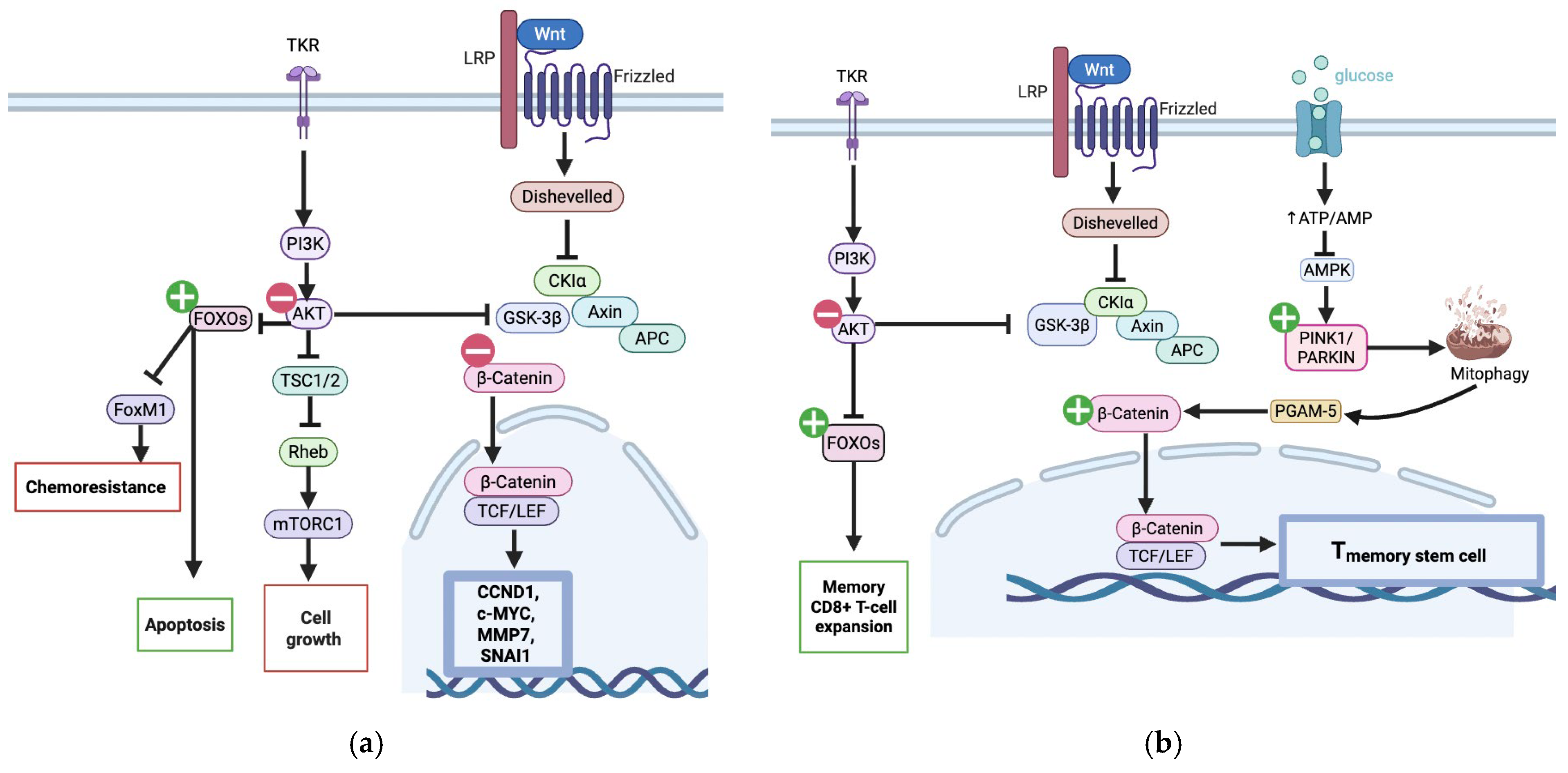
UA induces G2/M phase cell-cycle arrest by increasing p53 and its downstream effector p21, a cyclin-dependent kinase inhibitor [16,28]. Studies using p53-knockout HCT116 cells, in which the growth-inhibitory effects of UA were markedly reduced, illustrate the crucial role of UA in this p53 pathway [18]. Additionally, UA’s ability to suppress the PI3K/Akt/mTOR pathway contributes significantly to its antiproliferative effects in CRC. Tiwari et al. (2024) demonstrated, using network pharmacology and molecular docking, that UA binds to and inhibits the PI3K/AKT/mTOR pathway, leading to downstream suppression of oncogenic signaling (Figure 1a) [28].
UA may trigger apoptosis if prolonged cell cycle arrest occurs [29]. Both the intrinsic and extrinsic pathways are modulated by UA, as evidenced by the activation of initiator caspases 8 and 9, respectively (Figure 2) [16,28,30]. The anti-apoptotic pathway is affected by UA, as it alters mitochondrial membrane potential [30]. This results in the release of pro-apoptotic factors from the mitochondria, such as cytochrome c, which activate the intrinsic pathway by activating effector caspase-3 [31]. Caspase 3 then cleaves PARP, thereby promoting apoptosis by interfering with DNA repair [30,32]. UA also suppresses Bcl-2, an anti-apoptotic protein, in CRC cells (Figure 1) [33].
Additionally, UA-mediated inhibition of AKT allows FOXO transcription factors to translocate to the nucleus and upregulate pro-apoptotic genes [34]. Notably, in CD8+ memory T cells, FOXO1 activation supports T-cell maintenance and enhances anti-tumor immune surveillance (Figure 1b) [35].

3.1.2. Autophagy and Mitophagy

Zhao et al. (2018) demonstrated UA induced autophagy (without apoptosis) in SW620 CRC cells at submicromolar concentrations. The suppression of this process diminished the anti-metastatic effects of UA, suggesting that autophagy is integral to its mechanism of action at lower concentrations [36]. However, this contradicts studies indicating that, in CRC, inhibition of autophagy via Atg5 knockout enhances cancer cell sensitivity to apoptosis [37]. This suggests that UA-induced autophagy may be context-dependent—potentially supportive at low doses but possibly limiting apoptosis at higher concentrations (Figure 2).
Denk et al. (2022) demonstrated that UA promotes Wnt-dependent TSCM formation by inducing mitophagy, thereby releasing PGAM5 into the cytoplasm. PGAM5 is essential for upregulating TCF1 and CD95 and supporting T cell function. This pathway triggers mitochondrial biogenesis, enhancing T cell fitness in the tumor microenvironment (Figure 1b) [38].

3.1.3. Wnt/β-Catenin Suppression

Over 90% of CRCs harbor mutations that lead to overactivation of the Wnt signaling pathway [39]. Ghosh et al. (2022) demonstrated that UA can suppress this pathway in cancer cells resistant to Fluorouracil (FU) [21]. β-catenin downregulation reduces expression of proliferation and metastasis-associated genes, including SNAIL, MMP-7, cyclin D1, and c-MYC [33,40,41]. Thus, UA disrupts a central driver of CRC progression. In contrast, in CD8+ T cells, UA activates Wnt/β-catenin signaling to enhance anti-tumor immune function [38], illustrating context-dependent pathway effects.

3.1.4. Chemoresistance

A significant challenge in CRC treatment is the acquired resistance to standard chemotherapeutic agents such as 5-fluorouracil (5-FU). UA has demonstrated considerable potential to overcome this resistance and restore the efficacy of conventional drugs.
Multiple studies have shown that UA can sensitize CRC cells to 5-FU. In combination treatments, UA significantly lowers the IC50 of 5-FU, meaning a lower drug dose is required to achieve a therapeutic effect. Additionally, 5-FU alone primarily causes arrest in the S phase while co-treatment with UA adds another block at the G2/M phase too [42]. Chemoresistant cancer cells often overexpress ATP-binding cassette (ABC) transporters, such as MDR1, BCRP, and MRPs, which actively pump chemotherapy drugs out of the cell [20]. UA has been shown to downregulate the expression of these transporters in 5-FU-resistant CRC cells, thereby increasing intracellular drug accumulation and restoring cell death. This effect is mediated by the activation of FOXO3 and the inhibition of FOXM1, a key chemoresistance-associated oncogene (Figure 1a) [21]. In a study by Núñez-Sánchez et al. (2016), physiologically relevant urolithin mixtures, especially those enriched in UA (MPhA: ~85% UA), were shown to inhibit the growth of CSC-enriched colon spheres in Caco-2 cells and primary tumor-derived cells from a patient with CRC. In addition, the percentage of ALDH-high CSCs, a chemoresistant subpopulation, was significantly reduced by UA-rich mixtures [43].

3.1.5. Critical Appraisal and Translational Relevance of Current Evidence

While many studies provide detailed mechanistic insights, the majority rely on in vitro systems and often employ UA concentrations (10–100 μM) that exceed physiologically achievable levels in humans, where circulating free UA is typically low due to extensive phase-II conjugation (Table 1). In vivo data remain limited to xenograft and chemically induced models. Furthermore, sample sizes in animal studies are generally small, and few studies stratify outcomes by urolithin metabotype, a key determinant of endogenous UA exposure. These constraints highlight translational gaps that must be considered when interpreting biological effects.

3.2. Urolithin A and Type 2 Diabetes Mellitus

3.2.1. Apoptosis and Cellular Stress vs. Autophagy

Across all cell types, UA exhibits strong protective effects against cellular stress and apoptosis (Table 2). By blocking the action of Transglutaminase 2 (TGM2)—an enzyme that promotes the formation of contact sites between the ER and mitochondria, known as Mitochondria-associated membranes (MAMs)—UA lowers the excessive calcium influx from the endoplasmic reticulum (ER) into the mitochondria and avoids subsequent mitochondrial calcium overload. This regulation helps to reduce neuronal stress by reducing amyloid formation and Tau hyperphosphorylation, which are markers of neurodegenerative processes (Figure 3) [44,45]. Consistent with these findings, Xiao et al. (2023) demonstrated that UA alleviates cognitive impairment in T2DM mice by reducing Tau hyperphosphorylation, ER stress, and oxidative damage in the brain. In vitro, UA protected hippocampal neurons and HT22 cells from high-glucose and oxidative stress-induced apoptosis by downregulating Atp2a3, a regulator of calcium homeostasis and ER stress [46].
A primary mechanism by which UA exerts its beneficial effects, particularly in diabetic contexts, is by enhancing cellular quality-control processes, such as the targeted mitochondrial cleanup process, mitophagy. In T2DM, persistent metabolic stress and high glucose levels impair autophagy, leading to the accumulation of damaged cellular components and ultimately triggering cell death [47]. UA counters this by reactivating autophagic flux and stabilizing mitochondrial function.
The anti-apoptotic and protective effects of UA are observed across numerous cell types, often directly linked to the activation of autophagy and mitophagy (Table 2). In pancreatic β-cells of diabetic mice, UA decreased cleaved caspases, indicating a significant reduction in apoptosis. In vitro, UA protected MIN6 pancreatic cells from glucolipotoxicity-induced apoptosis by restoring mitochondrial membrane potential and activating autophagy [48].
These protective effects are directly mediated by its ability to activate autophagy via the AKT/mTOR signaling pathway, as confirmed in studies showing that blocking autophagy with inhibitors, such as chloroquine, eliminated the anti-apoptotic benefits of UA [49]. In podocytes exposed to high glucose, a model for diabetic kidney disease, UA improved cell survival and decreased apoptosis through the Bcl-2/caspase-3 pathway (Figure 4) and by increasing autophagy markers [50]. Similarly, UA has been shown to protect against diabetic kidney disease and hypoglycemia-induced heart injury by activating the PINK1/Parkin mitophagy pathway [51,52]. In the skeletal muscle of mice with diet-induced insulin resistance, UA has been shown to increase PINK1/PRKN (Parkin) mitophagy pathways (Figure 5) [53]. Paradoxically, in vascular smooth muscle cells, UA suppresses AKT phosphorylation, leading to downregulation of genes such as c-Myc and Cyclin D1, thereby preventing vascular smooth muscle proliferation [54,55]. In a study by Fang et al. (2025), UA demonstrated the ability to revitalize endothelial progenitor cells by activating Parkin-mediated mitophagy (Figure 5) [56]. Likewise, a study by Tang et al. (2023) found that UA alleviated T2DM-associated cognitive dysfunction in T2DM mice by restoring mitophagy in hippocampal neurons [57].
Table 2. Major studies exploring effects of UA in T2DM and Associated Complications.
System/
Complication
Model/SystemUA Dose/ExposureMechanism(s)Key
Findings
References
Pancreatic β-cell survivalDiabetic mice; MIN6 β-cells50 mg/kg/day; 11.5 μg/mLAutophagy (LC3II , SQSTM1/p62 );
Caspase-3/1;
ΔΨm restoration
β-cell
apoptosis;
β-cell viability and morphology
[48]
Obesity/insulin resistanceHFD & ob/ob mice; Adipocytes30 mg/kg/day; 20 μMBAT thermogenesis; iWAT browning; UCP1/PGC-1α;
Dio2 → T3
Adiposity; Insulin sensitivity[23]
Insulin resistance
(liver and skeletal muscle)
DBA/2J mice; Hepatocytes0.1% dietary UA; 10 μMMitophagy (PINK1/PRKN);
MFN2;
mtDNA;
Proton leak
Fasting glucose; Adiponectin; Improved IPTT/GTT[53]
Diabetic retinopathySTZ rats; HRECs2.5 mg/kg/day; 10 μMNrf2/HO-1;
IL-6, IL-1β, TNF-α; MDA;
SOD/GSH
Oxidative stress;
VEGF-mediated leakage; Improved retina
[58]
Cognitive dysfunctionT2DM mice; HT22 neurons200 mg/kg/day; 5 μMER stress (ATF6, CHOP);
Atp2a3;
Tau phosphorylation
Cognition; Apoptosis[46]
Gut barrier dysfunctionT2DM mice; Caco-2 cells200 mg/kg; 10 μMTight junctions (ZO-1, Occludin, Claudin 1); TLR4/Myd88;
NLRP3;
N-glycosylation genes
Gut integrity; Inflammation; Cognition[59]
Podocytopathy (kidney)Mouse podocytes (HG)10 μM (beneficial);
(cytotoxic at 100 μM)
ROS;
Bcl-2;
Autophagy (LC3B, ATG5);
Nephrin
Podocyte survival[50]
DKD—
tubular
injury
STZ mice; HK-2 cells50 mg/kg/day; 20 μMMitophagy
TRPC6–calpain-1; Cytokines
Tubular fibrosis & inflammation[51]
Wound healing (angiogenesis)STZ rats; EPCs25 mg/kg/day; 20 μMMitophagy (Parkin);
ROS
EPC proliferation
Wound closure;
Neovascularization
[56]
Vascular dysfunction (HG)STZ rats; VSMCsJuice → UA; 5–40 μMAKT phosphorylation
β-catenin (c-Myc, cyclin D1)
Endothelial function
VSMC proliferation
[54]
Diabetic cardiomyopathySTZ rats (T1D model)2.5 mg/kg/daySIRT1 → deacetylation of Nrf2, FOXO1, NF-κB; MnSOD, GSH;
ROS, TNF-α, IL-6
Cardiac function;
Fibrosis (TGF-β1, Smad3, Col1A1);
Apoptosis;
Effects abolished by SIRT1
inhibitor
[60]
indicates increase, indicates decrease.

3.2.2. Glucose Metabolism and Insulin Signaling

Modulating glucose metabolism and insulin signaling is another way UA exerts its anti-diabetic effects. Skeletal muscle, the primary site for glucose disposal, appears to be a main target. In rat skeletal muscle cells (L6 myotubes), UA increased glucose uptake in a dose-dependent manner, even in the absence of insulin [22]. Mechanistically, this occurs through the simultaneous activation of the PI3K/AKT and AMPK signaling pathways. Activation of these pathways promotes the movement of GLUT4 transporters to the plasma membrane, boosting glucose entry into the cell (Figure 3). These cellular effects result in tangible benefits in vivo. In diabetic KK-Ay/Ta mice, oral UA significantly lowered glucose levels at 90 and 120 min after glucose injection [22]. UA also influences glucose metabolism by targeting key enzymes involved in carbohydrate metabolism and insulin signaling. In silico and in vitro analyses have demonstrated that UA can bind to and inhibit α-amylase, α-glucosidase, and lipase, and thereby lower postprandial hyperglycemia [61,62]. UA-mediated inhibition of DPP-4 prolongs GLP-1 activity and enhances insulin secretion [62]. UA also inhibits aldose reductase [57], a key enzyme that contributes to diabetic complications by converting high glucose levels into sorbitol. This process produces ROS that may contribute to several diabetes-related issues, including nephropathy, cardiovascular disease, and neuropathy [63]. A randomized controlled trial by Barone Lumaga et al. (2024) reported a significant reduction in serum DPP-4 activity and lower fasting blood glucose levels among participants who consumed foods that increased their UA levels [64].
In silico analyses suggest that UA may inhibit protein tyrosine phosphatase 1B (PTP1B), a negative regulator of insulin receptor signaling (Figure 3) [65]. Computational docking studies further indicate that UA could interact with the insulin receptor (IR) and retinol-binding protein 4 (RBP4), suggesting a potential role in enhancing insulin receptor signaling and mitigating RBP4-associated insulin resistance [66].

3.2.3. Anti-Inflammatory and Antioxidant Effects

UA exhibits notable anti-inflammatory and antioxidant effects. In humans with T2DM, consuming red raspberries, which leads to UA production, significantly reduces the inflammatory marker hsCRP [67]. UA also prevents the formation of Advanced Glycation end products (AGEs), which are linked to diabetic complications due to increased production of ROS on AGE binding to its receptor (RAGE). It accomplishes this by scavenging reactive carbonyls like methylglyoxal and modifying protein structures to inhibit glycation [68]. In preclinical models, UA has been shown to lower inflammatory cytokines such as IL-6, TNF-α, and MCP-1 in diabetic kidney disease [51]. UA also demonstrated the ability to improve the integrity of tight junctions, thereby helping to inhibit inflammation caused by leaked LPS [59]. Additionally, in models of diabetic retinopathy, UA reduced inflammation (IL-6, IL-1β, TNF-α) and oxidative stress by increasing levels of SOD and GSH while decreasing MDA, effects associated with activation of the Nrf2/HO-1 pathway [58]. Consistently, studies show that UA suppresses pro-inflammatory cytokines such as IL-1β and TNF-α by downregulating NF-κB signaling in adipose tissue [68]. Additionally, UA promoted a phenotypic switch from pro-inflammatory M1 macrophages to anti-inflammatory M2 macrophages, reducing immune cell infiltration into adipose tissue [69,70].

3.2.4. Modulating Adipose Tissue and Systemic Health

UA shows significant potential in modulating adipose tissue and enhancing systemic health. Toney et al. (2019) demonstrated that UA supplementation improves systemic insulin sensitivity in high-fat diet-fed mice, reduces hepatic triglyceride accumulation, and promotes the upregulation of mitochondrial β-oxidation-related genes, such as Cpt1 and Sirt1 [69]. Other studies have shown that UA mediates the downregulation of adipogenesis-related genes, such as PPAR-γ, FABP4, and adiponectin [62]. In adipose tissue, UA attenuated adipocyte hypertrophy [62,69]. UA has been found to reduce both diet-induced and genetic obesity, which in turn improves overall glucose uptake and lowers endogenous hepatic glucose production. A key mechanism behind this is its ability to promote thermogenesis in brown adipose tissue (BAT) and the “browning” of white adipose tissue (IWAT). This effect occurs by increasing the local production of the active thyroid hormone T3 (triiodothyronine) within adipose tissue, which then triggers the expression of thermogenic genes, particularly UCP-1 and PGC-1α [23]. Together with the mechanistic evidence that UA reduces adiposity, enhances insulin sensitivity, and mitigates systemic inflammation, a cross-sectional study of Spanish adolescents reported that higher urinary microbial phenolic metabolites (including urolithin-related compounds) were inversely associated with metabolic-syndrome features. This inverse link could suggest a possible protective, early-life role for endogenous UA against the trajectory toward insulin resistance and T2DM [71].

3.3. Shared Mechanisms of UA’s Role in CRC and T2DM

UA’s capacity to modulate cellular stress responses via overlapping signaling pathways is a key component of its role in T2DM and CRC. Chronic inflammation, elevated oxidative stress, and mitochondrial dysfunction are the leading causes of both diseases. In T2DM, persistent hyperglycemia and lipotoxicity destabilize mitochondrial quality control, amplifying ROS production and ultimately leading to β-cell apoptosis and tissue damage [72]. Excess ROS also encourages additional DNA damage in CRC cells [73]. By stimulating the Nrf2 axis and increasing antioxidant enzymes, such as HO-1, UA reduces the burden of ROS in most tissues [68]. An important exception is the UA-mediated enhancement of ATRA-induced O2 production in macrophages, which promotes the phagocytosis of cancer cells [74]. In addition to fostering cardiomyocyte and podocyte survival in diabetic conditions (Table 1), this anti-oxidation action also inhibits the progression of cancer in CRC patients [35]
The regulation of mitophagy through the PINK1/Parkin pathway represents a second significant intersection. Insulin resistance and cell apoptosis are exacerbated in T2DM due to impaired mitophagy, leading to the accumulation of dysfunctional mitochondria in β-cells, podocytes, and cardiomyocytes. By encouraging the selective removal of damaged organelles, UA restores mitochondrial fidelity [48,51,52], Meanwhile, in CRC, the same pathway contributes to immunity: In CD8 T cells, UA-induced mitophagy improves depleted T cell populations, memory stem cell subsets, and overall anti-tumor immunity [39]. This highlights the context-dependent duality of UA by turning what is a cytoprotective mechanism in metabolically stressed tissues into an immunomodulatory lever in the tumor microenvironment.
Moreover, UA can regulate both PI3K/AKT/mTOR and β-catenin signalling pathways, which are disrupted in both CRC and T2DM. However, UA exhibits distinct therapeutic aims in the two conditions. UA inhibits AKT phosphorylation in CRC cells (Figure 1) while upregulating this growth-promoting pathway in T2DM (Figure 5). This reciprocal regulation demonstrates that UA can specifically alter pathways depending on the cellular environment, thereby limiting the growth of cancer cells and enhancing cell survival when metabolic homeostasis is disrupted.
Apoptosis is another typical mechanism modulated by UA, highlighting its ability to modulate the same pathway differently across environments. To guarantee tumor cell removal in CRC, UA induces apoptosis through intrinsic and extrinsic caspase cascades [56], On the other hand, UA prevents β-cells and podocytes from dying by preserving ΔΨm, thereby stabilizing mitochondrial membranes and reducing caspase activation [49,51]. Finally, in the context of inflammation, in diabetic tissues, UA suppresses IL-6, TNF-α, and MCP-1 while dampening TLR4/Myd88 and NLRP3 inflammasome activity, thereby curbing tissue injury [67,69]. In CRC, the same pathways are implicated in tumor-promoting inflammation, and UA’s dampening effect restricts cytokine-driven proliferation and metastasis [28].
The combination of these functions makes UA a molecular “balancer,” reducing the diabetic microenvironment that increases the risk of CRC while also directly preventing tumor growth (Table 3). In addition to explaining UA’s potential for managing comorbidities, these pleiotropic effects highlight the therapeutic complexity of a molecule that can reprogram cellular fate in response to context.

4. Discussion

UA’s anti-cancer actions are strongly supported by their positive effects on T2DM, which is a significant risk factor for CRC [77,78]. T2DM promotes colorectal tumor growth through mechanisms such as hyperinsulinemia, which activates the pro-growth PI3K/AKT/mTOR signaling pathway in colonic epithelial cells [79]. Mechanistically, UA intake by a diabetic patient downregulates the AKT/β-catenin pathway in colonic cells [21], thereby reducing the otherwise increased risk of developing CRC. Furthermore, UA reduces inflammation and activates the Nrf2 antioxidant pathway, which helps combat oxidative stress found in both T2DM and CRC (Table 3). Oxidative-stress modulation by UA is primarily protective (e.g., Nrf2/HO-1 activation in diabetic tissues and decreasing ROS levels in CRC cells), but context-specific pro-oxidant nudges may be therapeutically beneficial. For example, UA can amplify ATRA-stimulated superoxide production in macrophages, a cytotoxic burst that could conceivably aid tumor cell clearance [74]. This duality suggests the need for nuance: in patients with fragile tissues (e.g., advanced nephropathy), it is advisable to avoid combining UA with agents that exacerbate the respiratory burst [80]. Moreover, UA sensitizes CRC to standard therapies: it downregulates chemoresistance mechanisms (MDR1, BCRP, MRP transporters) via the FOXO3–FOXM1 pathway, thereby restoring 5-FU efficacy [21]. This chemosensitization is a double benefit in diabetic CRC, where patients often have worse chemo-response [81]. By targeting CRC cells while also disrupting the high-glucose, pro-inflammatory environment that fuels cancer, UA offers combined therapeutic potential for patients facing both conditions.
A significant property of UA that warrants further exploration for determining its therapeutic potential in various diseases is its pathway polarity. UA tends to favor glucose uptake via AMPK and PI3K/AKT signaling. Consistent with this, UA improves skeletal-muscle glucose handling and GLUT4 trafficking in preclinical models [22]. The clinical implication is that synergy with metabolic treatments that also act through the AMPK pathway can be expected, such as metformin [82]. In contrast, UA can inhibit downstream β-catenin outputs and dampen AKT activity in endothelial and CRC cells, thereby lowering pro-growth signaling [21,55]. This bidirectionality is not contradictory; it appears to be context-dependent and advantageous: UA can enhance insulin action in muscle tissues, while simultaneously suppressing oncogenic AKT/β-catenin activity in the colon and its vasculature. However, it is essential to monitor for UA’s pharmacodynamic interactions with oncologic agents that inhibit PI3K/AKT, as these agents, when given together, could lead to side effects such as hyperglycemia. Beyond muscle, UA drives thermogenic programming and browning of adipose tissue, a systemic lever for improving insulin sensitivity [23]
Despite its promises, a major translational limitation is the lack of human clinical trials evaluating UA’s efficacy in CRC or T2DM. Although human trials remain limited, several dietary and supplementation studies have clarified the translational landscape of UA metabolism, exposure, and safety. In healthy adults, Mosele et al. (2015) showed that four weeks of pomegranate-juice intake increased fecal UA and phenolic catabolites, yet only ~67% of participants were UA producers, underscoring large inter-individual variability in gut biotransformation [83]. In CRC patients, Núñez-Sánchez et al. (2014) confirmed tissue delivery of UA and Isourolithin A after 15 days of pomegranate-extract supplementation (900 mg/day), detecting up to ~7 µM UA in normal colon mucosa but markedly lower levels in tumor tissue, suggesting local metabolism and conjugation differences [84]. From a metabolic perspective, Zhang et al. (2020) reported that four weeks of red-raspberry consumption enhanced circulating microbial metabolites, including urolithins, in prediabetic subjects [85], while Barone Lumaga et al. (2024) demonstrated that a two-week high-fiber, walnut-enriched breakfast increased urinary UA-sulfate and modestly reduced fasting glucose and DPP-IV activity [64]. Collectively, these trials indicate that dietary ellagitannins can reproducibly enhance UA generation and yield mild improvements in metabolic biomarkers, but exposure remains modest and highly variable.
Quantitatively, human pharmacokinetic studies show that even after a single 500 mg oral UA dose (Mitopure), plasma UA-glucuronide peaks at approximately 480 ng/mL (~2 µM) within 6 h [86]. Hence, we can infer that systemic levels of UA aglycones would be much less than even 2 µM, due to extensive phase II metabolism. Colon-tissue concentrations after repeated pomegranate-extract dosing also lie in the low-µM range (~7 µM) [84], consistent with conjugated rather than free forms dominating systemic circulation. These levels are one to two orders of magnitude below the 10–100 µM concentrations commonly used in vitro to elicit cytotoxic, anti-inflammatory, or insulin-sensitizing effects in cell models (Table 1 and Table 2). Consequently, many mechanistic claims regarding direct UA cytotoxicity or insulin-signaling modulation remain speculative, as such exposures are unlikely to occur in vivo. Verified human outcomes are presently limited to measurable increases in conjugated UA metabolites, modest improvements in glycemic or inflammatory markers, and consistent reports of tolerability at doses up to 1 g/day over several months [87,88]. While extensive preclinical data demonstrate UA’s anticancer and metabolic benefits, these findings have yet to be validated in controlled human studies. Several studies have used UA concentrations as high as 100 μM to demonstrate the cytotoxic and anti-proliferative effects of UA on CRC cell lines in vitro [25,28]. However, at these concentrations, UA may also exert cytotoxic effects on podocytes and hepatocytes under high-glucose conditions (Table 2).
Exposure biology further complicates translation. UA production depends on microbiome “metabotypes,” with only a subset of individuals (metabotype A) efficiently converting ellagitannins to UA [89]. This capacity is reduced in insulin resistance and CRC, leading to lower circulating UA-derived metabolites [83,84]. Local glucuronidation also constrains free UA signaling; for example, UA metabolism proceeds more slowly under high-glucose conditions, potentially enhancing local protective signaling [50]. However, preclinical CRC studies show that physiologically relevant UA exposures can still modulate metabolic fitness and chemosensitivity [18,21,42].
Two pragmatic routes follow. (1) Low-dose, pathway-modulating strategy: use UA as chemopreventive or as an adjunct to standard therapies (5-FU, immunotherapy), aiming to improve mitophagy, barrier integrity, and T-cell fitness rather than to achieve direct tumor cytotoxicity. (2) High-local-dose, colon-targeted delivery: deploy pH-responsive or microbiota-activated formulations to release free UA at the tumor bed while keeping systemic levels within the protective range [90]. Given UA’s transporter-modulating and immune-conditioning properties, rational combinations should prioritize 5-FU–based regimens and T-cell–directed immunotherapies with pharmacodynamic readouts (e.g., mitophagy signatures, effector-memory phenotypes) captured alongside glycemic indices [21,24,42].
Even with optimized delivery, patient context matters. UA’s T-cell benefits are attractive for oncology, especially considering UA’s ability to re-prime CD8+ T cells towards memory-stem phenotypes, restoring depleted pools and enhancing persistence [38]. Practically speaking, this implies that UA may have a very significant effect on T2DM with CRC when administered in conjunction with immunotherapies, by strengthening the T-cell response. Still, in autoimmunity-prone settings (e.g., type 1 diabetes), any intervention that expands long-lived memory pools warrants caution and close monitoring due to the potential risk of promoting diseases like type 1 diabetes [91]. Vascular biology is another key consideration: short-term endothelial AKT inhibition may be anti-angiogenic and thus desirable for tumors [55] yet could be counterproductive in patients requiring collateral formation (e.g., limb ischemia). Prospectively stratifying by insulin-resistance status and urolithin metabotype, while capturing UA conjugates and free aglycone in plasma/stool, will be essential to connect exposure with antitumor and metabolic pharmacodynamics [24].
Implications for Future Research and Use:
  • Targeted delivery strategies: Advance UA formulations (e.g., pH-responsive polymers, nanoparticles) to concentrate high doses in colonic tumors while limiting systemic exposure. Nanocarriers designed to release UA in the tumor microenvironment could widen its therapeutic window [92].
  • Dose and regimen optimization: Map the dose–response continuum of UA’s effects. Careful titration is needed to separate its anti-CRC (high-dose) and anti-diabetic (low-dose) actions. For example, intermittent high-dose pulses might kill tumor cells while daily low-dose maintains metabolic control.
  • Combination therapy trials: Given UA’s ability to sensitize CRC to chemotherapy and to enhance antitumor T-cell responses, testing UA alongside standard chemo/immunotherapies is warranted. Early-phase clinical trials could assess the effects of UA supplementation in CRC patients with metabolic syndrome, monitoring tumor markers, glycemic indices, and immune phenotypes.
  • Patient stratification and biomarkers: Identify biomarkers (e.g., gut metabotype, insulin resistance status) to select patients most likely to benefit from UA. Only some individuals efficiently convert ellagitannins to UA.
  • Safety assessments: Evaluate UA’s effects in autoimmunity and healthy tissues. Its immune-boosting properties should be tested for safety in autoimmune disease models to ensure it does not exacerbate β-cell autoimmunity. Long-term toxicology should confirm that sustained UA levels (even via targeted delivery) are well tolerated.

5. Conclusions

Urolithin A, a gut microbiota–derived metabolite of dietary ellagitannins, emerges as a molecular bridge between CRC and T2DM. Both diseases share dysregulated insulin/IGF-1 signaling, chronic inflammation, oxidative stress, and mitochondrial dysfunction—mechanistic axes that UA can modulate through AMPK activation, PI3K/AKT/mTOR inhibition, and restoration of mitophagy and autophagy. This convergence positions UA as a dual-target compound that can simultaneously influence metabolic and oncogenic pathways.
Translationally, preclinical evidence supports UA’s antitumor and insulin-sensitizing effects. Still, human data remain limited to small trials demonstrating increased systemic UA exposure, modest glycemic improvements, and tissue accumulation without definitive clinical outcomes. Inter-individual differences in UA production (“metabotypes”), rapid conjugation, and uncertain effective doses represent major barriers to therapeutic application.
Future research should prioritize biomarker-guided, metabotype-stratified clinical trials that define optimal UA exposure, establish pharmacokinetic–pharmacodynamic relationships, and test colon-targeted or systemic delivery strategies. Integrating microbiome engineering with UA supplementation may enhance translational efficacy. Overall, UA exemplifies how microbial metabolites can reframe chronic disease prevention—linking diet, microbiota, and host signaling in the shared pathogenesis of CRC and T2DM.

Author Contributions

V.J. and D.B.: conceptualization; V.J.: writing—original draft preparation, review, and editing; S.H.: writing—review, and editing; P.K.: writing—review, and editing; D.B.: supervision and review and editing. All authors have read and agreed to the published version of the manuscript.

Funding

Open Access funding was provided by the Qatar National Library. This work was supported by a National Priorities Research Program grant (NPRP 14 S-0311–210033; awarded to Dietrich Büsselberg, January 2023-Current) from the Qatar National Research Fund (QNRF, a member of Qatar Foundation). The statements made herein are solely the responsibility of the authors.

Data Availability Statement

No datasets were generated or analyzed during the current study.

Conflicts of Interest

The authors declare no competing interests.

Abbreviations

The following abbreviations are used in this manuscript:
Amyloid-β
ATG5Autophagy related 5
ATRA All-trans retinoic acid
AKT Protein kinase B (AKT)
AMPK AMP-activated protein kinase
BAT Brown adipose tissue
BAX Bcl-2 associated X protein
BCL-2 B-cell lymphoma 2 (anti-apoptotic protein)
CRC Colorectal cancer
DPP-4 Dipeptidyl peptidase-4
DRP1 Dynamin-related protein 1 (mitochondrial fission regulator)
EPC(s) Endothelial progenitor cell(s)
ER Endoplasmic reticulum
GLUT4 Glucose transporter type 4
GSH Glutathione
HO-1 Heme oxygenase-1
IR Insulin receptor
IWAT Inguinal (subcutaneous) white adipose tissue
LC3 Microtubule-associated protein 1 light chain 3 (autophagy marker)
MAM(s) Mitochondria-associated membranes
MCP-1 Monocyte chemoattractant protein-1 (CCL2)
MDA Malondialdehyde
mtROS Mitochondrial reactive oxygen species
NLRP3 NOD-, LRR- and pyrin domain-containing protein 3 (inflammasome)
Nrf2 Nuclear factor erythroid 2-related factor 2
OCR Oxygen consumption rate
PI3K Phosphoinositide 3-kinase.
PINK1 PTEN-induced kinase 1
PRKN (Parkin) Parkin RBR E3 ubiquitin protein ligase (PRKN)
PTP1B Protein tyrosine phosphatase 1B
PSD95 Postsynaptic density protein 95
PTEN Phosphatase and tensin homolog (if used in text)
ROS Reactive oxygen species
SOD Superoxide dismutase
SQSTM1/p62 Sequestosome 1 (p62; autophagy adaptor)
T2DM Type 2 diabetes mellitus
TGM2 Transglutaminase 2
TSCM T memory stem cell (memory-stem CD8+ T cell)
UCP1 Uncoupling protein 1 (thermogenesis marker)
UA Urolithin A
ULK1 Unc-51 like autophagy activating kinase 1
ΔΨm Mitochondrial membrane potential (delta psi m)

References

  1. Home. Diabetes Atlas. Available online: https://diabetesatlas.org/ (accessed on 30 August 2025).
  2. Cancer of the Colon and Rectum—Cancer Stat Facts. SEER. Available online: https://seer.cancer.gov/statfacts/html/colorect.html (accessed on 22 September 2025).
  3. CDC. Health and Economic Benefits of Colorectal Cancer Interventions’; National Center for Chronic Disease Prevention and Health Promotion (NCCDPHP): Atlantan, GA, USA, 2025. Available online: https://www.cdc.gov/nccdphp/priorities/colorectal-cancer.html (accessed on 25 July 2025).
  4. Chen, Z.; Hong, Q. Correlation of serum IGF-1, AGEs and their receptors with the risk of colorectal cancer in patients with type 2 diabetes mellitus. Front. Oncol. 2023, 13, 1125745. [Google Scholar] [CrossRef]
  5. Gallagher, E.J.; LeRoith, D. The Proliferating Role of Insulin and Insulin-Like Growth Factors in Cancer. Trends Endocrinol. Metab. TEM 2010, 21, 610–618. [Google Scholar] [CrossRef]
  6. Alvarez-Leite, J.I. The Role of Bioactive Compounds in Human Health and Disease. Nutrients 2025, 17, 1170. [Google Scholar] [CrossRef]
  7. Cerdá, B.; Periago, P.; Espín, J.C.; Tomás-Barberán, F.A. Identification of Urolithin A as a Metabolite Produced by Human Colon Microflora from Ellagic Acid and Related Compounds. J. Agric. Food Chem. 2005, 53, 5571–5576. [Google Scholar] [CrossRef]
  8. Lin, I.-C.; Wu, J.-Y.; Fang, C.-Y.; Wang, S.-C.; Liu, Y.-W.; Ho, S.-T. Absorption and Metabolism of Urolithin A and Ellagic Acid in Mice and Their Cytotoxicity in Human Colorectal Cancer Cells. Evid.-Based Complement. Altern. Med. Ecam 2023, 2023, 8264716. [Google Scholar] [CrossRef]
  9. Aichinger, G. Natural Dibenzo-α-Pyrones: Friends or Foes? Int. J. Mol. Sci. 2021, 22, 13063. [Google Scholar] [CrossRef]
  10. He, F.; Bian, Y.; Zhao, Y.; Xia, M.; Liu, S.; Gui, J.; Hou, X.; Fang, Y. In vitro conversion of ellagic acid to urolithin A by different gut microbiota of urolithin metabotype A. Appl. Microbiol. Biotechnol. 2024, 108, 215. [Google Scholar] [CrossRef]
  11. Selma, M.V.; González-Sarrías, A.; Salas-Salvadó, J.; Andrés-Lacueva, C.; Alasalvar, C.; Örem, A.; Tomás-Barberán, F.A.; Espín, J.C. The gut microbiota metabolism of pomegranate or walnut ellagitannins yields two urolithin-metabotypes that correlate with cardiometabolic risk biomarkers: Comparison between normoweight, overweight-obesity and metabolic syndrome. Clin. Nutr. 2018, 37, 897–905. [Google Scholar] [CrossRef]
  12. Ares, A.M.; Toribio, L.; García-Villalba, R.; Villalgordo, J.M.; Althobaiti, Y.; Tomás-Barberán, F.A.; Bernal, J. Separation of Isomeric Forms of Urolithin Glucuronides Using Supercritical Fluid Chromatography. J. Agric. Food Chem. 2023, 71, 3033–3039. [Google Scholar] [CrossRef]
  13. Piwowarski, J.P.; Stanisławska, I.; Granica, S.; Stefańska, J.; Kiss, A.K. Phase II Conjugates of Urolithins Isolated from Human Urine and Potential Role of β-Glucuronidases in Their Disposition. Drug Metab. Dispos. 2017, 45, 657–665. [Google Scholar] [CrossRef]
  14. Totiger, T.M.; Srinivasan, S.; Jala, V.R.; Lamichhane, P.; Dosch, A.R.; Gaidarski, A.A.; Joshi, C.; Rangappa, S.; Castellanos, J.; Vemula, P.K.; et al. Urolithin A, a Novel Natural Compound to Target PI3K/AKT/mTOR Pathway in Pancreatic Cancer. Mol. Cancer Ther. 2019, 18, 301–311. [Google Scholar] [CrossRef]
  15. Lin, J.; Zhuge, J.; Zheng, X.; Wu, Y.; Zhang, Z.; Xu, T.; Meftah, Z.; Xu, H.; Wu, Y.; Tian, N.; et al. Urolithin A-induced mitophagy suppresses apoptosis and attenuates intervertebral disc degeneration via the AMPK signaling pathway. Free. Radic. Biol. Med. 2020, 150, 109–119. [Google Scholar] [CrossRef]
  16. El-Wetidy, M.S.; Ahmad, R.; Rady, I.; Helal, H.; Rady, M.I.; Vaali-Mohammed, M.-A.; Al-Khayal, K.; Traiki, T.B.; Abdulla, M.-H. Urolithin A induces cell cycle arrest and apoptosis by inhibiting Bcl-2, increasing p53-p21 proteins and reactive oxygen species zproduction in colorectal cancer cells. Cell Stress Chaperones 2021, 26, 473–493. [Google Scholar] [CrossRef]
  17. Sharma, M.; Li, L.; Celver, J.; Killian, C.; Kovoor, A.; Seeram, N.P. Effects of Fruit Ellagitannin Extracts, Ellagic Acid, and Their Colonic Metabolite, Urolithin A, on Wnt Signaling. J. Agric. Food Chem. 2010, 58, 3965–3969. [Google Scholar] [CrossRef]
  18. Norden, E.; Heiss, E.H. Urolithin A gains in antiproliferative capacity by reducing the glycolytic potential via the p53/TIGAR axis in colon cancer cells. Carcinogenesis 2019, 40, 93–101. [Google Scholar] [CrossRef]
  19. Giménez-Bastida, J.A.; Ávila-Gálvez, M.Á.; Espín, J.C.; González-Sarrías, A. The gut microbiota metabolite urolithin A, but not other relevant urolithins, induces p53-dependent cellular senescence in human colon cancer cells. Food Chem. Toxicol. 2020, 139, 111260. [Google Scholar] [CrossRef]
  20. Choi, Y.H.; Yu, A.-M. ABC Transporters in Multidrug Resistance and Pharmacokinetics, and Strategies for Drug Development. Curr. Pharm. Des. 2014, 20, 793–807. [Google Scholar] [CrossRef]
  21. Ghosh, S.; Singh, R.; Vanwinkle, Z.M.; Guo, H.; Vemula, P.K.; Goel, A.; Haribabu, B.; Jala, V.R. Microbial metabolite restricts 5-fluorouracil-resistant colonic tumor progression by sensitizing drug transporters via regulation of FOXO3-FOXM1 axis. Theranostics 2022, 12, 5574–5595. [Google Scholar] [CrossRef]
  22. Kondo, S.; Adachi, S.-I.; Komatsu, W.; Yoshizawa, F.; Yagasaki, K. Antidiabetic Effect of Urolithin A in Cultured L6 Myotubes and Type 2 Diabetic Model KK-A(y)/Ta Mice with Glucose Intolerance. Curr. Issues Mol. Biol. 2024, 46, 1078–1090. [Google Scholar] [CrossRef]
  23. Xia, B.; Shi, X.C.; Xie, B.C.; Zhu, M.Q.; Chen, Y.; Chu, X.Y.; Cai, G.H.; Liu, M.; Yang, S.Z.; Mitchell, G.A.; et al. Urolithin A exerts antiobesity effects through enhancing adipose tissue thermogenesis in mice. PLoS Biol. 2020, 18, e3000688. [Google Scholar] [CrossRef]
  24. Andreux, P.A.; Blanco-Bose, W.; Ryu, D.; Burdet, F.; Ibberson, M.; Aebischer, P.; Auwerx, J.; Singh, A.; Rinsch, C. The mitophagy activator urolithin A is safe and induces a molecular signature of improved mitochondrial and cellular health in humans. Nat. Metab. 2019, 1, 595–603. [Google Scholar] [CrossRef]
  25. González-Sarrías, A.; Giménez-Bastida, J.A.; Núñez-Sánchez, M.A.; Larrosa, M.; García-Conesa, M.T.; Tomás-Barberán, F.A.; Espín, J.C. Phase-II metabolism limits the antiproliferative activity of urolithins in human colon cancer cells. Eur. J. Nutr. 2014, 53, 853–864. [Google Scholar] [CrossRef]
  26. González-Sarrías, A.; Núñez-Sánchez, M.Á.; García-Villalba, R.; Tomás-Barberán, F.A.; Espín, J.C. Antiproliferative activity of the ellagic acid-derived gut microbiota isourolithin A and comparison with its urolithin A isomer: The role of cell metabolism. Eur. J. Nutr. 2017, 56, 831–841. [Google Scholar] [CrossRef]
  27. Tortora, K.; Femia, A.P.; Romagnoli, A.; Sineo, I.; Khatib, M.; Mulinacci, N.; Giovannelli, L.; Caderni, G. Pomegranate By-Products in Colorectal Cancer Chemoprevention: Effects in Apc-Mutated Pirc Rats and Mechanistic Studies In Vitro and Ex Vivo. Mol. Nutr. Food Res. 2018, 62, 1700401. [Google Scholar] [CrossRef]
  28. Tiwari, A.; Tiwari, V.; Verma, S.; Sharma, A.; Marisetti, A.L.; Kumar, M.; Kumar, G.; Qurtam, A.A.; Nasr, F.A.; Mubarak, M.; et al. Network Pharmacology, Molecular Docking, Simulation Studies of Urolithin A by Inhibiting AKT and Caspase Pathways against Colorectal Cancer. J. Biol. Regul. Homeost. Agents 2024, 38, 5175–5194. [Google Scholar] [CrossRef]
  29. Orth, J.D.; Loewer, A.; Lahav, G.; Mitchison, T.J. Prolonged mitotic arrest triggers partial activation of apoptosis, resulting in DNA damage and p53 induction. Mol. Biol. Cell 2012, 23, 567–576. [Google Scholar] [CrossRef]
  30. Cho, H.; Jung, H.; Lee, H.; Yi, H.C.; Kwak, H.; Hwang, K.T. Chemopreventive activity of ellagitannins and their derivatives from black raspberry seeds on HT-29 colon cancer cells. Food Funct. 2015, 6, 1675–1683. [Google Scholar] [CrossRef]
  31. Gupta, S.; Kass, G.E.; Szegezdi, E.; Joseph, B. The mitochondrial death pathway: A promising therapeutic target in diseases. J. Cell. Mol. Med. 2009, 13, 1004–1033. [Google Scholar] [CrossRef]
  32. Meza-Sosa, K.F.; Miao, R.; Navarro, F.; Zhang, Z.; Zhang, Y.; Hu, J.J.; Hartford, C.C.R.; Li, X.L.; Pedraza-Alva, G.; Pérez-Martínez, L.; et al. SPARCLE, a p53-induced lncRNA, controls apoptosis after genotoxic stress by promoting PARP-1 cleavage. Mol. Cell 2022, 82, 785–802.e10. [Google Scholar] [CrossRef]
  33. González-Sarrías, A.; Espín, J.-C.; Tomás-Barberán, F.A.; García-Conesa, M.-T. Gene expression, cell cycle arrest and MAPK signalling regulation in Caco-2 cells exposed to ellagic acid and its metabolites, urolithins. Mol. Nutr. Food Res. 2009, 53, 686–698. [Google Scholar] [CrossRef]
  34. Manning, B.D.; Cantley, L.C. AKT/PKB Signaling: Navigating Downstream. Cell 2007, 129, 1261–1274. [Google Scholar] [CrossRef]
  35. Ginefra, P.; Hope, H.C.; Chiang, Y.-H.; Nutten, S.; Blum, S.; Coukos, G.; Vannini, N. Urolithin-A Promotes CD8+ T Cell-mediated Cancer Immunosurveillance via FOXO1 Activation. Cancer Res. Commun. 2024, 4, 1189–1198. [Google Scholar] [CrossRef]
  36. Zhao, W.; Shi, F.; Guo, Z.; Zhao, J.; Song, X.; Yang, H. Metabolite of ellagitannins, urolithin A induces autophagy and inhibits metastasis in human sw620 colorectal cancer cells. Mol. Carcinog. 2018, 57, 193–200. [Google Scholar] [CrossRef] [PubMed]
  37. Qian, H.-R.; Shi, Z.-Q.; Zhu, H.-P.; Gu, L.-H.; Wang, X.-F.; Yang, Y. Interplay between apoptosis and autophagy in colorectal cancer. Oncotarget 2017, 8, 62759–62768. [Google Scholar] [CrossRef] [PubMed]
  38. Denk, D.; Petrocelli, V.; Conche, C.; Drachsler, M.; Ziegler, P.K.; Braun, A.; Kress, A.; Nicolas, A.M.; Mohs, K.; Becker, C.; et al. Expansion of T memory stem cells with superior anti-tumor immunity by Urolithin A-induced mitophagy. Immunity 2022, 55, 2059–2073.e8. [Google Scholar] [CrossRef]
  39. Giles, R.H.; van Es, J.H.; Clevers, H. Caught up in a Wnt storm: Wnt signaling in cancer. Biochim. Biophys. Acta (BBA) Rev. Cancer 2003, 1653, 1–24. [Google Scholar] [CrossRef]
  40. Brabletz, T.; Jung, A.; Dag, S.; Hlubek, F.; Kirchner, T. Beta-catenin regulates the expression of the matrix metalloproteinase-7 in human colorectal cancer. Am. J. Pathol. 1999, 155, 1033–1038. [Google Scholar] [CrossRef]
  41. Jung, A.; Schrauder, M.; Oswald, U.; Knoll, C.; Sellberg, P.; Palmqvist, R.; Niedobitek, G.; Brabletz, T.; Kirchner, T. The Invasion Front of Human Colorectal Adenocarcinomas Shows Co-Localization of Nuclear β-Catenin, Cyclin D1, and p16INK4A and Is a Region of Low Proliferation. Am. J. Pathol. 2001, 159, 1613–1617. [Google Scholar] [CrossRef]
  42. González-Sarrías, A.; Tomé-Carneiro, J.; Bellesia, A.; Tomás-Barberán, F.A.; Espín, J.C. The ellagic acid-derived gut microbiota metabolite, urolithin A, potentiates the anticancer effects of 5-fluorouracil chemotherapy on human colon cancer cells. Food Funct. 2015, 6, 1460–1469. [Google Scholar] [CrossRef]
  43. Núñez-Sánchez, M.T.; Karmokar, A.; González-Sarrías, A.; García-Villalba, R.; Tomás-Barberán, F.A.; García-Conesa, M.T.; Brown, K.; Espín, J.C. In vivo relevant mixed urolithins and ellagic acid inhibit phenotypic and molecular colon cancer stem cell features: A new potentiality for ellagitannin metabolites against cancer. Food Chem. Toxicol. 2016, 92, 8–16. [Google Scholar] [CrossRef]
  44. Lee, H.J.; Jung, Y.H.; Choi, G.E.; Kim, J.S.; Chae, C.W.; Lim, J.R.; Kim, S.Y.; Yoon, J.H.; Cho, J.H.; Lee, S.-J.; et al. Urolithin A suppresses high glucose-induced neuronal amyloidogenesis by modulating TGM2-dependent ER-mitochondria contacts and calcium homeostasis. Cell Death Differ. 2021, 28, 184–202. [Google Scholar] [CrossRef] [PubMed]
  45. Garcia-Alloza, M.; Bacskai, B.J.; Calvo-Rodriguez, M. Mitochondria-ER contacts and glucose: The powerhouse of Alzheimer’s disease? Cell Calcium 2021, 97, 102434. [Google Scholar] [CrossRef] [PubMed]
  46. Xiao, Y.; Li, K.; Bian, J.; Zhang, Y.; Li, J.; Liu, H.; Ye, Y.; Han, L.; Gong, L.; Wang, M. Urolithin A Protects Neuronal Cells against Stress Damage and Apoptosis by Atp2a3 Inhibition. Mol. Nutr. Food Res. 2023, 67, e2300146. [Google Scholar] [CrossRef]
  47. Mittal, R.; Lemos, J.R.N.; Chapagain, P.; Hirani, K. Interplay of hypoxia, immune dysregulation, and metabolic stress in pathophysiology of type 1 diabetes. Front. Immunol. 2025, 16, 1599321. [Google Scholar] [CrossRef] [PubMed]
  48. Zhang, Y.; Zhang, Y.; Halemahebai, G.; Tian, L.; Dong, H.; Aisker, G. Urolithin A, a pomegranate metabolite, protects pancreatic β cells from apoptosis by activating autophagy. J. Ethnopharmacol. 2021, 272, 113628. [Google Scholar] [CrossRef]
  49. Tuohetaerbaike, B.; Zhang, Y.; Tian, Y.; Zhang, N.N.; Kang, J.; Mao, X.; Zhang, Y.; Li, X. Pancreas protective effects of Urolithin A on type 2 diabetic mice induced by high fat and streptozotocin via regulating autophagy and AKT/mTOR signaling pathway. J. Ethnopharmacol. 2020, 250, 112479. [Google Scholar] [CrossRef]
  50. Kotewicz, M.; Krauze-Baranowska, M.; Daca, A.; Płoska, A.; Godlewska, S.; Kalinowski, L.; Lewko, B. Urolithins Modulate the Viability, Autophagy, Apoptosis, and Nephrin Turnover in Podocytes Exposed to High Glucose. Cells 2022, 11, 2471. [Google Scholar] [CrossRef]
  51. Liu, C.-C.; Ji, J.-L.; Wang, Z.; Zhang, X.-J.; Ding, L.; Zhang, Y.; Zhou, Y.; Zhang, D.-J.; Tang, Z.-L.; Cao, J.-Y.; et al. TRPC6-Calpain-1 Axis Promotes Tubulointerstitial Inflammation by Inhibiting Mitophagy in Diabetic Kidney Disease. Kidney Int. Rep. 2024, 9, 3301–3317. [Google Scholar] [CrossRef]
  52. Huang, C.; Huang, L.; Huang, Q.; Lin, L.; Wang, L.; Wu, Y.; Wu, K.; Gao, R.; Liu, X.; Liu, X.; et al. Mitophagy disorder mediates cardiac deterioration induced by severe hypoglycemia in diabetic mice. Mol. Cell. Endocrinol. 2023, 575, 111994. [Google Scholar] [CrossRef]
  53. Yang, J.; Guo, Y.; Henning, S.M.; Chan, B.; Long, J.; Zhong, J.; Acin-Perez, R.; Petcherski, A.; Shirihai, O.; Heber, D.; et al. Ellagic Acid and Its Microbial Metabolite Urolithin A Alleviate Diet-Induced Insulin Resistance in Mice. Mol. Nutr. Food Res. 2020, 64, e2000091. [Google Scholar] [CrossRef]
  54. Zhou, J.; Zhang, C.; Zheng, G.-H.; Qiu, Z. Emblic Leafflower (Phyllanthus emblica L.) Fruits Ameliorate Vascular Smooth Muscle Cell Dysfunction in Hyperglycemia: An Underlying Mechanism Involved in Ellagitannin Metabolite Urolithin A. Evid. Based Complement. Altern. Med. eCAM 2018, 2018, 8478943. [Google Scholar] [CrossRef]
  55. Dirimanov, S.; Högger, P. Screening of Inhibitory Effects of Polyphenols on Akt-Phosphorylation in Endothelial Cells and Determination of Structure-Activity Features. Biomolecules 2019, 9, 219. [Google Scholar] [CrossRef]
  56. Fang, X.; Shao, Z.; Ding, H.; Xu, H.; Tu, Z.; Wang, H.; Li, D.; Huang, C.; Jiang, C. Urolithin A enhances diabetic wound healing: Insights from parkin-mediated mitophagy in endothelial progenitor cells. Int. Immunopharmacol. 2025, 155, 114572. [Google Scholar] [CrossRef]
  57. Tang, W.; Yan, C.; He, S.; Du, M.; Cheng, B.; Deng, B.; Zhu, S.; Li, Y.; Wang, Q. Neuron-targeted overexpression of caveolin-1 alleviates diabetes-associated cognitive dysfunction via regulating mitochondrial fission-mitophagy axis. Cell Commun. Signal. 2023, 21, 357. [Google Scholar] [CrossRef]
  58. Xu, Z.; Li, S.; Li, K.; Wang, X.; Li, X.; An, M.; Yu, X.; Long, X.; Zhong, R.; Liu, Q.; et al. Urolithin A ameliorates diabetic retinopathy via activation of the Nrf2/HO-1 pathway. Endocr. J. 2022, 69, 971–982. [Google Scholar] [CrossRef]
  59. Xiao, Y.; Li, K.; Bian, J.; Liu, H.; Zhai, X.; El-Omar, E.; Han, L.; Gong, L.; Wang, M. Urolithin A Attenuates Diabetes-Associated Cognitive Impairment by Ameliorating Intestinal Barrier Dysfunction via N-glycan Biosynthesis Pathway. Mol. Nutr. Food Res. 2022, 66, e2100863. [Google Scholar] [CrossRef]
  60. Albasher, G.; Alkahtani, S.; Al-Harbi, L.N. Urolithin A prevents streptozotocin-induced diabetic cardiomyopathy in rats by activating SIRT1. Saudi J. Biol. Sci. 2022, 29, 1210–1220. [Google Scholar] [CrossRef]
  61. Han, L.; Zhao, D.; Li, Y.; Jin, J.; El-Kott, A.F.; Al-Saeed, F.A.; Eldib, A.M. Assessment of the Anti-Breast Cancer Effects of Urolithin with Molecular Docking Studies in the In Vitro Condition: Introducing a Novel Chemotherapeutic Drug. Mol. Biotechnol. 2024, 66, 554–566. [Google Scholar] [CrossRef] [PubMed]
  62. Les, F.; Arbonés-Mainar, J.M.; Valero, M.S.; López, V. Pomegranate polyphenols and urolithin A inhibit α-glucosidase, dipeptidyl peptidase-4, lipase, triglyceride accumulation and adipogenesis related genes in 3T3-L1 adipocyte-like cells. J. Ethnopharmacol. 2018, 220, 67–74. [Google Scholar] [CrossRef] [PubMed]
  63. Ramasamy, R.; Goldberg, I.J. Aldose reductase and cardiovascular diseases, creating human-like diabetic complications in an experimental model. Circ. Res. 2010, 106, 1449–1458. [Google Scholar] [CrossRef]
  64. Barone Lumaga, R.; Tagliamonte, S.; De Rosa, T.; Valentino, V.; Ercolini, D.; Vitaglione, P. Consumption of a Sourdough-Leavened Croissant Enriched with a Blend of Fibers Influences Fasting Blood Glucose in a Randomized Controlled Trial in Healthy Subjects. J. Nutr. 2024, 154, 2976–2987. [Google Scholar] [CrossRef]
  65. Pratama, R.A.; Astina, J.; Parikesit, A.A. In silico Screening of Potential Antidiabetic Phenolic Compounds from Banana (Musa spp.) Peel Against PTP1B Protein. J. Trop. Biodivers. Biotechnol. 2023, 8, 139. [Google Scholar] [CrossRef]
  66. Tolmie, M.; Bester, M.J.; Serem, J.C.; Nell, M.; Apostolides, Z. The potential antidiabetic properties of green and purple tea [Camellia sinensis (L.) O Kuntze], purple tea ellagitannins, and urolithins. J. Ethnopharmacol. 2023, 309, 116377. [Google Scholar] [CrossRef]
  67. Moreno Uclés, R.; González-Sarrías, A.; Espín, J.C.; Tomás-Barberán, F.A.; Janes, M.; Cheng, H.; Finley, J.; Greenway, F.; Losso, J.N. Effects of red raspberry polyphenols and metabolites on the biomarkers of inflammation and insulin resistance in type 2 diabetes: A pilot study. Food Funct. 2022, 13, 5166–5176. [Google Scholar] [CrossRef]
  68. Liu, W.; Ma, H.; Frost, L.; Yuan, T.; Dain, J.A.; Seeram, N.P. Pomegranate phenolics inhibit formation of advanced glycation endproducts by scavenging reactive carbonyl species. Food Funct. 2014, 5, 2996–3004. [Google Scholar] [CrossRef]
  69. Toney, A.M.; Fan, R.; Xian, Y.; Chaidez, V.; Ramer-Tait, A.E.; Chung, S. Urolithin A, a Gut Metabolite, Improves Insulin Sensitivity Through Augmentation of Mitochondrial Function and Biogenesis. Obesity 2019, 27, 612–620. [Google Scholar] [CrossRef]
  70. Boakye, Y.D.; Groyer, L.; Heiss, E.H. An increased autophagic flux contributes to the anti-inflammatory potential of urolithin A in macrophages. Biochim. Biophys. Acta Gen. Subj. 2018, 1862, 61–70. [Google Scholar] [CrossRef] [PubMed]
  71. Laveriano-Santos, E.P.; Quifer-Rada, P.; Marhuenda-Muñoz, M.; Arancibia-Riveros, C.; Vallverdú-Queralt, A.; Tresserra-Rimbau, A.; Ruiz-León, A.M.; Casas, R.; Estruch, R.; Bodega, P.; et al. Microbial Phenolic Metabolites in Urine Are Inversely Linked to Certain Features of Metabolic Syndrome in Spanish Adolescents. Antioxidants 2022, 11, 2191. [Google Scholar] [CrossRef]
  72. Caturano, A.; D’Angelo, M.; Mormone, A.; Russo, V.; Mollica, M.P.; Salvatore, T.; Galiero, R.; Rinaldi, L.; Vetrano, E.; Marfella, R.; et al. Oxidative Stress in Type 2 Diabetes: Impacts from Pathogenesis to Lifestyle Modifications. Curr. Issues Mol. Biol. 2023, 45, 6651–6666. [Google Scholar] [CrossRef] [PubMed]
  73. Perše, M. Oxidative stress in the pathogenesis of colorectal cancer: Cause or consequence? BioMed Res. Int. 2013, 2013, 725710. [Google Scholar] [CrossRef] [PubMed]
  74. Kikuchi, H.; Harata, K.; Madhyastha, H.; Kuribayashi, F. Ellagic acid and its fermentative derivative urolithin A show reverse effects on the gp91-phox gene expression, resulting in opposite alterations in all-trans retinoic acid-induced superoxide generating activity of U937 cells. Biochem. Biophys. Rep. 2021, 25, 100891. [Google Scholar] [CrossRef]
  75. Zhao, W.; Wang, Y.; Hao, W.; Yang, H.; Song, X.; Zhao, M.; Peng, S. Preparative isolation and purification of urolithins from the intestinal metabolites of pomegranate ellagitannins by high-speed counter-current chromatography. J. Chromatography. B Anal. Technol. Biomed. Life Sci. 2015, 990, 111–117. [Google Scholar] [CrossRef]
  76. Groestlinger, J.; Seidl, C.; Varga, E.; Del Favero, G.; Marko, D. Combinatory Exposure to Urolithin A, Alternariol, and Deoxynivalenol Affects Colon Cancer Metabolism and Epithelial Barrier Integrity in vitro. Front. Nutr. 2022, 9, 882222. [Google Scholar] [CrossRef] [PubMed]
  77. Dehal, A.N.; Newton, C.C.; Jacobs, E.J.; Patel, A.V.; Gapstur, S.M.; Campbell, P.T. Impact of Diabetes Mellitus and Insulin Use on Survival After Colorectal Cancer Diagnosis: The Cancer Prevention Study-II Nutrition Cohort. J. Clin. Oncol. 2012, 30, 53–59. [Google Scholar] [CrossRef]
  78. Lawler, T.; Walts, Z.L.; Steinwandel, M.; Lipworth, L.; Murff, H.J.; Zheng, W.; Warren Andersen, S. Type 2 Diabetes and Colorectal Cancer Risk. JAMA Netw. Open 2023, 6, e2343333. [Google Scholar] [CrossRef]
  79. Zhang, A.M.Y.; Wellberg, E.A.; Kopp, J.L.; Johnson, J.D. Hyperinsulinemia in Obesity, Inflammation, and Cancer. Diabetes Metab. J. 2021, 45, 285–311. [Google Scholar] [CrossRef]
  80. Ruiz, S.; Pergola, P.E.; Zager, R.A.; Vaziri, N.D. Targeting the transcription factor Nrf2 to ameliorate oxidative stress and inflammation in chronic kidney disease. Kidney Int. 2013, 83, 1029–1041. [Google Scholar] [CrossRef]
  81. Gaur, A.; Maity, R.; Dhali, A.; Biswas, J. Impact of poorly controlled type II diabetes mellitus on chemoresistance in colorectal cancer. World J. Gastroenterol. 2025, 31, 104065. [Google Scholar] [CrossRef]
  82. Zhou, G.; Myers, R.; Li, Y.; Chen, Y.; Shen, X.; Fenyk-Melody, J.; Wu, M.; Ventre, J.; Doebber, T.; Fujii, N.; et al. Role of AMP-activated protein kinase in mechanism of metformin action. J. Clin. Investig. 2001, 108, 1167–1174. [Google Scholar] [CrossRef] [PubMed]
  83. Mosele, J.I.; Gosalbes, M.-J.; Macià, A.; Rubió, L.; Vázquez-Castellanos, J.F.; Jiménez Hernández, N.; Moya, A.; Latorre, A.; Motilva, M.-J. Effect of daily intake of pomegranate juice on fecal microbiota and feces metabolites from healthy volunteers. Mol. Nutr. Food Res. 2015, 59, 1942–1953. [Google Scholar] [CrossRef]
  84. Nuñez-Sánchez, M.A.; García-Villalba, R.; Monedero-Saiz, T.; García-Talavera, N.V.; Gómez-Sánchez, M.B.; Sánchez-Álvarez, C.; García-Albert, A.M.; Rodríguez-Gil, F.J.; Ruiz-Marín, M.; Pastor-Quirante, F.A.; et al. Targeted metabolic profiling of pomegranate polyphenols and urolithins in plasma, urine and colon tissues from colorectal cancer patients. Mol. Nutr. Food Res. 2014, 58, 1199–1211. [Google Scholar] [CrossRef] [PubMed]
  85. Zhang, X.; Zhao, A.; Sandhu, A.K.; Edirisinghe, I.; Burton-Freeman, B.M. Functional Deficits in Gut Microbiome of Young and Middle-Aged Adults with Prediabetes Apparent in Metabolizing Bioactive (Poly)phenols. Nutrients 2020, 12, 3595. [Google Scholar] [CrossRef] [PubMed]
  86. Singh, A.; D’Amico, D.; Andreux, P.A.; Fouassier, A.M.; Blanco-Bose, W.; Evans, M.; Aebischer, P.; Auwerx, J.; Rinsch, C. Urolithin A improves muscle strength, exercise performance, and biomarkers of mitochondrial health in a randomized trial in middle-aged adults. Cell Rep. Med. 2022, 3, 100633. [Google Scholar] [CrossRef] [PubMed]
  87. Liu, S.; D’Amico, D.; Shankland, E.; Bhayana, S.; Garcia, J.M.; Aebischer, P.; Rinsch, C.; Singh, A.; Marcinek, D.J. Effect of Urolithin A Supplementation on Muscle Endurance and Mitochondrial Health in Older Adults: A Randomized Clinical Trial. JAMA Netw. Open 2022, 5, e2144279. [Google Scholar] [CrossRef]
  88. Singh, A.; D’Amico, D.; Andreux, P.A.; Dunngalvin, G.; Kern, T.; Blanco-Bose, W.; Auwerx, J.; Aebischer, P.; Rinsch, C. Direct supplementation with Urolithin A overcomes limitations of dietary exposure and gut microbiome variability in healthy adults to achieve consistent levels across the population. Eur. J. Clin. Nutr. 2022, 76, 297–308. [Google Scholar] [CrossRef] [PubMed]
  89. Mi, H.; Liu, S.; Hai, Y.; Yang, G.; Lu, J.; He, F.; Zhao, Y.; Xia, M.; Hou, X.; Fang, Y. Lactococcus garvieae FUA009, a Novel Intestinal Bacterium Capable of Producing the Bioactive Metabolite Urolithin A from Ellagic Acid. Foods 2022, 11, 2621. [Google Scholar] [CrossRef]
  90. Philip, A.K.; Philip, B. Colon Targeted Drug Delivery Systems: A Review on Primary and Novel Approaches. Oman Med. J. 2010, 25, 79–87. [Google Scholar] [CrossRef]
  91. Vignali, D.; Cantarelli, E.; Bordignon, C.; Canu, A.; Citro, A.; Annoni, A.; Piemonti, L.; Monti, P. Detection and Characterization of CD8+ Autoreactive Memory Stem T Cells in Patients With Type 1 Diabetes. Diabetes 2018, 67, 936–945. [Google Scholar] [CrossRef]
  92. Karumuru, V.; Dhasmana, A.; Mamidi, N.; Chauhan, S.C.; Yallapu, M.M. Unveiling the potential of Urolithin A in Cancer Therapy: Mechanistic Insights to Future Perspectives of Nanomedicine. Nanotheranostics 2025, 9, 121–143. [Google Scholar] [CrossRef]
Figure 1. Pathways modulated by UA in CRC: (a) In CRC cells, UA inhibits PI3K/AKT/mTOR and Wnt/β-catenin signaling, promoting apoptosis and reducing chemoresistance, proliferation, and metastasis. (Created in BioRender. Busselberg, D. (2025) https://BioRender.com/8z6f48a, accessed on 08 November 2025) (b) In T cells, UA enhances memory CD8+ T-cell expansion via PI3K/AKT/mTOR and Wnt/β-catenin pathway via mitophagy-associated signaling. (Created in BioRender. Busselberg, D. (2025) https://BioRender.com/ld15v1o accessed on 8 November 2025). Nutrients 17 03712 i001: UA mediated stimulation, Nutrients 17 03712 i002 UA mediated inhibition, : inhibition. c.
Nutrients 17 03712 g001
Figure 2. UA mediated modulation of apoptosis in CRC cells. UA inhibits the anti-apoptotic protein Bcl-2, promoting Bax-mediated cytochrome c release and caspase activation, leading to apoptosis. UA may also influence the ATG5–ATG12 pathway involved in autophagosome formation and autophagy regulation. Nutrients 17 03712 i001: UA mediated stimulation, Nutrients 17 03712 i002: UA mediated inhibition, : inhibition. (Created in BioRender. Busselberg, D. (2025) https://BioRender.com/oatsiyd accessed on 8 November 2025).
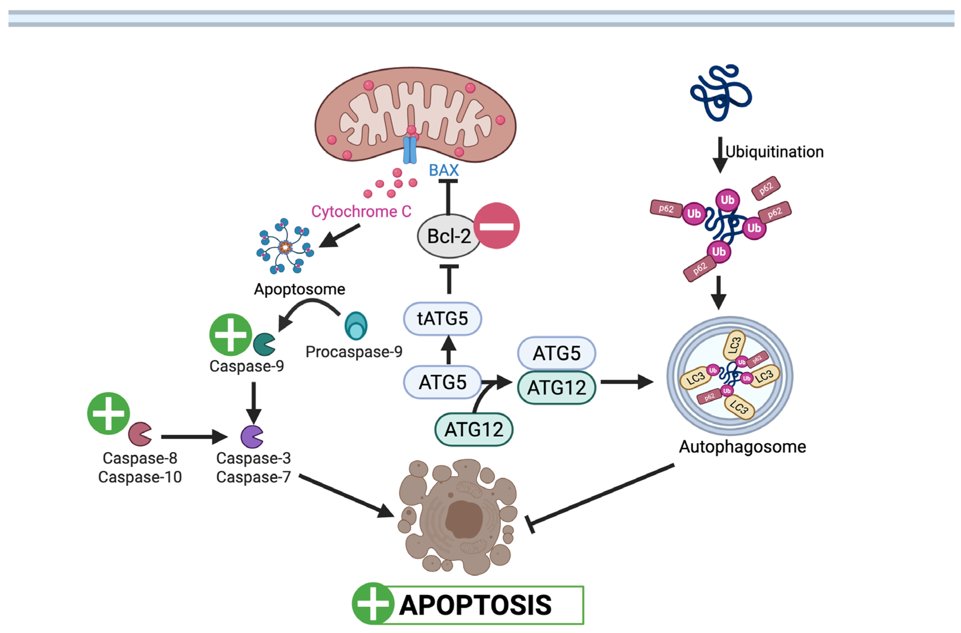
Nutrients 17 03712 g002
Figure 3. UA’s role in modulating mitochondrial function in a high (↑) glucose environment. UA reduces mitochondrial dysfunction by lowering ROS levels, preventing inflammasome activation, and β-cell apoptosis. UA also limits mitochondrial Ca2+ influx by inhibiting TGM-2, thereby reducing tau phosphorylation and amyloid formation. Nutrients 17 03712 i002: UA mediated inhibition (Created in BioRender. Busselberg, D. (2025) https://BioRender.com/x9iuf4l accessed on 8 November 2025).
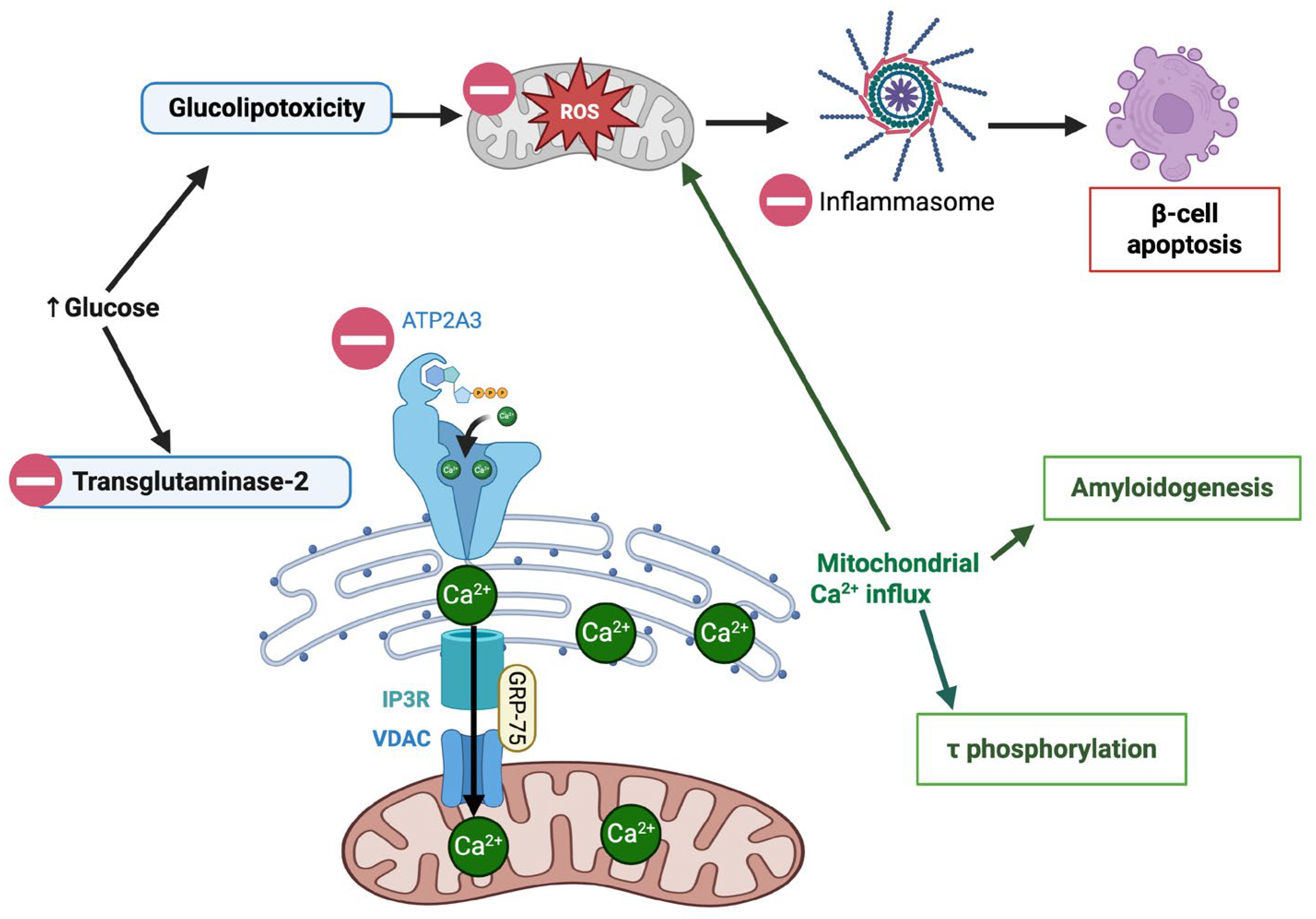
Nutrients 17 03712 g003
Figure 4. Urolithin-A mediated modulation of apoptosis in pancreatic β cells. UA stabilizes Bcl-2 to prevent cytochrome c release and caspase activation, thereby reducing apoptosis. UA also enhances autophagic clearance of damaged proteins through ATG5–ATG12 and p62 signaling. Nutrients 17 03712 i001: UA mediated stimulation, Nutrients 17 03712 i002: UA mediated inhibition, : inhibition. (Created in BioRender. Busselberg, D. (2025) https://BioRender.com/0v18m6c 8 November 2025).
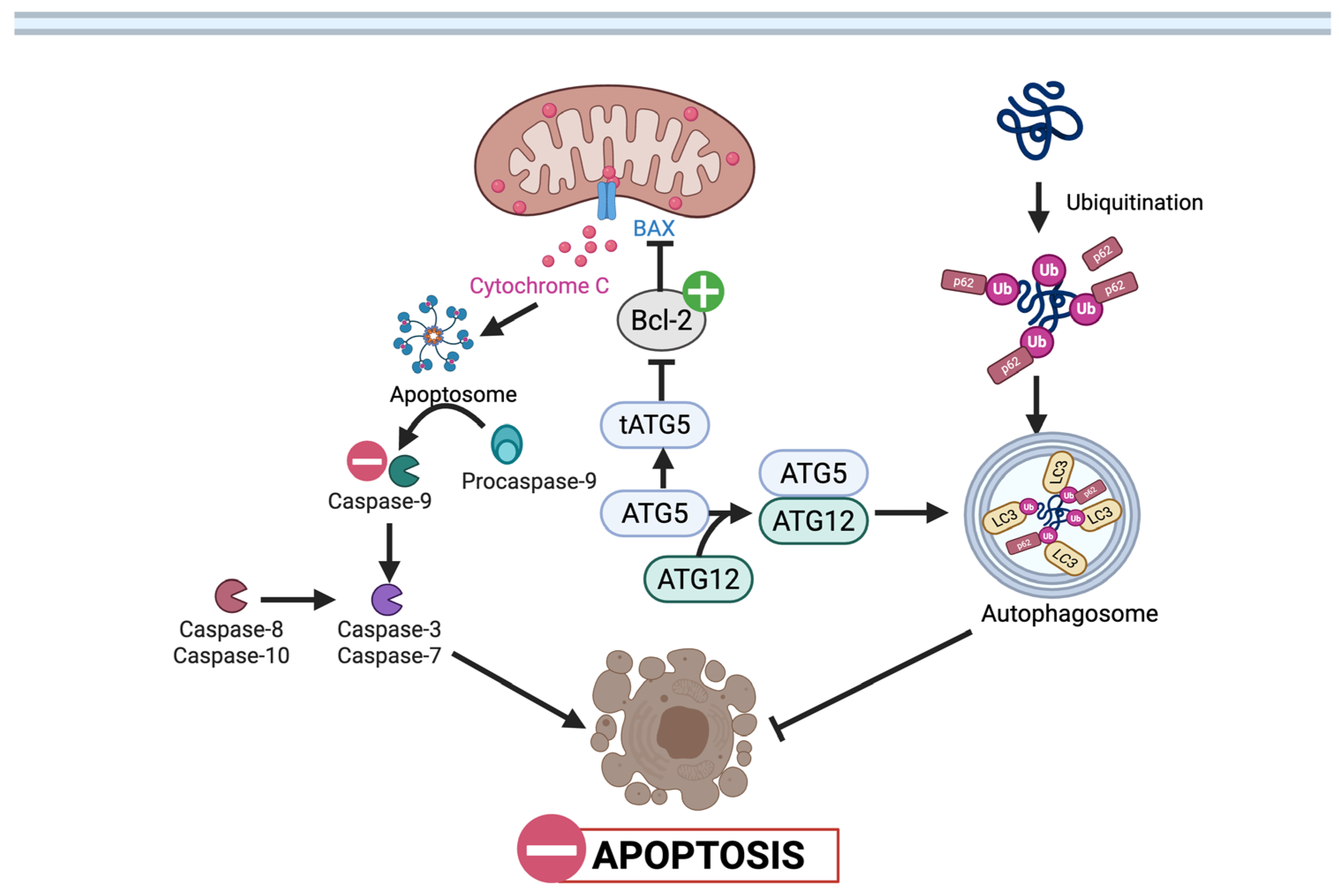
Nutrients 17 03712 g004
Figure 5. Pathways modulated by UA in T2DM: UA promotes mitophagy by modulating TRPC6–calpain signaling and activating the PINK1/Parkin pathway, supporting mitochondrial quality control. UA-induced activation of the AMPK and PI3K/AKT/mTOR pathways enhances glucose uptake via GLUT4, improves cell growth, and reduces PTP1B-mediated insulin resistance. Nutrients 17 03712 i001: UA mediated stimulation, Nutrients 17 03712 i002: UA mediated inhibition, : inhibition. (Created in BioRender. Busselberg, D. (2025) https://BioRender.com/3w2bhrn 8 November 2025).
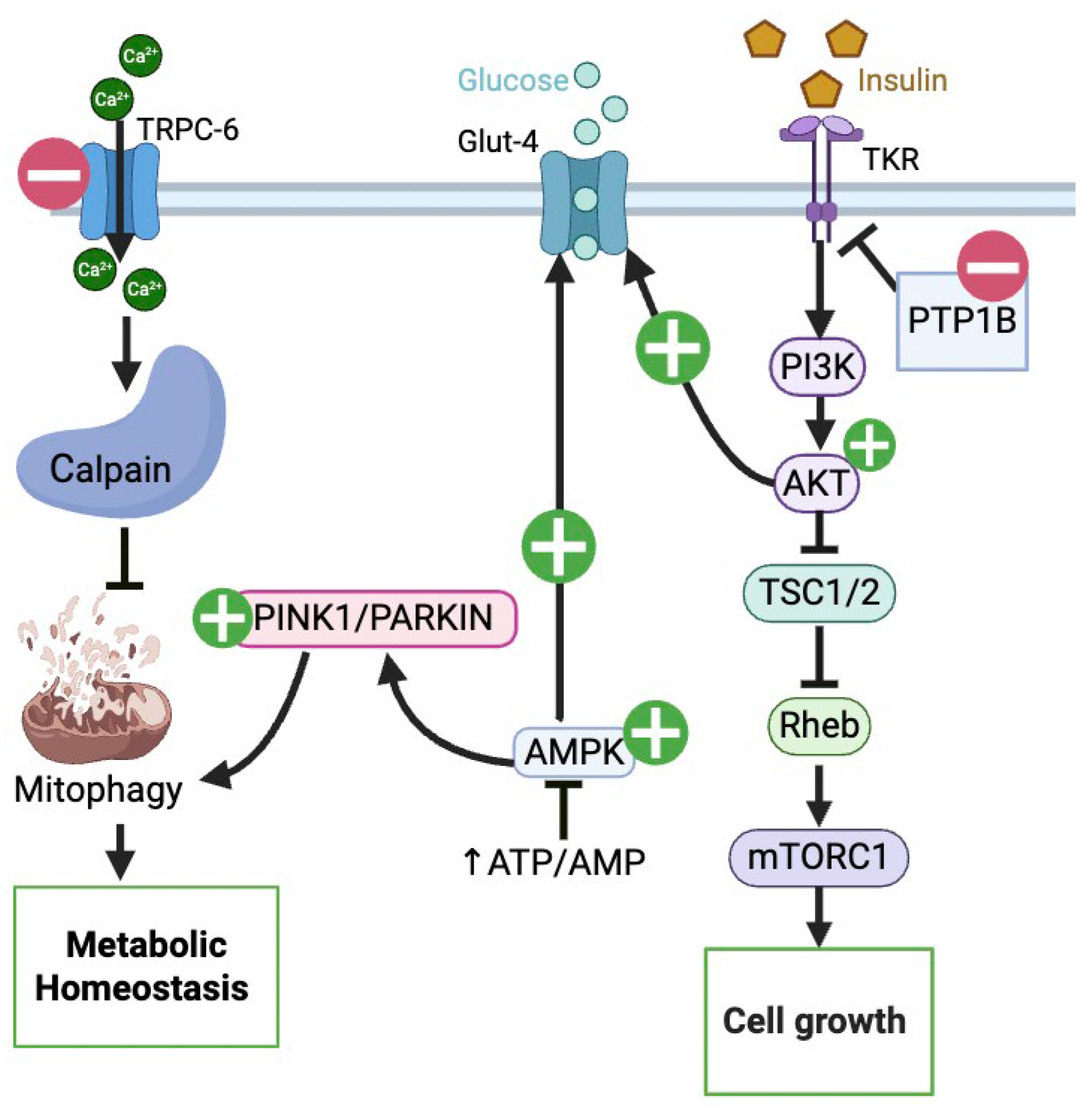
Nutrients 17 03712 g005
Table 1. Comparative Analysis of UA Studies in CRC.
StudyModelUA Dose Main Findings (*/**)AdvantagesLimitations
González-Sarrías et al., 2015 [42]Caco-2, SW480, HT-29 (human CRC)10 µM, 20 µM
(with 5-FU)
IC50 of 5-FU/5′DFUR;
chemosensitization
Multiple colon cancer cell lines;
Use of concentrations achievable in the colorectum
No in vivo validation; limited to combo treatment (UA + 5-FU)
Zhao et al., 2018 [36]SW620 (human CRC)0.05–15 µMAt sub-µM:
Autophagy
Shows activity at sub-micromolar doses—suggests plausible colonic relevancySingle CRC line only; no animal/human confirmation
Norden & Heiss (2019) [18]HCT116 (human CRC) WT vs. p53/IC50 ≈ 19 µM (72 h) p53, p21;
glycolysis
(via p53/TIGAR)
Mechanistic insight into p53 dependency—potential biomarker for responseEffects depend on tumor p53 status; no in vivo or clinical data
El-Wetidy et al., 2021
[16]
HT-29, SW480, SW620 (human CRC)IC50:
~25.5 µM HT-29; 38.1 µM SW480; 53.6 µM SW620
(at 48 h)
G2/M arrest;
p53 & p21;
Bcl-2;
cytochrome-c release;
caspase-3 activation;
Reactive Oxygen Species (ROS)
Multiple cell lines tested.No in vivo experiments;
physiological relevance questionable
Ghosh et al., 2022 [21]SW480, HCT116 (5-FU resistant lines); NRG mice xenografts; AOM/DSS miceIn vitro: 10–50 µM;
In vivo: UroA 20–40 mg/kg PO (mice);
5-FU 20 mg/kg i.p
UA ± 5-FU:
Viability,
Invasion,
EMT markers, drug transporters
(MDR1, BCRP, MRP2/7);
tumor growth in xenograft & AOM/DSS models
in vitro and in vivo model tested;
immunocompetent and immunodeficient in vivo mouse models tested.
UAS03 is synthetic (not naturally produced by gut microbiota), so human applicability depends on safety/PK in humans.
No direct patient data or clinical validation
González-Sarrías et al., 2009 [33]Caco-2 (human) + rat colon (in situ buffer experiments)40 µM
n = 3
Phase I/Phase II detox enzymes
(CYP1A1,
UGT1A10);
inhibited sulfotransferases (favoring glucuronidation)
It compared in vitro (Caco-2), in situ (rat colon perfusion), and in vivo (rat feeding) models.Inability to reproduce the in vitro findings in the in vivo animal colon, suggesting the in vitro model (compounds in buffer) lacks physiological relevance for this specific endpoint
González-Sarrías et al., 2014 [25]Caco-2, SW480, HT-29100 µM (aglycones & glucuronides tested)S and G2/M arrest
Uro-A most active. Glucuronidation: ↓ activity
Used multiple cell lines;
it directly tested and compared the biological activity of urolithin aglycones against their glucuronide metabolites.
High concentrations; No in vivo testing
González-Sarrías et al., 2017 [26]Caco-2 and CCD18-Co (normal colonic cells)IC50 ≈50 µM Uro-A, ~70 µM IsoUro-A) Proliferation,
S/G2 arrest,
Apoptosis at ~50–70 µM;
conjugated forms inactive;
higher glucuronidation in IsoUro-A
Compares isomeric forms and conjugation rates.High concentrations; physiologic relevance uncertain; lack of in vivo validation
Giménez-Bastida et al., 2020 [19]HCT116
(p53WT), Caco-2, HT-29
10 µM chronic exposure (up to >2 weeks) cellular senescence ( p53/p21) and anti-clonogenic effectsFocus on long-term low-dose exposure (physiologically relevant colonic concentrations)—strong translational logicNo animal data; emphasis on senescence rather than cell death
Núñez-Sánchez et al., 2016 [43]Caco-2 colonospheres; primary patient-derived CRC CSCsMixtures: C1 = 0.85 mM; C2 = 17 mM (UA ≈ 85% of mix)colonosphere
ALDH+
Uses patient-derived CSCs (more clinically relevant)Concentrations very high; unrealistic physiologic exposure; mixture study obscures single-compound effect; no in vivo data
Tortora et al., 2018 [27]Pirc rats (APC+/− model) + CRC cell lines; ex vivo tissueDiet-derived UA (pomegranate mesocarp)—in vitro:
UA 25 µM
+ 2.5 mM
sodium butyrate
precancerous lesions (MDF);
apoptosis in lesions
Combines in vitro & in vivo evidenceUA effect shown only with co-treatment (butyrate/diet matrix);
small n (rats: n = 4); Pirc model = FAP representation (not general CRC)
Tiwari et al., 2024 [28]HT-29 (human colon adenocarcinoma)IC50 ≈120 µM Viability (IC50 ≈ 120 µM);
p53/p21,
caspase activation; Bcl-2, AKT1/2
Detailed pathway predictions and in vitro validationIC50 extremely high (120 µM)—far above realistic plasma levels; single cell line;
no in vivo data
* indicates increase, ** indicates decrease.
Table 3. Pathways affected by UA that is common to its effect in CRC and T2DM.
Scheme
Molecule/Pathway
Effect in CRCEffect in T2DMReferences for CRCReferences for
T2DM
Wnt/β-cateninIn T-cells:
In CRC cells:
In VSMC:
In β-cells:
[21,38][22,49,54]
Apoptosis
(p53, p21, caspases, Bax/Bcl-2)
In C.R.C cells:In β-cells:
Podocytes:
[16,18,28][48,50]
Autophagy
& Mitophagy
(PINK1/Parkin, ULK1, LC3)
In T cells: In β-cells:
In Podocytes:
In EPC:
[36,38][48,51,56]
Inflammation
(TLR4/Myd88/NLRP3, cytokines)
In CRC cells: IL-6, TNF-αIn Retinal cells:
IL-6, IL-1β, and TNF-α
In Gut/Brain cells: TLR4/Myd88,NLRP-3
[21][51,58,59]
Oxidative Stress
(ROS, Nrf2/HO-1)
In CRC cells: R.O.S, MDA, S.O.D.In β-cells: MDA, GSH
In retinal cells:
Nrf2/HO-1
[75][49,58]
Aryl
Hydrocarbon Receptor (AhR) pathway
In colonic cells:
gut barrier integrity
In neurons:
neuroprotection
[76][45]
indicates increase, indicates decrease, ∴ indicates therefore.
Disclaimer/Publisher’s Note: The statements, opinions and data contained in all publications are solely those of the individual author(s) and contributor(s) and not of MDPI and/or the editor(s). MDPI and/or the editor(s) disclaim responsibility for any injury to people or property resulting from any ideas, methods, instructions or products referred to in the content.