Brief Report

A High-Fat Diet Induces Epigenetic 1-Carbon Metabolism, Homocystinuria, and Renal-Dependent HFpEF

Department of Physiology, University of Louisville School of Medicine, Louisville, KY 40202, USA
Nutrients 2025, 17(2), 216; https://doi.org/10.3390/nu17020216
Submission received: 12 October 2024 / Revised: 30 December 2024 / Accepted: 31 December 2024 / Published: 8 January 2025

Abstract

:
Background/Objectives: Chronic gut dysbiosis due to a high-fat diet (HFD) instigates cardiac remodeling and heart failure with preserved ejection fraction (HFpEF), in particular, kidney/volume-dependent HFpEF. Studies report that although mitochondrial ATP citrate lyase (ACLY) supports cardiac function, it decreases more in human HFpEF than HFrEF. Interestingly, ACLY synthesizes lipids and creates hyperlipidemia. Epigenetically, ACLY acetylates histone. The mechanism(s) are largely unknown. Methods/Results: One hypothesis is that an HFD induces epigenetic folate 1-carbon metabolism (FOCM) and homocystinuria. This abrogates dipping in sleep-time blood pressure and causes hypertension and morning heart attacks. We observed that probiotics/lactobacillus utilize fat/lipids post-biotically, increasing mitochondrial bioenergetics and attenuating HFpEF. We suggest novel and paradigm-shift epigenetic mitochondrial sulfur trans-sulfuration pathways that selectively target HFD-induced HFpEF. Previous studies from our laboratory, using a single-cell analysis, revealed an increase in the transporter (SLC25A) of s-adenosine–methionine (SAM) during elevated levels of homocysteine (Hcy, i.e., homocystinuria, HHcy), a consequence of impaired epigenetic recycling of Hcy back to methionine due to an increase in the FOCM methylation of H3K4, K9, H4K20, and gene writer (DNMT) and decrease in eraser (TET/FTO). Hcy is transported to mitochondria by SLC7A for clearance via sulfur metabolomic trans-sulfuration by 3-mercaptopyruvate sulfur transferase (3MST). Conclusions: We conclude that gut dysbiosis due to HFD disrupts rhythmic epigenetic memory via FOCM and increases in DNMT1 and creates homocystinuria, leading to a decrease in mitochondrial trans-sulfuration and bioenergetics. The treatment with lactobacillus metabolites fat/lipids post-biotically and bi-directionally produces folic acid and lactone–ketone body that mitigates the HFD-induced mitochondrial remodeling and HFpEF.

1. Introduction

Chronic gut symbiosis due to a high-fat diet (HFD) instigates cardiac remodeling and heart failure with preserved ejection fraction (HFpEF) [1,2,3,4]; however, its mechanisms are largely unknown. The therapeutic approaches focusing only on the oxidative stress, inflammation and hyperlipidemia have not yielded significant positive clinical outcomes for HFpEF. Although HFD contains lipids, fats, and proteins, there is not enough information regarding the role of animal protein intoxication in HFpEF. Interestingly, the recent studies report that the ATP citrate lyase (ACLY) supports cardiac function and decreases more in HFpEF than HFrEF. Paradoxically, ACLY creates hyperlipidemia. Because the inhibition of ACLY (ACLYi) mitigates hyperlipidemia and fatty acid declines ACLY expression [5], the connection is unclear. Epigenetically, ACLY acetylates histone [6]. It is known that the transcription is controlled by on/off promoters by rhythmic methylation/de-methylation during development, health and disease. The chromatin maturation, adaption, and accessibility are regulated by acetylation. The epigenetic folate 1-carbon metabolism (FOCM) pathways recycle Hcy back to methionine. The DNA methylation by FOCM [1,2,3,4,7] is the hallmark of epigenetic memory, i.e., rhythmic gene imprinting and off-printing during embryogenesis, development, health, remodeling, and diseases [8]. One hypothesis is that an HFD induces epigenetic FOCM and HFpEF. Probiotics utilize fat/lipids [9,10] which post-biotically increase mitochondrial bioenergetics and attenuate HFD-induced heart failure. Here, we review a novel and paradigm-shift epigenetic mitochondrial sulfur trans-sulfuration pathways that selectively targets HFD-induced HFpEF. A HFD via transport and metabolism pathways instigates cardiac remodeling.

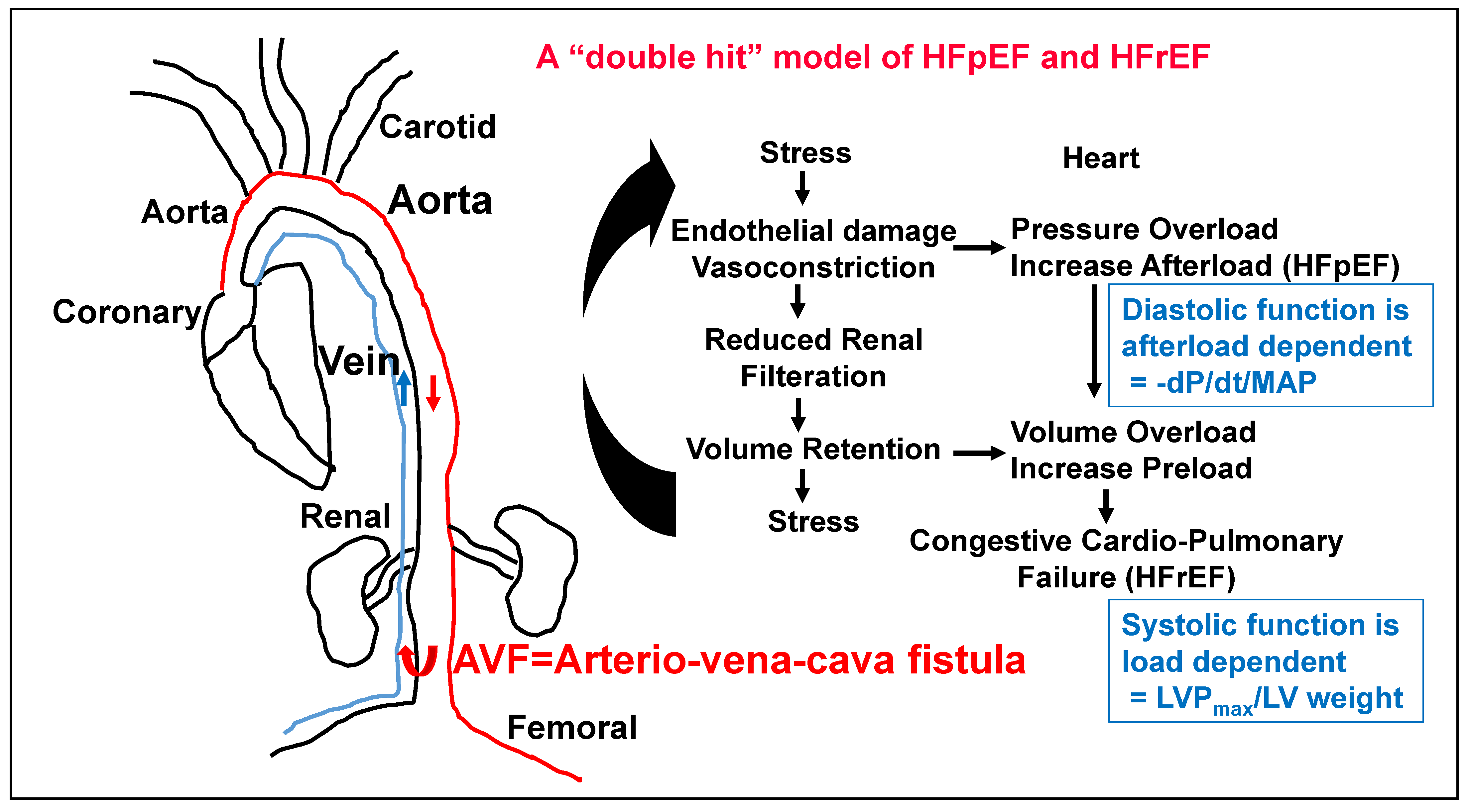
2. The Role of Kidney in HFpEF

Although the pathologies of obesity, hypertension, aging and diabetes are associated with kidney dysfunction, the role of kidney in HFpEF is not clear. Interestingly, we have shown the contribution of renal de-nervation in HFpEF [11]. The HFpEF is afterload and renal-dependent diastolic dysfunction; the cardiac relaxation rate, i.e., -dp/dt/MAP), normalized with MAP, is an effective and more appropriate test to determine levels of HFpEF. On the other hand, the HFrEF is pre-load dependent, leading to systolic dysfunction [12]; therefore, LV pressure should be used to normalize LV weight, in order to determine the levels of HFrEF (Figure 1). These studies will be a better index of HFpEF. To this end, it is important to address the role of epigenetics in HFpEF.

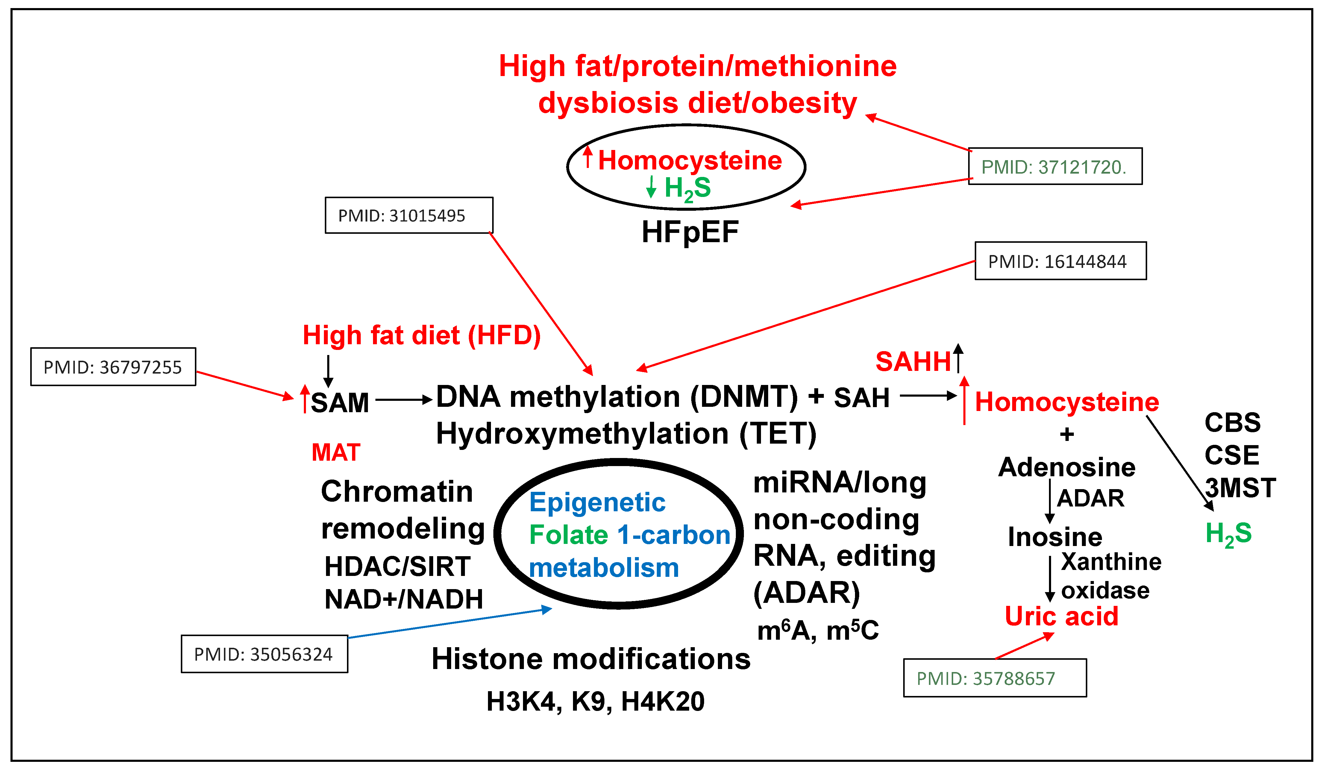
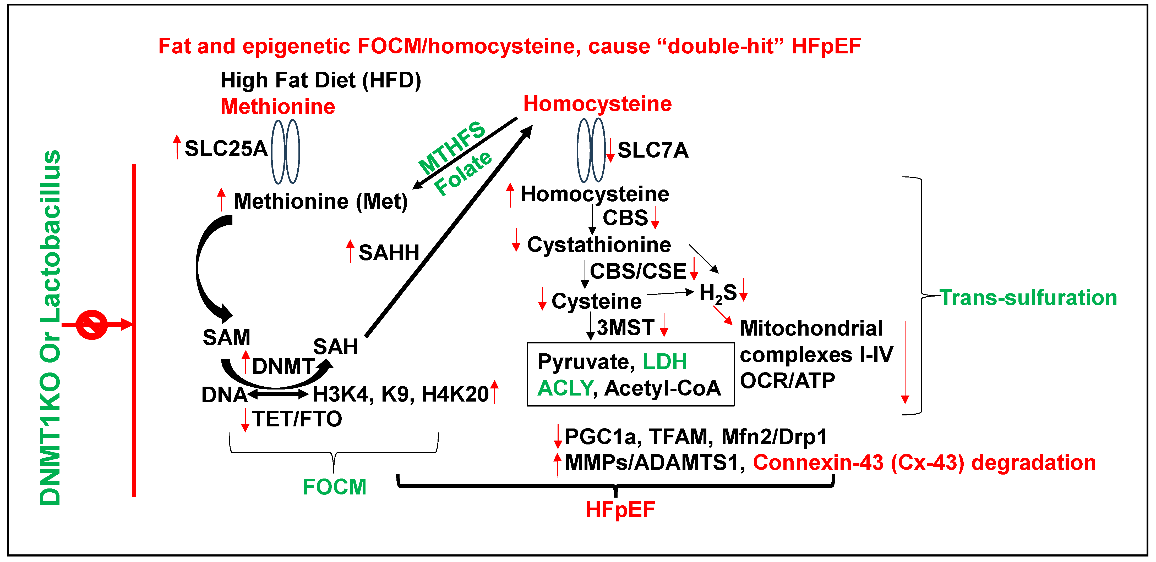
3. Epigenetics of HFpEF

Methionine (Met) is a substrate for epigenetic DNA/RNA/proteins, histone methylation, and the generation of homocysteine (Hcy) via SAM/SAH/SAHH pathways. Hcy are then converted to H2S via trans-sulfuration 3MST/CBS/CSE pathways [13,14,15]. The inhibition of methyl transferase restores metabolism and improves the regenerative capacity of the muscle [16]. The mutation in DNMT causes hypermethylation and causes growth retardation [17]. The accumulation of by-product uric acid causes HFrEF [18]. The activation of growth arrest DNA damage (GADD) is associated with the activation of nuclear metalloproteinases [19] (Figure 2). Collectively, these studies suggest the role of epigenetics in intra-nuclear, chromatin, histones, and extracellular remodeling.
Although a chronic high-fat diet (HFD) is associated with hypertension/obesity and “double hit” heart failure [20,21,22], its mechanism is unclear. There is ~1% of methionine (Met) in HFD [23], which is significantly high for a single amino acid. Interestingly, studies from our laboratory [1,2,3,4] observed an increase in dysbiosis due to an HFD (i.e., increase in ratio between polyunsaturated fatty acid/monounsaturated fatty acid in WT mice+HFD). The treatment with lactobacillus-probiotic (PB, a folate and lactone–ketone body producer [24,25,26] mitigated this increase in poly-unsaturated fatty acid [1,2,3,4]. Due to high methionine, gut–dysbiosis instigates a de-arrangement in the epigenetic rhythmic methylation/demethylation ratio on the DNA/RNA, and protein/histones, due to a DNMT1/TET2 ratio [27], and consequently, creates hyperhomocysteinemia (HHcy) [1,2,3,4,8]. Previous studies from our laboratory, using a single-cell analysis, revealed an increase in SLC25A (transporter of s-adenosine-methionine (SAM)) during elevated levels of homocysteine (Hcy) [28], caused by impaired recycling of homocysteine back to methionine via an increase in epigenetic methylation on H3K4, K9, and H4K20) via folate 1-carbon metabolism (FOCM) pathway by gene writer (DNMT) and decrease in eraser (TET/FTO) (Figure 2).
Although clinical trial shows that microbiome metabolizes cholesterol/lipids [9,10] and lactobacillus elicits cardiac benefits [9], it is unclear whether the treatment with Lactobacillus rhamnosus, a ketone body fuel for mitochondria [25,26,29,30,31,32,33,34] and a folic acid producing probiotic [24,25,26], reverses dysbiosis-induced cardiac complications, in part by increasing mitochondrial trans-sulfuration, H2S, and bioenergetics [26] (Figure 3). Therefore, it is important to determine, for the first time, that a probiotic can increase mitochondrial sulfur metabolism, trans-sulfuration and bioenergetics and mitigate HFD-induced obesity and heart failure.
Gut dysbiosis, FOCM, methylation, and HHcy via DNMT, BHMT, and PEMT are linked to microbiomes (Figure 4). We suggest an increase in DNMT and PEMT and decrease in BHMT in HFD-fed mice, which will increase Hcy [1,2,3,4]. The treatment with probiotics decreased levels of DNMT and PEMT; however, there were no changes in BHMT [1,2,3,4]. These results suggest differential mechanisms, i.e., betaine versus folate pathways of re-methylation of Hcy [33,34,35] (Figure 4). Interestingly, this also suggested a prominent central role of DNMT1 in Hcy formation by HFD. This incited us to employ DNMT1 knockout mice. We observed that gut–dysbiosis and altered Hcy metabolism represent the dominant mechanism wherein HFD induces DNMT1, causing detrimental cardiac remodeling and mitochondrial metabolic dysfunction (i.e., trans-sulfuration and bioenergetics) during HFD-induced HFpEF.
Others [7,36] have also supported our hypothesis that there is a rhythmic methylation/demethylation during the mitochondrial TCA cycle by epigenetic gene writer (DNMT) and erasers (TET and FTO). The premise of this study is also supported by Kay et al. [37,38], and Engler et al. [39,40,41], who reveal the role of transforming fibroblasts, and fibrosis in cardiac remodeling. The study by Dees et al. [42] suggests that DNA-methyltransferase 3A (DNMT3A) and DNMT1 in fibroblasts silence the expression of suppressor of cytokine signaling 3 (SOCS3) in a SMAD-dependent manner by promoting hypermethylation. The downregulation of SOCS3 facilitated the activation of signal transducers, and activators of transcription 3 (STAT3) to promote fibroblast-to-myofibroblast transition, collagen release, and fibrosis in vitro and in vivo. The re-establishment of the epigenetic control of STAT3 signaling via the genetic or pharmacological inactivation of DNMT3A reversed the phenotype of fibroblasts in tissue culture, inhibited TGFb-dependent fibroblast activation, and ameliorated experimental fibrosis in murine models [42,43,44]. Felisbino and McKinsey suggested the role of epigenetics in cardiac fibrosis with emphasis on inflammation and fibroblast activation [45,46]. The authors demonstrated the role of histone acetylation, and reader proteins using a small-molecule inhibitor of bromo-domain-containing protein 4 (BRD4) for mitigating cardiac fibrosis [47]. Interestingly, along the same line, we demonstrated that Hcy induces collagen expression in a dose- and time-dependent manner, as measured by Northern blot analysis [48]. Hcy causes fibroblast activation, and myofibroblast differentiation in murine aortic endothelial cells [49]. Previously, in human, we show the role of a dis-integrin and metalloproteinase (ADAM) in connexin-43 (Cx43) degradation in human end stage heart failure [50]. Others have shown that Cx-43 synchronizes myocyte-mitochondrial function [51,52]. Here, we focus on the significance of epigenetics via gene writer, eraser, and reader functions along with direct consequences of Hcy in cardiac remodeling.

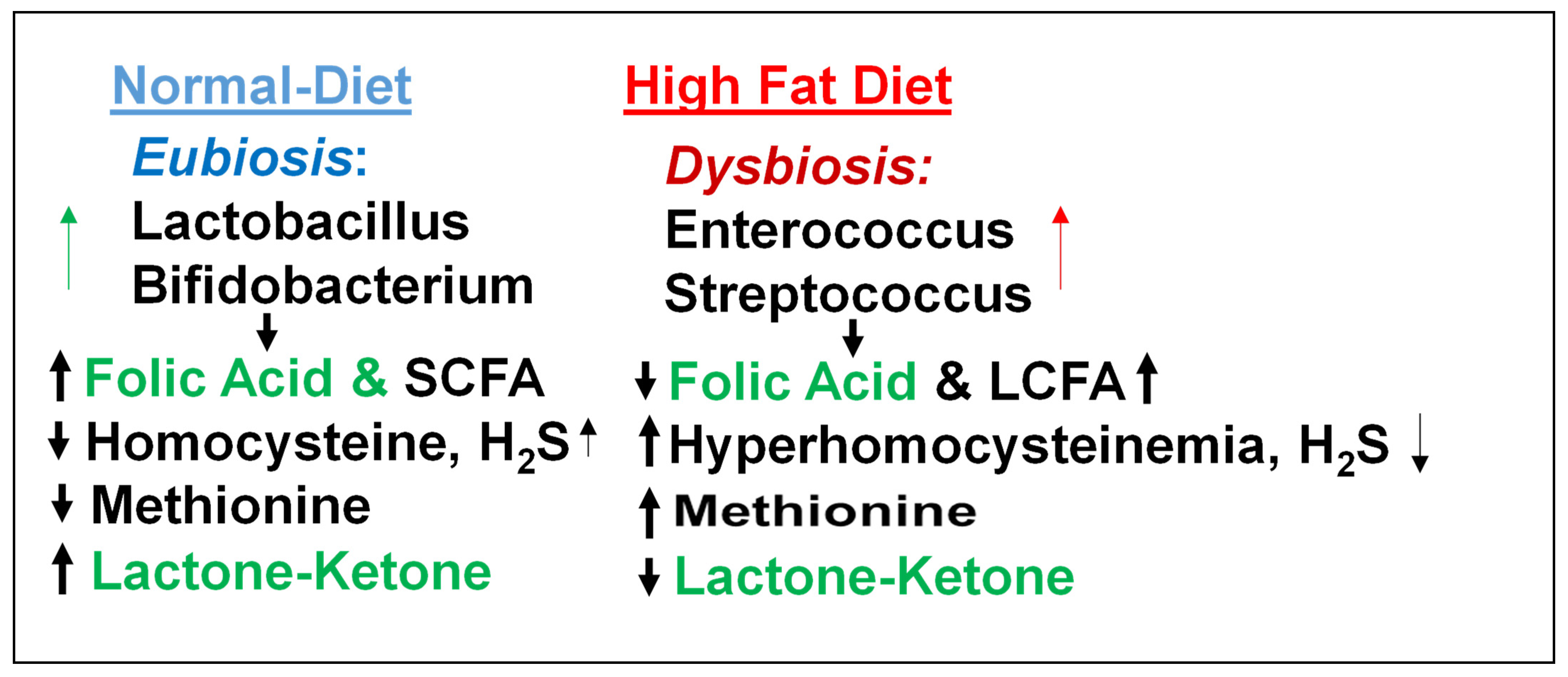
4. Homocysteine and Lipid Connection

Most of the human population is mildly HHcy and asymptomatic; however, when on an HFD, especially a red-meat-rich diet that is high in methionine (a substrate for Hcy generation during epigenetically regulated DNA methylation) [1,2,3,4], severe HHcy could develop, and that will ultimately cause symptomatic cardiovascular diseases. Interestingly, although folic acid and H2S reduce obesity and brown adipose fat [14,15], their mechanisms are unclear.
Homocysteine theory suggests a diet high in animal protein and low in B vitamins, which occur in many foods but are very easily destroyed by processing, i.e., a diet of meat, cheese, milk, white flour, and foods that are canned, boxed, refined, processed, or preserved. The theory suggests a strong connection between diet and CVD, but one that is different from cholesterol/lipids. The homocysteine theory considers CVD due to what McCully calls protein intoxication [53,54]. The cholesterol theory (sometimes called the lipid theory) instead demonizes fats. Since proteins and fats often occur in the same foods, the potential dietary treatments for high homocysteine and high cholesterol are similar, with the following distinction: the anti-homocysteine diet focuses on what should be eaten, as a preventive, while the anti-cholesterol diet focuses on what should be avoided, as a precipitator. Thus, a diet of lower homocysteine includes many natural sources of B vitamins, like fresh fruits and vegetables, and limits animal protein. A cholesterol-reducing diet limits foods high in saturated fats and cholesterol, like eggs, meat, and butter. Unfortunately, the latter is more commercially popular. Therefore, we use lactobacillus, which produces folic acid and lactone–ketone bodies (food for mitochondria) post-biotically and mitigates HFpEF.
The probiotic, post-biotically produced folic acid and lactone–ketone bodies mitigate HFD-induced heart failure and are therapeutically innovative (i.e., killing two pathologies in one strike). Interestingly, some probiotics also lower cholesterol [10]; however, their mechanisms are unclear. In this novel review article, we address the fact that lactobacillus eats lipids and post-biotically produces folic acid and lactone–ketone bodies, a bi-directional probiotic that mitigates HFD-induced mitochondrial remodeling and heart failure.

5. Viewpoint on the Role of Epigenetics and Future Perspective

This review presents an innovative hypothesis that the therapeutic effect of probiotic treatment should be tested to reverse gut–dysbiosis effects in HFpEF. Describing the mechanisms through which diet (particularly an HFD) and dysbiotic events in distant organs, such as the intestine, affect heart health is important, and positive findings will support the relevance and impact of systemic health on heart health. Although the use of probiotics as a therapeutic alternative to heart disease has been previously studied in pre-clinical models and clinical trials, there is no mechanism-based simple and safe therapy using probiotics. Here, we suggest that a probiotic that produces folic acid (a Hcy lowering agent) and lactate (a ketone body, fuel for mitochondria) post-biotically can eat fat and increases mitochondrial bioenergetics.
Based on this review, it is important to determine whether the lactobacillus downregulates SLC25A, and epigenetic gene writer DNMT1 upregulates eraser TET2/FTO and attenuates cardiac dysfunction by lowering homocysteine. The lactobacillus post biotically produces folic acid and attenuates HHcy via a decrease in epigenetic gene writer (DNMT1) and increase in eraser (TET2/FTO). Also, it will be novel to determine whether the lactobacillus increases SLC7A and mitochondrial H2S production by trans-sulfuration. Lactobacillus post-biotically produces a lactone/ketone body and improves mitochondrial bioenergetics via trans-sulfuration (Figure 5). The use of DNMT1KO and mitigation of HFD-induced epigenetic de-arrangement is novel. The use of transgenic mice overexpressing TET2, FTO (erasers), CBS (transsulfuration), and TFAM (mitochondrial) is mechanistically innovative.

Funding

National Institutes of Health, USA; HL139047; AR071789.

Institutional Review Board Statement

Not applicable.

Informed Consent Statement

Not applicable.

Data Availability Statement

Data are contained within the article.

Conflicts of Interest

The author declares no conflict of interest.

References

  1. Veeranki, S.; Tyagi, S.C. Dysbiosis and Disease: Many Unknown Ends, Is It Time to Formulate Guidelines for Dysbiosis Research? J. Cell. Physiol. 2017, 232, 2929–2930. [Google Scholar] [CrossRef] [PubMed]
  2. Singh, M.; Hardin, S.J.; George, A.K.; Eyob, W.; Stanisic, D.; Pushpakumar, S.; Tyagi, S.C. Epigenetics, 1-Carbon Metabolism, and Homocysteine During Dysbiosis. Front. Physiol. 2020, 11, 617953. [Google Scholar] [CrossRef] [PubMed]
  3. George, A.K.; Singh, M.; Pushpakumar, S.; Homme, R.P.; Hardin, S.J.; Tyagi, S.C. Dysbiotic 1-carbon metabolism in cardiac muscle remodeling. J. Cell. Physiol. 2020, 235, 2590–2598. [Google Scholar] [CrossRef] [PubMed]
  4. Singh, M.; Pushpakumar, S.; Zheng, Y.; Homme, R.P.; Smolenkova, I.; Mokshagundam, S.P.L.; Tyagi, S.C. Hydrogen sulfide mitigates skeletal muscle mitophagy-led tissue remodeling via epigenetic regulation of the gene writer and eraser function. Physiol. Rep. 2022, 10, e15422. [Google Scholar] [CrossRef] [PubMed]
  5. Meddeb, M.; Koleini, N.; Jun, S.; Keykhaei, M.; Farshidfar, F.; Zhao, L.; Kwon, S.; Lin, B.; Keceli, G.; Paolocci, N.; et al. ATP citrate lyase supports cardiac function and NAD+/NADH balance and os depressed in human heart failure. bioRxiv 2024. [Google Scholar] [CrossRef]
  6. Liu, S.; Gammon, S.T.; Tan, L.; Karlstaedt, A. ATP-dependent citrate lyase drives LV dysfunction by metabolic remodeling of the heart. bioRxiv 2024. [Google Scholar] [CrossRef]
  7. Arnold, P.K.; Finley, L.W. Regulation and function of the mammalian tricarboxylic acid cycle. J. Biol. Chem. 2023, 299, 102838. [Google Scholar] [CrossRef]
  8. Greco, C.M.; Cervantes, M.; Fustin, J.-M.; Ito, K.; Ceglia, N.; Samad, M.; Shi, J.; Koronowski, K.B.; Forne, I.; Ranjit, S.; et al. S-adenosyl-l-homocysteine hydrolase links methionine metabolism to the circadian clock and chromatin remodeling. Sci. Adv. 2020, 6, eabc5629. [Google Scholar] [CrossRef]
  9. da Silva, L.D.; de Oliveira, Y.; de Souza, E.L.; de Luna Freire, M.O.; de Andrade Braga, V.; Magnani, M.; de Brito Alves, J.L. Effects of probiotic therapy on cardio-metabolic parameters and autonomic modulation in hypertensive women: A randomized, triple-blind, placebo-controlled trial. Food Funct. 2020, 11, 7152–7163. [Google Scholar] [CrossRef] [PubMed]
  10. Li, C.; Stražar, M.; Mohamed, A.M.; Pacheco, J.A.; Walker, R.L.; Lebar, T.; Zhao, S.; Lockart, J.; Dame, A.; Thurimella, K.; et al. Gut microbiome and metabolome profiling in Framingham heart study reveals cholesterol-metabolizing bacteria. Cell 2024, 187, 1834–1852.e19. [Google Scholar] [CrossRef]
  11. Pushpakumar, S.; Singh, M.; Zheng, Y.; Akinterinwa, O.E.; Mokshagundam, S.P.L.; Sen, U.; Kalra, D.K.; Tyagi, S.C. Renal Denervation Helps Preserve the Ejection Fraction by Preserving Endocardial-Endothelial Function during Heart Failure. Int. J. Mol. Sci. 2023, 24, 7302. [Google Scholar] [CrossRef] [PubMed]
  12. Robinson, J.; Sehly, A.; Lan, N.S.R.; Abraham, A.; Dwivedi, G. Arteriovenous fistula is associated with diastolic dysfunction in end-stage renal failure patients. Clin. Exp. Nephrol. 2023, 27, 200–201. [Google Scholar] [CrossRef] [PubMed]
  13. Hayden, M.R.; Tyagi, S.C. Impaired Folate-Mediated One-Carbon Metabolism in Type 2 Diabetes, Late-Onset Alzheimer’s Disease and Long COVID. Medicina 2021, 58, 16. [Google Scholar] [CrossRef] [PubMed]
  14. Katsouda, A.; Szabo, C.; Papapetropoulos, A. Reduced adipose tissue H2S in obesity. Pharmacol. Res. 2018, 128, 190–199. [Google Scholar] [CrossRef]
  15. Han, W.; Li, M.; Yang, M.; Chen, S.; Lu, Y.; Tang, T.; Wang, R.; Zhang, C.; Qi, K. Dietary Folic Acid Supplementation Inhibits High Fat Diet Induced Body Weight Gain through Gut Microbiota-Associated Branched-Chain Amino Acids and Mitochondria in Mice. J. Nutr. Sci. Vitaminol. 2023, 69, 105–120. [Google Scholar] [CrossRef] [PubMed]
  16. Rajabian, N.; Ikhapoh, I.; Shahini, S.; Choudhury, D.; Thiyagarajan, R.; Shahini, A.; Kulczyk, J.; Breed, K.; Saha, S.; Mohamed, M.A.; et al. Methionine adenosyltransferase2A inhibition restores metabolism to improve regenerative capacity and strength of aged skeletal muscle. Nat. Commun. 2023, 14, 886. [Google Scholar] [CrossRef]
  17. Sendžikaitė, G.; Hanna, C.W.; Stewart-Morgan, K.R.; Ivanova, E.; Kelsey, G. A DNMT3A PWWP mutation leads to methylation of bivalent chromatin and growth retardation in mice. Nat. Commun. 2019, 10, 1884. [Google Scholar] [CrossRef]
  18. Doehner, W.; Anker, S.D.; Butler, J.; Zannad, F.; Filippatos, G.; Ferreira, J.P.; Salsali, A.; Kaempfer, C.; Brueckmann, M.; Pocock, S.J.; et al. Uric acid and sodium-glucose cotransporter-2 inhibition with empagliflozin in heart failure with reduced ejection fraction (HFrEF): The EMPEROR-reduced trial. Eur. Heart J. 2022, 43, 3435–3446. [Google Scholar] [CrossRef]
  19. Ijiri, K.; Zerbini, L.F.; Peng, H.; Correa, R.G.; Lu, B.; Walsh, N.; Zhao, Y.; Taniguchi, N.; Huang, X.-L.; Otu, H.; et al. A novel role for growth arrest DNA damage protein 45 (GADD45beta) as a mediator of MMP-13 gene expression. JBC 2005, 280, 38544–38555. [Google Scholar] [CrossRef]
  20. Saleem, T.H.; Algowhary, M.; Kamel, F.E.M.; I El-Mahdy, R. Plasma amino acid metabolomic pattern in heart failure patients with either preserved or reduced ejection fraction: The relation to established risk variables and prognosis. Biomed. Chromatogr. 2021, 35, e5012. [Google Scholar] [CrossRef]
  21. Liu, B.; Ma, S.; Wang, T.; Zhao, C.; Li, Y.; Yin, J.; Liu, C.; Gao, C.; Sun, L.; Yue, W.; et al. A novel rat model of heart failure induced by high methionine diet showing evidence of association between hyperhomocysteinemia and activation of NF-kappaB. Am. J. Transl. Res. 2016, 8, 117–124. [Google Scholar] [PubMed]
  22. Joseph, J.; Giczewska, A.; Alhanti, B.; Cheema, A.K.; Handy, D.E.; Mann, D.L.; Loscalzo, J.; Givertz, M.M. Associations of methyl donor and methylation inhibitor levels during anti-oxidant therapy in heart failure. J. Physiol. Biochem. 2021, 77, 295–304. [Google Scholar] [CrossRef] [PubMed]
  23. Service, N.H. Meat in your diet, (2021) & Tyagi SC. Clin. Exper Hypertens. 1999, 21, 181–198. [Google Scholar]
  24. Albano, C.; Silvetti, T.; Brasca, M. Screening of lactic acid bacteria producing folate and their potential use as adjunct cultures for cheese bio-enrichment. FEMS Microbiol. Lett. 2020, 367, fnaa059. [Google Scholar] [CrossRef]
  25. Guerrero-Encinas, I.; González-González, J.N.; Santiago-López, L.; Muhlia-Almazán, A.; Garcia, H.S.; Mazorra-Manzano, M.A.; Vallejo-Cordoba, B.; González-Córdova, A.F.; Hernández-Mendoza, A. Protective Effect of Lacticaseibacillus casei CRL 431 Postbiotics on Mitochondrial Function and Oxidative Status in Rats with Aflatoxin B(1)-Induced Oxidative Stress. Probiotics Antimicrob. Proteins 2021, 13, 1033–1043. [Google Scholar] [CrossRef]
  26. Gu, Y.; Xiao, X.; Pan, R.; Zhang, J.; Zhao, Y.; Dong, Y.; Cui, H. Lactobacillus plantarum dy-1 fermented barley extraction activates white adipocyte browning in high-fat diet-induced obese rats. J. Food Biochem. 2021, 45, e13680. [Google Scholar] [CrossRef]
  27. Hong, T.; Li, J.; Guo, L.; Cavalier, M.; Wang, T.; Dou, Y.; DeLaFuente, A.; Fang, S.; Guzman, A.; Wohlan, K.; et al. TET2 modulates spatial relocalization of heterochromatin in aged hematopoietic stem and progenitor cells. Nat. Aging 2023, 3, 1387–1400. [Google Scholar] [CrossRef]
  28. Singh, M.; George, A.K.; Homme, R.P.; Majumder, A.; Laha, A.; Sandhu, H.S.; Tyagi, S.C. Circular RNAs profiling in the cystathionine-β-synthase mutant mouse reveals novel gene targets for hyperhomocysteinemia induced ocular disorders. Exp. Eye Res. 2018, 174, 80–92. [Google Scholar] [CrossRef]
  29. Aubert, G.; Martin, O.J.; Horton, J.L.; Lai, L.; Vega, R.B.; Leone, T.C.; Koves, T.; Gardell, S.J.; Krüger, M.; Hoppel, C.L.; et al. The Failing Heart Relies on Ketone Bodies as a Fuel. Circulation 2016, 133, 698–705. [Google Scholar] [CrossRef]
  30. Panigrahi, P.; Parida, S.; Nanda, N.C.; Satpathy, R.; Pradhan, L.; Chandel, D.S.; Baccaglini, L.; Mohapatra, A.; Mohapatra, S.S.; Misra, P.R.; et al. A randomized synbiotic trial to prevent sepsis among infants in rural India. Nature 2017, 548, 407–412. [Google Scholar] [CrossRef]
  31. Brooks, G.A.; Osmond, A.D.; Arevalo, J.A.; Duong, J.J.; Curl, C.C.; Moreno-Santillan, D.D.; Leija, R.G. Lactate as a myokine and exerkine: Drivers and signals of physiology and metabolism. J. Appl. Physiol. 2023, 134, 529–548. [Google Scholar] [CrossRef] [PubMed]
  32. Matsuura, T.R.; Puchalska, P.; Crawford, P.A.; Kelly, D.P. Ketones and the Heart: Metabolic Principles and Therapeutic Implications. Circ. Res. 2023, 132, 882–898. [Google Scholar] [CrossRef] [PubMed]
  33. Balikcioglu, P.G.; Trub, C.J.; Balikcioglu, M.; Ilkayeva, O.; White, P.J.; Muehlbauer, M.; Bain, J.R.; Armstrong, S.; Freemark, M. Branched-chain α-keto acids and glutamate/glutamine: Biomarkers of insulin resistance in childhood obesity. Endocrinol. Diabetes Metab. 2023, 6, e388. [Google Scholar] [CrossRef] [PubMed]
  34. Chu, H.-N.; Kim, H.-R.; Jang, K.-A.; Hwang, Y.-J.; Kim, J.-S. Anti-obesity effects of yuja (Citrus junos Sieb ex Tanaka) pomace extract fermented with lactic acid bacteria on the hepatocytes and epididymal fat tissue of rats. Appl. Microsc. 2023, 53, 7. [Google Scholar] [CrossRef]
  35. Schaevitz, L.; Berger-Sweeney, J.; Ricceri, L. One-carbon metabolism in neurodevelopmental disorders: Using broad-based nutraceutics to treat cognitive deficits in complex spectrum disorders. Neurosci. Biobehav. Rev. 2014, 46 Pt 2, 270–284. [Google Scholar] [CrossRef]
  36. Tóth, M.E.; Sárközy, M.; Szűcs, G.; Dukay, B.; Hajdu, P.; Zvara, Á.; Puskás, L.G.; Szebeni, G.J.; Ruppert, Z.; Csonka, C.; et al. Exercise training worsens cardiac performance in males but does not change ejection fraction and improves hypertrophy in females in a mouse model of metabolic syndrome. Biol. Sex Differ. 2022, 13, 5. [Google Scholar] [CrossRef] [PubMed]
  37. Entcheva, E.; Kay, M.W. Cardiac optogenetics: A decade of enlightenment. Nat. Rev. Cardiol. 2021, 18, 349–367. [Google Scholar] [CrossRef]
  38. Zasadny, F.M.; Dyavanapalli, J.; Dowling, N.M.; Mendelowitz, D.; Kay, M.W. Cholinergic stimulation improves electrophysiological rate adaptation during pressure overload-induced heart failure in rats. Am. J. Physiol. Circ. Physiol. 2020, 319, H1358–H1368. [Google Scholar] [CrossRef]
  39. Whitehead, A.J.; Engler, A.J. Regenerative cross talk between cardiac cells and macrophages. Am. J. Physiol. Circ. Physiol. 2021, 320, H2211–H2221. [Google Scholar] [CrossRef]
  40. Yeoman, B.; Shatkin, G.; Beri, P.; Banisadr, A.; Katira, P.; Engler, A.J. Adhesion strength and contractility enable metastatic cells to become adurotactic. Cell Rep. 2021, 34, 108816. [Google Scholar] [CrossRef]
  41. Banisadr, A.; Eick, M.; Beri, P.; Parisian, A.D.; Yeoman, B.; Placone, J.K.; Engler, A.J.; Furnari, F. EGFRvIII uses intrinsic and extrinsic mechanisms to reduce glioma adhesion and increase migration. J. Cell Sci. 2020, 133, jcs247189. [Google Scholar] [CrossRef] [PubMed]
  42. Dees, C.; Pötter, S.; Zhang, Y.; Bergmann, C.; Zhou, X.; Luber, M.; Wohlfahrt, T.; Karouzakis, E.; Ramming, A.; Gelse, K.; et al. TGF-β–induced epigenetic deregulation of SOCS3 facilitates STAT3 signaling to promote fibrosis. J. Clin. Investig. 2020, 130, 2347–2363. [Google Scholar] [CrossRef]
  43. Kakoki, M.; Ramanathan, P.V.; Hagaman, J.R.; Grant, R.; Wilder, J.C.; Taylor, J.M.; Jennette, J.C.; Smithies, O.; Maeda-Smithies, N. Cyanocobalamin prevents cardiomyopathy in type 1 diabetes by modulating oxidative stress and DNMT-SOCS1/3-IGF-1 signaling. Commun. Biol. 2021, 4, 775. [Google Scholar] [CrossRef]
  44. Kanai, S.M.; Edwards, A.J.; Rurik, J.G.; Osei-Owusu, P.; Blumer, K.J. Proteolytic degradation of regulator of G protein signaling 2 facilitates temporal regulation of G(q/11) signaling and vascular contraction. J. Biol. Chem. 2017, 292, 19266–19278. [Google Scholar] [CrossRef]
  45. Felisbino, M.B.; McKinsey, T.A. Epigenetics in Cardiac Fibrosis. JACC Basic Transl. Sci. 2018, 3, 704–715. [Google Scholar] [CrossRef]
  46. Trial, J.; Cieslik, K.A. Changes in cardiac resident fibroblast physiology and phenotype in aging. Am. J. Physiol. Circ. Physiol. 2018, 315, H745–H755. [Google Scholar] [CrossRef]
  47. Stratton, M.S.; Bagchi, R.A.; Felisbino, M.B.; Hirsch, R.A.; Smith, H.E.; Riching, A.S.; Enyart, B.Y.; Koch, K.A.; Cavasin, M.A.; Alexanian, M.; et al. Dynamic Chromatin Targeting of BRD4 Stimulates Cardiac Fibroblast Activation. Circ. Res. 2019, 125, 662–677. [Google Scholar] [CrossRef]
  48. Tyagi, S.C.; Sood, H.S.; Hunt, M.J.; Mujumdar, V.S.; Tummalapalli, C.M.; Aru, G.M. Homocysteine redox receptor and regulation of extracellular matrix components in vascular cells. Am. J. Physiol. Physiol. 1998, 274, C396–C405. [Google Scholar] [CrossRef]
  49. Sen, U.; Moshal, K.S.; Tyagi, N.; Kartha, G.K.; Tyagi, S.C. Homocysteine-induced myofibroblast differentiation in mouse aortic endothelial cells. J. Cell. Physiol. 2006, 209, 767–774. [Google Scholar] [CrossRef]
  50. Hunt, M.J.; Aru, G.M.; Hayden, M.R.; Moore, C.K.; Hoit, B.D.; Tyagi, S.C. Induction of oxidative stress and disintegrin metalloproteinase in human heart end-stage failure. Am. J. Physiol. Lung Cell. Mol. Physiol. 2002, 283, L239–L245. [Google Scholar] [CrossRef] [PubMed]
  51. Miro-Casas, E.; Ruiz-Meana, M.; Agullo, E.; Stahlhofen, S.; Rodríguez-Sinovas, A.; Cabestrero, A.; Jorge, I.; Torre, I.; Vazquez, J.; Boengler, K.; et al. Connexin43 in cardiomyocyte mitochondria contributes to mitochondrial potassium uptake. Cardiovasc. Res. 2009, 83, 747–756. [Google Scholar] [CrossRef] [PubMed]
  52. Boengler, K.; Ruiz-Meana, M.; Gent, S.; Ungefug, E.; Soetkamp, D.; Miro-Casas, E.; Cabestrero, A.; Fernandez-Sanz, C.; Semenzato, M.; Di Lisa, F.; et al. Mitochondrial connexin 43 impacts on respiratory complex I activity and mitochondrial oxygen consumption. J. Cell. Mol. Med. 2012, 16, 1649–1655. [Google Scholar] [CrossRef] [PubMed]
  53. McCully, H. Pliny’s Pheromonic Abortifacients. Science 1969, 165, 236–237. [Google Scholar] [CrossRef] [PubMed]
  54. McCully, K.S. Vascular pathology of homocysteinemia: Implications for the pathogenesis of arteriosclerosis. Am. J. Pathol. 1969, 56, 111–128. [Google Scholar] [PubMed]
Figure 1. The chronic volume overload by aorta–vena cava fistula (AVF) creates HFpEF and HFrEF.
Nutrients 17 00216 g001
Figure 2. Metabolic regulation of epigenetic memory via FOCM by gene writer (DNMT), gene eraser (TET), and homocysteine in HFD-induced cardiac dysfunction [13,15,16,17,18,19].
Nutrients 17 00216 g002
Figure 3. Normal eubiosis contains beneficial microbiomes, lactobacillus, and SCFA, whereas dysbiosis contains enterococcus and LCFA. Therefore, it is important to increase eubiosis via lactobacillus.
Nutrients 17 00216 g003
Figure 4. A link between gut–dysbiosis and a decrease in betaine homocysteine methyltransferase (BHMT)-dependent re-methylation of homocysteine is elicited. The lactobacillus produces folate and ketone/lactate, post-biotically, and therefore decreases Hcy and increases mitochondrial bioenergetics, because lactate/ketone fuel for mitochondria increases H2S and mitigates HFD-induced cardiac dysfunction.
Nutrients 17 00216 g004
Figure 5. The hypothesis is that dysbiotic HFD increases transporter SLC25A and rhythmic bodies, i.e., methylation of histone lysine by gene writer and eraser, creating hyperhomocysteinemia (HHcy). The transporter SLC7A is decreased, causing disruption in mitochondrial sulfur metabolism H2S. The probiotic lactobacillus post-biotically produces folic acid and a lactone–ketone body, converting Hcy back to methionine and lactone (fuel for mitochondria), respectively, improving mitochondrial bioenergetics.
Nutrients 17 00216 g005
Disclaimer/Publisher’s Note: The statements, opinions and data contained in all publications are solely those of the individual author(s) and contributor(s) and not of MDPI and/or the editor(s). MDPI and/or the editor(s) disclaim responsibility for any injury to people or property resulting from any ideas, methods, instructions or products referred to in the content.

