Abstract
There is currently little research on the effects of reduced left ventricular ejection fraction and altered nutritional status in patients with acute myocardial infarction. We therefore examined the interrelationship between the parameters of left ventricular dysfunction after acute myocardial infarction and changes in the Geriatric Nutrition Risk Index (GNRI) and the Nutrition Status Control Index (CONUT). Based on the evidence, frailty is considered to be an important factor affecting the prognosis of cardiovascular disease, so it is important to detect malnutrition early to prevent adverse cardiovascular events. This study was an observational, prospective study that included a total of 73 subjects who presented at the 3-month AMI follow-up. All subjects were subjected to laboratory tests and the groups were divided as follows: group 1, in which we calculated the CONUT score, (CONUT < 3 points, n = 57) patients with normal nutritional status and patients with moderate to severe nutritional deficiency (CONUT ≥ 3, n = 16). In group 2, the GNRI score was calculated and out of the 73 patients we had: GNRI ≥ 98, n = 50, patients with normal nutritional status, and GNRI < 98, n = 23, patients with altered nutritional status. The results of this study showed that we had significant differences between LVEF values at 3 months post-infarction where, in the CONUT group, patients with altered nutritional status had lower LVEF values (46.63 ± 3.27% versus 42.94 ± 2.54%, p < 0.001) compared to CONUT < 3. Also, in the GNRI group, we had lower LVEF values in patients with impaired nutritional status (46.48 ± 3.35% versus 44.39 ± 3.35%, p = 0.01). It can be seen that LVEF values are improved at 3 months post infarction in both groups, in patients with impaired nutritional status and in patients with good nutritional status. Patients with impaired nutritional status have lower ejection fraction and worse outcomes in both the CONUT and GNRI groups at 3 months post acute myocardial infarction.
1. Introduction
Hospitalized patients with several chronic diseases are frequently malnourished, which is linked to weakened immune systems, poor wound healing, and a worsening prognosis [,,]. Research has consistently demonstrated that, despite this, clinical malnutrition has a negative impact on the healing process following illness, injury, or surgery. It is also typically linked to higher rates of morbidity and death in both acute and chronic conditions. Malnourished individuals stay in the hospital for far longer, and higher treatment expenses are associated with malnutrition []. It has been demonstrated that receiving proper nutritional care can lower hospitalization rates for malnutrition and associated expenses, therefore nutritional assessment is required to identify malnutrition early and start nutritional therapy on time [].
The current system used to detect undernutrition in hospitals depends on the sensitivity of the doctor and not even 10% of cases requiring intervention are detected. A systematic and standardized approach is needed to identify this condition, and this is where nutritional screening tools play an important role. Screening tools such as the Nutrition Status Control Index (CONUT) and the Geriatric Nutritional Risk Index (GNRI) are effective for early detection and ongoing control of hospital undernutrition [].
Apart from malnourishment, age and left ventricular (LV) systolic function continue to be significant predictors of survival following myocardial infarction in individuals []. In the general population with acute myocardial infarction, risk factors for adverse myocardial remodeling have included previous myocardial infarction, higher troponin levels, and LV ejection fraction at the time of myocardial infarction []; however, the need for more research is highlighted by the rising frequency of coronary artery disease risk factors and the particular difficulties associated with myocardial infarction in malnourished patients []. While it has been demonstrated that LV systolic function recovery following myocardial infarction predicts long-term outcomes, the prognostic significance of LV recovery in malnourished patients is not well understood []. Ejection fraction (EF) measured by echocardiography is considered the gold standard in the clinic; with all these limitations, echocardiography may have limited intra-observer and interobserver variability. According to Wu et al. [], the coefficient of variation for LV structural function is up to 15% [], restricting the slightest discernible meaningful change.
After myocardial infarction, left ventricular ejection fraction (LVEF) has both prognostic and therapeutic implications. This measurement is a performance benchmark recommended by both the American College of Cardiology and the American Heart Association.
The importance of this work lies in the fact that we evaluate the interrelationship between the alteration of ejection fraction at the time of myocardial infarction, 3 months after acute myocardial infarction (AMI) and nutritional status, taking into account the three control parameters of nutritional status, CONUT, and GNRI.
2. Materials and Methods
The present study includes 73 patients who presented for the 3-month AMI check-up at the Târgu Mureș Emergency County Clinical Hospital between 1 June and 15 August 2023. All 73 subjects had their CONUT and GNRI scores calculated to determine the degree of malnutrition and were divided as follows: In the CONUT group, we had patients with normal nutritional status or mild nutritional deficiency (CONUT < 3 points, n = 57), and patients with a dietary deficit that is mild to severe (CONUT ≥ 3, n = 16). In the GNRI group of the 73 patients, we had GNRI ≥ 98, n = 50, patients with normal nutritional status, and GNRI < 98, n = 23, patients with impaired nutritional status.
Demographic information and coronary risk factors were collected from patient observation charts. Blood samples were collected early in the morning after the overnight fast and dyslipidemia was defined as low-density lipoprotein (LDL-C) ≥ 140 mg/dL, high-density lipoprotein cholesterol (HDL-C) ≤ 40 mg/dL and triglycerides (TG) ≥ 150 mg/dL [,]. Also, from the blood samples, total blood cell count, blood glucose, serum albumin, creatinine, urea, aspartate aminotransferase (GOT), alanine aminotransferase (GPT), creatine kinase, ionogram, and gamma-glutamyltransferase (GGT) were determined, all of which were determined 3 months after admission. The left ventricular dysfunction parameters determined in this study were N-terminal B-type natriuretic peptide NTproBNP (pg/mL) determined at 1 day and 5 days after AMI using the PATHFAST™ (Polymedco, Cortlandt, NY, USA) equipment and LVEF (%). LVEF was measured by echocardiography and was determined both at 1 day after AMI and at the 3-month evaluation.
Assessment of nutritional status according to CONUT score was performed as shown in Table 1. A CONUT score < 3 indicates normal nutritional status, while a CONUT score ≥ 3 indicates moderate risk of malnutrition [,].

Table 1.
Evaluation scores by CONUT [].
The GNRI score was calculated according to the formula:
where WLo is the ideal weight and was calculated from the Lorentz equations: for males H − 100 − [(H − 150)/4]; for women H − 100 − [(H − 150)/2.5] (H: height) []. From these GNRI values, 2 grades of nutrition-related risk were classified: high risk of malnutrition (GNRI < 98) and (GNRI ≥ 98) normal nutritional status.
GNRI = [14.98 albumin (g/L)] + [41.7 (weight/WLo)],
2.1. Ethics
This study was approved by the ethics committee of the “George Emil Palade” University of Medicine, Pharmacy, Science and Technology of Târgu Mureș, and the Ethics Committee of the Târgu Mureș Emergency Clinical County Hospital. The 1975 Helsinki Declaration’s ethical guidelines were followed in the conduct of this investigation. Every patient has given their written, informed consent for this study to be published.
2.2. Statistical Analysis
Comparisons were performed using the Mann–Whitney test for continuous variables and the chi-square test or Fisher’s exact test for categorical data. Continuous variables were represented as means ± standard deviation, medians, and ranges. Logistic regression analysis was performed to investigate the interrelationship between nutritional status according to the 2 scores and ejection fraction alteration in the AMI patient. The data were analyzed using Graph Pad InStat 3.10 software (GraphPad Software, Inc., San Diego, CA, USA), with a threshold for statistical significance set at p < 0.05 [].
3. Results
The clinical characteristics of the total study sample by sex and age are shown in Table 2 and the demographic characteristics of patients after calculation of CONUT and GNRI score at 3 months after AMI are shown in Table 3. Also in Table 3, it can be seen that there are no statistically significant differences in terms of age either in the CONUT group (60.25 ± 10.29 years versus 57.69 ± 11.50 years; p = 0.39) or in the GNRI group (60.34 ± 11.61 years versus 58.26 ± 7.75 years; p = 0.37), but there were statistically significant differences in weight (76.00 ± 14.51 kg versus 84.43 ± 13.65 kg; p = 0.02) and body mass index (28.19 ± 4.24 kg/m2 versus 30.49 ± 2.95 kg/m2; p = 0.01) in the GNRI group. In the CONUT group, there were no significant differences in height, weight, BMI, or ideal weight, all with p> 0.05.

Table 2.
Clinical characteristics of the total study sample by sex and age.

Table 3.
Demographic data in patients with score CONUT and GNRI.
The laboratory biochemical data and blood cell counts in the CONUT and GNRI groups at 3 months post AMI are shown in Table 4. Paradoxically, urea and creatinine have higher values in the nutritionally deficient group compared to those with normal nutritional status in the CONUT group (all p < 0.05). Serum albumin values do not differ statistically significantly in the CONUT group, instead we have statistically significant differences in the GNRI group where values are lower in patients with impaired nutritional status (4.12 ± 0.23 g/dL versus 3.56 ± 0.14 g/dL; p < 0.001).

Table 4.
Biochemical profile and blood cell count are presented in patients with CONUT and GNRI at 3 months after AMI.
Lipid profile differs significantly in both the CONUT and GNRI groups, total cholesterol and LDL cholesterol values are lower among patients with altered nutritional status, and HDL cholesterol values are higher compared to CONUT < 3 and GNRI ≥ 98 (p < 0.01). Also, patients with an impaired status had lower triglyceride values in the CONUT group, where p = 0.002. Fasting blood glucose differed statistically significantly only in the GNRI group, where values were higher in patients with impaired nutritional status p = 0.02.
We assessed the parameters of left ventricular dysfunction immediately after acute myocardial infarction, and the NTproBNP values at 1 day and 5 days after infarction differed, but not statistically significantly, in both the CONUT group and the GNRI group. Left ventricular ejection fraction values at 1 day after infarction are also different in both groups, but not statistically significantly differently. What differs significantly are the LVEF values at 3 months post infarction, where in the CONUT group patients with impaired nutritional status had lower LVEF values (46.63 ± 3.27% versus 42.94 ± 2.54%, p < 0.001) compared to CONUT < 3. Also, in the GNRI group, we had lower LVEF values in patients with impaired nutritional status (46.48 ± 3.35% versus 44.39 ± 3.35%, p = 0.01). It can be seen that LVEF values improved at 3 months post infarction in both groups in patients with impaired nutritional status and in patients with good nutritional status. All of these results are shown in Table 5. The univariate and multivariate linear regression analysis of worsening LVEF are listed in Table 6.

Table 5.
Left ventricular dysfunction parameters in patients with CONUT and GNRI.

Table 6.
The univariate and multivariate linear regression analysis of worsening LVEF.
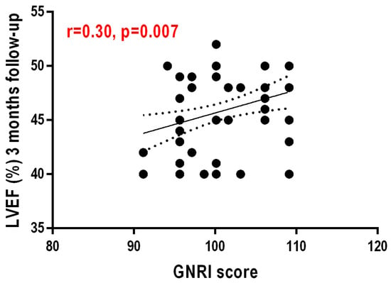
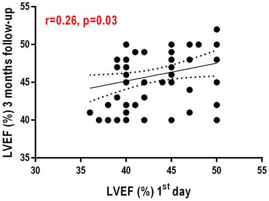
The GNRI score values showed a moderately positive, significant correlation with LVEF at 3 months post infarction (r = 0.310, p = 0.008)—Figure 1. Another weakly positive but statistically significant correlation exists between LVEF at 1 day post infarction and LVEF at 3 months post AMI (r = 0.259, p = 0.027)—Figure 2.

Figure 1.
Correlation between nutritional status evidenced by GNRI score and left ventricular ejection fraction value at 3 months after AMI.

Figure 2.
Correlation between left ventricular ejection fraction one day after AMI and ejection fraction 3 months after AMI.
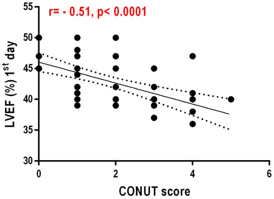
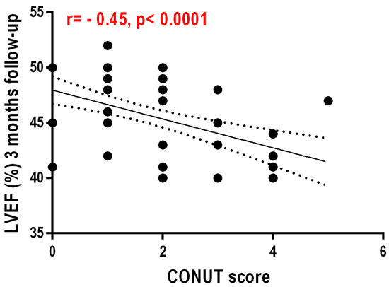
Regarding the CONUT values and the correlation with the GNRI score values at 3 months after AMI, these are negative and statistically insignificant (r = −0.087, p = 0.464). Another weak but still statistically significant negative correlation exists between the CONUT score and LVEF at 1 day after AMI (r = −0.51, p < 0.0001)—Figure 3. CONUT values showed a weak but statistically significant negative correlation with LVEF at 3 months post infarction (r = −0.453, p < 0.0001)—Figure 4.

Figure 3.
Correlation between left ventricular ejection fraction one day after AMI and CONUT score.

Figure 4.
Correlation between left ventricular ejection fraction at 3 months after AMI and CONUT score.
4. Discussion
Malnutrition is a frequent and significant problem and is seen particularly in elderly hospitalized patients, especially those with multiple chronic diseases. Even though it is quite difficult to determine the malnutrition status of the patient with AMI, it should be kept in mind that this problem may later lead to complications in terms of treatment and disease progression.
Left ventricular ejection fraction is an important determinant of the risk of heart failure and death in patients with myocardial infarction. The American College of Cardiology and the European Society of Cardiology recommend that LVEF be assessed in both ST-elevation (STEMI) and non-ST-elevation (NSTEMI) myocardial infarction patients before discharge [,,,,].
This study aimed to investigate the correlation between left ventricular ejection fraction and nutritional status, expressed by CONUT and GNRI scores. A percentage of 21% showed malnutrition, calculated using the CONUT score, and 31% of them were identified as malnourished, calculated using the GNRI score. Patients with malnutrition in both CONUT and GNRI had lower LVEF values both at 1 day post AMI and 3 months post AMI.
The existence of a wide range of blood-based biomarkers to identify malnutrition status can help us to highlight clinical risk. According to recent studies, low serum albumin levels are common in patients with heart failure (25–33%) due to several factors including lifestyle, absorption disorders, a complex malnutrition–inflammation syndrome, which produces excessive oxidative stress [,,,,].
And, in other studies, scores such as Prognostic Nutritional Index (PNI) and CONUT have been shown to be superior even in predicting mortality in patients with heart failure [,,,]. Another recently published study demonstrated the relationship between malnutrition and mortality in patients with LVEF ≥ 40%, as assessed by CONUT score in a cohort of patients undergoing coronary angiography []. Additionally, Iwakami et al. demonstrated that, within a group of 635 patients with acute heart failure, 78% of patients with an average age of 75 years in Japan had poor nutritional status based on the CONUT score []. Sze et al. demonstrated that worsening malnutrition was associated with worse outcomes in British heart failure patients using the three well-known scores, the GNRI, CONUT, and PNI scores []. Chen et al. showed that high CONUT values and neutrophil-to-lymphocyte ratio can be used to predict the occurrence of adverse clinical events and mortality in patients with AMI, and CONUT scores may be an independent predictor of major adverse cardiovascular events (MACEs) in patients with acute myocardial infarction []. An additional study on the relationship between nutritional status and recurrent major cardiovascular events after myocardial infarction showed that MACEs in post-MI patients were associated with nutritional status, which could be characterized by elevated inflammatory status and nutritional deficiency as determined by the CONUT score [,].
A study including 56 patients with AMI (44.64% with STEMI, 55.35% with NSTEMI) demonstrated that malnourished patients undergoing revascularization have an increased rate of in-hospital complications and a longer observation time in a tertiary cardiac intensive care unit, and assessment of nutritional status was calculated using the CONUT and PNI scores []. It has recently been shown that there is a strong relationship between low LVEF and the Naples Prognostic Score (NPS) in STEMI patients, with this study conducted on a cohort of 2.280 STEMI patients undergoing primary percutaneous coronary intervention between 2017 and 2022 []. Worsening nutritional status during hospitalization was linked to prognosis in patients with a low GNRI on admission, but not in patients with a high GNRI on admission, according to data from a recent study by Sunaga et al. that included 1.095 heart failure patients with preserved ejection fraction [,]. Our data indicate a correlation between parameters of left ventricular dysfunction after acute myocardial infarction and malnutrition status calculated using the CONUT and GNRI scores. LVEF had lower values in malnourished patients in both the CONUT and GNRI groups, and NTproBNP had lower values at both 1 day and 5 days after infarction in the CONUT ≥ 3 group.
The present study also has certain limitations, among which we first of all can list the small number of patients with acute myocardial infarction; however, while we were able to demonstrate that there is an association between altered nutritional status and left ventricular dysfunction, the follow-up period being a short one, only 3 months after AMI. In future studies, we may extend the follow-up period to observe differences in LVEF on day 1 post infarction in malnourished patients and 1 year post MI compared to patients with normal nutritional status. Secondly, we used only two screening scores for malnutrition, other scores we could have used include PNI or the Nutritional Risk Index (NRI). Third, patients included in the study were recruited from a single heart center, but we made every effort to enroll all patients with available data within the specified time frame. Further research including other biomarkers reflecting immunological activation such as troponin, tumor necrosis factor-α (TNF-α), interleukin (IL)-1, and (IL)-6, which may contribute to cardiac remodeling and left ventricular dysfunction by inducing cardiomyocyte hypertrophy, dilatation, fibrosis and apoptosis, may be of interest.
5. Conclusions
The correlation between the parameters of left ventricular dysfunction and nutritional status calculated by the two scores shows that patients with impaired nutritional status have worse outcomes at the 3-month post-infarction assessment, and lower values for LVEF (%) both at 1 day post infarction and at 3 months in patients with impaired nutritional status. Malnutrition is one of the risk factors for cardiovascular disease and in our study it was independently associated with adverse outcomes at 3 months post infarction. Future work would be needed to demonstrate the link between malnutrition and ventricular dysfunction.
Author Contributions
Conceptualization, M.G.C. and T.B.; methodology, M.G.C., V.N., L.-O.C. and T.P.; software, V.N., C.Ț. and T.N.; validation, L.-O.C., T.N. and T.B; formal analysis, V.N., V.B.H., C.Ț. and T.P.; investigation, M.G.C., V.N., V.B.H., C.Ț., L.-O.C., T.P. and T.N.; data curation, T.B.; writing—original draft preparation, M.G.C. and V.N.; writing—review and editing, V.B.H., C.Ț., L.-O.C., T.P., T.N. and T.B. supervision, T.B.; project administration, M.G.C. and T.B. All authors have read and agreed to the published version of the manuscript.
Funding
This research received no external funding.
Institutional Review Board Statement
This study was approved by the ethics committee of „George Emil Palade” University of Medicine, Pharmacy, Science and Technology of Târgu Mureș, România, and Ethics Committee of the Târgu Mureș Emergency Clinical County Hospital, 2112/24 February 2023.
Informed Consent Statement
Informed consent was obtained from all subjects involved in the study.
Data Availability Statement
Data are contained within the article.
Conflicts of Interest
The authors declare no conflict of interest.
References
- Pressoir, M.; Desné, S.; Berchery, D.; Rossignol, G.; Poiree, B.; Meslier, M.; Traversier, S.; Vittot, M.; Simon, M.I.S.D.S.; Gekiere, J.P.; et al. Prevalence, risk factors and clinical implications of malnutrition in French Comprehensive Cancer Centres. Br. J. Cancer 2010, 102, 966–971. [Google Scholar] [CrossRef]
- Bouillanne, O.; Morineau, G.; Dupont, C.; Coulombel, I.; Vincent, J.P.; Nicolis, I.; Benazeth, S.; Cynober, L.; Aussel, C. Geriatric nutritional risk index: A new index for evaluating at-risk elderly medical patients. Am. J. Clin. Nutr. 2005, 82, 777–783. [Google Scholar] [CrossRef]
- Takahashi, H.; Ito, Y.; Ishii, H.; Aoyama, T.; Kamoi, D.; Kasuga, H.; Yasuda, K.; Maruyama, S.; Matsuo, S.; Murohara, T.; et al. Geriatric nutritional risk index accurately predicts cardio- vascular mortality in incident hemodialysis patients. J. Cardiol. 2014, 64, 32–36. [Google Scholar] [CrossRef] [PubMed]
- Rus, V.; Chitu, M.; Cernea, S.; Benedek, I.; Hodas, R.; Zavate, R.; Nyulas, T.; Hintea, M.; Benedek, T. Altered nutritional status, inflammation and systemic vulnerability in patients with acute myocardial infarction undergoing percutaneous coronary revascularisation: A prospective study in a level 3 cardiac critical care unit. Nutr. Diet. 2020, 77, 212–222. [Google Scholar] [CrossRef]
- Ulibarri Pérez, J.I.; Picón César, M.J.; García Benavent, E.; Mancha Alvarez-Estrada, A. Early detection and control of hospital malnutrition. Nutr. Hosp. 2022, 17, 139–146. [Google Scholar]
- Sutton, N.R.; Li, S.; Thomas, L.; Wang, T.Y.; de Lemos, J.A.; Enriquez, J.R.; Shah, R.U.; Fonarow, G.C. The association of left ventricular ejection fraction with clinical outcomes after myocardial infarction: Findings from the Acute Coronary Treatment and Intervention Outcomes Network (ACTION) Registry-Get With the Guidelines (GWTG) Medicare-linked database. Am. Heart J. 2016, 178, 65–73. [Google Scholar] [CrossRef] [PubMed]
- Chew, D.S.; Wilton, S.B.; Kavanagh, K.; Southern, D.A.; Tan-Mesiatowsky, L.E.; Exner, D.V.; APPROACH Investigators. Left ventricular ejection fraction reassessment postmyocardial infarction: Current clinical practice and determinants of adverse remodeling. Am. Heart J. 2018, 198, 91–96. [Google Scholar] [CrossRef] [PubMed]
- Bangalore, S.; Fonarow, G.C.; Peterson, E.D.; Hellkamp, A.S.; Hernandez, A.F.; Laskey, W.; Peacock, W.F.; Cannon, C.P.; Schwamm, L.H.; Bhatt, D.L. Age and gender differences in quality of care and outcomes for patients with ST-segment elevation myocardial infarction. Am. J. Med. 2012, 125, 1000–1009. [Google Scholar] [CrossRef]
- Ahnve, S.; Gilpin, E.; Dittrich, H.; Nicod, P.; Henning, H.; Carlisle, J.; Ross, J., Jr. First myocardial infarction: Age and ejection fraction identify a low-risk group. Am Heart J. 1988, 116, 925–932. [Google Scholar] [CrossRef] [PubMed]
- Wu, W.Y.; Biery, D.W.; Singh, A.; Divakaran, S.; Berman, A.N.; Ayuba, G.; DeFilippis, E.M.; Nasir, K.; Januzzi, J.L.; Di Carli, M.F.; et al. Recovery of left ventricular systolic function and clinical outcomes in young adults with myocardial infarction. J. Am. Coll. Cardiol. 2020, 75, 2804–2815. [Google Scholar] [CrossRef]
- Hare, J.L.; Brown, J.K.; Marwick, T.H. Performance of conventional echocardiographic parameters and myocardial measurements in the sequential evaluation of left ventricular function. Am. J. Cardiol. 2008, 101, 706–711. [Google Scholar] [CrossRef] [PubMed]
- Robbie, L.; Libby, P. Inflammation and atherothrombosis. Ann. N. Y. Acad. Sci. 2001, 947, 167–179. [Google Scholar] [CrossRef] [PubMed]
- Teramoto, T.; Sasaki, J.; Ishibashi, S.; Birou, S.; Daida, H.; Dohi, S.; Egusa, G.; Hiro, T.; Hirobe, K.; Iida, M.; et al. Diagnostic criteria for dyslipidemia. J. Atheroscler. Thromb. 2013, 20, 655–660. [Google Scholar] [CrossRef] [PubMed]
- Kuroda, D.; Sawayama, H.; Kurashige, J.; Iwatsuki, M.; Eto, T.; Tokunaga, R.; Kitano, Y.; Yamamura, K.; Ouchi, M.; Nakamura, K.; et al. Controlling Nutritional Status (CONUT) score is a prognostic marker for gastric cancer patients after curative resection. Gastric Cancer 2018, 21, 204–212. [Google Scholar] [CrossRef] [PubMed]
- Cereda, E.; Pedrolli, C. The Geriatric Nutritional Risk Index. Curr. Opin. Clin. Nutr. Metab. Care 2009, 12, 1–7. [Google Scholar] [CrossRef] [PubMed]
- Avram, C.; Mărușteri, M. Normality assessment, few paradigms and use cases. Rev. Romana Med. Lab. 2022, 30, 251–260. [Google Scholar] [CrossRef]
- O’Gara, P.T.; Kushner, F.G.; Ascheim, D.D.; Casey, D.E., Jr.; Chung, M.K.; de Lemos, J.A.; Ettinger, S.M.; Fang, J.C.; Fesmire, F.M.; Franklin, B.A.; et al. 2013 ACCF/AHA guideline for the management of ST-elevation myocardial infarction: Executive summary: A report of the American College of Cardiology Foundation/American Heart Association Task Force on Practice Guidelines. Circulation 2013, 127, 529–555. [Google Scholar] [CrossRef] [PubMed]
- Steg, P.G.; James, S.K.; Atar, D.; Badano, L.P.; Lundqvist, C.B.; Borger, M.A.; Di Mario, C.; Dickstein, K.; Ducrocq, G.; Fernandez-Aviles, F. ESC Guidelines for the management of acute myocardial infarction in patients presenting with ST-segment elevation. Eur. Heart J. 2012, 33, 2569–2619. [Google Scholar] [CrossRef] [PubMed]
- Hamm, C.W.; Bassand, J.P.; Agewall, S.; Bax, J.; Boersma, E.; Bueno, H.; Caso, P.; Dudek, D.; Gielen, S.; Huber, K. ESC Guidelines for the management of acute coronary syndromes in patients presenting without persistent ST-segment elevation: The Task Force for the management of acute coronary syndromes (ACS) in patients presenting without persistent ST-segment elevation of the European Society of Cardiology (ESC). Eur. Heart J. 2011, 32, 2999–3054. [Google Scholar]
- Rodean, I.P.; Biris, C.I.; Halatiu, V.B.; Modiga, A.; Lazar, L.; Benedek, I.; Benedek, T. Is there a link between COVID-19 infection, periodontal disease and acute myocardial infarction? Life 2021, 11, 1050. [Google Scholar] [CrossRef]
- Călburean, P.A.; Grebenișan, P.; Nistor, I.A.; Pal, K.; Vacariu, V.; Drincal, R.K.; Ion, A.A.; Adorján, I.; Oltean, T.; Hadadi, L. Addition of eptifibatide and manual thrombus aspiration to ticagrelor does not improve long-term survival after STEMI treated with primary PCI. Front. Pharmacol. 2024, 15, 1415025. [Google Scholar] [CrossRef] [PubMed]
- Chien, S.C.; Chen, C.Y.; Lin, C.F.; Yeh, H.I. Crit-ical appraisal of the role of serum albu-min in cardiovascular disease. Biomark Res. 2017, 5, 31. [Google Scholar] [CrossRef] [PubMed]
- Arques, S.; Roux, E.; Stolidi, P.; Gelisse, R.; Ambrosi, P. Usefulness of serum albuminand serum total cholesterol in the pre-diction of hospital death in older pa-tients with severe, acute heart failure. Arch. Cardiovasc. Dis. 2011, 104, 502–508. [Google Scholar] [CrossRef] [PubMed]
- Gambardella, J.; Santulli, G. Integratingdiet and inflammation to calculate car-diovascular risk. Atherosclerosis 2016, 253, 258–261. [Google Scholar] [CrossRef] [PubMed]
- Gambardella, J.; Sardu, C.; Sacra, C.; DelGiudice, C.; Santulli, G. Quit smoking tooutsmart atherogenesis: Molecularmechanisms underlying clinical evi-dence. Atherosclerosis 2017, 257, 242–245. [Google Scholar] [CrossRef] [PubMed]
- Proboteanu, P.; Caloian, B.; Pop, D. Heart Failure as the First Clinical Manifestation of Basedow’s Disease. J. Cardiovasc. Emerg. 2024, 10, 78–82. [Google Scholar] [CrossRef]
- Yoshihisa, A.; Kanno, Y.; Watanabe, S.; Yokokawa, T.; Abe, S.; Miyata, M.; Sato, T.; Suzuki, S.; Oikawa, M.; Kobayashi, A.; et al. Impact of nutri-tional indices on mortality in patientswith heart failure. Open Heart 2018, 5, 000730. [Google Scholar] [CrossRef] [PubMed]
- Iwakami, N.; Nagai, T.; Furukawa, T.A.; Sugano, Y.; Honda, S.; Okada, A.; Asaumi, Y.; Aiba, T.; Noguchi, T.; Kusano, K.; et al. Prognostic value of malnutritionassessed by Controlling Nutritional Sta-tus score for long-term mortality in pa-tients with acute heart failure. Int. J. Cardiol. 2017, 230, 529–536. [Google Scholar] [CrossRef] [PubMed]
- Tromp, J.; Teng, T.H.; Tay, W.T.; Hung, C.L.; Narasimhan, C.; Shimizu, W.; Park, S.W.; Liew, H.B.; Ngarmukos, T.; Reyes, E.B.; et al. Heart fail-ure with preserved ejection fraction in Asia. Eur. J. Heart Fail. 2018, 21, 23–36. [Google Scholar] [CrossRef]
- Călburean, P.A.; Grebenișan, P.; Nistor, I.A.; Pal, K.; Vacariu, V.; Drincal, R.K.; Țepes, O.; Bârlea, I.; Șuș, I.; Somkereki, C.; et al. Prediction of 3-year all-cause and cardiovascular cause mortality in a prospective percutaneous coronary intervention registry: Machine learning model outperforms conventional clinical risk scores. Atherosclerosis 2022, 350, 33–40. [Google Scholar] [CrossRef]
- Mai, Z.; Huang, Z.; Lai, W.; Li, H.; Wang, B.; Huang, S.; Shi, Y.; Yu, S.; Hu, Q.; Liu, J.; et al. Corrigendum: Association of Malnutrition, Left Ventricular Ejection Fraction Category, and Mortality in Patients Undergoing Coronary Angiography: A Cohort With 45,826 Patients. Front. Nutr. 2022, 4, 890490. [Google Scholar] [CrossRef] [PubMed]
- Sze, S.; Zhang, J.; Pellicori, P.; Morgan, D.; Hoye, A.; Clark, A.L. Prognostic value of simple frailty and malnutrition screening tools in patients with acute heart failure due to left ventricular systolic dysfunction. Clin. Res. Cardiol. 2017, 106, 533–541. [Google Scholar] [CrossRef] [PubMed]
- Chen, B.; Yuan, L.; Chen, X.; Li, J.; Tao, J.; Li, W.; Zheng, R. Correlations and Prognostic Roles of the Nutritional Status and Neutrophil-to-lymphocyte Ratio in Elderly Patients with Acute Myocardial Infarction Undergoing Primary Coronary Intervention. Int. Heart J. 2020, 61, 1114–1120. [Google Scholar] [CrossRef] [PubMed]
- Czinege, M.; Halațiu, V.B.; Nyulas, V.; Cojocariu, L.O.; Ion, B.; Mașca, V.; Țolescu, C.; Benedek, T. Nutritional Status and Recurrent Major Cardiovascular Events Following Acute Myocardial Infarction—A Follow-Up Study in a Primary Percutaneous Coronary Intervention Center. Nutrients 2024, 16, 1088. [Google Scholar] [CrossRef] [PubMed]
- Blîndu, E.; Benedek, I.; Rodean, I.P.; Halatiu, V.B.; Rat, N.; Tolescu, C.; Mihăilă, T.; Rosca, A.; Mátyás, B.-B.; Szabó, E.; et al. Regional Differences in the Level of Inflammation Between the Right and Left Coronary Arteries—A Coronary Computed Tomography Angiography Study of Epicardial Fat Attenuation Index in Four Scenarios of Cardiovascular Emergencies. J. Cardiovasc. Emerg. 2023, 9, 111–119. [Google Scholar] [CrossRef]
- Rus, V.; Opincariu, D.; Hodas, R.; Nyulas, T.; Hintea, M.; Benedek, T. Interrelation between altered nutritional status and clinical outcomes in patients with acute myocardial infarction admitted in a tertiary intensive cardiac care unit. J. Cardiovasc. Emerg. 2018, 4, 32–40. [Google Scholar]
- Birdal, O.; Pay, L.; Aksakal, E.; Yumurtaş, A.Ç.; Çinier, G.; Yücel, E.; Tanboğa, İ.H.; Karagöz, A.; Oduncu, V. Naples Prognostic Score and Prediction of Left Ventricular Ejection Fraction in STEMI Patients. Angiology 2024, 75, 36–43. [Google Scholar] [CrossRef] [PubMed]
- Sunaga, A.; Hikoso, S.; Yamada, T.; Yasumura, Y.; Tamaki, S.; Yano, M.; Hayashi, T.; Nakagawa, Y.; Nakagawa, A.; Seo, M.; et al. Change in Nutritional Status during Hospitalization and Prognosis in Patients with Heart Failure with Preserved Ejection Fraction. Nutrients. 2022, 14, 4345. [Google Scholar] [CrossRef]
- Koller, L.; Kleber, M.; Goliasch, G.; Sulzgruber, P.; Scharnagl, H.; Silbernagel, G.; Grammer, T.; Delgado, G.; Tomaschitz, A.; Pilz, S.; et al. C-reactive protein predicts mortality in patients referred for coronary angiography and symptoms of heart failure with preserved ejection fraction. Eur. J. Heart Fail. 2014, 16, 758–766. [Google Scholar] [CrossRef]
Disclaimer/Publisher’s Note: The statements, opinions and data contained in all publications are solely those of the individual author(s) and contributor(s) and not of MDPI and/or the editor(s). MDPI and/or the editor(s) disclaim responsibility for any injury to people or property resulting from any ideas, methods, instructions or products referred to in the content. |
© 2024 by the authors. Licensee MDPI, Basel, Switzerland. This article is an open access article distributed under the terms and conditions of the Creative Commons Attribution (CC BY) license (https://creativecommons.org/licenses/by/4.0/).