Interaction Effect of Phase Angle and Age on Femoral Neck Bone Mineral Density in Patients with Non-Dialysis Chronic Kidney Disease Stage 5

 , , and
Abstract
1. Introduction
2. Materials and Methods
2.1. Patients and Data Collection
2.2. BMD Assessment
2.3. Body Composition Measurements
2.4. Statistical Analysis
3. Results
3.1. Patient Characteristics
3.2. Correlation of Body Composition and Laboratory Parameters with Femoral Neck BMD and T-Score
3.3. Multiple Linear Regression Analyses
3.4. Two-Way ANOVA
4. Discussion
5. Conclusions
Supplementary Materials
Author Contributions
Funding
Data Availability Statement
Conflicts of Interest
References
- Moe, S.M.; Nickolas, T.L. Fractures in Patients with CKD: Time for Action. Clin. J. Am. Soc. Nephrol. 2016, 11, 1929–1931. [Google Scholar] [CrossRef]
- Ketteler, M.; Block, G.A.; Evenepoel, P.; Fukagawa, M.; Herzog, C.A.; McCann, L.; Moe, S.M.; Shroff, R.; Tonelli, M.A.; Toussaint, N.D.; et al. Executive summary of the 2017 KDIGO Chronic Kidney Disease-Mineral and Bone Disorder (CKD-MBD) Guideline Update: What’s changed and why it matters. Kidney Int. 2017, 92, 26–36. [Google Scholar] [CrossRef] [PubMed]
- Ahn, S.H.; Lee, S.H.; Kim, H.; Kim, B.J.; Koh, J.M. Different relationships between body compositions and bone mineral density according to gender and age in Korean populations (KNHANES 2008-2010). J. Clin. Endocrinol. Metab. 2014, 99, 3811–3820. [Google Scholar] [CrossRef] [PubMed]
- Hars, M.; Trombetti, A. Body composition assessment in the prediction of osteoporotic fractures. Curr. Opin. Rheumatol. 2017, 29, 394–401. [Google Scholar] [CrossRef] [PubMed]
- Marinho, S.M.; Wahrlich, V.; Mafra, D. Association between body composition and bone mineral density in men on hemodialysis. Am. J. Med. Sci. 2015, 350, 286–289. [Google Scholar] [CrossRef]
- Fournie, C.; Pelletier, S.; Bacchetta, J.; Boutroy, S.; Confavreux, C.; Drai, J.; Arkouche, W.; Fouque, D.; Chapurlat, R.; Guebre-Egziabher, F. The relationship between body composition and bone quality measured with HR-pQCT in peritoneal dialysis patients. Perit. Dial. Int. 2017, 37, 548–555. [Google Scholar] [CrossRef] [PubMed]
- Ito, K.; Ookawara, S.; Hibino, Y.; Imai, S.; Fueki, M.; Bandai, Y.; Yasuda, M.; Kamimura, T.; Kakuda, H.; Kiryu, S.; et al. Skeletal muscle mass index is positively associated with bone mineral density in hemodialysis patients. Front. Med. (Lausanne) 2020, 7, 187. [Google Scholar] [CrossRef]
- Lukaski, H.C.; Kyle, U.G.; Kondrup, J. Assessment of adult malnutrition and prognosis with bioelectrical impedance analysis: Phase angle and impedance ratio. Curr. Opin. Clin. Nutr. Metab. Care 2017, 20, 330–339. [Google Scholar] [CrossRef]
- Norman, K.; Stobaus, N.; Pirlich, M.; Bosy-Westphal, A. Bioelectrical phase angle and impedance vector analysis–clinical relevance and applicability of impedance parameters. Clin. Nutr. 2012, 31, 854–861. [Google Scholar] [CrossRef]
- Akamatsu, Y.; Kusakabe, T.; Arai, H.; Yamamoto, Y.; Nakao, K.; Ikeue, K.; Ishihara, Y.; Tagami, T.; Yasoda, A.; Ishii, K.; et al. Phase angle from bioelectrical impedance analysis is a useful indicator of muscle quality. J. Cachexia Sarcopenia Muscle 2022, 13, 180–189. [Google Scholar] [CrossRef]
- Yamada, M.; Kimura, Y.; Ishiyama, D.; Nishio, N.; Otobe, Y.; Tanaka, T.; Ohji, S.; Koyama, S.; Sato, A.; Suzuki, M.; et al. Phase angle is a useful indicator for muscle function in older adults. J. Nutr. Health Aging 2019, 23, 251–255. [Google Scholar] [CrossRef] [PubMed]
- Kang, S.H.; Do, J.Y.; Kim, J.C. Impedance-derived phase angle is associated with muscle mass, strength, quality of life, and clinical outcomes in maintenance hemodialysis patients. PLoS ONE 2022, 17, e0261070. [Google Scholar] [CrossRef]
- Garlini, L.M.; Alves, F.D.; Ceretta, L.B.; Perry, I.S.; Souza, G.C.; Clausell, N.O. Phase angle and mortality: A systematic review. Eur. J. Clin. Nutr. 2019, 73, 495–508. [Google Scholar] [CrossRef] [PubMed]
- Di Vincenzo, O.; Marra, M.; Di Gregorio, A.; Pasanisi, F.; Scalfi, L. Bioelectrical impedance analysis (BIA) -derived phase angle in sarcopenia: A systematic review. Clin. Nutr. 2021, 40, 3052–3061. [Google Scholar] [CrossRef] [PubMed]
- Antunes, M.; Cyrino, E.S.; Silva, D.R.P.; Tomeleri, C.M.; Nabuco, H.C.G.; Cavalcante, E.F.; Cunha, P.M.; Cyrino, L.T.; Dos Santos, L.; Silva, A.M.; et al. Total and regional bone mineral density are associated with cellular health in older men and women. Arch. Gerontol. Geriatr. 2020, 90, 104156. [Google Scholar] [CrossRef]
- Ruperto, M.; Sanchez-Muniz, F.J.; Barril, G. Predictors of protein-energy wasting in haemodialysis patients: A cross-sectional study. J. Hum. Nutr. Diet. 2016, 29, 38–47. [Google Scholar] [CrossRef]
- Pimentel, A.; Urena-Torres, P.; Zillikens, M.C.; Bover, J.; Cohen-Solal, M. Fractures in patients with CKD-diagnosis, treatment, and prevention: A review by members of the European Calcified Tissue Society and the European Renal Association of Nephrology Dialysis and Transplantation. Kidney Int. 2017, 92, 1343–1355. [Google Scholar] [CrossRef]
- Ohnaka, S.; Yamada, S.; Tsujikawa, H.; Arase, H.; Taniguchi, M.; Tokumoto, M.; Tsuruya, K.; Nakano, T.; Kitazono, T. Association of normalized protein catabolic rate (nPCR) with the risk of bone fracture in patients undergoing maintenance hemodialysis: The Q-Cohort Study. Clin. Nutr. 2021, 40, 997–1004. [Google Scholar] [CrossRef]
- Tanaka, S.; Ando, K.; Kobayashi, K.; Hida, T.; Ito, K.; Tsushima, M.; Morozumi, M.; Machino, M.; Ota, K.; Seki, T.; et al. A low phase angle measured with bioelectrical impedance analysis is associated with osteoporosis and is a risk factor for osteoporosis in community-dwelling people: The Yakumo study. Arch. Osteoporos. 2018, 13, 39. [Google Scholar] [CrossRef]
- Lu, H.K.; Lai, C.L.; Lee, L.W.; Chu, L.P.; Hsieh, K.C. Assessment of total and regional bone mineral density using bioelectrical impedance vector analysis in elderly population. Sci. Rep. 2021, 11, 21161. [Google Scholar] [CrossRef]
- Kasher, M.; Gabdulina, G.; Beissebayeva, A.; Mussabaeva, D.; Tokarev, A.; Sarssenbayeva, M.; Omarova, K.; Mominova, G.; Livshits, G. Rheumatoid arthritis is associated with exacerbated body composition deterioration in Kazakh females. Nutrition 2019, 66, 219–226. [Google Scholar] [CrossRef] [PubMed]
- Keane, D.F.; Baxter, P.; Lindley, E.; Moissl, U.; Pavitt, S.; Rhodes, L.; Wieskotten, S. The Body Composition Monitor: A flexible tool for routine fluid management across the haemodialysis population. Biomed. Phys. Eng. Express 2017, 3, 035017. [Google Scholar] [CrossRef] [PubMed]
- Wang, K.; Zelnick, L.R.; Chertow, G.M.; Himmelfarb, J.; Bansal, N. Body composition changes following dialysis initiation and cardiovascular and mortality outcomes in CRIC (Chronic Renal Insufficiency Cohort): A bioimpedance analysis substudy. Kidney Med. 2021, 3, 327–334. [Google Scholar] [CrossRef] [PubMed]
- Hunter, G.R.; Plaisance, E.P.; Fisher, G. Weight loss and bone mineral density. Curr. Opin. Endocrinol. Diabetes Obes. 2014, 21, 358–362. [Google Scholar] [CrossRef]
- Migliaccio, S.; Greco, E.A.; Fornari, R.; Donini, L.M.; Lenzi, A. Is obesity in women protective against osteoporosis? Diabetes Metab. Syndr. Obes. 2011, 4, 273–282. [Google Scholar] [CrossRef]
- Kim, W.; Chung, S.G.; Kim, K.; Seo, H.G.; Oh, B.M.; Yi, Y.; Kim, M.J. The relationship between body fat and bone mineral density in Korean men and women. J. Bone Miner. Metab. 2014, 32, 709–717. [Google Scholar] [CrossRef]
- Ho-Pham, L.T.; Nguyen, N.D.; Lai, T.Q.; Nguyen, T.V. Contributions of lean mass and fat mass to bone mineral density: A study in postmenopausal women. BMC Musculoskelet. Disord. 2010, 11, 59. [Google Scholar] [CrossRef]
- Ho-Pham, L.T.; Nguyen, U.D.T.; Nguyen, T.V. Association between lean mass, fat mass, and bone mineral density: A meta-analysis. J. Clin. Endocrinol. Metab. 2014, 99, 30–38. [Google Scholar] [CrossRef]
- Jang, H.Y.; Choi, H.J.; Lee, K.B.; Cho, S.B.; Im, I.J.; Kim, H.J. The Association between muscle mass deficits estimated from bioelectrical impedance analysis and lumbar spine bone mineral density in Korean adults. J. Bone Metab. 2016, 23, 95–100. [Google Scholar] [CrossRef]
- Kim, I.J.; Kang, K.Y. Low skeletal muscle mass is associated with the risk of low bone mineral density in urban dwelling premenopausal women. Calcif. Tissue Int. 2017, 101, 581–592. [Google Scholar] [CrossRef]
- Kang, S.H.; Kim, A.Y.; Do, J.Y. Association between the appendicular lean mass index or handgrip strength and bone mineral density in patients undergoing peritoneal dialysis. Int. J. Med. Sci. 2022, 19, 1408–1416. [Google Scholar] [CrossRef] [PubMed]
- Park, J.H.; Song, Y.M.; Sung, J.; Lee, K.; Kim, Y.S.; Kim, T.; Cho, S.I. The association between fat and lean mass and bone mineral density: The Healthy Twin Study. Bone 2012, 50, 1006–1011. [Google Scholar] [CrossRef] [PubMed]
- Yamada, Y.; Buehring, B.; Krueger, D.; Anderson, R.M.; Schoeller, D.A.; Binkley, N. Electrical properties assessed by bioelectrical impedance spectroscopy as biomarkers of age-related loss of skeletal muscle quantity and quality. J. Gerontol. A Biol. Sci. Med. Sci. 2016, 72, 1180–1186. [Google Scholar] [CrossRef]
- Dittmar, M. Reliability and variability of bioimpedance measures in normal adults: Effects of age, gender, and body mass. Am. J. Phys. Anthropol. 2003, 122, 361–370. [Google Scholar] [CrossRef]
- Gonzalez, M.C.; Barbosa-Silva, T.G.; Bielemann, R.M.; Gallagher, D.; Heymsfield, S.B. Phase angle and its determinants in healthy subjects: Influence of body composition. Am. J. Clin. Nutr. 2016, 103, 712–716. [Google Scholar] [CrossRef]
- Barbosa-Silva, M.C.; Barros, A.J.; Wang, J.; Heymsfield, S.B.; Pierson, R.N., Jr. Bioelectrical impedance analysis: Population reference values for phase angle by age and sex. Am. J. Clin. Nutr. 2005, 82, 49–52. [Google Scholar] [CrossRef] [PubMed]
- Bosy-Westphal, A.; Danielzik, S.; Dorhofer, R.P.; Later, W.; Wiese, S.; Muller, M.J. Phase angle from bioelectrical impedance analysis: Population reference values by age, sex, and body mass index. JPEN J. Parenter. Enteral Nutr. 2006, 30, 309–316. [Google Scholar] [CrossRef] [PubMed]
- Stobaus, N.; Pirlich, M.; Valentini, L.; Schulzke, J.D.; Norman, K. Determinants of bioelectrical phase angle in disease. Br. J. Nutr. 2012, 107, 1217–1220. [Google Scholar] [CrossRef]
- Montenegro, J.; Klein, M.; Bregman, R.; Prado, C.M.; Barreto Silva, M.I. Osteosarcopenia in patients with non-dialysis dependent chronic kidney disease. Clin. Nutr. 2022, 41, 1218–1227. [Google Scholar] [CrossRef]
- Ngai, H.H.Y.; Cheung, C.L.; Yao, T.J.; Kung, A.W.C. Bioimpedance: Can its addition to simple clinical criteria enhance the diagnosis of osteoporosis? J. Bone Miner. Metab. 2009, 27, 372–378. [Google Scholar] [CrossRef]
- Riggs, B.L.; Khosla, S.; Melton III, L.J. A unitary model for involutional osteoporosis: Estrogen deficiency causes both type I and type II osteoporosis in postmenopausal women and contributes to bone loss in aging men. J. Bone Miner. Res. 1998, 13, 763–773. [Google Scholar] [CrossRef] [PubMed]
- Damasiewicz, M.J.; Nickolas, T.L. Rethinking bone disease in kidney disease. JBMR Plus 2018, 2, 309–322. [Google Scholar] [CrossRef] [PubMed]


| Variable | Total (n = 167) | Femoral Neck Bone Mineral Density | p Value (p for Trend) | ||
|---|---|---|---|---|---|
| Tertile 1 (n = 55) | Tertile 2 (n = 56) | Tertile 3 (n = 56) | |||
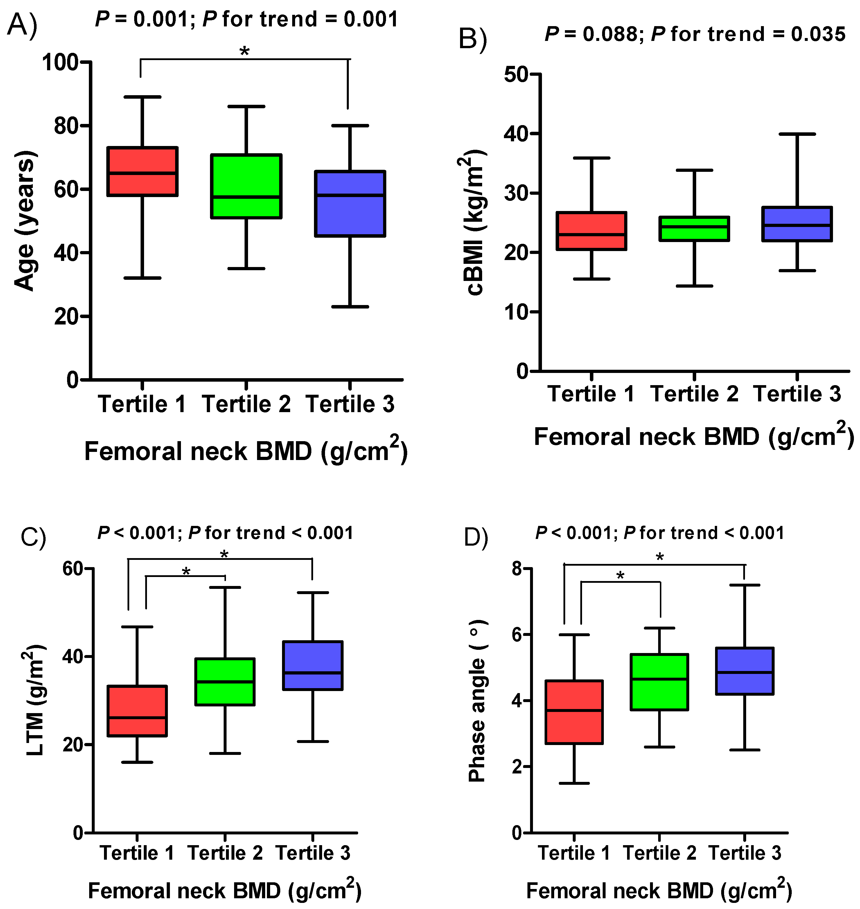
| Age, years | 59.65 ± 13.98 | 64.44 ± 13.70 | 59.66 ± 12.43 | 54.93 ± 14.32 b,c | 0.001 (0.001) |
| ≤65, years | 107 (64.1%) | 28 (26.2%) | 37 (34.6%) | 42 (39.3%) | 0.028 (0.008) |
| >65, years | 60 (35.9%) | 27 (45.0%) | 19 (31.7%) | 14 (23.3%) | |
| Sex | |||||
| Male | 100 (59.9%) | 17 (17.0%) | 41 (41.0%) | 42 (42.0%) | <0.001 (<0.001) |
| Female | 67 (40.1%) | 38 (56.7%) | 15 (22.4%) | 14 (20.9%) | |
| Phase angle, ° | 4.38 ± 1.19 | 3.68 ± 1.15 | 4.61 ± 0.98 a | 4.86 ± 1.10 b | <0.001 (<0.001) |
| Diabetes | |||||
| Yes | 105 (62.9%) | 35 (33.3%) | 36 (34.3%) | 34 (32.4%) | 0.917 (0.750) |
| No | 62 (37.1%) | 20 (32.3%) | 20 (32.3%) | 22 (35.5%) | |
| FN-BMD, g/cm2 | 0.64 ± 0.13 | 0.50 ± 0.10 | 0.64 ± 0.03 a | 0.78 ± 0.07 b,c | <0.001 (<0.001) |
| FN-BMD T-score | −1.59 ± 1.09 | −2.75 ± 0.71 | −1.60 ± 0.28 a | −0.44 ± 0.56 b,c | <0.001 (<0.001) |
| LS-BMD, g/cm2 | 0.96 ± 0.18 | 0.85 ± 0.14 | 0.97 ± 0.15 a | 1.07 ± 0.18 b,c | <0.001 (<0.001) |
| LS-BMD T-score | −0.47 ± 1.52 | −1.44 ± 1.13 | −0.41 ± 1.24 a | 0.41 ± 1.52 b,c | <0.001 (<0.001) |
| TBS, lumbar spine | 1.37 ± 0.10 | 1.32 ± 0.10 | 1.38 ± 0.08 a | 1.42 ± 0.08 b | <0.001 (<0.001) |
| cBMI, kg/m2 | 24.29 ± 4.05 | 23.35 ± 4.15 | 24.48 ± 3.56 | 25.02 ± 4.30 | 0.088 (0.035) |
| LTM, kg | 32.87 ± 8.52 | 27.30 ± 7.22 | 34.06 ± 7.88 a | 37.14 ± 7.42 b | <0.001 (<0.001) |
| ATM, kg | 30.86 ± 11.47 | 29.92 ± 11.22 | 30.80 ± 10.04 | 31.84 ± 13.08 | 0.680 (0.632) |
| Ferritin, ng/mL | 205.74 ± 222.50 | 229.10 ± 211.25 | 208.84 ± 281.53 | 179.71 ± 158.43 | 0.503 (0.306) |
| hs-CRP, mg/dL | 0.93 ± 2.47 | 0.71 ± 1.51 | 1.27 ± 3.31 | 0.79 ± 2.24 | 0.436 (0.763) |
| iPTH, pg/mL | 324.90 ± 241.58 | 388.15 ± 324.93 | 293.69 ± 212.24 | 294.00 ± 146.52 | 0.059 (0.200) |
| Vitamin D3, ng/mL | 15.19 ± 9.31 | 14.20 ± 8.11 | 15.04 ± 10.30 | 16.31 ± 9.41 | 0.496 (0.170) |
| Hemoglobin, g/dL | 9.05 ± 1.22 | 9.05 ± 1.37 | 8.91 ± 1.14 | 9.18 ± 1.14 | 0.508 (0.469) |
| Total protein, g/dL | 6.07 ± 0.73 | 6.07 ± 0.85 | 5.99 ± 0.68 | 6.14 ± 0.67 | 0.549 (0.486) |
| Albumin, g/dL | 3.54 ±0.50 | 3.52 ± 0.56 | 3.51 ± 0.51 | 3.60 ± 0.42 | 0.598 (0.365) |
| Alkaline phosphatase, U/L | 80.62 ± 36.94 | 87.16 ± 45.51 | 83.59 ± 38.65 | 71.23 ± 21.18 | 0.057 (0.132) |
| Calcium, mg/dL | 7.74 ± 1.09 | 7.80 ± 1.14 | 7.90 ± 1.00 | 7.53 ± 1.19 | 0.180 (0.286) |
| Phosphate, mg/dL | 5.98 ± 1.74 | 5.92 ± 1.68 | 5.92 ± 1.68 | 6.11 ± 1.74 | 0.798 (0.637) |
| Variables | Femoral Neck BMD | Femoral Neck BMD T-Score | ||
|---|---|---|---|---|
| Correlation Coefficient | p Value | Correlation Coefficient | p Value | |
| Age, years | −0.266 | 0.001 | −0.347 | <0.001 |
| Phase angle, ° | 0.477 | <0.001 | 0.463 | <0.001 |
| TBS, lumbar spine | 0.489 | <0.001 | 0.504 | <0.001 |
| LS-BMD, g/cm2 | 0.597 | <0.001 | 0.621 | <0.001 |
| LS-BMD T-score | 0.593 | <0.001 | 0.620 | <0.001 |
| cBMI, kg/m2 | 0.174 | 0.025 | 0.215 | 0.005 |
| LTM, kg | 0.466 | <0.001 | 0.482 | <0.001 |
| ATM, kg | 0.043 | 0.577 | 0.120 | 0.123 |
| Ferritin, ng/mL | −0.094 | 0.229 | −0.115 | 0.139 |
| hs-CRP, mg/dL | −0.029 | 0.709 | −0.032 | 0.679 |
| iPTH, pg/mL | −0.116 | 0.134 | −0.110 | 0.157 |
| Vitamin D3, ng/mL | 0.115 | 0.143 | 0.097 | 0.217 |
| Hemoglobin, g/dL | 0.067 | 0.388 | 0.057 | 0.469 |
| Total protein, g/dL | 0.120 | 0.123 | 0.106 | 0.171 |
| Albumin, g/dL | 0.162 | 0.036 | 0.124 | 0.111 |
| Alkaline phosphatase, U/L | −0.161 | 0.038 | −0.184 | 0.017 |
| Calcium, mg/dL | −0.007 | 0.929 | −0.039 | 0.616 |
| Phosphate, mg/dL | 0.006 | 0.934 | 0.083 | 0.286 |
| Unstandardized Coefficients | Standardized Coefficients | T | p Value | ||
|---|---|---|---|---|---|
| B (95% CI) | S.E. | Beta | |||
| Age, years | −0.001 (−0.003, 0.000) | 0.001 | −0.150 | −2.285 | 0.024 |
| iPTH, pg/mL | −7.44 × 10⁻⁵ (0.000, 0.000) | 0.000 | −0.133 | −2.031 | 0.044 |
| ALP, U/L | −0.001 (−0.001, 0.000) | 0.000 | −0.141 | −2.169 | 0.032 |
| LTM, kg | 0.004 ((0.002, 0.006) | 0.001 | 0.259 | 3.503 | 0.001 |
| Phase angle, ° | 0.038 (0.022, 0.055) | 0.008 | 0.337 | 4.550 | <0.001 |
| Source | Sum of Squares | df | Mean Square | F | p Value |
|---|---|---|---|---|---|
| Phase angle tertile | 0.561 | 2 | 0.281 | 19.430 | <0.001 |
| Age group | 0.068 | 1 | 0.068 | 4.725 | 0.031 |
| Interaction between phase angle and age | 0.106 | 2 | 0.053 | 3.660 | 0.028 |
| Error | 2.325 | 161 | 0.014 | ||
| Total | 72.035 | 167 |
| Age Group | Difference in Levels | Difference in Means | Difference in SE | 95% CI | p Value |
|---|---|---|---|---|---|
| Younger | Tertile 1–Tertile 2 | −0.068 | 0.031 | (−0.141, 0.005) | 0.072 |
| Tertile 1–Tertile 3 | −0.081 | 0.029 | (−0.150, −0.012) | 0.018 | |
| Tertile 2–Tertile 3 | −0.013 | 0.029 | (−0.082, 0.056) | 0.901 | |
| Elderly | Tertile 1–Tertile 2 | −0.133 | 0.034 | (−0.214, −0.051) | 0.001 |
| Tertile 1–Tertile 3 | −0.212 | 0.037 | (−0.300, −0.123) | 0.000 | |
| Tertile 2–Tertile 3 | −0.079 | 0.037 | (−0.168, 0.010) | 0.089 |
Disclaimer/Publisher’s Note: The statements, opinions and data contained in all publications are solely those of the individual author(s) and contributor(s) and not of MDPI and/or the editor(s). MDPI and/or the editor(s) disclaim responsibility for any injury to people or property resulting from any ideas, methods, instructions or products referred to in the content. |
© 2023 by the authors. Licensee MDPI, Basel, Switzerland. This article is an open access article distributed under the terms and conditions of the Creative Commons Attribution (CC BY) license (https://creativecommons.org/licenses/by/4.0/).
Share and Cite
Han, B.-G.; Pak, D.; Lee, J.Y.; Kim, J.-S.; Yang, J.-W.; Kim, S. Interaction Effect of Phase Angle and Age on Femoral Neck Bone Mineral Density in Patients with Non-Dialysis Chronic Kidney Disease Stage 5. Nutrients 2023, 15, 1680. https://doi.org/10.3390/nu15071680
Han B-G, Pak D, Lee JY, Kim J-S, Yang J-W, Kim S. Interaction Effect of Phase Angle and Age on Femoral Neck Bone Mineral Density in Patients with Non-Dialysis Chronic Kidney Disease Stage 5. Nutrients. 2023; 15(7):1680. https://doi.org/10.3390/nu15071680
Chicago/Turabian StyleHan, Byoung-Geun, Daewoo Pak, Jun Young Lee, Jae-Seok Kim, Jae-Won Yang, and Seongyup Kim. 2023. "Interaction Effect of Phase Angle and Age on Femoral Neck Bone Mineral Density in Patients with Non-Dialysis Chronic Kidney Disease Stage 5" Nutrients 15, no. 7: 1680. https://doi.org/10.3390/nu15071680
APA StyleHan, B.-G., Pak, D., Lee, J. Y., Kim, J.-S., Yang, J.-W., & Kim, S. (2023). Interaction Effect of Phase Angle and Age on Femoral Neck Bone Mineral Density in Patients with Non-Dialysis Chronic Kidney Disease Stage 5. Nutrients, 15(7), 1680. https://doi.org/10.3390/nu15071680