Abstract
The NOD-like receptor family pyrin domain-containing 3 (NLRP3) inflammasome is an essential component of the human innate immune system, and is closely associated with adaptive immunity. In most cases, the activation of the NLRP3 inflammasome requires priming and activating, which are influenced by various ion flux signals and regulated by various enzymes. Aberrant functions of intracellular NLRP3 inflammasomes promote the occurrence and development of autoimmune diseases, with the majority of studies currently focused on rheumatoid arthritis, systemic lupus erythematosus and systemic sclerosis. In recent years, a number of bioactive substances have shown new potentiality for regulating the NLRP3 inflammasome in autoimmune diseases. This review provides a concise overview of the composition, functions, and regulation of the NLRP3 inflammasome. Additionally, we focus on the newly discovered bioactive substances for regulating the NLRP3 inflammasome in autoimmune diseases in the past three years.
1. Introduction
Autoimmune diseases are characterized by the abnormal activation of the immune system, leading to an assault on the body. The pathophysiology of this illness is complex, and the clinical manifestations demonstrate a broad spectrum of variability [1]. Rheumatoid arthritis, systemic lupus erythematosus, and systemic sclerosis are three representative autoimmune diseases.
The NOD-like receptor family pyrin domain-containing 3 (NLRP3) inflammasome serves as a pattern recognition receptor within cellular systems, responsible for detecting and responding to both danger- and pathogen-associated molecular patterns (DAMPs and PAMPs) [2]. Upon stimulation, the NLRP3 inflammasome is primarily activated through the classical pathway, leading to pyroptosis and the release of the pro-inflammatory cytokines IL-1β and IL-18. This process has wide-ranging biological effects, impacting both innate and adaptive immunity, and is closely associated with autoimmune diseases.
In the past decade, the effectiveness of targeting the NLRP3 inflammasome for treating diseases has been tested in various models, holding vast prospects for development [3]. Particularly in the last three years, a large number of bioactive substances regulating the NLRP3 inflammasome for treating autoimmune diseases have been gradually discovered, and their therapeutic effects have been tested in various animal models. Hence, the practice of promptly summarizing and synthesizing information proves advantageous in guiding future clinical experiments with medications of similar nature.
2. NLRP3 Inflammasome
2.1. Structure of the NLRP3 Inflammasome
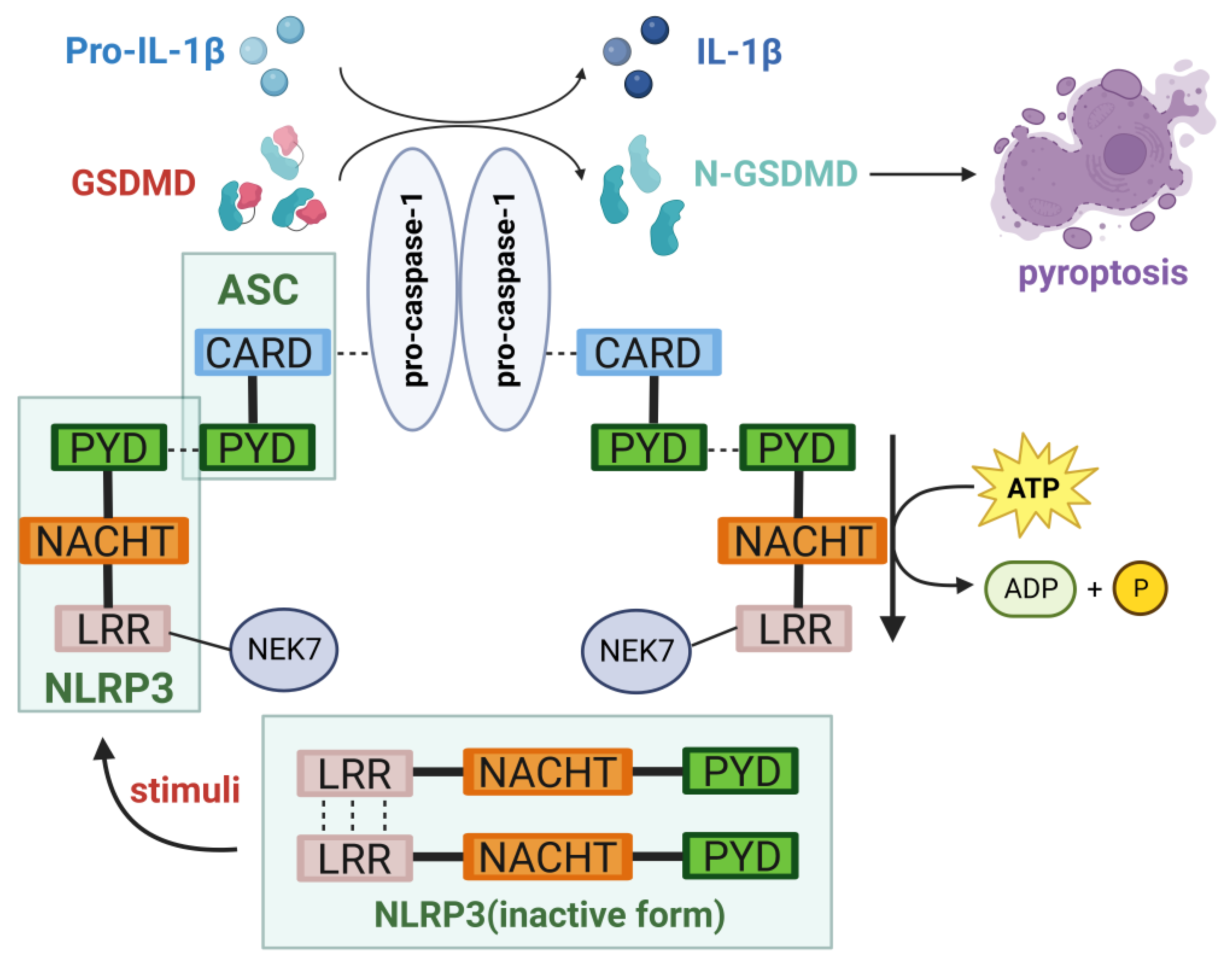
The primary constituents of the NLRP3 inflammasome include NLRP3, apoptosis-associated speck-like protein (ASC), and caspase-1. NLRP3 is an intracellular sensor in the NLRP3 inflammasome, which is a pattern recognition receptor (PRR) present within the cell responsible for recognizing the DAMPs and PAMPs among the NLRP family members [4].The NLRP3 protein comprises three distinct structural domains: the N-terminal pyrin domain (PYD), the central nucleotide-binding oligomerization domain (NACHT or NOD), and the C-terminal leucine-rich repeat (LRR) domain containing 12 leucine residues. The structure of each portion is intricately interconnected with its respective function. The PYD is associated with the recruitment of ASC [4]; the NACHT possesses the capability to bind nucleotides and hydrolyze ATP, and its activation can result in PYD interactions, thereby recruiting ASC [5]; and LRR serves as an additional molecular switch in NLRP3 activation, interacting with the NIMA-related kinase 7 (NEK7) located at the microtubule organizing center [6]. This interaction opens the NLRP3 cage formed on the trans-Golgi network (TGN) before stimulation by molecules like nigericin, enabling the formation of active NLRP3 [7].
Beyond PYD, ASC contains a caspase recruitment domain (CARD), which interacts with NLRP3 and procaspase-1 individually [5]. When given no stimulation, the NLRP3 protein forms oligomers and becomes inactive. This occurs through the C-terminal of the LRR domain, which prevents its binding with ASC [7]. Upon receiving appropriate signals, NLRP3 assembles with ASC and procaspase-1 to form the NLRP3 inflammasome [8]. Caspase-1, functioning as an effector molecule, has the ability to cleave and activate gasdermin D (GSDMD) responsible for pyroptosis, simultaneously generating mature pro-inflammatory cytokines, which is crucial for regulating immune responses [9,10,11]. Figure 1 provides a concise representation of the structure of the NLRP3 inflammasome.
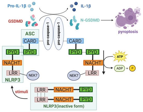
Figure 1.
The structure of the NLRP3 inflammasome. The NLRP3 inflammasome is composed of NLRP3, ASC, and caspase-1. Upon activation, it mediates cellular pyroptosis, leading to the production of IL-1β and IL-18, exerting biological functions (created with BioRender.com, accessed on 21 October 2023).
2.2. Activation of the NLRP3 Inflammasome
Currently, the categorization of NLRP3 inflammasome activation is generally classified into three types: canonical, non-canonical, and alternative pathways. In this review, we focus on the canonical pathway with a brief discussion of non-canonical and alternative pathways.
2.2.1. Canonical Pathway
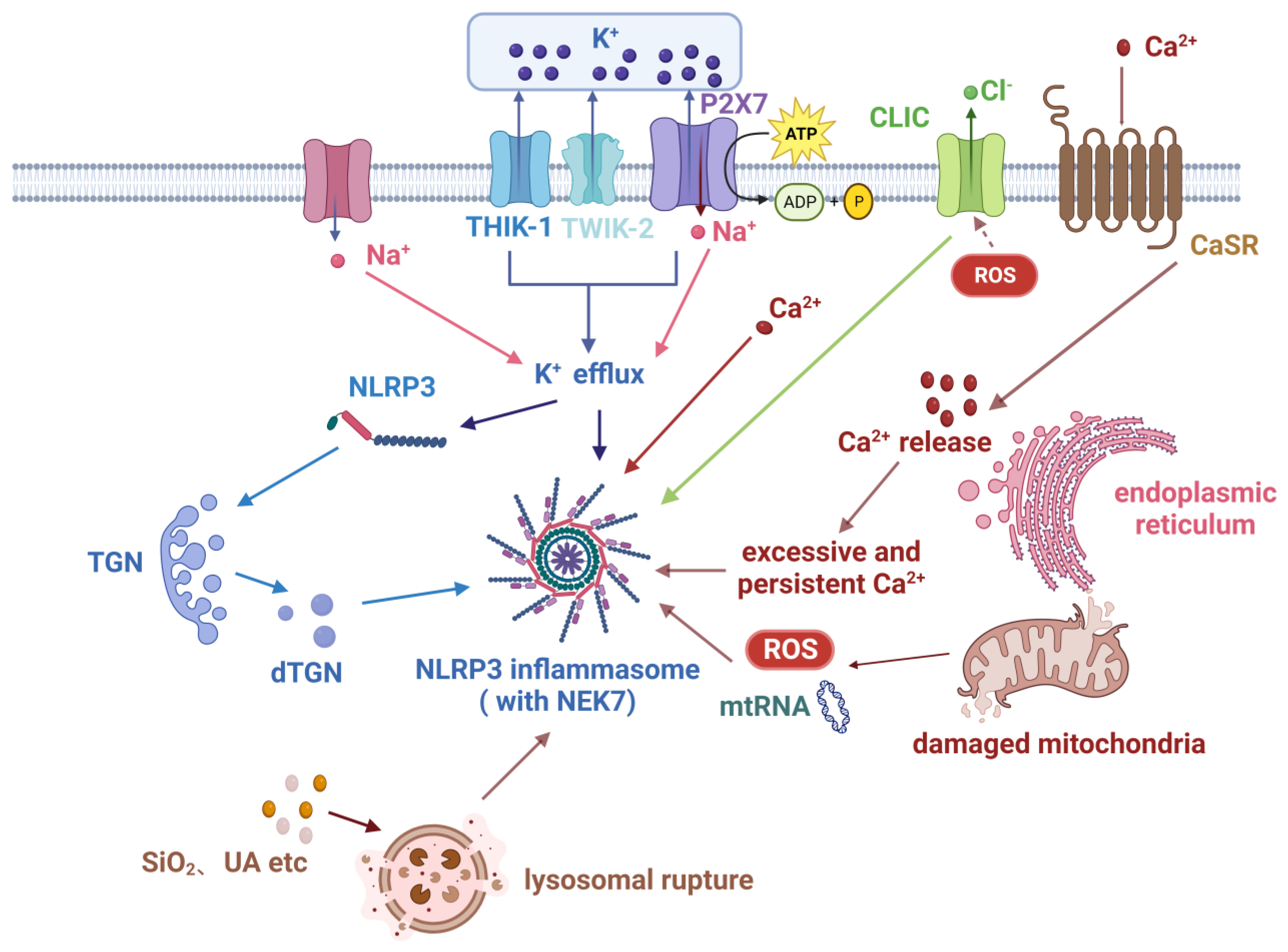
In the canonical pathway, the NLRP3 inflammasome necessitates the presence of both a priming signal and an activation signal [12]. The priming signal encompasses the regulation of NLRP3 expression and post-translational modifications (PTMs), although PTMs still maintain NLRP3 in a self-inhibited state [9,13]. The activation signal typically relies on changes in intracellular ion concentrations such as potassium () and calcium ().
The priming of NLRP3 is commonly dependent on the NF-κB signaling pathway. Surface receptors, including PRRs and TNF receptor (TNFR) superfamily members, respond to various stimuli primarily by activating the ubiquitin-dependent kinase TAK1 and then the multi-subunit IκB kinase (IKK). Subsequently, the IKK complex mediates the phosphorylation and subsequent degradation of IκBα, resulting in the transient nuclear translocation of NF-κB heterodimers [12]. In the context of research on rheumatoid arthritis (RA), it has been observed that lipopolysaccharide (LPS) has a role in the regulation of synovial fibroblast pyroptosis by activating the NF-κB signaling pathway [14]. Other transcription factors such as Sp17, c-Myb, AP-2, and c-Ets [2], together with sterol regulatory element binding transcription factor 3 [15], contribute to the expression of NLRP3. This indicates that the activation signals of the NLRP3 inflammasome exhibit a wide range of variations, and these signals have the capacity to mutually influence one another [16].
Following priming, NLRP3 remains in an inhibited state and requires the involvement of an activation signal. Once activated, NLRP3 assembles together with ASC and caspase-1 to form an inflammasome, resulting in the complete activation of the NLRP3 inflammasome. According to current research, the process of activation is known to transpire through multiple pathways, with the efflux pathway and mitochondrial dysfunction being the ones most thoroughly investigated [4].
efflux is considered a fundamental mechanism for the ATP-induced activation of the NLRP3 inflammasome. V P E’trilli et al. found that elevated extracellular levels inhibit NLRP3 activation in human monocytes, while intracellular reduction triggers NLRP3 inflammasome activation [17]. Furthermore, classical activators of the NLRP3 inflammasome, such as nigericin and ATP, lead to intracellular reduction, and channel blockers inhibit the release of IL-1β [18], providing further evidence that the efflux of promotes the activation of the NLRP3 inflammasome. Mechanistically, purinergic receptor P2X purinoceptor 7 (P2X7), two-pore channel THIK-1, and two-pore domain channel () TWIK2 have all been found to mediate efflux, subsequently activating the NLRP3 inflammasome [19,20]. In a model of PBMC-derived macrophages established to study RA, anti-citrullinated protein antibodies (ACPA) were found to activate pannexin channels, leading to ATP secretion and the subsequent activation of P2X7 receptors to promote efflux. This study first proved that ACPA can activate the NLRP3 inflammasome involved in the pathogenesis of RA [21]. NEK7 functions in the downstream pathway following efflux in the signal cascade of activation, mediating the assembly and activation of the NLRP3 inflammasome [22]. The latest research has demonstrated a notable rise in the development of the NEK7-NLRP3 complex in the context of Streptococcus pneumoniae infection, with an accompanying rapid phosphorylation of anaplastic lymphoma kinase and c-JunN-terminal kinase [23].
Additionally, influx can promote the activation of NLRP3 induced by certain agonists [24]. However, it should be noted that the only influx of pure is inadequate to activate NLRP3 [25]. A plausible explanation for this phenomenon is that the presence of leads to the stimulation of efflux. Alternatively, it is also possible that influx and efflux are regulated by a shared mechanism [26]. Chloride () efflux is another regulatory pathway for activating NLRP3. During the process of activation, the intracellular chloride intracellular channel (CLIC) functions in a downstream manner following the efflux and mitochondrial reactive oxygen species (mtROS) axis. The translocation of CLICs to the plasma membrane is induced by mtROS, which is then followed by efflux. This process facilitates the interaction between NEK7 and NLRP3 [4]. It is evident that NEK7 acts downstream of both efflux in the signal cascade and efflux, hence aiding the formation of inflammasomes. Furthermore, efflux can also promote inflammasome assembly by inducing ASC oligomerization. Similar to , this facilitative action likewise depends on efflux [27].
However, not all cellular activations of NLRP3 rely on efflux. For instance, certain imidazoquinoline amines such as imiquimod and CL097, as well as peptidoglycans, have the ability to activate NLRP3 in a manner that is not dependent on efflux [28,29]. Hence, there exist NLRP3 activation mechanisms that bypass efflux in certain cells.
Dysfunctional mitochondria release mtROS and mitochondrial DNA (mtDNA), contributing to the activation of the NLRP3 inflammasome [30]. Research indicates that mtROS is primarily generated through electron transport chain (ETC) complexes I and III, and is associated with NLRP3 activation [31]. Nevertheless, the precise role of mtROS in the activation of NLRP3 is still not well understood. In more specific studies, ETC complexes I or III were replaced with alternative oxidases maintaining normal ETC function but incapable of producing mtROS. In the absence of mtROS, cells exhibited normal NLRP3 activation and IL-1β release [32], suggesting that mtROS may not be essential for NLRP3 inflammasome activation. Cytidine monophosphate kinase 2 is an enzyme that provides deoxyribonucleotides for mtDNA synthesis. It induces CAMK1-dependent IRF2 transcription through Myd88 and TRIF TLR signaling, and CAMK2-mediated mtDNA synthesis is necessary for oxidative mtDNA generation induced by NLRP3 agonists [33]. The release of mtDNA from mitochondria that have been damaged can occur within the cytoplasm, hence aiding in the activation of NLRP3. For example, oxidized mtDNA can bind and activate NLRP3 [34]. Moreover, it is essential to note that TLR-mediated mtDNA synthesis plays a critical role in the activation of NLRP3, as genetic mtDNA depletion impairs NLRP3 activation [33]. However, NLRP3 activation may also lead to mtDNA release [30]. Studies suggest that ETC participates in NLRP3 activation through the production of phosphocreatine-dependent ATP [32]. Therefore, mtDNA depletion may indirectly impact NLRP3 activation through its influence on ETC activity. Overall, the significance of mtDNA in NLRP3 activation is still being debated.
, as an indispensable second messenger in many physiological processes, plays a crucial role in the activation of the NLRP3 inflammasome. The presence of extracellular ATP has the potential to facilitate influx. The physiological functions of calcium-binding protein S100A9 are mediated by its interaction with and can serve as an inflammation biomarker reflecting the severity of RA. A study revealed that IL-6 has the potential to facilitate the interaction between recombinant cathepsin B (CTSB) and NLRP3 via the CTSB/ATP pathway, thereby initiating the activation of the inflammasome and subsequently inducing RA. This mechanism was also validated in J774A.1 cells stimulated by IL-6 and ATP, resulting in an observed upregulation of S100A9 expression [35]. In another study investigating the pathogenesis of RA, it was discovered that colloidal calciprotein particles activate the calcium sensing receptor (CaSR) and induce the activation of phospholipase C through G proteins, leading to the hydrolysis of inositol triphosphate (IP3), among other effects. IP3 causes the release of calcium from intracellular calcium stores, leading to an elevation in intracellular calcium levels, perhaps facilitating the initiation of the NLRP3 inflammasome [36]. Acid-sensing ion channels (ASICs) represent cation channels activated by extracellular acidosis. Among them, ASIC1a possesses the distinct ability to facilitate the transportation of calcium ions. ASIC1a induces apoptosis of chondrocytes in RA by increasing intracellular mediated by extracellular acidosis [37]. Additionally, excessive or sustained influx can damage mitochondria and release mitochondrial mtROS, further activating the NLRP3 inflammasome [30,38].
Hypoxia-inducible factor-1α (HIF-1α) has also been found to be an inducer of NLRP3 inflammasome activation. It can be activated by various upstream signals, including ROS and the NF-κB signaling pathway [39]. In macrophages, the activation of HIF-1α is known to stimulate the synthesis of inflammatory factors, while inhibiting NF-κB can prevent excessive inflammation activation [40].
Finally, lysosomes and the TGN are also involved in the activation of the NLRP3 inflammasome. The engulfment of crystals by lysosomes leads to lysosomal damage and rupture, thereby activating the NLRP3 inflammasome. Self-dsDNA together with its autoantibodies can trigger the activation of the NLRP3 inflammasome in systemic lupus erythematosus (SLE) [41]. This activation process is potentially facilitated by the generation of ROS and the aforementioned efflux [7]. Recent research has identified that internalized C4b-binding protein (C4BP) acts as an inhibitor of the crystal- or particle-induced inflammasome response in primary human macrophages. In vivo, it has been observed that C4BP in mice has the ability to inhibit the augmentation of the inflammatory state [42].
The TGN is crucial for both efflux-dependent and independent activation of the NLRP3 inflammasome. efflux has been demonstrated to be necessary for the recruitment of NLRP3 to the TGN [43], possibly by reducing cellular ionic strength to facilitate ion binding. Additionally, studies have indicated that protein kinase IKKβ plays a critical role in recruiting NLRP3 to dispersed TGN (dTGN) [44]. NLRP3 agonists trigger the formation of dTGN, which then undergoes transportation to the microtubule-organizing center (MTOC). Through the formation of ionic bonds between the negatively charged phosphatidylinositol-4-phosphate and the positively charged polybasic region between the PYD and NACHT domains of NLRP3, NLRP3 is recruited to dTGN [43]. Subsequently, NLRP3 associates with NEK7 in the centrosome, resulting in the formation of active NLRP3 inflammasome specks. Following this, NLRP3 recruits ASC through PYD–PYD interactions, and ASC recruits caspase-1 through CARD–CARD interactions, ultimately forming the NLRP3 inflammasome [45]. Figure 2 presents a comprehensive depiction of the principal signaling pathways implicated in the classical activation of the NLRP3 inflammasome.
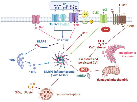
Figure 2.
Key signaling pathways in the canonical activation of the NLRP3 inflammasome. The participation of numerous signals is necessary for the activation of the NLRP3 inflammasome, with efflux being the most pivotal factor. In addition, influx and efflux are also synergistic signals. The endoplasmic reticulum, trans-Golgi network, mitochondria, and lysosomes may be involved in NLRP3 inflammasome activation partly through these signals (created with BioRender.com).
2.2.2. Other Pathways
In addition to the canonical pathway, there exist non-canonical and alternative pathways, which will be briefly outlined here.
The non-canonical inflammasome refers to the caspase-11-dependent inflammasome in mice (or caspase-4 and caspase-5 in humans), where Nur77 functions as an intracellular LPS receptor and activates the non-canonical NLRP3 inflammasome [46,47]. This leads to pyroptosis triggered by GSDMD cleavage [48]. During various bacterial infections, cytoplasmic LPS directly binds to mouse caspase-11 (or human caspase-4/caspase-5), leading to its oligomerization and activation [49]. Caspase-11 plays a crucial role in septic shock [50]. In human and porcine monocytes, alternative inflammasome activation is mediated by the upstream TLR1-TRIF-RIPK8-FADD-CASP4 signaling cascade. In contrast to canonical inflammasomes, the alternative response does not elicit the generation of pyroptotic entities or pyroptosis [51].
2.3. Inducing Pyroptosis
After the activation of the NLRP3 inflammasome, self-cleavage and activation of caspase-1 are induced. The activation of caspase-1 leads to the maturation of the pro-inflammatory cytokines IL-1β and IL-18. Simultaneously, it cleaves GSDMD and releases its N-terminal domain. This domain translocate to the cell membrane, forming pores that mediate the release of cellular contents and induce inflammatory cell death, known as pyroptosis [52]. The clearance of infected cells and resistance to pathogens are facilitated by this mechanism, wherein the activation of the NLRP3 inflammasome plays a critical role in the host’s defense against pathogenic invasions and the maintenance of homeostasis.
Nevertheless, an overabundance of NLRP3 inflammasome activation can contribute to the advancement of diverse inflammatory disorders, including the autoimmune diseases referenced in the following subsection.
2.4. Regulation of the NLRP3 Inflammasome
The NLRP3 inflammasome exhibits both a precise mechanism of activation and a dependence on meticulous regulation. The application of stringent regulatory mechanisms guarantees that the activation of the NLRP3 inflammasome elicits a targeted immune response that is both effective in providing protection and devoid of any detrimental effects on the organism. When regulatory mechanisms get disrupted, it can potentially result in the development of several autoimmune diseases. Numerous studies have demonstrated that the development of several autoimmune disorders is linked to the disruption of NLRP3 inflammasome activation [53]. Based on different PTMs regulating the NLRP3 inflammasome, the most common regulatory mechanisms currently studied involve phosphorylation and ubiquitination. The regulators of these two mechanisms act on different sites on NLRP3. We have compiled and listed them in Table 1 and Table 2.

Table 1.
The regulation of NLRP3 through dephosphorylation and phosphorylation.

Table 2.
The regulation of NLRP3 through ubiquitination and deubiquitination.
3. The Biological Functions of NLRP3 Inflammasome
The principal effectors of the biological response of the NLRP3 inflammasome are IL-1β and IL-18, which are generated through the cleavage of caspase-1. By affecting different immune cells and tissue cells, they play an essential role in immune response.
As a classical inflammatory mediator, IL-1β is primarily secreted by monocytes, macrophages and dendritic cells. Dendritic cells are of paramount importance in the process of recognizing antigens by naïve T cells and initiating specific immune responses. The interaction between IL-1β and IL-1R plays an essential part in the process of dendritic cell maturation [72]. IL-1β can promote the functionality of various T cell subsets. For example, it stimulates the secretion of IL-17 by CD4+T cells and γδT cells [73], facilitates the infiltration of Th17 cells [74], and promotes the proliferation and functional activation of CD8+ T cells [75]. The aforementioned impacts contribute positively to the host’s capacity to resist specific pathogens [76]. IL-1β also mediates the cross-talk between innate and adaptive immunity [77].
IL-18 is a cytokine that is also classified under the IL-1 family. Nevertheless, the expression of IL-1β is more prevalent in comparison [78]. IL-18 binding protein (IL-18BP) is a natural inhibitor of IL-18, which aids in regulating the function of IL-18. IL-12 performs a pivotal role in modulating the biological function of IL-18. In the presence of IL-12, IL-18 induces Th1-type responses and stimulates the production of IFN-γ. Conversely, IL-18 alone contributes to the induction of Th2-type responses. IL-18 is additionally involved in the stimulation of NK cells, hence contributing to the immune responses mounted against both infectious agents and tumors [79]. Therefore, IL-18 exhibits pleiotropic biological functions in the body and primarily participates in immune responses through T cells and NK cells. Several inflammatory diseases and autoimmune diseases are easily treated more manageably by decreasing the level of IL-18 in the body [80,81].
In summary, the NLRP3 inflammasome-mediated release of IL-1β and IL-18 simultaneously affects innate immunity and adaptive immunity. Dysfunctional NLRP3 inflammasomes are involved in the development of various autoimmune diseases.
4. NLRP3 Inflammasome in Autoimmune Diseases
4.1. Rheumatoid Arthritis
RA is a typical representative of the autoimmune disease. Its typical clinical manifestations include symmetrical swelling and pain in multiple joints, as well as involvement of extra-articular organs. Chronic synovial inflammation serves as the principal pathological alteration in RA [82]. Although the exact pathogenesis of RA has not been fully elucidated, multiple immune cells and cytokines are considered important drivers [83]. IL-1β and IL-18, generated upon NLRP3 inflammasome activation, emerge as critical pro-inflammatory mediators in innate immunity, exerting a pivotal influence on the initiation and advancement of RA [84].
The collagen-induced arthritis (CIA) mouse model is the most commonly used animal model for RA [85]. The model demonstrates an apparent augmentation in the expression of NLRP3 inside synovial tissues, exhibiting a favorable correlation with both clinical and radiographic assessments of arthritis [86]. The examination of peripheral blood cells in patients with RA indicates that individuals in the active phase of the disease have elevated baseline levels of NLRP3 inflammasomes [87]. Among them, monocytes exhibit a marked increase in the expression and activation of the NLRP3 inflammasome [88], whereas neutrophils demonstrate a notable decrease in NLRP3 inflammasome expression [89]. In spite of the activation of NLRP3 inflammasomes, there is no notable disparity in the peripheral concentrations of IL-1β in RA patients [88]. However, it may be elevated at specific infiltrated sites, including the lungs of RA patients with interstitial pneumonia [90] and synovial tissues [91]. Compared to IL-1β, it is plausible that IL-18 exhibits a stronger correlation with the potential joint involvement [92].
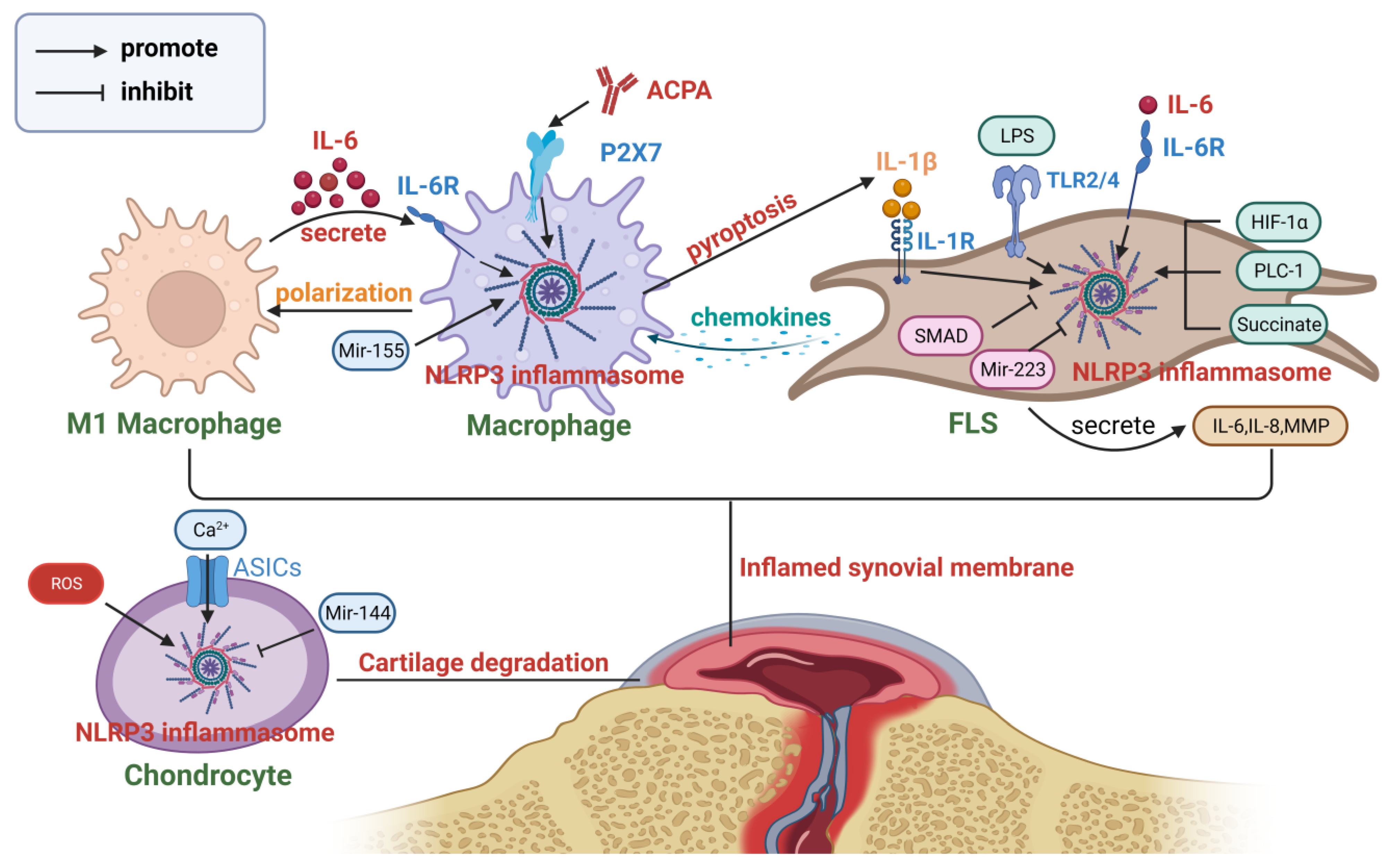
The main contributors responsible for the establishment and maintenance of a persistent and prolonged inflammatory milieu within the joints affected by RA are the infiltration of macrophages and the activation of fibroblasts. The infiltration of macrophages is positively correlated with the extent of joint erosion, which is considered an early sign of RA [93]. In RA patients, the activation level of the NLRP3 inflammasome in macrophages within synovial tissue is significantly increased [94], and finally, the pyroptosis can impact the biological characteristics of fibroblast-like synoviocytes (FLS) [95], demonstrating the bridging role of the NLRP3 inflammasome in connecting immune cells and non-immune cells in RA. ACPA serves as a significant biomarker within the population of RA patients. Previous studies have shown that ACPA has the capacity to trigger the activation of the P2X7 ion channel, consequently driving the activation of the NLRP3 inflammasome in macrophages of individuals diagnosed with RA. Consequently, this activation leads to the release of IL-1β and aggravates the symptoms and progression of RA [21]. Furthermore, Mir-155 functions as a critical regulatory factor of NLRP3 and has been found to regulate NLRP3-mediated cell pyroptosis in various diseases, including osteoarthritis. However, its effects vary across different cell types. Gen Li et al. [96] reported its inhibitory effect on chondrocyte pyroptosis, while Chen Li et al. [97] reported its promotion of macrophage pyroptosis. Mir-155 levels are elevated in RA patients, facilitating the polarization of M1 macrophages [98] and contributing to the activation of CD4+ T cells [99]. Cytokines secreted by M1 macrophages, such as IL-6, can further activate NLRP3 via the cathepsin B/S100A9 pathway, promoting the progression of RA [35].
FLS are the primary cellular constituents of the synovium, playing a crucial role in the initiation and progression of inflammation. The synovial tissue of RA patients expresses high levels of TLR2/4, providing a solid foundation for the priming of the NLRP3 inflammasome [100]. Multiple substances have been proven to activate the NLRP3 inflammasome within RA-FLS, including the metabolic product succinate [101], TNF-α [102], LPS [103], and phospholipase C-like 1 [104]. In addition, the synovial tissue of RA patients is known to be in a chronic hypoxic state [105], which facilitates the sustained activation of HIF-1α-related signaling pathways in FLS [106] and pyroptosis mediated by NLRP3 [107]. MiR-223 [108] and SMAD [95] have been shown to inhibit the activation of NLRP3 in RA-FLS, with the latter exerting its effects through the TGF-β pathway. Pyroptosis of RA-FLS may result in abnormal proliferation and migration, as well as the release of a large amount of inflammatory cytokines and chemokines [109]. IL-18 induces angiogenesis in RA synovial tissue, contributing to vascularization [110]. And due to high expression levels of IL-1R in the synovial tissue of RA patients [111], FLS exposed to IL-1β exhibit an enhanced secretion of inflammatory mediators, including IL-6, IL-8, and matrix metalloproteinases. This heightened release of inflammatory cytokines contributes to the degradation of connective tissues within the joints. Activated fibroblasts can also secrete chemokines and growth factors to recruit monocytes and promote their proliferation [112]. Therefore, there is a reciprocal interaction between FLS and monocytes/macrophages mediated by the NLRP3 inflammasome [113].
Moreover, the pyroptosis of chondrocytes should not be overlooked. One of the fundamental characteristics of RA is the presence of an acidic environment inside the joint cavity. In mouse models of arthritis, the presence of extracellular acidosis triggers the activation of NLRP3-mediated pyroptosis in chondrocytes. This activation may occur via two mechanisms: firstly, ASICs facilitate calcium influx [114], and secondly, it can be induced by an elevation in ROS levels [115]. Consequently, the aforementioned process culminates in the occurrence of pyroptosis in chondrocytes, thus indicating the potential role of NLRP3-mediated chondrocyte pyroptosis in the pathogenesis of cartilage tissue degradation. The inhibitory effects of Mir-144 on this pathway have been shown [116]. Figure 3 provides a comprehensive overview of the involvement of NLRP3 inflammasomes in the pathophysiology of RA.
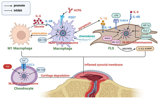
Figure 3.
The role of the NLRP3 inflammasome in the pathogenesis of RA. Chondrocytes, macrophages, and FLS interact with each other through the NLRP3 inflammasome, collectively promoting the occurrence and progression of RA (created with BioRender.com).
4.2. Systemic Lupus Erythematosus
SLE is an autoimmune disease characterized by immune dysregulation in patients, manifested by the formation of autoantibodies, deposition of immune complexes, and multi-organ damage, with the skin and kidneys being particularly affected [117]. Increasing evidence suggests that the NLRP3 inflammasome plays a crucial role in the pathogenesis of SLE, especially in relation to the abnormal activation of the innate and adaptive immune systems [118]. The abnormal activation of the NLRP3 inflammasome has been detected in various cell types within SLE patients, including macrophages [119], monocytes [120], renal tubular epithelial cells [121], and podocytes [122]. Additionally, several substances present in SLE patients contribute to the activation of the NLRP3 inflammasome within different cell types, including self-derived dsDNA [41], anti-dsDNA antibodies [123], and neutrophil extracellular traps (NETs) [124].
In addition, IL-1β and IL-18 in the blood of SLE patients also show significant alterations. Different studies have reported varying levels of IL-1β in SLE patients [125,126], while the levels of IL-18 are elevated and closely associated with active renal involvement [125]. At the organ level, the activation of the NLRP3 inflammasome can also be detected, primarily in the kidneys [127] and skin [128]. NLRP3 inflammasome activation often correlates with the severity of clinical indicators in patients, most commonly manifested as proteinuria due to renal involvement [129].
The pathogenesis of SLE involves the critical participation of neutrophils and IFN-γ in the innate immune system, with the NLRP3 inflammasome likely promoting SLE through them [130]. Upon activation, neutrophils release NETs [131], which contain diverse enzymes and antimicrobial peptides, and potentially contribute to autoimmunity [132]. Altered levels of NETs in SLE patients impact neutrophil bactericidal functions and facilitate the development of autoimmunity and fibrosis [133]. A decade ago, J Michelle Kahlenberg et al. discovered that NLRP3 activation in the macrophages of SLE patients contributes to the release of NETs mediated by neutrophils and impairs their clearance, resulting in NET accumulation [124]. This process triggers caspase-1 activation, initiating a detrimental cycle of inflammation [134] and potentially leading to severe complications [135]. IFN-I, an essential immunoregulatory factor, aids in the activation of adaptive immune cells and mediates immune dysregulation in SLE patients [136]. IFN-related genes are associated with genetic susceptibility to SLE, and elevated serum IFN levels enhance disease activity and increase the risk of relapse in SLE patients [137], thereby promoting the dysfunction of endothelial progenitor cells [138]. The interplay between IFN and the NLRP3 inflammasome has been gradually elucidated, including the induction of NLRP3 inflammasome activation by IFN-I during influenza A virus infection [139]. In SLE patients, IFN-I activates inflammasomes in monocytes through IRF-1 mediation [140]. Certain environmental pollutants, such as bisphenol A, also activate the NLRP3 inflammasome via IFN-I signaling [141]. Bisphenol A was previously identified to upregulate the expression of Nur77 [142], and recent studies have revealed that Nur77 serves as a critical mediator of NLRP3 activation through noncanonical pathways [143], potentially offering a novel perspective on the underlying mechanism of NLRP3 activation induced by bisphenol A. However, the activated NLRP3 inflammasome seems to negatively regulate IFN-I signaling, which may be associated with the SOCS1-mediated regulation of IRF-3 [144].
Another key characteristic of SLE in the adaptive immune response is the generation of autoantibodies. Autoantibodies against self-dsDNA directly bind to TLR or activate the NLRP3 inflammasome through promoting ROS or K+ efflux [145], thereby driving the excessive activation of B cells [146]. Recently, the NLRP3 inflammasome in Tfh cells has been found to involve B cell activation, leading to the production of high-affinity antibodies and correlating with disease activity [147].
Although the NLRP3 inflammasome plays a critical role in driving the development of SLE through various mechanisms, the relationship between the two is not straightforward. There is still controversy regarding the relationship between the activation level of NLRP3 and SLE. Furini et al. indicated that the activation level of NLRP3 inflammasomes and the expression level of P2X7 receptors are decreased in macrophages in SLE patients. It is suggested that IL-6 may play a more important role compared to IL-1β [148]. Qing-rui Yang et al. put forward the notion of a negative association between the activation level of the NLRP3 inflammasome in peripheral blood mononuclear cells (PBMCs) derived from patients with SLE and the severity of the disease, highlighting the involvement of IFN-I in this relationship [149]. Zhen-zhen Ma et al. also observed lower expression levels of NLRP3-related molecules in the serum of SLE patients [150].
Furthermore, the absence of NLRP3 can exacerbate the severity of SLE in certain circumstances. The direct blockade of IL-1β alone does not effectively reduce renal inflammation and overall survival rates in the NZM2328 mice, which even exacerbates the severity of proteinuria [151]. NLRP3/ASC promotes renal glomerular damage by increasing T cell infiltration [152], but mice lacking NLRP3 or ASC also develop severe lupus nephritis, which may be associated with other signaling pathways such as TGF-β [153]. This suggests that the NLRP3 inflammasome plays distinct roles in different pathways in SLE patients. Therefore, investigating how to selectively inhibit detrimental NLRP3 signaling holds potential significance for the treatment of SLE.
4.3. Systemic Sclerosis
Systemic sclerosis (SSc) is an autoimmune disease characterized by vascular abnormalities, inflammatory responses, and fibrosis. The pathogenesis of early-stage SSc primarily involves endothelial dysfunction, while in later stages, fibroblast hyperactivity leads to excessive extracellular matrix secretion, resulting in fibrosis in multiple organs. Inappropriate inflammatory responses are one of the core features of SSc, which ultimately facilitates irreversible organ damage [154]. Upon activation of the NLRP3 inflammasome, the secretion of IL-1β and IL-18 can promote vascular abnormalities, inflammatory responses, and extracellular matrix secretion [155]. Additionally, GSDMD-N-mediated pyroptosis has also been implicated in SSc [156].
Given the clinical characteristics of SSc, there are currently several models that demonstrate the typical features of fibrosis in SSc, including TSK gene mutation mice, Fra-2 transgenic mice, Fli-1 gene knockout mice, and bleomycin-induced fibrosis mice [157]. Among them, bleomycin-induced fibrosis mice are the most commonly used model. Bleomycin activates TLR2, mediates the release of various inflammatory cytokines, and ultimately leads to pulmonary fibrosis in mice [158]. In mice treated with bleomycin, the levels of caspase-1, IL-1β, and IL-18 are significantly increased in serum and lung tissue. This fibrosis can be alleviated by knocking out the caspase-1 and IL-18 genes [159]. The degree of change in NLRP3-related molecules in SSc patients varies in different studies. For example, some studies have indicated a significant increase in the content of caspase-1 in the skin tissue of SSc patients [160], while the level of caspase-1 in peripheral blood is significantly decreased [161]. In addition, Emily Lin et al. found a lack of significant alteration in the level of IL-1β in the serum of SSc patients [162], while Y.-J. Zhanget al. showed a significant increase [163]. The expression levels of NLRP3, IL-1β, and IL-18 in the skin of SSc patients are significantly elevated [160]. The results of these different studies may suggest that the activation of NLRP3 in different tissues of SSc patients varies and is related to the specific disease progression, such as higher expression levels of IL-1β and caspase-1 in the muscle tissue of SSc patients with myositis [164]. Furthermore, the levels of NLRP3-related cytokines in SSc patients are closely correlated with certain clinical indicators. High levels of IL-18 in the serum indicate poor pulmonary function in patients, while high levels of IL-1β in skin tissue indicate severe skin fibrosis [160] and relatively better lung function [162]. However, there are also studies that refute the clinical predictive value of IL-1β levels in the serum [163].
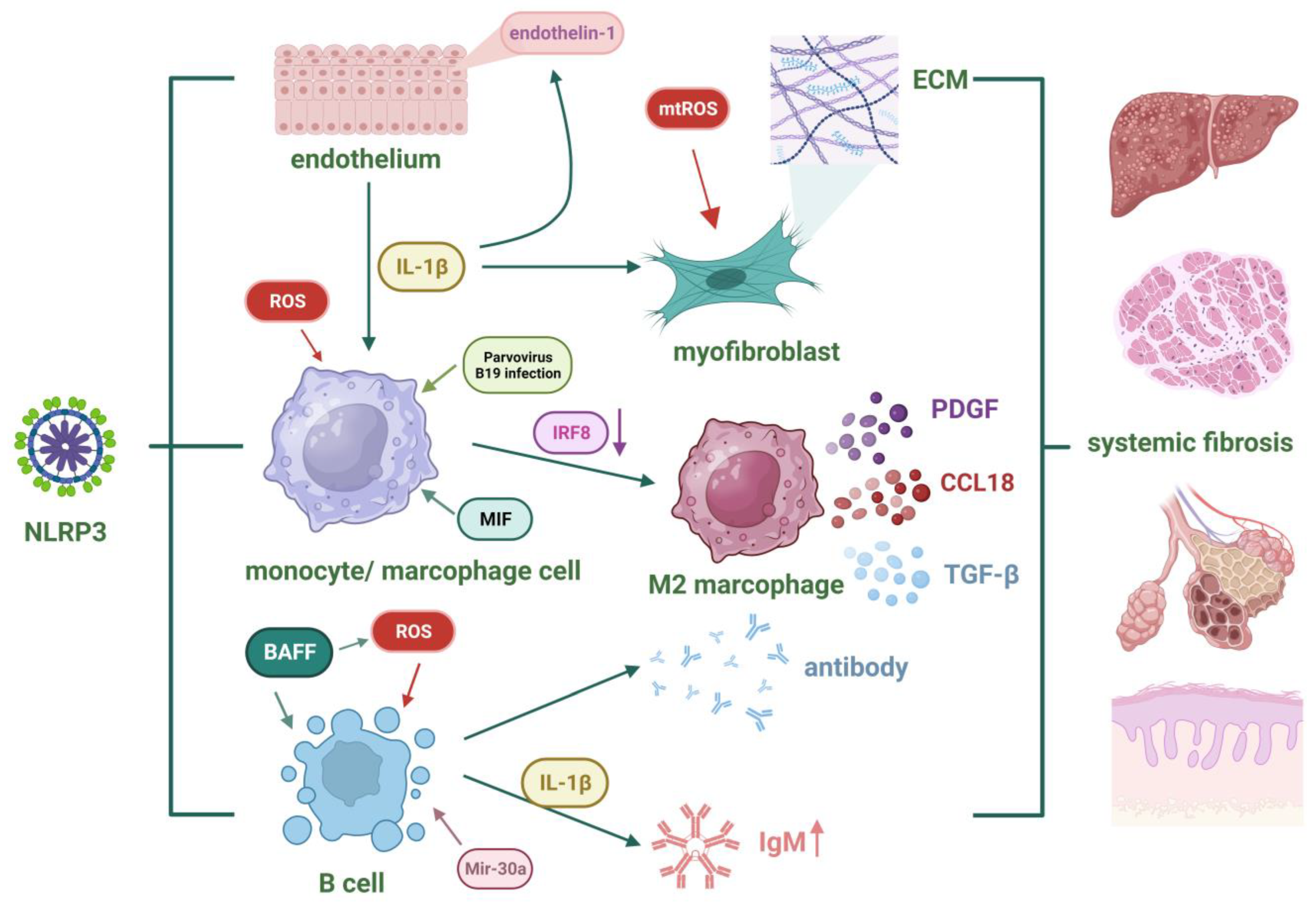
Mechanistically, the NLRP3 inflammasome may drive the pathogenesis of SSc by affecting monocytes/macrophages, B cells, endothelial cells, and fibroblasts. Monocytes circulate in the blood and serve as precursors for tissue macrophages. Studies have shown that monocytes from SSc patients produce more ROS [165], which contributes to the activation of the NLRP3 inflammasome. This may lead to the secretion of IL-1β and TNF-α [166]. Additionally, infection with parvovirus B19 can activate the NLRP3 inflammasome and enhance the activity of monocytes, resulting in increased release of IL-1β [167]. When monocytes migrate into tissues and differentiate into macrophages, they can also induce inflammation and promote fibrosis, highlighting the significant role of macrophages in the pathogenesis of SSc [168]. Macrophages can be classified into classically activated (M1) or alternatively activated (M2) macrophages based on their activation patterns. M2 macrophages secrete pro-fibrotic factors such as TGF-β, CCL18, and PDGF, exhibiting potential profibrotic functions [168]. An imbalance in the differentiation of M1/M2 macrophages is one of the mechanisms underlying SSc [169]. IRF8, a transcription factor promoting M1 macrophage differentiation, is reduced in SSc patients. Consequently, the proportion of M2 macrophages significantly increases, and artificially downregulating IRF8 expression worsens symptoms in SSc mice [170]. Recent studies have revealed the role of IRF8 in regulating the activation of the NLRP3 inflammasome, providing a new perspective on the association between NLRP3 inflammasome and fibrosis [171]. Upon sustained activation of the NLRP3 inflammasome, high levels of IL-1β in the bloodstream promote the differentiation of monocytes into macrophages through endothelial cell mediation. These macrophages then secrete high levels of pro-fibrotic factors such as CCL18, CCL2, and CXCL8, exacerbating skin fibrosis in SSc patients [172]. Furthermore, altered levels of macrophage migration inhibitory factor (MIF) have been observed in different tissues of localized and diffuse SSc patients [173]. MIF contributes to the interaction between NLRP3 and the intermediate filament protein vimentin, which facilitates NLRP3 inflammasome activation [174].
In addition to monocytes/macrophages, B cells are another group of immune cells that contribute to the pathogenesis of SSc. The dysregulation of B cell function in patients leads to their infiltration into affected organs, where they secrete antibodies and interact with other immune cells, promoting the fibrotic process in SSc [175]. B cell activating factor (BAFF) plays a pivotal role in bleomycin- and IL-17-mediated pulmonary fibrosis [176]. Subsequent studies have found that NLRP3 is one of the regulatory targets of BAFF. The upregulation of BAFF activates the NF-κB pathway, promoting the expression of NLRP3 while simultaneously activating ROS and K+ efflux, thereby activating the NLRP3 inflammasome in B cells [177]. However, this process can be inhibited by miR-30a [178]. Additionally, the secretion of IL-1β by activated B cells can also impact IgM synthesis [179].
The damage and dysfunction of endothelial cells in the vascular wall are evident throughout the entire process of SSc pathogenesis [180]. Endothelial dysfunction is typified by an aberration in the balance between vasodilation and vasoconstriction, accompanied by increasing ROS and inflammatory mediators [181], which are closely associated with the NLRP3 inflammasome. The release of IL-1β following activation can further induce the expression of endothelin-1 and adhesion molecules in endothelial cells, promoting leukocyte migration and maintaining vascular inflammation [181].
Fibrosis is the ultimate outcome of SSc, and myofibroblasts are key participants in this process. Epithelial–mesenchymal transition is one of the important features of fibrosis, characterized by the loss of polarity in epithelial cells and their transformation into mesenchymal cells. This is accompanied by an increased expression of α-SMA and a decreased expression of adhesion molecules, along with an increased deposition of extracellular matrix. The NLRP3 inflammasome promotes epithelial–mesenchymal transition [182]. Mitochondrial dysfunction is commonly present in fibrotic diseases, and the resulting mtROS is one of the important signals for activating NLRP3 molecules, which also has an effect on activating fibroblasts [183]. Upon IL-1β signaling through IL-1R on fibroblasts, it may form a positive feedback loop, ultimately resulting in increased collagen synthesis through the complex interplay of cytokines and cellular interactions [184]. In addition to fibroblasts, the NLRP3 inflammasome is also involved in lung fibrosis mediated by alveolar epithelial cells. The activation of the NLRP3 inflammasome in alveolar epithelial cells promotes the differentiation of mesenchymal stem cells into myofibroblasts during the pulmonary fibrosis process [185]. Figure 4 summarizes the role of the NLRP3 inflammasome in the pathogenesis of SSc.
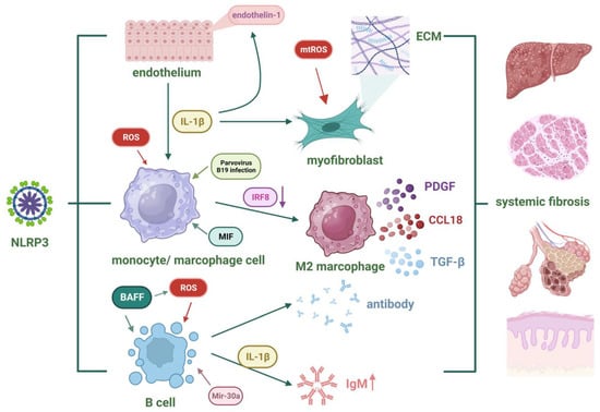
Figure 4.
The role of the NLRP3 inflammasome in the pathogenesis of SSc. Fibrosis represents the ultimate outcome of SSc, and NLRP3 facilitates fibrosis through monocytes/macrophages, B cells, endothelial cells, and myofibroblasts (created with BioRender.com).
5. Bioactive Substances Regulating the NLRP3 Inflammasome for the Treatment of Autoimmune Diseases
Considerable advancements have been achieved in the realm of pharmaceutical research pertaining to the targeting of the NLRP3 inflammasome as a therapeutic approach for the management of autoimmune disorders, which holds important practical significance and potential applications. Bioactive substances constitute a diverse family, encompassing active components extracted from plants as well as certain physiological substances that are essential for maintaining organismal life. Compared to chemically synthesized drugs, most of them exert milder effects [186]. Although the specific target sites and exact functions of many bioactive substances are not yet fully elucidated, it has been discovered in recent years that some of them indeed exhibit significant regulatory effects on NLRP3, including inhibiting the activation of the NLRP3 inflammasome or regulating NLRP3-associated signaling pathways. This review provides a concise overview of the bioactive substances that have been investigated for their potential to regulate the NLRP3 inflammasome in RA, SLE, and SSc within the past three years.
5.1. Inhibiting the Activation of the NLRP3 Inflammasome
The vitamin D receptor (VDR), expressed in nearly all immune cells, mediates the biological effects of vitamin D and regulates immune responses associated with autoimmune diseases [187]. VDR directly binds to NLRP3 and inhibits NLRP3 inflammasome activation by suppressing BRCC3-mediated ubiquitination. Furthermore, the inhibitory effect of VDR is enhanced by vitamin D [188]. Subsequent investigations have provided evidence indicating that vitamin D inhibits the activation of the NLRP3 inflammasome via a SIRT3-SOD2-mtROS signal [189]. These findings partially explain the protective effect of vitamin D in autoimmune diseases, including its ability to reduce the levels of inflammatory cytokines and ROS in RA patients [190]. Additionally, vitamin D improves renal damage in lupus mouse models by inhibiting the NF-κB and MAPK pathways and reducing autoantibody levels [191].
Compound K, a metabolite of ginsenoside derived from ginseng, inhibits NLRP3 inflammasome activation by suppressing NF-κB signaling and reducing ROS levels. This inhibition leads to the suppression of M1 macrophage activation and the induction of M2 macrophage activation [192]. Compound K also promotes SIRT1-mediated autophagy, which is one of the mechanisms by which it inhibits NLRP3 activation [193]. Notably, it demonstrates significant therapeutic effects in lupus nephritis (LN) [194].
Myrtenal and β-caryophyllene oxide are active components extracted from Myrica rubra. They alleviate symptoms in mice with antigen-induced arthritis (AIA) by inhibiting NLRP3 inflammasome activation [195].
5.2. Regulating the TLR4/NLRP3/NF-KB/GSDMD Signaling Pathway
Several bioactive substances with unidentified specific targets have been found to exert their effects by regulating the TLR4/NLRP3/NF-κB/GSDMD signaling pathway. These include traditional Chinese medicine formulas such as the Jinwujiangu capsule for treating RA [196], Baihu-Guizhi decoction [197], and Qi-Sai-Er-Sang-Dang-Song decoction [198]. Additionally, bioactive compounds extracted from plants, such as the monomer derivative of paeoniflorin [199], wedelolactone derived from Xanthium sibiricum [200], licorice-processed DGN products containing Huangruixiang [201], a combination of mangiferin and cinnamic acid [202], punicalagin [203], tectoridin [204], strychnine combined with Atractylodes Macrocephala [205], and pinocembrin from the genus Pinus [206], have also shown activity through this pathway.
5.3. Regulating Other Pathways Related to NLRP3
Honokiol is a bioactive biphenolic compound derived from plants. It has been shown to improve kidney function and reduce albuminuria in LN mice, exerting a preventive and therapeutic effect on LN. This may be achieved by inhibiting the NLRP3 inflammasome and sirtuin 1 signaling in the kidneys, thereby suppressing aberrant crosstalk between renal macrophages and tubular epithelial cells [207]. Subsequent studies have further elucidated the mechanism of the SIRT1-mediated negative regulation of NLRP3 inflammasome activation [208].
ASIC1a is an extracellular acid-activated cation channel that plays a role in chondrocyte pyroptosis by activating NLRP3 through calpain-2/calcineurin in the acidic microenvironment of extracellular acidosis [114]. Yiqi Yangyin Tongluo acts on this pathway to alleviate chondrocyte pyroptosis [209].
HIF-1α is an important mediator in the hypoxic synovial microenvironment of RA patients. It promotes the expression of the NLRP3 gene through the transcription factor STAT3. Monomeric derivatives of paeoniflorin extracted from paeoniflorin [107] and sodium Danshensu extracted from Salvia miltiorrhiza [210] alleviate the clinical manifestations of RA by regulating the HIF-1α/STAT3/NLRP3 pathway.
Er Miao San (EMS) is a formula consisting of equal proportions of Atractylodes Macrocephala rhizome and Cortex Phellodendri. It exhibits good anti-inflammatory effects in an animal model of RA, which is associated with the regulation of cytokine levels and the balance of Treg/Th17 cells [211]. Subsequent studies have found that EMS reduces macrophage polarization towards the M1 phenotype by regulating the miRNA-33/NLRP3 pathway [212], and serum containing EMS has a similar effect [213].
Table 3 provides a detailed summary of the relevant information on bioactive substances.

Table 3.
Bioactive substances regulating the NLRP3 inflammasome to treat autoimmune diseases.
6. Conclusions
The activation of the NLRP3 inflammasome is notably disrupted in autoimmune illnesses such as RA, SLE, and SSc. IL-1β and IL-18 play a crucial role in facilitating the initiation of immune responses, serving as a vital link between innate and adaptive immunity. Although the results are not completely consistent, most of the studies have shown that the level of NLRP3-related proteins has a significant association with diverse clinical features observed in autoimmune diseases. Within certain microenvironments, the activation of the NLRP3 inflammasome and subsequent tissue degradation exhibit a mutually reinforcing relationship, hence establishing a positive feedback loop. This intricate process involves the active participation of various cellular components, including macrophages, lymphocytes, and tissue cells such as fibroblasts. Therefore, it is imperative to investigate the activation and regulatory mechanisms underlying NLRP3 and devise appropriate tailored pharmaceutical interventions. In recent years, an increasing number of biologically active substances have been discovered to possess regulatory functions on NLRP3 inflammasomes. While this is not their exclusive function, it opens up new perspectives for the treatment of autoimmune diseases and warrants further exploration.
Author Contributions
B.C. and Y.W. wrote different sections of the manuscript, created the figures, and contributed to all subsequent revisions; G.C. supervised the overall writing, edited and contributed to the scientific content, and submitted the final manuscript. All authors have read and agreed to the published version of the manuscript.
Funding
The work was supported by grants from the National Nature Science Foundation of China (NSFC-81771731).
Data Availability Statement
Not Applicable.
Acknowledgments
The authors would like to acknowledge the use of Biorender.com, accessed on 21 October 2023, for the conception of all the figures.
Conflicts of Interest
The authors declare no conflict of interest.
References
- Wang, L.; Wang, F.-S.; Gershwin, M.E. Human Autoimmune Diseases: A Comprehensive Update. J. Intern. Med. 2015, 278, 369–395. [Google Scholar] [CrossRef]
- Kelley, N.; Jeltema, D.; Duan, Y.; He, Y. The NLRP3 Inflammasome: An Overview of Mechanisms of Activation and Regulation. Int. J. Mol. Sci. 2019, 20, 3328. [Google Scholar] [CrossRef]
- Zahid, A.; Li, B.; Kombe, A.J.K.; Jin, T.; Tao, J. Pharmacological Inhibitors of the NLRP3 Inflammasome. Front. Immunol. 2019, 10, 2538. [Google Scholar] [CrossRef]
- Zhan, X.; Li, Q.; Xu, G.; Xiao, X.; Bai, Z. The Mechanism of NLRP3 Inflammasome Activation and Its Pharmacological Inhibitors. Front. Immunol. 2023, 13, 1109938. [Google Scholar] [CrossRef]
- de Alba, E. Structure and Interdomain Dynamics of Apoptosis-Associated Speck-like Protein Containing a CARD (ASC). J. Biol. Chem. 2009, 284, 32932–32941. [Google Scholar] [CrossRef]
- Xiao, L.; Magupalli, V.G.; Wu, H. Cryo-EM Structures of the Active NLRP3 Inflammasome Disc. Nature 2023, 613, 595–600. [Google Scholar] [CrossRef]
- Andreeva, L.; David, L.; Rawson, S.; Shen, C.; Pasricha, T.; Pelegrin, P.; Wu, H. NLRP3 Cages Revealed by Full-Length Mouse NLRP3 Structure Control Pathway Activation. Cell 2021, 184, 6299–6312.e22. [Google Scholar] [CrossRef]
- Fu, J.; Wu, H. Structural Mechanisms of NLRP3 Inflammasome Assembly and Activation. Annu. Rev. Immunol. 2023, 41, 301–316. [Google Scholar] [CrossRef]
- Swanson, K.V.; Deng, M.; Ting, J.P.-Y. The NLRP3 Inflammasome: Molecular Activation and Regulation to Therapeutics. Nat. Rev. Immunol. 2019, 19, 477–489. [Google Scholar] [CrossRef]
- Liu, Z.; Wang, C.; Yang, J.; Chen, Y.; Zhou, B.; Abbott, D.W.; Xiao, T.S. Caspase-1 Engages Full-Length Gasdermin D through Two Distinct Interfaces That Mediate Caspase Recruitment and Substrate Cleavage. Immunity 2020, 53, 106–114.e5. [Google Scholar] [CrossRef] [PubMed]
- Xia, S.; Zhang, Z.; Magupalli, V.G.; Pablo, J.L.; Dong, Y.; Vora, S.M.; Wang, L.; Fu, T.-M.; Jacobson, M.P.; Greka, A.; et al. Gasdermin D Pore Structure Reveals Preferential Release of Mature Interleukin-1. Nature 2021, 593, 607–611. [Google Scholar] [CrossRef]
- Liu, T.; Zhang, L.; Joo, D.; Sun, S.-C. NF-κB Signaling in Inflammation. Signal Transduct. Target. Ther. 2017, 2, 17023. [Google Scholar] [CrossRef]
- Sharma, M.; de Alba, E. Structure, Activation and Regulation of NLRP3 and AIM2 Inflammasomes. Int. J. Mol. Sci. 2021, 22, 872. [Google Scholar] [CrossRef]
- Yücel, G.; Zhao, Z.; El-Battrawy, I.; Lan, H.; Lang, S.; Li, X.; Buljubasic, F.; Zimmermann, W.-H.; Cyganek, L.; Utikal, J.; et al. Lipopolysaccharides Induced Inflammatory Responses and Electrophysiological Dysfunctions in Human-Induced Pluripotent Stem Cell Derived Cardiomyocytes. Sci. Rep. 2017, 7, 2935. [Google Scholar] [CrossRef]
- Franchi, L.; Eigenbrod, T.; Núñez, G. Cutting Edge: TNF-α Mediates Sensitization to ATP and Silica via the NLRP3 Inflammasome in the Absence of Microbial Stimulation1. J. Immunol. 2009, 183, 792–796. [Google Scholar] [CrossRef]
- Gritsenko, A.; Green, J.P.; Brough, D.; Lopez-Castejon, G. Mechanisms of NLRP3 Priming in Inflammaging and Age Related Diseases. Cytokine Growth Factor Rev. 2020, 55, 15–25. [Google Scholar] [CrossRef]
- Pétrilli, V.; Papin, S.; Dostert, C.; Mayor, A.; Martinon, F.; Tschopp, J. Activation of the NALP3 Inflammasome Is Triggered by Low Intracellular Potassium Concentration. Cell Death Differ. 2007, 14, 1583–1589. [Google Scholar] [CrossRef]
- Walev, I.; Reske, K.; Palmer, M.; Valeva, A.; Bhakdi, S. Potassium-Inhibited Processing of IL-1 Beta in Human Monocytes. EMBO J. 1995, 14, 1607–1614. [Google Scholar] [CrossRef]
- Di, A.; Xiong, S.; Ye, Z.; Malireddi, R.K.S.; Kometani, S.; Zhong, M.; Mittal, M.; Hong, Z.; Kanneganti, T.-D.; Rehman, J.; et al. The TWIK2 Potassium Efflux Channel in Macrophages Mediates NLRP3 Inflammasome-Induced Inflammation. Immunity 2018, 49, 56–65.e4. [Google Scholar] [CrossRef]
- Drinkall, S.; Lawrence, C.B.; Ossola, B.; Russell, S.; Bender, C.; Brice, N.B.; Dawson, L.A.; Harte, M.; Brough, D. The Two Pore Potassium Channel THIK-1 Regulates NLRP3 Inflammasome Activation. Glia 2022, 70, 1301–1316. [Google Scholar] [CrossRef]
- Dong, X.; Zheng, Z.; Lin, P.; Fu, X.; Li, F.; Jiang, J.; Zhu, P. ACPAs Promote IL-1β Production in Rheumatoid Arthritis by Activating the NLRP3 Inflammasome. Cell Mol. Immunol. 2020, 17, 261–271. [Google Scholar] [CrossRef]
- Zhao, N.; Li, C.; Di, B.; Xu, L. Recent Advances in the NEK7-Licensed NLRP3 Inflammasome Activation: Mechanisms, Role in Diseases and Related Inhibitors. J. Autoimmun. 2020, 113, 102515. [Google Scholar] [CrossRef]
- Wang, X.; Zhao, Y.; Wang, D.; Liu, C.; Qi, Z.; Tang, H.; Liu, Y.; Zhang, S.; Cui, Y.; Li, Y.; et al. ALK-JNK Signaling Promotes NLRP3 Inflammasome Activation and Pyroptosis via NEK7 during Streptococcus Pneumoniae Infection. Mol. Immunol. 2023, 157, 78–90. [Google Scholar] [CrossRef]
- Surprenant, A.; Rassendren, F.; Kawashima, E.; North, R.A.; Buell, G. The Cytolytic P2Z Receptor for Extracellular ATP Identified as a P2X Receptor (P2X7). Science 1996, 272, 735–738. [Google Scholar] [CrossRef]
- Muñoz-Planillo, R.; Kuffa, P.; Martínez-Colón, G.; Smith, B.L.; Rajendiran, T.M.; Núñez, G. K+ Efflux Is the Common Trigger of NLRP3 Inflammasome Activation by Bacterial Toxins and Particulate Matter. Immunity 2013, 38, 1142–1153. [Google Scholar] [CrossRef]
- Schorn, C.; Frey, B.; Lauber, K.; Janko, C.; Strysio, M.; Keppeler, H.; Gaipl, U.S.; Voll, R.E.; Springer, E.; Munoz, L.E.; et al. Sodium Overload and Water Influx Activate the NALP3 Inflammasome. J. Biol. Chem. 2011, 286, 35–41. [Google Scholar] [CrossRef]
- Green, J.P.; Yu, S.; Martín-Sánchez, F.; Pelegrin, P.; Lopez-Castejon, G.; Lawrence, C.B.; Brough, D. Chloride Regulates Dynamic NLRP3-Dependent ASC Oligomerization and Inflammasome Priming. Proc. Natl. Acad. Sci. USA 2018, 115, E9371–E9380. [Google Scholar] [CrossRef]
- Groß, C.J.; Mishra, R.; Schneider, K.S.; Médard, G.; Wettmarshausen, J.; Dittlein, D.C.; Shi, H.; Gorka, O.; Koenig, P.-A.; Fromm, S.; et al. K+ Efflux-Independent NLRP3 Inflammasome Activation by Small Molecules Targeting Mitochondria. Immunity 2016, 45, 761–773. [Google Scholar] [CrossRef]
- Wolf, A.J.; Reyes, C.N.; Liang, W.; Becker, C.; Shimada, K.; Wheeler, M.L.; Cho, H.C.; Popescu, N.I.; Coggeshall, K.M.; Arditi, M.; et al. Hexokinase Is an Innate Immune Receptor for the Detection of Bacterial Peptidoglycan. Cell 2016, 166, 624–636. [Google Scholar] [CrossRef] [PubMed]
- Nakahira, K.; Haspel, J.A.; Rathinam, V.A.K.; Lee, S.-J.; Dolinay, T.; Lam, H.C.; Englert, J.A.; Rabinovitch, M.; Cernadas, M.; Kim, H.P.; et al. Autophagy Proteins Regulate Innate Immune Responses by Inhibiting the Release of Mitochondrial DNA Mediated by the NALP3 Inflammasome. Nat. Immunol. 2011, 12, 222–230. [Google Scholar] [CrossRef]
- Zhou, R.; Yazdi, A.S.; Menu, P.; Tschopp, J. A Role for Mitochondria in NLRP3 Inflammasome Activation. Nature 2011, 469, 221–225. [Google Scholar] [CrossRef]
- Billingham, L.K.; Stoolman, J.S.; Vasan, K.; Rodriguez, A.E.; Poor, T.A.; Szibor, M.; Jacobs, H.T.; Reczek, C.R.; Rashidi, A.; Zhang, P.; et al. Mitochondrial Electron Transport Chain Is Necessary for NLRP3 Inflammasome Activation. Nat. Immunol. 2022, 23, 692–704. [Google Scholar] [CrossRef]
- Zhong, Z.; Liang, S.; Sanchez-Lopez, E.; He, F.; Shalapour, S.; Lin, X.-J.; Wong, J.; Ding, S.; Seki, E.; Schnabl, B.; et al. New Mitochondrial DNA Synthesis Enables NLRP3 Inflammasome Activation. Nature 2018, 560, 198–203. [Google Scholar] [CrossRef]
- Shimada, K.; Crother, T.R.; Karlin, J.; Dagvadorj, J.; Chiba, N.; Chen, S.; Ramanujan, V.K.; Wolf, A.J.; Vergnes, L.; Ojcius, D.M.; et al. Oxidized Mitochondrial DNA Activates the NLRP3 Inflammasome during Apoptosis. Immunity 2012, 36, 401–414. [Google Scholar] [CrossRef]
- Wang, H.; Wang, Z.; Wang, L.; Sun, L.; Liu, W.; Li, Q.; Wang, J. IL-6 Promotes Collagen-Induced Arthritis by Activating the NLRP3 Inflammasome through the Cathepsin B/S100A9-Mediated Pathway. Int. Immunopharmacol. 2020, 88, 106985. [Google Scholar] [CrossRef]
- Jäger, E.; Murthy, S.; Schmidt, C.; Hahn, M.; Strobel, S.; Peters, A.; Stäubert, C.; Sungur, P.; Venus, T.; Geisler, M.; et al. Calcium-Sensing Receptor-Mediated NLRP3 Inflammasome Response to Calciprotein Particles Drives Inflammation in Rheumatoid Arthritis. Nat. Commun. 2020, 11, 4243. [Google Scholar] [CrossRef]
- Wu, X.; Ren, G.; Zhou, R.; Ge, J.; Chen, F.-H. The Role of Ca2+ in Acid-Sensing Ion Channel 1a-Mediated Chondrocyte Pyroptosis in Rat Adjuvant Arthritis. Lab. Investig. 2019, 99, 499–513. [Google Scholar] [CrossRef]
- Murakami, T.; Ockinger, J.; Yu, J.; Byles, V.; McColl, A.; Hofer, A.M.; Horng, T. Critical Role for Calcium Mobilization in Activation of the NLRP3 Inflammasome. Proc. Natl. Acad. Sci. USA 2012, 109, 11282–11287. [Google Scholar] [CrossRef]
- Li, X.; Zhang, H.; Huang, Z.; Zhang, N.; Zhang, L.; Xing, R.; Wang, P. Agnuside Alleviates Synovitis and Fibrosis in Knee Osteoarthritis through the Inhibition of HIF-1α and NLRP3 Inflammasome. Mediators Inflamm. 2021, 2021, e5534614. [Google Scholar] [CrossRef]
- Guo, X.; Chen, G. Hypoxia-Inducible Factor Is Critical for Pathogenesis and Regulation of Immune Cell Functions in Rheumatoid Arthritis. Front. Immunol. 2020, 11, 1668. [Google Scholar] [CrossRef]
- Shin, M.S.; Kang, Y.; Lee, N.; Wahl, E.R.; Kim, S.H.; Kang, K.S.; Lazova, R.; Kang, I. Self Double-Stranded (Ds)DNA Induces IL-1β Production from Human Monocytes by Activating NLRP3 Inflammasome in the Presence of Anti-dsDNA Antibodies. J. Immunol. Baltim. Md 1950 2013, 190, 1407–1415. [Google Scholar] [CrossRef]
- Bierschenk, D.; Papac-Milicevic, N.; Bresch, I.P.; Kovacic, V.; Bettoni, S.; Dziedzic, M.; Wetsel, R.A.; Eschenburg, S.; Binder, C.J.; Blom, A.M.; et al. C4b-Binding Protein Inhibits Particulate- and Crystalline-Induced NLRP3 Inflammasome Activation. Front. Immunol. 2023, 14, 1149822. [Google Scholar] [CrossRef]
- Chen, J.; Chen, Z.J. PtdIns4P on Dispersed Trans-Golgi Network Mediates NLRP3 Inflammasome Activation. Nature 2018, 564, 71–76. [Google Scholar] [CrossRef]
- Nanda, S.K.; Prescott, A.R.; Figueras-Vadillo, C.; Cohen, P. IKKβ Is Required for the Formation of the NLRP3 Inflammasome. EMBO Rep. 2021, 22, e50743. [Google Scholar] [CrossRef]
- Lee, B.; Hoyle, C.; Green, J.P.; Wellens, R.; Martin-Sanchez, F.; Williams, D.; Seoane, P.I.; Bennett, H.; Adamson, A.; Lopez-Castejon, G.; et al. NLRP3 Activation in Response to Disrupted Endocytic Traffic. bioRxiv 2021, 2021-09. [Google Scholar] [CrossRef]
- Zhu, F.; Ma, J.; Li, W.; Liu, Q.; Qin, X.; Qian, Y.; Wang, C.; Zhang, Y.; Li, Y.; Jiang, D.; et al. The Orphan Receptor Nur77 Binds Cytoplasmic LPS to Activate the Non-Canonical NLRP3 Inflammasome. Immunity 2023, 56, 753–767.e8. [Google Scholar] [CrossRef]
- Moretti, J.; Jia, B.; Hutchins, Z.; Roy, S.; Yip, H.; Wu, J.; Shan, M.; Jaffrey, S.R.; Coers, J.; Blander, J.M. Caspase-11 Interaction with NLRP3 Potentiates the Noncanonical Activation of the NLRP3 Inflammasome. Nat. Immunol. 2022, 23, 705–717. [Google Scholar] [CrossRef]
- Matikainen, S.; Nyman, T.A.; Cypryk, W. Function and Regulation of Noncanonical Caspase-4/5/11 Inflammasome. J. Immunol. Baltim. Md 1950 2020, 204, 3063–3069. [Google Scholar] [CrossRef]
- Shi, J.; Zhao, Y.; Wang, Y.; Gao, W.; Ding, J.; Li, P.; Hu, L.; Shao, F. Inflammatory Caspases Are Innate Immune Receptors for Intracellular LPS. Nature 2014, 514, 187–192. [Google Scholar] [CrossRef]
- Hagar, J.; Powell, D.; Aachoui, Y.; Ernst, R.; Miao, E. Cytoplasmic LPS Activates Caspase-11: Implications in TLR4-Independent Endotoxic Shock. Science 2013, 341, 1250–1253. [Google Scholar] [CrossRef]
- Gaidt, M.M.; Ebert, T.S.; Chauhan, D.; Schmidt, T.; Schmid-Burgk, J.L.; Rapino, F.; Robertson, A.A.B.; Cooper, M.A.; Graf, T.; Hornung, V. Human Monocytes Engage an Alternative Inflammasome Pathway. Immunity 2016, 44, 833–846. [Google Scholar] [CrossRef] [PubMed]
- Huang, Y.; Xu, W.; Zhou, R. NLRP3 Inflammasome Activation and Cell Death. Cell Mol. Immunol. 2021, 18, 2114–2127. [Google Scholar] [CrossRef] [PubMed]
- Xu, Y.; Biby, S.; Kaur, B.; Zhang, S. A Patent Review of NLRP3 Inhibitors to Treat Autoimmune Diseases. Expert Opin. Ther. Pat. 2023, 32, 401–421. [Google Scholar] [CrossRef]
- Stutz, A.; Kolbe, C.-C.; Stahl, R.; Horvath, G.L.; Franklin, B.S.; van Ray, O.; Brinkschulte, R.; Geyer, M.; Meissner, F.; Latz, E. NLRP3 Inflammasome Assembly Is Regulated by Phosphorylation of the Pyrin Domain. J. Exp. Med. 2017, 214, 1725–1736. [Google Scholar] [CrossRef]
- Huang, Y.; Wang, H.; Hao, Y.; Lin, H.; Dong, M.; Ye, J.; Song, L.; Wang, Y.; Li, Q.; Shan, B.; et al. Myeloid PTEN Promotes Chemotherapy-Induced NLRP3-Inflammasome Activation and Antitumour Immunity. Nat. Cell Biol. 2020, 22, 716–727. [Google Scholar] [CrossRef]
- Niu, T.; De Rosny, C.; Chautard, S.; Rey, A.; Patoli, D.; Groslambert, M.; Cosson, C.; Lagrange, B.; Zhang, Z.; Visvikis, O.; et al. NLRP3 Phosphorylation in Its LRR Domain Critically Regulates Inflammasome Assembly. Nat. Commun. 2021, 12, 5862. [Google Scholar] [CrossRef]
- Zhang, A.; Xing, J.; Xia, T.; Zhang, H.; Fang, M.; Li, S.; Du, Y.; Li, X.C.; Zhang, Z.; Zeng, M.-S. EphA2 Phosphorylates NLRP3 and Inhibits Inflammasomes in Airway Epithelial Cells. EMBO Rep. 2020, 21, e49666. [Google Scholar] [CrossRef]
- Zhao, W.; Shi, C.-S.; Harrison, K.; Hwang, I.-Y.; Nabar, N.R.; Wang, M.; Kehrl, J.H. AKT Regulates NLRP3 Inflammasome Activation by Phosphorylating NLRP3 Serine 5. J. Immunol. Baltim. Md 1950 2020, 205, 2255–2264. [Google Scholar] [CrossRef]
- Taatjes, D.J. The Continuing SAGA of TFIID and RNA Polymerase II Transcription. Mol. Cell 2017, 68, 1–2. [Google Scholar] [CrossRef][Green Version]
- Bittner, Z.A.; Liu, X.; Mateo Tortola, M.; Tapia-Abellán, A.; Shankar, S.; Andreeva, L.; Mangan, M.; Spalinger, M.; Kalbacher, H.; Düwell, P.; et al. BTK Operates a Phospho-Tyrosine Switch to Regulate NLRP3 Inflammasome Activity. J. Exp. Med. 2021, 218, e20201656. [Google Scholar] [CrossRef]
- Dufies, O.; Doye, A.; Courjon, J.; Torre, C.; Michel, G.; Loubatier, C.; Jacquel, A.; Chaintreuil, P.; Majoor, A.; Guinamard, R.R.; et al. Escherichia Coli Rho GTPase-Activating Toxin CNF1 Mediates NLRP3 Inflammasome Activation via P21-Activated Kinases-1/2 during Bacteraemia in Mice. Nat. Microbiol. 2021, 6, 401–412. [Google Scholar] [CrossRef]
- Zhang, Z.; Meszaros, G.; He, W.; Xu, Y.; de Fatima Magliarelli, H.; Mailly, L.; Mihlan, M.; Liu, Y.; Puig Gámez, M.; Goginashvili, A.; et al. Protein Kinase D at the Golgi Controls NLRP3 Inflammasome Activation. J. Exp. Med. 2017, 214, 2671–2693. [Google Scholar] [CrossRef] [PubMed]
- Py, B.F.; Kim, M.-S.; Vakifahmetoglu-Norberg, H.; Yuan, J. Deubiquitination of NLRP3 by BRCC3 Critically Regulates Inflammasome Activity. Mol. Cell 2013, 49, 331–338. [Google Scholar] [CrossRef] [PubMed]
- Palazón-Riquelme, P.; Worboys, J.D.; Green, J.; Valera, A.; Martín-Sánchez, F.; Pellegrini, C.; Brough, D.; López-Castejón, G. USP7 and USP47 Deubiquitinases Regulate NLRP3 Inflammasome Activation. EMBO Rep. 2018, 19, e44766. [Google Scholar] [CrossRef] [PubMed]
- Guo, Y.; Li, L.; Xu, T.; Guo, X.; Wang, C.; Li, Y.; Yang, Y.; Yang, D.; Sun, B.; Zhao, X.; et al. HUWE1 Mediates Inflammasome Activation and Promotes Host Defense against Bacterial Infection. J. Clin. Investig. 2020, 130, 6301–6316. [Google Scholar] [CrossRef]
- Ni, J.; Guan, C.; Liu, H.; Huang, X.; Yue, J.; Xiang, H.; Jiang, Z.; Tao, Y.; Cao, W.; Liu, J.; et al. Ubc13 Promotes K63-Linked Polyubiquitination of NLRP3 to Activate Inflammasome. J. Immunol. Baltim. Md 1950 2021, 206, 2376–2385. [Google Scholar] [CrossRef]
- Kawashima, A.; Karasawa, T.; Tago, K.; Kimura, H.; Kamata, R.; Usui-Kawanishi, F.; Watanabe, S.; Ohta, S.; Funakoshi-Tago, M.; Yanagisawa, K.; et al. ARIH2 Ubiquitinates NLRP3 and Negatively Regulates NLRP3 Inflammasome Activation in Macrophages. J. Immunol. 2017, 199, 3614–3622. [Google Scholar] [CrossRef]
- Xu, T.; Yu, W.; Fang, H.; Wang, Z.; Chi, Z.; Guo, X.; Jiang, D.; Zhang, K.; Chen, S.; Li, M.; et al. Ubiquitination of NLRP3 by Gp78/Insig-1 Restrains NLRP3 Inflammasome Activation. Cell Death Differ. 2022, 29, 1582–1595. [Google Scholar] [CrossRef]
- Tang, J.; Tu, S.; Lin, G.; Guo, H.; Yan, C.; Liu, Q.; Huang, L.; Tang, N.; Xiao, Y.; Pope, R.M.; et al. Sequential Ubiquitination of NLRP3 by RNF125 and Cbl-b Limits Inflammasome Activation and Endotoxemia. J. Exp. Med. 2020, 217, e20182091. [Google Scholar] [CrossRef]
- Wang, D.; Zhang, Y.; Xu, X.; Wu, J.; Peng, Y.; Li, J.; Luo, R.; Huang, L.; Liu, L.; Yu, S.; et al. YAP Promotes the Activation of NLRP3 Inflammasome via Blocking K27-Linked Polyubiquitination of NLRP3. Nat. Commun. 2021, 12, 2674. [Google Scholar] [CrossRef]
- Wan, P.; Zhang, Q.; Liu, W.; Jia, Y.; Ai, S.; Wang, T.; Wang, W.; Pan, P.; Yang, G.; Xiang, Q.; et al. Cullin1 Binds and Promotes NLRP3 Ubiquitination to Repress Systematic Inflammasome Activation. FASEB J. Off. Publ. Fed. Am. Soc. Exp. Biol. 2019, 33, 5793–5807. [Google Scholar] [CrossRef] [PubMed]
- Michelini, S.; Sarajlic, M.; Duschl, A.; Horejs-Hoeck, J. IL-1β Induces Expression of Costimulatory Molecules and Cytokines but Not Immune Feedback Regulators in Dendritic Cells. Hum. Immunol. 2018, 79, 610–615. [Google Scholar] [CrossRef] [PubMed]
- Lalor, S.J.; Dungan, L.S.; Sutton, C.E.; Basdeo, S.A.; Fletcher, J.M.; Mills, K.H.G. Caspase-1–Processed Cytokines IL-1β and IL-18 Promote IL-17 Production by Γδ and CD4 T Cells That Mediate Autoimmunity. J. Immunol. 2011, 186, 5738–5748. [Google Scholar] [CrossRef] [PubMed]
- Gao, Q.; Zhang, Y.; Han, C.; Hu, X.; Zhang, H.; Xu, X.; Tian, J.; Liu, Y.; Ding, Y.; Liu, J.; et al. Blockade of CD47 Ameliorates Autoimmune Inflammation in CNS by Suppressing IL-1-Triggered Infiltration of Pathogenic Th17 Cells. J. Autoimmun. 2016, 69, 74–85. [Google Scholar] [CrossRef]
- Ben-Sasson, S.Z.; Hogg, A.; Hu-Li, J.; Wingfield, P.; Chen, X.; Crank, M.; Caucheteux, S.; Ratner-Hurevich, M.; Berzofsky, J.A.; Nir-Paz, R.; et al. IL-1 Enhances Expansion, Effector Function, Tissue Localization, and Memory Response of Antigen-Specific CD8 T Cells. J. Exp. Med. 2013, 210, 491–502. [Google Scholar] [CrossRef]
- Galozzi, P.; Bindoli, S.; Doria, A.; Sfriso, P. The Revisited Role of Interleukin-1 Alpha and Beta in Autoimmune and Inflammatory Disorders and in Comorbidities. Autoimmun. Rev. 2021, 20, 102785. [Google Scholar] [CrossRef]
- Zhang, H.; Gao, J.; Tang, Y.; Jin, T.; Tao, J. Inflammasomes Cross-Talk with Lymphocytes to Connect the Innate and Adaptive Immune Response. J. Adv. Res. 2023. [Google Scholar] [CrossRef]
- Puren, A.J.; Fantuzzi, G.; Dinarello, C.A. Gene Expression, Synthesis, and Secretion of Interleukin 18 and Interleukin 1beta Are Differentially Regulated in Human Blood Mononuclear Cells and Mouse Spleen Cells. Proc. Natl. Acad. Sci. USA 1999, 96, 2256–2261. [Google Scholar] [CrossRef]
- Poznanski, S.M.; Lee, A.J.; Nham, T.; Lusty, E.; Larché, M.J.; Lee, D.A.; Ashkar, A.A. Combined Stimulation with Interleukin-18 and Interleukin-12 Potently Induces Interleukin-8 Production by Natural Killer Cells. J. Innate Immun. 2017, 9, 511–525. [Google Scholar] [CrossRef]
- Yu, S.; Chen, Z.; Mix, E.; Zhu, S.-W.; Winblad, B.; Ljunggren, H.-G.; Zhu, J. Neutralizing Antibodies to IL-18 Ameliorate Experimental Autoimmune Neuritis by Counter-Regulation of Autoreactive Th1 Responses to Peripheral Myelin Antigen. J. Neuropathol. Exp. Neurol. 2002, 61, 614–622. [Google Scholar] [CrossRef]
- Kanai, T.; Watanabe, M.; Okazawa, A.; Sato, T.; Yamazaki, M.; Okamoto, S.; Ishii, H.; Totsuka, T.; Iiyama, R.; Okamoto, R.; et al. Macrophage-Derived IL-18-Mediated Intestinal Inflammation in the Murine Model of Crohn’s Disease. Gastroenterology 2001, 121, 875–888. [Google Scholar] [CrossRef] [PubMed]
- Smolen, J.S.; Aletaha, D.; McInnes, I.B. Rheumatoid Arthritis. Lancet Lond. Engl. 2016, 388, 2023–2038. [Google Scholar] [CrossRef]
- Guo, Q.; Wang, Y.; Xu, D.; Nossent, J.; Pavlos, N.J.; Xu, J. Rheumatoid Arthritis: Pathological Mechanisms and Modern Pharmacologic Therapies. Bone Res. 2018, 6, 15. [Google Scholar] [CrossRef] [PubMed]
- Yin, H.; Liu, N.; Sigdel, K.R.; Duan, L. Role of NLRP3 Inflammasome in Rheumatoid Arthritis. Front. Immunol. 2022, 13, 931690. [Google Scholar] [CrossRef]
- Miyoshi, M.; Liu, S. Collagen-Induced Arthritis Models. Methods Mol. Biol. Clifton N.J. 2018, 1868, 3–7. [Google Scholar] [CrossRef]
- Zhang, Y.; Zheng, Y.; Li, H. NLRP3 Inflammasome Plays an Important Role in the Pathogenesis of Collagen-Induced Arthritis. Mediators Inflamm. 2016, 2016, e9656270. [Google Scholar] [CrossRef]
- Choulaki, C.; Papadaki, G.; Repa, A.; Kampouraki, E.; Kambas, K.; Ritis, K.; Bertsias, G.; Boumpas, D.T.; Sidiropoulos, P. Enhanced Activity of NLRP3 Inflammasome in Peripheral Blood Cells of Patients with Active Rheumatoid Arthritis. Arthritis Res. Ther. 2015, 17, 257. [Google Scholar] [CrossRef]
- Ruscitti, P.; Cipriani, P.; Di Benedetto, P.; Liakouli, V.; Berardicurti, O.; Carubbi, F.; Ciccia, F.; Alvaro, S.; Triolo, G.; Giacomelli, R. Monocytes from Patients with Rheumatoid Arthritis and Type 2 Diabetes Mellitus Display an Increased Production of Interleukin (IL)-1β via the Nucleotide-Binding Domain and Leucine-Rich Repeat Containing Family Pyrin 3(NLRP3)-Inflammasome Activation: A Possible Implication for Therapeutic Decision in These Patients. Clin. Exp. Immunol. 2015, 182, 35–44. [Google Scholar] [CrossRef]
- Wang, T.; Zhu, C.-L.; Wang, S.; Mo, L.-W.; Yang, G.-D.; Hu, J.; Zhang, F. Role of NLRP3 and NLRP1 Inflammasomes Signaling Pathways in Pathogenesis of Rheumatoid Arthritis. Asian Pac. J. Trop. Med. 2014, 7, 827–831. [Google Scholar] [CrossRef]
- Lasithiotaki, I.; Giannarakis, I.; Tsitoura, E.; Samara, K.D.; Margaritopoulos, G.A.; Choulaki, C.; Vasarmidi, E.; Tzanakis, N.; Voloudaki, A.; Sidiropoulos, P.; et al. NLRP3 Inflammasome Expression in Idiopathic Pulmonary Fibrosis and Rheumatoid Lung. Eur. Respir. J. 2016, 47, 910–918. [Google Scholar] [CrossRef]
- Kolly, L.; Busso, N.; Palmer, G.; Talabot-Ayer, D.; Chobaz, V.; So, A. Expression and Function of the NALP3 Inflammasome in Rheumatoid Synovium. Immunology 2010, 129, 178–185. [Google Scholar] [CrossRef]
- Bokarewa, M.; Hultgren, O. Is Interleukin-18 Useful for Monitoring Rheumatoid Arthritis? Scand. J. Rheumatol. 2005, 34, 433–436. [Google Scholar] [CrossRef] [PubMed]
- Sack, U.; Stiehl, P.; Geiler, G. Distribution of Macrophages in Rheumatoid Synovial Membrane and Its Association with Basic Activity. Rheumatol. Int. 1994, 13, 181–186. [Google Scholar] [CrossRef] [PubMed]
- Zhang, X.; Wang, Q.; Cao, G.; Luo, M.; Hou, H.; Yue, C. Pyroptosis by NLRP3/Caspase-1/Gasdermin-D Pathway in Synovial Tissues of Rheumatoid Arthritis Patients. J. Cell Mol. Med. 2023, 27, 2448–2456. [Google Scholar] [CrossRef] [PubMed]
- Mao, X.; Wu, W.; Nan, Y.; Sun, W.; Wang, Y. SMAD2 Inhibits Pyroptosis of Fibroblast-like Synoviocytes and Secretion of Inflammatory Factors via the TGF-β Pathway in Rheumatoid Arthritis. Arthritis Res. Ther. 2023, 25, 144. [Google Scholar] [CrossRef]
- Li, G.; Xiu, L.; Li, X.; Ma, L.; Zhou, J. miR-155 Inhibits Chondrocyte Pyroptosis in Knee Osteoarthritis by Targeting SMAD2 and Inhibiting the NLRP3/Caspase-1 Pathway. J. Orthop. Surg. 2022, 17, 48. [Google Scholar] [CrossRef]
- Li, C.; Yin, W.; Yu, N.; Zhang, D.; Zhao, H.; Liu, J.; Liu, J.; Pan, Y.; Lin, L. miR-155 Promotes Macrophage Pyroptosis Induced by Porphyromonas Gingivalis through Regulating the NLRP3 Inflammasome. ORAL Dis. 2019, 25, 2030–2039. [Google Scholar] [CrossRef]
- Kurowska-Stolarska, M.; Alivernini, S.; Ballantine, L.E.; Asquith, D.L.; Millar, N.L.; Gilchrist, D.S.; Reilly, J.; Ierna, M.; Fraser, A.R.; Stolarski, B.; et al. MicroRNA-155 as a Proinflammatory Regulator in Clinical and Experimental Arthritis. Proc. Natl. Acad. Sci. USA 2011, 108, 11193–11198. [Google Scholar] [CrossRef]
- Olsson, A.M.; Povoleri, G.A.M.; Somma, D.; Ridley, M.L.; Rizou, T.; Lalnunhlimi, S.; Macdonald, L.; Rajasekhar, M.; Martinez-Nunez, R.T.; Kurowska-Stolarska, M.; et al. miR-155-Overexpressing Monocytes Resemble HLAhighISG15+ Synovial Tissue Macrophages from Patients with Rheumatoid Arthritis and Induce Polyfunctional CD4+ T-Cell Activation. Clin. Exp. Immunol. 2022, 207, 188–198. [Google Scholar] [CrossRef]
- Radstake, T.R.D.J.; Roelofs, M.F.; Jenniskens, Y.M.; Oppers-Walgreen, B.; van Riel, P.L.C.M.; Barrera, P.; Joosten, L.A.B.; van den Berg, W.B. Expression of Toll-like Receptors 2 and 4 in Rheumatoid Synovial Tissue and Regulation by Proinflammatory Cytokines Interleukin-12 and Interleukin-18 via Interferon-γ. Arthritis Rheum. 2004, 50, 3856–3865. [Google Scholar] [CrossRef]
- Li, Y.; Zheng, J.-Y.; Liu, J.-Q.; Yang, J.; Liu, Y.; Wang, C.; Ma, X.-N.; Liu, B.-L.; Xin, G.-Z.; Liu, L.-F. Succinate/NLRP3 Inflammasome Induces Synovial Fibroblast Activation: Therapeutical Effects of Clematichinenoside AR on Arthritis. Front. Immunol. 2016, 7, 532. [Google Scholar] [CrossRef] [PubMed]
- Liu, Y.; Wei, W.; Wang, Y.; Wan, C.; Bai, Y.; Sun, X.; Ma, J.; Zheng, F. TNF-α/Calreticulin Dual Signaling Induced NLRP3 Inflammasome Activation Associated with HuR Nucleocytoplasmic Shuttling in Rheumatoid Arthritis. Inflamm. Res. Off. J. Eur. Histamine Res. Soc. Al 2019, 68, 597–611. [Google Scholar] [CrossRef] [PubMed]
- Yang, P.; Feng, W.; Li, C.; Kou, Y.; Li, D.; Liu, S.; Hasegawa, T.; Li, M. LPS Induces Fibroblast-like Synoviocytes RSC-364 Cells to Pyroptosis through NF-κB Mediated Dual Signalling Pathway. J. Mol. Histol. 2021, 52, 661–669. [Google Scholar] [CrossRef] [PubMed]
- Luo, S.; Li, X.-F.; Yang, Y.-L.; Song, B.; Wu, S.; Niu, X.-N.; Wu, Y.-Y.; Shi, W.; Huang, C.; Li, J. PLCL1 Regulates Fibroblast-like Synoviocytes Inflammation via NLRP3 Inflammasomes in Rheumatoid Arthritis. Adv. Rheumatol. Lond. Engl. 2022, 62, 25. [Google Scholar] [CrossRef]
- Akhavani, M.A.; Madden, L.; Buysschaert, I.; Sivakumar, B.; Kang, N.; Paleolog, E.M. Hypoxia Upregulates Angiogenesis and Synovial Cell Migration in Rheumatoid Arthritis. Arthritis Res. Ther. 2009, 11, R64. [Google Scholar] [CrossRef]
- Nonomura, Y.; Mizoguchi, F.; Suzuki, A.; Nanki, T.; Kato, H.; Miyasaka, N.; Kohsaka, H. Hypoxia-Induced Abrogation of Contact-Dependent Inhibition of Rheumatoid Arthritis Synovial Fibroblast Proliferation. J. Rheumatol. 2009, 36, 698–705. [Google Scholar] [CrossRef]
- Hong, Z.; Zhang, X.; Zhang, T.; Hu, L.; Liu, R.; Wang, P.; Wang, H.; Yu, Q.; Mei, D.; Xue, Z.; et al. The ROS/GRK2/HIF-1α/NLRP3 Pathway Mediates Pyroptosis of Fibroblast-Like Synoviocytes and the Regulation of Monomer Derivatives of Paeoniflorin. Oxid. Med. Cell Longev. 2022, 2022, 4566851. [Google Scholar] [CrossRef]
- Tian, J.; Zhou, D.; Xiang, L.; Liu, X.; Zhang, H.; Wang, B.; Xie, B. MiR-223-3p Inhibits Inflammation and Pyroptosis in Monosodium Urate-Induced Rats and Fibroblast-like Synoviocytes by Targeting NLRP3. Clin. Exp. Immunol. 2021, 204, 396–410. [Google Scholar] [CrossRef]
- Wu, T.; Zhang, X.-P.; Zhang, Q.; Zou, Y.-Y.; Ma, J.-D.; Chen, L.-F.; Zou, Y.-W.; Xue, J.-M.; Ma, R.-F.; Chen, Z.; et al. Gasdermin-E Mediated Pyroptosis-A Novel Mechanism Regulating Migration, Invasion and Release of Inflammatory Cytokines in Rheumatoid Arthritis Fibroblast-like Synoviocytes. Front. Cell Dev. Biol. 2021, 9, 810635. [Google Scholar] [CrossRef]
- Amin, M.A.; Mansfield, P.J.; Pakozdi, A.; Campbell, P.L.; Ahmed, S.; Martinez, R.J.; Koch, A.E. Interleukin-18 Induces Angiogenic Factors in Rheumatoid Arthritis Synovial Tissue Fibroblasts via Distinct Signaling Pathways. Arthritis Rheum. 2007, 56, 1787–1797. [Google Scholar] [CrossRef]
- Zhang, F.; Wei, K.; Slowikowski, K.; Fonseka, C.Y.; Rao, D.A.; Kelly, S.; Goodman, S.M.; Tabechian, D.; Hughes, L.B.; Salomon-Escoto, K.; et al. Defining Inflammatory Cell States in Rheumatoid Arthritis Joint Synovial Tissues by Integrating Single-Cell Transcriptomics and Mass Cytometry. Nat. Immunol. 2019, 20, 928–942. [Google Scholar] [CrossRef] [PubMed]
- Watanabe, H.; Mokuda, S.; Tokunaga, T.; Kohno, H.; Ishitoku, M.; Araki, K.; Sugimoto, T.; Yoshida, Y.; Yamamoto, T.; Matsumoto, M.; et al. Expression of Factor XIII Originating from Synovial Fibroblasts and Macrophages Induced by Interleukin-6 Signaling. Inflamm. Regen. 2023, 43, 2. [Google Scholar] [CrossRef] [PubMed]
- Demarco, B.; Danielli, S.; Fischer, F.A.; Bezbradica, J.S. How Pyroptosis Contributes to Inflammation and Fibroblast-Macrophage Cross-Talk in Rheumatoid Arthritis. Cells 2022, 11, 1307. [Google Scholar] [CrossRef] [PubMed]
- Zu, S.-Q.; Feng, Y.-B.; Zhu, C.-J.; Wu, X.-S.; Zhou, R.-P.; Li, G.; Dai, B.-B.; Wang, Z.-S.; Xie, Y.-Y.; Li, Y.; et al. Acid-Sensing Ion Channel 1a Mediates Acid-Induced Pyroptosis through Calpain-2/Calcineurin Pathway in Rat Articular Chondrocytes. Cell Biol. Int. 2020, 44, 2140–2152. [Google Scholar] [CrossRef]
- Zai, Z.; Xu, Y.; Qian, X.; Li, Z.; Ou, Z.; Zhang, T.; Wang, L.; Ling, Y.; Peng, X.; Zhang, Y.; et al. Estrogen Antagonizes ASIC1a-Induced Chondrocyte Mitochondrial Stress in Rheumatoid Arthritis. J. Transl. Med. 2022, 20, 561. [Google Scholar] [CrossRef]
- Jiang, J.-M.; Mo, M.-L.; Long, X.-P.; Xie, L.-H. MiR-144-3p Induced by SP1 Promotes IL-1β-Induced Pyroptosis in Chondrocytes via PTEN/PINK1/Parkin Axis. Autoimmunity 2022, 55, 21–31. [Google Scholar] [CrossRef]
- Kiriakidou, M.; Ching, C.L. Systemic Lupus Erythematosus. Ann. Intern. Med. 2020, 172, ITC81–ITC96. [Google Scholar] [CrossRef]
- Li, Z.; Guo, J.; Bi, L. Role of the NLRP3 Inflammasome in Autoimmune Diseases (2020). Biomed. Pharmacother. 2020, 130, 110542. [Google Scholar] [CrossRef]
- Yang, C.-A.; Huang, S.-T.; Chiang, B.-L. Sex-Dependent Differential Activation of NLRP3 and AIM2 Inflammasomes in SLE Macrophages. Rheumatol. Oxf. Engl. 2015, 54, 324–331. [Google Scholar] [CrossRef]
- da Cruz, H.L.A.; Cavalcanti, C.A.J.; de Azêvedo Silva, J.; de Lima, C.A.D.; Fragoso, T.S.; Barbosa, A.D.; Dantas, A.T.; de Ataíde Mariz, H.; Duarte, A.L.B.P.; Pontillo, A.; et al. Differential Expression of the Inflammasome Complex Genes in Systemic Lupus Erythematosus. Immunogenetics 2020, 72, 217–224. [Google Scholar] [CrossRef]
- Huang, T.; Yin, H.; Ning, W.; Wang, X.; Chen, C.; Lin, W.; Li, J.; Zhou, Y.; Peng, Y.; Wang, M.; et al. Expression of Inflammasomes NLRP1, NLRP3 and AIM2 in Different Pathologic Classification of Lupus Nephritis. Clin. Exp. Rheumatol. 2020, 38, 680–690. [Google Scholar] [PubMed]
- Fu, R.; Guo, C.; Wang, S.; Huang, Y.; Jin, O.; Hu, H.; Chen, J.; Xu, B.; Zhou, M.; Zhao, J.; et al. Podocyte Activation of NLRP3 Inflammasomes Contributes to the Development of Proteinuria in Lupus Nephritis: Podocyte nlrp3 activation in lupus nephritis. Arthritis Rheumatol. 2017, 69, 1636–1646. [Google Scholar] [CrossRef] [PubMed]
- Inoue, K.; Ishizawa, M.; Kubota, T. Monoclonal Anti-dsDNA Antibody 2C10 Escorts DNA to Intracellular DNA Sensors in Normal Mononuclear Cells and Stimulates Secretion of Multiple Cytokines Implicated in Lupus Pathogenesis. Clin. Exp. Immunol. 2020, 199, 150–162. [Google Scholar] [CrossRef] [PubMed]
- Kahlenberg, J.M.; Carmona-Rivera, C.; Smith, C.K.; Kaplan, M.J. Neutrophil Extracellular Trap-Associated Protein Activation of the NLRP3 Inflammasome Is Enhanced in Lupus Macrophages. J. Immunol. Baltim. Md 1950 2013, 190, 1217–1226. [Google Scholar] [CrossRef] [PubMed]
- Mende, R.; Vincent, F.B.; Kandane-Rathnayake, R.; Koelmeyer, R.; Lin, E.; Chang, J.; Hoi, A.Y.; Morand, E.F.; Harris, J.; Lang, T. Analysis of Serum Interleukin (IL)-1β and IL-18 in Systemic Lupus Erythematosus. Front. Immunol. 2018, 9, 1250. [Google Scholar] [CrossRef]
- Melamud, M.M.; Ermakov, E.A.; Boiko, A.S.; Kamaeva, D.A.; Sizikov, A.E.; Ivanova, S.A.; Baulina, N.M.; Favorova, O.O.; Nevinsky, G.A.; Buneva, V.N. Multiplex Analysis of Serum Cytokine Profiles in Systemic Lupus Erythematosus and Multiple Sclerosis. Int. J. Mol. Sci. 2022, 23, 13829. [Google Scholar] [CrossRef]
- Chen, F.-F.; Liu, X.-T.; Tao, J.; Mao, Z.-M.; Wang, H.; Tan, Y.; Qu, Z.; Yu, F. Renal NLRP3 Inflammasome Activation Is Associated with Disease Activity in Lupus Nephritis. Clin. Immunol. 2023, 247, 109221. [Google Scholar] [CrossRef]
- Mähönen, K.; Hau, A.; Bondet, V.; Duffy, D.; Eklund, K.K.; Panelius, J.; Ranki, A. Activation of NLRP3 Inflammasome in the Skin of Patients with Systemic and Cutaneous Lupus Erythematosus. Acta Derm. Venereol. 2022, 102, adv00708. [Google Scholar] [CrossRef]
- Zhang, C.; Boini, K.M.; Xia, M.; Abais, J.M.; Li, X.; Liu, Q.; Li, P.-L. Activation of Nod-like Receptor Protein 3 Inflammasomes Turns on Podocyte Injury and Glomerular Sclerosis in Hyperhomocysteinemia. Hypertens. Dallas Tex 1979 2012, 60, 154–162. [Google Scholar] [CrossRef]
- Gupta, S.; Kaplan, M.J. Bite of the Wolf: Innate Immune Responses Propagate Autoimmunity in Lupus. J. Clin. Investg. 2021, 131, e144918. [Google Scholar] [CrossRef]
- Brinkmann, V.; Reichard, U.; Goosmann, C.; Fauler, B.; Uhlemann, Y.; Weiss, D.S.; Weinrauch, Y.; Zychlinsky, A. Neutrophil Extracellular Traps Kill Bacteria. Science 2004, 303, 1532–1535. [Google Scholar] [CrossRef] [PubMed]
- Ohyama, A.; Osada, A.; Kawaguchi, H.; Kurata, I.; Nishiyama, T.; Iwai, T.; Ishigami, A.; Kondo, Y.; Tsuboi, H.; Sumida, T.; et al. Specific Increase in Joint Neutrophil Extracellular Traps and Its Relation to Interleukin 6 in Autoimmune Arthritis. Int. J. Mol. Sci. 2021, 22, 7633. [Google Scholar] [CrossRef] [PubMed]
- Lin, H.; Liu, J.; Li, N.; Zhang, B.; Nguyen, V.D.; Yao, P.; Feng, J.; Liu, Q.; Chen, Y.; Li, G.; et al. NETosis Promotes Chronic Inflammation and Fibrosis in Systemic Lupus Erythematosus and COVID-19. Clin. Immunol. 2023, 254, 109687. [Google Scholar] [CrossRef]
- Chen, K.W.; Monteleone, M.; Boucher, D.; Sollberger, G.; Ramnath, D.; Condon, N.D.; von Pein, J.B.; Broz, P.; Sweet, M.J.; Schroder, K. Noncanonical Inflammasome Signaling Elicits Gasdermin D-Dependent Neutrophil Extracellular Traps. Sci. Immunol. 2018, 3, eaar6676. [Google Scholar] [CrossRef] [PubMed]
- Han, X.; Zhang, X.; Song, R.; Li, S.; Zou, S.; Tan, Q.; Liu, T.; Luo, S.; Wu, Z.; Jie, H.; et al. Necrostatin-1 Alleviates Diffuse Pulmonary Haemorrhage by Preventing the Release of NETs via Inhibiting NE/GSDMD Activation in Murine Lupus. J. Immunol. Res. 2023, 2023, 4743975. [Google Scholar] [CrossRef]
- Kirchler, C.; Husar-Memmer, E.; Rappersberger, K.; Thaler, K.; Fritsch-Stork, R. Type I Interferon as Cardiovascular Risk Factor in Systemic and Cutaneous Lupus Erythematosus: A Systematic Review. Autoimmun. Rev. 2021, 20, 102794. [Google Scholar] [CrossRef]
- Kennedy, W.P.; Maciuca, R.; Wolslegel, K.; Tew, W.; Abbas, A.R.; Chaivorapol, C.; Morimoto, A.; McBride, J.M.; Brunetta, P.; Richardson, B.C.; et al. Association of the Interferon Signature Metric with Serological Disease Manifestations but Not Global Activity Scores in Multiple Cohorts of Patients with SLE. Lupus Sci. Med. 2015, 2, e000080. [Google Scholar] [CrossRef]
- Lee, P.Y.; Li, Y.; Richards, H.B.; Chan, F.S.; Zhuang, H.; Narain, S.; Butfiloski, E.J.; Sobel, E.S.; Reeves, W.H.; Segal, M.S. Type I Interferon as a Novel Risk Factor for Endothelial Progenitor Cell Depletion and Endothelial Dysfunction in Systemic Lupus Erythematosus. Arthritis Rheum. 2007, 56, 3759–3769. [Google Scholar] [CrossRef]
- Pothlichet, J.; Meunier, I.; Davis, B.K.; Ting, J.P.-Y.; Skamene, E.; von Messling, V.; Vidal, S.M. Type I IFN Triggers RIG-I/TLR3/NLRP3-Dependent Inflammasome Activation in Influenza A Virus Infected Cells. PLoS Pathog. 2013, 9, e1003256. [Google Scholar] [CrossRef]
- Liu, J.; Berthier, C.C.; Kahlenberg, J.M. Enhanced Inflammasome Activity in Systemic Lupus Erythematosus Is Mediated via Type I Interferon-Induced Up-Regulation of Interferon Regulatory Factor 1. Arthritis Rheumatol. 2017, 69, 1840–1849. [Google Scholar] [CrossRef]
- Panchanathan, R.; Liu, H.; Leung, Y.-K.; Ho, S.; Choubey, D. Bisphenol A (BPA) Stimulates the Interferon Signaling and Activates the Inflammasome Activity in Myeloid Cells. Mol. Cell Endocrinol. 2015, 415, 45–55. [Google Scholar] [CrossRef] [PubMed]
- Ahn, S.-W.; Nedumaran, B.; Xie, Y.; Kim, D.-K.; Kim, Y.D.; Choi, H.-S. Bisphenol A Bis(2,3-Dihydroxypropyl) Ether (BADGE.2H2O) Induces Orphan Nuclear Receptor Nur77 Gene Expression and Increases Steroidogenesis in Mouse Testicular Leydig Cells. Mol. Cells 2008, 26, 74–80. [Google Scholar] [PubMed]
- Bordon, Y. Nur77 Senses LPS and dsDNA for Non-Canonical Inflammasome Activation. Nat. Rev. Immunol. 2023, 23, 271. [Google Scholar] [CrossRef] [PubMed]
- Hu, Z.; Wu, D.; Lu, J.; Zhang, Y.; Yu, S.-M.; Xie, Y.; Li, H.; Yang, J.; Lai, D.-H.; Zeng, K.; et al. Inflammasome Activation Dampens Type I IFN Signaling to Strengthen Anti-Toxoplasma Immunity. mBio 2022, 13, e0236122. [Google Scholar] [CrossRef]
- Zhang, H.; Fu, R.; Guo, C.; Huang, Y.; Wang, H.; Wang, S.; Zhao, J.; Yang, N. Anti-dsDNA Antibodies Bind to TLR4 and Activate NLRP3 Inflammasome in Lupus Monocytes/Macrophages. J. Transl. Med. 2016, 14, 156. [Google Scholar] [CrossRef]
- Leal, V.N.C.; Reis, E.C.; Fernandes, F.P.; Soares, J.L.d.S.; Oliveira, I.G.C.; de Lima, D.S.; Lara, A.N.; Lopes, M.H.; Pontillo, A. Common Pathogen-Associated Molecular Patterns Induce the Hyper-Activation of NLRP3 Inflammasome in Circulating B Lymphocytes of HIV-Infected Individuals. AIDS Lond. Engl. 2021, 35, 899–910. [Google Scholar] [CrossRef]
- Zhao, Z.; Xu, B.; Wang, S.; Zhou, M.; Huang, Y.; Guo, C.; Li, M.; Zhao, J.; Sung, S.-S.J.; Gaskin, F.; et al. Tfh Cells with NLRP3 Inflammasome Activation Are Essential for High-Affinity Antibody Generation, Germinal Centre Formation and Autoimmunity. Ann. Rheum. Dis. 2022, 81, 1006–1012. [Google Scholar] [CrossRef]
- Furini, F.; Giuliani, A.L.; Parlati, M.E.; Govoni, M.; Di Virgilio, F.; Bortoluzzi, A. P2X7 Receptor Expression in Patients With Serositis Related to Systemic Lupus Erythematosus. Front. Pharmacol. 2019, 10, 435. [Google Scholar] [CrossRef]
- Yang, Q.; Yu, C.; Yang, Z.; Wei, Q.; Mu, K.; Zhang, Y.; Zhao, W.; Wang, X.; Huai, W.; Han, L. Deregulated NLRP3 and NLRP1 Inflammasomes and Their Correlations with Disease Activity in Systemic Lupus Erythematosus. J. Rheumatol. 2014, 41, 444–452. [Google Scholar] [CrossRef]
- Ma, Z.-Z.; Sun, H.-S.; Lv, J.-C.; Guo, L.; Yang, Q.-R. Expression and Clinical Significance of the NEK7-NLRP3 Inflammasome Signaling Pathway in Patients with Systemic Lupus Erythematosus. J. Inflamm. Lond. Engl. 2018, 15, 16. [Google Scholar] [CrossRef]
- Loftus, S.N.; Liu, J.; Berthier, C.C.; Gudjonsson, J.E.; Gharaee-Kermani, M.; Tsoi, L.C.; Kahlenberg, J.M. Loss of Interleukin-1 Beta Is Not Protective in the Lupus-Prone NZM2328 Mouse Model. Front. Immunol. 2023, 14, 1162799. [Google Scholar] [PubMed]
- Andersen, K.; Eltrich, N.; Lichtnekert, J.; Anders, H.-J.; Vielhauer, V. The NLRP3/ASC Inflammasome Promotes T-Cell-Dependent Immune Complex Glomerulonephritis by Canonical and Noncanonical Mechanisms. Kidney Int. 2014, 86, 965–978. [Google Scholar] [CrossRef] [PubMed]
- Lech, M.; Lorenz, G.; Kulkarni, O.P.; Grosser, M.O.O.; Stigrot, N.; Darisipudi, M.N.; Günthner, R.; Wintergerst, M.W.M.; Anz, D.; Susanti, H.E.; et al. NLRP3 and ASC Suppress Lupus-like Autoimmunity by Driving the Immunosuppressive Effects of TGF-β Receptor Signalling. Ann. Rheum. Dis. 2015, 74, 2224–2235. [Google Scholar] [CrossRef]
- Cutolo, M.; Soldano, S.; Smith, V. Pathophysiology of Systemic Sclerosis: Current Understanding and New Insights. Expert Rev. Clin. Immunol. 2019, 15, 753–764. [Google Scholar] [CrossRef] [PubMed]
- Lin, C.; Jiang, Z.; Cao, L.; Zou, H.; Zhu, X. Role of NLRP3 Inflammasome in Systemic Sclerosis. Arthritis Res. Ther. 2022, 24, 196. [Google Scholar] [CrossRef]
- Yang, H.; Shi, Y.; Liu, H.; Lin, F.; Qiu, B.; Feng, Q.; Wang, Y.; Yang, B. Pyroptosis Executor Gasdermin D Plays a Key Role in Scleroderma and Bleomycin-Induced Skin Fibrosis. Cell Death Discov. 2022, 8, 183. [Google Scholar] [CrossRef]
- Worrell, J.C.; O’Reilly, S. Bi-Directional Communication: Conversations between Fibroblasts and Immune Cells in Systemic Sclerosis. J. Autoimmun. 2020, 113, 102526. [Google Scholar] [CrossRef]
- Razonable, R.R.; Henault, M.; Paya, C.V. Stimulation of Toll-like Receptor 2 with Bleomycin Results in Cellular Activation and Secretion of pro-Inflammatory Cytokines and Chemokines. Toxicol. Appl. Pharmacol. 2006, 210, 181–189. [Google Scholar] [CrossRef]
- Hoshino, T.; Okamoto, M.; Sakazaki, Y.; Kato, S.; Young, H.A.; Aizawa, H. Role of Proinflammatory Cytokines IL-18 and IL-1beta in Bleomycin-Induced Lung Injury in Humans and Mice. Am. J. Respir. Cell Mol. Biol. 2009, 41, 661–670. [Google Scholar] [CrossRef]
- Martínez-Godínez, M.A.; Cruz-Domínguez, M.P.; Jara, L.J.; Domínguez-López, A.; Jarillo-Luna, R.A.; Vera-Lastra, O.; Montes-Cortes, D.H.; Campos-Rodríguez, R.; López-Sánchez, D.M.; Mejía-Barradas, C.M.; et al. Expression of NLRP3 Inflammasome, Cytokines and Vascular Mediators in the Skin of Systemic Sclerosis Patients. Isr. Med. Assoc. J. IMAJ 2015, 17, 5–10. [Google Scholar]
- Dziankowska-Bartkowiak, B.; Waszczykowska, E.; Zalewska, A.; Sysa-Jedrzejowska, A. Evaluation of Caspase 1 and sFas Serum Levels in Patients with Systemic Sclerosis: Correlation with Lung Dysfunction, Joint and Bone Involvement. Mediat. Inflamm. 2003, 12, 339–343. [Google Scholar] [CrossRef] [PubMed]
- Lin, E.; Vincent, F.B.; Sahhar, J.; Ngian, G.-S.; Kandane-Rathnayake, R.; Mende, R.; Morand, E.F.; Lang, T.; Harris, J. Analysis of Serum Interleukin(IL)-1α, IL-1β and IL-18 in Patients with Systemic Sclerosis. Clin. Transl. Immunol. 2019, 8, e1045. [Google Scholar] [CrossRef]
- Zhang, Y.-J.; Zhang, Q.; Yang, G.-J.; Tao, J.-H.; Wu, G.-C.; Huang, X.-L.; Duan, Y.; Li, X.-P.; Ye, D.-Q.; Wang, J. Elevated Serum Levels of Interleukin-1β and Interleukin-33 in Patients with Systemic Sclerosis in Chinese Population. Z. Rheumatol. 2018, 77, 151–159. [Google Scholar] [CrossRef]
- Mackiewicz, Z.; Hukkanen, M.; Povilenaite, D.; Sukura, A.; Fonseca, J.E.; Virtanen, I.; Konttinen, Y.T. Dual Effects of Caspase-1, Interleukin-1 Beta, Tumour Necrosis Factor-Alpha and Nerve Growth Factor Receptor in Inflammatory Myopathies. Clin. Exp. Rheumatol. 2003, 21, 41–48. [Google Scholar] [PubMed]
- Sambo, P.; Jannino, L.; Candela, M.; Salvi, A.; Donini, M.; Dusi, S.; Luchetti, M.M.; Gabrielli, A. Monocytes of Patients Wiht Systemic Sclerosis (Scleroderma Spontaneously Release in Vitro Increased Amounts of Superoxide Anion. J. Investig. Dermatol. 1999, 112, 78–84. [Google Scholar] [CrossRef][Green Version]
- Umehara, H.; Kumagai, S.; Murakami, M.; Suginoshita, T.; Tanaka, K.; Hashida, S.; Ishikawa, E.; Imura, H. Enhanced Production of Interleukin-1 and Tumor Necrosis Factor Alpha by Cultured Peripheral Blood Monocytes from Patients with Scleroderma. Arthritis Rheum. 1990, 33, 893–897. [Google Scholar] [CrossRef]
- Zakrzewska, K.; Arvia, R.; Torcia, M.G.; Clemente, A.M.; Tanturli, M.; Castronovo, G.; Sighinolfi, G.; Giuggioli, D.; Ferri, C. Effects of Parvovirus B19 In Vitro Infection on Monocytes from Patients with Systemic Sclerosis: Enhanced Inflammatory Pathways by Caspase-1 Activation and Cytokine Production. J. Investg. Dermatol. 2019, 139, 2125–2133.e1. [Google Scholar] [CrossRef]
- Bhandari, R.; Ball, M.S.; Martyanov, V.; Popovich, D.; Schaafsma, E.; Han, S.; ElTanbouly, M.; Orzechowski, N.M.; Carns, M.; Arroyo, E.; et al. Profibrotic Activation of Human Macrophages in Systemic Sclerosis. Arthritis Rheumatol. 2020, 72, 1160–1169. [Google Scholar] [CrossRef]
- Mohamed, M.E.; Gamal, R.M.; El-Mokhtar, M.A.; Hassan, A.T.; Abozaid, H.S.M.; Ghandour, A.M.; Abdelmoez A Ismail, S.; A Yousef, H.; H El-Hakeim, E.; S Makarem, Y.; et al. Peripheral Cells from Patients with Systemic Sclerosis Disease Co-Expressing M1 and M2 Monocyte/Macrophage Surface Markers: Relation to the Degree of Skin Involvement. Hum. Immunol. 2021, 82, 634–639. [Google Scholar] [CrossRef]
- Ototake, Y.; Yamaguchi, Y.; Asami, M.; Komitsu, N.; Akita, A.; Watanabe, T.; Kanaoka, M.; Kurotaki, D.; Tamura, T.; Aihara, M. Downregulated IRF8 in Monocytes and Macrophages of Patients with Systemic Sclerosis May Aggravate the Fibrotic Phenotype. J. Investig. Dermatol. 2021, 141, 1954–1963. [Google Scholar] [CrossRef]
- Karki, R.; Lee, E.; Sharma, B.R.; Banoth, B.; Kanneganti, T.-D. IRF8 Regulates Gram-Negative Bacteria-Mediated NLRP3 Inflammasome Activation and Cell Death. J. Immunol. Baltim. Md 1950 2020, 204, 2514–2522. [Google Scholar] [CrossRef]
- Laurent, P.; Lapoirie, J.; Leleu, D.; Levionnois, E.; Grenier, C.; Jurado-Mestre, B.; Lazaro, E.; Duffau, P.; Richez, C.; Seneschal, J.; et al. Interleukin-1β–Activated Microvascular Endothelial Cells Promote DC-SIGN–Positive Alternatively Activated Macrophages as a Mechanism of Skin Fibrosis in Systemic Sclerosis. Arthritis Rheumatol. 2022, 74, 1013–1026. [Google Scholar] [CrossRef]
- Baños-Hernández, C.J.; Bucala, R.; Hernández-Bello, J.; Navarro-Zarza, J.E.; Villanueva-Pérez, M.A.; Godínez-Rubí, M.; Parra-Rojas, I.; Vázquez-Villamar, M.; Pereira-Suárez, A.L.; Muñoz Valle, J.F. Expression of Macrophage Migration Inhibitory Factor and Its Receptor CD74 in Systemic Sclerosis. Cent.-Eur. J. Immunol. 2021, 46, 375–383. [Google Scholar] [CrossRef]
- Lang, T.; Lee, J.P.W.; Elgass, K.; Pinar, A.A.; Tate, M.D.; Aitken, E.H.; Fan, H.; Creed, S.J.; Deen, N.S.; Traore, D.A.K.; et al. Macrophage Migration Inhibitory Factor Is Required for NLRP3 Inflammasome Activation. Nat. Commun. 2018, 9, 2223. [Google Scholar] [CrossRef] [PubMed]
- Sakkas, L.I.; Katsiari, C.G.; Daoussis, D.; Bogdanos, D.P. The Role of B Cells in the Pathogenesis of Systemic Sclerosis: An Update. Rheumatol. Oxf. Engl. 2023, 62, 1780–1786. [Google Scholar] [CrossRef]
- François, A.; Gombault, A.; Villeret, B.; Alsaleh, G.; Fanny, M.; Gasse, P.; Adam, S.M.; Crestani, B.; Sibilia, J.; Schneider, P.; et al. B Cell Activating Factor Is Central to Bleomycin- and IL-17-Mediated Experimental Pulmonary Fibrosis. J. Autoimmun. 2015, 56, 1–11. [Google Scholar] [CrossRef] [PubMed]
- Lim, K.-H.; Chen, L.-C.; Hsu, K.; Chang, C.-C.; Chang, C.-Y.; Kao, C.-W.; Chang, Y.-F.; Chang, M.-C.; Chen, C.G. BAFF-Driven NLRP3 Inflammasome Activation in B Cells. Cell Death Dis. 2020, 11, 820. [Google Scholar] [CrossRef]
- Alsaleh, G.; François, A.; Philippe, L.; Gong, Y.-Z.; Bahram, S.; Cetin, S.; Pfeffer, S.; Gottenberg, J.-E.; Wachsmann, D.; Georgel, P.; et al. MiR-30a-3p Negatively Regulates BAFF Synthesis in Systemic Sclerosis and Rheumatoid Arthritis Fibroblasts. PLoS ONE 2014, 9, e111266. [Google Scholar] [CrossRef]
- Ali, M.F.; Dasari, H.; Van Keulen, V.P.; Carmona, E.M. Canonical Stimulation of the NLRP3 Inflammasome by Fungal Antigens Links Innate and Adaptive B-Lymphocyte Responses by Modulating IL-1β and IgM Production. Front. Immunol. 2017, 8, 1504. [Google Scholar] [CrossRef]
- Mostmans, Y.; Cutolo, M.; Giddelo, C.; Decuman, S.; Melsens, K.; Declercq, H.; Vandecasteele, E.; De Keyser, F.; Distler, O.; Gutermuth, J.; et al. The Role of Endothelial Cells in the Vasculopathy of Systemic Sclerosis: A Systematic Review. Autoimmun. Rev. 2017, 16, 774–786. [Google Scholar] [CrossRef]
- Dri, E.; Lampas, E.; Lazaros, G.; Lazarou, E.; Theofilis, P.; Tsioufis, C.; Tousoulis, D. Inflammatory Mediators of Endothelial Dysfunction. Life 2023, 13, 1420. [Google Scholar] [CrossRef] [PubMed]
- Alyaseer, A.A.A.; de Lima, M.H.S.; Braga, T.T. The Role of NLRP3 Inflammasome Activation in the Epithelial to Mesenchymal Transition Process During the Fibrosis. Front. Immunol. 2020, 11, 883. [Google Scholar] [CrossRef] [PubMed]
- Richter, K.; Kietzmann, T. Reactive Oxygen Species and Fibrosis: Further Evidence of a Significant Liaison. Cell Tissue Res. 2016, 365, 591–605. [Google Scholar] [CrossRef] [PubMed]
- Postlethwaite, A.E.; Raghow, R.; Stricklin, G.P.; Poppleton, H.; Seyer, J.M.; Kang, A.H. Modulation of Fibroblast Functions by Interleukin 1: Increased Steady-State Accumulation of Type I Procollagen Messenger RNAs and Stimulation of Other Functions but Not Chemotaxis by Human Recombinant Interleukin 1α and β. J. Cell Biol. 1988, 106, 311–318. [Google Scholar] [CrossRef]
- Ji, J.; Hou, J.; Xia, Y.; Xiang, Z.; Han, X. NLRP3 Inflammasome Activation in Alveolar Epithelial Cells Promotes Myofibroblast Differentiation of Lung-Resident Mesenchymal Stem Cells during Pulmonary Fibrogenesis. Biochim. Biophys. Acta BBA—Mol. Basis Dis. 2021, 1867, 166077. [Google Scholar] [CrossRef]
- Cao, S.; Weaver, C.M. Bioactives in the Food Supply: Effects on CVD Health. Curr. Atheroscler. Rep. 2022, 24, 655–661. [Google Scholar] [CrossRef]
- Daryabor, G.; Gholijani, N.; Kahmini, F.R. A Review of the Critical Role of Vitamin D Axis on the Immune System. Exp. Mol. Pathol. 2023, 132–133, 104866. [Google Scholar] [CrossRef]
- Rao, Z.; Chen, X.; Wu, J.; Xiao, M.; Zhang, J.; Wang, B.; Fang, L.; Zhang, H.; Wang, X.; Yang, S.; et al. Vitamin D Receptor Inhibits NLRP3 Activation by Impeding Its BRCC3-Mediated Deubiquitination. Front. Immunol. 2019, 10, 2783. [Google Scholar] [CrossRef]
- Dong, X.; He, Y.; Ye, F.; Zhao, Y.; Cheng, J.; Xiao, J.; Yu, W.; Zhao, J.; Sai, Y.; Dan, G.; et al. Vitamin D3 Ameliorates Nitrogen Mustard-Induced Cutaneous Inflammation by Inactivating the NLRP3 Inflammasome through the SIRT3-SOD2-mtROS Signaling Pathway. Clin. Transl. Med. 2021, 11, e312. [Google Scholar] [CrossRef]
- Aslam, M.M.; John, P.; Bhatti, A.; Jahangir, S.; Kamboh, M.I. Vitamin D as a Principal Factor in Mediating Rheumatoid Arthritis-Derived Immune Response. BioMed Res. Int. 2019, 2019, e3494937. [Google Scholar] [CrossRef]
- Li, X.; Liu, J.; Zhao, Y.; Xu, N.; Lv, E.; Ci, C.; Li, X. 1,25-Dihydroxyvitamin D3 Ameliorates Lupus Nephritis through Inhibiting the NF-κB and MAPK Signalling Pathways in MRL/Lpr Mice. BMC Nephrol. 2022, 23, 243. [Google Scholar] [CrossRef] [PubMed]
- Choi, S.; Kim, T. Compound K—An Immunomodulator of Macrophages in Inflammation. Life Sci. 2023, 323, 121700. [Google Scholar] [CrossRef]
- Wu, C.-Y.; Hua, K.-F.; Hsu, W.-H.; Suzuki, Y.; Chu, L.J.; Lee, Y.-C.; Takahata, A.; Lee, S.-L.; Wu, C.-C.; Nikolic-Paterson, D.J.; et al. IgA Nephropathy Benefits from Compound K Treatment by Inhibiting NF-κB/NLRP3 Inflammasome and Enhancing Autophagy and SIRT1. J. Immunol. Baltim. Md 1950 2020, 205, 202–212. [Google Scholar] [CrossRef] [PubMed]
- Lin, T.-J.; Wu, C.-Y.; Tsai, P.-Y.; Hsu, W.-H.; Hua, K.-F.; Chu, C.-L.; Lee, Y.-C.; Chen, A.; Lee, S.-L.; Lin, Y.-J.; et al. Accelerated and Severe Lupus Nephritis Benefits From M1, an Active Metabolite of Ginsenoside, by Regulating NLRP3 Inflammasome and T Cell Functions in Mice. Front. Immunol. 2019, 10, 1951. [Google Scholar] [CrossRef] [PubMed]
- Li, W.; Qian, P.; Guo, Y.; Gu, L.; Jurat, J.; Bai, Y.; Zhang, D. Myrtenal and β-Caryophyllene Oxide Screened from Liquidambaris Fructus Suppress NLRP3 Inflammasome Components in Rheumatoid Arthritis. BMC Complement. Med. Ther. 2021, 21, 242. [Google Scholar] [CrossRef]
- Ling, Y.; Xiao, M.; Huang, Z.-W.; Xu, H.; Huang, F.-Q.; Ren, N.-N.; Chen, C.-M.; Lu, D.M.; Yao, X.-M.; Xiao, L.-N.; et al. Jinwujiangu Capsule Treats Fibroblast-Like Synoviocytes of Rheumatoid Arthritis by Inhibiting Pyroptosis via the NLRP3/CAPSES/GSDMD Pathway. Evid.-Based Complement. Altern. Med. ECAM 2021, 2021, 4836992. [Google Scholar] [CrossRef]
- Li, W.; Mao, X.; Wang, X.; Liu, Y.; Wang, K.; Li, C.; Li, T.; Zhang, Y.; Lin, N. Disease-Modifying Anti-Rheumatic Drug Prescription Baihu-Guizhi Decoction Attenuates Rheumatoid Arthritis via Suppressing Toll-Like Receptor 4-Mediated NLRP3 Inflammasome Activation. Front. Pharmacol. 2021, 12, 743086. [Google Scholar] [CrossRef]
- Su, J.; Tao, Y.; Liu, J.; Sun, J.; Zeng, Y.; Meng, X.; Fan, G.; Zhang, Y. Tibetan Medicine Qi-Sai-Er-Sang-Dang-Song Decoction Inhibits TNF-α-Induced Rheumatoid Arthritis in Human Fibroblast-like Synoviocytes via Regulating NOTCH1/NF-κB/NLRP3 Pathway. J. Ethnopharmacol. 2023, 310, 116402. [Google Scholar] [CrossRef]
- Xu, L.; Wang, H.; Yu, Q.-Q.; Ge, J.-R.; Zhang, X.-Z.; Mei, D.; Liang, F.-Q.; Cai, X.-Y.; Zhu, Y.; Shu, J.-L.; et al. The Monomer Derivative of Paeoniflorin Inhibits Macrophage Pyroptosis via Regulating TLR4/NLRP3/GSDMD Signaling Pathway in Adjuvant Arthritis Rats. Int. Immunopharmacol. 2021, 101, 108169. [Google Scholar] [CrossRef]
- Cao, J.; Ni, Y.; Ning, X.; Zhang, H. Wedelolactone Ameliorates Synovial Inflammation and Cardiac Complications in a Murine Model of Collagen-Induced Arthritis by Inhibiting NF-κB/NLRP3 Inflammasome Activation. Folia Histochem. Cytobiol. 2022, 60, 301–310. [Google Scholar] [CrossRef]
- Meng, X.; Zhang, X.; Su, X.; Liu, X.; Ren, K.; Ning, C.; Zhang, Q.; Zhang, S. Daphnes Cortex and Its Licorice-Processed Products Suppress Inflammation via the TLR4/NF-κB/NLRP3 Signaling Pathway and Regulation of the Metabolic Profile in the Treatment of Rheumatoid Arthritis. J. Ethnopharmacol. 2022, 283, 114657. [Google Scholar] [CrossRef]
- Li, W.; Wang, K.; Liu, Y.; Wu, H.; He, Y.; Li, C.; Wang, Q.; Su, X.; Yan, S.; Su, W.; et al. A Novel Drug Combination of Mangiferin and Cinnamic Acid Alleviates Rheumatoid Arthritis by Inhibiting TLR4/NFκB/NLRP3 Activation-Induced Pyroptosis. Front. Immunol. 2022, 13, 912933. [Google Scholar] [CrossRef]
- Ge, G.; Bai, J.; Wang, Q.; Liang, X.; Tao, H.; Chen, H.; Wei, M.; Niu, J.; Yang, H.; Xu, Y.; et al. Punicalagin Ameliorates Collagen-Induced Arthritis by Downregulating M1 Macrophage and Pyroptosis via NF-κB Signaling Pathway. Sci. China Life Sci. 2022, 65, 588–603. [Google Scholar] [CrossRef] [PubMed]
- Niu, X.; Song, H.; Xiao, X.; Yang, Y.; Huang, Q.; Yu, J.; Yu, J.; Liu, Y.; Han, T.; Zhang, D.; et al. Tectoridin Ameliorates Proliferation and Inflammation in TNF-α-Induced HFLS-RA Cells via Suppressing the TLR4/NLRP3/NF-κB Signaling Pathway. Tissue Cell 2022, 77, 101826. [Google Scholar] [CrossRef] [PubMed]
- Gao, Y.; Xin, D.; Liang, X.-D.; Tang, Y. Effect of a Combination of <em>Atractylodes Macrocephala</Em> Extract with Strychnine on the TLR4/NF-κB/NLRP3 Pathway in MH7A Cells. Exp. Ther. Med. 2023, 25, 1–15. [Google Scholar] [CrossRef]
- Gan, W.; Li, X.; Cui, Y.; Xiao, T.; Liu, R.; Wang, M.; Wei, Y.; Cui, M.; Ren, S.; Helian, K.; et al. Pinocembrin Relieves Lipopolysaccharide and Bleomycin Induced Lung Inflammation via Inhibiting TLR4-NF-κB-NLRP3 Inflammasome Signaling Pathway. Int. Immunopharmacol. 2021, 90, 107230. [Google Scholar] [CrossRef] [PubMed]
- Ma, Q.; Xu, M.; Jing, X.; Qiu, J.; Huang, S.; Yan, H.; Yin, L.; Lou, J.; Zhao, L.; Fan, Y.; et al. Honokiol Suppresses the Aberrant Interactions between Renal Resident Macrophages and Tubular Epithelial Cells in Lupus Nephritis through the NLRP3/IL-33/ST2 Axis. Cell Death Dis. 2023, 14, 174. [Google Scholar] [CrossRef]
- Tian, J.; Huang, T.; Chen, J.; Wang, J.; Chang, S.; Xu, H.; Zhou, X.; Yang, J.; Xue, Y.; Zhang, T.; et al. SIRT1 Slows the Progression of Lupus Nephritis by Regulating the NLRP3 Inflammasome through ROS/TRPM2/Ca2+ Channel. Clin. Exp. Med. 2023. [Google Scholar] [CrossRef]
- Zhao, Y.; Chen, Y.; Wang, Z.; Peng, C.; Li, Q.; Wu, J.; Wang, L.; Liu, D.; Yue, Y.; Qing, Q.; et al. The Method of Yiqi Yangyin Tongluo Can Attenuate the Pyroptosis of Rheumatoid Arthritis Chondrocytes through the ASIC1a/NLRP3 Signaling Pathway. Ann. Transl. Med. 2022, 10, 145. [Google Scholar] [CrossRef]
- Wu, D.; Xu, J.; Jiao, W.; Liu, L.; Yu, J.; Zhang, M.; Chen, G. Suppression of Macrophage Activation by Sodium Danshensu via HIF-1 Alpha/STAT3/NLRP3 Pathway Ameliorated Collagen-Induced Arthritis in Mice. Molecules 2023, 28, 1551. [Google Scholar] [CrossRef]
- Dai, X.; Yang, D.; Bao, J.; Zhang, Q.; Ding, J.; Liu, M.; Ding, M.; Liu, M.; Liang, J.; Jia, X. Er Miao San, a Traditional Chinese Herbal Formula, Attenuates Complete Freund’s Adjuvant-Induced Arthritis in Rats by Regulating Th17/Treg Cells. Pharm. Biol. 2020, 58, 157–164. [Google Scholar] [CrossRef] [PubMed]
- Liu, M.; Meng, X.; Xuan, Z.; Chen, S.; Wang, J.; Chen, Z.; Wang, J.; Jia, X. Effect of Er Miao San on Peritoneal Macrophage Polarisation through the miRNA-33/NLRP3 Signalling Pathway in a Rat Model of Adjuvant Arthritis. Pharm. Biol. 2022, 60, 846–853. [Google Scholar] [CrossRef] [PubMed]
- Liu, M.; Wang, J.; Chen, S.; Meng, X.; Cheng, Z.; Wang, J.; Tan, Y.; Su, W.; Lu, Z.; Zhang, M.; et al. Exploring the Effect of Er Miao San-Containing Serum on Macrophage Polarization through miR-33/NLRP3 Pathway. J. Ethnopharmacol. 2023, 307, 116178. [Google Scholar] [CrossRef] [PubMed]
- Huang, J.; An, Q.; Ju, B.-M.; Zhang, J.; Fan, P.; He, L.; Wang, L. Role of Vitamin D/VDR Nuclear Translocation in down-Regulation of NF-κB/NLRP3/Caspase-1 Axis in Lupus Nephritis. Int. Immunopharmacol. 2021, 100, 108131. [Google Scholar] [CrossRef] [PubMed]
- Ye, Q.; Yan, T.; Shen, J.; Shi, X.; Luo, F.; Ren, Y. Sulforaphene Targets NLRP3 Inflammasome to Suppress M1 Polarization of Macrophages and Inflammatory Response in Rheumatoid Arthritis. J. Biochem. Mol. Toxicol. 2023, 37, e23362. [Google Scholar] [CrossRef]
- Jing, M.; Yang, J.; Zhang, L.; Liu, J.; Xu, S.; Wang, M.; Zhang, L.; Sun, Y.; Yan, W.; Hou, G.; et al. Celastrol Inhibits Rheumatoid Arthritis through the ROS-NF-κB-NLRP3 Inflammasome Axis. Int. Immunopharmacol. 2021, 98, 107879. [Google Scholar] [CrossRef]
- Zhou, M.; Tan, W.; Hasimu, H.; Liu, J.; Gu, Z.; Zhao, J. Euphorbium Total Triterpenes Improve Freund’s Complete Adjuvant-Induced Arthritis through PI3K/AKT/Bax and NF-κB/NLRP3 Signaling Pathways. J. Ethnopharmacol. 2023, 306, 116146. [Google Scholar] [CrossRef]
- Zhang, M.; Wu, D.; Xu, J.; Liu, L.; Jiao, W.; Yu, J.; Chen, G. Suppression of NLRP3 Inflammasome by Dihydroarteannuin via the HIF-1α and JAK3/STAT3 Signaling Pathway Contributes to Attenuation of Collagen-Induced Arthritis in Mice. Front. Pharmacol. 2022, 13, 884881. [Google Scholar] [CrossRef]
Disclaimer/Publisher’s Note: The statements, opinions and data contained in all publications are solely those of the individual author(s) and contributor(s) and not of MDPI and/or the editor(s). MDPI and/or the editor(s) disclaim responsibility for any injury to people or property resulting from any ideas, methods, instructions or products referred to in the content. |
© 2023 by the authors. Licensee MDPI, Basel, Switzerland. This article is an open access article distributed under the terms and conditions of the Creative Commons Attribution (CC BY) license (https://creativecommons.org/licenses/by/4.0/).

