Article

Folate and Vitamin B12 Deficiency Exacerbate Inflammation during Mycobacterium avium paratuberculosis (MAP) Infection

Division of Molecular Microbiology, Burnett School of Biomedical Sciences, College of Medicine, University of Central Florida, 4110 Libra Drive, Orlando, FL 32816, USA
*
Author to whom correspondence should be addressed.
Nutrients 2023, 15(2), 261; https://doi.org/10.3390/nu15020261
Submission received: 20 December 2022 / Accepted: 29 December 2022 / Published: 4 January 2023

Abstract

:
Folate and vitamin B12 deficiency is highly prevalent among Crohn’s disease (CD) patients. Furthermore, CD pathology can be mediated by Mycobacterium avium subsp. paratuberculosis (MAP) infection. However, the direct effect of folate (B9) and cobalamin (B12) deficiency during MAP infection remains uncharacterized. This study investigates how folate and B12 deficiency impedes macrophage apoptosis and exacerbates the inflammation in macrophages infected with MAP isolated from CD patients. Accordingly, we measured folate and B12 in ex vivo plasma samples collected from CD patients with or without MAP infection (N = 35 per group). We also measured the expression of the pro-inflammatory cytokines IL-1β and TNF-α, cellular apoptosis and viability markers, and bacterial viability in MAP-infected macrophages cultured in folate and B12 deficient media. We determined that MAP-positive CD patients have significantly lower plasma folate and B12 in comparison to MAP-negative CD patients [414.48 ± 94.60 pg/mL vs. 512.86 ± 129.12 pg/mL, respectively]. We further show that pro-inflammatory cytokines IL-1β and TNF-α are significantly upregulated during folate and vitamin B12 deprivation following MAP infection by several folds, while supplementation significantly reduces their expression by several folds. Additionally, depletion of folate, B12, and folate/B12 following MAP infection, led to decreased macrophage apoptosis from 1.83 ± 0.40-fold to 1.04 ± 0.08, 0.64 ± 0.12, and 0.45 ± 0.07 in folate-low, B12-low, and folate/B12-low cells, respectively. By contrast, folate and folate/B12 supplementation resulted in 3.38 ± 0.70 and 2.58 ± 0.14-fold increases in infected macrophages. Interestingly, changes in overall macrophage viability were only observed in folate-high, folate/B12-high, and folate/B12-low media, with 0.80 ± 0.05, 0.82 ± 0.02, and 0.91 ± 0.04-fold changes, respectively. Incubation of Caco-2 intestinal epithelial monolayers with supernatant from infected macrophages revealed that folate/B12 deficiency led to increased LDH release independent of oxidative stress. Overall, our results indicate that folate and B12 are key vitamins affecting cell survival and inflammation during MAP infection.

1. Introduction

Folate (vitamin B9) and cobalamin (vitamin B12) are essential cofactors in eukaryotic single-carbon metabolism [1,2]. Folate is naturally found in citrus fruits and leafy green vegetables; it is also supplemented in particular grains and synthesized by intestinal commensal bacteria such as Bifidobacterium strains [3,4,5]. In contrast, vitamin B12 is primarily found in animal products [6,7]. These two cofactors serve distinct roles in normal cellular metabolism, since folate transfer of methyl groups is necessary for the biosynthesis of purines, formyltransferase reactions, and the conversion of uridine to thymidylate [8,9,10]. Similarly, vitamin B12 is a cofactor for converting methylmalonyl-CoA to succinyl-CoA, thus proving a necessity for odd-chain fatty acid and amino acid metabolism [2]. Furthermore, the central cobalt moiety in the vitamin B12 corrin ring scavenges cyanide ions and reactive oxygen species (ROS) [11,12,13,14]. Folate and vitamin B12 overlap in function during the regeneration of methionine from homocysteine in a reaction catalyzed by methionine synthase [15]. During this reaction, L-methyltetrahydrofolate donates a methyl group to homocysteine using B12 as a cofactor [15]. The methionine generated this way can subsequently be incorporated into protein or adenosylated to generate S-adenosyl methionine (SAM), which is the universal methyl donor and precursor to polyamine biosynthesis [16,17].
Several studies have shown that folate and B12 deficiencies are associated with exacerbated inflammation and damage in the brain, vasculature, immune system, liver, and gastrointestinal tract [18,19,20,21,22,23,24,25,26,27]. This phenomenon is partly due to their metabolic roles in regenerating methionine from homocysteine; homocysteine mediates inflammation via cathepsin V activation and TXNIP-induced NLRP3 inflammasome activation [28,29]. However, macrophages supplemented with folate in excess of standard cell culture concentrations display reduced pro-inflammatory gene expression [30]. These effects are also observed in vivo during macrophage-mediated neuroinflammation [31]. Therefore, folate and vitamin B12 may link hypovitaminosis and chronic inflammatory disease, particularly when the disease progression impedes vitamin uptake [21,32].
Crohn’s disease (CD) is a chronic inflammatory bowel disease characterized by a pattern of relapse and remission and asymmetrical, segmental, and transmural inflammation [33]. CD patients are at elevated risk of malabsorptive vitamin deficiency due to chronic damage to the gastrointestinal wall [21,32,34,35]. Recently, a murine model of CD has indicated that a diet enriched in folate and cobalamin enhances the antibacterial response to pathogenic E. coli infection [36]. However, the clinical effect of folate and vitamin B12 supplementation on pathogens associated with CD needs further investigation.
Mycobacterium avium subspecies paratuberculosis (MAP) is an obligate intracellular pathogen that is known as a causative agent in Johne’s disease (JD), which is a chronic inflammatory disorder affecting the intestines of ruminants [37]. In genetically susceptible patients, MAP infection induces CD, leading to chronic gastrointestinal inflammation that requires the use of immunosuppressive agents to manage disease symptoms [38,39,40]. Since standard CD therapy involves anti-TNF-α biologic drugs that fail to alleviate symptoms in roughly half of CD patients and exacerbate MAP infection in tissue culture, exploring alternative immunomodulatory interventions for this patient subset is an ongoing priority [41,42]. In this context, our present study analyzes the effects of folate and B12 supplementation or deprivation on MAP infection and the resultant inflammatory response.

2. Materials and Methods

2.1. Measurement of Plasma Folate and Vitamin B12

Plasma samples from peripheral blood (4.0 mL K2-EDTA tube) were collected from 100 CD patients (CDAI ≥ 220 and ≤450). The presence of MAP was subsequently evaluated via IS900 PCR as described earlier [43]. We randomly selected 35 MAP-positive and 35 MAP-negative CD patients for this study. Within the selected samples, we used the Human Folic Acid ELISA Kit (Competitive EIA) (LifeSpan BioSciences, Seattle, WA, USA), the 5-Methyltetrahydrofolate ELISA Kit (LifeSpan BioSciences, Seattle, WA, USA), and the Human vitamin B12 (VB12) ELISA Kit (Aviva Systems Biology, San Diego, CA, USA) to determine levels of folate and vitamin B12, respectively. This study was approved by the University of Central Florida Institutional Review Board # STUDY00003468. All samples were de-identified before handling.

2.2. Culture and Infection of Monocyte-Derived Macrophages

THP-1 immortalized monocyte-like cells (ATCC TIB-202) were cultured in custom formulated folate and B12-free RPMI-1640 medium (ThermoFisher, Waltham, MA, USA) with 10% fetal bovine serum (FBS; Sigma Life Science, St. Louis, MO, USA). Pure folic acid and 1 mg/mL vitamin B12 solution in methanol (Sigma Life Science, St. Louis, MO) were then added to the medium to generate media with differential B-vitamin status (Table 1). The cells were grown to confluency in treated cell culture flasks in a humidified 5% CO2 incubator at 37 °C. A total of 2.0 mL of cell suspension was transferred to 12-well tissue culture plates with 1 × 10⁵ cells per well. We then differentiated the cells into monocyte-derived macrophages using 50 ng/mL phorbol 12-myristate 13-acetate (PMA; Sigma Life Science, St. Louis, MO, USA), followed by 48 h of incubation at 37 °C. In MAP-positive treatment groups, monocyte-derived macrophages were infected with clinical MAP UCF4 (1 × 10⁷ CFU/mL), followed by 24 h of incubation under the same conditions.

2.3. Measurement of IL-1β and TNF-α Expression in Cultured THP-1 Macrophages

RNA was isolated from each 2.0 mL sample of monocyte-derived macrophages following 24 h of infection with clinical MAP strain (UCF4). RNA was extracted using the RNeasy® Mini Kit (Qiagen, Hilden, Germany) according to manufacturer protocols. RNA concentrations were measured using NanoDrop (OD at 260 nm). RNA was then reverse-transcribed to cDNA cDNA was synthesized from 1000 ng of each RNA sample using 5.8 µL master mix made from the High-Capacity cDNA Reverse Transcription Kit (Applied Biosystems, Waltham, MA, USA) and then topped up to a total volume of 20 µL with RNase-free water, according to manufacturer protocols. A thermal cycler (MyGene Series Peltier Thermal Cycler) was used to perform the reactions for 5 min at 25 °C, 20 min at 46 °C, and 1 min at 95 °C. The cDNA samples were stored at −20 °C or used immediately for RT-qPCR analysis. Gene expression was measured using specific primers for GAPDH, TNF-α, and IL1β, obtained from Bio-rad (Hercules, CA, USA), followed by quantitative reverse transcription PCR (RT-qPCR) analysis. For each sample, 5 µL of cDNA was mixed with 10 µL of PowerUp SYBR Green Master Mix (ThermoFisher Scientific, Waltham, MA, USA), 1 µL primer mix, and 4 µL of DEPC-treated water. Samples were added in triplicate to a 96-well microamp RT-PCR reaction plate, and the experiment was run using the 7500 Fast Real-Time PCR System (Applied Biosystems, Waltham, MA, USA). GAPDH was the control used to obtain baseline CT readings. Relative mRNA expression levels were calculated using the equation (2−∆∆CT).

2.4. Quantification of THP-1 Macrophage Viability and Apoptosis during MAP Infection

THP-1 macrophages were cultured in 100 µL media on a 96 well opaque-sided plate. All cultured cells used the specialty RPMI 1640 formulations described previously. Macrophages were administered 50 ng/mL PMA and kept in a humidified incubator at 37 °C and 5% CO2 for 48 h. Macrophages were infected following this incubation with clinical MAP at 1 × 10⁷ CFU/mL and maintained for 24 h. We then conducted one of two assays on the plated cells. First, we used the RealTime-Glo™ MT Cell Viability Assay (Promega, Madison, WI, USA), an ATP-based luminescence assay according to manufacturer protocols. Briefly, we mixed MT Cell Viability Substrate and NanoLuc® Enzyme to 2X concentrations in folate and B12-free RPMI 1640 media and added 100 µL to each well. Cells were incubated with the reagent for 1 h, and then luminescence was measured using the GloMax Navigator system GM-2000 (Promega, Madison, WI., USA) Next, we used the RealTime-Glo™ Apoptosis Assay (Promega, Madison, WI, USA) an annexin-V-based luminescence assay. Briefly, we combined 2X concentrations of Annexin NanoBiT® Substrate, CaCl2, Annexin V-SmBiT®, and Annexin V-LgBiT in prewarmed folate and B12-free RPMI 1640. 100 µL of the assay mixture were administered to each well and incubated for 20 min, and then the same instrument was used to measure luminescence. Both luminescence readings were analyzed to determine cell viability and apoptosis.

2.5. Culture and Treatment of Caco-2 Monolayers with Infected THP-1 Supernatant

The impact of vitamin concentration on cell death and oxidative stress was examined in an immortalized enterocyte-like cell line (Caco-2 ATCC HTB-37.) Cells were cultured in ATCC-formulated Eagle’s Minimum Essential Medium (EMEM) supplemented with 20% FBS (ATCC, Manassas, VA, USA) and maintained at 37 °C in a humidified 5% CO2 incubator. 3 × 10⁵ cells were seeded in the base of clear-bottomed, opaque-sided 96-well plates with 200 μL media. The cells were allowed to differentiate for 14 days before the media was changed to the RPMI 1640 media with variable levels of folic acid and vitamin B12 (Table 1). On day 18, THP-1 macrophages were plated and differentiated for 48 h in RPMI 1640 media matching the vitamin concentrations of their paired Caco-2 monolayers. On day 20, the macrophages were infected with MAP as previously described. On day 21, supernatants were collected from each macrophage culture and centrifuged for 1 min at 8000 rcf to pellet debris and intact bacteria. Media was then removed from the Caco-2 wells and replaced with supernatant from infected macrophages with the same vitamin concentrations.

2.6. Quantification of Cytotoxicity and Oxidative Stress in Caco-2 Monolayers

After preparation of the Caco-2 monolayers and administration of the infected THP-1 macrophage supernatant, we used the LDH-Glo™ Cytotoxicity Assay and the NADP/NADPH-Glo™ Assay (Promega, Madison, WI, USA) to quantify plasma membrane damage and oxidative stress, respectively. Briefly, for the former assay, all components were combined in warm media and administered to intact, adherent cells inside their respective wells. The cells were incubated for 30 min, and luminescence was measured with the GloMax Navigator™ (Promega, Madison, WI, USA) For the latter assay, cells were lysed with 0.2 N NaOH solution with 1% DTAB, and the lysate was separated to be subjected to heat under acidic or basic conditions to decompose NADPH or NADP+, respectively, according to manufacturer protocols. The heated lysate was then cooled to room temperature and administered the assay components. After a 30 min incubation, luminescence was again quantified with the GloMax™ Navigator. Cells were not reused between assays. All experimental groups were plated and assayed in triplicate.

3. Statistical Analysis

GraphPad Prism V.9.4.0 (GraphPad, La Jolla, CA, USA) was used for statistical analysis. First, the Kolmogorov–Smirnov normality test was used to assess normal distribution for all values. Following this analysis, we used Student’s t-test was to assess significance between two groups of values. One-way ANOVA was used to assess significance in studies with multiple experimental groups, followed by Sidak’s multiple comparisons test. All data are expressed as average ± SD of the mean, and the difference between treated samples vs. controls was considered statistically significant at p-value < 0.05 and 95% confidence interval. All experiments save ELISAs of plasma samples were performed in triplicates unless noted otherwise.

4. Results

4.1. Folate and Vitamin B12 Are Reduced in MAP-Positive CD Patients

While previous studies have established that CD patients are at an elevated risk of folate and B12 deficiency, there is a lack of data on how MAP-positive and MAP-negative CD patient subsets compare. As such, we took plasma samples from patients previously confirmed to be MAP-negative or MAP-positive via IS900 PCR and compared their folate and B12 levels via ELISA. We found that average MAP-positive plasma folate levels were significantly reduced to 14.48 ± 13.88 ng/mL from MAP-positive 24.15 ± 25.74 ng/mL [Figure 1A] In addition, there was a significant decrease in average vitamin B12 to 414.48 ± 94.60 pg/mL from 512.86 ± 129.12 pg/mL in MAP-positive versus MAP-negative patients [Figure 1B]. Since the plasma samples were collected from two subsets of CD patients, these results indicate that the risk of malabsorptive folate and B12 deficiency may be correlated particularly with MAP infection in CD patients.

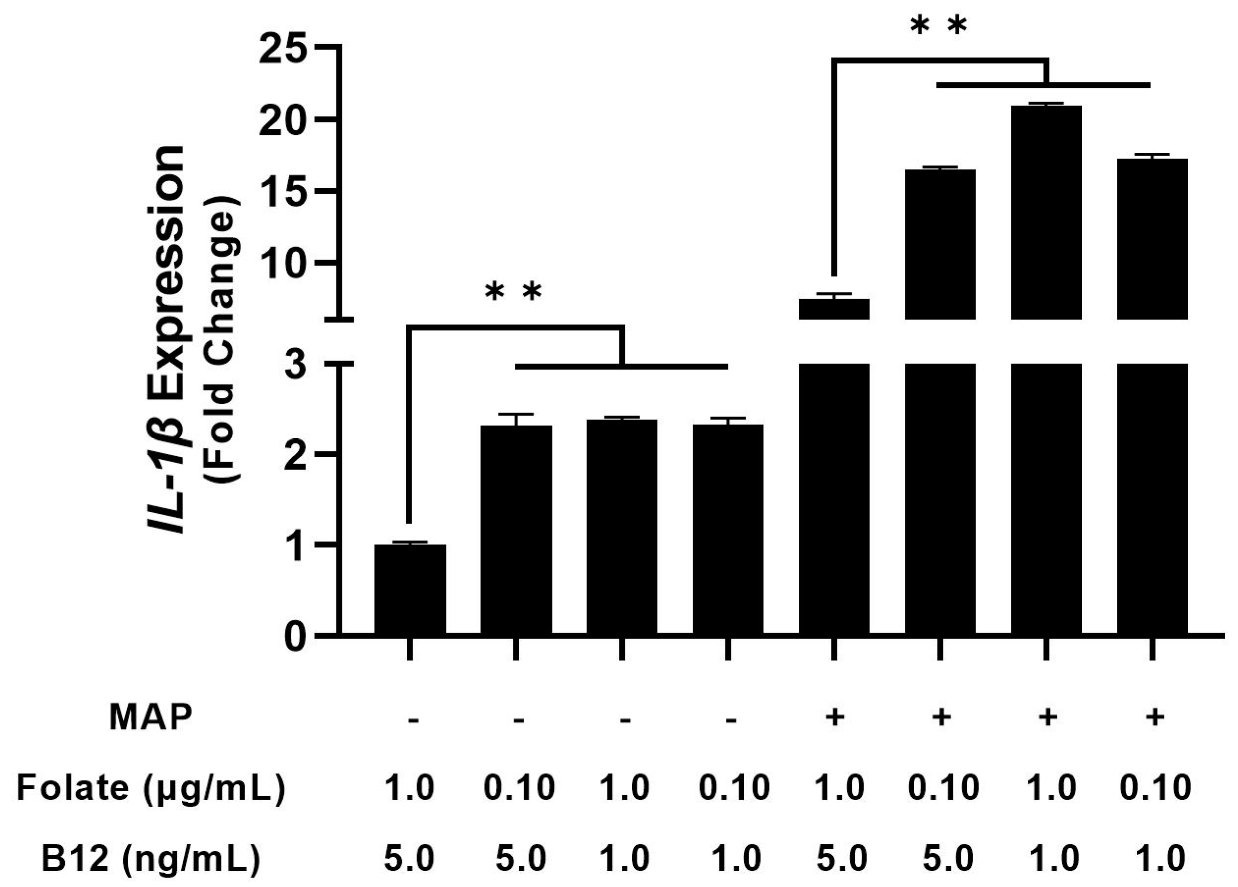
4.2. THP-1 Macrophages Cultured in Folate and B12 Supplemented and Deficient Media Show Altered Cytokine Expression

Next, we considered how differing vitamin levels in cell culture media alter macrophage cytokine expression. To examine this phenomenon, we took folate and B12-free RPMI 1640 and manually supplemented it with folate and B12 to yield folate-low media, folate-high media, B12-low media, B12-high media, folate/B12-low media, folate/B12-high media, and ordinary RPMI 1640. After 48 h of PMA stimulation and differentiation in the depleted media, we infected them with clinical MAP for 24 h and examined relative cytokine mRNA. folate, B12, and folate/B12 deficiency increased TNF-α expression in uninfected macrophages by 1.63 ± 0.08-fold, 1.91 ± 0.17-fold, and 1.79 ± 0.19-fold, respectively; however, these effects were not statistically significant [Figure 2]. In infected macrophages, B12-deficiency increased TNF-α expression from 20.37 ± 0.54-fold to 25.52 ± 1.17-fold [Figure 2]. However, no other vitamin deficiency, even concurrent folate/B12, significantly altered TNF-α expression. By contrast, folate, B12, and folate/B12 deficiency all had inhibitory effects on IL-1β expression in uninfected (2.31 ± 0.13, 2.38 ± 0.03, and 2.32 ± 0.07-fold, respectively) and infected macrophages (16.49 ± 0.16, 20.93 ± 0.17, and 17.26 ± 0.30-fold, respectively, compared with untreated 7.41 ± 0.38) [Figure 3].
We followed these experiments with another analysis to examine supplementation with folate and B12. Tenfold elevation of B12 in culture medium for infected macrophages reduced TNF-α expression from 12.49 ± 0.33 to 10.66 ± 0.39-fold, while folate had no significant effect (13.14 ± 0.50) [Figure 4]. Folate and B12 supplementation alone had no significant effect on TNF-α expression in uninfected macrophages (with a 0.89 ± 0.09 and a 1.06 ± 0.07-fold change, respectively) [Figure 4]. Interestingly, concurrent folate/B12 supplementation increased TNF-α expression by 1.34 ± 0.04-fold in uninfected macrophages but reduced it from 12.49 ± 0.33 to 11.31 ± 0.10 in infected macrophages [Figure 5]. Elevated folate reduced IL-1β expression from 1.00 ± 0.01 to 0.55 ± 0.10-fold in uninfected macrophages and from 10.75 ± 0.25 to 8.19 ± 0.11-fold in infected macrophages [Figure 5] Supplementation with B12 reduced IL-1β expression from 1.00 ± 0.04 to 0.80 ± 0.01-fold in uninfected macrophages and from 10.75 ± 0.25 to 9.39 ± 0.09-fold in infected macrophages [Figure 5]. Concurrent folate/B12 supplementation’s effect is more uniform in IL-1β than TNF-α, which shows a reduction from 1.00 ± 0.04 to 0.69 ± 0.02-fold in uninfected macrophages and from 10.75 ± 0.25 to 7.00 ± 0.10-fold in infected macrophages [Figure 5]. These results indicate that folate and B12 supplementation have an anti-inflammatory effect on macrophage cytokine expression during MAP infection, with results varying by specific cytokine. By contrast, folate and B12 deprivation enhance the inflammatory response in identically treated cells.

4.3. MAP Infection Increases Apoptosis during Folate and B12 Supplementation and Decreases Apoptosis during Depletion

Apoptosis of infected cells is a critical immune mechanism for countering intracellular pathogens, and MAP consequently uses a suite of anti-apoptotic proteins to impede it [44]. Accordingly, we hypothesized that infected macrophages supplemented with folate and B12 would increase apoptosis to control the infection. We used two methods to assess the apoptosis of MAP-infected macrophages. First, we directly quantified apoptosis using annexin V luminescence. MAP infection increased apoptosis by 1.83 ± 0.40 fold compared to uninfected controls, as anticipated. Furthermore, folate-enriched media increased macrophage apoptosis during infection by 3.38 ± 0.07-fold, a significant increase compared to MAP infection in control media [Figure 6A]. An increase in apoptosis to 2.58 ± 0.14-fold was also observed in concurrent folate and B12 supplemented media [Figure 6A]. We did not observe a significant change in media supplemented with B12 alone. Depletion of folate, B12, and both folate and B12 had the inverse effect: annexin V luminescence decreased to 1.04 ± 0.08, 0.64 ± 0.12, and 0.45 ± 0.07-fold, respectively [Figure 6A].
We followed this experiment with an ATP-based viability assay. General macrophage viability after MAP infection declined to 0.80 ± 0.05 and 0.82 ± 0.02-fold in folate and concurrent folate/B12 supplemented media, corresponding with the observed increases in annexin V luminescence [Figure 6B]. Interestingly, there was no significant change in macrophage viability in the MAP-infected cells compared with the uninfected cells. Similarly, decreased annexin V in folate, B12, and folate/B12 deficient media did not correspond with a change in overall viability in these groups [Figure 6B]. However, there was a small but statistically significant decrease in overall macrophage viability in the folate/B12-low group, from 1.00 ± 0.05-fold to 0.91 ± 0.04-fold.

4.4. Folate and Vitamin B12 Deficiency Individually Exacerbate Cytotoxicity in Caco-2 Monolayers

After assessing the effects of folate and vitamin deficiency on macrophage inflammation, we considered the impact of altered macrophage inflammation on enterocytes. We hypothesized that changes in macrophage survival and pro-inflammatory cytokine expression would have a detrimental impact on co-cultured Caco-2 cells. Accordingly, we differentiated Caco-2 cells into monolayers and maintained them in culture for 7 days in RPMI 1640 with altered vitamin levels. administered supernatant collected from macrophages cultured and infected in media with comparably altered vitamin levels. 24 h post-treatment, we examined LDH release and NADP+/Total NADP in the monolayers.
Treating Caco-2 cells with supernatant from infected macrophages increased LDH luminescence 3.16 ± 0.95-fold compared with uninfected controls, though this effect did not reach statistical significance [Figure 7]. By contrast, administration of infected macrophage supernatant in vitamin B12-low conditions increased LDH luminescence 7.73 ± 1.01-fold, which was significant compared to both infected and uninfected cells in control media [Figure 7]. Similarly, folate-low media increased luminescence by 5.87 ± 1.78-fold compared with the control [Figure 7]. Interestingly, concurrent folate and vitamin B12 deprivation did not significantly alter LDH luminescence in culture. Treating Caco-2 cells with supernatant from infected macrophages did not significantly alter the percentage of NADPH compared with the control in any group, suggesting that oxidative stress did not mediate the damage observed in the LDH assay [Table 2].

5. Discussion

CD patients are at an elevated risk of malabsorptive folate and vitamin B12 deficiency [21,32]. Furthermore, attempts to avoid foods that patients believe to be triggers for CD relapse can lead to self-imposed dietary restriction and reduced nutrient intake [45]. Accordingly, periodic screenings for these vitamins are indicated for CD patients, particularly those who received ileal resection [40]. However, there is a dearth of information on the effects of folate and vitamin B12 supplementation on CD symptoms and inflammation. Furthermore, the state of the literature is murky on the effects of folate and B12 supplementation on common CD pathogens like MAP. Physicians with CD patients are left without guidance about the risks of prolonged folate and B12 deficiency in this patient subset and a potential tool to mitigate inflammation.
Gastrointestinal health and resilience against infection have been correlated with folate and vitamin B12 intake. Rat models of methyl donor deficiency indicate that methyl donor-low diets impede bowel development and barrier function while aggravating induced colitis [18,46]. By contrast, folate-producing lactic acid bacteria have an anti-inflammatory effect on induced murine mucositis [47]. Additionally, murine diets supplemented with folate and other methyl-donating nutrients improved antimicrobial gene expression and resilience against adherent invasive E. coli in a mouse model of CD [36]. These findings concur with a study establishing that neonatal folate deprivation sensitizes adult guinea pigs to Shigella infection [48]. In humans, folate-associated metabolic pathways are perturbed in pediatric CD patients, and a review of meta-analyses for CD environmental risk factors indicated that high folate levels were protective [49,50]. Furthermore, low folate levels in CD were associated with increased CD activity in a Swiss cohort of patients [26].
The results of our investigation add to these findings by showing that folate and B12 alter inflammation during infection with a common CD pathogen. MAP-positive CD patients have significantly lower plasma folate and B12 than MAP-negative CD patients. We further show that pro-inflammatory cytokines IL-1β and TNF-α are significantly upregulated during folate and vitamin B12 deprivation in after MAP infection, while supplementation significantly reduces their expression. Macrophages in folate and B12-deficient media are less likely to undergo apoptosis during MAP infection, suggesting this key mechanism to counteract intracellular pathogens is inhibited during vitamin deficiency. Folate supplementation, by contrast, increased apoptosis of infected macrophages. Finally, the treatment of enterocytes with folate-low or B12-low supernatant from infected macrophages leads to increased LDH release without significant alterations in oxidative stress.
Our findings on folate and B12 in CD patient plasma cannot at this time be compared with normal clinical serum ranges for these vitamins, as the ELISAs used to quantify folate and vitamin B12 availability are not approved for clinical practice. However, these results indicate that even within a patient group at risk for folate and B12 deficiency, distinct subsets of folate and B12 availability can be characterized based on MAP infection. Furthermore, the data on cytokine availability indicate that folate and B12 levels affect macrophage-mediated inflammation after only 48 of h prophylactic exposure. Since monocyte-derived macrophages constitutively infiltrate the gut, these results suggest that clinical trials of folate and B12 supplementation may gradually impact inflammation patterns as older, vitamin-restricted cells are replaced [51]. Importantly, our work builds on the findings of Samblas and colleagues by showing that the anti-inflammatory effects of folate and B12 can be observed during an ongoing bacterial infection, not just after stimulation with a TLR agonist like LPS [30]. Since these findings are paired with improved apoptosis of infected macrophages, we propose that MAP-infected CD patients taking folate and B12 supplements may experience reduced inflammation upon the initial macrophage encounter with MAP. Altered inflammation may be followed by apoptosis of the pro-inflammatory, MAP-infected cells, resulting in the removal of both pathogen and inflammatory mediator.
Future studies are required to explore not merely how folate and B12 alter MAP infection in cell culture, but in animal and clinical studies. Our study centered on monocyte derived macrophages because prior studies have highlighted macrophages as a crucial innate immune mediator for both MAP destruction and proliferation [52]. However, we do not analyze other phagocytic immune cells or determine if the 48 h differentiation period is the only relevant window to mediate folate and B12’s anti-inflammatory effects. Furthermore, macrophages frequently interact with components of the adaptive immune system, thereby alter adaptive immunity. We do not explore how folate and B12 might affect this function to contribute to long-term inflammatory CD patterns. Accordingly, further investigation is warranted.

Author Contributions

Conceptualization, J.A.V. and S.A.N.; formal analysis, J.A.V.; funding acquisition, S.A.N.; methodology, J.A.V., A.Q. and S.A.N.; supervision, A.Q. and S.A.N.; writing—original draft, J.A.V.; writing—review and editing, A.Q. and S.A.N. All authors have read and agreed to the published version of the manuscript.

Funding

This study was supported in part by the Florida legislative grant.

Institutional Review Board Statement

Experimentation regarding human subjects were reviewed and approved by University of Central Florida Institutional Review Board # STUDY00003468.

Informed Consent Statement

Written informed consent has been obtained from study participants for research and publication purposes.

Data Availability Statement

The original contributions presented in this study are included in the article. Further inquiries may be directed to the corresponding author.

Acknowledgments

We would like to thank our colleagues in Naser’s laboratory for their ongoing support. Article processing charges were provided in part by the UCF College of Graduate Studies Open Access Publishing Fund.

Conflicts of Interest

The authors declare that the research was conducted in the absence of any commercial or financial relationships that could be construed as a potential conflict of interest.

References

  1. Ebara, S. Nutritional role of folate. Congenit. Anom. 2017, 57, 138–141. [Google Scholar] [CrossRef] [PubMed]
  2. Allen, L.H.; Miller, J.W.; de Groot, L.; Rosenberg, I.H.; Smith, A.D.; Refsum, H.; Raiten, D.J. Biomarkers of Nutrition for Development (BOND): Vitamin B-12 Review. J. Nutr. 2018, 148, 1995s–2027s. [Google Scholar] [CrossRef] [PubMed] [Green Version]
  3. Mosley, B.S.; Cleves, M.A.; Siega-Riz, A.M.; Shaw, G.M.; Canfield, M.A.; Waller, D.K.; Werler, M.M.; Hobbs, C.A.; for the National Birth Defects Prevention Study. Neural tube defects and maternal folate intake among pregnancies conceived after folic acid fortification in the United States. Am. J. Epidemiol. 2009, 169, 9–17. [Google Scholar] [CrossRef] [Green Version]
  4. Konings, E.J.M.; Roomans, H.H.S.; Dorant, E.; Goldbohm, R.A.; Saris, W.H.M.; van den Brandt, P.A. Folate intake of the Dutch population according to newly established liquid chromatography data for foods. Am. J. Clin. Nutr. 2001, 73, 765–776. [Google Scholar] [CrossRef] [Green Version]
  5. Rossi, M.; Amaretti, A.; Raimondi, S. Folate production by probiotic bacteria. Nutrients 2011, 3, 118–134. [Google Scholar] [CrossRef] [PubMed] [Green Version]
  6. Banerjee, D.K.; Chatterjea, J.B. Serum vitamin B12 in vegetarians. Br. Med. J. 1960, 2, 992–994. [Google Scholar] [CrossRef] [PubMed] [Green Version]
  7. Herrmann, W.; Schorr, H.; Obeid, R.; Geisel, J. Vitamin B-12 status, particularly holotranscobalamin II and methylmalonic acid concentrations, and hyperhomocysteinemia in vegetarians. Am. J. Clin. Nutr. 2003, 78, 131–136. [Google Scholar] [CrossRef] [Green Version]
  8. Wilson, R.S.; Mertes, M.P. Chemical model for thymidylate synthetase catalysis. J. Am. Chem. Soc. 1972, 94, 7182–7183. [Google Scholar] [CrossRef]
  9. Himes, R.H.; Rabinowitz, J.C. Formyltetrahydrofolate synthetase. II. Characteristics of the enzyme and the enzymic reaction. J. Biol. Chem. 1962, 237, 2903–2914. [Google Scholar] [CrossRef]
  10. Wasserman, G.F.; Mueller, W.T.; Benkovic, S.J.; Liao, W.S.L.; Taylor, J. Evidence that the folate-requiring enzymes of de novo purine biosynthesis are encoded by individual mRNAs. Biochemistry 1984, 23, 6704–6710. [Google Scholar] [CrossRef]
  11. Mahgoub, A.E.; Deliwala, S.S.; Bachuwa, G. Important reminder: Nitrous oxide (N2O) as a cause of functional vitamin B12 deficiency. BMJ Case Rep. 2021, 14, e241414. [Google Scholar] [CrossRef] [PubMed]
  12. Moreira, E.S.; Brasch, N.E.; Yun, J. Vitamin B12 protects against superoxide-induced cell injury in human aortic endothelial cells. Free Radic. Biol. Med. 2011, 51, 876–883. [Google Scholar] [CrossRef] [PubMed] [Green Version]
  13. Shepherd, G.; Velez, L.I. Role of Hydroxocobalamin in Acute Cyanide Poisoning. Ann. Pharmacother. 2008, 42, 661–669. [Google Scholar] [CrossRef] [PubMed]
  14. Wheatley, C. A scarlet pimpernel for the resolution of inflammation? The role of supra-therapeutic doses of cobalamin, in the treatment of systemic inflammatory response syndrome (SIRS), sepsis, severe sepsis, and septic or traumatic shock. Med. Hypotheses 2006, 67, 124–142. [Google Scholar] [CrossRef] [PubMed]
  15. Krebs, H.A.; Hems, R.; Tyler, B. The regulation of folate and methionine metabolism. Biochem. J. 1976, 158, 341–353. [Google Scholar] [CrossRef] [PubMed]
  16. Shou, L.; Pan, F.; Chin, S.-f. Pancreatic Hormones and Hepatic Methionine Adenosyltransferase in the Rat. Proc. Soc. Exp. Biol. Med. 1969, 131, 1012–1018. [Google Scholar] [CrossRef] [PubMed]
  17. Soda, K. Polyamine Metabolism and Gene Methylation in Conjunction with One-Carbon Metabolism. Int. J. Mol. Sci. 2018, 19, 3106. [Google Scholar] [CrossRef] [Green Version]
  18. Bressenot, A.; Pooya, S.; Bossenmeyer-Pourie, C.; Gauchotte, G.; Germain, A.; Chevaux, J.-B.; Coste, F.; Vignaud, J.-M.; Guéant, J.-L.; Peyrin-Biroulet, L. Methyl donor deficiency affects small-intestinal differentiation and barrier function in rats. Br. J. Nutr. 2013, 109, 667–677. [Google Scholar] [CrossRef] [Green Version]
  19. Kunisawa, J.; Hashimoto, E.; Ishikawa, I.; Kiyono, H. A pivotal role of vitamin B9 in the maintenance of regulatory T cells in vitro and in vivo. PLoS ONE 2012, 7, e32094. [Google Scholar] [CrossRef] [Green Version]
  20. Lee, Y.-J.; Wang, M.-Y.; Lin, M.-C.; Lin, P.-T. Associations between Vitamin B-12 Status and Oxidative Stress and Inflammation in Diabetic Vegetarians and Omnivores. Nutrients 2016, 8, 118. [Google Scholar] [CrossRef]
  21. Ao, M.; Tsuji, H.; Shide, K.; Kosaka, Y.; Noda, A.; Inagaki, N.; Nakase, H.; Tanaka, K. High prevalence of vitamin B-12 insufficiency in patients with Crohn’s disease. Asia Pac. J. Clin. Nutr. 2017, 26, 1076–1081. [Google Scholar] [CrossRef] [PubMed]
  22. Zheng, S.; Yang, W.; Wu, C.; Sun, L.; Lin, D.; Lin, X.; Jiang, L.; Ding, R.; Jiang, Y. Association of ulcerative colitis with transcobalamin II gene polymorphisms and serum homocysteine, vitamin B12, and folate levels in Chinese patients. Immunogenetics 2017, 69, 421–428. [Google Scholar] [CrossRef] [PubMed]
  23. Harb, Z.; Deckert, V.; Bressenot, A.M.; Christov, C.; Guéant-Rodriguez, R.-M.; Raso, J.; Alberto, J.M.; de Barros, J.-P.P.; Umoret, R.; Peyrin-Biroulet, L.; et al. The deficit in folate and vitamin B12 triggers liver macrovesicular steatosis and inflammation in rats with dextran sodium sulfate-induced colitis. J. Nutr. Biochem. 2020, 84, 108415. [Google Scholar] [CrossRef] [PubMed]
  24. Holland, N.; Harmatz, P.; Golden, D.; Hubbard, A.; Wu, Y.-Y.; Bae, J.; Chen, C.; Huen, K.; Heyman, M.B. Cytogenetic Damage in Blood Lymphocytes and Exfoliated Epithelial Cells of Children with Inflammatory Bowel Disease. Pediatr. Res. 2007, 61, 209–214. [Google Scholar] [CrossRef] [PubMed]
  25. Kolb, A.F.; Petrie, L. Folate deficiency enhances the inflammatory response of macrophages. Mol. Immunol. 2013, 54, 164–172. [Google Scholar] [CrossRef]
  26. Madanchi, M.; Fagagnini, S.; Fournier, N.; Biedermann, L.; Zeitz, J.; Battegay, E.; Zimmerli, L.; Vavricka, S.R.; Rogler, G.; Scharl, M.; et al. The Relevance of Vitamin and Iron Deficiency in Patients with Inflammatory Bowel Diseases in Patients of the Swiss IBD Cohort. Inflamm. Bowel Dis. 2018, 24, 1768–1779. [Google Scholar] [CrossRef] [Green Version]
  27. Scalabrino, G. Vitamin-regulated cytokines and growth factors in the CNS and elsewhere. J. Neurochem. 2009, 111, 1309–1326. [Google Scholar] [CrossRef]
  28. Arumugam, M.K.; Paal, M.C.; Donohue, T.M., Jr.; Ganesan, M.; Osna, N.A.; Kharbanda, K.K. Beneficial Effects of Betaine: A Comprehensive Review. Biology 2021, 10, 456. [Google Scholar] [CrossRef]
  29. Leng, Y.-P.; Ma, Y.-S.; Li, X.-G.; Chen, R.-F.; Zeng, P.-Y.; Li, X.-H.; Qiu, C.-F.; Li, Y.-P.; Zhang, Z.; Chen, A.F. L-Homocysteine-induced cathepsin V mediates the vascular endothelial inflammation in hyperhomocysteinaemia. Br. J. Pharmacol. 2018, 175, 1157–1172. [Google Scholar] [CrossRef] [Green Version]
  30. Samblas, M.; Martínez, J.A.; Milagro, F. Folic Acid Improves the Inflammatory Response in LPS-Activated THP-1 Macrophages. Mediat. Inflamm. 2018, 2018, 1312626. [Google Scholar] [CrossRef]
  31. Ehmedah, A.; Nedeljkovic, P.; Dacic, S.; Repac, J.; Draskovic-Pavlovic, B.; Vučević, D.; Pekovic, S.; Nedeljkovic, B.B. Effect of Vitamin B Complex Treatment on Macrophages to Schwann Cells Association during Neuroinflammation after Peripheral Nerve Injury. Molecules 2020, 25, 5426. [Google Scholar] [CrossRef] [PubMed]
  32. Hoffbrand, A.V.; Stewart, J.S.; Booth, C.C.; Mollin, D.L. Folate deficiency in Crohn’s disease: Incidence, pathogenesis, and treatment. Br. Med. J. 1968, 2, 71–75. [Google Scholar] [CrossRef] [PubMed] [Green Version]
  33. Torres, J.; Mehandru, S.; Colombel, J.-F.; Peyrin-Biroulet, L. Crohn’s disease. Lancet 2017, 389, 1741–1755. [Google Scholar] [CrossRef]
  34. Bermejo, F.; Algaba, A.; Guerra, I.; Chaparro, M.; De-La-Poza, G.; Valer, P.; Piqueras, B.; Bermejo, A.; García-Alonso, J.; Pérez, M.-J.; et al. Should we monitor vitamin B12 and folate levels in Crohn’s disease patients? Scand. J. Gastroenterol. 2013, 48, 1272–1277. [Google Scholar] [CrossRef] [PubMed]
  35. Weisshof, R.; Chermesh, I. Micronutrient deficiencies in inflammatory bowel disease. Curr. Opin. Clin. Nutr. Metab. Care 2015, 18, 576–581. [Google Scholar] [CrossRef] [PubMed]
  36. Gimier, E.; Chervy, M.; Agus, A.; Sivignon, A.; Billard, E.; Privat, M.; Viala, S.; Minet-Quinard, R.; Buisson, A.; Vazeille, E.; et al. Methyl-donor supplementation prevents intestinal colonization by Adherent-Invasive E. coli in a mouse model of Crohn’s disease. Sci. Rep. 2020, 10, 12922. [Google Scholar] [CrossRef] [PubMed]
  37. Rathnaiah, G.; Zinniel, D.K.; Bannantine, J.P.; Stabel, J.R.; Gröhn, Y.T.; Collins, M.T.; Barletta, R.G. Pathogenesis, Molecular Genetics, and Genomics of Mycobacterium avium subsp. paratuberculosis, the Etiologic Agent of Johne’s Disease. Front. Vet. Sci. 2017, 4, 187. [Google Scholar] [CrossRef] [Green Version]
  38. Naser, S.A.; Ghobrial, G.; Romero, C.; Valentine, J.F. Culture of Mycobacterium avium subspecies paratuberculosis from the blood of patients with Crohn’s disease. Lancet 2004, 364, 1039–1044. [Google Scholar] [CrossRef]
  39. Naser, S.A.; Sagramsingh, S.R.; Naser, A.S.; Thanigachalam, S. Mycobacterium avium subspecies paratuberculosis causes Crohn’s disease in some inflammatory bowel disease patients. World. J. Gastroenterol. 2014, 20, 7403–7415. [Google Scholar] [CrossRef]
  40. Veauthier, B.; Hornecker, J.R. Crohn’s Disease: Diagnosis and Management. Am. Fam. Physician 2018, 98, 661–669. [Google Scholar]
  41. Qasem, A.; Naser, A.E.; Naser, S.A. The alternate effects of anti-TNFα therapeutics and their role in mycobacterial granulomatous infection in Crohn’s disease. Expert Rev. Anti-Infect. Ther. 2017, 15, 637–643. [Google Scholar] [CrossRef] [PubMed]
  42. Qasem, A.; Naser, S.A. TNFα inhibitors exacerbate Mycobacterium paratuberculosis infection in tissue culture: A rationale for poor response of patients with Crohn’s disease to current approved therapy. BMJ Open Gastroenterol. 2018, 5, e000216. [Google Scholar] [CrossRef] [PubMed] [Green Version]
  43. Qasem, A.; Ramesh, S.; Naser, S.A. Genetic polymorphisms in tumour necrosis factor receptors (TNFRSF1A/1B) illustrate differential treatment response to TNFα inhibitors in patients with Crohn’s disease. BMJ Open Gastroenterol. 2019, 6, e000246. [Google Scholar] [CrossRef] [PubMed]
  44. Abendaño, N.; Juste, R.A.; Alonso-Hearn, M. Anti-inflammatory and antiapoptotic responses to infection: A common denominator of human and bovine macrophages infected with Mycobacterium avium subsp. paratuberculosis. BioMed. Res. Int. 2013, 2013, 908348. [Google Scholar] [CrossRef] [Green Version]
  45. Cox, S.R.; Clarke, H.; O’Keeffe, M.; Dubois, P.; Irving, P.M.; Lindsay, J.O.; Whelan, K. Nutrient, Fibre, and FODMAP Intakes and Food-related Quality of Life in Patients with Inflammatory Bowel Disease, and Their Relationship with Gastrointestinal Symptoms of Differing Aetiologies. J. Crohn’s Colitis 2021, 15, 2041–2053. [Google Scholar] [CrossRef]
  46. Chen, M.; Peyrin-Biroulet, L.; George, A.; Coste, F.; Bressenot, A.; Bossenmeyer-Pourie, C.; Alberto, J.-M.; Xia, B.; Namour, B.; Guéant, J.-L. Methyl deficient diet aggravates experimental colitis in rats. J. Cell. Mol. Med. 2011, 15, 2486–2497. [Google Scholar] [CrossRef] [Green Version]
  47. Levit, R.; de Giori, G.S.; de Moreno de LeBlanc, A.; LeBlanc, J.G. Folate-producing lactic acid bacteria reduce inflammation in mice with induced intestinal mucositis. J. Appl. Microbiol. 2018, 125, 1494–1501. [Google Scholar] [CrossRef]
  48. Nelson, J.D.; Haltalin, K.C. Effect of neonatal folic acid deprivation on later growth and susceptibility to Shigella infection in the guinea pig. Am. J. Clin. Nutr. 1972, 25, 992–996. [Google Scholar] [CrossRef]
  49. Kolho, K.-L.; Pessia, A.; Jaakkola, T.; de Vos, W.M.; Velagapudi, V. Faecal and Serum Metabolomics in Paediatric Inflammatory Bowel Disease. J. Crohn’s Colitis 2017, 11, 321–334. [Google Scholar] [CrossRef] [Green Version]
  50. Piovani, D.; Danese, S.; Peyrin-Biroulet, L.; Nikolopoulos, G.K.; Lytras, T.; Bonovas, S. Environmental Risk Factors for Inflammatory Bowel Diseases: An Umbrella Review of Meta-analyses. Gastroenterology 2019, 157, 647–659. [Google Scholar] [CrossRef] [Green Version]
  51. Bain, C.C.; Mowat, A.M. Macrophages in intestinal homeostasis and inflammation. Immunol. Rev. 2014, 260, 102–117. [Google Scholar] [CrossRef] [PubMed]
  52. Arsenault, R.J.; Maattanen, P.; Daigle, J.; Potter, A.; Griebel, P.; Napper, S. From mouth to macrophage: Mechanisms of innate immune subversion by Mycobacterium avium subsp. paratuberculosis. Vet. Res. 2014, 45, 54. [Google Scholar] [CrossRef] [PubMed]
Figure 1. Levels of plasma folate (A) and B12 (B) in MAP-negative and MAP-positive CD patients, N = 35 for both groups. All measurements were performed in duplicates. ** Indicates p-value of less than 0.01.
Nutrients 15 00261 g001
Figure 2. Effect of folate and B12 deficiency on TNF-α expression in MAP-infected and uninfected macrophages. * Indicates p-value of less than 0.05.
Nutrients 15 00261 g002
Figure 3. Effect of folate and B12 deficiency on IL-1β expression in MAP-infected and uninfected macrophages. ** Indicates p-value of less than 0.01.
Nutrients 15 00261 g003
Figure 4. Effect of folate and B12 supplementation on TNF-α expression in MAP-infected and uninfected macrophages. * Indicates p-value of less than 0.05. ** Indicates p-value of less than 0.01.
Nutrients 15 00261 g004
Figure 5. Effect of folate and B12 supplementation on IL-1β expression in MAP-infected and uninfected macrophages. * Indicates p-value of less than 0.05. ** Indicates a p-value of less than 0.01.
Nutrients 15 00261 g005
Figure 6. Effect of folate and B12 supplementation or depletion on macrophage apoptosis (A) and general viability (B) during MAP infection. * Indicates p-value of less than 0.05. ** Indicates p-value of less than 0.01.
Nutrients 15 00261 g006
Figure 7. Effect of folate and vitamin B12 concentration on LDH release from Caco-2 monolayers after treatment with supernatant from infected macrophages. * Indicates p-value less than 0.05. ** Indicates p-value less than 0.01.
Nutrients 15 00261 g007
Table 1. Concentrations of folate and B12 in modified RPMI 1640 media for macrophage cell culture and infection.
Culture ConditionFolate (μg/mL)B12 (ng/mL)
Control RPMI 16401.05.0
Folate-High10.05.0
B12-High1.050.0
Folate + B12-High10.050.0
Folate-Low0.105.0
B12-Low1.01.0
Folate + B12-Low0.101.0
Table 2. Effect of folate and vitamin B12 concentration on NADP release from Caco-2 monolayers after treatment with supernatant from infected macrophages.
Infection and TreatmentNADP+/(NADPH + NADP+) × 100 ± SD
Control (no infection)6.23 ± 0.396
MAP infection (1 × 10⁷ CFU/mL)6.89 ± 0.761
Folate-low + MAP infection7.74 ± 0.750
B12-low + MAP infection8.24 ± 0.657
Folate/B12-low + MAP infection7.07 ± 0.801
Folate-high + MAP infection7.63 ± 1.04
B12-high + MAP infection6.59 ± 1.00
Folate/B12-high + MAP6.80 ± 0.716
Disclaimer/Publisher’s Note: The statements, opinions and data contained in all publications are solely those of the individual author(s) and contributor(s) and not of MDPI and/or the editor(s). MDPI and/or the editor(s) disclaim responsibility for any injury to people or property resulting from any ideas, methods, instructions or products referred to in the content.

