Article

Preparation of Corn Peptides with Anti-Adhesive Activity and Its Functionality to Alleviate Gastric Injury Induced by Helicobacter pylori Infection In Vivo

1
Heilongjiang Provincial Key Laboratory of Corn Deep Processing Theory and Technology, College of Food and Bioengineering, Qiqihar University, Qiqihar 161006, China
2
College of Food Science, Heilongjiang Bayi Agricultural University, Daqing 163319, China
*
Author to whom correspondence should be addressed.
Nutrients 2023, 15(15), 3467; https://doi.org/10.3390/nu15153467
Submission received: 5 July 2023 / Revised: 28 July 2023 / Accepted: 3 August 2023 / Published: 5 August 2023
(This article belongs to the Special Issue Nutrition Intervention on Digestive Diseases)

Abstract

:
More than 50% of the world population is infected with Helicobacter pylori (H. pylori), which is classified as group I carcinogen by the WHO. H. pylori surface adhesins specifically recognize gastric mucosal epithelial cells’ (GES-1 cells) receptor to complete the adhesion. Blocking the adhesion with an anti-adhesion compound is an effective way to prevent H. pylori infection. The present study found that corn protein hydrolysate, hydrolyzed by Neutral, effectively alleviated gastric injury induced by H. pylori infection through anti-adhesive and anti-inflammatory effects in vitro and in vivo. The hydrolysate inhibited H. pylori adhesion to GES-1 cells significantly, and its anti-adhesive activity was 50.44 ± 0.27% at 4 mg/mL, which indicated that the hydrolysate possessed a similar structure to the GES-1 cells’ receptor, and exhibited anti-adhesive activity in binding to H. pylori. In vivo, compared with the H. pylori infection model group, the medium and high dose of the hydrolysate (400–600 mg/kg·bw) significantly decreased (p < 0.05) the amount of H. pylori colonization, pro-inflammatory cytokines (IL-6, IL-1β, TNF-α and MPO), chemokines (KC and MCP-1) as well as key metabolites of NF-κB signaling pathway levels (TLR4, MyD88 and NF-κB), and it increased antioxidant enzyme contents (SOD and GSH-Px) and the mitigation of H. pylori-induced pathological changes in the gastric mucosa. Taken together, these results indicated that the hydrolysate intervention can prevent H. pylori-induced gastric injury by anti-adhesive activity and inhibiting the NF-κB signaling pathway’s induction of inflammation. Hence, the corn protein hydrolysate might act as a potential anti-adhesive agent to prevent H. pylori infection.

1. Introduction

Helicobacter pylori (H. pylori) is able to colonize the human gastric mucosa, which is a Gram-negative, spiral-shaped, and microaerophilic bacterium. It is reported that more than 50% of the world population has been infected with H. pylori, and the current infection rate in China is 60–70%, which has been classified as a group I carcinogen by the World Health Organization (WHO) [1]. H. pylori specifically adheres to gastric mucosal epithelial cells (GES-1 cells); it is recognized as one of the important causative agents of peptic ulcers, chronic gastritis and other gastric disorders; and it is also closely associated with the development of gastric cancer. H. pylori infection causes oxidative stress and neutrophil infiltration in gastric tissue, which promotes levels of pro-inflammatory cytokines and chemokines. In addition, overexpression of pro-inflammatory cytokines activates the NF-κB signaling pathway, which is one of the major pathways inducing gastric cell inflammation [2]. To date, antibiotics are the most widely used agents for the treatment of H. pylori infection, with the eradication rates ranging from 60–90% [3]. Although antibiotics have good effects, they still possess some side effects and resistance to H. pylori; extensive research has been conducted to develop alternative approaches to prevent and treat H. pylori [4], and anti-adhesion of H. pylori is an effective strategy.
Adhesion proteins, outer membrane proteins (OMPs) of the H. pylori, could specifically recognize receptors on the GES-1 cells’ surface to achieve colonization of H. pylori [5]. Many adhesins are expressed by H. pylori, of which sialic acid-binding adhesin (SabA) and Lewis-b blood group antigen-binding adhesin (BabA) are dominant adhesins. SabA and BabA specifically recognize sialyl-Lewis-a and Lewis-b, respectively [6]. Anti-adhesion therapy, a promising strategy, could effectively prevent infection by inhibiting H. pylori adhesion to GES-1 cells [7]. Specifically, the anti-adhesion component acts as GES-1 cell receptor analogs or H. pylori surface adhesin analogs to block the adhesion between the H. pylori and the GES-1 cells [8]. Recent studies found that a variety of natural products possessed anti-adhesive effects to prevent H. pylori infection in vitro, such as pea peptides [9], pectin from apples [10], polyphenols from cranberries [11], 3′-sialyllactose [12] and wheat germ-derived peptides [8]. Despite the above natural products showing anti-adhesive activities in vitro, their anti-adhesion effects were not evaluated in vivo.
Corn gluten meal (CGM) is a co-product of the wet milling process; its total protein content is 60–70% (w/w), including zein, glutenin, globulin and albumin [13]. Due to CGM’s unique amino acid composition and partial amino acid sequence, it is also a quality protein source for preparing bioactive peptides [14]. By enzymatic hydrolysis, the solubility of corn protein is elevated significantly, and the functional sequences may be released. The reported major bioactive peptides from CGM included antioxidant activity [15], antagonism alcohol-induced liver injury [16] and angiotensin converting enzyme inhibitory activity [17]. In the literature available, no reports could be found about corn protein-derived peptides with anti-adhesive activity preventing H. pylori infection. The aim of this study is to effectively prepare corn protein-derived peptides with anti-adhesive activity, evaluate their functionality to alleviate gastric injury induced by H. pylori infection in vivo and investigate the possible underlying mechanisms.

2. Materials and Methods

2.1. Materials and Chemicals

Corn gluten meal (CGM) was purchased from FuFeng Development Co., Ltd. with a total protein content of 60.44% (w/w) (Qiqihar, Heilongjiang, China). Neutral (21,000 U/mL), Alcalase (23,000 U/g), Protamex (38,000 U/g), Trypsin (21,000 U/g) and Papain (11,000 U/g) were obtained from Novo Nordisk (Bagsvaerd, Denmark). Protease P (48,000 U/g), Protease N (48,000 U/g) and Protease M (61,000 U/g) were purchased from Amano Pharmaceutical Co., Ltd. (Nagoya, Japan). Dimethyl sulfoxide (DMSO) and Fluorescein isothiocyanate (FITC) were bought from Solarbio Life Sciences (Beijing, China). H. pylori (43,504) was bought from ATCC. GES-1 cells were purchased from JianCheng Co., Ltd. (Nanjing, Jiangsu, China). Fetal bovine serum (FBS) and Dulbecco’s modified eagle medium (DMEM) were bought from GE Healthcare (Pittsburgh, PA, USA).

2.2. Starch Removal of CGM

The starch was removed from the CGM as previously described [14]. Briefly, CGM was dispersed in distilled water (10%, w/v), adjusted pH to 6.5 with 0.1 M HCl, then α-amylase (30 U/g of protein) was added and incubated at 65 °C for 120 min. Next, the mixture was heated to 95 °C for 5 min to inactivate the enzyme and centrifuged at 4000× g for 15 min. The precipitate was washed three times with distilled water, and the pretreated CGM was obtained by drying for preparing the corn protein hydrolysates.

2.3. Preparation of Corn Protein Hydrolysates

The pH of the 15% suspension (w/v, protein/water) of pretreated CGM was adjusted to the optimum pH of protease with 0.1M HCl. Enzymatic hydrolysis of the pretreated CGM was catalyzed at the optimum temperature and pH of all the proteases (Table 1). The hydrolysis time was 150 min, and the enzyme/substrate ratio (U/g·protein) was 400 U/g for all the proteases. Following the hydrolysate reaction, the hydrolysate mixture was heated to 95 °C for 10 min and then centrifuged at 4000× g for 15 min. The hydrolysate was collected and lyophilized to be used for the determination of anti-adhesive activity.

2.4. H. pylori and GES-1 Cell Line Culture Conditions

The culture conditions of H. pylori ATCC 43,504 and GES-1 cells were as described in previous report [8].

2.5. Labeling of H. pylori with FITC

An amount of 2 mg FITC was dissolved in 1 mL of DMSO, then the FITC solution was sterilized with a 0.22 μm filter. The H. pylori suspension and the FITC solution were mixed (1:1, v/v) and incubated in the dark for 30 min for the combination to occur. The H. pylori was precipitated to end the labeling operation (4500× g, 3 min) and washed three times with 0.1 M PBS buffer (pH 7.4) to remove free FITC. Finally, the FITC-labeled H. pylori solution was diluted with Medium no. 18 until the OD600 value was about 0.1 (108 cfu/mL), which was further used in the anti-adhesion assay.

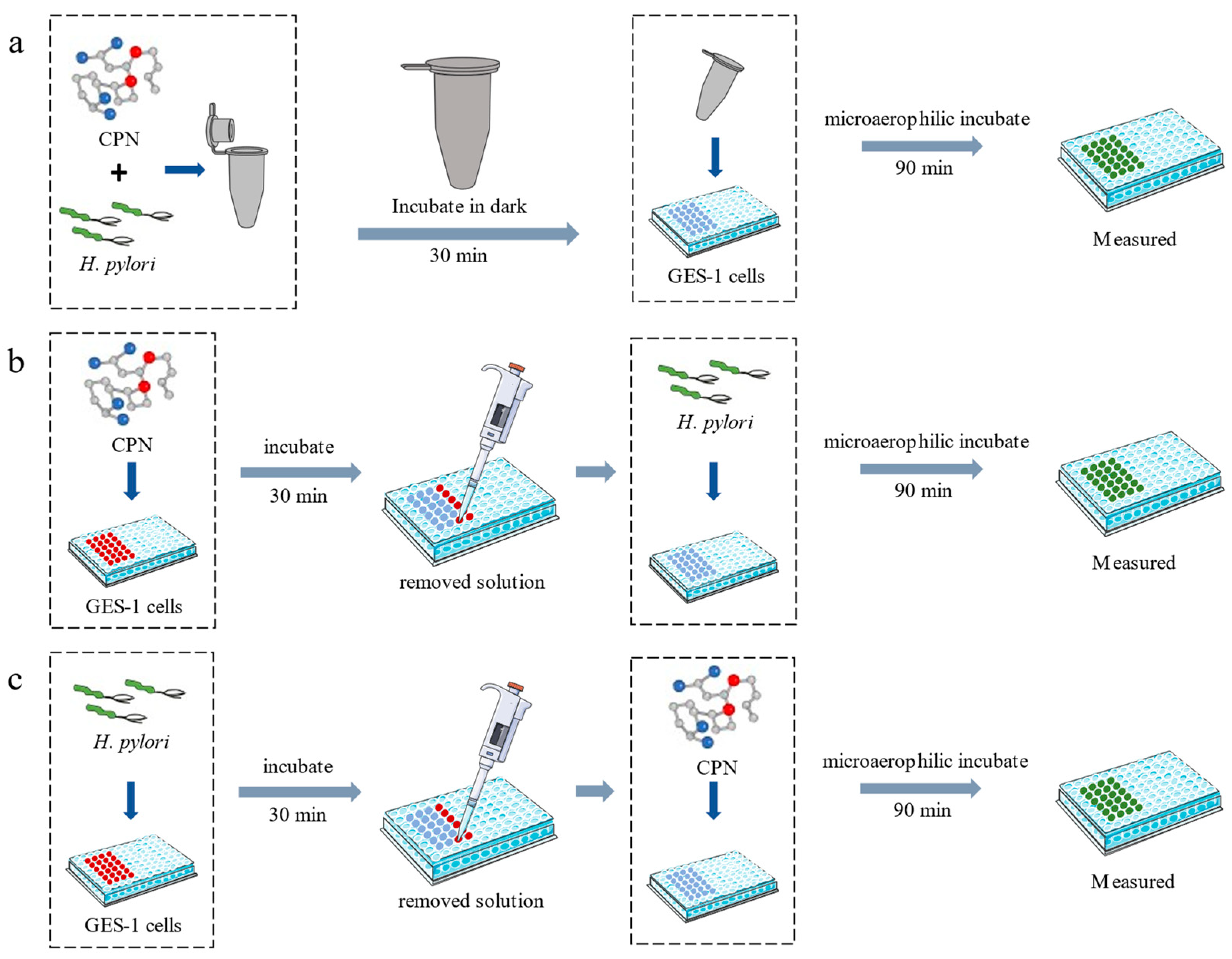
2.6. Anti-Adhesion Assay

The anti-adhesive activity was measured as described in the previous report with minor modifications [8]. The anti-adhesion assay was performed in three different manners (Figure 1) and involved Neutral corn protein hydrolysate (CPN), H. pylori and GES-1 cells. CPN was treated with a 0.22 μm filter for sterilization. (1) CPN (8 mg/mL) and FITC-labeled H. pylori (108 cfu/mL) were mixed together (1:1, v/v) and incubated for 30 min in the dark. Then, 100 μL of the H. pylori-CPN solution was added to the GES-1 cells inside the 96-well plate (3 × 10⁵ cells/well), then the 96-well plate was incubated at 37 °C for 1.5 h under microaerophilic conditions (5% O2, 10% CO2 and 85% N2). (2) An amount of 100 μL of CPN was added to each well with GES-1 cells and incubated at 37 °C for 0.5 h under microaerophilic conditions, then the CPN solution was removed and 100 μL of FITC-labeled H. pylori was added to the GES-1 cells at the same conditions and incubated for 1.5 h. (3) An amount of 100 μL of FITC-labeled H. pylori was added to the GES-1 cells and incubated at 37 °C for 0.5 h under microaerophilic conditions. After the FITC-labeled H. pylori solution was removed, 100 μL of CPN solution was added to the GES-1 cells, then it was incubated under microaerophilic conditions for 1.5 h.
The treated GES-1 cells in the above three manners were washed with 200 μL of 0.1 M sterile PBS buffer (pH 7.4) three times to remove free H. pylori, then 100 μL of 0.1 M PBS buffer (pH 7.4) was added to the 96-well plate. Finally, the fluorescence intensity of the 96-well plate was measured at the excitation wavelength of 485 nm and emission wavelength of 530 nm. The standard curve was set up by the value of OD600 and fluorescence intensity, and then the concentration of H. pylori was calculated. The CPN was replaced by 100 μg/mL of rebamipide and DMEM incomplete medium as the positive control and negative control, respectively.
I n h i b i t i o n % = n u m b e r   o f   H .   p y l o r i   n e g a t i v e   c o n t r o l n u m b e r   o f   H .   p y l o r i   s a m p l e n u m b e r   o f   H . p y l o r i   n e g a t i v e   c o n t r o l × 100
The percent inhibition (anti-adhesive activity) of CPN was calculated as follows:
Figure 1. The anti-adhesion assays of CPN. (a) CPN and H. pylori were pre-incubated; (b) CPN and GES-1 cells were pre-incubated; (c) H. pylori and GES-1 cells were pre-incubated.
Nutrients 15 03467 g001

2.7. Animals and Experimental Design

Six-week-old male KM mice were obtained from the Changchun Yisi Experimental Animal Research Center (Changchun, China). The animal certificate number is SCXK (Ji)-2018–0007. The animal study protocol was approved on 8 June 2021 by the Animal Ethics Committee of the College of Food and Bioengineering, Qiqihar University (Approval No. 2021-004). The temperature and humidity conditions were 22 ± 1 °C and 55 ± 5%, respectively, and the mice were fed with free access to water and food.
After acclimation for 1 week, mice were randomly divided into six groups (n = 8). Normal control (NC), H. pylori infection model (HM), 200 mg/kg·bw CPN group (CPN-200), 400 mg/kg·bw CPN group (CPN-400), 600 mg/kg·bw CPN group (CPN-600) and positive group (14.25 mg/kg amoxicillin +7.15 mg/kg Clarithromycin + 14.2 mg/kg Metronidazole). Body weight was measured once per day during the experimental period.
At 1–2 weeks, NC and HM groups were given 0.3 mL 0.9% normal saline and the positive group was given 0.3 mL the mixed antibiotics, whereas the CPN groups were given 200 mg/kg·bw, 400 mg/kg·bw and 600 mg/kg·bw CPN, respectively.
At 3–4 weeks, all groups continued to be given different doses of CPN or 0.9% normal saline or antibiotics. Except NC group, other groups were given 0.4 mL H. pylori (about 109 cfu/mL) after 30 min. The H. pylori was given every other day, 7 times in total.
At 5 weeks, all groups continued to be given different doses of CPN or 0.9% normal saline or antibiotics.

2.8. Analysis of H. pylori Enumeration of Gastric Tissue

The sterile gastric homogenate serially diluted in 0.1 M PBS buffer, 100 μL of each dilution, was spread-plated onto Medium no. 260, then the plates were incubated at 37 °C for 48 h under microaerophilic conditions. The colonies were counted and expressed as CFU/g gastric tissue.

2.9. Measurement of Biochemical Parameters by ELISA

The mice were fed in the experiment for five weeks. The mice were fasted for 12 h before being sacrificed through cervical dislocation. After the gastric tissue was weighed, it was split into two sections: one for histopathologic study, the other for homogenization in ice-cold 0.9% normal saline to generate a 10% gastric homogenate under aseptic conditions.
According to the explanatory memorandum, tumor necrosis factor-α (TNF-α), malondialdehyde (MDA), monocyte chemoattractant protein-1 (MCP-1), Superoxide dismutase (SOD) activities, lactate dehydrogenase (LDH), toll-like receptor 4 (TLR4), GSH peroxidase (GSH-Px) activities, interleukin (IL)-1β, myeloperoxidase (MPO) activity, keratinocyte chemokine (KC), interleukin (IL)-6, myeloid differentiation factor88 (MyD88) and NF-κB levels in the gastric homogenates were measured by enzyme-linked immunosorbent assay kits (JiangLai bio, Shanghai, China).

2.10. Histopathologic Analysis

The gastric tissue samples were fixed in 10% buffered formalin solution, and then were embedded in paraffin. The 4 μm tissue sections were made, then the sections were stained with hematoxylin and eosin and visualized using microscope (Olympus BX43 microscope).

2.11. Statistical Analysis

All results were expressed as mean ± standard deviations (SD), and ANOVA and Tukey’s tests were used to analyze the statistical comparisons. SPSS statistics 19 (SPSS Inc., Chicago, IL, USA) was used to analyze the data. Significant differences were considered at p < 0.05.

3. Results and Discussion

3.1. Corn Protein-Derived Peptides Preparation

CGM contains mainly zein (68%) and glutelin (22%) [13]. Zein has a high ratio of hydrophobic amino acids (Leu (20%), Ala (10%), Pro (10%)) [14], leading to a high frequency of hydrophobic amino acid residue sequences in its protein primary structure. For example, α-zein (22 kDa) has some typical fragments, such as LLAL11–14, APIA30–33, LLPP35–38, AIAA61–64 and LAAA181–184 (Table 2). Corn glutelin has a high glutamine (Gln, Q) content of 33% [14] and many sequences of QQ or QQQ located in its primary structure, for example, QQ104–105,153–154,157–158,199–200,208–209 and QQQ181–183 in glutelin (26.3 kDa) (Table 2).
Alcalase, Protamex and Protease M are endoproteases; they act on hydrophobic and aromatic amino acid residues, such as Ala, Leu and Phe. Protease N and Protease P mainly act on hydrophobic and hydroxyl amino acids, such as Thr, Ser and Pro. Neutral is also an endoprotease, and it mainly hydrolyzes the aromatic amino acids. According to the structural characteristics of corn protein, and taking into account the substrate specificity of the proteases, the above proteases were chosen to hydrolyze corn protein, expecting to obtain corn protein hydrolysates with anti-adhesive activity. Thus, the effect of the protease on the anti-adhesive activity of the hydrolysates was investigated. Eight proteases were applied for producing anti-adhesive hydrolysates from CGM at the enzyme/substrate ratio of 400 U/g·protein and hydrolysis time of 120 min.
As shown in Table 1, the anti-adhesive activities of different protease hydrolysates had significant differences (p < 0.05). All hydrolysates, except the hydrolysate hydrolyzed by papain, showed anti-adhesive activities, and the hydrolysate (CPN) hydrolyzed by Neutral had the best anti-adhesive activity (50.44 ± 0.27%) at the concentration of 4 mg/mL; hydrolysates hydrolyzed by Protease N and Protease P also showed better anti-adhesive activities. It was speculated that hydrophobic and aromatic amino acids in the peptides had an important effect on the anti-adhesive activity of the hydrolysates.
Moreover, trypsin and papain hydrolysates showed the lowest anti-adhesive activities. Trypsin and papain have similarity in the cleavage of proteins, and they could act on basic amino acid residues such as Arg and Lys; it is possible that Arg and Lys have less of an effect on the anti-adhesive activity of hydrolysates. This finding was consistent with Sun et al. [8], where it was also found that the trypsin hydrolysate had low anti-adhesive activity.

3.2. Assay of Anti-Adhesive Activity of CPN

H. pylori could specifically adhere to the GES-1 cells. Adhesion proteins, OMPs of H. pylori, specifically recognize receptors of GES-1 cells’ surface to achieve H. pylori infection [5]. Anti-adhesion therapy, a promising strategy, could effectively prevent infection by inhibiting H. pylori adhesion to GES-1 cells. Specifically, the anti-adhesion component acts as GES-1 cell receptor analogs or H. pylori surface adhesin analogs to block the adhesion between the H. pylori and the GES-1 cells.
To analyze the possible anti-adhesive mechanism of CPN, three inhibition assays were performed in the present study. CPN (as receptor analogs) and H. pylori, CPN (as adhesin analogs) and GES-1 cells, and H. pylori and GES-1 cells were mixed, respectively. When CPN and H. pylori were pre-mixed (Figure 1a) and incubated for 0.5 h in dark, then the CPN-H. pylori solution was added to the 96-well plate with GES-1 cells (3 × 10⁵ cells/well) and incubated at 37 °C for 1.5 h under microaerophilic conditions, it was found excitedly that the measured fluorescence intensity of the CPN group decreased sharply, compared with that of control group, indicating that the number of FITC-labeled H. pylori adhering to the GES-1 cells’ surface significantly reduced. The anti-adhesive activity of CPN was 50.44 ± 0.27%, showing that CPN was able to block the H. pylori adhesion to GES-1 cells as receptor analogs; this might be a possible mechanism.
When CPN and GES-1 cells were pre-mixed (Figure 1b) and incubated for 0.5 h under microaerophilic conditions, then the solution was removed and the FITC-labeled H. pylori was added to each well and incubated for 1.5 h, the fluorescence intensity of each well was measured. Compared with that of control group, it was found that the fluorescence intensity of the CPN group did not change significantly, showing that the number of FITC-labeled H. pylori adhering to the GES-1 cells’ surface did not reduce. CPN did not exhibit anti-adhesive activity, suggesting that CPN could not inhibit the adhesion of H. pylori as an adhesion analog.
When H. pylori and GES-1 cells were pre-mixed (Figure 1c) and incubated at 37 °C for 0.5 h under microaerophilic conditions, then after the solution was removed, CPN was added to GES-1 cells and incubated for 1.5 h, it was found that the fluorescence intensity of the CPN group did not change significantly, compared with that of control group, and CPN did not remove H. pylori that was already bound to GES-1 cells, indicating that CPN could not destroy the binding between GES-1 cells and H. pylori.
Based on the above, the CPN could inhibit the adhesion of H. pylori, when used as receptor analogs, indicating that CPN could prevent H. pylori infection as a safe and cost-effective anti-adhesive agent.

3.3. Effect of CPN on the Colonization of H. pylori in the Mice Gastric Mucosa

In the process of H. pylori infection, the successful colonization of H. pylori in the gastric mucosa is the first and most important step [18]. Therefore, in order to evaluate the effect of CPN against H. pylori infection, the amount of H. pylori colonization in the mice gastric mucosa was analyzed. After the adaptive feeding of mice, CPN doses of 200 mg/kg·bw, 400 mg/kg·bw and 600 mg/kg·bw were given by intragastric administration for two weeks. From the third week, in addition to feeding the CPN, 0.4 mL high-concentration H. pylori (109 cfu/mL) was fed by intragastric administration every other day for 7 times in total to establish the H. pylori infection model. Then, we continued to feed for a week to empty the H. pylori that does not colonize in the mice gastric mucosa.
As shown in Table 3, compared with that of the NC group, the number of H. pylori increased significantly in the HM group (p < 0.05), indicating that the model is successful. Compared with that of the HM group, it was found that CPN intervention significantly reduced the number of H. pylori in the mice mucosa (p < 0.05). Concerning the number of H. pylori in the gastric mucosa of mice in the CPN-200, CPN-400 and CPN-600 groups, they had an H. pylori content of 1.63–2.08 × 10⁵ cfu/g. The content of the three CPN groups was significantly lower than that of the HM group (p < 0.05), and the CPN-400 group showed the best result (1.63 × 10⁵ cfu/g). It is possible that the CPN showed significant anti-adhesive activity in vitro. A certain dose of CPN could inhibit the colonization of most of the H. pylori, so the number of H. pylori colonization did not change significantly with increasing CPN concentration. These results indicated that the amount of H. pylori colonization in the gastric mucosa could be effectively reduced by CPN intervention. It is possible that the underlying mechanism of the antagonistic effect against H. pylori infection is associated with the anti-adhesive effect of CPN.

3.4. CPN Alleviates Oxidative Stress with H. pylori Infection-Induced Gastric Injury in Mice

When the mice are infected with H. pylori, the H. pylori colonizes and multiplies in the stomach. This leads to the release of virulence factors, such as VacA, CagA and urease [19]. These factors then stimulate gastric cells, causing excessive production of oxygen radicals, which in turn increase cellular lipid peroxidation. This process ultimately injures the structure of the cell membrane, leading to injury of the gastric mucosa [20,21]. In order to evaluate the effect of CPN on alleviating oxidative stress induced by H. pylori infection, the activities of gastric tissue SOD and GSH-Px and the contents of MDA and LDH were measured. The results are shown in Table 4.
Table 4 shows that compared with the NC group, the activities of SOD and GSH-Px in gastric tissue decreased significantly in the HM group (p < 0.05), and MDA as well as LDH contents increased significantly (p < 0.05). The activities of SOD and GSH-Px decreased by 40.62% and 35.62%, while MDA and LDH content were elevated by 44.88% and 41.94%, respectively. These results indicated that H. pylori infection caused the depletion of antioxidant enzymes (SOD and GSH-Px) and the excessive production of MDA and LDH in the gastric tissue. In other words, H. pylori infection led to oxidative stress in the gastric tissue.
SOD is an important endogenous antioxidant enzyme, which could scavenge reactive oxygen species in vivo [22]. Meanwhile, SOD could catalyze the generation of hydrogen peroxide by superoxide anions, reduce the damage of superoxide anions to cells and protect the gastric mucosa from free radical damage. SOD activity of the CPN-400 and CPN-600 groups significantly increased (p < 0.05), compared with the HM group. SOD activity of the CPN-600 was group elevated significantly by 53.0% (p < 0.05), which was higher than that of positive control group. These results suggest that CPN is effective to alleviate the oxidative stress with H. pylori infection-induced gastric injury in mice.
GSH-Px, as an endogenous antioxidant enzyme in the body’s antioxidant system, plays an important role in the antioxidant process [23]. The activity of GSH-Px in the HM group decreased significantly (p < 0.05), indicating that H. pylori infection could reduce GSH-Px content in gastric tissue. Compared to those of the NC group, GSH-Px activity of the HM group, CPN-200, CPN-400, CPN-600 and positive control groups decreased by 35.62%, 20.22%, 11.61%, 9.95% and 5.46%. Specifically, there was no significant difference between the middle as well as high dose of the CPN groups and positive group. Compared with the HM group, the activity of GSH-Px in the CPN-400 and CPN-600 groups was significantly increased by 37.30% and 39.86% (p < 0.05), which was comparable to the positive group, which suggested that CPN intervention significantly increased the GSH-Px level (p < 0.05).
MDA is a reactive aldehyde produced after lipid peroxidation of polyunsaturated fatty acids on cell membranes, and it is a marker of oxidative stress. MDA content directly reflects the degree of peroxidation of the biofilm, and excessive accumulation of it will cause the loss of cell membrane function and cause damage to the body [24]. As shown in Table 4, MDA content in the HM group was 289.03 nmol/g, which was 1.45-fold higher than the NC group, indicating that the level of oxidative stress was increased by H. pylori infection and led to gastric tissue injury. Compared with the HM group, the lipid peroxidation level of gastric tissue in the CPN-400, CPN-600 and positive control groups was lower, and MDA content decreased significantly by 11.72%, 22.72% and 22.69% (p < 0.05). Specifically, the CPN-400 group showed the best effect of all CPN groups, which indicated that CPN could effectively remove free radicals, restore the antioxidant capacity of gastric tissue, and relieve lipid peroxidation on the surface of gastric cell membranes.
LDH is a glycolytic enzyme in the cytoplasm. The action of the LDH that catalyzes pyruvate produces lactic acid in cells. Generally, LDH exists stably in cells. When the gastric tissue is infected by H. pylori, the NADPH oxidase in cells is stimulated to generate endogenous stress factors, which attack and lead to lipid peroxidation on the surface of the cell membrane, which destroys the cell membrane, releasing and increasing LDH content. Compared with the NC group, LDH content in the HM group increased significantly by 41.94% (p < 0.05), showing that the gastric tissue was injured by H. pylori infection, and LDH content was extremely elevated. Compared with the HM group, there was a significant reduction in LDH content following different doses of CPN intervention (p < 0.05), of which the CPN-600 group had the best effect, with a reduction of 25.19%, which was close to the level of the NC group and comparable to that of the positive control group. These results indicate that the oxidative stress injury of gastric tissue is significantly alleviated by CPN intervention, and the gastric tissue membrane possesses integrity.
Based on these results, CPN intervention could improve the activities of SOD and GSH-Px, inhibit the degree of lipid peroxidation (MDA), as well as reduce the release of LDH. Therefore, the CPN could improve the total antioxidant capacity of the body and then achieve preventive and protective effects on gastric injury induced by H. pylori infection. Some studies have shown that bioactive peptides can alleviate gastric injury by reducing oxidative stress, such as wheat peptides [24] and cod collagen peptides [25]. Similarly, some recent studies indicate that corn peptides (glycopeptides) possess excellent antioxidant activities in vitro, such as metal-ion chelating capacity and free radical scavenging ability [16,26]. Wang et al. reported that glycosylated zein peptides (250 mg/kg·bw) significantly increase endogenous antioxidant enzyme activities and GSH levels and relieve oxidative stress caused by alcoholic liver injury [27]. These results suggested that corn peptides can also reduce oxidative stress, but the protective effect of corn peptides on H. pylori infection-induced gastric injury has not been reported.

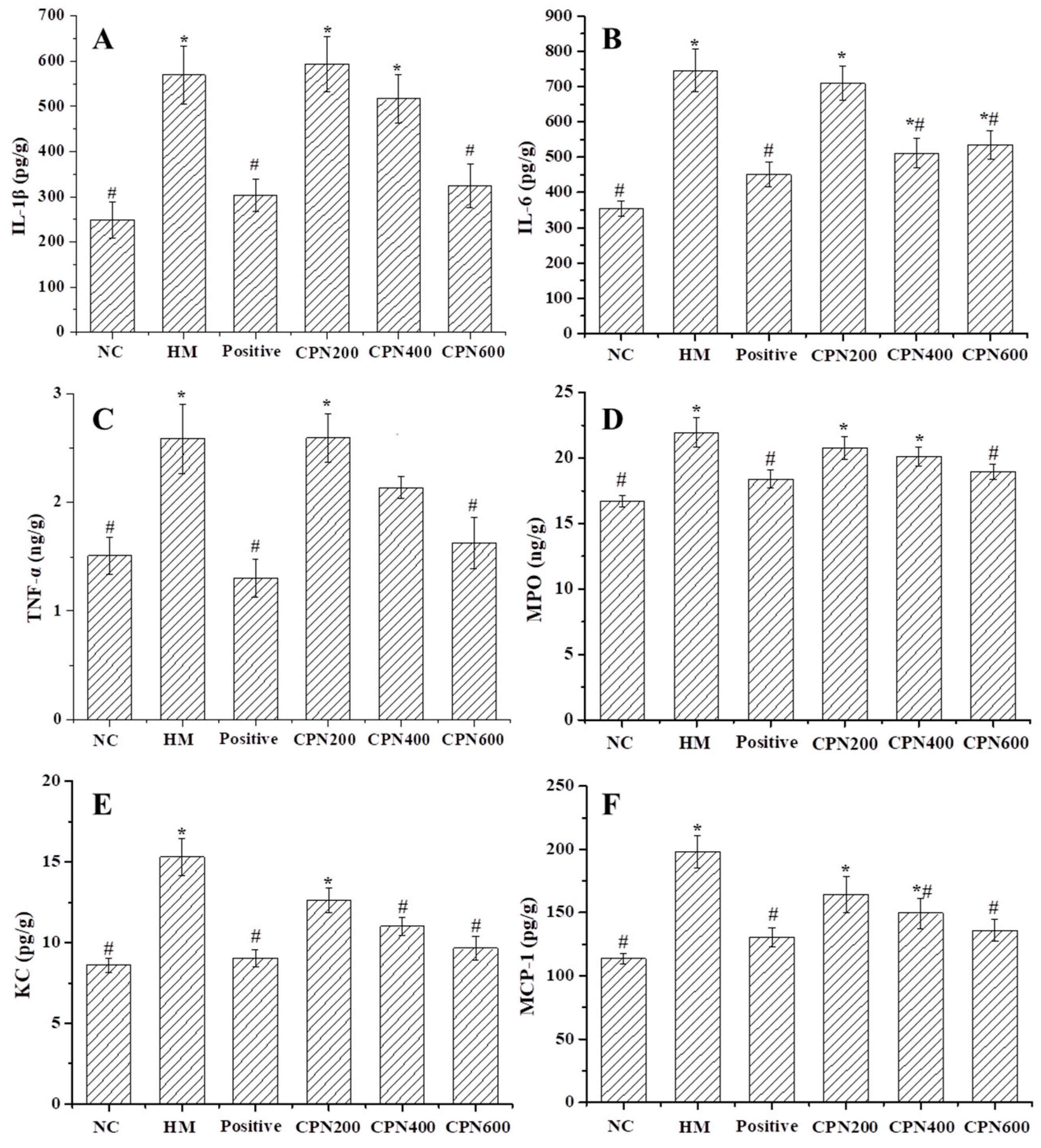
3.5. Effects of CPN on the Expression of Gastric Pro-Inflammatory Cytokines and Chemokines

After infecting gastric mucosal epithelial cells, H. pylori releases virulence factors that can directly injure the cells, reduce gastric mucus secretion and cause bleeding and other symptoms. The infection also activates the immune system, leading to the production of cellular inflammatory factors such as TNF-α, IL-1β and MPO, which mediate the occurrence of inflammatory reactions [28]. The content of gastric pro-inflammatory cytokines (IL-1β, IL-6, TNF-α and MPO), as well as chemokines (KC and MCP-1), was measured to evaluate the effectiveness of CPN on the inflammatory response induced by H. pylori infection. As shown in Figure 2, the contents of IL-1β, IL-6, TNF-α, MPO, KC and MCP-1 in gastric tissue significantly increased in H. pylori-infected mice, compared with the NC group (p < 0.05), increasing by 128.66%, 110.55%, 72.00%, 31.45%, 77.70% and 74.10%, respectively, which indicated that H. pylori-infected mice could abnormally metabolize pro-inflammatory factors and chemokines in the gastric mucosa. These molecules can cause inflammatory gastric injury with mitochondrial dysfunction and increased endoplasmic reticulum stress.
Compared with the HM group, all CPN groups could decrease the contents of IL-1β and IL-6 in the gastric tissue (Figure 2A,B). Specifically, the high-dose CPN could significantly decrease the IL-1β and IL-6 contents by 43.08% and 46.77%, respectively. Additionally, compared with the NC group, there is no significant difference in the effect of high-dose CPN. These results showed that the levels of IL-1β and IL-6 in the gastric tissue were effectively reduced with CPN intervention, and the inflammatory response in the gastric tissue was alleviated.
As an important cytokine with multiple functions, TNF-α activation is closely associated with a series of pro-inflammatory cytokines, which is related to inflammatory diseases [29]. Studies have shown that the activation of the pro-inflammatory factors (e.g., IL-6 and IL-1β) could increase TNF-α content again, which ultimately aggravates inflammation. TNF-α is one of the strongest pro-inflammatory cytokines; thus, it is often used as an important indicator to judge the degree of inflammation [30]. As shown in Figure 2C, CPN intervention could effectively reduce the TNF-α content in a dose-dependent manner compared with the HM group, while only high-dose CPN could significantly decrease the TNF-α content close to the normal level (p < 0.05).
MPO is a heme protein, and it exists in neutrophils. Neutrophils are stimulated by external factors, which aggregate and release MPO. The activation and infiltration of neutrophils are involved in the gastric inflammation that can be assessed by the increased MPO content [31]. Consequently, MPO is a crucial indicator for assessing the extent of neutrophil infiltration and inflammation. MPO content in the HM group, CPN-200, CPN-400, CPN-600 and positive control groups was 1.31-, 1.24-, 1.20-, 1.13- and 1.10-fold higher compared with that of the NC group, respectively, indicating that H. pylori infection caused a significant increase in MPO content in the gastric tissue (p < 0.05). This finding is consistent with previous reports [32]. Compared with the HM group, all dose groups could reduce the MPO content, but only the CPN-600 group had a significant difference (p < 0.05), decreasing by 13.67%, and compared with the NC group, there was no significant difference. These results showed that treatment with varying doses of CPN effectively improved the infiltration of neutrophils, leading to a reduction in MPO release. This resulted in the alleviation of gastric mucosal injury and inflammation, ultimately maintaining the integrity of the gastric mucosa. A similar result was reported [32]. Li et al. also found that tricholoma matsutake-derived peptides have the ability to counteract alcohol-induced acute gastric mucosal injury by decreasing the MPO levels in gastric tissue [33]. This suggests that CPN intervention can alleviate gastric mucosal injury by reducing the MPO content in the gastric tissue.
The gastric epithelial cytoskeleton rearrangement is induced by H. pylori infection, which activates NF-κB, and the expression and release of chemokines (MCP-1, KC) are induced, which lead to gastric epithelial cell apoptosis and mucosal injury [33]. KC and MCP-1 are two important chemokines. Currently, many types of cells, such as macrophages, endothelial cells and smooth muscle cells are known to be affected by chemicals or bacteria, being induced to secrete chemokines [30]. The studies have reported that chemokines have chemotactic activity on cells, which activates monocytes and macrophages, which increase the concentration of Ca2+ in the cytoplasm and release oxygen free radicals and lysozyme, up-regulating pro-inflammatory cytokines [34]. The effects of CPN on the content of KC and MCP-1 are illustrated in Figure 2E, F. Compared with the NC group, the KC and MCP-1 content of the HM group increased significantly (p < 0.05) from 8.61 ± 0.43 and 113.71 ± 4.26 pg/g to 15.31 ± 1.13 and 197.97 ± 12.43 pg/g, indicating that the chemokine content was greatly elevated by H. pylori infection. Compared with the HM group, the KC and MCP-1 contents of three dose groups gradually decreased in a dose-dependent manner, and the CPN-400 and CPN-600 groups showed a significant decrease in KC and MCP-1 contents by 27.97% and 36.93% and 24.46% and 31.33%, respectively. Additionally, the content of the high-dose group was close to the normal level and the CPN effects were comparable to the positive control. The results showed that the possible underlying mechanism of the antagonistic effect against H. pylori infection is associated with the anti-inflammatory property of CPN.
Based on these results, this study found the dose-dependent attenuation of these levels following CPN intervention. In particular, the level of pro-inflammatory cytokines and chemokines returned to normal with the CPN-600 treatment. In other words, CPN intervention could down-regulate the production of chemokines and pro-inflammatory cytokines, which block the development of inflammation and alleviate a series of metabolic changes linked to inflammation, such as oxidative stress and lipid peroxidation, which exert gastric protective effects against H. pylori infection. Kan et al. reported a novel wheat peptide that could reduce ethanol-induced gastric mucosal injury through antioxidant and anti-inflammatory activity [24]. These results indicated that the CPN possesses anti-inflammatory properties, which may be an underlying mechanism of the protective effect inhibiting H. pylori infection.

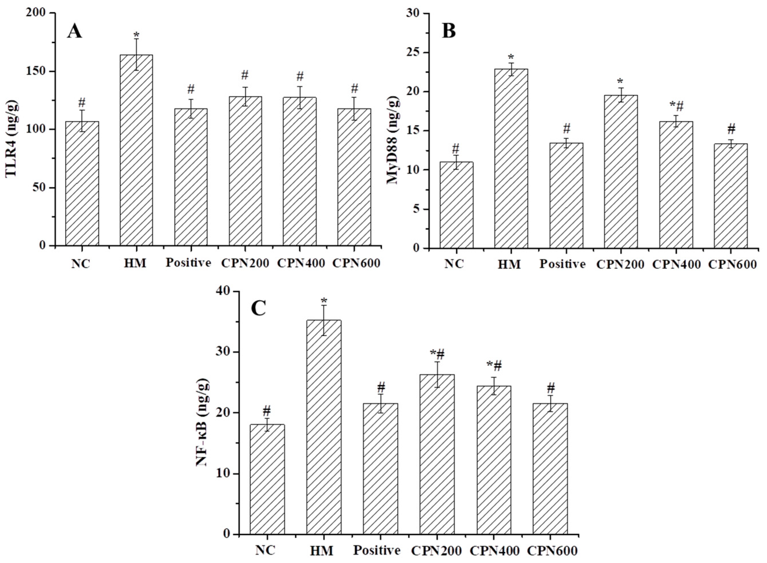
3.6. Effects of CPN on Key Metabolites in NF-κB Signaling Pathway

NF-κB plays a key role in regulating the immune response to H. pylori infection [35]. The pattern recognition receptor TLR4 of gastric mucosal epithelial cells’ surface can specifically recognize the virulence factors released by H. pylori, which promote the increased aggregation of MyD88 and the formation of TLR4/MyD88 complexes, which activate the signaling pathway, leading to an excessive increase in NF-κB content, resulting in an inflammatory reaction [36,37]. Effects of CPN on the important metabolites TLR4, MyD88 and NF-κB in the NF-κB signaling pathway are shown in Figure 3. TLR4, MyD88 and NF-κB contents in the HM group significantly increased (p < 0.05) by 53.36%, 108.18% and 95.39%, respectively, compared with the NC group. It showed that H. pylori infection promoted the overexpression of TLR4 receptor and MyD88, which activated the NF-κB signaling pathway and caused an inflammatory response. Compared with the HM group, the levels of TLR4, MyD88 and NF-κB in all CPN groups were significantly decreased (p < 0.05), indicating that CPN was effective in reducing the levels of TLR4, MyD88 and NF-κB as well as alleviating the inflammatory response. It is worth noting that TLR4 and NF-κB levels were significantly lowered in the 200 mg/kg·bw dose (p < 0.05). Moreover, compared with the NC group, the TLR4, MyD88 and NF-κB levels were not significantly different. These results showed that CPN might suppress expression of inflammatory mediators by inhibiting NF-κB activation in the gastric tissues of H. pylori-infected mice, which may be another possible mechanism of the protective antagonistic effect of H. pylori infection. The previous reports indicated that artemisinin has the potential to reduce the production of ROS induced by H. pylori and effectively inhibit the activation of the NF-κB signaling pathway, thus preventing gastric carcinogenesis [34], and Kim et al. found that the astaxanthin could inhibit NF-κB activation of the H. pylori in gastric cells by down-regulating TLR4 and MyD88 contents [38]. These results showed that the inhibition of the NF-κB signaling pathway activation is extremely important to prevent H. pylori infection-induced gastric injury. Moreover, more studies are required to elucidate the exact mechanism by which CPN inhibits NF-κB signaling pathway activation.

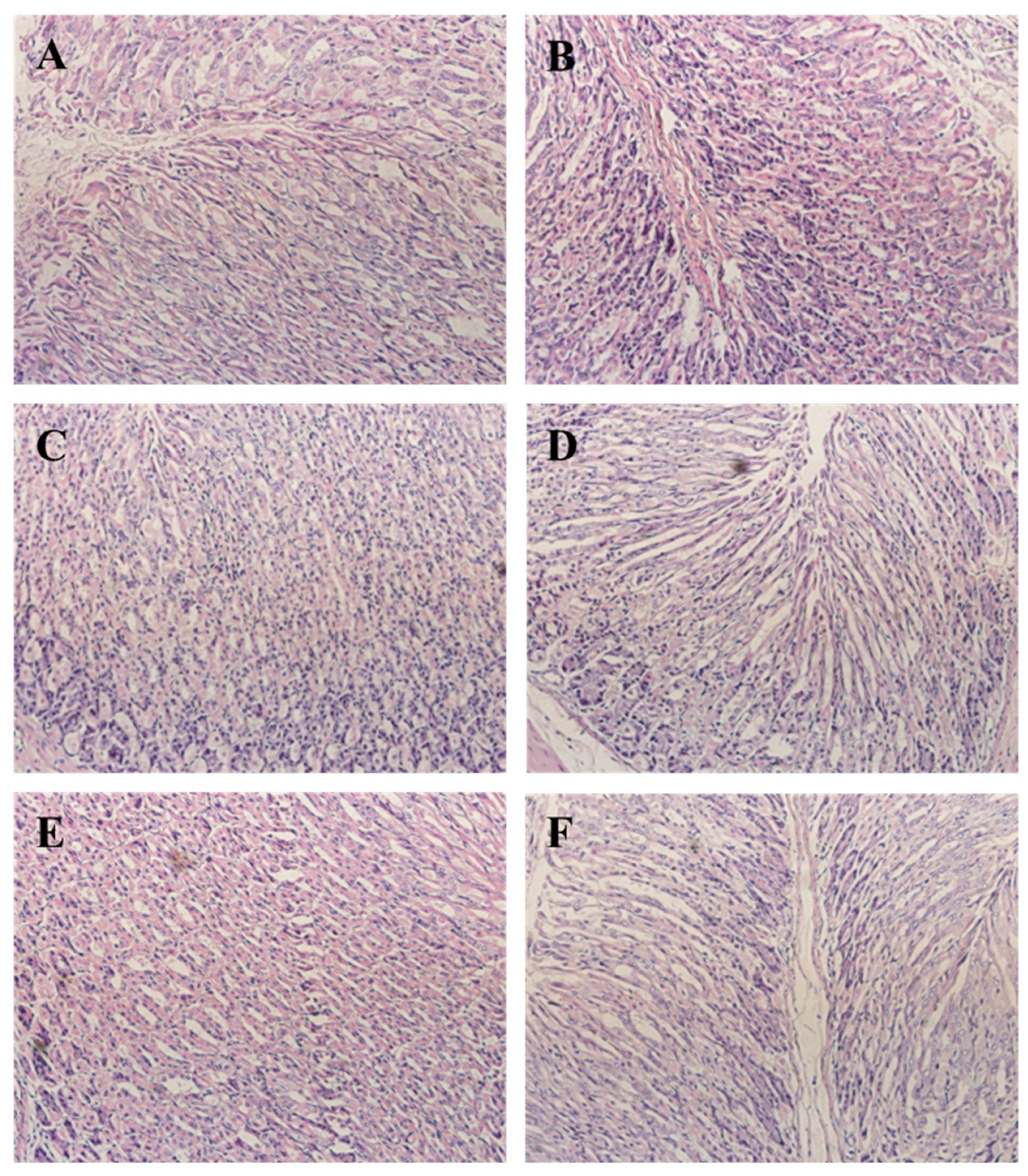
3.7. CPN Antagonism H. pylori-Induced Histological Changes in Gastric Tissue

H. pylori infection induced macroscopic morphological changes in gastric tissue. Compared with the NC group, the HM group had mucosal injury characterized by neutrophil infiltration, whereas the gastric mucosal regions of the NC group showed neither inflammatory lesions nor neutrophil infiltration (Figure 4); the study demonstrated that H. pylori infection induced an inflammatory response in gastric epithelial cells, which corroborates with prior findings on this particular model [39]. The study found that CPN intervention significantly improved the injury of gastric mucosal cells in mice infected with H. pylori, compared to the model group. There was still evidence of cell damage and swelling, but it was relieved compared to the model group. As the dose of CPN increased, neutrophil infiltration in the gastric mucosa of the mice decreased significantly, resulting in a weakened inflammatory response. The high-dose group had a similar effect as the positive control group. These pathological results indicate that CPN can inhibit histological changes related to neutrophil infiltration in mice, suggesting that CPN intervention can counteract H. pylori-induced gastric mucosal inflammation. These findings are consistent with the detection results of gastric tissue biochemical indicators in mice.

4. Conclusions

This study indicated that CPN possesses anti-adhesive activities in vitro and protective antagonistic effects against H. pylori-induced gastric injury in vivo, as shown by the decreased amount of H. pylori colonization, pro-inflammatory cytokines, chemokines and key metabolites of NF-κB signaling pathway levels and increased antioxidant enzyme contents and mitigation of H. pylori-induced pathological changes in gastric mucosa, as well as inhibition of the NF-κB signaling pathway activation when compared with the HM group. Specifically, CPN intervention at 600 mg/kg·bw could significantly alleviate gastric injury induced by H. pylori infection parameters, restoring levels to those observed in NC group. Overall, this study indicated that the use of corn protein-derived peptides as food products or drugs could establish preventive strategies against gastric H. pylori infection.

Author Contributions

G.L.: Writing—original draft; X.L.: Writing—review and editing; Z.M.: Analyzed the data; N.H.: Formal analysis; X.Z.: Experimental design. All authors have read and agreed to the published version of the manuscript.

Funding

This study was funded by Talent Foundation of the Central Government Supporting Local Universities (No. 2020GSP08), National Key Research and Development Project of China (No. 2021YFD2100904), Open project of Heilongjiang Key Laboratory of Corn Deep Processing Theory and Technology (No. SPKF202022) and Heilongjiang Provincial Foundation for the Characteristic Discipline of Processing Technology of Plant Foods (No. YSTSXK201839).

Institutional Review Board Statement

The animal study protocol was approved by the Animal Ethics Committee of the College of Food and Bioengineering, Qiqihar University. The KM mice were maintained in accordance with the Qiqihar University Regulations on Animal Experimentation (Approval No. 2021-004).

Informed Consent Statement

Not applicable.

Data Availability Statement

Data are contained within the article.

Conflicts of Interest

The authors declare that they have no conflict of interest.

References

  1. Neshani, A.; Zare, H.; Akbari Eidgahi, M.R.; Hooshyar Chichaklu, A.; Movaqar, A.; Ghazvini, K. Review of antimicrobial peptides with anti-Helicobacter pylori activity. Helicobacter 2019, 24, e12555. [Google Scholar] [CrossRef] [PubMed] [Green Version]
  2. Shibata, W.; Hirata, Y.; Maeda, S.; Ogura, K.; Ohmae, T.; Yanai, A. CagA protein secreted by the intact type IV secretion system leads to gastric epithelial inflammation in the Mongolian gerbil model. J. Pathol. 2006, 210, 306–314. [Google Scholar] [CrossRef]
  3. Hu, Y.; Zhu, Y.; Lu, N.H. Novel and effective therapeutic regimens for Helicobacter pylori in an era of increasing antibiotic resistance. Front. Cell. Infect. Microbiol. 2017, 7, 168. [Google Scholar] [CrossRef] [PubMed] [Green Version]
  4. Cong, Y.; Geng, J.; Wang, H.; Su, J.; Arif, M.; Dong, Q.; Chi, Z.; Liu, C. Ureido-modified carboxymethyl chitosan-graft-stearic acid polymeric nano-micelles as a targeted delivering carrier of clarithromycin for Helicobacter pylori: Preparation and in vitro evaluation. Int. J. Biol. Macromol. 2019, 129, 686–692. [Google Scholar] [CrossRef] [PubMed]
  5. Liu, Q.; Meng, X.; Li, Y.; Zhao, C.N.; Tang, G.Y.; Li, S.; Gan, R.Y.; Li, H.B. Natural products for the prevention and management of Helicobacter pylori infection. Compr. Rev. Food Sci. Food Saf. 2018, 17, 937–952. [Google Scholar] [CrossRef] [Green Version]
  6. Petersson, C.; Forsberg, M.; Aspholm, M.; Olfat, F.O.; Forslund, T.; Boren, T.; Magnusson, K.E. Helicobacter pylori SabA adhesin evokes a strong inflammatory response in human neutrophils which is down-regulated by the neutrophil-activating protein. Med. Microbiol. Immun. 2006, 195, 195–206. [Google Scholar] [CrossRef]
  7. Sun, X.H.; Wu, J. Food derived anti-adhesive components against bacterial adhesion: Current progresses and future perspectives. Trends Food Sci. Technol. 2017, 69, 148–156. [Google Scholar] [CrossRef]
  8. Sun, X.H.; Zhang, S.Y.; Udenigwe, C.C. Wheat germ-derived peptides exert antiadhesive activity against Helicobacter pylori: Insights into structural characteristics of identified peptides. J. Agric. Food Chem. 2020, 68, 11954–11974. [Google Scholar] [CrossRef] [PubMed]
  9. Niehues, M.; Euler, M.; Georgi, G.; Mank, M.; Stahl, B.; Hensel, A. Peptides from Pisum sativum L. enzymatic protein digest with anti-adhesive activity against Helicobacter pylori: Structure activity and inhibitory activity against BabA, SabA, HpaA and a fibronectin-binding adhesin. Mol. Nutr. Food Res. 2010, 54, 1851–1861. [Google Scholar] [CrossRef]
  10. Gottesmann, M.; Paraskevopoulou, V.; Mohammed, A.; Falcone, F.H.; Hensel, A. BabA and LPS inhibitors against Helicobacter pylori: Pectins and pectin-like rhamnogalacturonans as adhesion blockers. Appl. Microbiol. Biotechnol. 2020, 104, 351–363. [Google Scholar] [CrossRef]
  11. Burger, O.; Ofek, I.; Tabak, M.; Weiss, E.I.; Sharon, N.; Neeman, I. A high molecular mass constituent of cranberry juice inhibits Helicobacter pylori adhesion to human gastric mucus. Fems Immunol. Med. Microbiol. 2000, 29, 295–301. [Google Scholar] [CrossRef] [PubMed] [Green Version]
  12. Simon, P.; Goode, P.; Mobasseri, A.; Zopf, D. Inhibition of Helicobacter pylori binding to gastrointestinal epithelial cells by sialic acid-containing oligosaccharides. Infect. Immun. 1997, 65, 750–757. [Google Scholar] [CrossRef] [PubMed]
  13. Liu, X.L.; Wang, J.T.; Liu, Y.; Cui, N.; Wang, D.Y.; Zheng, X.Q. Conjugation of the glutelin hydrolysates-glucosamine by transglutaminase and functional properties and antioxidant activity of the products. Food Chem. 2022, 380, 132210. [Google Scholar] [CrossRef] [PubMed]
  14. Liu, X.L.; Song, C.L.; Chen, J.P.; Liu, X.; Ren, J.; Zheng, X.Q. Preparation and evaluation of new glycopeptides obtained by proteolysis from corn gluten meal followed by transglutaminase-induced glycosylation with glucosamine. Foods 2020, 9, 555. [Google Scholar] [CrossRef] [PubMed]
  15. Jiang, Y.; Zhang, M.D.; Lin, S.Y.; Cheng, S. Contribution of specific amino acid and secondary structure to the antioxidant property of corn gluten proteins. Food Res. Int. 2018, 105, 836–844. [Google Scholar] [CrossRef]
  16. Wang, X.J.; Liu, X.L.; Zheng, X.Q.; Qu, Y. Preparation of corn glycopeptides and evaluation of their antagonistic effects on alcohol-induced liver injury in rats. J. Funct. Foods 2020, 66, 103776. [Google Scholar] [CrossRef]
  17. Zhou, C.S.; Ma, H.L.; Ding, Q.Z.; Lin, L.; Yu, X.J.; Luo, L.; Yagoub, A.E.G.A. Ultrasonic pretreatment of corn gluten meal proteins and neutrase: Effect on protein conformation and preparation of ACE (angiotensin converting enzyme) inhibitory peptides. Food Bioprod. Process. 2013, 91, 665–671. [Google Scholar] [CrossRef]
  18. Lee, J.H.; Park, E.K.; Uhm, C.S.; Chung, M.S.; Kim, K.H. Inhibition of Helicobacter pylori adhesion to human gastric adenocarcinoma epithelial cells by acidic polysaccharides from artemisia capillaris and panax ginseng. Planta Med. 2004, 70, 615–619. [Google Scholar] [CrossRef]
  19. Keilberg, D.; Ottemann, K.M. How Helicobacter pylori senses, targets and interacts with the gastric epithelium. Environ. Microbiol. 2016, 18, 791–806. [Google Scholar] [CrossRef]
  20. Brown, J.C.; Wang, J.; Kasman, L.; Jiang, X.; Haley-Zitlin, V. Activities of muscadine grape skin and quercetin against Helicobacter pylori infection in mice. J. Appl. Microbiol. 2011, 110, 139–146. [Google Scholar] [CrossRef]
  21. Jung, D.H.; Park, M.H.; Kim, C.J.; Lee, J.Y.; Keum, C.Y.; Kim, I.S.; Yun, C.H.; Kim, S.K.; Kim, W.H.; Lee, Y.C. Effect of beta-caryophyllene from cloves extract on Helicobacter pylori eradication in mouse model. Nutrients 2020, 12, 1000. [Google Scholar] [CrossRef] [PubMed] [Green Version]
  22. Zou, S.; Wang, Y.; Zhou, Q.; Lu, Y.; Zhang, Y.; Zhang, J. Protective effect of kinsenoside on acute alcohol-induced liver injury in mice. Rev. Bras. Farmacogn. 2019, 29, 637–643. [Google Scholar] [CrossRef]
  23. Teng, G.G.; Liu, Y.; Wu, T.; Wang, W.H.; Wang, H.H.; Hu, F.L. Efficacy of sucralfate-combined quadruple therapy on gastric mucosal injury induced by Helicobacter pylori and its effect on gastrointestinal flora. Biomed Res. Int. 2020, 2020, 4936318. [Google Scholar] [CrossRef]
  24. Kan, J.T.; Hood, M.; Burns, C.; Scholten, J.; Chuang, J.; Tian, F.; Pan, X.C.; Du, J.; Gui, M. A novel combination of wheat peptides and fucoidan attenuates ethanol-induced gastric mucosal damage through anti-oxidant, anti-inflammatory, and pro-survival mechanisms. Nutrients 2017, 9, 978. [Google Scholar] [CrossRef] [Green Version]
  25. Niu, H.N.; Wang, Z.C.; Hou, H.; Zhang, Z.H.; Li, B.F. Protective effect of cod (Gadus macrocephalus) skin collagen peptides on acetic acid-induced gastric ulcer in rats. J. Food Sci. 2016, 81, 1807–1815. [Google Scholar] [CrossRef]
  26. Ren, X.F.; Liang, Q.F.; Zhang, X.; Hou, T.; Li, S.Y.; Ma, H.L. Stability and antioxidant activities of corn protein hydrolysates under simulated gastrointestinal digestion. Cereal Chem. 2018, 95, 760–769. [Google Scholar] [CrossRef]
  27. Wang, L.; Ding, L.; Xue, C.; Ma, S.; Du, Z.; Zhang, T.; Liu, J. Corn gluten hydrolysate regulates the expressions of antioxidant defense and ROS metabolism relevant genes in H2O2-induced HepG2 cells. J. Funct. Foods 2018, 42, 362–370. [Google Scholar] [CrossRef]
  28. Yu, L.L.; Li, R.J.; Liu, W.; Zhou, Y.L.; Li, Y.; Qin, Y.; Chen, Y.H.; Xu, Y.J. Protective effects of wheat peptides against ethanol-induced gastric mucosal lesions in rats: Vasodilation and anti-inflammation. Nutrients 2020, 12, 2355. [Google Scholar] [CrossRef] [PubMed]
  29. Huang, J.N.; Wen, B.; Zhu, J.G.; Zhang, Y.S.; Gao, J.Z.; Chen, Z.Z. Exposure to microplastics impairs digestive performance, stimulates immune response and induces microbiota dysbiosis in the gut of juvenile guppy (Poecilia reticulata). Sci. Total Environ. 2020, 733, 138929. [Google Scholar] [CrossRef]
  30. Shi, M.Q.; He, J.Y.; Wang, X.; Shu, H.; Ngwa, A.N.; Chen, Y.T.; Peng, X.; Zhang, J.H.; Chen, M.H. Protective effect of total triterpenoids from Chaenomeles speciosa against Helicobacter pylori-induced gastritis in mice. J. Chin. Mat. Med. 2021, 46, 4782–4792. [Google Scholar]
  31. Da Silva, D.T.; Rodrigues, R.F.; Machado, N.M.; Maurer, L.H.; Ferreira, L.F.; Somacal, S.; Veiga, M.L.; Da Rocha, M.I.D.M.; Vizzotto, M.; Rodrigues, E.; et al. Natural deep eutectic solvent (NADES)-based blueberry extracts protect against ethanol-induced gastric ulcer in rats. Food Res. Int. 2020, 138, 109718. [Google Scholar] [CrossRef] [PubMed]
  32. Han, H.; Lim, J.W.; Kim, H. Astaxanthin inhibits Helicobacter pylori-induced inflammatory and oncogenic responses in gastric mucosal tissues of mice. J. Cancer Prev. 2020, 25, 244–251. [Google Scholar] [CrossRef] [PubMed]
  33. Li, M.Q.; Lv, R.Z.; Xu, X.M.; Ge, Q.; Lin, S.Y. Tricholoma matsutake-Derived Peptides Show Gastroprotective Effects against Ethanol-Induced Acute Gastric Injury. J. Agric. Food Chem. 2021, 69, 14985–14994. [Google Scholar] [CrossRef] [PubMed]
  34. Su, T.; Li, F.Y.; Guan, J.J.; Liu, L.X.; Huang, P.; Wang, Y.; Qi, X.X.; Liu, Z.Q.; Lu, L.L.; Wang, D.W. Artemisinin and its derivatives prevent Helicobacter pylori-induced gastric carcinogenesis via inhibition of NF-κB signaling. Phytomedicine 2019, 63, 152968. [Google Scholar] [CrossRef]
  35. Santos, A.M.; Lopes, T.; Oleastro, M.; Gato, I.V.; Floch, P.; Benejat, L.; Chaves, P.; Pereira, T.; Seixas, E.; Machado, J.; et al. Curcumin inhibits gastric inflammation induced by Helicobacter pylori infection in a mouse model. Nutrients 2015, 7, 306–320. [Google Scholar] [CrossRef]
  36. Yang, X.; Yang, L.G.; Pan, D.; Liu, H.C.; Xia, H.; Wang, S.K.; Sun, G.J. Wheat peptide protects against ethanol-induced gastric mucosal damage through downregulation of TLR4 and MAPK. J. Funct. Foods 2020, 75, 104271. [Google Scholar] [CrossRef]
  37. Tong, X.H.; Li, B.Q.; Li, J.; Li, L.; Zhang, R.Q.; Du, Y.Q.; Zhang, Y. Polyethylene microplastics cooperate with Helicobacter pylori to promote gastric injury and inflammation in mice. Chemosphere 2021, 288, 132579. [Google Scholar] [CrossRef]
  38. Kim, S.H.; Lim, J.W.; Kim, H. Astaxanthin inhibits mitochondrial dysfunction and interleukin-8 expression in Helicobacter pylori infected gastric epithelial cells. Nutrients 2018, 10, 1320. [Google Scholar] [CrossRef] [Green Version]
  39. Kim, A.; Lim, J.W.; Kim, H.; Kim, H. Supplementation with angelica keiskei inhibits expression of inflammatory mediators in the gastric mucosa of Helicobacter pylori–infected mice. Nutr. Res. 2016, 36, 488–497. [Google Scholar] [CrossRef]
Figure 2. Effects of CPN on gastric contents of IL-1β (A), IL-6 (B), TNF-α (C), MPO (D), KC (E) and MCP-1 (F) in H. pylori-infected mice. Data are expressed as the mean ± SD, n = 8. * Denotes significant difference to NC group, p < 0.05; # denotes significant difference to HM group, p < 0.05.
Nutrients 15 03467 g002
Figure 3. Effects of CPN on gastric contents of TLR4 (A), MyD88 (B), NF-κB (C) in H. pylori-infected mice. Data are expressed as the mean±SD, n = 8. * Denotes significant difference to NC group, p < 0.05; # denotes significant difference to HM group, p < 0.05.
Nutrients 15 03467 g003
Figure 4. Histopathological sections of the gastric mucosa (H&E, ×400). (A) NC group; (B) HM group; (C) CPN-200; (D) CPN-400; (E) CPN-600; (F) positive control.
Nutrients 15 03467 g004
Table 1. Enzymatic conditions and anti-adhesive activity of hydrolysates.
EnzymepHTemperature (°C)Anti-Adhesive Activity (%)
Trypsin8.03712.37 ± 0.61 f
Alcalase8.56040.72 ± 2.61 c
Protamex7.05539.79 ± 4.71 c
Neutral7.04550.44 ± 0.27 a
Protease N7.05044.16 ± 1.99 b
Protease P7.04547.44 ± 4.50 b
Protease M6.05039.93 ± 3.76 c
Papain8.0500
Rebamipide 22.30 ± 0.62 e
Note: different lowercase letters represent significant differences (p < 0.05).
Table 2. Sequence information of corn protein.
No.SequencePosition in ProteinProtein NameAccession No. on Uniprot
1LL8–9, 11–12, 35–36, 155–156ZeinP04703
2LLAL11–14ZeinP04703
3APIA30–33ZeinP04703
4LLPP35–38ZeinP04703
5AIAA61–64ZeinP04703
6VALA106–109ZeinP04703
7LAAL161–164ZeinP04703
8LAAA181–184ZeinP04703
9QQ104–105, 153–154, 157–158, 199–200, 208–209GlutelinP04706
10QQQ181–183GlutelinP04706
Table 3. Animal experimental design and the amount of H. pylori colonization in mice gastric mucosa.
Group1–2 Weeks3–4 Weeks5 Weekscfu/g Gastric Tissue
NC0.3 mL saline0.3 mL saline0.3 mL saline0
HM0.3 mL saline0.3 mL saline
+ 0.4 mL H. pylori
0.3 mL saline(1.44 ± 0.42) × 107 c
CPN-200200 mg/kg·bw200 mg/kg·bw
+ 0.4 mL H. pylori
200 mg/kg·bw(2.08 ± 0.93) × 105 a
CPN-400400 mg/kg·bw400 mg/kg·bw
+ 0.4 mL H. pylori
400 mg/kg·bw(1.63 ± 0.75) × 105 b
CPN-600600 mg/kg·bw600 mg/kg·bw
+ 0.4 mL H. pylori
600 mg/kg·bw(1.79 ± 0.77) × 105 b
Positive control 10.3 mL antibiotic0.3 mL antibiotic
+ 0.4 mL H. pylori
0.3 mL antibiotic(0.70 ± 0.31) × 104 d
1 Mixed antibiotics: 14.25 mg/kg amoxicillin + 7.15 mg/kg Clarithromycin + 14.2 mg/kg Metronidazole. Different lowercase letters represent significant differences (p < 0.05).
Table 4. Effects of CPN activities of gastric tissue SOD and GSH-Px and MDA and LDH content.
Groups 1SOD
(U/g)
GSH-Px
(U/g)
MDA
(nmol/g)
LDH
(ng/g)
NC2124.35 ± 102.76 #3257.91 ± 133.91 #199.49 ± 7.62 #111.28 ± 4.83 #
HM1264.23 ± 89.33 *2097.31 ± 150.49 *289.03 ± 10.70 *157.96 ± 6.59 *
CPN-2001436.88 ± 107.18 *2599.14 ± 128.78 *293.98 ± 9.26 *137.14 ± 7.65 *
CPN-4001758.01 ± 107.93 #2879.65 ± 125.19 #255.13 ± 11.69 *133.48 ± 7.47
CPN-6001934.29 ± 110.75 #2933.47 ± 179.52 #223.34 ± 10.84 #118.16 ± 6.96 #
Positive control1909.61 ± 66.88 #3079.95 ± 139.21 #223.46 ± 8.62 #102.35 ± 3.14 #
1 Data are expressed as the mean ± SD, n = 8. * Denotes significant difference to NC group, p < 0.05; # denotes significant difference to HM group, p < 0.05.
Disclaimer/Publisher’s Note: The statements, opinions and data contained in all publications are solely those of the individual author(s) and contributor(s) and not of MDPI and/or the editor(s). MDPI and/or the editor(s) disclaim responsibility for any injury to people or property resulting from any ideas, methods, instructions or products referred to in the content.

