Review

Unraveling the Impact of Gut and Oral Microbiome on Gut Health in Inflammatory Bowel Diseases

1
Department of Medical Microbiology and Immunology, College of Medicine and Health Sciences, United Arab Emirates University, Al Ain 15551, United Arab Emirates
2
Zayed Center for Health Sciences, United Arab Emirates University, Al Ain 15551, United Arab Emirates
*
Author to whom correspondence should be addressed.
Nutrients 2023, 15(15), 3377; https://doi.org/10.3390/nu15153377
Submission received: 27 June 2023 / Revised: 24 July 2023 / Accepted: 27 July 2023 / Published: 29 July 2023

Abstract

:
Inflammatory bowel disease (IBD) is a complex disorder characterized by chronic inflammation of the gastrointestinal tract (GIT). IBD mainly includes two distinct diseases, namely Crohn’s disease and ulcerative colitis. To date, the precise etiology of these conditions is not fully elucidated. Recent research has shed light on the significant role of the oral and gut microbiome in the development and progression of IBD and its collective influence on gut health. This review aims to investigate the connection between the oral and gut microbiome in the context of IBD, exploring the intricate interplay between these microbial communities and their impact on overall gut health. Recent advances in microbiome research have revealed a compelling link between the oral and gut microbiome, highlighting their pivotal role in maintaining overall health. The oral cavity and GIT are two interconnected ecosystems that harbor complex microbial communities implicated in IBD pathogenesis in several ways. Reduction in diversity and abundance of beneficial bacterial species with the colonization of opportunistic pathogens can induce gut inflammation. Some of these pathogens can arise from oral origin, especially in patients with oral diseases such as periodontitis. It is essential to discern the mechanisms of microbial transmission, the impact of oral health on the gut microbiome, and the potential role of dysbiosis in disease development. By elucidating this relationship, we can enhance our understanding of IBD pathogenesis and identify potential therapeutic avenues for managing the disease. Furthermore, innovative strategies for modulating the oral and gut microbiome can promote health and prevent disease occurrence and progression.

1. Introduction

Inflammatory bowel diseases (IBDs) are chronic idiopathic disorders with relapsing inflammation of the gastrointestinal tract (GIT). More than six million patients are suffering from IBD, and more cases are diagnosed worldwide [1]. In the 20th century, IBD was not prevalent in Asia, Africa, Eastern Europe, or South America but was more common in North America and Europe. Therefore, IBD has been recognized as a Western disease in the past [2]. Nowadays, IBD incidence and prevalence have significantly grown all over the world, spreading to all continents.
IBD includes two main types, which are Crohn’s disease (CD) and ulcerative colitis (UC). There are several distinctions between CD and UC despite the similarities between the symptoms of these two diseases [3]. While UC predominantly affects the colon and rectum, CD affects mostly the terminal ileum and the large intestine, with transmural inflammation involving any part of the GIT from the oral cavity to the anal tract [1]. The symptoms of IBD may be mild to severe, intermittent with a flare-up period [4]. Although recurrent diarrhea, abdominal discomfort, gastrointestinal bleeding, and weight loss are the predominant symptoms of IBD, many patients also experience extraintestinal problems, often in the form of oral, skin, joint, eye, and bone lesions. It is interesting to note that oral lesions can occasionally occur months or years before intestinal signs [5]. In children, the disease is more severe and characterized by rapid progression, more extraintestinal involvement, and the need for surgery in addition to the obvious retardation of growth and puberty [6]. Children with IBD may respond to therapy more favorably than adults. Inflammation should be treated early on in the course of the illness in order to avoid long-term complications including strictures, blockage, the need for surgery, and hospitalization [7].
In recent years, the captivating topic of the microbiome and its profound significance for human health has gained much attention. Research on the human microbiome has expanded rapidly, aided by advancements in culture-independent technologies such as metagenomics, transcriptomics, metabolomics, and proteomics. Thus, microbiota functional dynamics, interaction with the host, and contribution to the pathophysiology of diseases have been explored comprehensively using these multi-omics techniques [8]. Imbalance or disruption of the microbiome has been related to various health conditions, including obesity, diabetes, mental disorders, and IBD, among others [9]. The precise etiology of IBD is not fully understood; however, emerging evidence suggests that alterations of GIT microbiota play a significant role in the development and progression of IBD [10]. Recently, the oral microbiome has gained more attention in relation to many diseases [11], including IBD [12]. It is still debatable whether the alterations of microbiota represent the cause of IBD or if they happen as a consequence of the disease’s pathological changes [13].
In this review, we delve into the gut and oral microbiome, their connections, and the detrimental effects of their dysbiosis on IBD pathogenesis and progression. Furthermore, we shed light into the potential avenues for restoring the microbiome to treat IBD.

2. Overview of the Significance of Normal Microbiome

The microbiome refers to the set of microorganisms residing at a particular niche and working together in a mutualistic relationship with the human host [14]. The human microbiome consists of trillions of microbial cells inhabiting various parts of the body. Most of the microbiota present in the human body are concentrated in the GIT. The human digestive system microbiome is composed of hundreds of bacterial and fungal species, and these microorganisms have 150 times more genes than the human genome [15]. The gut microbiota, the body’s richest reservoir of bacteria, coexists with its host in varying densities throughout the GIT, peaking in the colon with up to 1012 bacteria/g of gut luminal contents [16]. The four major bacterial phyla that are most frequently seen (90%) are Firmicutes, Bacteroidetes, Proteobacteria, and Actinobacteria, with 500–1000 different species residing in the gut. Intestinal microbes can be classified into aerobic, anaerobic, and facultative anaerobic bacteria according to aerobiosis or anaerobiosis. Anaerobic bacteria are the most prevalent intestinal bacteria. The distal parts of the small intestine and colon are where these microbes are most found. The majority of these bacteria are attached to the intestinal mucosa, adherent to the epithelial cells to create a bacterial layer, which eventually has an impact on how well the intestinal system works [17].
The gut microbiome has a pivotal role in maintaining overall health and well-being. This ecosystem influences various physiological processes, including digestion, metabolism, absorption of nutrients, and production of vitamins. It is also involved in immune system development and regulation, ensuring its effective response to pathogens while preventing excessive inflammation [18]. Furthermore, the gut microbiome acts as a protective shield, preventing harmful microorganisms from colonizing and causing infections. It protects the host against bacterial infections by competing with pathogens for space and nutrition and by creating antimicrobial peptides, such as bacteriocin, as well as hydrogen peroxide [8]. Furthermore, microbiota generate a variety of metabolites, such as short-chain fatty acids (SCFAs), specifically butyrate, acetate, and propionate, from dietary products. These metabolites influence host health and physiological functions and have a significant effect on gut barrier function and immune responses [19].
Overall, host physiology is impacted significantly by the composition, function, and metabolites of gut microbiota.

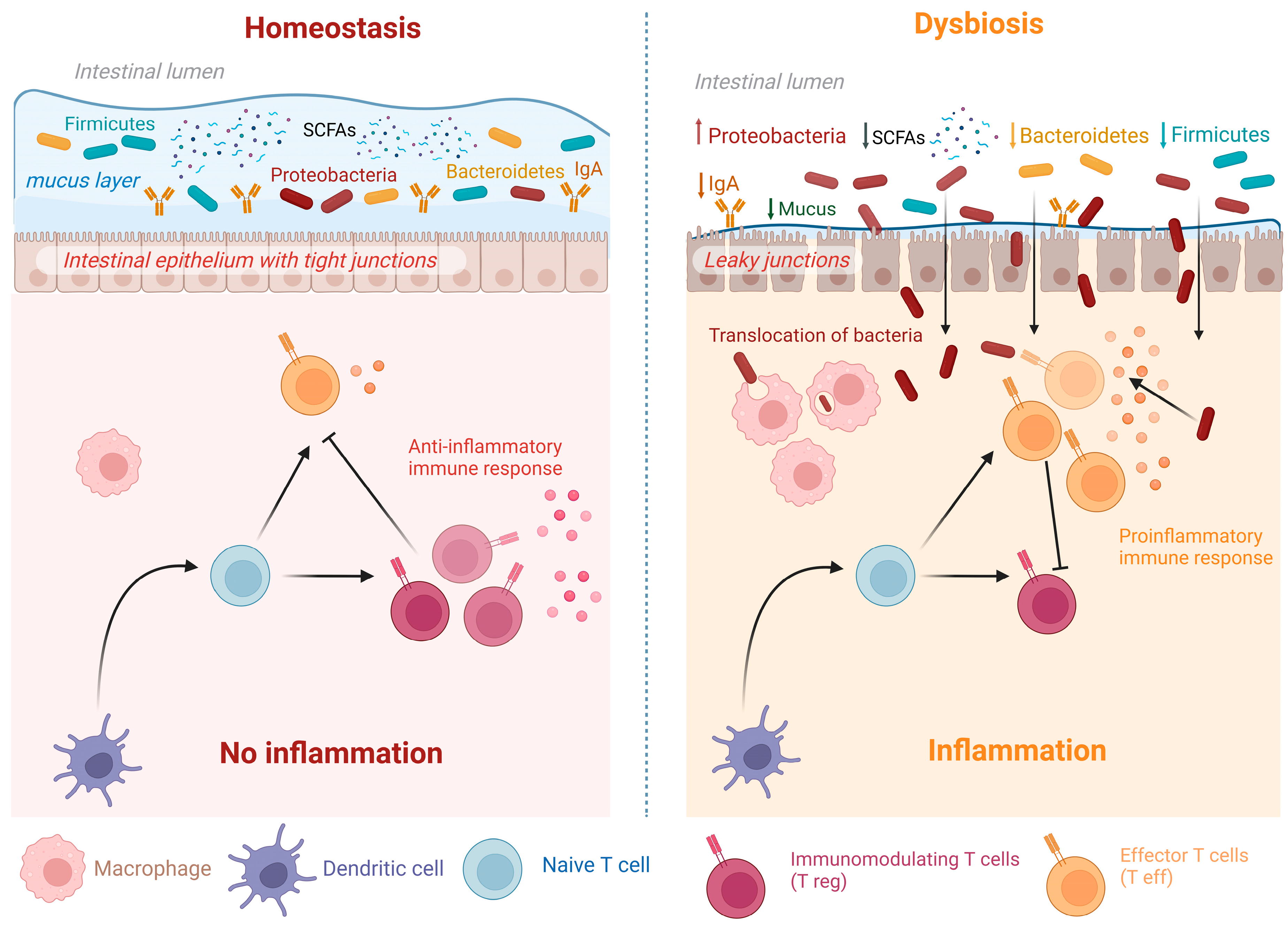
3. Dysbiosis and Relation to Diseases

If the normal microbiome is essential for maintaining suitable health, indeed, its disturbance is harmful [20]. Dysbiosis refers to an imbalance or disruption in the microbial abundance and/or function caused by alterations in microbiota composition and diversity in a particular ecosystem, such as the GIT [21]. Noteworthy, dysbiosis is a complex and multifactorial phenomenon, and its contribution to various diseases is still being actively researched. Research has shown that dysbiosis has significant implications for health. The disrupted microbiota may lead to a loss of beneficial functions performed by the microbes, impairing the metabolism of nutrients, affecting the intestinal barrier integrity, and altering the functions of innate and adaptive intestinal immunity compromising immune system regulation [18]. This can result in a broad array of diseases, including but not limited to metabolic disorders and gastrointestinal and inflammatory conditions [8,20]. Figure 1 summarizes the changes that occur in dysbiosis compared to normal homeostasis and the contribution of microbiota to both processes.
As shown in Figure 1, dysbiosis can cause disturbance of intestinal mucosal homeostasis. These changes are driven by several intrinsic and extrinsic factors, collectively and collaboratively leading to dysbiosis. Among these factors are genetic predisposition, diet, lifestyle, ecological factors, and therapeutic interventions such as antibiotics, lipid-lowering drugs, laxatives, proton pump inhibitors, ACE inhibitors, beta blockers, antidiabetic agents such as metformin, and others [22]. Furthermore, several studies reported that dysbiotic microbiota can affect human response to therapeutic intervention. The best example is microbiota influence on cancer immunotherapy, as the gut microbiome was shown to have the potential to modulate the efficacy and toxicity of several antitumor drugs against different types of cancer, thus impacting the outcome of such therapies [23]. In the coronavirus disease 2019 (COVID-19) era, some studies reported that gut microbes influence the immune response to severe acute respiratory syndrome coronavirus 2 (SARS-CoV-2) infection. Increased susceptibility to COVID-19 with more severe symptoms was noted among patients with dysbiotic gut microbiota [24,25]. This was explained by the ability of SARS-CoV-2 to replicate in the enterocytes, interacting with the gut microbiota, which in turn affected host immune responses. On the other hand, COVID-19 itself caused dysbiosis in infected patients [26], with persistent microbiota perturbations even after the clearance of infection [25].
Taken together, dysbiosis not only predisposes to diseases, but also impacts host’s response to infections, and medications. Thus, microbiome homeostasis is essential for suitable health while dysbiosis has a detrimental effect on health.

4. Gut Microbiota Alterations in IBD and Relation to IBD Pathogenesis

Inflammatory bowel illness is frequently a polygenic condition involving barrier dysfunction, gut microbiota, and dysregulated host responses to microbial stimulation. The precise etiology and pathogenesis of IBD remain unclear. The most accepted hypothesis explaining IBD is that chronic intestinal inflammation coupled with aberrant immune responses are derived from complex interactions between genetics, environmental factors, and the host immune system [27]. IBD is genetically connected to host pathways that suggest an underlying role for abnormal immune responses to gut microbiota [28]. The initiating stimuli for immune dysregulation are not fully explained by genetic predisposition. Genetic loci associated with the development of IBD can alter the delicate relationship between the microbiota in the GIT and the host, leading to a dysregulated immune response and intestinal inflammation [29]. In a meta-analysis of IBD genome-wide association in more than 75,000 cases and controls, many genes associated with IBD were found responsible for immune system recognition of microbes, suggesting that these patients are unable to either nurture beneficial bacteria or destroy the harmful ones [30].
Recently, deep-sequencing analyses of the microbiota in IBD patients have been instrumental in proving the strong association between dysbiosis and IBD development [20], but more research is indeed needed to identify the key species leading to IBD or contributing to the activity of the disease. In healthy people, the gut microbiome is highly individualized in both its condition and its dynamics. Nevertheless, cross-sectional research has shown that IBD is associated with gut microbiota dysbiosis [31]. In a previous study, Illumina HiSeq 2000 platform was used for the sequencing of the V4 amplicons [32]. By sequencing 248 million 16S rRNA gene amplicons, the microbiome composition in each sample was identified. Research has shown that individuals with IBD have distinct microbial profiles and has shown that IBD patients’ gut microbiota considerably varied from microbiota inhabiting the gut of healthy individuals.
Additionally, many identified microbial metabolites are reduced in people with IBD compared to healthy people, according to metabolomic studies. Research has speculated that IBD can be caused by the increase in certain proinflammatory bacterial metabolites and their producing species in IBD patients with the depletion of other anti-inflammatory metabolites and associated species that might have a protective role [33]. SCFAs are produced by Firmicutes and Bacteroidetes from carbohydrates, which triggers the production of associated proteins necessary for the development of tight junctions and supports intestinal mechanical barriers. Moreover, polysaccharide A generated by Bacteroidetes has anti-inflammatory effects by induction of Treg cells to release interleukin-10 (IL-10) which has a protective role on the intestinal barrier. However, the intestinal barrier function is destroyed and IBD vulnerability is increased because these microbiota are depleted in the colon of IBD patients [34]. Furthermore, research in germ-free mice indicates that gut bacteria impact body fat accumulation, metabolism, and immunological function [16]. The function of the microbiome in the pathogenesis of IBD is still being contested; however, the illness involves a significant inflammatory response that can be induced by acquired infection or alterations in the host’s own microbiome [35].
The mucosal layer in the gut is composed of a mucin complex that is rich in O-glycosylation. It has an imperative role in controlling germs growth, in addition to provision of food and space for intestinal microbiota to live. The stratification of the mucus layers and mucin synthesis are both influenced by gut bacteria (Figure 1). Any anomalies could lead to a malfunction in the gut population, which would lead to inflammation-related harm [36].
Bacterial 16S rRNA gene sequencing has improved our understanding of the microbiota makeup in various regions of the human body as well as the complex bacterial communities in IBD. Next-generation sequencing technologies helped in the precise identification of gut microbiota alterations in CD patients. An overabundance of bacteria from Enterobacteriaceae has been observed in individuals with IBD. On the other hand, beneficial bacteria like Faecalibacterium prausnitzii, which produces anti-inflammatory substances [37], are often depleted in individuals with CD. Gut dysbiosis, which includes enrichment of Proteobacteria, depletion of Firmicutes and Bacteroidetes, and a reduction in gut biodiversity, is definitely associated with CD [5]. Studies have pointed to the possibility of having certain infections implicated in CD pathogenesis, such as infections caused by Mycobacterium avium (subspecies paratuberculosis) and infections with adhesive-invasive strains of E. coli. These infections are most likely causing an imbalance in the total microbiota community, which is more significant for CD development [16]. Another intriguing finding is that the gut microbiota in CD patients is more unstable than in healthy persons. However, it is unclear whether the observed dysbiosis is the cause or the result of intestinal inflammation in CD, with growing evidence supporting the hypothesis that both hereditary and environmental variables are directly connected to dysbiosis [38]. In a recent Chinese cohort, patients with a recent diagnosis of CD possessed gut microbiota deficient in SCFAs-producing bacteria, such as Clostridium IV, Fusicatenibacter, Coprococcus, Blautia, and Dorea, with enrichment of Gram-negative pathogens such as Proteus, Escherichia, and Shigella when compared to healthy controls [39]. There was a significant correlation between the levels of some of these genera and various clinical parameters, inflammatory indicators such as fecal calprotectin, C-reactive protein, erythrocyte sedimentation rate, and counts of gland and goblet cells. These findings are important as they reflect microbiota perturbation early during the course of the disease with a progressive increase in the abundance of opportunistic pathogens. The latter can exacerbate microbiome dysbiosis in CD patients, hence, impacting the disease progression and outcome.
As for UC, alterations in gut commensals involved in microbiota-mediated defenses have been reported. When 16S rRNA microbial profiling was used for the characterization of the microbiota in UC patients, a significant decrease in Bifidobacteria was noted, especially the species Bifidobacterium bifidum, suggesting that this taxon may be biologically involved in UC pathogenies [40]. As for CD, the complex pathogenesis of UC is thought to be influenced by the interactive effect of environmental, genetic, and microbial variables. Mucosal permeability rises in UC due to an excessive inflammatory response within the lymphoid tissues. There is still much to learn about gut microbiota and its composition in UC, including various phyla such as Bacteroidetes, Firmicutes, Actinomycetes, and many others. UC status had been linked to microbial dysbiosis with observed temporal variations in microbiota composition as reported by multiple genomic-based microbiome studies. Although new initiatives went beyond genomics to profile the microbiota in UC, it is still vague whether these changes are causal or associative in nature [41]. To fill this knowledge gap, the host–microbiota interactions were examined in a big cohort of IBD patients to compare the metagenomic-metaproteomic strategy to more traditional 16S rRNA gene amplicon sequencing, fecal metabolomics, and serum proteomics techniques. Multiple bioinformatic tools as QIIME, DIAMOND, and STAMP were used to conduct a thorough, unbiased meta-analysis of the gut microbiome data from five distinct cohorts of IBD patients who originated from five different countries. Similar microbial species depletion in CD patients was verified by the analysis. It did, however, show a divergent enrichment in the UC cases. Following a thorough analysis, multi-omics data revealed that some proteases from the genus Bacteroides may play a role in UC pathogenesis. Meta-omics data specifically identified Bacteroides vulgatus proteases as possible treatment targets for UC [41]. Notably, butyrate, an essential SCFA made by some species, such as F. prausnitzii, was found to be increased in UC patients. This increase may signify an adaptive enrichment due to the vital role of butyrate in maintaining the epithelial cells in the gut [42]. In the induced UC model, intestinal microecology is significant, and the composition of the gut microbiome varies rapidly during the onset and progression of the disease. Researchers have demonstrated that Lactobacillus abundance decreased in UC. Anaerobic probiotic Lactobacillus produces vitamins and amino acids in the intestinal system. It may induce regulatory T cells (Treg) differentiation, reduce proinflammatory cytokines secretion, stop the growth of pathogenic organisms, and prevent the release of endotoxins, in order to balance the intestinal immune system and stop the progression of UC [43]. Saccharomycetales enrichment with microbiome diversity depletion was demonstrated as typical of UC gut dysbiosis using both fungal internal transcribed spacer 2 sequencing and bacterial 16S rRNA sequencing of fecal samples from 30 UC compared to 13 healthy controls [44]. Aside from that, it was discovered that four unique microbial community states (MCSs) verified their presence in a separate UC cohort and showed how they were related to the ethnicity of the patient as well as the severity of the illness [44]. Clinical, endoscopic, radiological, and histological results were used to make the diagnosis of UC. Through the sequencing of the V3-V4 region of the 16S rRNA gene and with the use of bioinformatic techniques, the makeup of the gut microbiota was identified. The functional makeup of the gut microbiota was anticipated by the Phylogenetic Investigation of Communities by Reconstruction of Unobserved States (PICRUSt) program. Intestinal flora in UC patients was considerably less diverse and rich than in healthy control subjects. When UC patients with active disease were compared to patients in remission status, alpha diversity did not show statistically significant variations, although intestinal flora compositions varied noticeably based on beta diversity analysis [45].
Comparatively to UC, the genetic effects of CD on the microbiota have received more attention, particularly those involving the NOD2 gene [15]. However, investigations involving mice and humans have also been conducted to shed light on UC variations connected to the microbiota. MHCII, HLA, ATG16L1, LRRK2, CARD9, CLEC7A, MUC5AC, and cytokine-related pathways (IL-17, IL-22) are specifically implicated in UC. Since this may be the only component not shared with CD, changes in the expression of mucus-related variations, including MUC5AC, seem relevant to determining peculiarities of UC development. As a result, this characteristic draws attention to the potential connections between microbiota dysbiosis, mucus composition, epithelial barrier disruption, and the development of UC [46]. Indeed, more research is needed to elucidate these links in IBD patients.

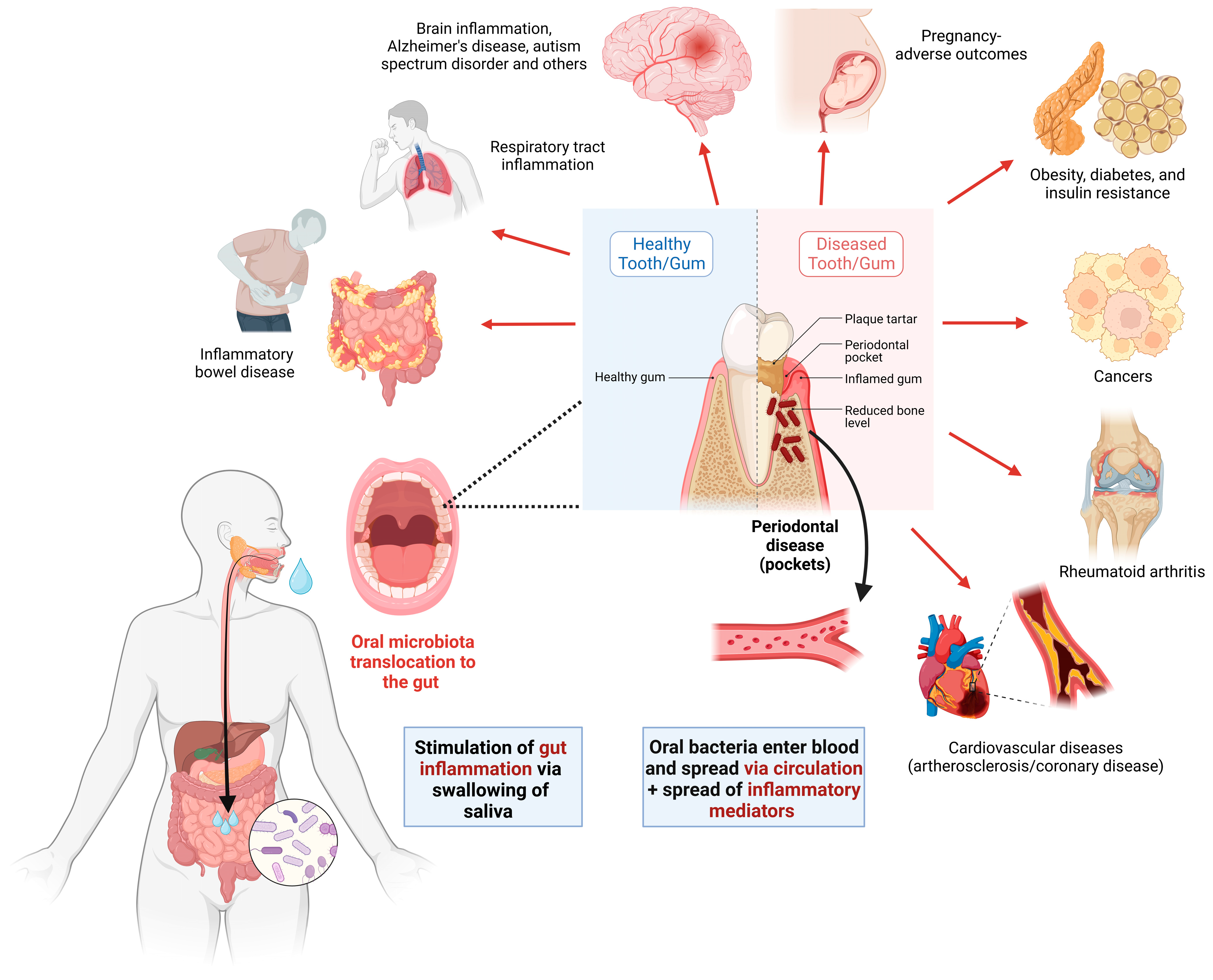
5. Oral–Gut Axis and the Role of the Oral Microbiome in IBD Pathogenesis

Although the mechanisms of the gut microbiota interaction with the immune system have received considerable attention [47], little is known about the connection between the oral microbiota and host immunity in IBD, and much less is known about IBD-related oral microbiota dysbiosis [47]. It is important to consider that the oral microbiome ranked as second after the gut microbiome in terms of having huge number of species, with high diversity and complexity of the microbial community [48]. The complex microbiota that inhabits the mouth includes more than 700 prominent species and contributes greatly to the host’s oral and extra-oral health. The oral and gut microbiomes, contain most of the microbial biodiversity in the human microbiome. However, these two groups are significantly different in terms of makeup. The most dominant bacteria in the oral cavity are facultative, sugar fermenting organisms such as Streptococcus and Actinomyces, whereas the most dominant organisms in the gut are metabolically varied population of anaerobic bacteria such as Clostridium and Bacteroides. Bacteriodetes, Firmicutes, Proteobacteria, Synergistetes, Fusobacteria, Spirochaetes, Actinobacteria, SR-1, and TM-7 are among the most prevalent bacterial phyla discovered in the oral cavity, using the human oral microbe identification microarray [47]. Surprisingly, the oral microbiome has less interindividual variance than the gut microbiome. The increased interindividual variance reported in gut microbiomes seems to be connected to the larger influence of variables such as nutrition and antibiotic use, whereas the oral microbiome appears to be more robust to similar challenges [47].
Since the GIT and the oral cavity are one continuous system, oral health may be directly related to gut health. Recent studies have shed light on the role of the oral microbiome in shaping the gut microbiome and subsequently impacting gut health. This is not surprising, as shown in the Human Microbiome Project, whereby oral and stool microbiota overlapped in approximately half of the subjects [49]. Some studies showed that the oral microbiota can be translocated from the mouth into the intestines causing microbial dysbiosis and inflammation but other studies on the other hand suggested that the oral microbiota dysbiosis will occur as a consequence of the gut inflammation so that it can be used as a biomarker of IBD [50]. Oral bacteria can enter the digestive system through swallowing, aspiration, or translocation, thus potentially influencing the microbial ecosystem in the gut [51]. Microorganisms may pass from the oral cavity to the gut while still being alive, despite the barrier of the gastric acidic pH, suggesting that oral–gut transit of microbes is likely common [50]. Pathological alterations of the oral microbiota, such as changes caused by periodontal disease, are associated with multiple inflammatory disorders, including IBD [52]. Inflammatory processes in the oral cavity can release inflammatory mediators that may affect the gut’s immune response and exacerbate intestinal inflammation. Furthermore, ulcerous periodontal pockets can serve as a portal of entry for subgingival microbiota with their components into circulation, leading to low-grade systemic inflammation, which can induce various systemic diseases [53].
Figure 2 shows oral microbiota translocation methods, impact on the systemic health, and relation to various diseases, including IBD.
Recent studies suggest crosstalk between gut and oral microbiomes, which may also contribute to IBD pathogenesis [52]. Metagenomic studies within the last 10 years have revealed comprehensive information on microbiome dysbiosis and surprisingly found a higher abundance of common oral taxa (such as Veillonella, Haemophilus, and Eikenella) in the gut of IBD patients, while many bacteria producing SCFAs were reduced. Microbiota translocation from the mouth to the gut may be a unique phenomenon leading to microbial dysbiosis, which is a special signature of IBD [50]. Pathobionts can move between the mouth and gut following a local inflammation starting in the oral cavity, then disseminating to the intestine [54]. A recent study in mice has shown that inflammation of the oral mucosa combined with oral pathobionts’ proliferation led to colitis via gut colonization and the induction and migration of bacteria-reactive T cells (Th17) to the gut. Thus, both pathobionts and pathogenic T cells derived from oral inflammation and translocated to the gut can collectively cause exacerbation of intestinal inflammation [55]. Another study reported that inoculation of saliva from children with CD to germ-free mice led to the accumulation of inflammatory IFN-γ+ CD4+ TH1 cells in the intestinal lamina propria and enrichment of Fusobacterium, Veillonella, and Klebsiella spp. in the fecal microbiome of the inoculated mice [12]. It was reported that multidrug-resistant Klebsiella strains isolated from the saliva of IBD patients can colonize the gut of mice when the intestinal microbiota is dysbiotic, eliciting a high-grade inflammatory response in the gut of genetically susceptible hosts [12]. These observations provide strong evidence supporting the connections between the oral–gut axis, oral microbiome, and immune-mediated mechanisms in IBD pathogenesis.
By collecting oral samples, it has been possible to assess the health of the gut by utilizing the oral–gut axis. Since each person produces between 1 and 2 L of saliva each day, saliva has been considered a desirable biospecimen for sampling and a valuable source of disease biomarkers [56,57]. Utilizing saliva as a biofluid has several benefits, including a quick, simple, non-invasive, and affordable collecting procedure that is suited outside of the laboratory and does not require medical professionals. Even though it is still in its early stages, research on the oral–gut axis shows promise [58].

6. Oral Dysbiosis Link to Oral Diseases and Contribution to IBD

It is speculated that the microbiology of the oral cavity may be unique in IBD patients [59]. W.D. Miller, a pioneering oral microbiologist, postulated in the 1890s that bacteria in the mouth and their products might have a dramatic impact on several illnesses, both local and systemic, owing to dental bacteremia, which was dubbed “oral focal infection hypothesis” [5]. Even though the mouth is constantly exposed to a bombardment of host and environmental toxins, the oral microbiome in healthy people remains reasonably consistent over time. Given this, alterations in the oral microbiota profile may provide correlative insight into illness initiation, progression, and recurrence.
Numerous studies have demonstrated the link between oral flora imbalance and inflammation, and how this link may contribute to the development of systemic diseases (Figure 2) such as Alzheimer’s disease, diabetes mellitus, cardiovascular disease, preterm birth, and obesity as well as gastrointestinal disorders like colorectal cancer and IBD and autoimmune disorders like rheumatoid arthritis through bacteremia [60]. One key aspect of the oral–gut connection is the transmission of specific microorganisms from the mouth to the gut. The reciprocal flow of bacterial pathogens provides more evidence of commonalities between the oral and gut microbiomes [61]. Oral microbiome disruptions caused by poor oral hygiene or induced by excessive sugar intake may result in oral diseases such as dental caries, and gum diseases including periodontitis. The oral microbiome has been well characterized in terms of its role in oral diseases, but its members have also been implicated as contributing factors in a variety of extra-oral diseases [51,62].
Some pathogenic oral bacteria, such as Fusobacterium nucleatum and Porphyromonas gingivalis, have been detected in gut biopsies of IBD patients, suggesting a potential role in disease progression. Dysregulated host–oral microbiota interaction is crucial in the pathogenesis of many oral diseases. IBD patients also appear to have an increased risk for oral diseases such as dental caries and periodontitis [63]. Moreover, studies have shown that individuals with periodontal disease are at a higher risk of developing IBD. Periodontitis is an inflammatory oral disease with a high global prevalence and association with multiple health issues [11,56,64]. It is a progressive chronic inflammatory condition that can lead to localized bone loss. In patients with periodontitis, several systemic markers of inflammation are increased; thus, it may represent an indirect mechanism explaining how the oral bacteria participate in the pathogenesis of systemic diseases, such as IBD [52]. Furthermore, gut microbiota dysbiosis can be induced by periodontitis via salivary microbiota. In a previous study, saliva-sourced microbes were enriched in the fecal samples of patients with severe periodontitis. When saliva from the latter group of patients was orally administered to mice, enrichment of oral bacteria from Porphyromonadaceae and Fusobacterium was evident. This was accompanied with pathological changes in the gut, as the colon tissues showed significantly higher expression of proinflammatory cytokines, chemokines and tight junction proteins with low zonula occludens-1 expression and crypt depth [65]. Thus, it is important to investigate oral health, IBD activity, and their relation to oral microbiota dysbiosis.

6.1. Oral Microbiome Alterations in CD

Although the host–microbe interaction has been linked to the pathophysiology of CD in genetically predisposed hosts, little is known about oral microorganisms in CD. Recent microbiome research suggests that the translocation of oral bacteria to the gut leads to microbial dysbiosis, which is a hallmark of CD [5]. Few studies have looked specifically at the influence of CD on the oral microbiota and its relation to oral health [50]. A common extraintestinal sign of CD is oral pathology, such as angular cheilitis, linear ulcerations, mucogingivitis, cobblestoning of mucosa, and persistent mucosal swelling [66]. Oral signs found in CD patients imply a link between oral microbiota and such manifestations; nevertheless, little is known about the oral microbiota of CD patients [31]. Regardless of the presence of oral symptoms, the oral mucosa is an immunologically active surface with higher cytokine generation. It has previously been reported that particular oral bacteria, such as a subset of Porphyromonas gingivalis, Streptococcus mutans, Fusobacterium nucleatum, Campylobacter concisus, and Klebsiella pneumoniae, may aggravate inflammation in CD [47]. Ectopic colonization by these oral bacteria may break down the intestinal epithelial barrier, generate excessive release of inflammatory cytokines, disturb the host immune system, promote immunological escape, and create gut microbiota dysbiosis, ultimately exacerbating chronic intestinal inflammation. Previous research focused only on changes in the makeup of the oral microbiota while ignoring the relationship between oral microbiota dysbiosis and the inflammatory state in the gut [47]. Although the makeup of the communities in the mouth and gut differs, the amount of species richness in both settings is comparable, and a single individual may house over 100 unique species at each site [47].
A significant reduction in both total microbial diversity and particular phylum levels was found in CD. Furthermore, the loss of certain phyla, such as Fusobacteria and Firmicutes, has been demonstrated in investigations of the gut microbiome in CD. The oral microbiota is changed in IBD patients, particularly in CD [59]. Remarkably, patients with CD and oral symptoms had significantly greater anti-Saccharomyces cerevisiae antibody (ASCA) titers than those without oral signs [59]. Salivary microbiota dysbiosis is associated with inflammatory responses in IBD patients, indicating that it is probably related to gut microbiota dysbiosis [47].
In a recent study, the differences in taxonomic and predicted functional pathways were clearly observed in saliva samples collected from patients with CD during active and remission phases of the disease compared to healthy controls. Both alpha and beta diversities were significantly lower during the active phase in contrast with the remission phase and healthy controls [27]. A total of 30 bacterial taxa were significantly enriched in the active phase. In contrast, remission phase and healthy controls exhibited significant enrichment of other 24 and 22 bacterial taxa, respectively. It has become evident that typical mouth-resident bacteria, such as Fusobacteriaceae, Pasteurellaceae, and Veillonellaceae, are enriched in the mucosal tissues of CD patients [28]. Another study reported that the salivary microbiota in CD patients was significantly different from that of healthy controls [31]. The relative abundance of Bacteroidetes was significantly higher, while that of Proteobacteria was significantly lower in the saliva of CD patients compared to the healthy controls. Several important pathways, such as ribosome biogenesis and energy metabolism, were depleted in the active phase. Eleven differentially abundant pathways were also identified; four were significantly enriched in healthy controls, whereas another seven were significantly enriched in the active phase of the disease. This study has highlighted several taxa and functional categories that could be implicated with the onset of CD and thus have the potential to serve as biomarkers of the active disease [27].
A substantial reduction in the total variety of tongue microbiota was seen in CD, according to a pediatric study that included 40 children with CD and 43 control patients without IBD. A phylum-level study of salivary microbiota showed that individuals with IBD had higher concentrations of Bacteroidetes and lower concentrations of Proteobacteria. These investigations have revealed oral microbial variations in individuals with IBD, suggesting the importance of the oral microbiome in IBD diagnosis and patient monitoring [67].

6.2. Oral Microbiome Alterations in UC

Numerous studies have examined the modifications in the microbiome of patients with UC. There have been reports of, among other variations, a decline in biodiversity and depletion of the phyla Firmicutes and Bacteroidetes. Changes in microbial composition have an impact on the amount of butyrate and other metabolites like H2S produced in the gut as well as the metabolites that are created there because of microbial activity [68].
Although oral lesions have sporadically been linked to UC, a recurrent pathology with a complicated etiology, little is known about the general makeup of the oral microbiome in UC patients or its significance in the pathogenesis of the illness. The oral microbiomes of healthy people and UC patients were compared to determine any potential changes in the oral microbial communities related to UC. For this, 16S rRNA gene sequencing was used to examine the salivary microbiota of 10 patients with UC diagnosed in the active period and 11 healthy controls [69]. A metataxonomic study of the oral core microbiome in UC patients showed a decline in alpha diversity and an imbalance in the relative proportions of some important members. In addition, four distinct species or phylotypes of Staphylococcus and other organisms were only found in UC patients and were undetectable in healthy individuals. A comprehensive picture of the existence of oral dysbiosis related to UC and the potential existence of oral biomarkers was shown before [69].
Using Illumina sequencing of the V3-V4 region of the 16S rRNA gene, the salivary microbiomes of 54 UC patients, 13 CD patients, and 25 healthy controls were compared. Comparing taxa abundances showed enrichment of Streptococcus and Enterobacteriaceae in UC and enrichment of Veillonellaceae in CD, with depletion of Lachnospiraceae and Prevotella in UC and depletion of Neisseriaceae and Haemophilus in CD [5].
In comparison to healthy people, the detection frequency of the specified species was noticeably greater in UC patients. Colitis was made worse by administering the specific Streptococcus mutans strains that were identified in patients. One potential risk factor for the worsening of UC is infection with some strains of very pathogenic S. mutans [70]. Only in gingivitis areas, UC patients had a significantly higher abundance of S. aureus and P. anaerobius than controls. Peptostreptococcus anaerobius, Staphylococcus aureus, Streptococcus anginosus, Streptococcus intermedius, Streptococcus mitis, Streptococcus mutans, and Treponema denticola were among the bacteria with different counts between the groups, according to a multiple comparison analysis. In either gingivitis or periodontitis areas, CD patients had significantly greater amounts of these microorganisms than UC patients. Only in gingivitis areas did UC patients have greater concentrations of S. aureus and P. anaerobius than controls. IBD patients had increased concentrations of opportunistic infection-related bacteria in inflammatory subgingival regions, which could be detrimental to the vital microbe–host connection [71].
Table 1 summarizes the studies exploring the oral microbiome in CD and UC patients, with microbiota alteration reported in each group. Indeed, fewer studies were conducted in the pediatric population compared to the adult population.

7. Factors Influencing Microbiome Composition and Their Impact on IBD Pathogenesis

The microbiota benefit from their mutualistic interaction with the human host while living in a nutrient-rich environment [76]. Indeed, many factors can affect the residing microbiota. Studies have shown that a diet missing essential nutrients needed for commensal survival, the use of drugs, physical exercise, smoking, and mental and emotional stress are some environmental factors that may lead to intestinal inflammatory disorders [58]. We summarized the most important factors contributing to microbiota dysbiosis in relation to IBD as follows.

7.1. Early Life Factors

During the first three years of life, the gut microbiome is gradually established [77]. Previous studies have shown that alterations in the gut microbiome in infants are associated with gastrointestinal disorders and other long-term health issues [78]. A few years after birth, the gut microbiota progressively takes shape to reach its mature composition, with temporal variations in the richness and order of dominating species being mostly influenced by food and host physiological conditions [31]. Early childhood “healthy” gut microbiota are necessary for microbiota–host interactions that have a long-lasting effect on adult health and disease status [6].
Some of the most crucial factors influencing the colonization of the early-life microbiome are antibiotic use, feeding habit, and mode of delivery [79]. In fact, early-life antibiotic exposure may potentially be a significant risk factor for developing IBD later in life. Moreover, it is also possible that prolonged use of broad-spectrum antibiotics, not just in early life, may be associated with the development of IBD [79]. Breastfeeding, especially prolonged breastfeeding, was preventative of the development of CD or UC. Bioactive compounds and immunological cells found in human breast milk serve important roles in forming the newborn immune system as well as passive immunity. Additionally, human milk oligosaccharides function as prebiotics, encouraging beneficial intestinal flora. Numerous research studies examined the relationship between the manner of delivery and IBD. These studies produced contradictory findings, with the majority of the studies finding no relationship. Although studies have indicated that Cesarean section causes distinct microbial community structure and function, these variations vanish within 6 weeks of age. C-sections may, thus, not be a sufficient prenatal risk factor for IBD [79]. Although research has been performed on genetically vulnerable animal models to assess the impact of particular environmental conditions on immune responses and microbiota, research on human subjects is very few. These linkages will be clarified in the future by human research intended to assess the effects of environmental variables on microbiota–immune interactions in individuals with established genetic risk factors for IBD [80].

7.2. Effect of Diet and Supplements

Indeed, gut dysbiosis can be induced or aggravated by the intake of some dietary components. Based on several case–control dietary studies, the link between a high-fat diet (HFD) and IBD is now well established, and an HFD is seen as a possible risk factor for developing IBD. Higher intestinal permeability and altered gut flora may contribute to the increased risk brought on by the HFD [76]. A Western diet is thought to play a significant role in both the obesity pandemic and the development of IBD; thus, it is not unexpected that Westernized nations have the greatest prevalence of IBD [58]. In contrast, high intakes of fibers from vegetables and fruits were linked to a lower risk of UC and CD [80]. While Bacteroides species are more prevalent in children who eat Western diets high in fat and poor in fiber, Prevotella and Succinivibrio are more prevalent in diets high in plants and fiber. Western diets rich in fat may have an influence on the development of inflammatory bowel illness by reducing biodiversity and SCFA production [50].
Additionally, a diet rich in heme (such as red meat and beef) is characteristic of the Western diet with a distinct impact on gut microbiota, increasing the risk of IBD [81,82,83]. Research on mice has shown that a diet rich in heme can cause disturbance of the gut homeostasis due to the overgrowth of pathogenic bacteria (Proteobacteria, especially Enterobacteriaceae), depletion of useful microbial groups (Firmicutes), decrease in essential SCFAs (butyrate) and a drop in the alpha diversity. This is accompanied by intense intestinal inflammation, which is very similar to the chemically induced colitis (dextran sodium sulfate; DSS) used in animal models of IBD [84].
Iron deficiency anemia is one of the complications of IBD due to intestinal bleeding, poor absorption caused by gut ulceration, and loss of appetite leading to less dietary intake of natural sources of iron. As iron supplementation may be needed in IBD patients with severe anemia, it must be administered with caution [85]. Oral iron administration may not be well tolerated in many patients and can have a negative impact on the gut and the microbiome, thus triggering inflammation and contributing to dysbiosis. This was proven experimentally in DSS-induced colitis, whereby administration of dietary iron exacerbates colitis. This was explained by the ability of iron to induce oxidative stress through the Fenton reaction, coupled with inflammation due to the activation of the nuclear factor kappa-B (NF-κB) pathway leading to upregulation of the expression of proinflammatory cytokines [86,87]. Another study proved the harmful effect of iron on the gut microbiome in DSS-induced colitis, in which a high dose of dietary iron caused a significant depletion of fecal Firmicutes and Bacteroidetes, concomitant with enrichment of Actinobacteria and Proteobacteria. Furthermore, a significant increase in fecal calprotectin level was noted, reflecting more intense inflammation correlated with increased disease activity in colitis [85]. Moreover, the harmful effect of oral administration of iron was shown in patients with IBD. A clinical trial was conducted to compare the effect of oral versus intravenous (IV) iron replacement therapy on the gut microbiome of IBD patients. However, both oral and IV therapies ameliorated iron deficiency, oral iron therapy induced gut dysbiosis due to the depletion of beneficial microbiota, namely Faecalibacterium prausnitzii, Ruminococcus bromii, Collinsella aerofaciens, and Dorea species [88,89].
On the other hand, there are some therapeutic modalities that rely on exclusive enteral nutrition (EEN) to alleviate IBD symptoms [90]. EEN involves the use of a nutritionally complete liquid diet provided exclusively for up to 8 weeks instead of usual solids. The capability to employ EEN as a substitute for corticosteroids to produce remission in CD is unique to children with IBD, especially after the first diagnosis of the disease. EEN is a successful, non-pharmacologic method of inducing remission that also enables doctors to address any dietary requirements of the kid whose health has been impaired by an inflammatory condition. EEN is thought to modify the microbiota and create an anti-inflammatory environment in the gut while also aiding mucosal repair; however, the precise method by which it functions is yet unknown. This dietary intervention is an alternative for the treatment of children and young adults since it does not have the negative effects or immunosuppression associated with many other treatments [7].
Noteworthy, dietary modulation of the gut microbiome in IBD has shown promising results. For instance, the Crohn’s disease exclusion diet (CDED), which is a low-fat, high-protein diet, has been effective for managing children with mild to moderate CD, as well as for patients with an unfavorable response to biological therapy. Changes in microbial diversity, depletion of Proteobacteria, and enrichment of Firmicutes, particularly Clostridiales, were noted following the intake of this diet [91]. Furthermore, studies in mice have shown that a ketogenic diet (KD) and low-carbohydrate diet (LCD) can cause modulation of specific composition and function of gut microbiota and metabolites. KD dramatically increased the abundance of Akkermansia and Roseburia associated with improved immune responses and protective barrier functions. Moreover, significantly fewer inflammatory responses with less expression of inflammatory cytokines were shown following KD. On the contrary, LCD caused opposite effects compared to KD [92]. Another study in UC patients confirmed that consuming a low-fat, high-fiber diet helped to resolve both dysbiosis and gut inflammation with prolonged remission. This improvement was correlated with the increase in the abundance of beneficial microbiota such as F. prausnitzii after regular consumption of this diet. Interestingly, remission was also accompanied by an increase in fecal acetate levels, which is one of the beneficial SCFAs with anti-inflammatory effects [93].

7.3. Effect of Exercise

Exercise can increase the microbiome diversity; increase the Bacteroidetes/Firmicutes ratio, which may help to reduce weight, obesity-related diseases, and gut disorders; and stimulate the growth of bacteria that can control mucosal immunity and improve barrier functions. Particularly, the Firmicutes phylum has more diversity, which supports the maintenance of a healthy intestinal environment [94]. Exercise altered the makeup of the gut microbiota in one study in which women engaged in physical activity to at least the level advised by the WHO, and 11 genera were substantially different between active and inactive women. The healthier bacteria Faecalibacterium prausnitzii, Roseburia hominis, and Akkermansia muciniphila were more prevalent in the active women. Metagenomic analysis indicated taxonomic modifications, including an increase in Akkermansia and a decrease in Proteobacteria, in another 6-week endurance exercise research without dietary changes [95]. However, increasing the training load (i.e., lengthening the exercise period or raising the level of physical activity) may lead to gut dysbiosis and have a detrimental impact on the digestive system and result in symptoms including nausea, vomiting, diarrhea, colic, flatulence, and stomach discomfort [96].
The role of exercise in preventing and restoring gut dysbiosis in patients with IBD is well established [97]. Physical exercise was thought to be a modulator of the intestinal microbiome, leading to positive clinical outcomes in IBD patients. Research has suggested that crosstalk between muscles and the gut microbiota is induced by exercise through the 5′ adenosine monophosphate-activated protein kinase and fasting-induced adipose factor pathways as well a reduction in fecal bile acids, an increase in SCFAs production, an increase in gut luminal IgA, a reduction in luminal transit time, and the activation of the stress hypothalamic–pituitary–adrenal axis [97].

7.4. IBD Drugs and Microbiota

Since there is currently no medication that may cure IBD, treatment focuses on symptom management, mucosal healing, inducing and maintaining remission of the active illness, preventing relapses, hospitalizations, and surgeries, and generally enhancing the patient’s quality of life [98]. Management of CD is decided by considering several factors, including the patient’s age, disease location, and behavior [99]. Most current therapies for IBD rely on suppressing the host immune system without direct effect on the microbes that cause or contribute to inflammation. Complete remission induced by these therapies occurs in less than half of patients; thus, discovering new therapeutic targets is needed [100]. Antibiotics, anti-inflammatory agents such as corticosteroids, and 5-aminosalicylic acid (5-ASA) are among the medications used to treat IBD. Corticosteroids are not always effective in maintaining remission and usually contribute to growth impairment in pediatric patients [101]. Immunosuppressive medications such as thiopurines are commonly used for the maintenance of remission in steroid-dependent and steroid-refractory IBD [102]. Immunomodulators are used for the maintenance of remission in moderate to severe IBD [4]. Biological therapies, including monoclonal antibodies such as anti-TNF (IFX, adalimumab, and certolizumab pegol), anti-integrin (VDZ), and anti-interleukin ustekinumab (blocking p40 subunit of IL12/23), are used to suppress the immune response. They are usually considered in IBD patients with high disease activity, features indicating poor prognosis, and inadequate response to standard therapy [99]. The widespread use of biological therapies and immunomodulators in pediatric patients has altered the natural course of the disease and impeded the progression of complications. The introduction of therapeutic monoclonal antibodies directed against TNF, a major proinflammatory pathogenic cytokine in IBD, has revolutionized the treatment of CD. The use of TNF-α antagonist (infliximab) was found effective in inducing and prolonging remission in children with IBD, helping in mucosal healing in this population, and reversing growth failure in children with severe refractory CD [103]. Interestingly, it has been reported that initiation of medical treatment with anti-TNF-α in pediatric patients with active IBD caused changes in the fecal microbial composition [104]. The abundance of six bacteria, including Eubacterium rectale and Bifidobacterium spp., predicted the response to anti-TNF-α medication. Another study demonstrated that the change in the treatment regimen influenced the gut microbiome of IBD patients, including those with CD [32]. Patients who had received a course of oral corticosteroids at disease flare had more microbiome fluctuations than patients on stable medications [32]. These findings indicate that the dynamics of the gut microbiome composition can be influenced by the medications used to treat the disease.
As for the oral microbiome, subgingival microbiota was investigated in pediatric CD patients. About 17 genera were reported as candidate biomarkers for the disease. Of them, Capnocytophaga, Rothia, and TM7 were more abundant in CD compared to healthy controls. Alloprevotella, Fusobacterium, Porphyromonas, and Prevotella were consistently decreased in the antibiotic-exposed CD patients compared to CD subjects not using antibiotics. CD-associated genera were not present in samples obtained after 8 weeks of treatment (either with corticosteroids, immunomodulators, enteral therapy, or 5-ASAs), suggesting alterations in CD microbiome community structure in response to successful therapy [74].

8. Microbiome Modulation to Treat IBD

Indeed, any treatment utilizing gut microbiota relies on the reversal of its dysbiosis. Addressing dysbiosis often involves restoring a healthy balance of the microbiome [19]. Microbiome-based personalized treatments are being explored to target specific imbalances or dysfunctions within microbial communities. This can be achieved through interventions such as dietary modifications, probiotic supplementation, prebiotics, postbiotics and lifestyle changes that support a diverse and balanced microbial community.
Probiotics are living non-pathogenic microbes administered in food or dietary supplements to offer health benefits to the host. Several microorganisms, such as Lactobacillus, Bifidobacterium, Saccharomyces, and Streptococcus, have been recognized with useful probiotic properties [105]. These microbes have multiple benefits to the gut via modulation of microbiota composition, modulation of the immune response, and enhancement of intestinal barrier functions [106]. In inflamed mucosa of active IBD or experimental UC, downregulation of the expression of proinflammatory cytokines like TNF-α, IL-6, and IFN-γ, nitric oxide synthase, and matrix metalloproteinase activity was shown after probiotic treatment [107]. Moreover, the secretion of vitamins and other metabolites by probiotic bacteria can indirectly modulate intestinal microflora, which can be induced to produce beneficial SCFAs, ultimately leading to the alleviation or improvement of IBD [108]. A recent study reported the use of programmable probiotics to modulate inflammation and gut microbiota for IBD treatment. In the latter study, effective oral delivery of genetically engineered bacteria (Escherichia coli Nissle 1917) that overexpress catalase and superoxide dismutase was used to clear reactive oxygen species in the inflamed tissue in mice. This method was successful in restoring intestinal homeostasis by improving the abundance of Lachnospiraceae_NK4A136 and Odoribacter in the intestinal flora. Treated mice showed rapid recovery of body weight and alleviated colonic mucosa damage with less colonic MPO activity. Moreover, probiotic effectiveness was proven in human model of IBD, more in UC than in CD [108]. Most of these studies reported improved symptoms, reduced recovery time, maintaining remission and preventing recurrence, and reduction in disease activity index and intestinal permeability. This was coupled with the cessation of inflammation marked by lower IL-1β expression and higher expression of IL-10 and IgA in the colonic mucosa [108]. Nevertheless, a few studies reported no therapeutic effect, as reported in one of the old studies showing the ineffectiveness of Lactobacillus johnsonii LA1 to prevent postoperative recurrence in CD [109]. There has been a significant improvement in patient symptoms in several trials looking at probiotic supplements for the treatment of IBD. Numerous studies have shown the benefits of certain probiotic bacteria, including strains from the genera Lactobacillus and Bifidobacterium. Researchers discovered a reduction in inflammation because of metabolic changes resulting from altered gut microbial populations in a trial using fermented milk products containing Bifidobacterium lactis in mouse models of colitis. According to these data, a change to the gut environment with a more powerful commensal bacterial population may protect the gut lining and make it unfavorable for opportunistic strains [58]. According to the American Gastroenterological Association Institute, probiotics may be used to alleviate functional symptoms of IBD. By preventing the growth of harmful bacteria and encouraging the growth of suitable species, probiotics may have anti-inflammatory effects, enhance barrier function, and positively regulate the microbiome composition. In individuals with CD and UC, the probiotic cocktail VSL#3 (a combination of one strain of Streptococcus, plus three strains of Bifidobacteria, and four strains of Lactobacilli) decreased recurrence and maintained remission [33].
Postbiotics are composed mainly of SCFAs, enzymes, peptides, vitamins, peptidoglycans, and polysaccharides produced from microbial fermentation [110]. Among them, SCFAs are the most promising adjuvant therapy in the clinical management of IBD patients with active disease. Acetate, propionate, and butyrate were used either individually or in combination, mainly in patients with UC. The results were inconsistent as some studies reported beneficial effects and improvement in the clinical parameters, while others could not find any significant improvement [111]. Nevertheless, a recent study reported a better modulatory effect on the gut microbiome induced by postbiotics than probiotics (from Bifidobacterium adolescentis B8589) in a mouse colitis model [112].
Other microbial-based therapies for IBD include fecal microbiota transplantation (FMT), which is a technique of transferring healthy fecal microbiota to the gut of patients with dysbiosis-related gut disorders in order to restore a healthy gut microbiome. A large body of evidence proved the efficacy of FMT for the treatment of recurrent Clostridium difficile infection, but the effect was variable depending on the delivery method and the frequency of administrations [113]. In terms of clinical remission, frozen fecal material produced better results compared to fresh material. FMT can be delivered by oral capsules, enemas, nasogastric or nasojejunal tubes [107]. Current evidence on the effectiveness of FMT for IBD is inconclusive, according to a recent systematic review and meta-analysis [114]. However, there are studies supporting the use of FMT coupled with other treatment modalities. In a recent a randomized controlled trial, FMT provided with an anti-inflammatory diet followed by an anti-inflammatory diet alone was effective in inducing and maintaining remission over one year in cases of UC with mild to moderate disease [115]. Another recent randomized, double-blind, placebo-controlled trial reported that the administration of antibiotics followed by oral FMT was associated with the induction of remission in patients with active UC. Furthermore, continuing FMT was well tolerated, with better clinical, endoscopic, and histological scores [116]. There are reports proving the effect of FMT on gut microbiota restoration, mainly in UC. Several genera, such as Bacteroides, Proteus, and Prevotella, were significantly enriched, while pathogenic bacteria from the genera Klebsiella and Streptococcus decreased significantly after FMT [107].
Noteworthy, the vast majority of the previous studies had significant limitations related to the low sample size and difficulty in having proper study groups; for example, some patients may have concomitant treatment with immunosuppressants and steroids, which can affect outcomes [111]. This indeed pinpoints the need for conducting more comprehensive studies in the future.

9. Summary and Outlook

The microbiome encompasses a vast ecosystem of microorganisms that profoundly influence the health of their hosts [31]. Dysbiosis is the consequence of disruptions in the microbiome, which is described as changes in the organization of a microbial community. The resulting dysbiosis manifests as an altered balance of microbiota elements. This may impede key microbiome functions, including resistance to harmful bacteria. The study of the microbiome in different niches has greatly advanced our understanding of various diseases, including IBD. Dysbiosis in IBD is not limited to the gut but also includes the oral microbiome. Indeed, the oral–gut connection is a crucial component of IBD pathogenesis. Bidirectional communications between these two environments have implications for human health. Since the oral and gut microbiomes account for the majority of the overall human microbial load, they offer unique prospects for improving human health, diagnosis, prognosis, outcome prediction, and discovery of targeted therapies [61]. Further understanding of these microbiomes and their interactions with the human body has the potential to revolutionize healthcare. Renewed research efforts employing next-generation sequencing for high-resolution characterization of the composition, function, and ecology of microbiota have improved our overall knowledge of the role of microbiota in health, which is required for the study of disease-related dysbiosis.
Delving into the mechanisms of microbial interaction and their implications for disease pathogenesis indeed contributes to the growing body of knowledge in the field. Ultimately, a better understanding of the oral–gut microbiome relationship in IBD can pave the way for targeted interventions and personalized approaches to improve gut health and disease outcomes for affected individuals. Elucidating the intricate interplay between the oral and gut microbiome in IBD patients holds promise for novel therapeutic interventions. Strategies that target the oral microbiome, such as improved oral hygiene practices and periodontal disease treatment, may help in modulating the gut microbiome and alleviating IBD symptoms. Furthermore, manipulating the gut microbiome through dietary interventions, prebiotics, probiotics, and FMT could have a beneficial impact on oral and gut health.

Author Contributions

Conceptualization, F.A.-M., H.E. and G.M.; methodology, F.A.-M.; software, F.A.-M. and H.E.; formal analysis, F.A.-M., H.E. and G.M.; investigation, F.A.-M., H.E. and G.M.; resources, F.A.-M.; data curation, F.A.-M.; writing—original draft preparation, F.A.-M., H.E. and G.M.; writing—review and editing, F.A.-M.; visualization, F.A.-M.; supervision, F.A.-M.; project administration, F.A.-M.; funding acquisition, F.A.-M. All authors have read and agreed to the published version of the manuscript.

Funding

This work was supported by a grant from the Zayed Bin Sultan Charitable and Humanitarian Foundation (ZCHF), United Arab Emirates University, UAE (grant number G00004262), awarded to FA.

Institutional Review Board Statement

Not applicable.

Informed Consent Statement

Not applicable.

Data Availability Statement

Not applicable.

Acknowledgments

We acknowledge the support of the Office of Research and Graduate Studies, College of Medicine and Health Sciences to the master’s projects of the students Hala Elzayat and Ghaidaa Mesto. Figures were prepared using Biorender (https://www.biorender.com/ (accessed on 26 June 2023)).

Conflicts of Interest

The authors declare no conflict of interest.

References

  1. Alatab, S.; Sepanlou, S.G.; Ikuta, K.; Vahedi, H.; Bisignano, C.; Safiri, S.; Sadeghi, A.; Nixon, M.R.; Abdoli, A.; Abolhassani, H.; et al. The global, regional, and national burden of inflammatory bowel disease in 195 countries and territories, 1990–2017: A systematic analysis for the Global Burden of Disease Study 2017. Lancet Gastroenterol. Hepatol. 2020, 5, 17–30. [Google Scholar] [CrossRef] [Green Version]
  2. Park, J.; Cheon, J.H. Incidence and Prevalence of Inflammatory Bowel Disease across Asia. Yonsei Med. J. 2021, 62, 99. [Google Scholar] [CrossRef]
  3. Seyedian, S.S.; Nokhostin, F.; Malamir, M.D. A review of the diagnosis, prevention, and treatment methods of inflammatory bowel disease. J. Med. Life 2019, 12, 113–122. [Google Scholar] [CrossRef] [PubMed]
  4. Diefenbach, K.A.; Breuer, C.K. Pediatric inflammatory bowel disease. World J. Gastroenterol. 2006, 12, 3204–3212. [Google Scholar] [CrossRef]
  5. Xun, Z.; Zhang, Q.; Xu, T.; Chen, N.; Chen, F. Dysbiosis and Ecotypes of the Salivary Microbiome Associated With Inflammatory Bowel Diseases and the Assistance in Diagnosis of Diseases Using Oral Bacterial Profiles. Front. Microbiol. 2018, 9, 1136. [Google Scholar] [CrossRef] [PubMed] [Green Version]
  6. Cococcioni, L.; Panelli, S.; Varotto-Boccazzi, I.; Carlo, D.D.; Pistone, D.; Leccese, G.; Zuccotti, G.V.; Comandatore, F. IBDs and the pediatric age: Their peculiarities and the involvement of the microbiota. Dig. Liver. Dis. 2021, 53, 17–25. [Google Scholar] [CrossRef]
  7. Carroll, M.W.; Kuenzig, M.E.; Mack, D.R.; Otley, A.R.; Griffiths, A.M.; Kaplan, G.G.; Bernstein, C.N.; Bitton, A.; Murthy, S.K.; Nguyen, G.C.; et al. The Impact of Inflammatory Bowel Disease in Canada 2018: Children and Adolescents with IBD. J. Can. Assoc. Gastroenterol. 2019, 2, S49–S67. [Google Scholar] [CrossRef]
  8. Shreiner, A.B.; Kao, J.Y.; Young, V.B. The gut microbiome in health and in disease. Curr. Opin. Gastroenterol. 2015, 31, 69–75. [Google Scholar] [CrossRef] [PubMed]
  9. Kittana, M.; Ahmadani, A.; Al Marzooq, F.; Attlee, A. Dietary Fat Effect on the Gut Microbiome, and Its Role in the Modulation of Gastrointestinal Disorders in Children with Autism Spectrum Disorder. Nutrients 2021, 13, 3818. [Google Scholar] [CrossRef] [PubMed]
  10. Wilson, D.C.; Russell, R.K. Overview of paediatric IBD. Semin. Pediatr. Surg. 2017, 26, 344–348. [Google Scholar] [CrossRef]
  11. Rahman, B.; Al-Marzooq, F.; Saad, H.; Benzina, D.; Al Kawas, S. Dysbiosis of the Subgingival Microbiome and Relation to Periodontal Disease in Association with Obesity and Overweight. Nutrients 2023, 15, 826. [Google Scholar] [CrossRef] [PubMed]
  12. Atarashi, K.; Suda, W.; Luo, C.; Kawaguchi, T.; Motoo, I.; Narushima, S.; Kiguchi, Y.; Yasuma, K.; Watanabe, E.; Tanoue, T.; et al. Ectopic colonization of oral bacteria in the intestine drives TH1 cell induction and inflammation. Science 2017, 358, 359–365. [Google Scholar] [CrossRef] [PubMed]
  13. Khan, I.; Ullah, N.; Zha, L.; Bai, Y.; Khan, A.; Zhao, T.; Che, T.; Zhang, C. Alteration of Gut Microbiota in Inflammatory Bowel Disease (IBD): Cause or Consequence? IBD Treatment Targeting the Gut Microbiome. Pathogens 2019, 8, 126. [Google Scholar] [CrossRef] [PubMed] [Green Version]
  14. Setubal, J.C.; Dias-Neto, E. Microbiomes. In Reference Module in Life Sciences; Elsevier: Amsterdam, The Netherlands, 2022; ISBN 978-0-12-809633-8. [Google Scholar]
  15. Zhu, B.; Wang, X.; Li, L. Human gut microbiome: The second genome of human body. Protein Cell 2010, 1, 718–725. [Google Scholar] [CrossRef] [Green Version]
  16. Kostic, A.D.; Xavier, R.J.; Gevers, D. The microbiome in inflammatory bowel disease: Current status and the future ahead. Gastroenterology 2014, 146, 1489–1499. [Google Scholar] [CrossRef] [Green Version]
  17. Shen, Z.-H.; Zhu, C.-X.; Quan, Y.-S.; Yang, Z.-Y.; Wu, S.; Luo, W.-W.; Tan, B.; Wang, X.-Y. Relationship between intestinal microbiota and ulcerative colitis: Mechanisms and clinical application of probiotics and fecal microbiota transplantation. World J. Gastroenterol. 2018, 24, 5. [Google Scholar] [CrossRef]
  18. Levy, M.; Kolodziejczyk, A.A.; Thaiss, C.A.; Elinav, E. Dysbiosis and the immune system. Nat. Rev. Immunol. 2017, 17, 219–232. [Google Scholar] [CrossRef]
  19. Ghosh, S.; Whitley, C.S.; Haribabu, B.; Jala, V.R. Regulation of Intestinal Barrier Function by Microbial Metabolites. Cell. Mol. Gastroenterol. Hepatol. 2021, 11, 1463–1482. [Google Scholar] [CrossRef]
  20. DeGruttola, A.K.; Low, D.; Mizoguchi, A.; Mizoguchi, E. Current understanding of dysbiosis in disease in human and animal models. Inflamm. Bowel Dis. 2016, 22, 1137–1150. [Google Scholar] [CrossRef] [Green Version]
  21. Walker, W.A. Chapter 25—Dysbiosis. In The Microbiota in Gastrointestinal Pathophysiology; Floch, M.H., Ringel, Y., Allan Walker, W., Eds.; Academic Press: Boston, MA, USA, 2017; pp. 227–232. ISBN 978-0-12-804024-9. [Google Scholar]
  22. Weersma, R.K.; Zhernakova, A.; Fu, J. Interaction between drugs and the gut microbiome. Gut 2020, 69, 1510–1519. [Google Scholar] [CrossRef]
  23. Huang, J.; Liu, W.; Kang, W.; He, Y.; Yang, R.; Mou, X.; Zhao, W. Effects of microbiota on anticancer drugs: Current knowledge and potential applications. eBioMedicine 2022, 83, 104197. [Google Scholar] [CrossRef] [PubMed]
  24. Ng, H.Y.; Leung, W.K.; Cheung, K.S. Association between Gut Microbiota and SARS-CoV-2 Infection and Vaccine Immunogenicity. Microorganisms 2023, 11, 452. [Google Scholar] [CrossRef] [PubMed]
  25. Yeoh, Y.K.; Zuo, T.; Lui, G.C.-Y.; Zhang, F.; Liu, Q.; Li, A.Y.; Chung, A.C.; Cheung, C.P.; Tso, E.Y.; Fung, K.S.; et al. Gut microbiota composition reflects disease severity and dysfunctional immune responses in patients with COVID-19. Gut 2021, 70, 698–706. [Google Scholar] [CrossRef] [PubMed]
  26. Wang, B.; Zhang, L.; Wang, Y.; Dai, T.; Qin, Z.; Zhou, F.; Zhang, L. Alterations in microbiota of patients with COVID-19: Potential mechanisms and therapeutic interventions. Signal Transduct. Target. Ther. 2022, 7, 143. [Google Scholar] [CrossRef]
  27. Zhang, T.; Kayani, M.U.R.; Hong, L.; Zhang, C.; Zhong, J.; Wang, Z.; Chen, L. Dynamics of the Salivary Microbiome During Different Phases of Crohn’s Disease. Front. Cell. Infect. Microbiol. 2020, 10, 544704. [Google Scholar] [CrossRef]
  28. Gevers, D.; Kugathasan, S.; Denson, L.A.; Vázquez-Baeza, Y.; Van Treuren, W.; Ren, B.; Schwager, E.; Knights, D.; Song, S.J.; Yassour, M.; et al. The treatment-naive microbiome in new-onset Crohn’s disease. Cell Host Microbe 2014, 15, 382–392. [Google Scholar] [CrossRef] [Green Version]
  29. Wright, E.K.; Kamm, M.A.; Teo, S.M.; Inouye, M.; Wagner, J.; Kirkwood, C.D. Recent Advances in Characterizing the Gastrointestinal Microbiome in Crohn’s Disease: A Systematic Review. Inflamm. Bowel Dis. 2015, 21, 1219–1228. [Google Scholar] [CrossRef] [Green Version]
  30. Jostins, L.; Ripke, S.; Weersma, R.K.; Duerr, R.H.; McGovern, D.P.; Hui, K.Y.; Lee, J.C.; Schumm, L.P.; Sharma, Y.; Anderson, C.A.; et al. Host-microbe interactions have shaped the genetic architecture of inflammatory bowel disease. Nature 2012, 491, 119–124. [Google Scholar] [CrossRef] [Green Version]
  31. Said, H.S.; Suda, W.; Nakagome, S.; Chinen, H.; Oshima, K.; Kim, S.; Kimura, R.; Iraha, A.; Ishida, H.; Fujita, J.; et al. Dysbiosis of Salivary Microbiota in Inflammatory Bowel Disease and Its Association With Oral Immunological Biomarkers. DNA Res. 2014, 21, 15–25. [Google Scholar] [CrossRef] [Green Version]
  32. Halfvarson, J.; Brislawn, C.J.; Lamendella, R.; Vázquez-Baeza, Y.; Walters, W.A.; Bramer, L.M.; D’Amato, M.; Bonfiglio, F.; McDonald, D.; Gonzalez, A.; et al. Dynamics of the human gut microbiome in Inflammatory Bowel Disease. Nat. Microbiol. 2017, 2, 17004. [Google Scholar] [CrossRef] [Green Version]
  33. Liu, S.; Zhao, W.; Lan, P.; Mou, X. The microbiome in inflammatory bowel diseases: From pathogenesis to therapy. Protein Cell 2021, 12, 331–345. [Google Scholar] [CrossRef] [PubMed]
  34. Xia, K.; Gao, R.; Wu, X.; Sun, J.; Wan, J.; Wu, T.; Fichna, J.; Yin, L.; Chen, C. Characterization of Specific Signatures of the Oral Cavity, Sputum, and Ileum Microbiota in Patients With Crohn’s Disease. Front. Cell. Infect. Microbiol. 2022, 12, 864944. [Google Scholar] [CrossRef] [PubMed]
  35. de Alencar Junior, H.; Paiotti, A.P.R.; de Araújo Filho, H.B.; Oshima, C.T.F.; Miszputen, S.J.; Ambrogini-Júnior, O. The relationship between the commensal microbiota levels and Crohn’s disease activity. JGH Open 2020, 4, 784–789. [Google Scholar] [CrossRef]
  36. Fang, J.; Wang, H.; Zhou, Y.; Zhang, H.; Zhou, H.; Zhang, X. Slimy partners: The mucus barrier and gut microbiome in ulcerative colitis. Exp. Mol. Med. 2021, 53, 772–787. [Google Scholar] [CrossRef]
  37. Parsaei, M.; Sarafraz, N.; Moaddab, S.Y.; Ebrahimzadeh Leylabadlo, H. The importance of Faecalibacterium prausnitzii in human health and diseases. New Microbes New Infect. 2021, 43, 100928. [Google Scholar] [CrossRef] [PubMed]
  38. Nikitakis, N.G.; Papaioannou, W.; Sakkas, L.I.; Kousvelari, E. The autoimmunity-oral microbiome connection. Oral Dis. 2017, 23, 828–839. [Google Scholar] [CrossRef]
  39. Ma, X.; Lu, X.; Zhang, W.; Yang, L.; Wang, D.; Xu, J.; Jia, Y.; Wang, X.; Xie, H.; Li, S.; et al. Gut microbiota in the early stage of Crohn’s disease has unique characteristics. Gut Pathog. 2022, 14, 46. [Google Scholar] [CrossRef]
  40. Duranti, S.; Gaiani, F.; Mancabelli, L.; Milani, C.; Grandi, A.; Bolchi, A.; Santoni, A.; Lugli, G.A.; Ferrario, C.; Mangifesta, M.; et al. Elucidating the gut microbiome of ulcerative colitis: Bifidobacteria as novel microbial biomarkers. FEMS Microbiol. Ecol. 2016, 92, fiw191. [Google Scholar] [CrossRef] [Green Version]
  41. Mills, R.H.; Dulai, P.S.; Vázquez-Baeza, Y.; Sauceda, C.; Daniel, N.; Gerner, R.R.; Batachari, L.E.; Malfavon, M.; Zhu, Q.; Weldon, K.; et al. Multi-omics analyses of the ulcerative colitis gut microbiome link Bacteroides vulgatus proteases with disease severity. Nat. Microbiol. 2022, 7, 262–276. [Google Scholar] [CrossRef]
  42. Sankarasubramanian, J.; Ahmad, R.; Avuthu, N.; Singh, A.B.; Guda, C. Gut Microbiota and Metabolic Specificity in Ulcerative Colitis and Crohn’s Disease. Front. Med. 2020, 7, 606298. [Google Scholar] [CrossRef]
  43. Gu, W.; Zhang, L.; Han, T.; Huang, H.; Chen, J. Dynamic Changes in Gut Microbiome of Ulcerative Colitis: Initial Study from Animal Model. JIR 2022, 15, 2631–2647. [Google Scholar] [CrossRef]
  44. Mar, J.S.; LaMere, B.J.; Lin, D.L.; Levan, S.; Nazareth, M.; Mahadevan, U.; Lynch, S.V. Disease Severity and Immune Activity Relate to Distinct Interkingdom Gut Microbiome States in Ethnically Distinct Ulcerative Colitis Patients. mBio 2016, 7, e01072-16. [Google Scholar] [CrossRef] [PubMed] [Green Version]
  45. Zhu, S.; Han, M.; Liu, S.; Fan, L.; Shi, H.; Li, P. Composition and diverse differences of intestinal microbiota in ulcerative colitis patients. Front. Cell. Infect. Microbiol. 2022, 12, 953962. [Google Scholar] [CrossRef]
  46. do Nascimento, R.D.P.; da Fonseca Machado, A.P.; Galvez, J.; Cazarin, C.B.B.; Maróstica Junior, M.R. Ulcerative colitis: Gut microbiota, immunopathogenesis and application of natural products in animal models. Life Sci. 2020, 258, 118129. [Google Scholar] [CrossRef]
  47. Qi, Y.; Zang, S.; Wei, J.; Yu, H.; Yang, Z.; Wu, H.; Kang, Y.; Tao, H.; Yang, M.; Jin, L.; et al. High-throughput sequencing provides insights into oral microbiota dysbiosis in association with inflammatory bowel disease. Genomics 2021, 113, 664–676. [Google Scholar] [CrossRef]
  48. Abdullah, N.; Al-Marzooq, F.; Mohamad, S.; Abd Rahman, N.; Chi Ngo, H.; Perera Samaranayake, L. Intraoral appliances for in situ oral biofilm growth: A systematic review. J. Oral Microbiol. 2019, 11, 1647757. [Google Scholar] [CrossRef] [PubMed] [Green Version]
  49. Segata, N.; Haake, S.K.; Mannon, P.; Lemon, K.P.; Waldron, L.; Gevers, D.; Huttenhower, C.; Izard, J. Composition of the adult digestive tract bacterial microbiome based on seven mouth surfaces, tonsils, throat and stool samples. Genome Biol. 2012, 13, R42. [Google Scholar] [CrossRef] [Green Version]
  50. Elmaghrawy, K.; Hussey, S.; Moran, G.P. The Oral Microbiome in Pediatric IBD: A Source of Pathobionts or Biomarkers? Front. Pediatr. 2021, 8, 620254. [Google Scholar] [CrossRef] [PubMed]
  51. Olsen, I.; Yamazaki, K. Can oral bacteria affect the microbiome of the gut? J. Oral Microbiol. 2019, 11, 1586422. [Google Scholar] [CrossRef] [Green Version]
  52. Read, E.; Curtis, M.A.; Neves, J.F. The role of oral bacteria in inflammatory bowel disease. Nat. Rev. Gastroenterol. Hepatol. 2021, 18, 731–742. [Google Scholar] [CrossRef]
  53. Cecoro, G.; Annunziata, M.; Iuorio, M.T.; Nastri, L.; Guida, L. Periodontitis, Low-Grade Inflammation and Systemic Health: A Scoping Review. Medicina 2020, 56, 272. [Google Scholar] [CrossRef] [PubMed]
  54. Ray, K. The oral–gut axis in IBD. Nat. Rev. Gastroenterol. Hepatol. 2020, 17, 532. [Google Scholar] [CrossRef] [PubMed]
  55. Kitamoto, S.; Nagao-Kitamoto, H.; Jiao, Y.; Gillilland, M.G.; Hayashi, A.; Imai, J.; Sugihara, K.; Miyoshi, M.; Brazil, J.C.; Kuffa, P.; et al. The Intermucosal Connection between the Mouth and Gut in Commensal Pathobiont-Driven Colitis. Cell 2020, 182, 447–462.e14. [Google Scholar] [CrossRef] [PubMed]
  56. Al-Rawi, N.; Al-Marzooq, F. The Relation between Periodontopathogenic Bacterial Levels and Resistin in the Saliva of Obese Type 2 Diabetic Patients. J. Diabetes Res. 2017, 2017, 2643079. [Google Scholar] [CrossRef] [PubMed] [Green Version]
  57. Al-Rawi, N.H.; Al-Marzooq, F.; Al-Nuaimi, A.S.; Hachim, M.Y.; Hamoudi, R. Salivary microRNA 155, 146a/b and 203: A pilot study for potentially non-invasive diagnostic biomarkers of periodontitis and diabetes mellitus. PLoS ONE 2020, 15, e0237004. [Google Scholar] [CrossRef]
  58. Bartlett, A.; Gullickson, R.G.; Singh, R.; Ro, S.; Omaye, S.T. The Link between Oral and Gut Microbiota in Inflammatory Bowel Disease and a Synopsis of Potential Salivary Biomarkers. Appl. Sci. 2020, 10, 6421. [Google Scholar] [CrossRef]
  59. Docktor, M.J.; Paster, B.J.; Abramowicz, S.; Ingram, J.; Wang, Y.E.; Correll, M.; Jiang, H.; Cotton, S.L.; Kokaras, A.S.; Bousvaros, A. Alterations in diversity of the oral microbiome in pediatric inflammatory bowel disease. Inflamm. Bowel Dis. 2012, 18, 935–942. [Google Scholar] [CrossRef] [PubMed]
  60. Han, Y.; Wang, B.; Gao, H.; He, C.; Hua, R.; Liang, C.; Xin, S.; Wang, Y.; Xu, J. Insight into the Relationship between Oral Microbiota and the Inflammatory Bowel Disease. Microorganisms 2022, 10, 1868. [Google Scholar] [CrossRef]
  61. Kodukula, K.; Faller, D.V.; Harpp, D.N.; Kanara, I.; Pernokas, J.; Pernokas, M.; Powers, W.R.; Soukos, N.S.; Steliou, K.; Moos, W.H. Gut Microbiota and Salivary Diagnostics: The Mouth Is Salivating to Tell Us Something. BioResearch Open Access 2017, 6, 123–132. [Google Scholar] [CrossRef]
  62. Yin, C.; Chen, J.; Wu, X.; Liu, Y.; He, Q.; Cao, Y.; Huang, Y.-E.; Liu, S. Preterm Birth Is Correlated With Increased Oral Originated Microbiome in the Gut. Front. Cell. Infect. Microbiol. 2021, 11, 537. [Google Scholar] [CrossRef]
  63. Tan, C.X.W.; Brand, H.S.; Kalender, B.; De Boer, N.K.H.; Forouzanfar, T.; de Visscher, J.G.A.M. Dental and periodontal disease in patients with inflammatory bowel disease. Clin. Oral Investig. 2021, 25, 5273–5280. [Google Scholar] [CrossRef]
  64. Nazir, M.A. Prevalence of periodontal disease, its association with systemic diseases and prevention. Int. J. Health Sci. 2017, 11, 72–80. [Google Scholar]
  65. Bao, J.; Li, L.; Zhang, Y.; Wang, M.; Chen, F.; Ge, S.; Chen, B.; Yan, F. Periodontitis may induce gut microbiota dysbiosis via salivary microbiota. Int. J. Oral Sci. 2022, 14, 32. [Google Scholar] [CrossRef]
  66. Harikishan, G.; Reddy, N.R.; Prasad, H.; Anitha, S. Oral Crohn’s disease without intestinal manifestations. J. Pharm. Bioallied Sci. 2012, 4, S431–S434. [Google Scholar] [CrossRef]
  67. Somineni, H.K.; Weitzner, J.H.; Venkateswaran, S.; Dodd, A.; Prince, J.; Karikaran, A.; Sauer, C.G.; Abramowicz, S.; Zwick, M.E.; Cutler, D.J.; et al. Site- and Taxa-Specific Disease-Associated Oral Microbial Structures Distinguish Inflammatory Bowel Diseases. Inflamm. Bowel Dis. 2021, 27, 1889–1900. [Google Scholar] [CrossRef] [PubMed]
  68. Lucas López, R.; Grande Burgos, M.J.; Gálvez, A.; Pérez Pulido, R. The human gastrointestinal tract and oral microbiota in inflammatory bowel disease: A state of the science review. APMIS 2017, 125, 3–10. [Google Scholar] [CrossRef] [PubMed]
  69. Molinero, N.; Taladrid, D.; Zorraquín-Peña, I.; de Celis, M.; Belda, I.; Mira, A.; Bartolomé, B.; Moreno-Arribas, M.V. Ulcerative Colitis Seems to Imply Oral Microbiome Dysbiosis. Curr. Issues Mol. Biol. 2022, 44, 1513–1527. [Google Scholar] [CrossRef]
  70. Kojima, A.; Nakano, K.; Wada, K.; Takahashi, H.; Katayama, K.; Yoneda, M.; Higurashi, T.; Nomura, R.; Hokamura, K.; Muranaka, Y.; et al. Infection of specific strains of Streptococcus mutans, oral bacteria, confers a risk of ulcerative colitis. Sci. Rep. 2012, 2, 332. [Google Scholar] [CrossRef] [PubMed] [Green Version]
  71. Brito, F.; Zaltman, C.; Carvalho, A.T.P.; Fischer, R.G.; Persson, R.; Gustafsson, A.; Figueredo, C.M.S. Subgingival microflora in inflammatory bowel disease patients with untreated periodontitis. Eur. J. Gastroenterol. Hepatol. 2013, 25, 239–245. [Google Scholar] [CrossRef] [PubMed]
  72. Hu, S.; Mok, J.; Gowans, M.; Ong, D.E.H.; Hartono, J.L.; Lee, J.W.J. Oral Microbiome of Crohn’s Disease Patients With and Without Oral Manifestations. J. Crohn’s Colitis 2022, 16, 1628–1636. [Google Scholar] [CrossRef]
  73. Abdelbary, M.M.H.; Hatting, M.; Bott, A.; Dahlhausen, A.; Keller, D.; Trautwein, C.; Conrads, G. The oral-gut axis: Salivary and fecal microbiome dysbiosis in patients with inflammatory bowel disease. Front. Cell. Infect. Microbiol. 2022, 12, 1010853. [Google Scholar] [CrossRef] [PubMed]
  74. Kelsen, J.; Bittinger, K.; Pauly-Hubbard, H.; Posivak, L.; Grunberg, S.; Baldassano, R.; Lewis, J.D.; Wu, G.D.; Bushman, F.D. Alterations of the Subgingival Microbiota in Pediatric Crohn’s Disease Studied Longitudinally in Discovery and Validation Cohorts. Inflamm. Bowel Dis. 2015, 21, 2797–2805. [Google Scholar] [CrossRef] [PubMed] [Green Version]
  75. Elmaghrawy, K.; Fleming, P.; Fitzgerald, K.; Cooper, S.; Dominik, A.; Hussey, S.; Moran, G.P. The Oral Microbiome in Treatment-Naïve Paediatric IBD Patients Exhibits Dysbiosis Related to Disease Severity that Resolves Following Therapy. J. Crohn’s Colitis 2022, 17, 553–564. [Google Scholar] [CrossRef] [PubMed]
  76. Mentella, M.C.; Scaldaferri, F.; Pizzoferrato, M.; Gasbarrini, A.; Miggiano, G.A.D. Nutrition, IBD and Gut Microbiota: A Review. Nutrients 2020, 12, 944. [Google Scholar] [CrossRef] [Green Version]
  77. Niu, J.; Xu, L.; Qian, Y.; Sun, Z.; Yu, D.; Huang, J.; Zhou, X.; Wang, Y.; Zhang, T.; Ren, R.; et al. Evolution of the Gut Microbiome in Early Childhood: A Cross-Sectional Study of Chinese Children. Front. Microbiol. 2020, 11, 439. [Google Scholar] [CrossRef] [Green Version]
  78. Sarkar, A.; Yoo, J.Y.; Valeria Ozorio Dutra, S.; Morgan, K.H.; Groer, M. The Association between Early-Life Gut Microbiota and Long-Term Health and Diseases. J. Clin. Med. 2021, 10, 459. [Google Scholar] [CrossRef]
  79. Agrawal, M.; Sabino, J.; Frias-Gomes, C.; Hillenbrand, C.M.; Soudant, C.; Axelrad, J.E.; Shah, S.C.; Ribeiro-Mourão, F.; Lambin, T.; Peter, I.; et al. Early life exposures and the risk of inflammatory bowel disease: Systematic review and meta-analyses. EClinicalMedicine 2021, 36, 100884. [Google Scholar] [CrossRef] [PubMed]
  80. Glassner, K.L.; Abraham, B.P.; Quigley, E.M.M. The microbiome and inflammatory bowel disease. J. Allergy Clin. Immunol. 2020, 145, 16–27. [Google Scholar] [CrossRef] [PubMed] [Green Version]
  81. Rizzello, F.; Spisni, E.; Giovanardi, E.; Imbesi, V.; Salice, M.; Alvisi, P.; Valerii, M.C.; Gionchetti, P. Implications of the Westernized Diet in the Onset and Progression of IBD. Nutrients 2019, 11, 1033. [Google Scholar] [CrossRef] [Green Version]
  82. Chiba, M.; Nakane, K.; Komatsu, M. Westernized Diet is the Most Ubiquitous Environmental Factor in Inflammatory Bowel Disease. Perm. J. 2019, 23, 18–107. [Google Scholar] [CrossRef] [Green Version]
  83. Lee, J.-E.; Kim, K.S.; Koh, H.; Lee, D.-W.; Kang, N.J. Diet-Induced Host–Microbe Interactions: Personalized Diet Strategies for Improving Inflammatory Bowel Disease. Curr. Dev. Nutr. 2022, 6, nzac110. [Google Scholar] [CrossRef]
  84. Constante, M.; Fragoso, G.; Calvé, A.; Samba-Mondonga, M.; Santos, M.M. Dietary Heme Induces Gut Dysbiosis, Aggravates Colitis, and Potentiates the Development of Adenomas in Mice. Front. Microbiol. 2017, 8, 1809. [Google Scholar] [CrossRef] [PubMed] [Green Version]
  85. Mahalhal, A.; Williams, J.M.; Johnson, S.; Ellaby, N.; Duckworth, C.A.; Burkitt, M.D.; Liu, X.; Hold, G.L.; Campbell, B.J.; Pritchard, D.M.; et al. Oral iron exacerbates colitis and influences the intestinal microbiome. PLoS ONE 2018, 13, e0202460. [Google Scholar] [CrossRef] [Green Version]
  86. Reifen, R.; Matas, Z.; Zeidel, L.; Berkovitch, Z.; Bujanover, Y. Iron Supplementation May Aggravate Inflammatory Status of Colitis in a Rat Model. Dig. Dis. Sci. 2000, 45, 394–397. [Google Scholar] [CrossRef]
  87. Carrier, J.C.; Aghdassi, E.; Jeejeebhoy, K.; Allard, J.P. Exacerbation of dextran sulfate sodium-induced colitis by dietary iron supplementation: Role of NF-κB. Int. J. Color. Dis. 2006, 21, 381–387. [Google Scholar] [CrossRef]
  88. Malesza, I.J.; Bartkowiak-Wieczorek, J.; Winkler-Galicki, J.; Nowicka, A.; Dzięciołowska, D.; Błaszczyk, M.; Gajniak, P.; Słowińska, K.; Niepolski, L.; Walkowiak, J.; et al. The Dark Side of Iron: The Relationship between Iron, Inflammation and Gut Microbiota in Selected Diseases Associated with Iron Deficiency Anaemia—A Narrative Review. Nutrients 2022, 14, 3478. [Google Scholar] [CrossRef]
  89. Lee, T.; Clavel, T.; Smirnov, K.; Schmidt, A.; Lagkouvardos, I.; Walker, A.; Lucio, M.; Michalke, B.; Schmitt-Kopplin, P.; Fedorak, R.; et al. Oral versus intravenous iron replacement therapy distinctly alters the gut microbiota and metabolome in patients with IBD. Gut 2017, 66, 863–871. [Google Scholar] [CrossRef] [PubMed]
  90. Adamji, M.; Day, A.S. An overview of the role of exclusive enteral nutrition for complicated Crohn’s disease. Intest. Res. 2019, 17, 171–176. [Google Scholar] [CrossRef] [PubMed]
  91. Gubatan, J.; Boye, T.L.; Temby, M.; Sojwal, R.S.; Holman, D.R.; Sinha, S.R.; Rogalla, S.R.; Nielsen, O.H. Gut Microbiome in Inflammatory Bowel Disease: Role in Pathogenesis, Dietary Modulation, and Colitis-Associated Colon Cancer. Microorganisms 2022, 10, 1371. [Google Scholar] [CrossRef]
  92. Kong, C.; Yan, X.; Liu, Y.; Huang, L.; Zhu, Y.; He, J.; Gao, R.; Kalady, M.F.; Goel, A.; Qin, H.; et al. Ketogenic diet alleviates colitis by reduction of colonic group 3 innate lymphoid cells through altering gut microbiome. Signal Transduct. Target. Ther. 2021, 6, 154. [Google Scholar] [CrossRef]
  93. Fritsch, J.; Garces, L.; Quintero, M.A.; Pignac-Kobinger, J.; Santander, A.M.; Fernández, I.; Ban, Y.J.; Kwon, D.; Phillips, M.C.; Knight, K.; et al. Low-Fat, High-Fiber Diet Reduces Markers of Inflammation and Dysbiosis and Improves Quality of Life in Patients With Ulcerative Colitis. Clin. Gastroenterol. Hepatol. 2021, 19, 1189–1199.e30. [Google Scholar] [CrossRef]
  94. Monda, V.; Villano, I.; Messina, A.; Valenzano, A.; Esposito, T.; Moscatelli, F.; Viggiano, A.; Cibelli, G.; Chieffi, S.; Monda, M.; et al. Exercise Modifies the Gut Microbiota with Positive Health Effects. Oxidative Med. Cell. Longev. 2017, 2017, 3831972. [Google Scholar] [CrossRef]
  95. Clauss, M.; Gérard, P.; Mosca, A.; Leclerc, M. Interplay Between Exercise and Gut Microbiome in the Context of Human Health and Performance. Front. Nutr. 2021, 8, 637010. [Google Scholar] [CrossRef] [PubMed]
  96. Dziewiecka, H.; Buttar, H.S.; Kasperska, A.; Ostapiuk–Karolczuk, J.; Domagalska, M.; Cichoń, J.; Skarpańska-Stejnborn, A. Physical activity induced alterations of gut microbiota in humans: A systematic review. BMC Sports Sci. Med. Rehabil. 2022, 14, 122. [Google Scholar] [CrossRef] [PubMed]
  97. Koutouratsas, T.; Philippou, A.; Kolios, G.; Koutsilieris, M.; Gazouli, M. Role of exercise in preventing and restoring gut dysbiosis in patients with inflammatory bowel diseases: A review. World J. Gastroenterol. 2021, 27, 5037–5046. [Google Scholar] [CrossRef] [PubMed]
  98. Franzin, M.; Stefančič, K.; Lucafò, M.; Decorti, G.; Stocco, G. Microbiota and Drug Response in Inflammatory Bowel Disease. Pathogens 2021, 10, 211. [Google Scholar] [CrossRef]
  99. Alkhatry, M.; Al-Rifai, A.; Annese, V.; Georgopoulos, F.; Jazzar, A.N.; Khassouan, A.M.; Koutoubi, Z.; Nathwani, R.; Taha, M.S.; Limdi, J.K. First United Arab Emirates consensus on diagnosis and management of inflammatory bowel diseases: A 2020 Delphi consensus. World J. Gastroenterol. 2020, 26, 6710–6769. [Google Scholar] [CrossRef]
  100. Caruso, R.; Lo, B.C.; Núñez, G. Host–microbiota interactions in inflammatory bowel disease. Nat. Rev. Immunol. 2020, 20, 411–426. [Google Scholar] [CrossRef]
  101. Lahad, A.; Weiss, B. Current therapy of pediatric Crohn’s disease. World J. Gastrointest. Pathophysiol. 2015, 6, 33–42. [Google Scholar] [CrossRef] [Green Version]
  102. Stournaras, E.; Qian, W.; Pappas, A.; Hong, Y.Y.; Shawky, R.; Investigators, U.I.B.; Raine, T.; Parkes, M. Thiopurine monotherapy is effective in ulcerative colitis but significantly less so in Crohn’s disease: Long-term outcomes for 11928 patients in the UK inflammatory bowel disease bioresource. Gut 2021, 70, 677–686. [Google Scholar] [CrossRef]
  103. Cuffari, C. Diagnostic Considerations in Pediatric Inflammatory Bowel Disease Management. Gastroenterol. Hepatol. 2009, 5, 775–783. [Google Scholar]
  104. Kolho, K.-L.; Korpela, K.; Jaakkola, T.; Pichai, M.V.A.; Zoetendal, E.G.; Salonen, A.; de Vos, W.M. Fecal Microbiota in Pediatric Inflammatory Bowel Disease and Its Relation to Inflammation. Am. J. Gastroenterol. 2015, 110, 921–930. [Google Scholar] [CrossRef] [PubMed]
  105. Fijan, S. Microorganisms with Claimed Probiotic Properties: An Overview of Recent Literature. Int. J. Environ. Res. Public Health 2014, 11, 4745–4767. [Google Scholar] [CrossRef] [PubMed] [Green Version]
  106. Di Stefano, M.; Santonocito, S.; Polizzi, A.; Mauceri, R.; Troiano, G.; Lo Giudice, A.; Romano, A.; Mascitti, M.; Isola, G. A Reciprocal Link between Oral, Gut Microbiota during Periodontitis: The Potential Role of Probiotics in Reducing Dysbiosis-Induced Inflammation. Int. J. Mol. Sci. 2023, 24, 1084. [Google Scholar] [CrossRef]
  107. Selvamani, S.; Mehta, V.; Ali El Enshasy, H.; Thevarajoo, S.; El Adawi, H.; Zeini, I.; Pham, K.; Varzakas, T.; Abomoelak, B. Efficacy of Probiotics-Based Interventions as Therapy for Inflammatory Bowel Disease: A Recent Update. Saudi J. Biol. Sci. 2022, 29, 3546–3567. [Google Scholar] [CrossRef]
  108. Liang, Y.; Liu, M.; Pu, J.; Zhu, Z.; Gao, Z.; Zhou, Q.; Gu, Q.; Li, P. Probiotics and Their Metabolites Ameliorate Inflammatory Bowel Disease: A Critical Review. Infect. Microbes Dis. 2021, 3, 4. [Google Scholar] [CrossRef]
  109. Marteau, P.; Lémann, M.; Seksik, P.; Laharie, D.; Colombel, J.F.; Bouhnik, Y.; Cadiot, G.; Soulé, J.C.; Bourreille, A.; Metman, E.; et al. Ineffectiveness of Lactobacillus johnsonii LA1 for prophylaxis of postoperative recurrence in Crohn’s disease: A randomised, double blind, placebo controlled GETAID trial. Gut 2006, 55, 842–847. [Google Scholar] [CrossRef]
  110. Vinderola, G.; Sanders, M.E.; Salminen, S. The Concept of Postbiotics. Foods 2022, 11, 1077. [Google Scholar] [CrossRef]
  111. Martyniak, A.; Medyńska-Przęczek, A.; Wędrychowicz, A.; Skoczeń, S.; Tomasik, P.J. Prebiotics, Probiotics, Synbiotics, Paraprobiotics and Postbiotic Compounds in IBD. Biomolecules 2021, 11, 1903. [Google Scholar] [CrossRef]
  112. Zhang, T.; Zhang, W.; Feng, C.; Kwok, L.-Y.; He, Q.; Sun, Z. Stronger gut microbiome modulatory effects by postbiotics than probiotics in a mouse colitis model. Npj Sci. Food 2022, 6, 53. [Google Scholar] [CrossRef]
  113. Baunwall, S.M.D.; Lee, M.M.; Eriksen, M.K.; Mullish, B.H.; Marchesi, J.R.; Dahlerup, J.F.; Hvas, C.L. Faecal microbiota transplantation for recurrent Clostridioides difficile infection: An updated systematic review and meta-analysis. EClinicalMedicine 2020, 29–30, 100642. [Google Scholar] [CrossRef] [PubMed]
  114. de Fátima Caldeira, L.; Borba, H.H.; Tonin, F.S.; Wiens, A.; Fernandez-Llimos, F.; Pontarolo, R. Fecal microbiota transplantation in inflammatory bowel disease patients: A systematic review and meta-analysis. PLoS ONE 2020, 15, e0238910. [Google Scholar] [CrossRef]
  115. Kedia, S.; Virmani, S.; Vuyyuru, S.K.; Kumar, P.; Kante, B.; Sahu, P.; Kaushal, K.; Farooqui, M.; Singh, M.; Verma, M.; et al. Faecal microbiota transplantation with anti-inflammatory diet (FMT-AID) followed by anti-inflammatory diet alone is effective in inducing and maintaining remission over 1 year in mild to moderate ulcerative colitis: A randomised controlled trial. Gut 2022, 71, 2401–2413. [Google Scholar] [CrossRef] [PubMed]
  116. Haifer, C.; Paramsothy, S.; Kaakoush, N.O.; Saikal, A.; Ghaly, S.; Yang, T.; Luu, L.D.W.; Borody, T.J.; Leong, R.W. Lyophilised oral faecal microbiota transplantation for ulcerative colitis (LOTUS): A randomised, double-blind, placebo-controlled trial. Lancet Gastroenterol. Hepatol. 2022, 7, 141–151. [Google Scholar] [CrossRef] [PubMed]
Figure 1. Dysbiosis versus homeostasis. GIT homeostasis is the balance between the functions of the cells in the epithelial lining of the gut and the immune responses to microbes whereby beneficial microbiota predominate, with tolerance of commensal bacteria residing on a healthy epithelium and intact tight junctions. In this case, inflammation is suppressed by the action of immunomodulatory T cells (regulatory T cells; Treg), which counteract the action of proinflammatory T cells (effector T cells). The reverse is true in the case of dysbiosis. Mucus layer is lost, and epithelial cells’ tight junctions are damaged and become leaky to harmful microbiota, which interact with immune cells to induce inflammation.
Nutrients 15 03377 g001
Figure 2. Oral microbiota translocation. Oral bacteria can reach the gut through swallowing of saliva loaded with the oral microbiota, which can induce gut inflammation. It can also enter the blood via ulcered gingival tissues if the person is having periodontal disease. Systemic spread of the oral microbiota and their interaction with the immune system can lead to inflammatory changes and contribute to the pathogenesis of many diseases (examples are shown in the figure).
Nutrients 15 03377 g002
Table 1. Oral microbiome alterations in CD and UC patients.
Sample TypeIBD TypeAge GroupSexSample SizeGeographical RegionBacteriaFinding:
Increased/Decreased
Reference
SalivaCDAdultsSex was not considered—not mentioned2JapanKlebsiella, Fusobacterium, and VeillonellaIncreased[12]
SalivaCDAdults34 males and 31 females65SingaporeActinobacteria and ProteobacteriaIncreased[72]
Firmicutes, BacteriodetesDecreased
SalivaCDAdults392 males and 276 females668North AmericaFusobacterium nucleatum, Heamophilus parainfluenzae,
Veillonella parvula, Eikenella corrodens, and Gemella moribillum
Increased[28]
Bacteroides vulgatus and Bacteroides caccaeDecreased
SalivaCD and UCAdults38 males and 21 females59JapanNeisseria (phy. Proteobacteria.), Gemella (phy. Firmicutes), Proteobacteria, Neisseria, and HaemophilusDecreased[31]
Bacteroidetes and PrevotellaIncreased
SalivaCDAdults57 males and 34 females91ChinaPhyla: Firmicutes, Bacteroidetes, and Proteobacteria
Genera: Streptococcus, Neisseria, Prevotella, Haemophilus, and Veillonella
Increased[27]
SalivaCD and UCAdults14 males and 12 females26GermanyPhyla: Fusobacteria, Proteobacteria, and Patescibacteria
Genera: Neisseria, Streptococcus, Haemophilus, Porphyromonas, and Fusobacterium
Decreased[73]
Phyla: Firmicutes, Bacteroidetes, and Actinobacteria
Genera: Veillonella and Prevotella
Increased
SalivaUCAdultsSex was not considered—not mentioned21SpainStaphylococcus and NeisseriaIncreased[69]
Peptostreptococcaceae, Atopobiaceae, Lachnospiraceae, and RuminococcaceaeDecreased
SalivaUCAdultsSex was not considered—not mentioned92ChinaStreptococcus and EnterobacteriaceaeIncreased[5]
Lachnospiraceae and PrevotellaDecreased
CDVillanellaIncreased
Neisseriaceae and HaemophilusDecreased
SalivaCDAdults18 males and 13 females30ChinaSaccharibacteria (TM7), Absconditabacteria (SR1), Actinobacteria, Bulleidia, Parvimonas, and PrevotellaIncreased[47]
Rothia, Corynebacterium, and MycoplasmaDecreased
UCSaccharibacteria (TM7), Absconditabacteria (SR1), Actinobacteria, Leptotrichia, and AtopobiumIncreased
Rothia, Corynebacterium, and MycoplasmaDecreased
Subgingival plaque samplesCDAdults22 males and 23 females45BrazilPeriodontitis sites: Bacteroides ureolyticus, Campylobacter gracilis, P. melaninogenica, S. aureus, S. anginosus, Streptococcus intermedius, S. mitis, and S. mutans
Gingivitis sites: Parvimonas
micra, Prevotella melaninogenica, Peptostreptococcus anaerobius,
Staphylococcus aureus, Streptococcus anginosus, Streptococcus mitis, S. mutans, and Treponema denticola
Increased[71]
UCPeriodontitis sites: Bacteroides
ureolyticus, Campylobacter gracilis, P. melaninogenica, S. aureus, S. anginosus, Streptococcus intermedius, and S. mutans
Gingivitis sites: P. anaerobius and S. aureus
Increased
Gingivitis sites: P. micra, S. anginosus, and S. mitis,Decreased
Tongue and buccal mucosal brushingsCDPediatrics62 males and 52 females114USAFusobacteria and Firmicutes.Decreased[59]
UCFusobacteriaDecreased
Spirochaetes, Synergistetes, and BacteroidetesIncreased
Subgingival plaque samplesCDPediatricsSex was not considered—not mentioned156USAAlloprevotella, Campylobacter, Catonella, Fusobacterium, Porphyromonas, Prevotella, Selenomonas, and VeillonellaDecreased[74]
Capnocytophaga, Rothia, and TM7.Increased
Tongue and buccal mucosal
swabs
CDPediatricsMale:female ratio:
2.6:1 in IBD
2:1 in healthy control
248IrelandPrevotella, Fusobacterium, Leptotrichia, Rothia, Porphyromonas Veillonella, Oribacterium, Peptostreptococcaceae, and LachnoanaerobaculumDecreased[75]
Lachnospiraceae, Oribacterium, Catonella, Stomatobaculum, and RuminococcaceaeDecreased in association with severe IBD
‘IBD-associated’ taxa Eikenella and Pseudopropionibacterium spp.Decreased after therapy
Ottowia, Pseudopropionobacterium, Lautropia, Staphylococcus, Pseudomonas and Corynebacterium species, Eikenella, and Streptococcus species.Increased
Lactobacillus, Streptococcus, Staphylococcus, and Klebsiella spp.Increased in those with severe IBD
‘Health-associated’ taxa: Veillonella spp. and Oribacterium spp.Increased after therapy
Disclaimer/Publisher’s Note: The statements, opinions and data contained in all publications are solely those of the individual author(s) and contributor(s) and not of MDPI and/or the editor(s). MDPI and/or the editor(s) disclaim responsibility for any injury to people or property resulting from any ideas, methods, instructions or products referred to in the content.

