Review

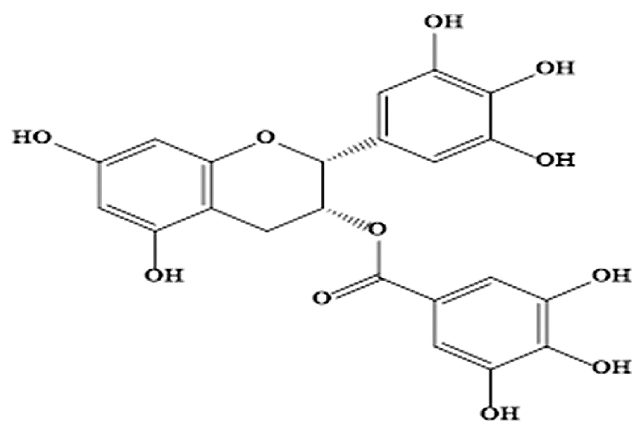
Therapeutic Activity of Green Tea Epigallocatechin-3-Gallate on Metabolic Diseases and Non-Alcoholic Fatty Liver Diseases: The Current Updates

1
Key Laboratory of Geriatric Nutrition and Health, Ministry of Education, Beijing Technology and Business University, Beijing 100048, China
2
Tanzania Agricultural Research Institute (TARI), Makutupora Center, Dodoma P.O. Box 1676, Tanzania
3
Rizhao Huawei Institute of Comprehensive Health Industries, Shandong Keepfit Biotech. Co., Ltd., Rizhao 276800, China
*
Author to whom correspondence should be addressed.
Nutrients 2023, 15(13), 3022; https://doi.org/10.3390/nu15133022
Submission received: 26 May 2023 / Revised: 26 June 2023 / Accepted: 29 June 2023 / Published: 3 July 2023
(This article belongs to the Section Phytochemicals and Human Health)

Abstract

:
Green tea polyphenols have numerous functions including antioxidation and modulation of various cellular proteins and are thus beneficial against metabolic diseases including obesity, type 2 diabetes, cardiovascular and non-alcoholic fatty liver diseases, and their comorbidities. Epigallocatechin-3-gallate (EGCG) is the most abundant polyphenol in green tea and is attributed to antioxidant and free radical scavenging activities, and the likelihood of targeting multiple metabolic pathways. It has been shown to exhibit anti-obesity, anti-inflammatory, anti-diabetic, anti-arteriosclerotic, and weight-reducing effects in humans. Worldwide, the incidences of metabolic diseases have been escalating across all age groups in modern society. Therefore, EGCG is being increasingly investigated to address the problems. This review presents the current updates on the effects of EGCG on metabolic diseases, and highlights evidence related to its safety. Collectively, this review brings more evidence for therapeutic application and further studies on EGCG and its derivatives to alleviate metabolic diseases and non-alcoholic fatty liver diseases.

1. Introduction

Green tea is an important beverage in most Asian countries and is popularly enjoyed by the Chinese and the Japanese. It is produced from Camellia sinensis fresh leaves. Green tea contains antioxidant compounds, vitamins, carbohydrates, proteins, minerals, chlorophyll, and polyphenols, which provide health benefits [1,2]. Green tea polyphenols have strong antioxidants and display the ability to quench and scavenge reactive oxygen species (ROS). The most prominent effects of green tea on human health are mainly attributed to catechins including epicatechin, epigallocatechin, epicatechin gallate, and epigallocatechin-3-gallate (EGCG) [3].
EGCG is the most efficacious compound and is key to the biological activities of green tea (Figure 1). Green tea contains a high concentration of active EGCG, which accounts for about 50% of total polyphenols in tea leaves [4]. The EGCG, a flavone-3-ol polyphenol is the most promising bioactive phytochemical due to its strong antioxidant activity and prebiotic function [5,6]. EGCG is mainly absorbed in the intestine, and gut microbiota plays a critical role prior to absorption as microbiota can metabolize large molecular tea polyphenols to bioactive and bioactive microbial metabolites [7]. Oral administration of EGCG showed good bioavailability as analyzed in the plasma when administered to healthy individuals after overnight fasting [8]. In a randomized crossover experiment, Van Amelsvoort et al. [9] reported an insignificant amount of EGCG excreted in urine when healthy individuals were administered with EGCG, which suggests that EGCG is transported to the liver or metabolized by the gut microbiota to other potential metabolites. Indeed, it has been established that EGCG can be detected in plasma after ingestion and its metabolites are identified in bile, plasma, and urine.
Metabolic syndrome or syndrome X is indicated by elevated blood sugar, elevated blood cholesterol, hyperlipidemia or dyslipidemia, excessive body fat, insulin resistance, visceral obesity, and arterial hypertension. Moreover, metabolic disease patients have been diagnosed with hyperuricemia, while serum urate stands as the biomarker for metabolic syndrome [10,11]. Consequently, elevated serum urate is closely associated with gout, coronary heart disease, hypertension, non-alcoholic fatty liver disease (NAFLD), and type 2 diabetes [12,13]. The modern world is characterized by highly processed high-sugar and high-fat diets together with a sedentary lifestyle exposing the population across all age groups to an unprecedented increase in metabolic diseases. The increasing incidences of metabolic diseases and associated comorbidities have become a hot spot of research globally. From obesity, type 2 diabetes, cardiovascular diseases, kidney diseases, and gout to NAFLD, EGCG has been examined for its alleviating effect on the diseases. EGCG has been reported to have a uric acid-lowering effect on metabolic disease patients, particularly through reduced uric acid production or increased uric acid excretion. Notably, the EGCG anti-hyperuricemia effect is mediated by xanthine oxidase (XOD) inhibition, an enzyme that catalyzes hypoxanthine to xanthine to uric acid [14]. Moreover, EGCG increases energy expenditure, improves fat oxidation, and reduces respiratory quotient, thereby influencing body mass index (BMI) and total body fat, and is a preventive agent for obesity and oxidative stress [15]. The ability of EGCG to bind several biological molecules, influence enzyme activities, and signal transduction pathways may explain its health benefits.
On the other hand, EGCG may be harmful in high doses when taken in the extract forms, and reports on toxicity are emerging. High doses of EGCG not only cause cytotoxicity in vitro but also may result in hepatoxicity, nephrotoxicity, and gastrointestinal disorders such as vomiting and diarrhea. Therefore, this review aims to provide current knowledge on the potential activity of EGCG as a component of green tea or extract supplements in the prevention and alleviation of metabolic syndrome, metabolic diseases, and NALFD.

2. The Role of EGCG on Metabolic Syndrome

Metabolic syndrome includes a group of risk factors: elevated blood sugar, insulin resistance, excessive body fat, visceral obesity, elevated blood cholesterol, hyperlipidemia, and arterial hypertension, which has become one of the major public health challenges worldwide [16,17]. The development of one of these conditions increases the risk of obesity, type 2 diabetes, hypertension, and renal and cardiovascular diseases (CVD) [17,18]. Abdominal obesity and insulin resistance have gained increased attention as the core manifestation of metabolic syndrome. Other abnormalities contributing to metabolic syndrome include chronic inflammatory and prothrombotic states, NAFLD, and sleep apnea [19]. In addition, high serum urate or hyperuricemia is a suggested metabolic syndrome indicator and a risk factor for the progression of metabolic diseases [11,20].
A patient with metabolic syndrome is in constant inflammation due to the associated concentration of chemokines, adipokines, and pro-inflammatory cytokines [21,22]. At the same time, metabolic syndrome-related chronic inflammations are implicated in a delayed and inferior immune response, with increased activation of immunosuppressive macrophages which exacerbate metabolic dysfunction [23,24]. Additionally, both humoral and cellular immune memory are impaired weakening the adaptive response of the immune system to diseases. The inflammatory markers of metabolic syndrome include increased interleukin IL-1, 6, and 8, tumor necrosis factor-α (TNF-α), resistin, white blood cell count, and C-creative protein as well as a decreased adiponectin [25,26,27]. Studies have revealed that some inflammatory cytokines, such as IL-1β and IL-18 play a crucial role in the development of arteriosclerotic plaques in patients with metabolic syndrome [28,29]. Moreover, hyperglycemia contributes to increased glucose levels in the endothelial cells, which favors the oxidative degradation of glucose metabolites with consequent oxidative stress. Costa et al. [30] conducted a systematic review and revealed that metabolic syndrome is a risk factor for the progression and prognosis of coronavirus disease 2019 (COVID-19) and elaborated that patients with metabolic disorders may face a higher risk of infection, thus complicated treatment. Overall, integrated and proper functioning metabolic homeostasis is paramount to innate immune responses.
The increasing risk and prevalence of metabolic syndrome demand therapeutic food-based treatment intervention. Although, the fundamental approach is lifestyle changes to reduce or remove the underlying problems, weight loss, increased physical activities, drug treatment, and a healthy diet could alleviate metabolic syndrome. Thus, green tea EGCG forms a food-based approach and a disease-preventive supplement, as it may aid in the reduction of metabolic syndrome and the onset of age-related non-communicable diseases (Figure 2). Several clinical studies have associated green tea EGCG consumption with a significant reduction in body weight, body mass index (BMI), and abdominal fat [27]. The pro-antioxidant properties of EGCG suppress gene and protein expression of adenosine monophosphate-activated protein kinases (AMPK) and transcription factors involved in adipogenesis and lipogenesis alleviating insulin sensitivity, thus leading to a reduction in body weight [31]. Recent studies suggest that EGCG exerts its beneficial effect by modulating mitochondrial functions impacting mitochondrial biogenesis, bioenergetic control in adenosine triphosphate (ATP) production, alteration of the cell cycle, and mitochondrial-related apoptosis [32,33]. In addition, EGCG appears to have a beneficial effect on the gut microbiota as increases the number of beneficial species of Bifidobacterium, thereby improving the energy metabolism. Simultaneously, EGCG stimulates enzymes involved in lipolysis and modulation of serum urate as well as copious pathways (Table 1). Therefore, EGCG is a promising therapy for weight management, BMI, and waist circumference reduction, as well as improving lipid metabolism.

2.1. Effects of EGCG on Insulin Resistance and High Blood Pressure

Insulin resistance is identified as an impaired biological response to insulin stimulation of the target tissues, prominently in the muscles, liver, and adipose tissue [51]. It impairs sensitivity to insulin-mediated glucose disposal, resulting in a compensatory increase in pancreatic β-cell insulin production to maintain normal blood glucose levels. Consequently, it results in a cluster of abnormalities including hyperinsulinemia, hyperglycemia, dyslipidemia, visceral adiposity, obesity, hyperuricemia, hypertension, endothelial dysfunction, and elevated inflammatory markers [52].
Insulin increases glucose uptake in the muscles and liver and inhibits hepatic gluconeogenesis and lipolysis. However, insulin resistance impairs insulin-mediated inhibition of lipolysis in adipose tissues leading to increased circulating lipids, which further inhibit the antilipolytic effect of insulin [51]. Non-esterified fatty acids or free fatty acids (FFAs) inhibit protein kinase activation in the muscles leading to reduced glucose uptake. Conversely, FFAs increase the activation of protein kinases in the liver, promoting hepatic gluconeogenesis and stimulating adipose lipogenesis as well as lipolysis [53,54,55]. Thus, higher levels of circulating FFAs directly affect the liver and muscle metabolism, and further aggravate insulin resistance [56]. Overall, the progression of insulin resistance may lead to metabolic syndrome, polycystic ovary syndrome, non-alcoholic fatty liver disease (NAFLD), type 2 diabetes, sleep apnea, and certain forms of cancer.
The anti-insulin resistance and glucose homeostasis effects of EGCG have been consistently described. Yan et al. [57] revealed that green tea catechins significantly decreased glucose levels and improved glucose tolerance in the animal experiment. Green tea EGCG reduced ROS in adipocytes, attenuated dexamethasone, and TNF-α as a result of ROS, and increased glucose uptake ability hence alleviating adipose insulin resistance [57]. Fasting serum glucose, insulin levels, and insulin resistance were reduced significantly in obese hypertensive patients following the uptake of green tea extract in the randomized double-blind, placebo control clinical trial study [58]. Similarly, Liu et al. [44] conducted a randomized double-blind, placebo control clinical trial experiment involving 92 subjects with type 2 diabetes and lipid abnormalities; found that green tea extract (GTE) significantly alleviated insulin resistance, increased glucagon-like peptide-1 and high-density lipoprotein (HDL) levels, and decreased triglycerides levels. At the same time, EGCG ameliorated insulin resistance by upregulating and increasing phosphorylation of the insulin receptor substrate-1 (IRS-1), which is essential for the stimulation of glucose uptake in response to insulin [59]. For instance, EGCG reversed high glucose- and glucosamine-induced insulin resistance in SH-SY5Y neuronal cells by improving the oxidized cellular status and mitochondrial function [59]. Similarly, a study that employed a GTE in mice, showed that EGCG attenuated insulin resistance induced by a high-fat diet [47]. Additionally, EGCG was shown to improve glucose tolerance in mice [60]. According to Lee et al. [61] green tea-derived products such as extracts and water-soluble polysaccharides exhibit hypoglycemic effects as they caused delayed intestinal absorption of glucose. The hypoglycemic mechanism of EGCG has been contributed by its inhibitory effect on α-glucosidase activity, enhancement of glucose uptake, and promotion of glucose transporter-4 (GLUT4) translocation to the plasma membrane through a phosphatidylinositide-3-kinase/activated protein kinase B (PI3K/AKT) signaling pathway in skeletal muscle cells [62,63]. When EGCG uptake was combined with regular exercise in overweight or obese postmenopausal women, it resulted in reduced plasma glucose concentration in subjects with impaired glucose tolerance [64]. Collectively, green tea EGCG alleviates insulin resistance, increases glucose uptake, and lowers blood glucose, which are important for glucose homeostasis.

2.2. Effects of EGCG on Adipose Mass, Blood Cholesterol, and Triglycerides

Several studies suggest that EGCG can decrease energy and food intake, lipogenesis as well as preadipocyte differentiation and proliferation, while increasing lipolysis, and fat oxidation. Green tea EGCG was revealed to reduce tissue and blood lipid accumulation in the FFAs-induced human liver hepatocellular carcinoma cell line (HepG2) via activation of the AMPK pathway [43]. Consequently, AMPK activation shifts some FFAs toward oxidation, away from lipid and triglycerides storage, and suppresses hepatic gluconeogenesis, which is implicated in the reduction of adipose mass and body weight.
Findings from a systematic review by Asbaghi et al. [65] revealed that, supplementing >800 mg GTE/day for eight or more weeks significantly improved lipid profile by reducing serum triglycerides and total cholesterol concentrations in patients with type 2 diabetes. Similarly, the consumption of green tea EGCG significantly lowered low-density lipoprotein (LDL) as well as total cholesterol levels in normal weight and obese individuals [66]. Moreover, supplementing EGCG for four to eight weeks to patients with obesity reduced plasma triglycerides and serum kisspeptin levels [67].
A study involving healthy Japanese women revealed that elevated plasma and urinary concentration of green tea catechins was associated with improved plasma lipid profile [68]. Randomized double-blind placebo-controlled clinical trials involving obese women in Taiwan, reported a significant decrease in total cholesterol, LDL, and triglyceride, and increased levels of HDL as well as plasma adiponectin in groups administered with GTE for 12 weeks [69,70]. Furthermore, a combination of EGCG and caffeine produced a synergistic effect on gut microbiota: increasing Bifidobacterium count and fecal short-chain fatty acid (SCFAs) levels and enhanced fecal bile acids excretion in experimental rats [71]. At the same time, the combination effect increased the expression of hepatic G-coupled protein receptor 1 and activation of intestinal farnesoid X receptor (FXR). The activation of intestinal and hepatic FXR induces endocrine hormone fibroblast growth factor 15 (FGF15) and small intestine heterodimer partner production, which collectively inhibits hepatic bile acid biosynthesis via signaling cascades [71]. A randomized double-blind parallel placebo-controlled clinical trial showed that administering 400, 600, or 800 g EGCG (depending on body weight) for 12 months in men with Down syndrome resulted in weight loss, reduced body fat, and improved lipid profile [72]. In addition, Choi. et al. [60] revealed that EGCG regulates lipid catabolism through AMPK-mediated mechanisms increasing lipolysis and suppressing lipogenesis in the adipocytes. Therefore, EGCG reduces visceral adiposity by activating autophagy and lipolysis in white adipose tissue through an AMPK-mediated mechanism.

2.3. Effects of EGCG on Hyperuricemia and Uric Acid Metabolism

Uric acid is the final catabolic product of the enzymatic degradation of purines as well as other dietary components and can scavenge ROS, thus protecting the erythrocytes membrane from oxidation in humans [73]. Hyperuricemia is considered a metabolic disease, while elevated serum uric acid is the metabolic disease biomarker [20]. Hyperuricemia induces oxidative stress and endothelial dysfunction, resulting in the development of a series of diseases including insulin resistance, type 2 diabetes, coronary artery diseases, chronic kidney diseases, kidney stone, and gout, thus becoming a metabolic disease that threatens human health [13,74]. Moreover, hyperuricemia has been reported to involve in the manifestation of NAFLD. Maintaining serum urate levels below 7 and 6 mg/dL in men and women, respectively, is clinically important for the prevention of hyperuricemia, type 2 diabetes, cerebrovascular, cardiovascular diseases, and gout [75].
At present drugs such as allopurinol, oxypurinol, febuxostat, and topiroxostat, which are xanthine oxidase inhibitors, and the recombinant uricase (rasburicase), uricosuric agent (probenecid) are currently used to treat hyperuricemia [76,77]. However, these drugs have side effects such as causing uric acid stones, liver and kidney stones, liver damage, and/or may lead to hypersensitivity reactions, which may not be tolerated by the patient [77,78]. Of interest, EGCG has been studied to have a uric acid-lowering effect. Thus, can be used to manage or develop nutraceutical drugs for hyperuricemia and alleviate metabolic diseases.
In animal studies using mice, tea ranging from green, yellow, black, or dark tea extract significantly increased uric acid excretion by upregulating the expression of uric acid secretion transporters ABCG2, organic anion transporter 1 (OTA1), organic anion transporter 3 (OTA3) and organic cation transporter 1 (OCT1), and by downregulating the expression of uric acid reabsorption transporter; urate transporter 1 (URAT1) and glucose transporter 9 (GLUT9) in the kidney [79,80]. Likewise, Li et al. [14] reported that EGCG significantly promoted the expression of OAT1 and downregulated the expression of GLUT9 in renal tissues of hyperuricemia rats. At the same time, tea extract significantly lowers serum urate levels through the inhibition of XOD and ADA to produce uric acid [38]. Moreover, interventions using tea extract revealed that tea components upregulate the expression of the intestinal ABCG2 protein and alleviated hyperuricemia by modulating the gut microbiota. The study of Sang et al. [80] reports yellow tea to be the best in alleviating hyperuricemia in mice and indicated 50% EGCG oral bioavailability.
Using human normal liver cell line HL-7702 (L-02), Wu et al. [81] revealed that tea extract limit uric acid production via inhibition of XOD activity, with green tea showing the strongest inhibitory activity followed by yellow, oolong, white, black, and dark tea. Zhang et al. [37], using spectroscopic and computer simulation methods, found that EGCG at a concentration of 0.13 mmol/L inhibited 80% XOD activity by binding to the vicinity of flavine adenine dinucleotide (FAD) in XOD, hindering the entry of the substrate. In xanthine-stimulated BRL 3A rat liver cells, EGCG significantly reduced uric acid levels in vitro. Additionally, it was revealed that EGCG significantly reduced serum uric acid and inhibited XOD activity in rats treated with potassium oxonate [14].
A randomized cross-over study in Japan revealed a significant increase in the excretion of uric acid and uric acid precursor (xanthine and hypoxanthine) in the group of healthy men receiving distilled spirit (Shōchū) with catechin-enriched green tea [82]. Similarly, Jatuworapruk et al. [83] reported the hypouricemic effect of green tea in healthy individuals, that serum uric acid decreased with decreased uric acid clearance after two weeks of the randomized study. The studies on healthy individuals reflected the idea of green tea extract components particularly EGCG to inhibit XOD activity and thus reduce the production of uric acid and increase the excretion of uric acid precursor (xanthine and hypoxanthine) with urine.

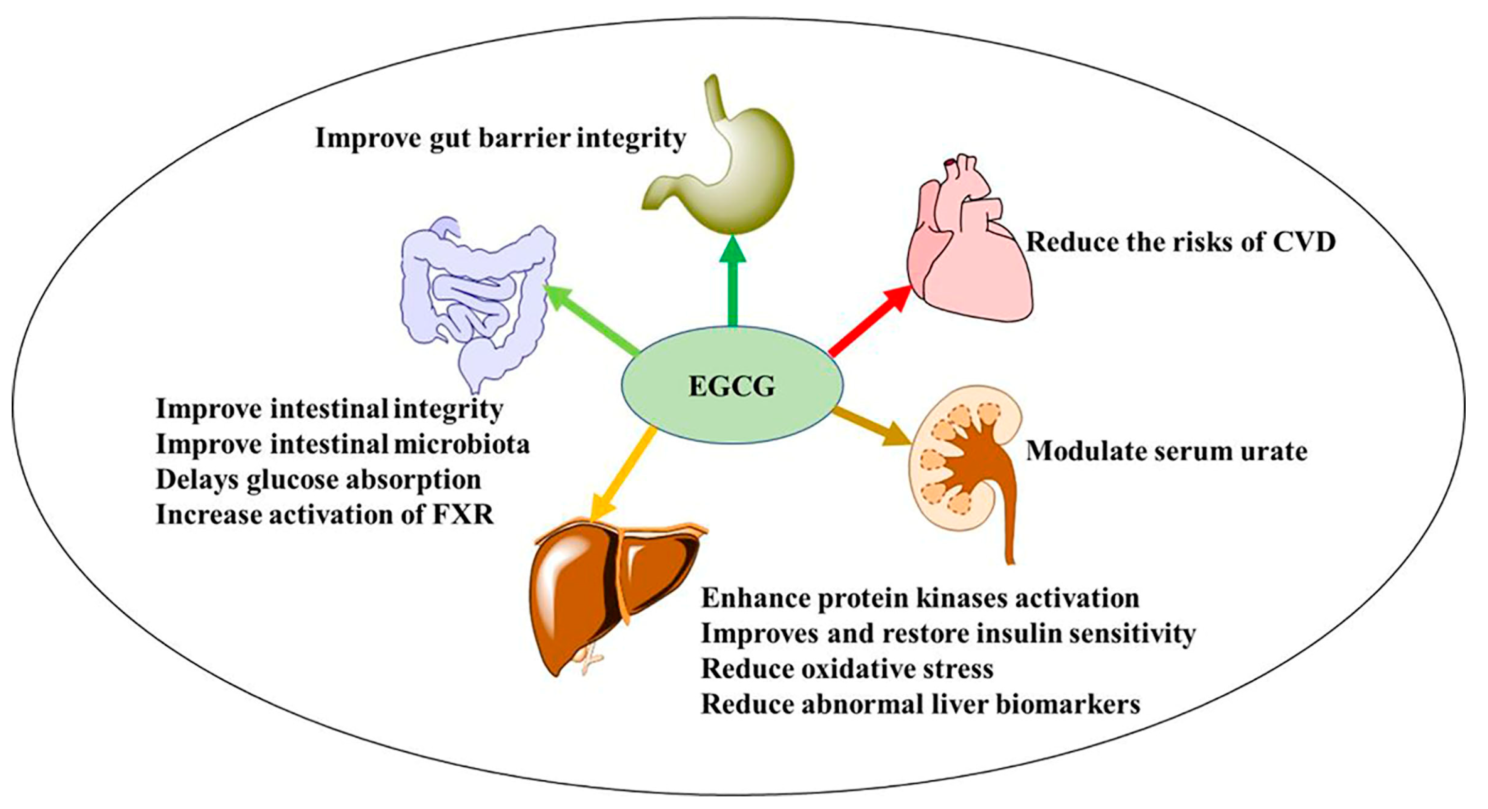
3. Effect of EGCG on Metabolic Diseases

3.1. Obesity

Obesity is a major public health burden that leads to chronic inflammation and metabolic disorders in both peripheral tissues and the central nervous system. Obesity is mainly caused by an energy imbalance between calorie intake and utilization, which results in adipose tissue dysfunction with adipocyte hypertrophy, excessive accumulation of adipose tissue, and to an extent that impairs physical health and psychological well-being [84]. Obesity comprises several metabolic alterations accompanied by a state of chronic inflammations and an increased oxidative state that contributes to the development of an array of health complications. It increases the risk of insulin resistance, high blood glucose levels, dyslipidemia (high triglyceride and cholesterol levels), age-related cognitive impairment, arteriosclerosis progression, and peripheral inflammation.
Obesity is one of the major risk factors for type 2 diabetes, hypertension, and CVD. Worldwide, >35% of adults are considered to be obese, and in some countries, obesity prevalence exceeds 40% [85,86]. Additionally, obesity is estimated to reach 18% and 21% in men and women, respectively, by 2025 [71,84]. Interestingly, obesity had previously been identified as a risk factor for viral infections due to its influence on the immune response. For instance, during the 2009 H1N1 outbreak, obese patients presented severe complications [87,88]. Likewise, during the recent COVID-19 pandemic, obese patients exhibited a high rate of complications and a need for hospitalization [89,90].
The intake of EGCG appears to be a promising strategy for the prevention and management of obesity and its complications. The study by Chatree et al. [67] showed that EGCG supplementation for eight weeks significantly decreased fasting plasma triglyceride levels, blood pressure, and serum kisspeptin levels in humans. A meta-analysis of randomized controlled clinical trials on the influence of green tea intake on obesity indices in humans revealed a significant reduction in body weight, BMI, and a reduced waist circumference at a dosage of <500 mg of green tea per day for 12 weeks [91]. In a randomized placebo-controlled trial involving 60 healthy Japanese people in Japan, administration of a combined dose of 146 mg EGCG in green tea and citrus polyphenol (178 mg α-glucosyl hesperidin) for 12 weeks prevented weight gain and reduced the BMI [92]. Similarly, the obesity-related indicators including triglycerides levels, visceral and body fat percentage, as well as blood LDL/HDL ratio decreased in the <50 years group [92].
In the mice experiment, it was revealed that EGCG significantly ameliorated insulin resistance and cognitive disorder by upregulating IRS-1 and extracellular signal-regulated kinases (ERK)/cAMP response element-binding protein (CREB)/brain-derived neurotrophic factor (BDNF) signaling pathways [59]. Zhou et al. [93] showed that GTE inhibits the release of the inflammatory cytokine TNF-α, IL-1β and IL-6 in palmitic acid-induced BV-2 microglial cells by suppressing the JAK2/signal transducer and activator of transcription-3 (STAT3) signaling pathway. Furthermore, the animal experiment that recruited obese rats treated with GTE showed a significant reduction in obesity indicators through AMPK activation, restored insulin sensitivity, and stimulated fatty acid oxidation in the plasma and liver [94].

3.2. Type 2 Diabetes Mellitus

Diabetes is a group of metabolic diseases characterized by hyperglycemia, resulting from defects in insulin secretion, insulin action, or both. A predisposition to glucose intolerance depends on various factors that share an ability to stress the glucose homeostasis profile, with the current explosion of sedentary lifestyles, obesity, and insulin resistance being the major causes of type 2 diabetes [95]. Failure of insulin to induce an adequate response of the target tissues and the level of insulin receptors contributes to metabolic abnormalities in glucose homeostasis [96]. The skeletal muscles, adipose tissues, liver, pancreatic β-cell, brain, and vascular endothelium are the major insulin targets. Acquired defects in glucose homeostasis cause blood glucose levels to rise to a range of intolerable impaired glucose tolerance. The rise of blood glucose causes additional deterioration of beta-cell function along with further insulin resistance and elevated hepatic glucose; subsequently, blood glucose rises to full-blown type 2 diabetes. In addition to suppressing the patient’s immunity, type 2 diabetes can cause metabolic dysfunction that directly affects homeostasis and is associated with a range of debilitating complications such as CVD, chronic kidney diseases, and visual disability or blindness [55,96]. Thus, the global prevalence of type 2 diabetes is estimated at 6.28%, which is equivalent to 462 million individuals [97], and is projected to reach 700 million individuals by 2045 [98].
Several drugs, both insulin and non-insulin formulations, have been in use to manage and alleviate diabetes. They include different formulations of exogenous insulin, insulin simulants (glimepiride, glipizide, repaglinide, nateglinide, sitagliptin, and saxagliptin), α-glucosidase inhibitors (acarbose and miglitol), glucagon-like peptide-1 receptor agonistics (sulfonylureas, meglitinides, exenatide, and semaglutide) and insulin sensitivity enhancers (metformin and rosiglitazone) among others [99,100]. However, the drugs may display adverse effects and complications in patients such as risks of hypoglycemia, weight gain, headache, genitourinary tract infection, gastrointestinal tract disturbances, and cardiovascular events including heart failure [101,102]. Additionally, the use of the existing drugs requires prior diagnosis adding up to the healthcare costs to manage the disease. In addition, diabetic patients present complicated individualized treatment strategies due to associated side effects, contraindications, and underlying comorbidities, thus a major challenge to the health care systems. With this in mind, improving metabolic control to normal glucose homeostasis through the intake of EGCG as a supplement or part of green tea can greatly benefit a long-term, sustainable, and safe intervention.
Green tea EGCG has been demonstrated to improve insulin sensitivity and glycemic control, and significantly decrease serum triglycerides and total cholesterol levels following a long-term supplementation at ≤800 mg/day [65]. In addition, green tea EGCG decreased triglycerides and significantly increased HDL and glucagon-like peptide 1 levels in a randomized double-blinded placebo control clinical trial involving patients with type 2 diabetes and lipid abnormalities for 16 weeks in Taiwan [44]. According to Liu et al. [103] EGCG can reverse pancreatic β-cell damage or apoptosis and enhance glucose-stimulated insulin secretion or insulin sensitivity by decreasing the expression of microRNA (miR-16-5p), which targets the anti-apoptotic β-cell lymphoma-2 (BCL-2). The cell surface protein 67-kDa laminin receptor was revealed to act as the sensor for EGCG inducing the production of nitric oxide in the endothelial cells while downregulating the production of inducible nitric oxide synthase (iNOS) enzymes, thereby mediating the beneficial effect of EGCG through cyclic guanosine monophosphate (cGMP) dependent pathway and cellular nitric oxide production [104,105,106]. Therefore, EGCG improves endothelial function and reduced oxidative stress through decreased nitric oxide production and decreased expression of iNOS. Likewise, the EGCG-derived autoxidation products have been revealed to improve insulin sensitivity through suppression of liver-derived secretory selenocysteine-containing selenoprotein P (SELENOP) implicated to cause insulin resistance [107]. For instance, theasinensin A an oxidation product of EGCG displayed high cellular uptake on HePG2 cells as well as higher antioxidant capacity compared to the monomer EGCG [108]. Thus, EGCG can be effective in controlling hyperglycemia and alleviating the complications of diabetes by improving insulin sensitivity and reducing the risk factors for type 2 diabetes. In addition, a study by Hadi et al. [109] which involved 50 diabetic patients concluded that consuming 300 mg EGCG/day for eight weeks significantly decreases fasting blood glucose, body weight, and the high-sensitive C-reactive proteins, thus alleviating type 2 diabetes. A systematic review and meta-analysis reported that consumption of green tea for more than eight weeks significantly decreased body weight, BMI, and body fat in diabetic patients [39].
A high-fat diet may activate the nucleotide-binding oligomerization domain-like receptor protein 3 (NLRP3) inflammasome in macrophages and participate in immune dysfunction leading to chronic inflammation and insulin resistance. The pro-inflammatory cytokines interleukin IL-1β and IL-18 play important roles in inflammatory diseases and type 2 diabetes. They are regulated by the inflammasomes, which process inactive pro- IL-1β and pro-IL-18 proteins into active IL-1β and IL-18 proteins, respectively [110]. Inflammasomes are the key targets for EGCG. In the same way, EGCG may exert anti-inflammatory effects against NLRP3, which enhances insulin signaling [111]. According to Zhan et al. [112] administration of EGCG in a type 2 diabetic mouse model provided an anti-inflammasome effect and improved glucose tolerance in vivo. Therefore, the suppression of NLRP3 inflammasome-mediated inflammation is a possible mode of action of EGCG for alleviation and treatment of type 2 diabetes.

3.3. Cardiovascular Diseases

Cardiovascular diseases (CVD) are implicated in the abnormal function of the heart and blood vessels. It includes coronary heart diseases, peripheral arterial diseases, congenital heart diseases, rheumatic heart diseases, and cerebrovascular diseases [113]. Arteriosclerosis is a disease of arteries that is caused by endothelial dysfunction, inflammatory vascular cells, and lipid accumulation. Plaque which is mostly composed of lipid, calcified, fibrous, fibrolipidic, or necrotic cores is the culprit that causes arteriosclerosis, and can partially or completely block the blood flow in the arteries [45]. Cardiovascular diseases occur as a complication and are comorbid with other metabolic diseases. For instance, approximately 34.8% of CVD occurs concurrently with type 2 diabetes across countries as revealed in the cross-sectional study on CVD across continents [114]. The prevalence of CVD in South Asian countries is as high as 49.6% among adult individuals [115] and contributes to a third of overall death in the Americas [116]. Globally, CVD has contributed to about 17.4 and 17.8 million deaths in 2012 and 2017, respectively [117,118].
There are a number of studies assessing green tea EGCG consumption with respect to CVD. Consumption of green tea and administration of GTE or EGCG has been reviewed to reduce the risks and mortality rate owing to CVD [119]. The therapeutic effects of EGCG on CVD are associated with the inhibition of LDL cholesterol, inhibition of NF-κB, reduction of plasma glucose and glycated hemoglobin levels, inhibition of myeloperoxidase activity, reduction of inflammatory markers and inhibition of ROS generation. For instance, when EGCG uptake was combined with regular exercise in overweight or obese postmenopausal women reduced the resting heart rate [64]. Lange [120] presented a review of population-based and epidemiological studies in Japan, North America, and Europe, and reported that habitual green tea intake of two to six cups a day was associated with reduced risks of CVD. In a clinical trial in Iran, Mozaffari-Khosravi et al. [121] reported a significant decrease in systolic and diastolic blood pressure in mildly hypertensive type 2 diabetic individuals who consumed three glasses of green or sour tea daily for four consecutive weeks. Likewise, Peng et al. [122] performed a meta-analysis of randomized controlled trials, and suggested that consumption of green tea or a low dose of green tea polyphenols significantly decreases systolic and diastolic blood pressure by 1.98 and 1.92 mmHg, respectively, in humans. In a randomized double-blind placebo-controlled cross-over experiment, it was revealed that a single dose of 300 mg EGCG alleviated endothelial function and improved arterial-mediated dilation in patients with coronary arterial diseases, but no significant effect on administering 150 mg of EGCG (twice a day) for two weeks [123].
In animal experiments, rats were fed a diet containing 2 and 4 g/kg GTE with added salt (35 g/kg) to induce hypertension for 42 days; green tea indicated beneficial effects on blood pressure, markers of inflammation (TNF-α), and serum antioxidants status [124]. Similarly, EGCG was shown to attenuated salt-induced hypertension and renal injury in rats after six weeks of oral administration [125]. Ocular hypertensive patients were administered with EGCG for three months in a randomized, placebo-controlled, double-blind, cross-over clinical trial, which suggested that EGCG favorably influences inner retinal function in the eyes with early to moderately advanced glaucomatous damage, although the observed effect was small [126]. Additionally, supplementing green tea extract for three months in obese hypertensive patients significantly reduced the risks of blood pressure such as insulin resistance, inflammation and oxidative stress [58]. The reported effect may be linked to increased insulin sensitivity and suppression of leucocytes adhesion to the endothelium and transmigration through inhibition of transcription factor NF-κB-mediated production of cytokines and adhesion molecules, in both vascular endothelial and inflammatory cells [58,127]. For instance, EGCG chelates metal ions to form an inactivated complex that reduces the catalytic effects of metal ions in the oxidation reaction and effectively removes surplus active free radicals from the body, thus reducing oxidative vascular endothelium damage and the possibility of thrombosis. In contrast, a study involving elderly women and men aged ≥80 years in China revealed that green tea intake was associated with a 38% increase in the risk of developing hypertension in men, although it had no impact on women [128].

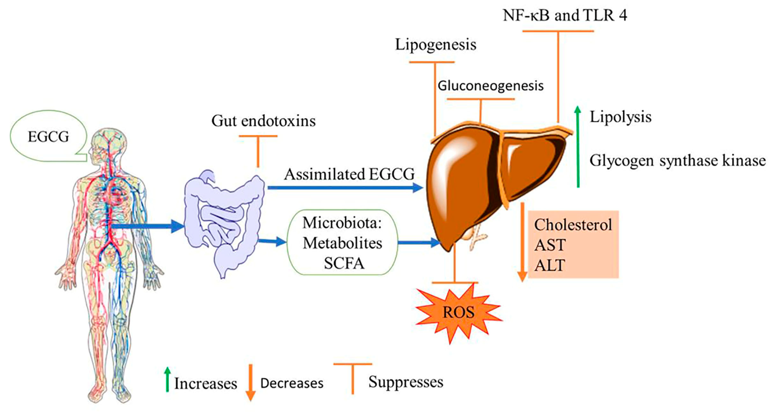
4. Effects of EGCG on Non-Alcoholic Fatty Liver Disease

Non-alcoholic fatty liver disease (NAFLD) is the predominant hepatic disorder worldwide affecting about 25% of the general population, which is estimated at one billion people worldwide [129,130]. Relatively, the prevalence of NAFLD in China, European countries, Japan, and the United States of America (USA) were reported at 17.6%, 17.9–25.4%, 17.9%, and 26.3%, respectively [130].
The NAFLD describes a spectrum of progressive liver conditions ranging from relatively benign liver steatosis with inflammation and advancing to non-alcoholic steatohepatitis (NASH), fibrosis, and cirrhosis [46,131]. The NASH features are indicated as fatty hepatocytes and inflammatory cell infiltrates in association with increased activation of hepatic NF-κB, which exacerbates liver injury. It is the necessary stage of NAFLD for the development of simple steatosis to cirrhosis and hepatocellular carcinoma. Recent studies have highlighted that iron loading contributes to liver damage, whereas the accumulation of free cholesterol can exacerbate NASH. While the etiology of NAFLD is unclear: genetic factors, lifestyle, ageing, and environmental factors are implicated [132]. It is generally considered as the liver component of metabolic syndrome which is associated with insulin resistance as the main pathogenetic mechanism to trigger NASH. It presents high degree of comorbidities with obesity, type 2 diabetes, and hypertension [133,134]. For instance, the metabolic associated NAFLD affects about 50.7% of obese or overweight adults globally [129]. The longitudinal cohort studies conducted in Beijing, China concluded that hyperuricemia precedes NAFLD and contributes to the development of the disease [135]. Additionally, Li et al. [136] revealed a significant association of hyperuricemia with NAFLD in a cross-sectional study in Ningbo, China. Thus, lowering serum uric acid levels and alleviating metabolic syndrome may prevent NAFLD as described in previous sections.
As the treatment options are limited and there are no effective pharmacological treatments for NAFLD, dietary approaches have been emphasized to manage NASH risks. Various studies have revealed that EGCG targets and activate the cellular AMPK pathway as well as insulin receptor substrate-1 (IRS-1) attenuating insulin resistance [94,137]. The activation of AMPK leads to increased fatty acid oxidation in the liver, and simultaneously inhibits hepatic lipogenesis and cholesterol synthesis. Subsequently, the activated AMPK pathway reduce the activity of enzymes involved in fatty deposits and triglyceride accumulation in the liver, thus alleviating NAFLD [138].
The ability of EGCG to attenuate intracellular redox alterations and anti-inflammatory bioactivity responses downstream of NF-κB activation from extracellular receptors has been studied. EGCG exerts its effect indirectly through gut microbiota-derived metabolites, which limits NF-κB activation and NASH-associated liver injuries [137]. Increasing evidence suggests that EGCG may prevent and mitigate NAFLD through antioxidant activity, inhibition of endotoxins, and restoring redox homeostasis (Figure 3). The GTE or EGCG protects against NAFLD and reduces liver steatosis by reducing hepatic oxidative stress and endotoxins toll-like receptor-4 nuclear factor κB (TLR4/NF-κB) inflammation [47,139]. The EGCG benefits are linked, at least in part, to alleviating gut microbiota and improved gut barrier integrity, which limits endotoxins translocation and absorption. In addition, the protective effect against NASH has been linked to certain enzymes from the microbiome and microbiota beneficial short chain fatty acids [132,140]. According to the cross-sectional data of adult individuals from the 2009–2014 United States National Health and Nutrition Examination Survey, consumption of green tea was associated with reduced odds of having one or more abnormal liver biomarkers, namely bilirubin, alkaline phosphatase, gamma-glutamyl transferase, aspartate aminotransferase (AST), and serum alanine aminotransferase (ALT) [141]. Similar reduction of transaminases (ALT and AST) were reported in the animal model when diabetic mice were administered with EGCG [104].

5. Safety Implications of EGCG

The increasing incidences of metabolic diseases have promoted the increased use of food supplements and therapeutic agents including EGCG. Recently, EGCG has become one of the most studied green tea catechins due to their associated health-promoting benefits. As EGCG comes as a component of green tea, intake of green tea is considered as safe in the range of historical use in China and Japan despite high consumption levels. However, EGCG extracts safety implications have to be scrutinized and communicated as higher doses are achievable in the context of dietary supplements, particularly for weight loss formulations. A number of studies have reported events of liver toxicity either caused by green tea extracts or ingestion of EGCG supplements. The acceptable daily intake (ADI) for a 70 kg adult human was reported to be 322 mg EGCG per day [142]. The no observed adverse effect level (NOAEL) was reported to be 600 mg per day and the European Union Food Safety Authority (EFSA) indicates an intake of equal or above 800 mg EGCG a day could lead to human liver damage as indicated by elevated transaminases [142,143]. A review by Dekant et al. [144] reported no liver toxicity observed after the intake of EGCG below 600 mg EGCG per person a day with green tea infusion or tea GTE-based beverages. Recently, a randomized prospective cohort study involving 39 women (18 ≤ age ≤ 40 years) recommended a dose of 720 mg EGCG (for at least a month) alone or in combination with uterine fibroids management drugs to be tolerable without associated liver toxicity [145].
Moreover, it was demonstrated that EGCG induced liver toxicity as the function of dose, administration route, and treatment period in the animal experiments. In the animal model, subcutaneous injection of EGCG at a dose rate of 500 mg EGCG/kg body weight per day resulted in liver toxicity and 75% of experimental mice died after the first injection. The oral gavage dose at 200 mg EGCG/kg body weight in lactating mice was recorded as NOAEL, however the same dose via subcutaneous injection induced liver cell necrosis and renal tubule damage [146]. Intraperitoneal injection of 100 mg EGCG/kg per day for four consecutive days induce mice renal toxicity as indicated by elevated serum cystatin C and neutrophil gelatinase-associated lipocalin and inflammatory markers, and caused 60% mortality in streptozotocin-induced diabetic mice [147]. Moreover, an increase in nicotinamide dinucleotide phosphate (NADPH) oxidase was observed, which potentiated the production of ROS and exacerbated oxidative stress in diabetic mice injected with EGCG [147]. The daily tolerance dose for 14 consecutive days in mice was established at 21.1 or 67.8 mg EGCG/kg intraperitoneal injection and oral administration, respectively [148]. Assessing genotoxicity in mice, up to 50 and 1200 mg EGCG/kg per day by intravenous injection or oral gavage, respectively, was regarded as safe [149].
On the other hand, the chemical structure of EGCG makes it susceptible to autooxidation degradation, which may have implications regarding toxicity. Under normal physiological conditions, EGCG can be auto-oxidized to o-quinone through non-enzymatic dehydrogenation of the phenolic hydroxyl groups, which are further oxidized by oxygen to yield superoxide radicals [150]. The peroxide radicals further function as oxidants of EGCG to form o-quinone and hydrogen peroxide (H2O2), subsequently generating ROS [108]. As a consequence, oxidative stress occurs as the ROS level exceeds cellular antioxidant capacity. The prooxidant effect of EGCG and ROS generation might further display cellular or DNA-damaging activities.
Overall, an individualized safe intake level of EGCG as an extract or supplement is recommended to check on the label and calculate the intake amount as the presented ADI stands for a 70 kg body weight adult individual. Additionally, further clinical studies are recommended to clarify toxicity levels of EGCG intake in humans as most of the presented findings are based on animal experiments.

6. Conclusions

The safety of EGCG is well documented in animal and clinical studies as to the established acceptable daily intake (ADI) of 322 mg/day. However, intake of an amount higher than 800 mg EGCG a day may cause liver injuries. Moreover, different methods of preparing a cup of green tea, tea extract, EGCG purity, and associated catechin compounds or experimental designs applied in different studies might have contributed to some reported discrepancies in the activity of EGCG on metabolic diseases and NAFLD. Therefore, further studies to understand signaling pathways and molecular events associated with EGCG in alleviating metabolic diseases and NAFLD are recommended.
Bringing it all together, EGCG can be useful to alleviate metabolic diseases and their related malaise. Lastly, long-term use of EGCG either alone or in combination with conventional therapies for the prevention, management, and treatment of metabolic diseases and non-alcoholic fatty liver diseases remains to be an area for further research.

Author Contributions

Conceptualization, validation, writing—original draft preparation, writing—review and editing, A.J. and K.W.; fund acquisition, investigation, supervision, writing—review and editing, Y.W. All authors have read and agreed to the published version of the manuscript.

Funding

This research was funded by the National Natural Science Foundation of China, grant number 31972127 and 31471626 and The Natural Science Foundation of Rizhao, grant number 202143.

Institutional Review Board Statement

Not applicable.

Informed Consent Statement

Not applicable.

Data Availability Statement

Not applicable.

Conflicts of Interest

The authors declare no conflict of interest.

References

  1. Narotzki, B.; Reznick, A.Z.; Aizenbud, D.; Levy, Y. Green tea: A promising natural product in oral health. Arch. Oral Biol. 2012, 57, 429–435. [Google Scholar] [CrossRef] [PubMed]
  2. Xu, C.; Liang, L.; Li, Y.; Yang, T.; Fan, Y.; Mao, X.; Wang, Y. Studies of quality development and major chemical composition of green tea processed from tea with different shoot maturity. LWT 2021, 142, 111055. [Google Scholar] [CrossRef]
  3. Xu, R.; Bai, Y.; Yang, K.; Chen, G. Effects of green tea consumption on glycemic control: A systematic review and meta-analysis of randomized controlled trials. Nutr. Metab. 2020, 17, 56. [Google Scholar] [CrossRef] [PubMed]
  4. De Oliveira, M.R.; Nabavi, S.F.; Daglia, M.; Rastrelli, L.; Nabavi, S.M. Epigallocatechin gallate and mitochondria—A story of life and death. Pharmacol. Res. 2016, 104, 70–85. [Google Scholar] [CrossRef]
  5. Gan, R.Y.; Li, H.B.; Sui, Z.Q.; Corke, H. Absorption, metabolism, anti-cancer effect and molecular targets of epigallocatechin gallate (EGCG): An updated review. Crit. Rev. Food Sci. Nutr. 2018, 58, 924–941. [Google Scholar] [CrossRef]
  6. Roychoudhury, S.; Agarwal, A.; Virk, G.; Cho, C.L. Potential role of green tea catechins in the management of oxidative stress-associated infertility. Reprod. Biomed. Online 2017, 34, 487–498. [Google Scholar] [CrossRef] [Green Version]
  7. Zhang, Y.; Cheng, L.; Liu, Y.; Wu, Z.; Weng, P. The Intestinal Microbiota Links Tea Polyphenols with the Regulation of Mood and Sleep to Improve Immunity. Food Rev. Int. 2021, 39, 1485–1498. [Google Scholar] [CrossRef]
  8. Fernández, V.A.; Toledano, L.A.; Lozano, N.P.; Tapia, E.N.; Roig, M.D.G.; Fornell, R.D.L.T.; Algar, Ó.G. Bioavailability of Epigallocatechin Gallate Administered with Different Nutritional Strategies in Healthy Volunteers. Antioxidants 2020, 9, 440. [Google Scholar] [CrossRef]
  9. Van Amelsvoort, J.M.M.; Van Het Hof, K.H.; Mathot, J.N.J.J.; Mulder, T.P.J.; Wiersma, A.; Tijburg, L.B.M. Plasma concentrations of individual tea catechins after a single oral dose in humans. Xenobiotica 2008, 31, 891–901. [Google Scholar] [CrossRef]
  10. Raya-Cano, E.; Vaquero-Abellán, M.; Molina-Luque, R.; De Pedro-Jiménez, D.; Molina-Recio, G.; Romero-Saldaña, M. Association between metabolic syndrome and uric acid: A systematic review and meta-analysis. Sci. Rep. 2022, 12, 18412. [Google Scholar] [CrossRef]
  11. Diniz, M.D.; Beleigoli, A.M.; Galvão, A.I.; Telles, R.W.; Schmidt, M.I.; Duncan, B.B.; Benseñor, I.M.; Ribeiro, A.L.; Vidigal, P.G.; Barreto, S.M. Serum uric acid is a predictive biomarker of incident metabolic syndrome at the Brazilian longitudinal study of adult Health (ELSA—Brasil). Diabetes Res. Clin. Pract. 2022, 191, 110046. [Google Scholar] [CrossRef] [PubMed]
  12. Cicero, A.F.G.; Fogacci, F.; Giovannini, M.; Grandi, E.; Rosticci, M.; D’Addato, S.; Borghi, C. Serum uric acid predicts incident metabolic syndrome in the elderly in an analysis of the Brisighella Heart Study. Sci. Rep. 2018, 8, 11529. [Google Scholar] [CrossRef] [PubMed] [Green Version]
  13. Ali, N.; Miah, R.; Hasan, M.; Barman, Z.; Mou, A.D.; Hafsa, J.M.; Trisha, A.; Das Hasan, A.; Islam, F. Association between serum uric acid and metabolic syndrome: A cross-sectional study in Bangladeshi adults. Sci. Rep. 2020, 10, 1–7. [Google Scholar] [CrossRef] [PubMed]
  14. Li, F.; Liu, Y.; Xie, Y.; Liu, Z.; Zou, G. Epigallocatechin gallate reduces uric acid levels by regulating xanthine oxidase activity and uric acid excretion in vitro and in vivo. Ann. Palliat. Med. 2020, 9, 331–338. [Google Scholar] [CrossRef]
  15. Xu, X.Y.; Zhao, C.N.; Li, B.Y.; Tang, G.Y.; Shang, A.; Gan, R.Y.; Feng, Y.; Li, H.B. Effects and mechanisms of tea on obesity. Crit. Rev. Food Sci. Nutr. 2021, 63, 3716–3733. [Google Scholar] [CrossRef]
  16. Saklayen, M.G. The Global Epidemic of the Metabolic Syndrome. Curr. Hypertens. Rep. 2018, 20, 1–8. [Google Scholar] [CrossRef] [PubMed] [Green Version]
  17. Eckel, R.H.; Grundy, S.M.; Zimmet, P.Z. The metabolic syndrome. In Proceedings of the Lancet; Elsevier Limited: Amsterdam, The Netherlands, 2005; Volume 365, pp. 1415–1428. [Google Scholar]
  18. Rochlani, Y.; Pothineni, N.V.; Kovelamudi, S.; Mehta, J.L. Metabolic syndrome: Pathophysiology, management, and modulation by natural compounds. Ther. Adv. Cardiovasc. Dis. 2017, 11, 215–225. [Google Scholar] [CrossRef] [PubMed] [Green Version]
  19. Kassi, E.; Pervanidou, P.; Kaltsas, G.; Chrousos, G. Metabolic syndrome: Definitions and controversies. BMC Med. 2011, 9, 48. [Google Scholar] [CrossRef] [Green Version]
  20. James, A.; Ke, H.; Yao, T.; Wang, Y. The Role of Probiotics in Purine Metabolism, Hyperuricemia and Gout: Mechanisms and Interventions. Food Rev. Int. 2023, 39, 261–277. [Google Scholar] [CrossRef]
  21. Al-Mansoori, L.; Al-Jaber, H.; Prince, M.S.; Elrayess, M.A. Role of Inflammatory Cytokines, Growth Factors and Adipokines in Adipogenesis and Insulin Resistance. Inflammation 2022, 45, 31–44. [Google Scholar] [CrossRef]
  22. Reddy, P.; Lent-Schochet, D.; Ramakrishnan, N.; McLaughlin, M.; Jialal, I. Metabolic syndrome is an inflammatory disorder: A conspiracy between adipose tissue and phagocytes. Clin. Chim. Acta 2019, 496, 35–44. [Google Scholar] [CrossRef] [PubMed]
  23. Thapa, B.; Lee, K. Metabolic influence on macrophage polarization and pathogenesis. BMB Rep. 2019, 52, 360. [Google Scholar] [CrossRef]
  24. Chawla, A.; Nguyen, K.D.; Goh, Y.P.S. Macrophage-mediated inflammation in metabolic disease. Nat. Rev. Immunol. 2011, 11, 738. [Google Scholar] [CrossRef] [PubMed] [Green Version]
  25. Alexopoulos, N.; Katritsis, D.; Raggi, P. Visceral adipose tissue as a source of inflammation and promoter of atherosclerosis. Atherosclerosis 2014, 233, 104–112. [Google Scholar] [CrossRef] [PubMed]
  26. Dekker, M.J.; Lee, S.J.; Hudson, R.; Kilpatrick, K.; Graham, T.E.; Ross, R.; Robinson, L.E. An exercise intervention without weight loss decreases circulating interleukin-6 in lean and obese men with and without type 2 diabetes mellitus. Metabolism 2007, 56, 332–338. [Google Scholar] [CrossRef]
  27. Noce, A.; Di Lauro, M.; Di Daniele, F.; Pietroboni Zaitseva, A.; Marrone, G.; Borboni, P.; Di Daniele, N. Natural Bioactive Compounds Useful in Clinical Management of Metabolic Syndrome. Nutrients 2021, 13, 630. [Google Scholar] [CrossRef]
  28. Noce, A.; Canale, M.P.; Capria, A.; Rovella, V.; Tesauro, M.; Splendiani, G.; Annicchiarico-Petruzzelli, M.; Manzuoli, M.; Simonetti, G.; Di Daniele, N. Coronary artery calcifications predict long term cardiovascular events in non diabetic Caucasian hemodialysis patients. Aging 2015, 7, 269–279. [Google Scholar] [CrossRef] [Green Version]
  29. Wang, J.; Sun, C.; Gerdes, N.; Liu, C.; Liao, M.; Liu, J.; Shi, M.A.; He, A.; Zhou, Y.; Sukhova, G.K.; et al. Interleukin 18 function in atherosclerosis is mediated by the interleukin 18 receptor and the Na-Cl co-transporter. Nat. Med. 2015, 21, 820–826. [Google Scholar] [CrossRef] [Green Version]
  30. Costa, F.F.; Rosário, W.R.; Ribeiro Farias, A.C.; de Souza, R.G.; Duarte Gondim, R.S.; Barroso, W.A. Metabolic syndrome and COVID-19: An update on the associated comorbidities and proposed therapies. Diabetes Metab. Syndr. Clin. Res. Rev. 2020, 14, 809–814. [Google Scholar] [CrossRef]
  31. Suzuki, T.; Pervin, M.; Goto, S.; Isemura, M.; Nakamura, Y. Beneficial Effects of Tea and the Green Tea Catechin Epigallocatechin-3-gallate on Obesity. Molecules 2016, 21, 1305. [Google Scholar] [CrossRef] [Green Version]
  32. Hang, L.; Basil, A.H.; Lim, K.L. Nutraceuticals in Parkinson’s Disease. NeuroMolecular Med. 2016, 18, 306–321. [Google Scholar] [CrossRef] [PubMed] [Green Version]
  33. Peter, B.; Bosze, S.; Horvath, R. Biophysical characteristics of proteins and living cells exposed to the green tea polyphenol epigallocatechin-3-gallate (EGCg): Review of recent advances from molecular mechanisms to nanomedicine and clinical trials. Eur. Biophys. J. 2017, 46, 1–24. [Google Scholar] [CrossRef]
  34. Olson, K.R.; Briggs, A.; Devireddy, M.; Iovino, N.A.; Skora, N.C.; Whelan, J.; Villa, B.P.; Yuan, X.; Mannam, V.; Howard, S.; et al. Green tea polyphenolic antioxidants oxidize hydrogen sulfide to thiosulfate and polysulfides: A possible new mechanism underpinning their biological action. Redox Biol. 2020, 37, 101731. [Google Scholar] [CrossRef] [PubMed]
  35. Hodges, J.K.; Zhu, J.; Yu, Z.; Vodovotz, Y.; Brock, G.; Sasaki, G.Y.; Dey, P.; Bruno, R.S. Intestinal-level anti-inflammatory bioactivities of catechin-rich green tea: Rationale, design, and methods of a double-blind, randomized, placebo-controlled crossover trial in metabolic syndrome and healthy adults. Contemp. Clin. Trials Commun. 2020, 17, 100495. [Google Scholar] [CrossRef] [PubMed]
  36. Zhong, Y.; Shahidi, F. Lipophilised epigallocatechin gallate (EGCG) derivatives and their antioxidant potential in food and biological systems. Food Chem. 2012, 131, 22–30. [Google Scholar] [CrossRef]
  37. Zhang, G.; Zhu, M.; Liao, Y.; Gong, D.; Hu, X. Action mechanisms of two key xanthine oxidase inhibitors in tea polyphenols and their combined effect with allopurinol. J. Sci. Food Agric. 2022, 102, 7195–7208. [Google Scholar] [CrossRef]
  38. Yuan, D.; Lin, L.; Peng, Y.; Zhou, Y.; Li, L.; Xiao, W.; Gong, Z. Effects of black tea and black brick tea with fungal growth on lowering uric acid levels in hyperuricemic mice. J. Food Biochem. 2022, 46, e14140. [Google Scholar] [CrossRef] [PubMed]
  39. Asbaghi, O.; Fouladvand, F.; Gonzalez, M.J.; Aghamohammadi, V.; Choghakhori, R.; Abbasnezhad, A. Effect of Green Tea on Anthropometric Indices and Body Composition in Patients with Type 2 Diabetes Mellitus: A Systematic Review and Meta-Analysis. Complement. Med. Res. 2021, 28, 244–251. [Google Scholar] [CrossRef]
  40. Baggio, L.L.; Drucker, D.J. Glucagon-like peptide-1 receptor co-agonists for treating metabolic disease. Mol. Metab. 2021, 46, 101090. [Google Scholar] [CrossRef]
  41. Lin, C.-L.; Lin, J.-K. Epigallocatechin gallate (EGCG) attenuates high glucose-induced insulin signaling blockade in human hepG2 hepatoma cells. Mol. Nutr. Food Res. 2008, 52, 930–939. [Google Scholar] [CrossRef]
  42. Ma, S.; Zhang, R.; Miao, S.; Gao, B.; Lu, Y.; Hui, S.; Li, L.; Shi, X.-P.; Wen, A.-D. Epigallocatechin-3-gallate ameliorates insulin resistance in hepatocytes. Mol. Med. Rep. 2017, 15, 3803–3809. [Google Scholar] [CrossRef] [Green Version]
  43. Liu, Z.; Li, Q.; Huang, J.; Liang, Q.; Yan, Y.; Lin, H.; Xiao, W.; Lin, Y.; Zhang, S.; Tan, B.; et al. Proteomic analysis of the inhibitory effect of epigallocatechin gallate on lipid accumulation in human HepG2 cells. Proteome Sci. 2013, 11, 32. [Google Scholar] [CrossRef] [PubMed] [Green Version]
  44. Liu, C.Y.; Huang, C.J.; Huang, L.H.; Chen, I.J.; Chiu, J.P.; Hsu, C.H. Effects of Green Tea Extract on Insulin Resistance and Glucagon-Like Peptide 1 in Patients with Type 2 Diabetes and Lipid Abnormalities: A Randomized, Double-Blinded, and Placebo-Controlled Trial. PLoS ONE 2014, 9, e91163. [Google Scholar] [CrossRef] [PubMed]
  45. Eng, Q.Y.; Thanikachalam, P.V.; Ramamurthy, S. Molecular understanding of Epigallocatechin gallate (EGCG) in cardiovascular and metabolic diseases. J. Ethnopharmacol. 2018, 210, 296–310. [Google Scholar] [CrossRef]
  46. Hodges, J.K.; Sasaki, G.Y.; Bruno, R.S. Anti-inflammatory activities of green tea catechins along the gut–liver axis in nonalcoholic fatty liver disease: Lessons learned from preclinical and human studies. J. Nutr. Biochem. 2020, 85, 108478. [Google Scholar] [CrossRef]
  47. Dey, P.; Olmstead, B.D.; Sasaki, G.Y.; Vodovotz, Y.; Yu, Z.; Bruno, R.S. Epigallocatechin gallate but not catechin prevents nonalcoholic steatohepatitis in mice similar to green tea extract while differentially affecting the gut microbiota. J. Nutr. Biochem. 2020, 84, 108455. [Google Scholar] [CrossRef] [PubMed]
  48. Zhang, X.; Chen, Y.; Zhu, J.; Zhang, M.; Ho, C.T.; Huang, Q.; Cao, J. Metagenomics Analysis of Gut Microbiota in a High Fat Diet–Induced Obesity Mouse Model Fed with (−)-Epigallocatechin 3-O-(3-O-Methyl) Gallate (EGCG3″Me). Mol. Nutr. Food Res. 2018, 62, 1800274. [Google Scholar] [CrossRef]
  49. Ma, H.; Hu, Y.; Zhang, B.; Shao, Z.; Roura, E.; Wang, S. Tea polyphenol—Gut microbiota interactions: Hints on improving the metabolic syndrome in a multi-element and multi-target manner. Food Sci. Hum. Wellness 2022, 11, 11–21. [Google Scholar] [CrossRef]
  50. Sheng, L.; Jena, P.K.; Liu, H.-X.; Hu, Y.; Nagar, N.; Bronner, D.N.; Settles, M.L.; Bäumler, A.J.; Wan, Y.-J.Y. Obesity treatment by epigallocatechin-3-gallate−regulated bile acid signaling and its enriched Akkermansia muciniphila. FASEB J. 2018, 32, 6371–6384. [Google Scholar] [CrossRef] [Green Version]
  51. Courtney, C.H.; Olefsky, J.M. Insulin Resistance. In Mechanisms of Insulin Action; Medical Intelligence Unit; Springer: New York, NY, USA, 2021; pp. 185–209. [Google Scholar] [CrossRef]
  52. Reaven, G. The metabolic syndrome or the insulin resistance syndrome? Different names, different concepts, and different goals. Endocrinol. Metab. Clin. 2004, 33, 283–303. [Google Scholar] [CrossRef]
  53. Santoleri, D.; Titchenell, P.M. Resolving the Paradox of Hepatic Insulin Resistance. Cell. Mol. Gastroenterol. Hepatol. 2019, 7, 447–456. [Google Scholar] [CrossRef] [PubMed] [Green Version]
  54. Raje, V.; Ahern, K.W.; Martinez, B.A.; Howell, N.L.; Oenarto, V.; Granade, M.E.; Kim, J.W.; Tundup, S.; Bottermann, K.; Gödecke, A.; et al. Adipocyte lipolysis drives acute stress-induced insulin resistance. Sci. Rep. 2020, 10, 18166. [Google Scholar] [CrossRef] [PubMed]
  55. Petersen, M.C.; Shulman, G.I. Mechanisms of insulin action and insulin resistance. Physiol. Rev. 2018, 98, 2133–2223. [Google Scholar] [CrossRef] [PubMed] [Green Version]
  56. Fryk, E.; Olausson, J.; Mossberg, K.; Strindberg, L.; Schmelz, M.; Brogren, H.; Gan, L.M.; Piazza, S.; Provenzani, A.; Becattini, B.; et al. Hyperinsulinemia and insulin resistance in the obese may develop as part of a homeostatic response to elevated free fatty acids: A mechanistic case-control and a population-based cohort study. eBioMedicine 2021, 65, 103264. [Google Scholar] [CrossRef]
  57. Yan, J.; Zhao, Y.; Suo, S.; Liu, Y.; Zhao, B. Green tea catechins ameliorate adipose insulin resistance by improving oxidative stress. Free Radic. Biol. Med. 2012, 52, 1648–1657. [Google Scholar] [CrossRef]
  58. Bogdanski, P.; Suliburska, J.; Szulinska, M.; Stepien, M.; Pupek-Musialik, D.; Jablecka, A. Green tea extract reduces blood pressure, inflammatory biomarkers, and oxidative stress and improves parameters associated with insulin resistance in obese, hypertensive patients. Nutr. Res. 2012, 32, 421–427. [Google Scholar] [CrossRef]
  59. Mi, Y.; Qi, G.; Fan, R.; Qiao, Q.; Sun, Y.; Gao, Y.; Liu, X. EGCG ameliorates high-fat- and high-fructose-induced cognitive defects by regulating the IRS/AKT and ERK/CREB/BDNF signaling pathways in the CNS. FASEB J. 2017, 31, 4998–5011. [Google Scholar] [CrossRef] [Green Version]
  60. Choi, C.; Song, H.D.; Son, Y.; Cho, Y.K.; Ahn, S.Y.; Jung, Y.S.; Yoon, Y.C.; Kwon, S.W.; Lee, Y.H. Epigallocatechin-3-Gallate Reduces Visceral Adiposity Partly through the Regulation of Beclin1-Dependent Autophagy in White Adipose Tissues. Nutrients 2020, 12, 3072. [Google Scholar] [CrossRef]
  61. Lee, Y.E.; Yoo, S.H.; Chung, J.O.; Park, M.Y.; Hong, Y.D.; Park, S.H.; Park, T.S.; Shim, S.M. Hypoglycemic effect of soluble polysaccharide and catechins from green tea on inhibiting intestinal transport of glucose. J. Sci. Food Agric. 2020, 100, 3979–3986. [Google Scholar] [CrossRef]
  62. Bakhtiyari, S.; Zaherara, M.; Haghani, K.; Khatami, M.; Rashidinejad, A. The Phosphorylation of IRS1S307 and AktS473 Molecules in Insulin-Resistant C2C12 Cells Induced with Palmitate Is Influenced by Epigallocatechin Gallate from Green Tea. Lipids 2019, 54, 141–148. [Google Scholar] [CrossRef]
  63. Xu, L.; Li, W.; Chen, Z.; Guo, Q.; Wang, C.; Santhanam, R.K.; Chen, H. Inhibitory effect of epigallocatechin-3-O-gallate on α-glucosidase and its hypoglycemic effect via targeting PI3K/AKT signaling pathway in L6 skeletal muscle cells. Int. J. Biol. Macromol. 2019, 125, 605–611. [Google Scholar] [CrossRef] [PubMed]
  64. Hill, A.M.; Coates, A.M.; Buckley, J.D.; Ross, R.; Thielecke, F.; Howe, P.R.C. Can EGCG Reduce Abdominal Fat in Obese Subjects? J. Am. Coll. Nutr. 2013, 26, 396S–402S. [Google Scholar] [CrossRef] [PubMed]
  65. Asbaghi, O.; Fouladvand, F.; Moradi, S.; Ashtary-Larky, D.; Choghakhori, R.; Abbasnezhad, A. Effect of green tea extract on lipid profile in patients with type 2 diabetes mellitus: A systematic review and meta-analysis. Diabetes Metab. Syndr. Clin. Res. Rev. 2020, 14, 293–301. [Google Scholar] [CrossRef]
  66. Momose, Y.; Maeda-Yamamoto, M.; Nabetani, H. Systematic review of green tea epigallocatechin gallate in reducing low-density lipoprotein cholesterol levels of humans. Int. J. Food Sci. Nutr. 2016, 67, 606–613. [Google Scholar] [CrossRef] [PubMed]
  67. Chatree, S.; Sitticharoon, C.; Maikaew, P.; Pongwattanapakin, K.; Keadkraichaiwat, I.; Churintaraphan, M.; Sripong, C.; Sririwichitchai, R.; Tapechum, S. Epigallocatechin gallate decreases plasma triglyceride, blood pressure, and serum kisspeptin in obese human subjects. Exp. Biol. Med. 2021, 246, 163–176. [Google Scholar] [CrossRef]
  68. Takechi, R.; Alfonso, H.; Hiramatsu, N.; Ishisaka, A.; Tanaka, A.; Tan, L.; Lee, A.H. Elevated plasma and urinary concentrations of green tea catechins associated with improved plasma lipid profile in healthy Japanese women. Nutr. Res. 2016, 36, 220–226. [Google Scholar] [CrossRef]
  69. Chen, I.J.; Liu, C.Y.; Chiu, J.P.; Hsu, C.H. Therapeutic effect of high-dose green tea extract on weight reduction: A randomized, double-blind, placebo-controlled clinical trial. Clin. Nutr. 2016, 35, 592–599. [Google Scholar] [CrossRef]
  70. Hsu, C.H.; Tsai, T.H.; Kao, Y.H.; Hwang, K.C.; Tseng, T.Y.; Chou, P. Effect of green tea extract on obese women: A randomized, double-blind, placebo-controlled clinical trial. Clin. Nutr. 2008, 27, 363–370. [Google Scholar] [CrossRef]
  71. Zhu, M.Z.; Zhou, F.; Ouyang, J.; Wang, Q.Y.; Li, Y.L.; Wu, J.L.; Huang, J.A.; Liu, Z.H. Combined use of epigallocatechin-3-gallate (EGCG) and caffeine in low doses exhibits marked anti-obesity synergy through regulation of gut microbiota and bile acid metabolism. Food Funct. 2021, 12, 4105–4116. [Google Scholar] [CrossRef]
  72. Bag, S.; Mondal, A.; Majumder, A.; Banik, A. Tea and its phytochemicals: Hidden health benefits & modulation of signaling cascade by phytochemicals. Food Chem. 2022, 371, 131098. [Google Scholar] [CrossRef]
  73. Li, C.; Hsieh, M.C.; Chang, S.J. Metabolic syndrome, diabetes, and hyperuricemia. Curr. Opin. Rheumatol. 2013, 25, 210–216. [Google Scholar] [CrossRef]
  74. Huang, G.; Xu, J.; Zhang, T.; Cai, L.; Liu, H.; Yu, X.; Wu, J. Hyperuricemia is associated with metabolic syndrome in the community very elderly in Chengdu. Sci. Rep. 2020, 10, 8678. [Google Scholar] [CrossRef] [PubMed]
  75. Lytvyn, Y.; Perkins, B.A.; Cherney, D.Z.I. Uric Acid as a Biomarker and a Therapeutic Target in Diabetes. Can. J. Diabetes 2015, 39, 239–246. [Google Scholar] [CrossRef] [PubMed]
  76. Burns, C.; Wortmann, R. Gout therapeutics: New drugs for an old disease. Lancet 2011, 377, 165–177. [Google Scholar] [CrossRef] [PubMed]
  77. Strilchuk, L.; Fogacci, F.; Cicero, A.F. Safety and tolerability of available urate-lowering drugs: A critical review. Expert Opin. Drug Saf. 2019, 18, 261–271. [Google Scholar] [CrossRef]
  78. Gliozzi, M.; Malara, N.; Muscoli, S.; Mollace, V. The treatment of hyperuricemia. Int. J. Cardiol. 2016, 213, 23–27. [Google Scholar] [CrossRef] [Green Version]
  79. Chen, Y.; You, R.; Wang, K.; Wang, Y. Recent Updates of Natural and Synthetic URAT1 Inhibitors and Novel Screening Methods. Evid. Based Complement. Altern. Med. 2021, 2021, 5738900. [Google Scholar] [CrossRef]
  80. Sang, S.; Wang, L.; Liang, T.; Su, M.; Li, H. Potential role of tea drinking in preventing hyperuricaemia in rats: Biochemical and molecular evidence. Chin. Med. 2022, 17, 108. [Google Scholar] [CrossRef]
  81. Wu, D.; Chen, R.; Zhang, W.; Lai, X.; Sun, L.; Li, Q.; Zhang, Z.; Cao, J.; Wen, S.; Lai, Z.; et al. Tea and its components reduce the production of uric acid by inhibiting xanthine oxidase. Food Nutr. Res. 2022, 66, 8239. [Google Scholar] [CrossRef]
  82. Kawakami, Y.; Yasuda, A.; Hayashi, M.; Akiyama, M.; Asai, T.; Hosaka, T.; Arai, H. Acute effect of green tea catechins on uric acid metabolism after alcohol ingestion in Japanese men. Clin. Rheumatol. 2021, 40, 2881–2888. [Google Scholar] [CrossRef]
  83. Jatuworapruk, K.; Srichairatanakool, S.; Ounjaijean, S.; Kasitanon, N.; Wangkaew, S.; Louthrenoo, W. Effects of green tea extract on serum uric acid and urate clearance in healthy individuals. J. Clin. Rheumatol. 2014, 20, 310–313. [Google Scholar] [CrossRef] [PubMed]
  84. Carrasco-Pozo, C.; Cires, M.J.; Gotteland, M. Quercetin and Epigallocatechin Gallate in the Prevention and Treatment of Obesity: From Molecular to Clinical Studies. J. Med. Food 2019, 22, 753–770. [Google Scholar] [CrossRef]
  85. Chew, N.W.S.; Ng, C.H.; Tan, D.J.H.; Kong, G.; Lin, C.; Chin, Y.H.; Lim, W.H.; Huang, D.Q.; Quek, J.; Fu, C.E.; et al. The global burden of metabolic disease: Data from 2000 to 2019. Cell Metab. 2023, 35, 414–428. [Google Scholar] [CrossRef] [PubMed]
  86. Jaacks, L.M.; Vandevijvere, S.; Pan, A.; McGowan, C.J.; Wallace, C.; Imamura, F.; Mozaffarian, D.; Swinburn, B.; Ezzati, M. The obesity transition: Stages of the global epidemic. Lancet Diabetes Endocrinol. 2019, 7, 231–240. [Google Scholar] [CrossRef]
  87. Jain, S.; Kamimoto, L.; Bramley, A.M.; Schmitz, A.M.; Benoit, S.R.; Louie, J.; Sugerman, D.E.; Druckenmiller, J.K.; Ritger, K.A.; Chugh, R.; et al. Hospitalized Patients with 2009 H1N1 Influenza in the United States, April–June 2009. N. Engl. J. Med. 2009, 361, 1935–1944. [Google Scholar] [CrossRef] [Green Version]
  88. Cui, W.; Zhao, H.; Lu, X.; Wen, Y.; Zhou, Y.; Deng, B.; Wang, Y.; Wang, W.; Kang, J.; Liu, P. Factors associated with death in hospitalized pneumonia patients with 2009 H1N1 influenza in Shenyang, China. BMC Infect. Dis. 2010, 10, 145. [Google Scholar] [CrossRef] [Green Version]
  89. Farrell, E.; Hollmann, E.; Le Roux, C.; Nadglowski, J.; McGillicuddy, D. At home and at risk: The experiences of Irish adults living with obesity during the COVID-19 pandemic. eClinicalMedicine 2022, 51, 101568. [Google Scholar] [CrossRef] [PubMed]
  90. Kompaniyets, L.; Goodman, A.B.; Wiltz, J.L.; Shrestha, S.S.; Grosse, S.D.; Boehmer, T.; Blanck, H.M. Inpatient care cost, duration, and acute complications associated with BMI in children and adults hospitalized for COVID-19. Obesity 2022, 30, 2055–2063. [Google Scholar] [CrossRef]
  91. Lin, Y.; Shi, D.; Su, B.; Wei, J.; Găman, M.A.; Sedanur Macit, M.; Borges do Nascimento, I.J.; Guimaraes, N.S. The effect of green tea supplementation on obesity: A systematic review and dose–response meta-analysis of randomized controlled trials. Phyther. Res. 2020, 34, 2459–2470. [Google Scholar] [CrossRef]
  92. Yoshitomi, R.; Yamamoto, M.; Kumazoe, M.; Fujimura, Y.; Yonekura, M.; Shimamoto, Y.; Nakasone, A.; Kondo, S.; Hattori, H.; Haseda, A.; et al. The combined effect of green tea and α-glucosyl hesperidin in preventing obesity: A randomized placebo-controlled clinical trial. Sci. Rep. 2021, 11, 19067. [Google Scholar] [CrossRef]
  93. Zhou, J.; Lin, H.; Xu, P.; Yao, L.; Xie, Q.; Mao, L.; Wang, Y. Matcha green tea prevents obesity-induced hypothalamic inflammation via suppressing the JAK2/STAT3 signaling pathway. Food Funct. 2020, 11, 8987–8995. [Google Scholar] [CrossRef]
  94. Rocha, A.; Bolin, A.P.; Cardoso, C.A.L.; Otton, R. Green tea extract activates AMPK and ameliorates white adipose tissue metabolic dysfunction induced by obesity. Eur. J. Nutr. 2015, 55, 2231–2244. [Google Scholar] [CrossRef]
  95. Targher, G.; Corey, K.E.; Byrne, C.D.; Roden, M. The complex link between NAFLD and type 2 diabetes mellitus—Mechanisms and treatments. Nat. Rev. Gastroenterol. Hepatol. 2021, 18, 599–612. [Google Scholar] [CrossRef] [PubMed]
  96. Kharroubi, A.T.; Darwish, H.M. Diabetes mellitus: The epidemic of the century. World J. Diabetes 2015, 6, 850. [Google Scholar] [CrossRef]
  97. Khan, M.A.B.; Hashim, M.J.; King, J.K.; Govender, R.D.; Mustafa, H.; Kaabi, J. Al Epidemiology of Type 2 Diabetes—Global Burden of Disease and Forecasted Trends. J. Epidemiol. Glob. Health 2020, 10, 107. [Google Scholar] [CrossRef] [Green Version]
  98. Teo, Z.L.; Tham, Y.C.; Yu, M.; Chee, M.L.; Rim, T.H.; Cheung, N.; Bikbov, M.M.; Wang, Y.X.; Tang, Y.; Lu, Y.; et al. Global Prevalence of Diabetic Retinopathy and Projection of Burden through 2045: Systematic Review and Meta-analysis. Ophthalmology 2021, 128, 1580–1591. [Google Scholar] [CrossRef] [PubMed]
  99. Artasensi, A.; Pedretti, A.; Vistoli, G.; Fumagalli, L. Type 2 Diabetes Mellitus: A Review of Multi-Target Drugs. Molecules 2020, 25, 1987. [Google Scholar] [CrossRef]
  100. Tahrani, A.A.; Barnett, A.H.; Bailey, C.J. Pharmacology and therapeutic implications of current drugs for type 2 diabetes mellitus. Nat. Rev. Endocrinol. 2016, 12, 566–592. [Google Scholar] [CrossRef] [Green Version]
  101. Wan, C.; Ouyang, J.; Li, M.; Rengasamy, K.R.R.; Liu, Z. Effects of green tea polyphenol extract and epigallocatechin-3-O-gallate on diabetes mellitus and diabetic complications: Recent advances. Crit. Rev. Food Sci. Nutr. 2022, 29, 877–887. [Google Scholar] [CrossRef] [PubMed]
  102. Marín-Peñalver, J.J.; Martín-Timón, I.; Sevillano-Collantes, C.; del Cañizo-Gómez, F.J. Update on the treatment of type 2 diabetes mellitus. World J. Diabetes 2016, 7, 354. [Google Scholar] [CrossRef]
  103. Liu, H.; Wang, L.; Li, F.; Jiang, Y.; Guan, H.; Wang, D.; Sun-Waterhouse, D.; Wu, M.; Li, D. The synergistic protection of EGCG and quercetin against streptozotocin (STZ)-induced NIT-1 pancreatic β cell damage via upregulation of BCL-2 expression by miR-16-5p. J. Nutr. Biochem. 2021, 96, 108748. [Google Scholar] [CrossRef] [PubMed]
  104. Bulboaca, A.E.; Boarescu, P.M.; Porfire, A.S.; Dogaru, G.; Barbalata, C.; Valeanu, M.; Munteanu, C.; Râjnoveanu, R.M.; Nicula, C.A.; Stanescu, I.C. The Effect of Nano-Epigallocatechin-Gallate on Oxidative Stress and Matrix Metalloproteinases in Experimental Diabetes Mellitus. Antioxidants 2020, 9, 172. [Google Scholar] [CrossRef] [Green Version]
  105. Kumazoe, M.; Fujimura, Y.; Tachibana, H. 67-kDa Laminin Receptor Mediates the Beneficial Effects of Green Tea Polyphenol EGCG. Curr. Pharmacol. Rep. 2020, 6, 280–285. [Google Scholar] [CrossRef]
  106. Serreli, G.; Deiana, M. Role of Dietary Polyphenols in the Activity and Expression of Nitric Oxide Synthases: A Review. Antioxidants 2023, 12, 147. [Google Scholar] [CrossRef] [PubMed]
  107. Wu, X.; Yang, M.; He, Y.; Wang, F.; Kong, Y.; Ling, T.J.; Zhang, J. EGCG-derived polymeric oxidation products enhance insulin sensitivity in db/db mice. Redox Biol. 2022, 51, 102259. [Google Scholar] [CrossRef]
  108. Alfke, J.; Esselen, M. Cellular Uptake of Epigallocatechin Gallate in Comparison to Its Major Oxidation Products and Their Antioxidant Capacity In Vitro. Antioxidants 2022, 11, 1746. [Google Scholar] [CrossRef]
  109. Hadi, S.; Alipour, M.; Aghamohammadi, V.; Shahemi, S.; Ghafouri-Taleghani, F.; Pourjavidi, N.; Foroughi, M.; Chraqipoor, M. Improvement in fasting blood sugar, anthropometric measurement and hs-CRP after consumption of epigallocatechin-3-gallate (EGCG) in patients with type 2 diabetes mellitus. Nutr. Food Sci. 2020, 50, 348–359. [Google Scholar] [CrossRef]
  110. Dinarello, C.A. Immunological and Inflammatory Functions of the Interleukin-1 Family. Annu. Rev. Immunol. 2009, 27, 519–550. [Google Scholar] [CrossRef]
  111. Sehgal, A.; Behl, T.; Kaur, I.; Singh, S.; Sharma, N.; Aleya, L. Targeting NLRP3 inflammasome as a chief instigator of obesity, contributing to local adipose tissue inflammation and insulin resistance. Environ. Sci. Pollut. Res. 2021, 28, 43102–43113. [Google Scholar] [CrossRef]
  112. Zhang, C.; Li, X.; Hu, X.; Xu, Q.; Zhang, Y.; Liu, H.; Diao, Y.; Zhang, X.; Li, L.; Yu, J.; et al. Epigallocatechin-3-gallate prevents inflammation and diabetes-Induced glucose tolerance through inhibition of NLRP3 inflammasome activation. Int. Immunopharmacol. 2021, 93, 107412. [Google Scholar] [CrossRef]
  113. Lopez, E.O.; Ballard, B.D.; Jan, A. Cardiovascular Disease; StatPearls: Tampa, FL, USA, 2021. [Google Scholar]
  114. Mosenzon, O.; Alguwaihes, A.; Leon, J.L.A.; Bayram, F.; Darmon, P.; Davis, T.M.E.; Dieuzeide, G.; Eriksen, K.T.; Hong, T.; Kaltoft, M.S.; et al. CAPTURE: A multinational, cross-sectional study of cardiovascular disease prevalence in adults with type 2 diabetes across 13 countries. Cardiovasc. Diabetol. 2021, 20, 154. [Google Scholar] [CrossRef]
  115. Raheem, A.; Ahmed, S.; Kakar, A.W.; Majeed, H.; Tareen, I.; Tariq, K.; Rehman, Z.U.; Karim, M. Burden of Cardiovascular Diseases in South Asian Region from 1990 to 2019: Findings from the Global Burden of Disease Study. Pak. Heart J. 2022, 55, 15–21. [Google Scholar] [CrossRef]
  116. Ordunez, P.; Lombardi, C.; Picone, D.S.; Brady, T.M.; Campbell, N.R.C.; Moran, A.E.; Padwal, R.; Rosende, A.; Whelton, P.K.; Sharman, J.E. HEARTS in the Americas: A global example of using clinically validated automated blood pressure devices in cardiovascular disease prevention and management in primary health care settings. J. Hum. Hypertens. 2022, 37, 126–129. [Google Scholar] [CrossRef]
  117. Kaptoge, S.; Pennells, L.; De Bacquer, D.; Cooney, M.T.; Kavousi, M.; Stevens, G.; Riley, L.M.; Savin, S.; Khan, T.; Altay, S.; et al. World Health Organization cardiovascular disease risk charts: Revised models to estimate risk in 21 global regions. Lancet Glob. Health 2019, 7, e1332–e1345. [Google Scholar] [CrossRef] [PubMed] [Green Version]
  118. Mendis, S. Global progress in prevention of cardiovascular disease. Cardiovasc. Diagn. Ther. 2017, 7, S32. [Google Scholar] [CrossRef] [Green Version]
  119. Reygaert, W.C. An Update on the Health Benefits of Green Tea. Beverages 2017, 3, 6. [Google Scholar] [CrossRef] [Green Version]
  120. Lange, K.W. Tea in cardiovascular health and disease: A critical appraisal of the evidence. Food Sci. Hum. Wellness 2022, 11, 445–454. [Google Scholar] [CrossRef]
  121. Mozaffari-Khosravi, H.; Ahadi, Z.; Barzegar, K. The Effect of Green Tea and Sour Tea on Blood Pressure of Patients with Type 2 Diabetes: A Randomized Clinical Trial. J. Diet. Suppl. 2013, 10, 105–115. [Google Scholar] [CrossRef]
  122. Peng, X.; Zhou, R.; Wang, B.; Yu, X.; Yang, X.; Liu, K.; Mi, M. Effect of green tea consumption on blood pressure: A meta-analysis of 13 randomized controlled trials. Sci. Rep. 2014, 4, srep06251. [Google Scholar] [CrossRef] [Green Version]
  123. Widlansky, M.E.; Hamburg, N.M.; Anter, E.; Holbrook, M.; Kahn, D.F.; Elliott, J.G.; Keaney, J.F.; Vita, J.A. Acute EGCG Supplementation Reverses Endothelial Dysfunction in Patients with Coronary Artery Disease. J. Am. Coll. Nutr. 2013, 26, 95–102. [Google Scholar] [CrossRef]
  124. Szulińska, M.; Stępień, M.; Kręgielska-Narożna, M.; Suliburska, J.; Skrypnik, D.; Bąk-Sosnowska, M.; Kujawska-Łuczak, M.; Grzymisławska, M.; Bogdański, P. Effects of green tea supplementation on inflammation markers, antioxidant status and blood pressure in NaCl-induced hypertensive rat model. Food Nutr. Res. 2017, 61, 1295525. [Google Scholar] [CrossRef] [PubMed] [Green Version]
  125. Luo, D.; Xu, J.; Chen, X.; Zhu, X.; Liu, S.; Li, J.; Xu, X.; Ma, X.; Zhao, J.; Ji, X. (−)-Epigallocatechin-3-gallate (EGCG) attenuates salt-induced hypertension and renal injury in Dahl salt-sensitive rats. Sci. Rep. 2020, 10, 4783. [Google Scholar] [CrossRef] [Green Version]
  126. Falsini, B.; Marangoni, D.; Salgarello, T.; Stifano, G.; Montrone, L.; Landro, S.; Guccione, L.; Balestrazzi, E.; Colotto, A. Effect of epigallocatechin-gallate on inner retinal function in ocular hypertension and glaucoma: A short-term study by pattern electroretinogram. Graefe’s Arch. Clin. Exp. Ophthalmol. 2009, 247, 1223–1233. [Google Scholar] [CrossRef] [PubMed]
  127. Ntamo, Y.; Jack, B.; Ziqubu, K.; Mazibuko-Mbeje, S.E.; Nkambule, B.B.; Nyambuya, T.M.; Mabhida, S.E.; Hanser, S.; Orlando, P.; Tiano, L.; et al. Epigallocatechin gallate as a nutraceutical to potentially target the metabolic syndrome: Novel insights into therapeutic effects beyond its antioxidant and anti-inflammatory properties. Crit. Rev. Food Sci. Nutr. 2022, 1–23. [Google Scholar] [CrossRef]
  128. Peng, X.; Zhang, M.; Wang, X.; Wu, K.; Li, Y.; Li, L.; Yang, J.; Ruan, Y.; Bai, R.; Ma, C.; et al. Sex differences in the association between green tea consumption and hypertension in elderly Chinese adults. BMC Geriatr. 2021, 21, 486. [Google Scholar] [CrossRef]
  129. Liu, J.; Ayada, I.; Zhang, X.; Wang, L.; Li, Y.; Wen, T.; Ma, Z.; Bruno, M.J.; de Knegt, R.J.; Cao, W.; et al. Estimating Global Prevalence of Metabolic Dysfunction-Associated Fatty Liver Disease in Overweight or Obese Adults. Clin. Gastroenterol. Hepatol. 2022, 20, e573–e582. [Google Scholar] [CrossRef] [PubMed]
  130. Stefan, N.; Cusi, K. A global view of the interplay between non-alcoholic fatty liver disease and diabetes. Lancet Diabetes Endocrinol. 2022, 10, 284–296. [Google Scholar] [CrossRef] [PubMed]
  131. Williamson, R.M.; Price, J.F.; Glancy, S.; Perry, E.; Nee, L.D.; Hayes, P.C.; Frier, B.M.; Van Look, L.A.F.; Johnston, G.I.; Reynolds, R.M.; et al. Prevalence of and Risk Factors for Hepatic Steatosis and Nonalcoholic Fatty Liver Disease in People with Type 2 Diabetes: The Edinburgh Type 2 Diabetes Study. Diabetes Care 2011, 34, 1139–1144. [Google Scholar] [CrossRef] [Green Version]
  132. Ning, K.; Lu, K.; Chen, Q.; Guo, Z.; Du, X.; Riaz, F.; Feng, L.; Fu, Y.; Yin, C.; Zhang, F.; et al. Epigallocatechin gallate protects mice against methionine-choline-deficient-diet-induced nonalcoholic steatohepatitis by improving gut microbiota to attenuate hepatic injury and regulate metabolism. ACS Omega 2020, 5, 20800–20809. [Google Scholar] [CrossRef]
  133. Yang, S.; Kwak, S.; Lee, J.H.; Kang, S.; Lee, S.P. Nonalcoholic fatty liver disease is an early predictor of metabolic diseases in a metabolically healthy population. PLoS ONE 2019, 14, e0224626. [Google Scholar] [CrossRef]
  134. Sookoian, S.; Pirola, C.J. Review article: Shared disease mechanisms between non-alcoholic fatty liver disease and metabolic syndrome—Translating knowledge from systems biology to the bedside. Aliment. Pharmacol. Ther. 2019, 49, 516–527. [Google Scholar] [CrossRef] [PubMed]
  135. Ma, Z.; Zhang, J.; Kang, X.; Xu, C.; Sun, C.; Tao, L.; Zheng, D.; Han, Y.; Li, Q.; Guo, X.; et al. Hyperuricemia precedes non-alcoholic fatty liver disease with abdominal obesity moderating this unidirectional relationship: Three longitudinal analyses. Atherosclerosis 2020, 311, 44–51. [Google Scholar] [CrossRef] [PubMed]
  136. Li, Y.; Xu, C.; Yu, C.; Xu, L.; Miao, M. Association of serum uric acid level with non-alcoholic fatty liver disease: A cross-sectional study. J. Hepatol. 2009, 50, 1029–1034. [Google Scholar] [CrossRef]
  137. Tang, G.; Xu, Y.; Zhang, C.; Wang, N.; Li, H.; Feng, Y. Green Tea and Epigallocatechin Gallate (EGCG) for the Management of Nonalcoholic Fatty Liver Diseases (NAFLD): Insights into the Role of Oxidative Stress and Antioxidant Mechanism. Antioxidants 2021, 10, 1076. [Google Scholar] [CrossRef] [PubMed]
  138. Naito, Y.; Ushiroda, C.; Mizushima, K.; Inoue, R.; Yasukawa, Z.; Abe, A.; Takagi, T. Epigallocatechin-3-gallate (EGCG) attenuates non-alcoholic fatty liver disease via modulating the interaction between gut microbiota and bile acids. J. Clin. Biochem. Nutr. 2020, 67, 2–9. [Google Scholar] [CrossRef] [PubMed]
  139. Li, J.; Sapper, T.N.; Mah, E.; Rudraiah, S.; Schill, K.E.; Chitchumroonchokchai, C.; Moller, M.V.; Mcdonald, J.D.; Rohrer, P.R.; Manautou, J.E.; et al. Green tea extract provides extensive Nrf2-independent protection against lipid accumulation and NFκB pro- inflammatory responses during nonalcoholic steatohepatitis in mice fed a high-fat diet. Mol. Nutr. Food Res. 2016, 60, 858–870. [Google Scholar] [CrossRef] [Green Version]
  140. Wen, J.J.; Li, M.Z.; Chen, C.H.; Hong, T.; Yang, J.R.; Huang, X.J.; Geng, F.; Hu, J.L.; Nie, S.P. Tea polyphenol and epigallocatechin gallate ameliorate hyperlipidemia via regulating liver metabolism and remodeling gut microbiota. Food Chem. 2023, 404, 134591. [Google Scholar] [CrossRef]
  141. Fallah, S.; Musa-Veloso, K.; Cao, J.; Venditti, C.; Lee, H.Y.; Hamamji, S.; Hu, J.; Appelhans, K.; Frankos, V. Liver biomarkers in adults: Evaluation of associations with reported green tea consumption and use of green tea supplements in U.S. NHANES. Regul. Toxicol. Pharmacol. 2022, 129, 105087. [Google Scholar] [CrossRef]
  142. Yates, A.A.; Erdman, J.W.; Shao, A.; Dolan, L.C.; Griffiths, J.C. Bioactive nutrients—Time for tolerable upper intake levels to address safety. Regul. Toxicol. Pharmacol. 2017, 84, 94–101. [Google Scholar] [CrossRef]
  143. Younes, M.; Aggett, P.; Aguilar, F.; Crebelli, R.; Dusemund, B.; Filipič, M.; Frutos, M.J.; Galtier, P.; Gott, D.; Gundert-Remy, U.; et al. Scientific opinion on the safety of green tea catechins. EFSA J. 2018, 16, e05239. [Google Scholar] [CrossRef] [Green Version]
  144. Dekant, W.; Fujii, K.; Shibata, E.; Morita, O.; Shimotoyodome, A. Safety assessment of green tea based beverages and dried green tea extracts as nutritional supplements. Toxicol. Lett. 2017, 277, 104–108. [Google Scholar] [CrossRef] [PubMed]
  145. Siblini, H.; Al-Hendy, A.; Segars, J.; González, F.; Taylor, H.S.; Singh, B.; Flaminia, A.; Flores, V.A.; Christman, G.M.; Huang, H.; et al. Assessing the Hepatic Safety of Epigallocatechin Gallate (EGCG) in Reproductive-Aged Women. Nutr. 2023, 15, 320. [Google Scholar] [CrossRef] [PubMed]
  146. Isbrucker, R.A.; Edwards, J.A.; Wolz, E.; Davidovich, A.; Bausch, J. Safety studies on epigallocatechin gallate (EGCG) preparations. Part 3: Teratogenicity and reproductive toxicity studies in rats. Food Chem. Toxicol. 2006, 44, 651–661. [Google Scholar] [CrossRef] [PubMed]
  147. Rasheed, N.O.A.; Ahmed, L.A.; Abdallah, D.M.; El-Sayeh, B.M. Nephro-toxic effects of intraperitoneally injected EGCG in diabetic mice: Involvement of oxidative stress, inflammation and apoptosis. Sci. Rep. 2017, 7, 40617. [Google Scholar] [CrossRef] [Green Version]
  148. Ramachandran, B.; Jayavelu, S.; Murhekar, K.; Rajkumar, T. Repeated dose studies with pure Epigallocatechin-3-gallate demonstrated dose and route dependant hepatotoxicity with associated dyslipidemia. Toxicol. Rep. 2016, 3, 336–345. [Google Scholar] [CrossRef] [Green Version]
  149. Isbrucker, R.A.; Bausch, J.; Edwards, J.A.; Wolz, E. Safety studies on epigallocatechin gallate (EGCG) preparations. Part 1: Genotoxicity. Food Chem. Toxicol. 2006, 44, 626–635. [Google Scholar] [CrossRef]
  150. Lambert, J.D.; Sang, S.; Yang, C.S. Possible controversy over dietary polyphenols: Benefits vs risks. Chem. Res. Toxicol. 2007, 20, 583–585. [Google Scholar] [CrossRef]
Figure 1. Epigallocatechin-3-gallate (EGCG): a catechin ester of epigallocatechin and gallic acid with three aromatic rings linked by a pyran ring, which contributes to its functional benefits.
Nutrients 15 03022 g001
Figure 2. EGCG and metabolic syndrome, target the functioning of the gastrointestinal tract, the liver, the kidneys, and the heart. CVD: cardiovascular diseases and FXR: farnesoid X receptor.
Nutrients 15 03022 g002
Figure 3. EGCG targets various pathways and metabolic processes to alleviate non-alcoholic fatty liver disease (NAFLD): SCFA: shorty chain fatty acids, ROS: reactive oxygen species, AST: aspartate aminotransferase, ALT: alanine aminotransaminase, NF-κB: nuclear factor kappa B, TLR 4: toll-like receptor 4.
Nutrients 15 03022 g003
Table 1. Molecular targets of EGCG for the prevention and therapeutic effect on metabolic syndrome/diseases.
Molecular MechanismMolecular TargetsReferences
Antioxidation Protect against metal-induced oxidation by chelating metal ions
Cell sulfur metabolism
[34]
ROS scavenging including superoxide anion and hydroxyl radicals [35,36]
Uric acid metabolismInhibitory activity on xanthine oxidase (XOD) and adenosine deaminase (ADA) enzymes[14,37]
Upregulation of uric acid secretion transporters and downregulating uric acid reabsorption[14,38]
Glucose metabolismInhibition of α-glucosidase, an enzyme that hydrolyzes disaccharide and oligosaccharides
Increase glucagon-like peptide-1 for lowering blood glucose and improving glucose homeostasis
[39,40]
Insulin sensitivityRestores phosphorylation of protein kinase B (AKT)/glycogen synthetase kinase and insulin receptor substrate-1 (IRS-1) through AMPK-activated pathway
Restore and decrease ROS-induced pathway
Stimulate glycogen synthesis
[41,42]
Hepatic gluconeogenesisPhosphorylation of insulin receptor substrate-1 (IRS-1) and suppresses hepatic gluconeogenesis [43,44]
Lipid metabolismIncrease serum glucagon-like peptide-1[44]
Reduction of total cholesterol
Increase the HDL levels
Reduce LDL cell uptake and inhibit LDL cholesterol oxidation
[36,45]
Free fatty acid oxidationActivation of AMPK and upregulation of succinate dehydrogenase activity to maintain cell energy homeostasis [43]
Anti-inflammatoryInterferes and suppresses activation of inflammatory nuclear factor kappa-B (NF-κB) and tumor necrosis factor-α (TNF-α)[45,46]
Liver functioningLower expression of hepatic alanine aminotransaminase (ALT) and aspartate transaminase (AST), the hepatic enzymes indicative of liver injury [46]
Gut microbiota Targets gut microbiota by providing prebiotics function.
Protects against the gut barrier
Enhance the formation of beneficial short chain fatty acids (SCFAs) and amino acids
[47,48]
Bile acids metabolismTargets bile acids metabolism, which mediates various metabolic pathways via G-protein-coupled receptor 1 and farnesoid X-activated receptor (FXR)[49,50]
Disclaimer/Publisher’s Note: The statements, opinions and data contained in all publications are solely those of the individual author(s) and contributor(s) and not of MDPI and/or the editor(s). MDPI and/or the editor(s) disclaim responsibility for any injury to people or property resulting from any ideas, methods, instructions or products referred to in the content.

