Article

Faecalibacterium prausnitzii in Differentiated Thyroid Cancer Patients Treated with Radioiodine

1
Department of Nuclear Medicine, Centro Hospitalar Universitário de São João, E.P.E., 4200-319 Porto, Portugal
2
Department of Biomedicine, Faculdade de Medicina da Universidade do Porto, 4200-319 Porto, Portugal
3
i3S—Instituto de Investigação e Inovação em Saúde, Universidade do Porto, 4200-135 Porto, Portugal
4
Department of Pharmaceutical Science, Faculdade de Ciências da Saúde da Universidade Fernando Pessoa, 4249-004 Porto, Portugal
5
Department of Pathology, Centro Hospitalar Universitário do Porto, 4099-001 Porto, Portugal
*
Author to whom correspondence should be addressed.
Nutrients 2023, 15(12), 2680; https://doi.org/10.3390/nu15122680
Submission received: 12 May 2023 / Revised: 5 June 2023 / Accepted: 7 June 2023 / Published: 8 June 2023
(This article belongs to the Section Micronutrients and Human Health)

Abstract

:
Background: Faecalibacterium prausnitzii, one of the most important bacteria of the human gut microbiota, produces butyrate (a short-chain fatty acid). Short-chain fatty acids are known to influence thyroid physiology and thyroid cancer’s response to treatment. We aimed to analyze the relative abundance of Faecalibacterium prausnitzii on the gut microbiota of differentiated thyroid cancer patients compared to controls and its variation after radioiodine therapy (RAIT). Methods: Fecal samples were collected from 37 patients diagnosed with differentiated thyroid cancer before and after radioiodine therapy and from 10 volunteers. The abundance of F. prausnitzii was determined using shotgun metagenomics. Results: Our study found that the relative abundance of F. prausnitzii is significantly reduced in thyroid cancer patients compared to volunteers. We also found that there was a mixed response to RAIT, with an increase in the relative and absolute abundances of this bacterium in most patients. Conclusions: Our study confirms that thyroid cancer patients present a dysbiotic gut microbiota, with a reduction in F. prausnitzii’s relative abundance. In our study, radioiodine did not negatively affect F. prausnitzii, quite the opposite, suggesting that this bacterium might play a role in resolving radiation aggression issues.

1. Introduction

Faecalibacterium prausnitzii (F. prausnitzii; NCBI:txid853) is a butyrate-producing bacterium and one of the most important species in the human gut microbiota [1,2].
Previously known as Fusobacterium prausnitzii [3,4], F. prausnitzii belongs to the Clostridia cluster IV [5,6,7] and to the Faecalibacterium genus. It is an anaerobic, non-spore-forming, and non-motile Gram-positive bacterium. F. prausnitzii is one of the most common species of the healthy gut microbiota, representing over 5% (5–15%) of the total fecal microbiota in adults [1,3,4,8,9]. Its most important functions include the synthesis of butyrate (an essential short-chain fatty acid (SCFA)), the production of energy for the colonocytes, and the production of anti-inflammatory metabolites such as salicylic acid. F. prausnitzii may prevent inflammation by inducing the production of IL-10 and inhibiting the synthesis of pro-inflammatory cytokines (IL-6 and IL-12) [10,11]. It can also produce salicylic acid [12] and restore serotonin [13] to normal levels [11]. A reduction in F. prausnitzii has been reported in several diseases characterized by gut dysbiosis, including Crohn’s disease [14], ulcerative colitis [15], celiac disease [16], and obesity [17]. Importantly, F. prausnitzii has been suggested to serve as a biomarker for monitoring thyroid health [18].
Butyrate is known to suppress cancer cells’ activity and life cycle [19], supports a healthy intestinal environment, and is the preferred energy source for normal colonocytes [20]. It also presents anti-inflammatory properties [21,22,23,24] and pleiotropic effects in the intestinal cell life cycle, among other beneficial health effects [3]. As with other dietary antioxidants that promote the proliferation of beneficial gut microbiota, butyrate has been used as a dietary supplement.
Recently, finding butyrate-producing bacteria depletion in multiple diseases (mainly colorectal cancer (CRC) and inflammatory bowel diseases) [25] has prompted the hypothesis of considering them as new-generation probiotics [26].
Thyroid cancer is the most common head and neck malignancy [27]. According to the International Agency for Research on Cancer, it is the fifth most common cancer worldwide [28]. Differentiated thyroid cancers of follicular cell origin (DTC) are the most frequent thyroid cancers, representing more than 90%. Patients with DTC are frequently treated with radioiodine (I-131) therapy (RAIT), consisting of the systemic administration of high doses of I-131 for the ablation of thyroid tissue remnant after surgery for the treatment of recurrence or metastatic disease [29]. Changes in the gut microbiota composition have been associated with thyroid cancer [30,31,32,33]. Short-chain fatty acids, especially butyrate, can inhibit histone deacetylase and activate sodium/iodide symporter (NIS) expression in thyroid cancer cells, thereby enhancing iodine uptake [34]. Additionally, previous research has revealed that SCFAs may act as epigenetic modifiers interfering with NIS expression and iodine uptake [35] and, therefore, affecting the success of therapy.
Ionizing radiation (IR) is a type of energy capable of removing electrons from atoms and molecules due to its energy and penetrating capacity. DNA is considered the critical target for radiation damage; however, proteins, lipids, and metabolites may also have their structure altered, affecting their function or even causing their destruction [36,37].
Ionizing radiation is known to damage the intestinal epithelial barrier and mucus layer, leading to the translocation of bacteria that can trigger an inflammatory response. The resulting dysbiosis can influence both local and systemic immune responses [38]. Both systemic radiation delivery via radiopharmaceuticals (including RAIT) and radiotherapy use ionizing radiation. However, the treatments present major differences concerning dosimetry, dose rate, linear energy transfer, duration of treatment delivery, and target volume, among other parameters. The variety of conditions induces distinct radiation-induced biological responses, thus, making it almost impossible to extrapolate from radiotherapy to nuclear medicine [36,39,40]. Moreover, although there are studies on the changes in the abundance of gut microbiota bacteria, including butyrate-producing bacteria, after abdominal and pelvic radiotherapy [41,42,43,44,45], we could not retrieve any data regarding the effect of RAIT.
I-131 has been used for both the diagnosis and treatment of benign and malign thyroid diseases since 1941. It consists of the systemic administration of sodium or potassium iodide for selective irradiation of thyroid tissue. After oral administration, I-131 is absorbed rapidly in the upper gastrointestinal tract and is distributed after entering the bloodstream [29]. I-131 is excreted mainly through the urinary system, but a small quantity is secreted from the blood into the colon. Some early effects, mostly transient, may include gastrointestinal symptoms, namely diarrhea, indicating that I-131 might alter the gut microbiota homeostasis [29].
Due to its recognized importance and relation to thyroid cancer, in this prospective study, we specifically evaluated the changes in the abundance of the most important butyrate-producing bacterium, Faecalibacterium prausnitzii, after I-131 treatment in 37 differentiated thyroid cancer patients. Additionally, we compared the gut microbiota composition of the thyroid cancer patients with ten volunteers. To do this, we compared fecal samples collected from patients two to three days before treatment and eight to ten days after treatment and compared the samples from the thyroid cancer patients with the samples from volunteers. Understanding the impact of radioiodine therapy on this main bacterium will allow us to predict and minimize side effects by embracing prophylactic/therapeutic attitudes.

2. Materials and Methods

2.1. Study Population and Treatment

In this prospective cohort study, stool samples from patients with differentiated thyroid cancer were collected to investigate the relative abundance of F. prausnitzii before and after radioiodine therapy.
We included consecutive patients with differentiated thyroid cancer treated with RAIT at the Centro Hospitalar e Universitário de São João, E.P.E, between February 2019 and April 2021.
Patients were eligible for enrolment if they met the following inclusion criteria: histologically confirmed differentiated thyroid carcinoma, age 18 years or older, and had undergone thyroidectomy. Patients with previous radioiodine treatment or patients incapable of providing consent were excluded. Patients with recent treatment with antibiotics, steroids, immune suppressants, and pre- or pro-biotics were also excluded.
The patients were asked to provide fecal samples within 1 to 2 days before the RAIT and 8 to 10 days post-treatment. The dose rate of the neck and abdomen from each patient at a distance of one meter and the dose rate from the sample at a distance of one centimeter were measured with a detector (Victoreen 190).
Additionally, ten volunteers provided fecal samples to serve as controls. Exclusion criteria included a history of thyroid pathologies, previous nuclear medicine treatments or radiotherapy, and treatments with antibiotics, steroids, or immunosuppressors six months prior to the study.
This work was approved by the Ethics Committee of the Centro Hospitalar e Universitário de São João, E.P.E. and the Faculdade de Medicina da Universidade do Porto (FMUP) on 27th February 2018 (number: 52/18). Furthermore, all subjects gave written informed consent in accordance with the Declaration of Helsinki.

2.2. Sample Collection

Patients collected stool samples at two different time points. The first was 1 to 2 days before the treatment (pre-RAIT samples), and the second was collected 8 to 10 days after the treatment (post-RAIT samples). In addition, patients completed a questionnaire at each time point to establish bowel patterns, concomitant medication, and dietary changes. All samples were collected into a sterile plastic container (VWR Specimen Containers, PP) and stored at the laboratory at −80 °C until further processing.

2.3. DNA Extraction and Sequencing

DNA from fecal samples was obtained utilizing the Invitrogen™ PureLink™ Microbiome Kit following the manufacturer’s proposed methodology.
The Illumina Nextera XT DNA Library Preparation Kit (San Diego, CA, USA) was used to prepare DNA Libraries and IDT Unique Dual Indexes, with total DNA input of 1ng. Genomic DNA was fragmented with a proportional amount of fragmentation enzyme. To each sample, unique dual indexes were added, and 12 cycles of PCR were run to construct libraries which were purified using AMpure magnetic beads (Beckman Coulter) and eluted in QIAGEN EB buffer. The DNA libraries were quantified using fluorometry using the Qubit 4 and the Qubit dsDNA HS Assay Kit (Thermo Fisher Scientific, Wilmington, DE, USA). Libraries were then sequenced on the Illumina NovaSeq platform 2 × 150 bp.

2.4. Metagenomic Profiling and Statiscal Analysis

Unassembled sequencing reads were directly analyzed using the CosmosID bioinformatics platform (CosmosID Inc., Rockville, MD, USA) for multi-kingdom microbiome analysis to quantify the relative and absolute abundances of the bacteria.
A high-performance data-mining k-mer algorithm was used. This algorithm disambiguates millions of short sequence reads into genomes that engender particular sequences. The pipeline includes two separable comparators: one consists of a pre-computation phase for reference databases, and the other is a per-sample computation.
The inputs to the pre-computation phase are databases of reference genomes that are continuously curated by CosmosID, and the output is a phylogeny tree of microbes combined with sets of biomarkers uniquely associated with distinct branches and leaves of the tree.
The second computational phase consists of searching the millions of short sequence reads, or, alternatively, contigs, from draft de novo assemblies against the biomarkers sets. This allows highly precise and sensitive detection and taxonomic classification of the NGS microbial reads. The obtained statistics are then analyzed to return the fine-grain taxonomic and relative abundance estimates for the microbial NGS datasets. These results are filtered using a threshold based on an internal statistical score, determined by analysis of multiple metagenomes, to exclude false positive identifications. The same approach is applied to enable the sensitive and accurate detection of genetic markers for virulence and resistance to antibiotics.
Assessment of significant differences in the alpha diversity was determined by the Wilcoxon rank-sum test. Alpha diversity was estimated using three indexes: Chao1 for microbial species richness and the Simpson and Shannon indexes for biodiversity and observed species. Additionally, permutational multivariate analysis of variance (PERMANOVA) was used for statistical significance testing of beta dissimilarities between selected groups. Beta diversity was assessed using Bray–Curtis distance-based non-metric multidimensional scaling analysis and the Jaccard dissimilarity index through 999 permutations. Diversity analyses were performed using species taxonomy level. A taxa bar plot was performed to distinguish discrepant abundant species between the different taxa of the groups, from the phylum to species level. To further explore key phylotypes that may contribute to the observed differences in microbial communities between cohorts, linear discriminant analysis (LDA) effect size (LEfSe) was performed to estimate differentially abundant features with biological consistency and statistical significance [46], with an LDA threshold value of >3.0. Statistical significance was considered for p < 0.05 values.
Statistical analysis was performed in R v4.1.2. The normality of the relative abundance difference distribution was tested using the Shapiro–Wilk test and by observing the histogram. Differences between relative bacterial abundance pre- and post-RAIT were tested using the Wilcoxon signals rank. Aiming to investigate whether there were variables related to the differences in the relative abundance of each bacterium prior- and post-administration of the radiopharmaceutical, several linear regression models were developed and interpreted.

3. Results

3.1. Study Population

A total of 37 patients with differentiated thyroid cancer of follicular cell origin met the inclusion criteria, providing 74 samples collected during the study period. In total, 532,242,746 reads with an average of 7,192,470 reads per sample were generated after initial quality filtering (median length: 150 bp).
The median age of the 37 included patients (11 males, 26 females) was 51 ± 17 years (range: 25–81). All patients received RAIT, 7 with 1110 MBq, 22 with 3700 MBq, and 8 with 5550 MBq. One patient had follicular thyroid carcinoma, and the rest had papillary thyroid carcinoma. Basic demographics and clinical characteristics of the study population are presented in Table 1. One patient used thyrotropin alfa, and all others induced hypothyroidism by suspending hormone substitution for four weeks. None had inflammatory intestinal diseases. The volunteers had a mean age of 42 ± 15 years, 4 males and 6 females.

3.2. Gut Microbiota Diversity, Richness, and Taxonomic Distribution from Thyroid Cancer Patients before Treatment Compared with Healthy Controls

At the phylum level, the two prominent phyla were found to be Bacteroidetes (39.26% in thyroid cancer patients; 33.31% in controls) and Firmicutes (46.25% in thyroid cancer; 50.78% in controls). The two represented over 80% of the total phyla (Figure 1 and Table S1 from the Supplementary Material).
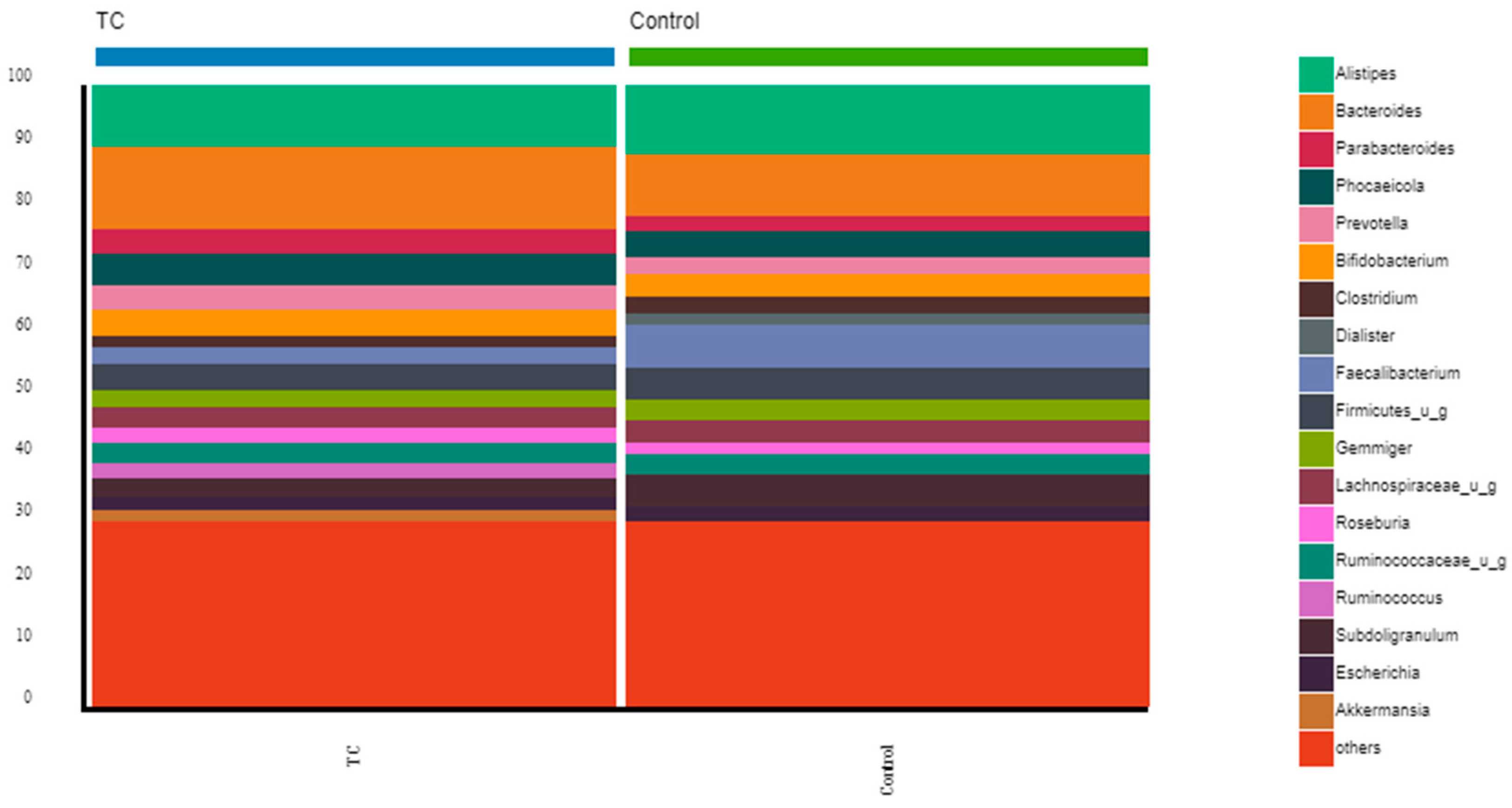
At the genus level, the genera that represented more than 5% in thyroid cancer patients were Bacteroides (13.25%), Alistipes (10.03%), and Phocaeicola (5.11%); in the volunteers, they were Alistipes (11.04%), Bacteroides (10.00%), Faecalibacterium (7.06%), and Subdoligranulum (5.03%) (Figure 2, and Tables S2 and S3 from the Supplementary Materials).
Finally, at the species level, the most abundant species (above 4%) in thyroid cancer patients were Bacteroides_u_s (5.15%) and Firmicutes_u_s (4.23%); in the volunteers, they were Subdoligranulum_u_s (5.00%), Firmicutes_u_s (4.99%), Faecalibacterium prausnitzii (4.50%), and Alistipes putredinis (4.39%) (Figure 3, and Tables S4 and S5 from the Supplementary Materials).
In the present study, alpha diversity, evaluated using Chao1 and the Simpson and Shannon indexes, was not significantly different between thyroid cancer patients and control sets of samples (p = 0.483, p = 0.815, and p = 0.406, respectively). Regarding beta diversity, no separating trends between the bacterial communities in pre- and post-treatment groups were observed in terms of Jaccard distance and Bray–Curtis dissimilarity (p = 0.322 and p = 0.110, respectively).

3.3. The Relative Abundance of F. prausnitzii in Differentiated Thyroid Cancer Patients Is Decreased Compared with Controls

In our population, we observed significant differences between the mean relative abundance of F. prausnitzii from control compared to pre-RAIT samples (p = 0.02) and post-RAIT samples (p = 0.002) from thyroid cancer patients, which is illustrated in the box-plots in Figure 4 and Figure 5, respectively.
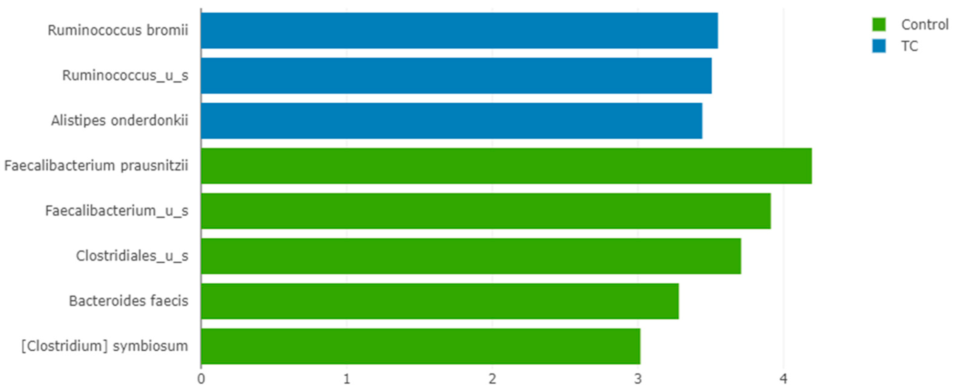
Interestingly, LEfSe analysis found that F. prausnitzii was the most significantly enriched species in controls compared to thyroid cancer patients, with an LDA score > 4 and p < 0.001 (Figure 6).

3.4. Impact of Radioiodine Therapy on the Richness, Diversity, and Composition

Alpha diversity was not significantly different between the pre- and post-treatment sets of samples, using Chao1 and the Simpson and Shannon indexes (p = 0.456, p = 0.273, and p = 0.208, respectively). Regarding beta diversity, no separating trends between the bacterial communities in pre- and post-treatment groups were observed in terms of Jaccard distance and Bray–Curtis dissimilarity (p = 1.000 and p = 0.994, respectively).
No significant changes at the phylum and genus levels were found after RAIT. At the species level, through LEfSe analysis, we found that Ruminococcus bicirculans was enriched before treatment (LDA of 3.082, p = 0.005).

3.5. Impact of RAIT on the Relative Abundance of F. prausnitzii

Our results showed that the mean relative abundance of F. prausnitzii increased in the patients’ samples post-RAIT (1.76 ± 1.86%) compared to pre-RAIT samples (1.70 ± 1.96%) but without significance (p = 0.1485). The same pattern was observed in the absolute abundances (p = 0.1646). In two patients, F. prausnitzii was not detected in either sample.
When analyzing each patient, an increase in the relative abundance of F. prausnitzii after RAIT was seen in nineteen patients, and a decrease was observed in sixteen patients; in two, it was unchanged. The relative abundance of F. Prausnitzii pre- and post-treatment from each patient is summarized in Figure 7.

4. Discussion

To the best of our knowledge, our study was the first to identify the variation of the relative abundance of F. prausnitzii after RAIT using shotgun metagenomics. We also compared the gut microbiota from thyroid cancer patients with volunteers. Faecalibacterium prausnitzii is the most important and explored butyrate-producing bacteria and one of the most abundant species in the human gut microbiota.

4.1. Composition of the Gut Microbiota of Differentiated Thyroid Cancer Patients before Treatment: The Relative Abundance of F. prausnitzii Is Decreased in Thyroid Cancer Patients Compared to Controls

The gut microbiota in our population of thyroid cancer patients was characterized by a dominance of Bacteroidetes and Firmicutes at the phylum level, which are known to be the most prominent phyla of the human gut microbiota.
Previous studies have evaluated the gut microbiota composition of thyroid cancer patients and found a dominance of the genera Prevotella, Roseburia, Coprococcus, Anaerostipes, Ruminococcus, Neisseria, Streptococcus, and Prophyromonas [37]. Conversely, other studies found a decrease in Prevotella [27,30].
At the genus level, in both groups, we found a dominance of Alistipes and Bacteroides. However, the other most represented genera in thyroid cancer patients were Phocaeicola. In contrast, in the control group, Faecalibacterium and Subdoligranulum were, together with Alistipes and Bacteroides, the most represented genera, and both are butyrate-producing bacteria [47].
The connection between gut microbiota and thyroid diseases, including thyroid cancer, has been suggested by numerous studies [30,35,48,49]. In our study, the thyroid cancer patients (before and after RAIT) who also presented with temporary hypothyroidism had a significant reduction in the relative abundance of F. prausnitzii compared to controls. This result is in line with previous studies since these bacteria were also reported to be significantly less present in multiple other diseases, including Crohn’s disease [14,50,51], rheumatoid arthritis [52], ulcerative colitis [15,53], celiac disease [16], and obesity [17]. The low relative abundance may be due to hypothyroidism or thyroid cancer, or both. Previous research found a decrease in F. prausnitzii in hypothyroid Hashimoto’s thyroiditis [18]; contrarily, Lauritano et al. and Ishaq et al. found that hypothyroidism is associated with bacterial overgrowth development [54,55].
The link between thyroid diseases and short-chain fatty acids has also been explored. Zhang J et al. found that Butyricimonas, which produce short-chain fatty acids, are significantly lower in thyroid cancer patients [37]. Microbial products, particularly short-chain fatty acids, can serve as an energy source for enterocytes and, combined with thyroid hormones, may enhance enterocyte differentiation and strengthen intercellular tight junctions [48,56]. The microbiota may also play a role in I uptake and in the enterohepatic cycling of thyroid hormones [48].
Interestingly, Yu X et al. suggested that the loss of SCFAs-producing bacteria may promote the development of thyroid cancer [27]. They believe that a low abundance of F. prausnitzii may result in gut microbial dysbiosis before or after developing thyroid cancer [27]. Our study reinforces that thyroid cancer patients present with dysbiotic gut microbiota with a low abundance of one of the most important butyrate-producing bacterium.

4.2. Effects of Radioiodine Therapy

Previous studies in patients with different types of cancers exposed to abdominal and pelvic radiotherapy reported that there were decreases in the relative abundance of F. prausnitzii [41,42,43,44] and an increase in one study [45]. Previous similar studies that did not report changes in these bacteria used different, less sensitive analytical methodologies that may have failed to detect F. prausnitzii. The difference between our results and previously reported results from radiotherapy reinforces the position of the radiobiology European Association of Nuclear Medicine working group that “radiopharmaceutical treatments are well tolerated and that some regimens could undertreat patients” [57].
Our study shows a mixed response, with an overall increase in the relative abundance of F. prausnitzii after radioiodine therapy, but without significance. In most patients, we found an increase in F. prausnitzii after radioiodine therapy.
Lapiere et al. demonstrated that prophylactic treatment with F. prausnitzii protects the colonic mucosal barrier from radiation exposure by maintaining its functional integrity through the regulation of the intestinal permeability, the enhancement of epithelial self-renewal, and through the preservation of differentiated epithelial cells [58].
Ionizing radiation refers to high-energy radiation that can ionize atoms by removing electrons from them, causing damage to molecules and cells. Some molecular effects of ionizing radiation include DNA and protein damage, oxidative stress, and cell cycle arrest. Overall, the molecular effects of ionizing radiation depend on the type and dose of radiation and on the type of cells being exposed.
Ionizing radiation has several effects on gut microbiota, the most relevant being the induction of oxidative stress. It generates reactive oxygen and nitrogen species that can damage cellular components, impair cellular functions, and change the redox state of the intestinal environment. These changes affect microbial growth, survival, and metabolism and, therefore, alter the gut microbiota’s composition. In addition to the previously described effects, ionizing radiation is capable of modulating microbial gene expression as it can alter the expression of genes involved in functions including DNA repair, response to stress, and energy metabolism. These gene expression changes may lead to relevant alterations in microbial function and metabolism, which may affect host health. The gut microbiota is a complex and diverse ecosystem of microorganisms, and ionizing radiation can promote certain microbial species’ growth and suppress others.
When exposed to ionizing radiation, the intestine experiences damage to epithelial cells and disruption of the microbiota, leading to inflammation and increased susceptibility to infection. However, SCFAs produced by gut microbiota inhibit inflammation and promote the survival and repair of epithelial cells.
Short-chain fatty acids have been shown to reduce oxidative stress and DNA damage in intestinal cells and enhance the repair of damaged DNA. Additionally, short-chain fatty acids can improve intestinal barrier function, which protects against the infiltration of harmful bacteria and toxins.
Given the positive effects of short-chain fatty acids in metabolism and immune modulation, the increase in the relative abundance in most patients might indicate that these bacteria could be important in resolving radiation aggression.

5. Conclusions

In conclusion, in this study, we found a significant reduction in the relative abundance of F. prausnitzii, the most important butyrate-producing bacterium, in thyroid cancer patients compared to controls.
Regarding the effect of radioiodine therapy, it does not appear to negatively affect the human gut microbiota. Moreover, we found that most patients had a higher relative abundance of F. prausnitzii after treatment, which, in line with previous studies, could indicate that F. prausnitzii, due to its anti-inflammatory characteristics, could play an important role in resolving radiation aggression.
Further studies are needed to discern whether and to what extent F. prausnitzii effectively plays a role in the outcome of the patients.

Supplementary Materials

The following supporting information can be downloaded at: https://www.mdpi.com/article/10.3390/nu15122680/s1, Table S1: Relative abundance Phylum-level before/after RAIT and volunteers; Table S2: Genus level-relative abundance before/after RAIT; Table S3: Relative abundance genus level in volunteers; Table S4: Species relative abundance from controls; Table S5: Species-level relative abundance before/after RAIT; Table S6: LefSe analysis Controls versus Thyroid cancer; Table S7: Change in absolute abundance phylum level before/after RAIT. after the Bonferroni correction; Table S8: Change in absolute abundance genus level before/after RAIT. p-values after the Bonferroni correction; Table S9: Change in absolute abundance species-level before/after RAIT. p-values after the Bonferroni correction; Table S10: Change in relative abundance phylum level before/after RAIT. p-values after the Bonferroni correction; Table S11: Change in relative abundance genus-level before/after RAIT. p-values after the Bonferroni correction; Table S12: Change in relative abundance species-level before/after RAIT. p-value after the Bonferroni correction.

Author Contributions

Conceptualization, A.F., R.S. and P.B.; methodology, A.F.; formal analysis, A.F. and P.B.; investigation, A.L.C. and A.F.; writing—original draft preparation, A.F.; writing—review and editing, A.F., A.O., R.S. and P.B.; supervision, R.S. and P.B.; funding acquisition, R.S. All authors have read and agreed to the published version of the manuscript.

Funding

This work was partially supported by FCT—Fundação para a Ciência e Tecnologia (Grant number UID/BIM/04293/2019).

Institutional Review Board Statement

The study was conducted in accordance with the Declaration of Helsinki and approved by the Institutional Ethics Committee Centro Hospitalar Universitário de São João and of Faculdade de Medicina da Universidade do Porto (27 February 2018/number: 52/18).

Informed Consent Statement

Informed consent was obtained from all subjects involved in the study.

Data Availability Statement

The data that support the findings of this study are available in Mendeley Data, V1 at http://doi.org/10.17632/3nfhkdmmzy.1, (accessed on 11 May 2023).

Acknowledgments

The authors would like to thank the members of the nuclear medicine department that helped in this research.

Conflicts of Interest

The authors declare no conflict of interest.

References

  1. Louis, P.; Flint, H.J. Diversity, metabolism and microbial ecology of butyrate-producing bacteria from the human large intestine. FEMS Microbiol. Lett. 2009, 294, 1–8. [Google Scholar] [CrossRef] [PubMed] [Green Version]
  2. Mahowald, M.A.; Rey, F.E.; Seedorf, H.; Turnbaugh, P.J.; Fulton, R.S.; Wollam, A.; Shah, N.; Wang, C.; Magrini, V.; Wilson, R.K.; et al. Characterizing a model human gut microbiota composed of members of its two dominant bacterial phyla. Proc. Natl. Acad. Sci. USA 2009, 106, 5859–5864. [Google Scholar] [CrossRef] [PubMed] [Green Version]
  3. Miquel, S.; Martin, R.; Rossi, O.; Bermúdez-Humarán, L.G.; Chatel, J.M.; Sokol, H.; Thomas, M.; Wells, J.M.; Langella, P. Faecalibacterium prausnitzii and human intestinal health. Curr. Opin. Microbiol. 2013, 16, 255–261. [Google Scholar] [CrossRef]
  4. Duncan, S.H.; Hold, G.L.; Harmsen, H.J.M.; Stewart, C.S.; Flint, H.J. Growth requirements and fermentation products of Fusobacterium prausnitzii, and a proposal to reclassify it as Faecalibacterium prausnitzii gen. nov., comb. nov. Int. J. Syst. Evol. Microbiol. 2002, 52 Pt 6, 2141–2146. [Google Scholar] [PubMed] [Green Version]
  5. Franks, A.H.; Harmsen, H.J.; Raangs, G.C.; Jansen, G.J.; Schut, F.; Welling, G.W. Variations of bacterial populations in human feces measured by fluorescent in situ hybridization with group-specific 16S rRNA-targeted oligonucleotide probes. Appl. Environ. Microbiol. 1998, 64, 3336–3345. [Google Scholar] [CrossRef] [Green Version]
  6. Suau, A.; Bonnet, R.; Sutren, M.; Godon, J.J.; Gibson, G.R.; Collins, M.D.; Doré, J. Direct analysis of genes encoding 16S rRNA from complex communities reveals many novel molecular species within the human gut. Appl. Environ. Microbiol. 1999, 65, 4799–4807. [Google Scholar] [CrossRef] [Green Version]
  7. Eckburg, P.B.; Bik, E.M.; Bernstein, C.N.; Purdom, E.; Dethlefsen, L.; Sargent, M.; Gill, S.R.; Nelson, K.E.; Relman, D.A. Diversity of the human intestinal microbial flora. Science 2005, 308, 1635–1638. [Google Scholar] [CrossRef] [Green Version]
  8. Wang, R.F.; Cao, W.W.; Cerniglia, C.E. Phylogenetic analysis of Fusobacterium prausnitzii based upon the 16S rRNA gene sequence and PCR confirmation. Int. J. Syst. Bacteriol. 1996, 46, 341–343. [Google Scholar] [CrossRef] [Green Version]
  9. Stewart, L. Faecalibacterium Prausnitzii: The Gut Microbe That Prevents Inflammation. 2021. Available online: https://atlasbiomed.com/blog/faecalibacterium-prausnitzii/ (accessed on 20 February 2022).
  10. Carlsson, A.H.; Yakymenko, O.; Olivier, I.; Håkansson, F.; Postma, E.; Keita, Å.V.; Söderholm, J.D. Faecalibacterium prausnitzii supernatant improves intestinal barrier function in mice DSS colitis. Scand. J. Gastroenterol. 2013, 48, 1136–1144. [Google Scholar] [CrossRef]
  11. Martín, R.; Miquel, S.; Chain, F.; Natividad, J.M.; Jury, J.; Lu, J.; Sokol, H.; Theodorou, V.; Bercik, P.; Verdu, E.F.; et al. Faecalibacterium prausnitzii prevents physiological damages in a chronic low-grade inflammation murine model. BMC Microbiol. 2015, 15, 67. [Google Scholar] [CrossRef] [Green Version]
  12. Miquel, S.; Leclerc, M.; Martin, R.; Chain, F.; Lenoir, M.; Raguideau, S.; Hudault, S.; Bridonneau, C.; Northen, T.; Bowen, B.; et al. Identification of metabolic signatures linked to anti-inflammatory effects of Faecalibacterium prausnitzii. MBio 2015, 6, e00300-15. [Google Scholar] [CrossRef] [PubMed] [Green Version]
  13. Ohman, L.; Simrén, M. New insights into the pathogenesis and pathophysiology of irritable bowel syndrome. Dig. Liver Dis. 2007, 39, 201–215. [Google Scholar] [CrossRef] [PubMed]
  14. Sokol, H.; Pigneur, B.; Watterlot, L.; Lakhdari, O.; Bermúdez-Humarán, L.G.; Gratadoux, J.J.; Blugeon, S.; Bridonneau, C.; Furet, J.P.; Corthier, G.; et al. Faecalibacterium prausnitzii is an anti-inflammatory commensal bacterium identified by gut microbiota analysis of Crohn disease patients. Proc. Natl. Acad. Sci. USA 2008, 105, 16731–16736. [Google Scholar] [CrossRef] [PubMed] [Green Version]
  15. Sokol, H.; Seksik, P.; Furet, J.P.; Firmesse, O.; Nion-Larmurier, I.; Beaugerie, L.; Cosnes, J.; Corthier, G.; Marteau, P.; Doré, J. Low counts of Faecalibacterium prausnitzii in colitis microbiota. Inflamm. Bowel Dis. 2009, 15, 1183–1189. [Google Scholar] [CrossRef]
  16. De Palma, G.; Nadal, I.; Medina, M.; Donat, E.; Ribes-Koninckx, C.; Calabuig, M.; Sanz, Y. Intestinal dysbiosis and reduced immunoglobulin-coated bacteria associated with coeliac disease in children. BMC Microbiol. 2010, 10, 63. [Google Scholar] [CrossRef] [Green Version]
  17. Furet, J.P.; Kong, L.C.; Tap, J.; Poitou, C.; Basdevant, A.; Bouillot, J.L.; Mariat, D.; Corthier, G.; Doré, J.; Henegar, C.; et al. Differential adaptation of human gut microbiota to bariatric surgery-induced weight loss: Links with metabolic and low-grade inflammation markers. Diabetes 2010, 59, 3049–3057. [Google Scholar] [CrossRef] [Green Version]
  18. Zhao, F.; Feng, J.; Li, J.; Zhao, L.; Liu, Y.; Chen, H.; Jin, Y.; Zhu, B.; Wei, Y. Alterations of the Gut Microbiota in Hashimoto’s Thyroiditis Patients. Thyroid 2018, 28, 175–186. [Google Scholar] [CrossRef]
  19. Davie, J.R. Inhibition of histone deacetylase activity by butyrate. J. Nutr. 2003, 133 (Suppl. 7), 2485s–2493s. [Google Scholar] [CrossRef] [Green Version]
  20. Gibson, P.R.; Rosella, O.; Wilson, A.J.; Mariadason, J.M.; Rickard, K.; Byron, K.; Barkla, D.H. Colonic epithelial cell activation and the paradoxical effects of butyrate. Carcinogenesis 1999, 20, 539–544. [Google Scholar] [CrossRef] [Green Version]
  21. Lopez-Siles, M.; Duncan, S.H.; Garcia-Gil, L.J.; Martinez-Medina, M. Faecalibacterium prausnitzii: From microbiology to diagnostics and prognostics. ISME J. 2017, 11, 841–852. [Google Scholar] [CrossRef]
  22. Inan, M.S.; Rasoulpour, R.J.; Yin, L.; Hubbard, A.K.; Rosenberg, D.W.; Giardina, C. The luminal short-chain fatty acid butyrate modulates NF-kappaB activity in a human colonic epithelial cell line. Gastroenterology 2000, 118, 724–734. [Google Scholar] [CrossRef] [PubMed]
  23. Schwab, M.; Reynders, V.; Loitsch, S.; Steinhilber, D.; Stein, J.; Schröder, O. Involvement of different nuclear hormone receptors in butyrate-mediated inhibition of inducible NF kappa B signalling. Mol. Immunol. 2007, 44, 3625–3632. [Google Scholar] [CrossRef] [PubMed]
  24. Klampfer, L.; Huang, J.; Sasazuki, T.; Shirasawa, S.; Augenlicht, L. Inhibition of interferon gamma signaling by the short chain fatty acid butyrate. Mol. Cancer Res. 2003, 1, 855–862. [Google Scholar]
  25. Roediger, W.E. Utilization of nutrients by isolated epithelial cells of the rat colon. Gastroenterology 1982, 83, 424–429. [Google Scholar] [CrossRef] [PubMed]
  26. He, X.; Zhao, S.; Li, Y. Faecalibacterium prausnitzii: A Next-Generation Probiotic in Gut Disease Improvement. Can. J. Infect. Dis. Med. Microbiol. 2021, 2021, 6666114. [Google Scholar] [CrossRef]
  27. Yu, X.; Jiang, W.; Kosik, R.O.; Song, Y.; Luo, Q.; Qiao, T.; Tong, J.; Liu, S.; Deng, C.; Qin, S.; et al. Gut microbiota changes and its potential relations with thyroid carcinoma. J. Adv. Res. 2021, 35, 61–70. [Google Scholar] [CrossRef]
  28. Cancer IAfRo. Available online: https://gco.iarc.fr/today/data/factsheets/populations/620-portugal-fact-sheets.pdf (accessed on 15 March 2022).
  29. Haugen, B.R.; Alexander, E.K.; Bible, K.C.; Doherty, G.M.; Mandel, S.J.; Nikiforov, Y.E.; Pacini, F.; Randolph, G.W.; Sawka, A.M.; Schlumberger, M.; et al. 2015 American Thyroid Association Management Guidelines for Adult Patients with Thyroid Nodules and Differentiated Thyroid Cancer: The American Thyroid Association Guidelines Task Force on Thyroid Nodules and Differentiated Thyroid Cancer. Thyroid 2016, 26, 2-92. [Google Scholar] [CrossRef] [Green Version]
  30. Feng, J.; Zhao, F.; Sun, J.; Lin, B.; Zhao, L.; Liu, Y.; Jin, Y.; Li, S.; Li, A.; Wei, Y. Alterations in the gut microbiota and metabolite profiles of thyroid carcinoma patients. Int. J. Cancer 2019, 144, 2728–2745. [Google Scholar] [CrossRef]
  31. Vought, R.L.; Brown, F.A.; Sibinovic, K.H.; McDaniel, E.G. Effect of changing intestinal bacterial flora on thyroid function in the rat. Horm. Metab. Res. 1972, 4, 43–47. [Google Scholar] [CrossRef]
  32. Kasaikina, M.V.; Kravtsova, M.A.; Lee, B.C.; Seravalli, J.; Peterson, D.A.; Walter, J.; Legge, R.; Benson, A.K.; Hatfield, D.L.; Gladyshev, V.N. Dietary selenium affects host selenoproteome expression by influencing the gut microbiota. FASEB J. 2011, 25, 2492–2499. [Google Scholar] [CrossRef] [Green Version]
  33. Zhou, L.; Li, X.; Ahmed, A.; Wu, D.; Liu, L.; Qiu, J.; Yan, Y.; Jin, M.; Xin, Y. Gut microbe analysis between hyperthyroid and healthy individuals. Curr. Microbiol. 2014, 69, 675–680. [Google Scholar] [CrossRef] [PubMed]
  34. Jiang, W.; Lu, G.; Gao, D.; Lv, Z.; Li, D. The relationships between the gut microbiota and its metabolites with thyroid diseases. Front. Endocrinol. 2022, 13, 943408. [Google Scholar] [CrossRef] [PubMed]
  35. Samimi, H.; Haghpanah, V. Gut Microbiome and Radioiodine-Refractory Papillary Thyroid Carcinoma Pathophysiology. Trends Endocrinol. Metab. 2020, 31, 627–630. [Google Scholar] [CrossRef] [PubMed]
  36. Aerts, A.; Eberlein, U.; Holm, S.; Hustinx, R.; Konijnenberg, M.; Strigari, L.; van Leeuwen, F.W.; Glatting, G.; Lassmann, M. EANM position paper on the role of radiobiology in nuclear medicine. Eur. J. Nucl. Med. Mol. Imaging 2021, 48, 3365–3377. [Google Scholar] [CrossRef]
  37. Zhang, J.; Zhang, F.; Zhao, C.; Xu, Q.; Liang, C.; Yang, Y.; Wang, H.; Shang, Y.; Wang, Y.; Mu, X.; et al. Dysbiosis of the gut microbiome is associated with thyroid cancer and thyroid nodules and correlated with clinical index of thyroid function. Endocrine 2019, 64, 564–574. [Google Scholar] [CrossRef]
  38. Hollingsworth, B.A.; Cassatt, D.R.; DiCarlo, A.L.; Rios, C.I.; Satyamitra, M.M.; Winters, T.A.; Taliaferro, L.P. Acute Radiation Syndrome and the Microbiome: Impact and Review. Front. Pharmacol. 2021, 12, 643283. [Google Scholar] [CrossRef]
  39. Morris, Z.S.; Wang, A.Z.; Knox, S.J. The Radiobiology of Radiopharmaceuticals. Semin. Radiat. Oncol. 2021, 31, 20–27. [Google Scholar] [CrossRef]
  40. Terry, S.Y.A.; Nonnekens, J.; Aerts, A.; Baatout, S.; de Jong, M.; Cornelissen, B.; Pouget, J.-P. Call to arms: Need for radiobiology in molecular radionuclide therapy. Eur. J. Nucl. Med. Mol. Imaging 2019, 46, 1588–1590. [Google Scholar] [CrossRef]
  41. Wang, A.; Ling, Z.; Yang, Z.; Kiela, P.R.; Wang, T.; Wang, C.; Cao, L.; Geng, F.; Shen, M.; Ran, X.; et al. Gut microbial dysbiosis may predict diarrhea and fatigue in patients undergoing pelvic cancer radiotherapy: A pilot study. PLoS ONE 2015, 10, e0126312. [Google Scholar] [CrossRef] [Green Version]
  42. Yi, Y.; Shen, L.; Shi, W.; Xia, F.; Zhang, H.; Wang, Y.; Zhang, J.; Wang, Y.; Sun, X.; Zhang, Z.; et al. Gut microbiome components predict response to neoadjuvant chemoradiotherapy in patients with locally advanced rectal cancer: A prospective, longitudinal study. Clin. Cancer Res. 2021, 27, 1329–1340. [Google Scholar] [CrossRef]
  43. Sahly, N.; Moustafa, A.; Zaghloul, M.; Salem, T.Z. Effect of radiotherapy on the gut microbiome in pediatric cancer patients: A pilot study. PeerJ 2019, 2019, e7683. [Google Scholar] [CrossRef] [PubMed]
  44. Shi, W.; Shen, L.; Zou, W.; Wang, J.; Yang, J.; Wang, Y.; Liu, B.; Xie, L.; Zhu, J.; Zhang, Z. The Gut Microbiome Is Associated with Therapeutic Responses and Toxicities of Neoadjuvant Chemoradiotherapy in Rectal Cancer Patients-A Pilot Study. Front. Cell. Infect. Microbiol. 2020, 10, 562463. [Google Scholar] [CrossRef] [PubMed]
  45. Nam, Y.D.; Kim, H.J.; Seo, J.G.; Kang, S.W.; Bae, J.W. Impact of pelvic radiotherapy on gut microbiota of gynecological cancer patients revealed by massive pyrosequencing. PLoS ONE 2013, 8, e82659. [Google Scholar] [CrossRef] [PubMed] [Green Version]
  46. Segata, N.; Izard, J.; Waldron, L.; Gevers, D.; Miropolsky, L.; Garrett, W.S.; Huttenhower, C. Metagenomic biomarker discovery and explanation. Genome Biol. 2011, 12, R60. [Google Scholar] [CrossRef] [Green Version]
  47. Van Hul, M.; Le Roy, T.; Prifti, E.; Dao, M.C.; Paquot, A.; Zucker, J.-D.; Delzenne, N.M.; Muccioli, G.G.; Clément, K.; Cani, P.D. From correlation to causality: The case of Subdoligranulum. Gut Microbes 2020, 12, 1849998. [Google Scholar] [CrossRef]
  48. Frohlich, E.; Wahl, R. Microbiota and Thyroid Interaction in Health and Disease. Trends Endocrinol. Metab. 2019, 30, 479–490. [Google Scholar] [CrossRef] [Green Version]
  49. Knezevic, J.; Starchl, C.; Tmava Berisha, A.; Amrein, K. Thyroid-Gut-Axis: How Does the Microbiota Influence Thyroid Function? Nutrients 2020, 12, 1769. [Google Scholar] [CrossRef]
  50. Gevers, D.; Kugathasan, S.; Denson, L.A.; Vázquez-Baeza, Y.; Van Treuren, W.; Ren, B.; Schwager, E.; Knights, D.; Song, S.J.; Yassour, M.; et al. The treatment-naive microbiome in new-onset Crohn’s disease. Cell Host Microbe 2014, 15, 382–392. [Google Scholar] [CrossRef] [Green Version]
  51. Takahashi, K.; Nishida, A.; Fujimoto, T.; Fujii, M.; Shioya, M.; Imaeda, H.; Inatomi, O.; Bamba, S.; Andoh, A.; Sugimoto, M. Reduced Abundance of Butyrate-Producing Bacteria Species in the Fecal Microbial Community in Crohn’s Disease. Digestion 2016, 93, 59–65. [Google Scholar] [CrossRef]
  52. Vaahtovuo, J.; Munukka, E.; Korkeamäki, M.; Luukkainen, R.; Toivanen, P. Fecal microbiota in early rheumatoid arthritis. J. Rheumatol. 2008, 35, 1500–1505. [Google Scholar]
  53. Vermeiren, J.; Abbeele, P.V.D.; Laukens, D.; Vigsnaes, L.K.; De Vos, M.; Boon, N.; Van De Wiele, T. Decreased colonization of fecal Clostridium coccoides/Eubacterium rectale species from ulcerative colitis patients in an in vitro dynamic gut model with mucin environment. FEMS Microbiol. Ecol. 2012, 79, 685–696. [Google Scholar] [CrossRef] [PubMed]
  54. Ishaq, H.M.; Mohammad, I.S.; Guo, H.; Shahzad, M.; Hou, Y.J.; Ma, C.; Naseem, Z.; Wu, X.; Shi, P.; Xu, J. Molecular estimation of alteration in intestinal microbial composition in Hashimoto’s thyroiditis patients. Biomed. Pharmacother. 2017, 95, 865–874. [Google Scholar] [CrossRef] [PubMed]
  55. Lauritano, E.C.; Bilotta, A.L.; Gabrielli, M.; Scarpellini, E.; Lupascu, A.; Laginestra, A.; Novi, M.; Sottili, S.; Serricchio, M.; Cammarota, G.; et al. Association between Hypothyroidism and Small Intestinal Bacterial Overgrowth. J. Clin. Endocrinol. Metab. 2007, 92, 4180–4184. [Google Scholar] [CrossRef] [PubMed] [Green Version]
  56. Bargiel, P.; Szczuko, M.; Stachowska, L.; Prowans, P.; Czapla, N.; Markowska, M.; Petriczko, J.; Kledzik, J.; Jędrzejczyk-Kledzik, A.; Palma, J.; et al. Microbiome Metabolites and Thyroid Dysfunction. J. Clin. Med. 2021, 10, 3609. [Google Scholar] [CrossRef] [PubMed]
  57. Pouget, J.-P.; Konijnenberg, M.; Eberlein, U.; Glatting, G.; Gabina, P.M.; Herrmann, K.; Holm, S.; Strigari, L.; van Leeuwen, F.W.B.; Lassmann, M. An EANM position paper on advancing radiobiology for shaping the future of nuclear medicine. Eur. J. Nucl. Med. Mol. Imaging 2023, 50, 242–246. [Google Scholar]
  58. Lapiere, A.; Geiger, M.; Robert, V.; Demarquay, C.; Auger, S.; Chadi, S.; Benadjaoud, M.; Fernandes, G.; Milliat, F.; Langella, P.; et al. Prophylactic Faecalibacterium prausnitzii treatment prevents the acute breakdown of colonic epithelial barrier in a preclinical model of pelvic radiation disease. Gut Microbes 2020, 12, 1812867. [Google Scholar] [CrossRef]
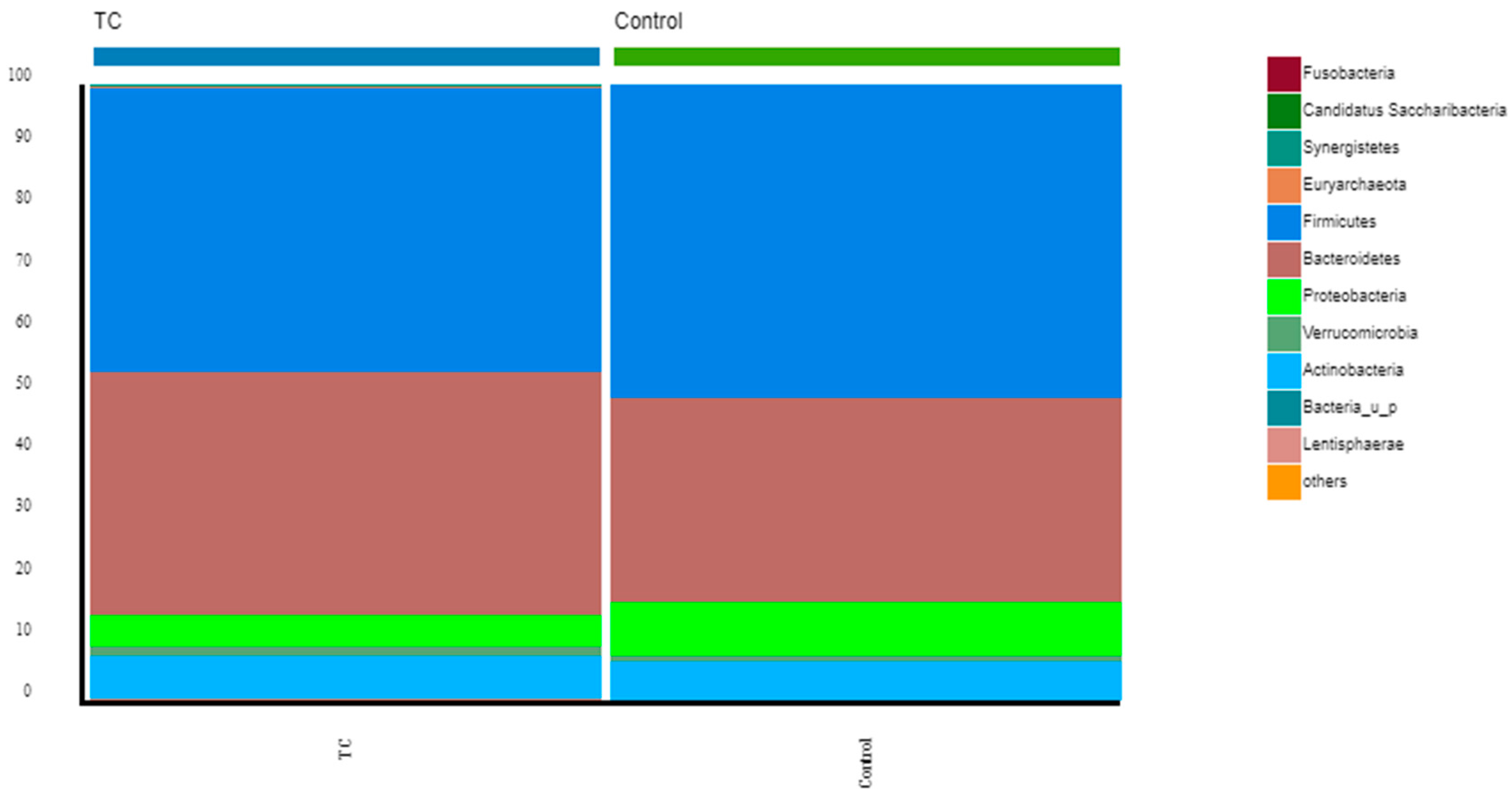
Figure 1. Comparison of bacterial communities between thyroid cancer patients and controls. Stacked bar chart representing the relative abundance of the most frequent phyla from the thyroid cancer patients before RAIT compared with the controls.
Nutrients 15 02680 g001
Figure 2. Comparison of bacterial communities between thyroid cancer patients and controls. Stacked bar charts represent the relative abundance of the most frequent genera from the thyroid cancer patients before RAIT and volunteers. Others represent the bottom 30% of the less abundant genera.
Nutrients 15 02680 g002
Figure 3. Comparison of bacterial communities between thyroid cancer patients and controls. Stacked bar chart representing the relative abundance at the species level from the thyroid cancer patients before RAIT and controls. Others represent the bottom 50% of the less abundant genera.
Nutrients 15 02680 g003
Figure 4. Comparison of F. prausnitzii’s relative abundance between thyroid cancer patients pre-RAIT samples and controls. Box-plot representing the relative abundance of F. prausnitzii in thyroid cancer patients (blue) and controls (red). Significant differences were found between the relative abundances, p = 0.02.
Nutrients 15 02680 g004
Figure 5. Comparison of F. prausnitzii’s relative abundance between thyroid cancer patients post-RAIT samples and controls. Box-plot representing the relative abundance of F. prausnitzii in post-RAIT samples from thyroid cancer patients (orange) and controls (red). Significant differences were found between the relative abundances, p = 0.002.
Nutrients 15 02680 g005
Figure 6. LEfSe chart representing the species that are significantly enriched in thyroid cancer patients (blue) and controls (green). All had LDA scores> 3 and p < 0.05.
Nutrients 15 02680 g006
Figure 7. Differences in the relative abundance (%) of Faecalibacterium prausnitzii before and after treatment from each patient (n = 37).
Nutrients 15 02680 g007
Table 1. Patient baseline characteristics (ND—not determined; SD—standard deviation).
Baseline Characteristicsn = 37
Age (mean, SD)51 ± 17
Gender
 Male11
 Female26
BMI (mean, SD)28.3 ± 6.6
 Concomitant diseases
 Diabetes7
 Cardiovascular diseases16
 Other (psoriasis, chronic kidney disease, hyperuricemia, gastritis, etc.)8
Number of chronic medications (mean, SD)3.1 ± 2.4
Tumor Type
 Follicular1
 Papillar36
Clinical T stage
 T116
 T213
 T36
 T41
 ND1
Clinical N stage
 Nx25
 N04
 N18
Clinical M stage
 M034
 M13
R
 R030
 R15
 R21
Age (mean, SD)51 ± 17
Gender
 Male11
 Female26
I-131 dose
 1110 MBq7
 3700 MBq22
 5550 MBq8
Disclaimer/Publisher’s Note: The statements, opinions and data contained in all publications are solely those of the individual author(s) and contributor(s) and not of MDPI and/or the editor(s). MDPI and/or the editor(s) disclaim responsibility for any injury to people or property resulting from any ideas, methods, instructions or products referred to in the content.