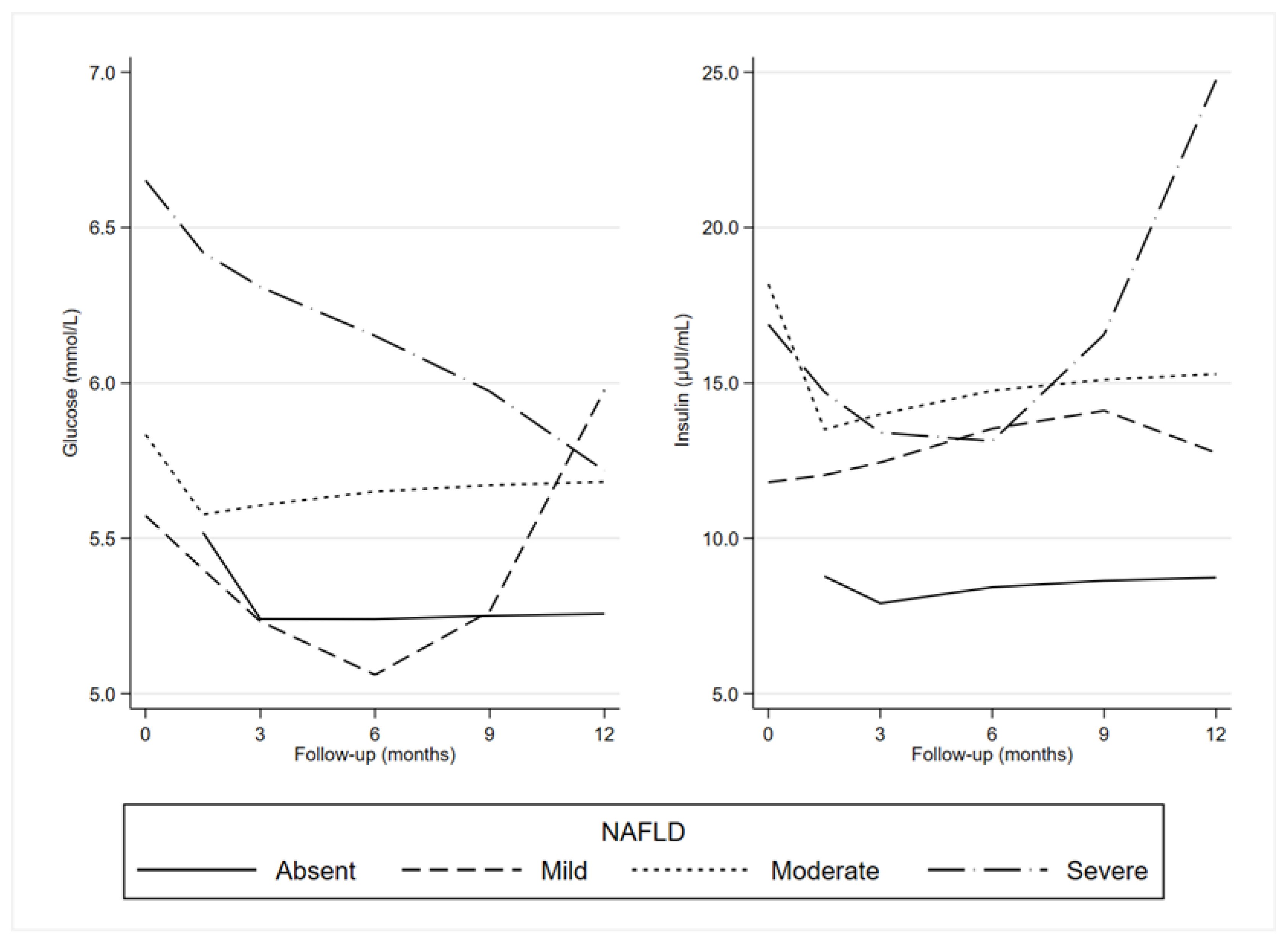
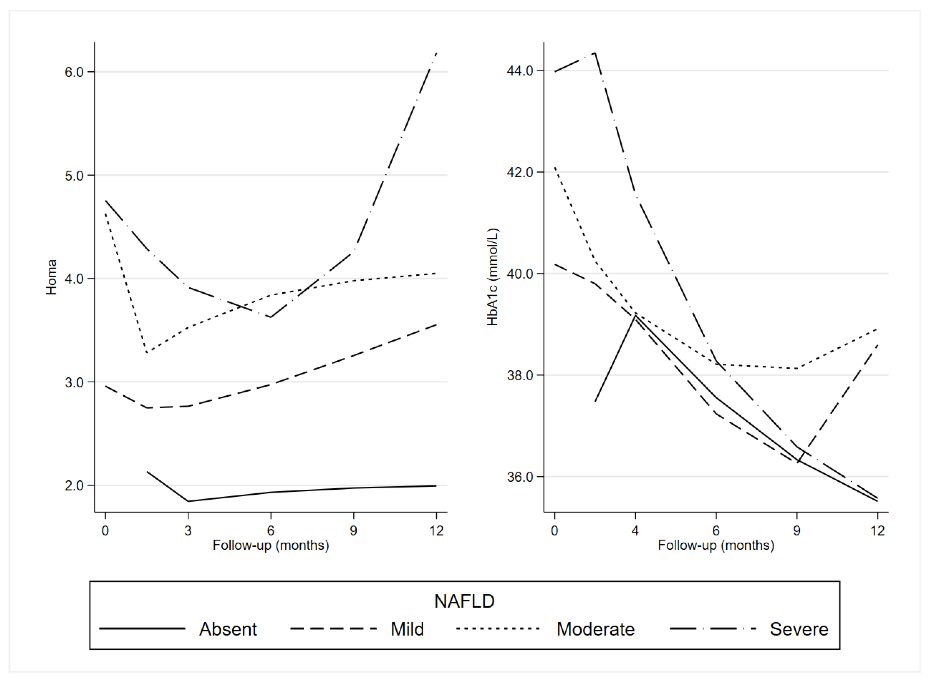
Diet and Exercise Exert a Differential Effect on Glucose Metabolism Markers According to the Degree of NAFLD Severity
Abstract
1. Introduction
2. Materials and Methods
2.1. Participants
2.2. Study Design
2.3. Data Collection
2.4. Measurements
2.5. Dietary Plan
2.6. Exercise Protocol
2.6.1. Fitness Assessment Tests
2.6.2. Aerobic Exercise and Resistance Training Combination
2.7. Statistical Analysis
3. Results
4. Discussion
5. Conclusions
Author Contributions
Funding
Institutional Review Board Statement
Informed Consent Statement
Data Availability Statement
Conflicts of Interest
Appendix A
References
- Mundi, M.S.; Velapati, S.; Patel, J.; Kellogg, T.A.; Abu Dayyeh, B.K.; Hurt, R.T. Evolution of NAFLD and its management. Nutr. Clin. Pract. 2020, 35, 72–84. [Google Scholar] [CrossRef] [PubMed]
- Smith, B.W.; Adams, L.A. Nonalcoholic fatty liver disease and diabetes mellitus: Pathogenesis and treatment. Nat. Rev. Endocrinol. 2011, 7, 456–465. [Google Scholar] [CrossRef] [PubMed]
- Ballestri, S.; Zona, S.; Targher, G.; Romagnoli, D.; Baldelli, E.; Nascimbeni, F.; Roverato, A.; Guaraldi, G.; Lonardo, A. Nonalcoholic fatty liver disease is associated with an almost twofold increased risk of incident type 2 diabetes and metabolic syndrome. Evidence from a systematic review and meta-analysis. J. Gastroenterol. Hepatol. 2016, 31, 936–944. [Google Scholar] [CrossRef]
- Kunutsor, S.K.; Apekey, T.A.; Walley, J. Liver aminotransferases and risk of incident type 2 diabetes: A systematic review and meta-analysis. Am. J. Epidemiol. 2013, 178, 159–171. [Google Scholar] [CrossRef] [PubMed]
- Wannamethee, S.G.; Shaper, A.G.; Lennon, L.; Whincup, P.H. Hepatic enzymes, the metabolic syndrome, and the risk of type 2 diabetes in older men. Diabetes Care 2005, 28, 2913–2918. [Google Scholar] [CrossRef]
- Brar, G.; Tsukamoto, H. Alcoholic and non-alcoholic steatohepatitis: Global perspective and emerging science. J. Gastroenterol. 2019, 54, 218–225. [Google Scholar] [CrossRef]
- Smith, G.I.; Shankaran, M.; Yoshino, M.; Schweitzer, G.G.; Chondronikola, M.; Beals, J.W.; Okunade, A.L.; Patterson, B.W.; Nyangau, E.; Field, T. Insulin resistance drives hepatic de novo lipogenesis in nonalcoholic fatty liver disease. J. Clin. Investig. 2020, 130, 1453–1460. [Google Scholar] [CrossRef] [PubMed]
- Day, C.P.; James, O.F. Steatohepatitis: A Tale of Two “Hits”? Elsevier: Amsterdam, The Netherlands, 1998; Volume 114, pp. 842–845. [Google Scholar]
- Utzschneider, K.M.; Kahn, S.E. The role of insulin resistance in nonalcoholic fatty liver disease. J. Clin. Endocrinol. Metab. 2006, 91, 4753–4761. [Google Scholar] [CrossRef]
- Misciagna, G.; Del Pilar Diaz, M.; Caramia, D.V.; Bonfiglio, C.; Franco, I.; Noviello, M.R.; Chiloiro, M.; Abbrescia, D.I.; Mirizzi, A.; Tanzi, M.; et al. Effect of a Low Glycemic Index Mediterranean Diet on Non-Alcoholic Fatty Liver Disease. A Randomized Controlled Clinici Trial. J. Nutr. Health Aging 2017, 21, 404–412. [Google Scholar] [CrossRef]
- Franco, I.; Bianco, A.; Dìaz, M.D.P.; Bonfiglio, C.; Chiloiro, M.; Pou, S.A.; Becaria Coquet, J.; Mirizzi, A.; Nitti, A.; Campanella, A.; et al. Effectiveness of two physical activity programs on non-alcoholic fatty liver disease. a randomized controlled clinical trial. Rev. Fac. Cienc. Médicas Córdoba 2019, 76, 26. [Google Scholar] [CrossRef]
- Franco, I.; Bianco, A.; Mirizzi, A.; Campanella, A.; Bonfiglio, C.; Sorino, P.; Notarnicola, M.; Tutino, V.; Cozzolongo, R.; Giannuzzi, V.; et al. Physical Activity and Low Glycemic Index Mediterranean Diet: Main and Modification Effects on NAFLD Score. Results from a Randomized Clinical Trial. Nutrients 2020, 13, 66. [Google Scholar] [CrossRef] [PubMed]
- Calabrese, F.M.; Disciglio, V.; Franco, I.; Sorino, P.; Bonfiglio, C.; Bianco, A.; Campanella, A.; Lippolis, T.; Pesole, P.L.; Polignano, M.; et al. A Low Glycemic Index Mediterranean Diet Combined with Aerobic Physical Activity Rearranges the Gut Microbiota Signature in NAFLD Patients. Nutrients 2022, 14, 1773. [Google Scholar] [CrossRef] [PubMed]
- Curci, R.; Bianco, A.; Franco, I.; Campanella, A.; Mirizzi, A.; Bonfiglio, C.; Sorino, P.; Fucilli, F.; Di Giovanni, G.; Giampaolo, N.; et al. The Effect of Low Glycemic Index Mediterranean Diet and Combined Exercise Program on Metabolic-Associated Fatty Liver Disease: A Joint Modeling Approach. J. Clin. Med. 2022, 11, 4339. [Google Scholar] [CrossRef] [PubMed]
- Swain, D.P.; Brawner, C.A.; Medicine, A.C.O.S. ACSM’s Resource Manual for Guidelines for Exercise Testing and Prescription; Wolters Kluwer Health/Lippincott Williams & Wilkins: Amsterdam, The Netherlands, 2014. [Google Scholar]
- Craig, C.L.; Marshall, A.L.; Sjöström, M.; Bauman, A.E.; Booth, M.L.; Ainsworth, B.E.; Pratt, M.; Ekelund, U.; Yngve, A.; Sallis, J.F.; et al. International physical activity questionnaire: 12-country reliability and validity. Med. Sci. Sports Exerc. 2003, 35, 1381–1395. [Google Scholar] [CrossRef] [PubMed]
- Riboli, E.; Hunt, K.J.; Slimani, N.; Ferrari, P.; Norat, T.; Fahey, M.; Charrondiere, U.R.; Hemon, B.; Casagrande, C.; Vignat, J.; et al. European Prospective Investigation into Cancer and Nutrition (EPIC): Study populations and data collection. Public Health Nutr. 2002, 5, 1113–1124. [Google Scholar] [CrossRef] [PubMed]
- Brazier, J.E.; Harper, R.; Jones, N.M.; O’Cathain, A.; Thomas, K.J.; Usherwood, T.; Westlake, L. Validating the SF-36 health survey questionnaire: New outcome measure for primary care. BMJ 1992, 305, 160–164. [Google Scholar] [CrossRef]
- Chiloiro, M.; Misciagna, G. Ultrasonographic Anthropometry: An Application to the Measurement of Liver and Abdominal Fat. In Handbook of Anthropometry: Physical Measures of Human Form in Health and Disease; Springer: Berlin/Heidelberg, Germany, 2012; pp. 2227–2242. [Google Scholar]
- Skinner, H.A.; Sheu, W.J. Reliability of alcohol use indices. The Lifetime Drinking History and the MAST. J. Stud. Alcohol. 1982, 43, 1157–1170. [Google Scholar] [CrossRef]
- Chiloiro, M.; Caruso, M.G.; Cisternino, A.M.; Inguaggiato, R.; Reddavide, R.; Bonfiglio, C.; Guerra, V.; Notarnicola, M.; De Michele, G.; Correale, M.; et al. Ultrasound evaluation and correlates of fatty liver disease: A population study in a Mediterranean area. Metab. Syndr. Relat. Disord. 2013, 11, 349–358. [Google Scholar] [CrossRef]
- Veronese, N.; Notarnicola, M.; Cisternino, A.M.; Inguaggiato, R.; Guerra, V.; Reddavide, R.; Donghia, R.; Rotolo, O.; Zinzi, I.; Leandro, G.; et al. Trends in adherence to the Mediterranean diet in South Italy: A cross sectional study. Nutr. Metab. Cardiovasc. Dis. NMCD 2019, 30, 410–417. [Google Scholar] [CrossRef]
- Laukkanen, R.; Oja, P.; Pasanen, M.; Vuori, I. Validity of a two kilometre walking test for estimating maximal aerobic power in overweight adults. Int. J. Obes. Relat. Metab. Disord. J. Int. Assoc. Study Obes. 1992, 16, 263–268. [Google Scholar]
- Canadian Society for Exercise Physiology. The Canadian Physical Activity, Fitness and Lifestyle Approach (CPAFLA): CSEP—Health and Fitness Program’s Health-Related Appraisal and Counselling Strategy; Canadian Society for Exercise Physiology: Ottawa, ON, Canada, 2003. [Google Scholar]
- Hoeger, W.W.; Hopkins, D.R. A comparison of the sit and reach and the modified sit and reach in the measurement of flexibility in women. Res. Q. Exerc. Sport 1992, 63, 191–195. [Google Scholar] [CrossRef]
- Tanaka, H.; Monahan, K.D.; Seals, D.R. Age-predicted maximal heart rate revisited. J. Am. Coll. Cardiol. 2001, 37, 153–156. [Google Scholar] [CrossRef] [PubMed]
- Colberg, S.R.; Sigal, R.J.; Fernhall, B.; Regensteiner, J.G.; Blissmer, B.J.; Rubin, R.R.; Chasan-Taber, L.; Albright, A.L.; Braun, B. Exercise and type 2 diabetes: The American College of Sports Medicine and the American Diabetes Association: Joint position statement. Diabetes Care 2010, 33, e147–e167. [Google Scholar] [CrossRef] [PubMed]
- Liang, K.-Y.; Zeger, S.L. Longitudinal data analysis using generalized linear models. Biometrika 1986, 73, 13–22. [Google Scholar] [CrossRef]
- De Silva, N.M.G.; Borges, M.C.; Hingorani, A.D.; Engmann, J.; Shah, T.; Zhang, X.; Luan, J.A.; Langenberg, C.; Wong, A.; Kuh, D. Liver function and risk of type 2 diabetes: Bidirectional Mendelian randomization study. Diabetes 2019, 68, 1681–1691. [Google Scholar] [CrossRef]
- Fujiwara, N.; Qian, T.; Koneru, B.; Hoshida, Y. Omics-derived hepatocellular carcinoma risk biomarkers for precision care of chronic liver diseases. Hepatol. Res. 2020, 50, 817–830. [Google Scholar] [CrossRef]
- Brouwers, M.C.; Simons, N.; Stehouwer, C.D.; Isaacs, A. Non-alcoholic fatty liver disease and cardiovascular disease: Assessing the evidence for causality. Diabetologia 2020, 63, 253–260. [Google Scholar] [CrossRef]
- Koo, B.K.; Allison, M.A.; Criqui, M.H.; Denenberg, J.O.; Wright, C.M. The association between liver fat and systemic calcified atherosclerosis. J. Vasc. Surg. 2020, 71, 204–211. e204. [Google Scholar] [CrossRef]
- Byrne, C.D.; Targher, G. NAFLD as a driver of chronic kidney disease. J. Hepatol. 2020, 72, 785–801. [Google Scholar] [CrossRef] [PubMed]
- Wild, S.H.; Walker, J.J.; Morling, J.R.; McAllister, D.A.; Colhoun, H.M.; Farran, B.; McGurnaghan, S.; McCrimmon, R.; Read, S.H.; Sattar, N. Cardiovascular disease, cancer, and mortality among people with type 2 diabetes and alcoholic or nonalcoholic fatty liver disease hospital admission. Diabetes Care 2018, 41, 341–347. [Google Scholar] [CrossRef]
- Stefan, N.; Fritsche, A.; Schick, F.; Häring, H.-U. Phenotypes of prediabetes and stratification of cardiometabolic risk. Lancet Diabetes Endocrinol. 2016, 4, 789–798. [Google Scholar] [CrossRef]
- Stefan, N.; Schick, F.; Häring, H.-U. Causes, characteristics, and consequences of metabolically unhealthy normal weight in humans. Cell Metab. 2017, 26, 292–300. [Google Scholar] [CrossRef]
- Keating, S.E.; George, J.; Johnson, N.A. The benefits of exercise for patients with non-alcoholic fatty liver disease. Expert Rev. Gastroenterol. Hepatol. 2015, 9, 1247–1250. [Google Scholar] [CrossRef]
- Anania, C.; Perla, F.M.; Olivero, F.; Pacifico, L.; Chiesa, C. Mediterranean diet and nonalcoholic fatty liver disease. World J. Gastroenterol. 2018, 24, 2083. [Google Scholar] [CrossRef]
- Bullón-Vela, V.; Abete, I.; Tur, J.A.; Pintó, X.; Corbella, E.; Martínez-González, M.A.; Toledo, E.; Corella, D.; Macías, M.; Tinahones, F. Influence of lifestyle factors and staple foods from the Mediterranean diet on non-alcoholic fatty liver disease among older individuals with metabolic syndrome features. Nutrition 2020, 71, 110620. [Google Scholar] [CrossRef]
- Errazuriz, I.; Dube, S.; Slama, M.; Visentin, R.; Nayar, S.; O’Connor, H.; Cobelli, C.; Das, S.K.; Basu, A.; Kremers, W.K. Randomized controlled trial of a MUFA or fiber-rich diet on hepatic fat in prediabetes. J. Clin. Endocrinol. Metab. 2017, 102, 1765–1774. [Google Scholar] [CrossRef] [PubMed]
- Boyraz, M.; Pirgon, Ö.; Dündar, B.; Çekmez, F.; Hatipoğlu, N. Long-term treatment with n-3 polyunsaturated fatty acids as a monotherapy in children with nonalcoholic fatty liver disease. J. Clin. Res. Pediatr. Endocrinol. 2015, 7, 121. [Google Scholar] [CrossRef] [PubMed]
- Martínez-González, M.A.; Salas-Salvadó, J.; Estruch, R.; Corella, D.; Fitó, M.; Ros, E.; Investigators, P. Benefits of the Mediterranean diet: Insights from the PREDIMED study. Prog. Cardiovasc. Dis. 2015, 58, 50–60. [Google Scholar] [CrossRef] [PubMed]
- Abenavoli, L.; Greco, M.; Milic, N.; Accattato, F.; Foti, D.; Gulletta, E.; Luzza, F. Effect of Mediterranean diet and antioxidant formulation in non-alcoholic fatty liver disease: A randomized study. Nutrients 2017, 9, 870. [Google Scholar] [CrossRef]
- Babu, A.F.; Csader, S.; Lok, J.; Gómez-Gallego, C.; Hanhineva, K.; El-Nezami, H.; Schwab, U. Positive Effects of Exercise Intervention without Weight Loss and Dietary Changes in NAFLD-Related Clinical Parameters: A Systematic Review and Meta-Analysis. Nutrients 2021, 13, 3135. [Google Scholar] [CrossRef]
- Sargeant, J.A.; Gray, L.J.; Bodicoat, D.H.; Willis, S.A.; Stensel, D.J.; Nimmo, M.A.; Aithal, G.P.; King, J.A. The effect of exercise training on intrahepatic triglyceride and hepatic insulin sensitivity: A systematic review and meta-analysis. Obes. Rev. Off. J. Int. Assoc. Study Obes. 2018, 19, 1446–1459. [Google Scholar] [CrossRef]
- Clark, J.S.; Simpson, B.S.; Murphy, K.J. The role of a Mediterranean diet and physical activity in decreasing age-related inflammation through modulation of the gut microbiota composition. Br. J. Nutr. 2022, 128, 1299–1314. [Google Scholar] [CrossRef]
- Guan, L.; Liu, R. The Role of Diet and Gut Microbiota Interactions in Metabolic Homeostasis. Adv. Biol. 2023, e2300100. [Google Scholar] [CrossRef] [PubMed]
- Takahashi, H.; Kotani, K.; Tanaka, K.; Egucih, Y.; Anzai, K. Therapeutic Approaches to Nonalcoholic Fatty Liver Disease: Exercise Intervention and Related Mechanisms. Front. Endocrinol. 2018, 9, 588. [Google Scholar] [CrossRef]
- Orci, L.A.; Gariani, K.; Oldani, G.; Delaune, V.; Morel, P.; Toso, C. Exercise-based Interventions for Nonalcoholic Fatty Liver Disease: A Meta-analysis and Meta-regression. Clin. Gastroenterol. Hepatol. Off. Clin. Pract. J. Am. Gastroenterol. Assoc. 2016, 14, 1398–1411. [Google Scholar] [CrossRef] [PubMed]
- Thompson, P.D.; Arena, R.; Riebe, D.; Pescatello, L.S. ACSM’s new preparticipation health screening recommendations from ACSM’s guidelines for exercise testing and prescription, ninth edition. Curr. Sports Med. Rep. 2013, 12, 215–217. [Google Scholar] [CrossRef]
- Li, G.; Zhang, P.; Wang, J.; Gregg, E.W.; Yang, W.; Gong, Q.; Li, H.; Li, H.; Jiang, Y.; An, Y.; et al. The long-term effect of lifestyle interventions to prevent diabetes in the China Da Qing Diabetes Prevention Study: A 20-year follow-up study. Lancet 2008, 371, 1783–1789. [Google Scholar] [CrossRef] [PubMed]
- Bae, J.C.; Suh, S.; Park, S.E.; Rhee, E.J.; Park, C.Y.; Oh, K.W.; Park, S.W.; Kim, S.W.; Hur, K.Y.; Kim, J.H.; et al. Regular exercise is associated with a reduction in the risk of NAFLD and decreased liver enzymes in individuals with NAFLD independent of obesity in Korean adults. PLoS ONE 2012, 7, e46819. [Google Scholar] [CrossRef]
- Bergström, J.; Hermansen, L.; Hultman, E.; Saltin, B. Diet, muscle glycogen and physical performance. Acta Physiol. Scand. 1967, 71, 140–150. [Google Scholar] [CrossRef]
- Cartee, G.; Farrar, R. Exercise training induces glycogen sparing during exercise by old rats. J. Appl. Physiol. 1988, 64, 259–265. [Google Scholar] [CrossRef]
- Liu, Y.; Ye, W.; Chen, Q.; Zhang, Y.; Kuo, C.-H.; Korivi, M. Resistance exercise intensity is correlated with attenuation of HbA1c and insulin in patients with type 2 diabetes: A systematic review and meta-analysis. Int. J. Environ. Res. Public Health 2019, 16, 140. [Google Scholar] [CrossRef]
- Umpierre, D.; Ribeiro, P.; Schaan, B.; Ribeiro, J. Volume of supervised exercise training impacts glycaemic control in patients with type 2 diabetes: A systematic review with meta-regression analysis. Diabetologia 2013, 56, 242–251. [Google Scholar] [CrossRef] [PubMed]
- Houmard, J.A.; Tanner, C.J.; Slentz, C.A.; Duscha, B.D.; McCartney, J.S.; Kraus, W.E. Effect of the volume and intensity of exercise training on insulin sensitivity. J. Appl. Physiol. 2004, 96, 101–106. [Google Scholar] [CrossRef]
- Franz, M.J.; VanWormer, J.J.; Crain, A.L.; Boucher, J.L.; Histon, T.; Caplan, W.; Bowman, J.D.; Pronk, N.P. Weight-loss outcomes: A systematic review and meta-analysis of weight-loss clinical trials with a minimum 1-year follow-up. J. Am. Diet. Assoc. 2007, 107, 1755–1767. [Google Scholar] [CrossRef] [PubMed]
- Gelli, C.; Tarocchi, M.; Abenavoli, L.; Di Renzo, L.; Galli, A.; De Lorenzo, A. Effect of a counseling-supported treatment with the Mediterranean diet and physical activity on the severity of the non-alcoholic fatty liver disease. World J. Gastroenterol. 2017, 23, 3150–3162. [Google Scholar] [CrossRef]
- O’sullivan, C.; Hynes, N.; Mahendran, B.; Andrews, E.; Avalos, G.; Tawfik, S.; Lowery, A.; Sultan, S. Haemoglobin A1c (HbA1C) in non-diabetic and diabetic vascular patients. Is HbA1C an independent risk factor and predictor of adverse outcome? Eur. J. Vasc. Endovasc. Surg. 2006, 32, 188–197. [Google Scholar] [CrossRef]
- Cavero-Redondo, I.; Peleteiro, B.; Álvarez-Bueno, C.; Rodriguez-Artalejo, F.; Martínez-Vizcaíno, V. Glycated haemoglobin A1c as a risk factor of cardiovascular outcomes and all-cause mortality in diabetic and non-diabetic populations: A systematic review and meta-analysis. BMJ Open 2017, 7, e015949. [Google Scholar] [CrossRef]
- Cavero-Redondo, I.; Peleteiro, B.; Álvarez-Bueno, C.; Garrido-Miguel, M.; Artero, E.G.; Martinez-Vizcaino, V. The effects of physical activity interventions on glycated haemoglobin A1c in non-diabetic populations: A protocol for a systematic review and meta-analysis. BMJ Open 2017, 7, e015801. [Google Scholar] [CrossRef]
- Richter, E.A.; Hargreaves, M. Exercise, GLUT4, and skeletal muscle glucose uptake. Physiol. Rev. 2013, 93, 993–1017. [Google Scholar] [CrossRef] [PubMed]
- Tahara, Y.; Shima, K. Kinetics of HbA1c, glycated albumin, and fructosamine and analysis of their weight functions against preceding plasma glucose level. Diabetes Care 1995, 18, 440–447. [Google Scholar] [CrossRef]
- Wrench, E.; Rattley, K.; Lambert, J.E.; Killick, R.; Hayes, L.D.; Lauder, R.M.; Gaffney, C.J. There is no dose-response relationship between the amount of exercise and improvement in HbA1c in interventions over 12 weeks in patients with type 2 diabetes: A meta-analysis and meta-regression. Acta Diabetol. 2022, 59, 1399–1415. [Google Scholar] [CrossRef] [PubMed]
- Sherwani, S.I.; Khan, H.A.; Ekhzaimy, A.; Masood, A.; Sakharkar, M.K. Significance of HbA1c test in diagnosis and prognosis of diabetic patients. Biomark. Insights 2016, 11, BMI.S38440. [Google Scholar] [CrossRef] [PubMed]
- WHO. International Classification of Diseases, 10th ed.; WHO: Geneva, Switzerland, 2010. [Google Scholar]


| Period | Intensity |
|---|---|
| Conditioning | Intensity at 50–55% HRmax a + exercises at basic level |
| 1st Step | 5% increase in intensity (55/60% HRmax) + introduction of intermediate level exercises |
| 2nd Step | Increased duration of aerobic work by 5′ (tot.15′) + reduced conditioning |
| 3rd Step | 5% increase in intensity (60/65% HRmax) and maintenance of intermediate level exercises |
| 4th Step | Increased duration by 5′ of the strengthening work (tot.15′) + reduction in cool down time |
| 5th Step | 5% increase in intensity (65/70% HRmax) + intermediate and advanced level exercises |
| 6th Step | Increased duration by 5′ of aerobic work (tot.20′) |
| 7th Step | 5% increase in intensity (70/75% HRmax) + advanced level exercises |
| 8th Step | Increased duration by 5′ of the strengthening work (tot.20′) + reduction in all the other phases (aerobic excluded) |
| Whole Sample | NAFLD | ||||
|---|---|---|---|---|---|
| Mild | Moderate | Severe | p Value § | ||
| N | 58 | 23 | 24 | 11 | |
| Age (years) * | 55.16 (7.36) | 55.75 (7.07) | 56.33 (6.58) | 51.33 (8.90) | 0.16 |
| Sex ** | |||||
| Female | 29 (50%) | 12 (52%) | 12 (50%) | 5 (45%) | 0.94 |
| Male | 29 (50%) | 11 (48%) | 12 (50%) | 6 (55%) | |
| SBP (mmHg) * | 128.77 (12.22) | 126.52 (10.27) | 131.74 (14.59) | 127.27 (10.09) | 0.32 |
| DBP (mmHg) * | 81.58 (9.12) | 81.09 (7.68) | 82.39 (11.27) | 80.91 (7.35) | 0.86 |
| BMI (kg/m2) * | 31.84 (5.00) | 30.20 (4.28) | 32.37 (5.17) | 34.11 (5.27) | 0.078 |
| Triglycerides (mmol/L) * | 1.74 (1.01) | 1.69 (1.01) | 1.66 (1.08) | 2.04 (0.86) | 0.55 |
| Total Cholesterol (mmol/L) * | 5.32 (0.98) | 5.49 (0.85) | 5.13 (0.94) | 5.40 (1.31) | 0.45 |
| HDL-C (mmol/L) * | 1.16 (0.29) | 1.19 (0.32) | 1.15 (0.28) | 1.12 (0.27) | 0.79 |
| LDL-C (mmol/L) * | 3.36 (0.83) | 3.52 (0.60) | 3.22 (0.78) | 3.34 (1.27) | 0.46 |
| ALT (μkat/L) * | 0.52 (0.29) | 0.36 (0.10) | 0.58 (0.31) | 0.75 (0.36) | <0.001 |
| GGT (U/L) * | 0.45 (0.42) | 0.43 (0.47) | 0.39 (0.31) | 0.61 (0.49) | 0.36 |
| AST (μkat/L) * | 0.43 (0.16) | 0.35 (0.08) | 0.46 (0.17) | 0.53 (0.21) | 0.003 |
| Glucose (mmol/L) * | 5.88 (1.52) | 5.41 (0.83) | 5.84 (1.46) | 6.97 (2.21) | 0.017 |
| Hb1Ac (mmol/L) * | 41.50 (10.30) | 38.52 (5.43) | 42.50 (11.53) | 45.55 (13.91) | 0.15 |
| Homa-IR * | 3.94 (2.29) | 2.73 (1.16) | 4.60 (2.85) | 5.06 (1.58) | 0.003 |
| Insulin * (µUI/mL) | 15.33 (9.25) | 11.30 (4.55) | 18.08 (11.84) | 17.73 (7.60) | 0.024 |
| Smoker ** | |||||
| Never/Former | 52 (90%) | 21 (91%) | 21 (88%) | 10 (91%) | 0.90 |
| Current | 6 (10%) | 2 (9%) | 3 (13%) | 1 (9%) | |
| Resistent Insulin ** | |||||
| Homa < 2.5 | 17 (29%) | 13 (57%) | 3 (13%) | 1 (9%) | 0.001 |
| Homa ≥ 2.5 | 41 (71%) | 10 (43%) | 21 (88%) | 10 (91%) | |
| Diabetes ** | |||||
| No | 41 (71%) | 20 (87%) | 15 (63%) | 6 (55%) | 0.078 |
| Yes | 17 (29%) | 3 (13%) | 9 (38%) | 5 (45%) |
| NAFLD | |||||
|---|---|---|---|---|---|
| Absent | Mild # | Moderate | Severe | p-Value § | |
| Mean (SD) | Mean (SD) | Mean (SD) | Mean (SD) | ||
| BMI | |||||
| Baseline | 30.20 (4.28) | 32.37 (5.17) | 34.11 (5.27) | 0.078 | |
| 3 months | 28.44 (4.42) | 30.22 (3.23) | 31.77 (5.35) | 31.49 (3.47) | 0.31 |
| 6 months | 29.51 (7.92) | 29.12 (3.83) | 31.97 (4.11) | 32.64 (2.29) | 0.42 |
| 9 months | 27.09 (3.91) | 30.16 (4.54) | 32.17 (5.68) | 35.12 (5.34) | 0.040 |
| 12 months | 29.17 (4.13) | 31.98 | 34.54 (6.01) | 32.01 (4.13) | 0.34 |
| SBP (mmHg) | |||||
| Baseline | 126.52 (10.27) | 131.74 (14.59) | 127.27 (10.09) | 0.32 | |
| 3 months | 120.00 (11.55) | 127.50 (9.57) | 121.43 (12.39) | 120.00 (12.65) | 0.75 |
| 6 months | 131.00 (15.95) | 121.43 (13.45) | 125.56 (14.23) | 126.00 (11.40) | 0.59 |
| 9 months | 128.00 (10.33) | 127.50 (5.00) | 125.38 (8.77) | 122.50 (12.58) | 0.77 |
| 12 months | 118.00 (10.95) | 130.00 | 129.55 (10.11) | 132.50 (9.57) | 0.16 |
| DBP (mmHg) | |||||
| Baseline | 81.09 (7.68) | 82.39 (11.27) | 80.91 (7.35) | 0.86 | |
| 3 months | 77.50 (5.40) | 73.75 (4.79) | 77.50 (7.64) | 75.00 (5.48) | 0.66 |
| 6 months | 81.00 (9.66) | 78.57 (9.00) | 78.89 (5.57) | 84.00 (5.48) | 0.52 |
| 9 months | 78.00 (6.32) | 76.25 (7.50) | 78.85 (5.83) | 83.75 (7.50) | 0.38 |
| 12 months | 77.00 (10.95) | 80.00 | 81.36 (7.10) | 82.50 (5.00) | 0.71 |
| Glucose (mmol/L) | |||||
| Baseline | 5.41 (0.83) | 5.84 (1.46) | 6.97 (2.21) | 0.017 | |
| 3 months | 5.19 (0.69) | 4.93 (0.37) | 5.65 (1.05) | 6.86 (1.28) | 0.008 |
| 6 months | 5.42 (1.05) | 5.11 (0.81) | 5.74 (1.22) | 6.34 (1.53) | 0.30 |
| 9 months | 5.03 (0.40) | 5.30 (0.82) | 5.55 (0.78) | 5.30 (0.38) | 0.33 |
| 12 months | 5.07 (0.63) | 5.16 | 5.70 (0.72) | 5.32 (0.22) | 0.32 |
| Homa-IR | |||||
| Baseline | 2.73 (1.16) | 4.60 (2.85) | 5.06 (1.58) | 0.003 | |
| 3 months | 1.88 (1.55) | 1.83 (0.37) | 3.83 (4.41) | 5.12 (2.26) | 0.25 |
| 6 months | 1.78 (1.05) | 2.53 (1.46) | 5.77 (7.80) | 3.56 (1.10) | 0.27 |
| 9 months | 1.37 (0.50) | 5.09 (2.25) | 3.01 (1.34) | 4.94 (2.73) | <0.001 |
| 12 months | 1.47 (0.89) | 1.64 | 3.08 (1.26) | 6.79 (3.19) | 0.002 |
| Insulin (µUI/mL) | |||||
| Baseline | 11.30 (4.55) | 18.08 (11.84) | 17.73 (7.60) | 0.024 | |
| 3 months | 7.68 (5.37) | 8.41 (1.81) | 14.40 (12.77) | 17.23 (8.46) | 0.22 |
| 6 months | 7.13 (3.61) | 10.59 (4.65) | 19.67 (19.60) | 12.74 (3.75) | 0.13 |
| 9 months | 6.06 (2.05) | 22.90 (13.79) | 12.20 (5.25) | 21.22 (12.62) | <0.001 |
| 12 months | 6.25 (3.12) | 7.16 | 12.21 (4.89) | 29.10 (14.46) | 0.001 |
| Hb1Ac (mmol/L) | |||||
| Baseline | 38.52 (5.43) | 42.50 (11.53) | 45.55 (13.91) | 0.15 | |
| 3 months | 38.80 (7.36) | 37.75 (7.41) | 38.46 (6.71) | 52.17 (13.29) | 0.003 |
| 6 months | 37.50 (4.06) | 37.14 (5.01) | 38.39 (10.26) | 40.00 (5.29) | 0.92 |
| 9 months | 35.50 (5.06) | 41.00 (12.08) | 40.54 (7.00) | 32.50 (3.70) | 0.12 |
| 12 months | 35.60 (3.36) | 29.00 | 40.45 (8.62) | 32.75 (5.44) | 0.19 |
| ALT (μkat/L) | |||||
| Baseline | 0.35 (0.08) | 0.46 (0.17) | 0.53 (0.21) | 0.003 | |
| 3 months | 0.36 (0.08) | 0.33 (0.03) | 0.40 (0.11) | 0.46 (0.08) | 0.14 |
| 6 months | 0.39 (0.10) | 0.40 (0.11) | 0.40 (0.10) | 0.40 (0.05) | 0.99 |
| 9 months | 0.33 (0.08) | 0.34 (0.04) | 0.38 (0.10) | 0.50 (0.19) | 0.066 |
| 12 months | 0.35 (0.09) | 0.32 | 0.33 (0.08) | 0.45 (0.12) | 0.17 |
| AST (μkat/L) | |||||
| Baseline | 0.36 (0.10) | 0.58 (0.31) | 0.75 (0.36) | <0.001 | |
| 3 months | 0.36 (0.14) | 0.36 (0.14) | 0.42 (0.16) | 0.59 (0.15) | 0.035 |
| 6 months | 0.37 (0.11) | 0.41 (0.14) | 0.45 (0.23) | 0.53 (0.14) | 0.39 |
| 9 months | 0.27 (0.05) | 0.40 (0.17) | 0.42 (0.24) | 0.72 (0.40) | 0.015 |
| 12 months | 0.33 (0.09) | 0.25 | 0.36 (0.11) | 0.61 (0.38) | 0.10 |
| GGT (U/L) | |||||
| Baseline | 0.43 (0.47) | 0.39 (0.31) | 0.61 (0.49) | 0.36 | |
| 3 months | 0.28 (0.24) | 0.37 (0.23) | 0.37 (0.22) | 0.33 (0.09) | 0.76 |
| 6 months | 0.21 (0.08) | 0.37 (0.29) | 0.38 (0.21) | 0.41 (0.12) | 0.15 |
| 9 months | 0.27 (0.15) | 0.33 (0.14) | 0.38 (0.28) | 0.69 (0.55) | 0.10 |
| 12 months | 0.25 (0.05) | 0.40 | 0.31 (0.17) | 0.71 (0.51) | 0.058 |
| Triglycerides (mmol/L) | |||||
| Baseline | 1.69 (1.01) | 1.66 (1.08) | 2.04 (0.86) | 0.55 | |
| 3 months | 1.10 (0.49) | 1.85 (0.67) | 1.69 (1.19) | 1.09 (0.64) | 0.26 |
| 6 months | 1.35 (0.97) | 1.40 (0.67) | 1.64 (0.57) | 1.51 (0.71) | 0.74 |
| 9 months | 1.14 (0.71) | 1.00 (0.19) | 1.88 (1.03) | 1.77 (0.81) | 0.12 |
| 12 months | 1.24 (0.19) | 1.38 | 1.61 (0.98) | 3.53 (1.73) | 0.021 |
| Total Cholesterol (mmol/L) | |||||
| Baseline | 5.49 (0.85) | 5.13 (0.94) | 5.40 (1.31) | 0.45 | |
| 3 months | 5.10 (0.91) | 5.13 (0.90) | 5.08 (0.79) | 3.69 (0.74) | 0.004 |
| 6 months | 5.30 (0.81) | 4.71 (1.08) | 5.00 (1.03) | 5.53 (1.36) | 0.50 |
| 9 months | 5.18 (0.89) | 3.96 (2.08) | 5.47 (0.87) | 4.44 (0.64) | 0.076 |
| 12 months | 5.62 (0.82) | 4.73 | 5.29 (1.32) | 5.47 (0.97) | 0.89 |
| HDL | |||||
| Baseline | 1.19 (0.32) | 1.15 (0.28) | 1.12 (0.27) | 0.79 | |
| 3 months | 1.24 (0.27) | 0.98 (0.24) | 1.12 (0.32) | 1.16 (0.19) | 0.48 |
| 6 months | 1.21 (0.16) | 1.07 (0.39) | 1.11 (0.21) | 1.23 (0.39) | 0.58 |
| 9 months | 1.29 (0.32) | 0.98 (0.16) | 1.20 (0.33) | 1.01 (0.26) | 0.27 |
| 12 months | 1.36 (0.29) | 0.93 | 1.23 (0.38) | 0.92 (0.20) | 0.24 |
| LDL | |||||
| Baseline | 3.52 (0.60) | 3.22 (0.78) | 3.34 (1.27) | 0.46 | |
| 3 months | 3.36 (0.66) | 3.30 (0.81) | 3.19 (0.85) | 2.03 (0.43) | 0.008 |
| 6 months | 3.46 (0.63) | 3.00 (0.93) | 3.14 (0.88) | 3.62 (1.32) | 0.53 |
| 9 months | 3.37 (0.59) | 2.53 (2.03) | 3.40 (0.61) | 2.62 (0.62) | 0.19 |
| 12 months | 3.70 (0.51) | 3.18 | 3.40 (0.84) | 2.94 (1.31) | 0.64 |
| Glucose | Insulin | Homa-IR | Hb1Ac | |||||
|---|---|---|---|---|---|---|---|---|
| β | 95% CI | β | 95% CI | β | 95% CI | β | 95% CI | |
| Follow-up | ||||||||
| Baseline | 0.00 | 0.00 | 0.00 | 0.00 | ||||
| 3 month | −4.65 | −9.74; 0.44 | −2.39 * | −4.50; −0.28 | −0.59 | −1.28; 0.09 | −1.93 * | −3.59; −0.27 |
| 6 month | −3.74 | −9.05; 1.57 | −1.72 | −3.88; 0.44 | −0.39 | −1.09; 0.31 | −3.84 ** | −5.55; −2.13 |
| 9 month | −8.56 ** | −14.34; −2.77 | −0.91 | −3.38; 1.57 | −0.39 | −1.16; 0.39 | −4.80 ** | −6.68; −2.91 |
| 12 month | −8.54 ** | −15.15; −1.94 | −1.82 | −4.62; 0.99 | −0.72 | −1.57; 0.13 | −5.41 ** | −7.51; −3.32 |
| NAFLD | ||||||||
| Absent | 0.00 | 0.00 | 0.00 | 0.00 | ||||
| Mild | −3.47 | −9.73; 2.78 | 0.96 | −1.26; 3.17 | 0.23 | −0.44; 0.89 | −1.72 | −3.80; 0.36 |
| Moderate | −0.11 | −6.32; 6.09 | 2.39 | −0.02; 4.80 | 0.92 ** | 0.19; 1.66 | −1.14 | −3.23; 0.95 |
| Severe | 10.29 * | 1.89; 18.68 | 2.28 | −1.13; 5.69 | 1.35 * | 0.22; 2.47 | 0.32 | −2.46; 3.09 |
| Glucose | Insulin | Homa-IR | HbA1c | |||||
|---|---|---|---|---|---|---|---|---|
| Contrast | β | 95% CI | β | 95% CI | β | 95% CI | β | 95% CI |
| (3 ms vs. t0) Mild | −7.75 | −20.46; 4.96 | −1.49 | −5.58; 2.60 | −0.45 | −1.64; 0.74 | 0.91 | −3.50; 5.32 |
| (6 ms vs. t0) Mild | −8.85 | −18.66; 0.96 | −0.23 | −3.83; 3.36 | −0.23 | −1.25; 0.79 | −2.87 | −6.02; 0.29 |
| (9 ms vs. t0) Mild | −7.17 | −19.98; 5.63 | 3.73 | −2.77; 10.23 | 0.74 | −1.16; 2.63 | −5.35 * | −9.22; −1.48 |
| (12 ms vs. t0) Mild | 0.38 | −26.48; 27.24 | −6.41 | −14.09; 1.27 | −1.62 | −4.01; 0.76 | −4.23 | −12.07; 3.61 |
| (3 ms vs. t0) Moderate | −2.19 | −9.09; 4.71 | −4.09 * | −7.42; −0.76 | −0.45 | −1.64; 0.74 | −2.41 * | −4.58; −0.23 |
| (6 ms vs. t0) Moderate | 0.64 | −7.30; 8.58 | −2.27 | −6.16; 1.63 | 0.03 | −1.33; 1.39 | −3.90 ** | −6.32; −1.48 |
| (9 ms vs. t0) Moderate | −7.27 | −15.61; 1.07 | 3.54 | −7.72; 0.64 | −1.16 | −2.46; 0.14 | −4.19 ** | −6.82; −1.57 |
| (12 ms vs. t0) Moderate | −7.02 | −16.01; 1.97 | −5.33 * | −9.59; −1.07 | −1.58 * | −2.90; −0.26 | −4.10 * | −6.92; −1.28 |
| (3 ms vs. t0) Severe | −6.26 | −20.23; 7.71 | −3.05 | −8.40; 2.30 | −0.85 | −2.81; 1.10 | −2.79 | −6.83; 1.25 |
| (6 ms vs. t0) Severe | −6.25 | −21.24; 8.74 | −2.11 | −8.16; 3.93 | −0.78 | −2.91; 1.36 | −5.50 ** | −9.65; −1.35 |
| (9 ms vs. t0) Severe | −15.17 * | −30.57; 0.23 | −2.67 | −9.71; 4.38 | −1.00 | −3.40; 1.39 | −7.34 ** | −11.68;−3.00 |
| (12 ms vs. t0) Severe | −16.74 * | −32.33; −1.15 | 7.42 | −3.11; 17.96 | 1.26 | −2.12; 4.6 | −9.36 ** | −13.71;−5.01 |
| Glucose Pre | Glucose Post | |||
|---|---|---|---|---|
| β | 95% CI | β | 95% CI | |
| Months of activity | ||||
| 1 | 0.00 | 0.00 | ||
| 2 | 1.78 | −0.09, 3.65 | −2.28 ** | −3.62, −0.95 |
| 3 | −3.20 ** | −5.04, −1.36 | −2.94 ** | −4.28, −1.60 |
| 4 | 1.04 | −1.07, 3.15 | −2.73 ** | −4.23, −1.24 |
| 5 | −5.81 ** | −7.91, −3.71 | −2.45 ** | −3.99, −0.92 |
| 6 | −5.76 ** | −8.09, −3.44 | −3.30 ** | −4.99, −1.60 |
| 7 | −5.30 ** | −7.57, −3.02 | −5.07 ** | −6.70, −3.44 |
| 8 | −5.23 ** | −7.78, −2.68 | −4.15 ** | −5.98, −2.32 |
| 9 | −6.13 ** | −8.67, −3.59 | −2.42 * | −4.27, −0.56 |
| 10 | −9.36 ** | −11.95, −6.78 | −3.04 ** | −4.94, −1.14 |
| 11 | −10.28 ** | −13.06, −7.50 | −5.67 ** | −7.69, −3.65 |
| 12 | −7.33 ** | −10.95, −3.70 | −0.10 | −2.81, 2.61 |
| NAFLD | ||||
| Absent | 0.00 | 0.00 | ||
| Mild | 6.36 ** | 3.40, 9.32 | 2.73 * | 0.58, 4.88 |
| Moderate | 5.18 ** | 2.49, 7.87 | 2.26 * | 0.29, 4.24 |
| Severe | 2.46 | −0.78, 5.70 | 3.60 ** | 1.21, 0.99 |
Disclaimer/Publisher’s Note: The statements, opinions and data contained in all publications are solely those of the individual author(s) and contributor(s) and not of MDPI and/or the editor(s). MDPI and/or the editor(s) disclaim responsibility for any injury to people or property resulting from any ideas, methods, instructions or products referred to in the content. |
© 2023 by the authors. Licensee MDPI, Basel, Switzerland. This article is an open access article distributed under the terms and conditions of the Creative Commons Attribution (CC BY) license (https://creativecommons.org/licenses/by/4.0/).
Share and Cite
Bianco, A.; Franco, I.; Curci, R.; Bonfiglio, C.; Campanella, A.; Mirizzi, A.; Fucilli, F.; Di Giovanni, G.; Giampaolo, N.; Pesole, P.L.; et al. Diet and Exercise Exert a Differential Effect on Glucose Metabolism Markers According to the Degree of NAFLD Severity. Nutrients 2023, 15, 2252. https://doi.org/10.3390/nu15102252
Bianco A, Franco I, Curci R, Bonfiglio C, Campanella A, Mirizzi A, Fucilli F, Di Giovanni G, Giampaolo N, Pesole PL, et al. Diet and Exercise Exert a Differential Effect on Glucose Metabolism Markers According to the Degree of NAFLD Severity. Nutrients. 2023; 15(10):2252. https://doi.org/10.3390/nu15102252
Chicago/Turabian StyleBianco, Antonella, Isabella Franco, Ritanna Curci, Caterina Bonfiglio, Angelo Campanella, Antonella Mirizzi, Fabio Fucilli, Giuseppe Di Giovanni, Nicola Giampaolo, Pasqua Letizia Pesole, and et al. 2023. "Diet and Exercise Exert a Differential Effect on Glucose Metabolism Markers According to the Degree of NAFLD Severity" Nutrients 15, no. 10: 2252. https://doi.org/10.3390/nu15102252
APA StyleBianco, A., Franco, I., Curci, R., Bonfiglio, C., Campanella, A., Mirizzi, A., Fucilli, F., Di Giovanni, G., Giampaolo, N., Pesole, P. L., & Osella, A. R. (2023). Diet and Exercise Exert a Differential Effect on Glucose Metabolism Markers According to the Degree of NAFLD Severity. Nutrients, 15(10), 2252. https://doi.org/10.3390/nu15102252


