Review

Intestinal ‘Infant-Type’ Bifidobacteria Mediate Immune System Development in the First 1000 Days of Life

1
State Key Laboratory of Food Science and Technology, Jiangnan University, Wuxi 214122, China
2
Zhongshan Bo’ai Hospital, Southern Medical University, Zhongshan 528400, China
3
Wuxi Maternity and Child Health Care Hospital, Wuxi 214002, China
4
School of Food Science and Technology, Jiangnan University, Wuxi 214122, China
5
(Yangzhou) Institute of Food Biotechnology, Jiangnan University, Yangzhou 225004, China
6
National Engineering Center of Functional Food, Jiangnan University, Wuxi 214122, China
7
Wuxi Translational Medicine Research Center, Jiangsu Translational Medicine Research Institute Wuxi Branch, Wuxi 214122, China
*
Authors to whom correspondence should be addressed.
Nutrients 2022, 14(7), 1498; https://doi.org/10.3390/nu14071498
Submission received: 8 February 2022 / Revised: 27 March 2022 / Accepted: 31 March 2022 / Published: 2 April 2022
(This article belongs to the Special Issue The Role of Breastfeeding: From Nutrition to Immunological Action)

Abstract

:
Immune system maturation begins early in life, but few studies have examined how early-life gut microbiota colonization educates the neonatal immune system. Bifidobacteria predominate in the intestines of breastfed infants and metabolize human milk oligosaccharides. This glycolytic activity alters the intestinal microenvironment and consequently stimulates immune system maturation at the neonatal stage. However, few studies have provided mechanistic insights into the contribution of ‘infant-type’ Bifidobacterium species, especially via metabolites such as short-chain fatty acids. In this review, we highlight the first 1000 days of life, which provide a window of opportunity for infant-type bifidobacteria to educate the neonatal immune system. Furthermore, we discuss the instrumental role of infant-type bifidobacteria in the education of the neonatal immune system by inducing immune tolerance and suppressing intestinal inflammation, and the potential underlying mechanism of this immune effect in the first 1000 days of life. We also summarize recent research that suggests the administration of infant-type bifidobacteria helps to modify the intestinal microecology and prevent the progress of immune-mediated disorders.

1. Introduction

During the first 1000 days of life, immune system maturation and gut microbiota establishment occur concurrently, and this phenomenon has become one of the most exciting areas of immunological research. Due to potential microbiological contamination, it has not yet been possible to determine the exact bacterial composition of prenatal meconium [1,2,3]. However, it is well known that extensive microbial colonization begins rapidly after delivery [4]. In the first 1000 days of life, the gut microbiota of healthy breastfed infants is typically dominated by ‘infant-type’ bifidobacteria, including Bifidobacterium longum subsp. infantis (B. longum subsp. infantis), B. bifidum, B. breve and B. longum subsp. longum [5,6]. It is increasingly believed that the first 1000 days of life provide a critical window of opportunity for the shaping of the early immune system; thus, it might influence short-term and long-term host health by infant-type bifidobacterial colonization (Figure 1).
To utilize host-derived nutrients, intestinal infant-type bifidobacteria have evolved a series of complex genetic pathways to metabolize human milk oligosaccharides (HMOs) [7,8]. This glycolytic activity leads to an anaerobic and acidic intestinal environment that stimulates immune development, controls pathogens, and affects the development of many organs (e.g., the liver and the brain) [9,10,11]. Moreover, a plethora of studies has provided evidence to support the association of the reduced abundance of infant-type bifidobacterial species with immune disorders in children or adults, such as pathogen infection, necrotizing enterocolitis (NEC), allergy, asthma, atopic dermatitis, type 1 diabetes mellitus (T1D), and obesity [12,13]. In addition, impaired microbiota development has been observed in preterm infants, infants delivered by cesarean section, and infants exposed to antibiotics in early life, which is often characterized by a reduced abundance of infant-type Bifidobacterium species [14,15]. Infant-type bifidobacteria are thus widely considered critical for the healthy development of the intestinal microbiota and immune system in the first 1000 days of life. Therefore, gut microbiome manipulation strategies are receiving increasing attention, especially those involving the generation or supplementation of infant-type bifidobacteria to help restore immune system development [9,16,17,18,19].
The purpose of this review is to highlight the core immunomodulatory role of infant-type bifidobacteria as keystone intestinal flora in the first 1000 days of life. The review also provides mechanistic insights into the application of infant-type Bifidobacterium species as a promising microbiome regulator to lay the foundation for intestinal health and immune homeostasis in children.

2. What Are the ‘Infant-Type’ Bifidobacterium Species and the Effect of Cross-Feeding?

The bifidobacteria isolated from the human gastrointestinal tract can be divided into two major categories: ‘adult-type’ Bifidobacterium species derived from the adult feces (including B. adolescentis, B. catenulatum and B. pseudocatenulatum), and infant-type Bifidobacterium species, encompassing other bifidobacteria that naturally inhabit the infant gut (including B. longum subsp. infantis, B. bifidum, B. breve and B. longum subsp. longum) [5,6,20]. B. longum subsp. longum is predominant in infant, adult, and elderly intestines [21]. Of note, B. longum subsp. longum is more common in formula-fed infants, while B. longum subsp. infantis is more common in breastfed infants [22]. Infant-type Bifidobacterium species possess genetic and enzymatic toolsets that are specific for HMO utilization; this confers upon them a growth advantage over other bacterial species, such as adult-type bifidobacteria and pathogens, enabling them to thrive in the intestines of infants in early life [7,8].
Notably, the utilization of HMOs by bifidobacteria shows species-specific differences. B. longum subsp. infantis possesses all the critical genes required for the complete internal degradation of HMOs and has been found to consume HMOs over other carbohydrates preferentially [8,23]. B. bifidum and B. breve are endowed with only very limited capabilities in utilizing HMOs [23]. It is evident that B. longum subsp. infantis is the most effective consumer of HMOs, and B. bifidum and B. breve can also partially consume HMOs, which are commonly found in breastfed infants [23]. However, B. longum subsp. longum and B. adolescentis mainly consume plant-derived, animal-derived, and host-metabolized carbohydrates, which results in their predominance in adults [8].
Moreover, certain Bifidobacterium species’ abundance in infants is associated with feeding patterns. HMOs are abundant in breast milk but are not or are very uncommon in infant formula [24]. Nowadays, HMOs are emerging in infant formulae to mimic natural human milk [25]. However, the infant formulae currently on the market, even the most premium ones, cannot provide the full range of HMOs [23]. More than 200 HMOs have been identified in breast milk, while only 2′-fucosyllactose (2′FL) and lacto-N-neotetraose (LNnT) are the two major HMOs applied to high-end and ultra-high-end infant formulae [25,26]. In addition, for many valid reasons, not all bottle-fed babies are fortunate enough to consume formula supplemented with HMOs [27]. As a result, most formula-fed infants are routinely not receiving adequate and abundant amounts of HMOs, which causes a decrease in the abundance of infant-type bifidobacteria.
Furthermore, the bifidobacterial population in the infant’s gut is composed of a co-group of multiple Bifidobacterium strains, rather than one strain dominating and competing to the exclusion of all others [28]. On the one hand, the cross-feeding effect among bifidobacterial species/strains is associated with the ability to thrive in HMOs of multiple Bifidobacterium members in the infant’s gut [29]. According to a study, the total abundance of Bifidobacteriaceae became very high when the abundance of B. bifidum exceeded 10% of the total microbiota in the feces of breastfed infants, suggesting the cross-feeding of HMO degraders in the bifidobacterial taxa [30]. It was found that B. bifidum SC555, as a fucose provider by hydrolyzing 2′FL, fulfilled the cross-feeding function of B. breve [31]. Although B. breve has no direct access to HMOs as a carbon source, host-derived monosaccharides/oligosaccharides could be vigorously consumed by the hydrolytic activity of other bifidobacteria [32]. In this context, the moderate growth of B. breve in total HMO and good growth in lacto-N-tetraose (LNT) and LNnT were observed [33]. As a result, B. breve was isolated from the feces of breastfed infants at a high frequency. Similarly, B. pseudocatenulatum and B. kashiwanohense can barely consume HMOs and require further growth based on the degradation products of other bifidobacterial HMO utilizers [32]. On the other hand, certain bifidobacterial taxa cooperate with non-bifidobacterial taxa (including HMO consumers and non-HMO consumers) to maximize the nutrient consumption of HMOs, thus contributing to increased bifidobacterial diversity and dominance-gaining [29]. Considering the ability to digest HMOs, recent studies have confirmed that Lactobacillus, Bacteroides and Fragilis might also dominate the gut microbiota of infants to some extent [34]. Although other genera of neonatal intestinal microbiota (such as Clostridium, Enterococcus, Escherichia, Staphylococcus, and Streptococcus) cannot degrade HMOs on their own, partial decomposition products or fermentation end-products might be produced in combination with Bifidobacterium and/or Bacteroides [35,36]. Taken as a whole, this highlights the cooperation of the bacterial community in the neonatal intestine to maximize the utilization of HMOs, so as to maintain the intestinal immune balance of newborns.
This suggests a clear evolutionary link between infant-type bifidobacteria, neonatal immune systems and HMO metabolism. Overall, infant-type Bifidobacterium species are well adapted to the infant gut and efficiently consume HMOs, and their presence influences both immediate and long-term health outcomes.

3. Establishment and Evolution of Infant-Type Bifidobacteria

When it comes to the establishment and evolution of infant-type bifidobacteria, we naturally think about where it comes from. Is it prenatal or postpartum? For more than a century, it was believed that the fetal environment in the womb is sterile, and that the fetal immune system is immature and inactive [37,38]. Recent discoveries made via the next-generation sequencing of the placenta samples, amniotic fluid, meconium, and fetal tissue have challenged this sterile uterus dogma [37]. However, while the critical roles of bifidobacteria as commensals and symbionts in modulating the immune system are understood, research on whether prenatal mother–fetal bifidobacterial exchange occurs has been inconclusive [1,2,3]. This area remains largely unexplored, despite the finding that maternal microorganisms can influence fetal immune development. The initial antibodies in the fetus appear due to transplacental circulation during pregnancy and play a vital role in protecting the newborn from infection before and after birth [39]. Antibodies are also transferred to infants through their mother’s milk, especially immunoglobulin (Ig) A, which protects preterm infants from NEC [40]. In addition, maternal microorganisms produce myriad metabolites with a wide range of immunomodulatory functions, some of which are transmitted from the mother to the child during pregnancy and lactation [39,41]. The most common metabolites that are transferred are short-chain fatty acids (SCFAs), which reduce cytokine storms by promoting the development of regulatory T cells (Tregs) [39]. The above findings suggest that before the birth of an infant, the maternal microbiome provides a ‘distance’ education that stimulates, to some extent, the development of the infant’s immunity.
Regardless of intrauterine exposure, a wide variety of bacteria infiltrate the neonatal gastrointestinal tract, actively or passively, via the mouth and nose (through breathing, feeding, crying) and anus (defecating), within a few hours after delivery. Maternal microbiome acquisition in early life might occur through several mechanisms, including early exposure to the vaginal and fecal microflora of the mother, the transmission of maternal skin-related microorganisms, and the transfer of microorganisms through breast milk [14,15]. Birth via cesarean section is known to interrupt the vertical transmission of the maternal gut and vaginal microbiota to infants. Most of the intestinal bacteria of vaginally delivered infants originate from the mother’s gut and/or vagina, whereas those of cesarean-delivered infants mainly originate from the mother’s oral cavity and skin and from the hospital environment, all of which have a higher abundance of Enterococcus, Enterobacter and Klebsiella species and a lower abundance of bifidobacteria [14,15]. Moreover, compared with vaginal delivery, cesarean delivery might delay breastfeeding initiation and reduce breast milk supply and receptivity to breastfeeding, which might further compromise the development of the infant microbiome in early life [42].
The infant gut microbiota initially shows a low diversity and then increases with early development. Immediately after birth, the infant’s gut is first colonized by facultative anaerobes, such as Staphylococcus spp., Streptococcus spp., Enterobacter spp., and other members of the Enterobacteriaceae family. These anaerobes gradually deplete intestinal oxygen and reduce intestinal oxidation-reduction potential within 48 h of birth, which facilitates the colonization of absolute anaerobic bacteria such as Bifidobacterium, Clostridia, and Bacteroides [43,44]. Approximately one week after delivery, Bifidobacterium is the most predominant genus, and represents a significant proportion of the gut microbiota of a healthy infant throughout the breastfeeding period, accounting for 40–80% of the total intestinal microbiota [45,46]. The colonization of infant-type bifidobacterial strains in the intestinal tract of infants is not deterministic but is instead strongly driven by early-life nutrition, especially by breast milk [47]. The third-largest solid component in breast milk, HMOs have a bifidogenic effect on the infant’s microbiota by supporting the growth of intestinal infant-type bifidobacteria [48]. In addition, breastfed babies also have diverse gut microbiotas; these depend on the mother’s nutrition during pregnancy, as well as physical fitness, etc. For example, the high-fat diet of breastfeeding mothers is linked to gut flora dysbiosis in their infants [49]. Before weaning, Bifidobacterium in the infant’s intestine gradually decreases due to competition from Bacteroides [34]. After weaning, bifidobacteria decrease, together with Clostridium perfringens and Clostridium difficile, while the abundance of other strictly anaerobic Clostridium increases [50]. Subsequently, with the increase in the number and variety of supplementary foods, the bifidobacterial composition of the intestinal flora of infants begins to gradually change from infant-type to adult-type, becoming progressively more complex during the first year of life and approaching that of an adult at approximately three years of age [51,52]. Aside from the mode of delivery and type of diet, the two most important factors shaping the intestinal flora of newborns, pregnancy (term delivery vs. preterm delivery), environment (rural vs. urban), household exposure (e.g., siblings and furry pets), antibiotic use and lifestyle are known to have far-reaching effects on the abundance and species composition of bifidobacteria. These factors are investigated in detail in the literature [53].

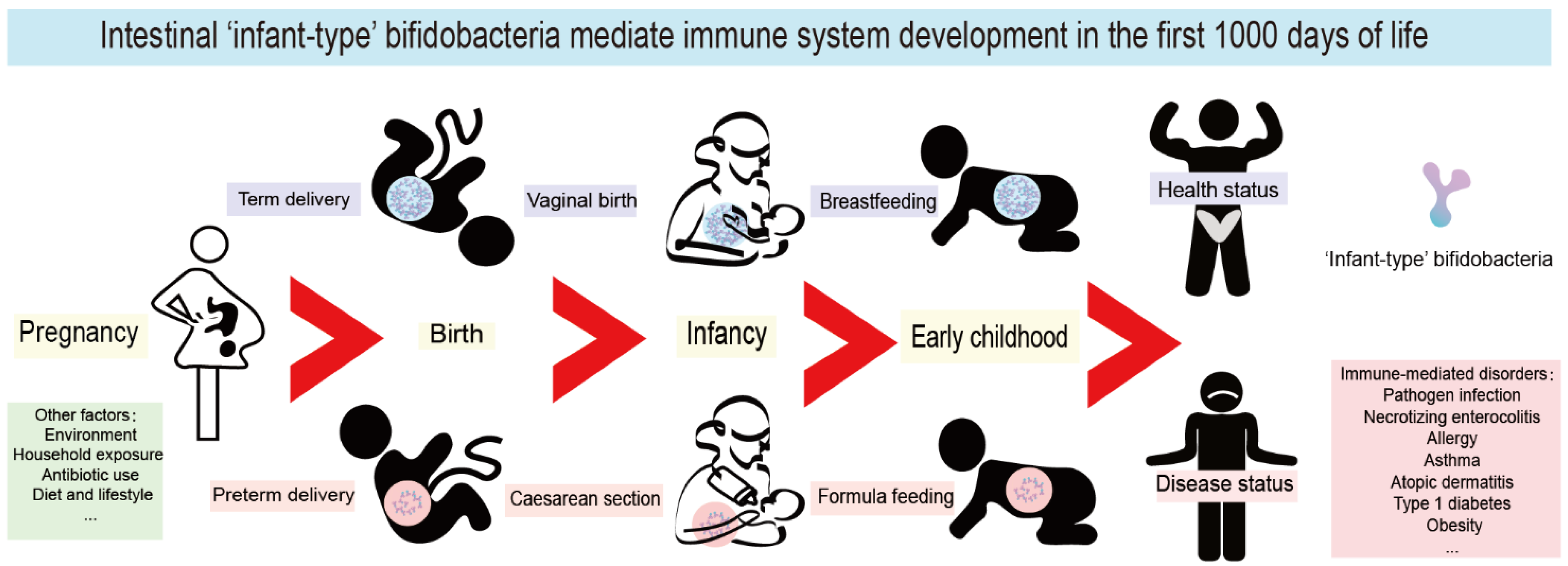
4. The First 1000 Days of Life Are a Key Window of Opportunity for Immune System Maturation

As shown in Figure 1, the first 1000 days of life represent the initial period from the formation of the fertilized ovum and the intrauterine growth of the fetus to the second year after birth. The presence of infant-type bifidobacteria strains in newborns is considered to be a sign of healthy microbiota development and immune system maturation during the first 1000 days of life. Mounting evidence shows this period to be a critical window of opportunity for the colonization and establishment of the first intestinal microorganisms and the development and maturation of the immune system, both of which influence host health in adolescence and even in adulthood [4].
In 2008, Victora et al. [54] reported the effect of maternal and infant malnutrition on infant health. They found that the critical foundations for lifetime health outcomes are laid down from the fetal period to 2 years after birth, thus proposing the concept of the first 1000 days of life [54]. Subsequently, in 2013, Balbus et al. [55] noted that more attention should be paid to early postpartum interventions, nutrition optimization and the prevention of exposure to toxins to reduce the prevalence of non-communicable diseases. Since then, there has been an increasing research focus on the first 1000 days of life as the starting point for a healthy future.
Nowadays, breastfeeding alone is insufficient to protect infants against pathogens and develop a healthy immune system, suggesting the need to develop infant-type bifidobacteria supplements. Recent studies have reported that breastfed infants are increasingly found to have manifestations of intestinal microbiota dysfunction [56,57]. The abundance of bifidobacteria in breastfed infants in resource-rich countries has decreased with the generations over the past 100 years, accompanied by increased levels of intestinal pathogens and fecal pH [56]. This might be attributed to the individual differences of cofactors in breast milk, the use of antibiotics, the frequency of cesarean sections and the increase in potential pathogens [57]. More worryingly, not all babies are fortunate enough to be fed with their mother’s breast milk for many legitimate reasons, but formulae are fully or partially HMO-free. A growing number of studies have shown that infants should be fed a diet that supplements infant-type bifidobacteria early to maintain intestinal health, inhibit the abundance of harmful bacteria, and reduce the risk of allergies and autoimmune diseases [17,57,58].

5. Infant-Type Bifidobacteria Affect the Establishment of Immunity in Early Life

As mentioned, the first 1000 days of life provide a critical window of opportunity for immune system maturation, including the establishment of adaptive immunity and immune tolerance. During this period, the intestinal microbiome of the fetus and the infant undergo substantial development: from being in a near-sterile environment in the womb to being rapidly colonized by a large number of microbes at birth. There is mounting evidence that intestinal infant-type bifidobacteria are ideal probiotics for infants and are vital to early-life immunological development [9,59,60,61]. Herein, we discuss what is known about the role of intestinal infant-type bifidobacteria in establishing immune function in the early stages of life.

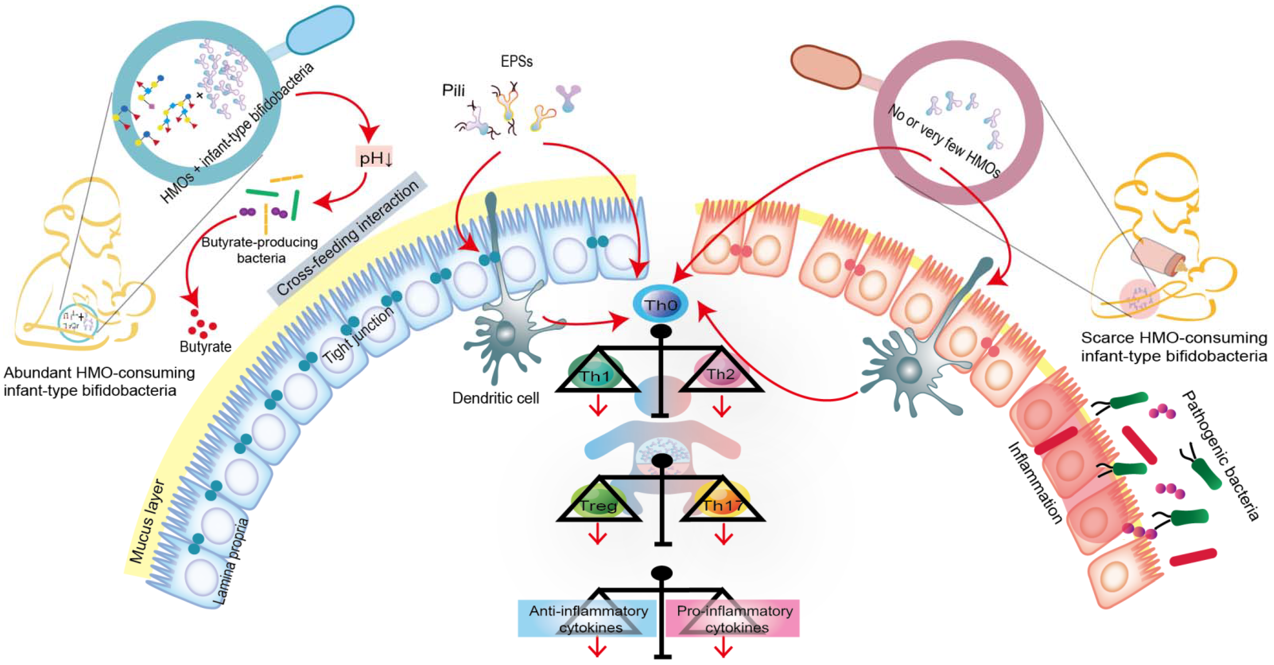
5.1. Infant-Type Bifidobacteria Occupy Intestinal Ecological Sites

Humans cannot digest HMOs due to the lack of the necessary enzyme (glucosidase); this provides a selective nutritional advantage for the growth of beneficial bacteria that specifically consume HMOs [9,62]. Moreover, intestinal infant-type bifidobacteria, the main drivers of HMO metabolism, have evolved complex genetic pathways to metabolize the different glycans in human milk [7,8]. Intestinal infant-type bifidobacteria, therefore, have a competitive advantage in colonization over other gastrointestinal bacteria. In addition, the saccharolytic activities of bifidobacteria generate an anaerobic and acidic intestinal environment that protects the intestine from pathogenic infection. For example, compared to the stool samples of breastfed infants who received no supplementation, the stool samples from breastfed infants who were supplemented with B. longum subsp. infantis EVC001 exhibited a decrease in the abundance of Gram-negative Proteobacteria and Bacteroides and a four-fold decrease in the levels of endotoxin [17]. This indicates that infant-type bifidobacteria play a critical role in mediating immunity by occupying intestinal ecological sites and preventing pathogens from invading the intestine (Figure 2).

5.2. Infant-Type Bifidobacteria Facilitate Breast-Milk Metabolism

Infant-type bifidobacteria can affect host physiology in multiple ways, such as by regulating breast-milk metabolism. As mentioned above, it is now widely accepted that the co-evolution of infant-type bifidobacteria and the host immune system, mediated by HMOs, profoundly shapes early intestinal flora colonization and strongly influences the neonatal immune system. However, little is known about infant-type bifidobacterial metabolites, such as SCFAs, that mediate host-microbe interactions [63].
We focused on three common short-chain fatty acids (acetic acid, propionic acid and butyric acid) and lactic acid (also produced by gut bacteria and playing an important role in gut health) to learn how they contribute to gut health and immune development early in life. Infant-type bifidobacteria convert HMOs into acidic end-products (acetic acid and lactic acid) that affect fecal pH, reduce intestinal permeability, and increase the stability of tight junction proteins, all of which are characteristic of some intestinal diseases, such as NEC (Figure 2) [10,11,63]. Moreover, the acetic acid produced by infant-type bifidobacteria indirectly stimulates the growth, function and immune response of butyric acid-producing microorganisms through a mutually beneficial cross-feeding interaction (Figure 2) [64]. Butyric acid is the preferred fuel for intestinal epithelial cells, and its production further promotes the maintenance of intestinal barrier function by increasing mucin production and improving tight junction integrity [63,65]. In addition to their role in the intestinal tract, SCFAs produced by intestinal infant-type bifidobacteria can affect host physiology outside of the intestine, e.g., in the lung, brain, liver and adipose tissue [10,11]. For example, acetic acid, as the most abundant SCFA in the peripheral circulation, plays a direct role in central appetite regulation by crossing the blood-brain barrier and being absorbed by the brain, resulting in appetite suppression and hypothalamic neuron activation [10]. Although the concentrations of propionic acid and butyric acid are low in peripheral circulation, they indirectly affect the peripheral organs by activating hormone production and the nervous system [11].
Recent studies have also demonstrated the importance of aromatic amino acids, a type of metabolite produced by infant-type bifidobacteria, in early life [9,59]. Infant-type bifidobacteria produce large amounts of aromatic lactic acids, such as tryptophan-derived indole-3-lactic acid (ILA), in the intestinal tract of infants, through the action of aromatic lactate dehydrogenase [59]. Recently, ILA was shown to bind to both the aryl hydrocarbon receptor and hydrocarboxylic acid receptor 3, regulate the response of monocytes to lipopolysaccharides, and block the transcription of inflammatory cytokines such as interleukin (IL)-8 [59]. ILA is also an anti-inflammatory molecule that can promote the development of immature intestinal epithelial cells [59]. Similarly, Henrick et al. observed that ILA produced by B. longum subsp. infantis silences the T helper cell 2 (Th2) and Th17 immune responses that are required to induce immune tolerance and inhibit intestinal inflammation during early life [9]. These new findings suggest that tryptophan metabolic pathways, particularly the ILA pathway, might be crucial for early immune development mediated by infant-type bifidobacteria, which maintains the inflammatory/anti-inflammatory balance of neonatal immunity, blocks the production of inflammatory cytokines and mitigates inflammatory damage.

5.3. Infant-Type Bifidobacteria Promote Immune Development and Prime the Anti-Inflammatory Gene Pool

As mentioned above, the immune system is immature in the early stages of life. Intestinal infant-type bifidobacteria represent one of the earliest antigens to activate the host defense mechanism, which helps to promote immune development and prime the anti-inflammatory gene pool (Figure 2). Recent studies have shown that infant-type bifidobacteria present different cell surface structures, such as exopolysaccharides (EPSs) and pili/fimbriae, allowing bifidobacteria to colonize the infant’s gut, adhere to intestinal cells and participate in the development of the host immune system [66,67]. Consequently, the administration of EPSs from B. breve UCC2003 might serve as a new strategy for promoting infant health [68]. In addition, EPSs produced by B. longum subsp. longum 35624 plays an essential role in inhibiting the pro-inflammatory response of the host (especially the local Th17 response), thus preventing inflammatory diseases [69]. Pili represent a fundamental cell-surface structure considered to be crucial in the interaction between Bifidobacterium and its host, and B. bifidum PRL2010 is equipped with the most functionally characteristic sortase-dependent pili [70,71].
Furthermore, the colonized intestinal microflora can trigger various immune responses by activating T cells and B cells in the intestinal mucosa. In the neonatal period, inflammatory responses are actively suppressed. This phenomenon occurs because neonatal T cells have an internal mechanism wherein the immune system develops toward the characteristics of sensitizing Th2 cytokines, while Th1 cell proliferation and interferon-γ production are inhibited (Figure 2) [38,72]. A recent study showed that the absence of bifidobacteria in the gut flora of newborns leads to the activation of innate and adaptive immunity, which increases the populations of neutrophils, basophils, plasmablasts and memory CD8+ T cells [9]. Additionally, intestinal infant-type bifidobacteria affect the intestinal Th cell response, especially that of Th0 and Th17 cells [9]. In particular, these bifidobacteria stimulate the early immune response for defense while avoiding the overactivation of Th2 and Th17 responses [9]. This study found that the increase in infant-type bifidobacteria abundance is positively associated with a decrease in pro-inflammatory markers (such as memory Tregs and pro-inflammatory monocytes) and an increase in inflammatory markers (such as Tregs, IL-10 and IL-27) [9]. Another study showed that the growth of HMO-consuming B. longum subsp. infantis stimulates the activity of the intestinal epithelium via T cells [61]. Specifically, B. longum subsp. infantis, grown on HMO, increased the expression of the anti-inflammatory cytokine IL-10 in Caco-2 cells and the expression of junctional adhesion molecules and occludin in Caco-2 cells and HT-29 cells [61]. Furthermore, the infant-type bifidobacteria might affect T cells directly or indirectly, via dendritic cells [66,73]. Recent research provides new evidence of the crucial role of breast milk in promoting neonatal Bifidobacterium colonization and B cell activation [74]. It shows that core-fucosylated N-glycans in the mother’s milk selectively promote the colonization of bifidobacteria in infants, whereas their absence from the mother’s milk reduces the proportion of spleen CD19+ CD69+ B cells in offspring mice. In vitro studies also found that the L-fucose metabolites, lactic acid and 1,2-propanediol promote B cell activation through the signaling pathway mediated by B cell receptors [74].

5.4. The Potential Role of Infant-Type Bifidobacteria in Early Neuroimmune Development

Recent studies have emphasized the potential role of intestinal infant-type bifidobacteria in neuroregulation in early life [75,76]. A growing body of evidence supports that the gut-brain connection already exists at birth and enables the gut microbiome to interact with and affect the central nervous system [75,76]. The accumulated data show that neonatal intestinal microbial colonization activates the immune system, sends signals to the central nervous system through the afferent vagus nerve, and produces microbial metabolites (SCFAs, such as acetic acid) that can directly or indirectly affect brain function [75]. Thus, the intestinal microbiota that colonizes infants during their early life shapes the steady-state development and balance of postnatal neural connection [75]. Intestinal infant-type bifidobacteria might also shape host neural circuits by regulating synaptic gene expression and microglial functionality during postnatal development [75]. Currently, however, there is limited information on the mechanism by which gut microbes communicate with the host brain. Further studies are warranted to further clarify the effects of infant-type bifidobacteria on brain function and the underlying mechanism of their action in newborns.
In short, intestinal infant-type bifidobacteria trigger immunomodulatory responses through multiple mechanisms to maintain host health (Figure 2). For example, bifidobacteria occupy the microbial nutritional niche in the gut, release metabolic by-products of HMOs either directly or indirectly (cross-feeding interaction), present extracellular structures (such as EPSs and pili) that interact with host cells, and inhibit inflammatory responses, including intestinal Th cell responses and B cell-induction responses. Observational studies have confirmed a link between the loss of infant-type bifidobacteria and early intestinal inflammation, although little is known about the underlying mechanisms [9,59,76,77].

6. Infant-Type Bifidobacteria Supplementation Is a Promising Strategy for Immune-Mediated Disorders

In fact, some immune-mediated diseases are associated with gut dysbiosis in infancy, often as a result of absent or insufficient breastfeeding. Indeed, breastfeeding is considered to be the best form of infant feeding, which has the potential to overcome immune-mediated disorders. However, over the past 100 years, breastfed infants have experienced a disturbing upheaval in the gut microbiota, with a reduction in fecal bifidobacteria and a dramatic rise in pH to 6.5 [56]. Accumulated evidence suggested that supplementation with B. longum subsp. infantis was recommended for breastfed infants to promote healthy intestinal development [17,57,58]. Therefore, it is emphasized here that infant-type bifidobacteria supplementation is a promising strategy to avoid immune-mediated disorders, even in breast milk-fed babies. Given that the intestinal microbiome is modifiable, optimizing its composition in early life might be a potential approach for treating immune-mediated disease. Here, we review the latest studies that have investigated the critical role of the early-stage microbiota in immune-mediated disease progression. We also discuss whether infant-type bifidobacteria supplementation could alter the composition of the early intestinal microbiota and thereby prevent the progression of immune-mediated disease (Table 1).

6.1. Effects of Infant-Type Bifidobacteria on Pathogen Infection and NEC

The neonatal gut is immature and is susceptible to colonization by pathogenic bacteria; therefore, gut colonization by healthy microflora in early life helps to protect infants from pathogenic infection. Rotavirus is a common pathogen that can cause severe diarrhea and fatal dehydration in infants and young children. Studies have shown that the administration of B. breve M-16V to Lewis rats 2–14 days after birth can effectively reduce the severity and incidence of infectious diarrhea caused by rotavirus [79]. In addition, it has been suggested that the administration of infant-type bifidobacteria could prevent and treat rotaviral infection [80,81]. Moreover, early gut colonization by Bifidobacterium, especially B. longum subsp. infantis, enhances the response to early vaccination: the average abundance of bifidobacteria in early infancy was positively correlated with the CD4 T-cell responses to Bacillus Calmette-Guérin, tetanus toxoid and hepatitis B virus at 15 weeks [73]. Interestingly, the positive response of CD4 T cells to Bacillus Calmette-Guérin and tetanus toxoid was found to be maintained for over two years [73]. Moreover, the mean abundance of bifidobacteria was positively correlated with plasma tetanus toxoid-specific IgG concentrations and stool polio-specific IgA concentrations at 2 years [73]. A similar result was obtained in the dominant subspecies, B. longum subsp. infantis [73].
NEC is one of the most common and severe gastrointestinal tract diseases; it has high morbidity and mortality rates and mainly occurs in premature infants. In NEC, excessive inflammation is caused by the high immunoreactivity of the intestine and affects distant organs such as the brain, liver and lungs [86,87]. Infants who recover from NEC have increased risks of developing short bowel syndrome, parenteral nutrition-related liver disease, pulmonary hypertension, microcephaly and severe neurodevelopmental delays [86,87]. Many microbiological and molecular biological studies have been conducted on the intestines of infants with NEC, but the cause and mechanism of NEC remain incompletely understood. According to the hypothesis proposed by Claud and Walker, the occurrence of NEC in premature infants results from the synergistic effects of multiple risk factors, with inappropriate initial microbial colonization of the gut being one of the most critical risk factors [88]. Specifically, it is thought that inappropriate initial microbial colonization exacerbates the risk of inflammation in the intestine, which is also a crucial neonatal risk factor for NEC [89]. Studies have confirmed the positive role of infant-type bifidobacteria in preventing and treating NEC. Hoyos was the first to describe infant-type bifidobacteria as probiotics that had therapeutic effects on NEC [78]. In 1999, she found that the oral administration of prophylactic B. longum subsp. infantis and Lactobacillus acidophilus (at a dose of 2.5 × 10⁸ CFU of each organism) for one year decreased the incidence of and mortality due to NEC in 1237 neonates in an intensive-care unit [78]. Subsequent follow-up studies have found that the relative risk of NEC reduced significantly in infants on probiotic treatment regimens that consisted of infant-type bifidobacteria, although the optimal strains, treatment durations and dosages have not yet been determined [16,82,83,84,85]. The administration of antibiotics to preterm and low-birthweight infants disrupts the gut colonization pattern of bifidobacteria, thereby reducing infants’ intestinal resistance to pathogenic bacteria and increasing their risk of NEC [90,91,92]. Therefore, antibiotic treatment for infants should be avoided unless absolutely essential. In summary, changing the composition of early gut flora, especially by increasing the proportion of infant-type bifidobacteria, is a promising strategy for reducing the risk of early life-threatening infections in infants.

6.2. Effects of Infant-Type Bifidobacteria on Allergic Diseases

The prevalence of allergic diseases has been increasing globally in recent decades, especially in low- and middle-income countries, and these diseases currently affect approximately 25% of the world’s population [93,94]. In high-income countries, deficiencies in intestinal infant-type bifidobacteria are common in newborns and are likely to contribute to an increase in the incidence of allergic diseases [19,95]. In contrast, in developing countries, children with high rates of B. longum subsp. infantis colonization were found to be at low risk of atopic diseases [19]. Although the pathogenesis of allergic diseases might be multifactorial, many studies have provided evidence suggesting that inappropriate early intestinal flora colonization is the main reason for the disruption of normal postnatal immune regulation. For example, an imbalance in the composition of intestinal flora leading to a low abundance of bifidobacteria in newborns predates the development of atopic diseases [96]. In addition, compared with newborns, in older children, there is a less consistent association between the composition of the gut microbiota and the development of allergic diseases or asthma [97]. Hence, it appears that disturbances in the early gut microbiota, or dysbiosis, might drive the occurrence of allergic diseases, which might last a lifetime. Regulating the early intestinal microbiota through the administration of infant-type bifidobacteria has thus emerged as a potential strategy to prevent allergic diseases in infants. For example, it was proposed that the addition of specific B. breve strains to infant formula might assist with allergy management [98]. In summary, the administration of infant-type bifidobacteria, whether as a single species or as a synbiotic, might contribute to the prevention and treatment of allergic diseases.

6.3. Effects of Infant-Type Bifidobacteria on T1D and Obesity

In the context of immune-mediated diseases, the gut microbial community of infants is emerging as a critical early-life factor that is capable of influencing long-term health outcomes, such as T1D and obesity. Recent evidence suggests that an imbalance of gut microbiota composition is highly related to the pathogenesis of insulin dysfunction and T1D [99,100]. Although the exact pathogenesis of T1D is not completely clear, maladjustments in the gut microbiota composition and intestinal barrier properties have been documented in T1D subjects [99,100]. Recently, SINT1A, the Global Platform for the Prevention of Autoimmune Diabetes, investigated whether the daily administration of B. longum subsp. infantis reduces the cumulative incidence of β-cell autoantibodies in children who have a high genetic risk of developing T1D [18]. Although the study results are not yet known, it is believed that this approach holds much therapeutic potential.
Furthermore, modulation of the gut microflora associated with early rapid weight gain has been proposed as a potential strategy for curbing the growing global obesity epidemic, as evidence suggests that childhood and adult obesity is significantly associated with excessive prenatal and infant weight gain [101]. Recent studies have emphasized the potential role of bifidobacteria in obesity in early life [101,102,103]. A longitudinal study showed that normal-weight children had a higher abundance of Bifidobacterium in their stool samples from infancy than overweight children, who instead had a higher abundance of Staphylococcus aureus in their stool samples from infancy [101]. Other studies have also suggested that the abundance of bifidobacteria is inversely related to plasma glucose tolerance level, body mass index and lipopolysaccharide concentration [103]. Therefore, regulation of the intestinal microbiota via the supplementation of infant-type bifidobacteria in early life might be a strategy for the prevention of immune-mediated disorders.

7. Conclusions

The first 1000 days of life is a critical window for immune development and a crucial period during which the initial large-scale colonization of the gut flora of newborns occurs. Over the past 100 years, modern practices have been developed and widely applied for delivering and feeding babies. These deviations from the natural delivery and feeding processes might affect infant microbiome acquisition, particularly the bifidobacterial colonization of the gut, which is increasingly seen as a potential risk factor for immune diseases such as allergies. The gut Bifidobacterium population can be broadly divided into infant type (i.e., predominant in early life) and adult type (i.e., predominant in adult life). The physicochemical properties of HMOs in breast milk-fed infants confer a strong selective advantage for the gut colonization of infant-type Bifidobacterium species. This finding further underscores the importance of age-appropriate probiotics in the development of the immune system in the first 1000 days of life. Despite much research, it remains unclear how intestinal infant-type bifidobacteria mediate immunity in early life. Infant-type bifidobacteria not only enhance neonatal immunity by establishing important short-term protection against pathogenic infection but also provide long-term metabolic and immunological benefits. Thus, increasing the abundance of intestinal infant-type bifidobacteria has been proposed as a promising early-life health-development strategy. There is sufficient evidence to support the administration of infant-type Bifidobacterium strains to children with immune-mediated diseases. However, further studies are required to investigate the mechanism by which infant-type bifidobacteria influence immune system development in early life and to explore this keystone taxon and its critical ecological functions that affect the short- and long-term health outcomes of infants.

8. Outlook: How Do We Accelerate the Colonization of Infant-Type Bifidobacteria in the Intestinal Flora of Infants during Early Life?

An increasing body of research has investigated the role of the infant gut microbiome in long-term health outcomes throughout individuals’ lifetimes. The link between intestinal infant-type bifidobacteria and early health has been established, but the causative direction of this relationship remains unknown. Moreover, due to the limitations of the current metagenomics approaches used to evaluate the detailed compositions of complex intestinal communities, the actual contribution of infant-type bifidobacteria to the biodiversity of infant intestinal microbiota might be underestimated. Therefore, it is critical to developing optimized protocols to determine the composition of the infant gut microbiota and assess the actual contribution of infant-type bifidobacteria to early-life health and disease. Furthermore, fetal life might not be as devoid of environmental stimuli as previously thought, suggesting that infant-type bifidobacteria supplementation, both to mothers during pregnancy and to newborns in early life, might help to develop the immunity of infants. Nevertheless, several parameters of infant-type bifidobacterial supplementation remain to be determined, e.g., the optimal strain(s), single strain or synbiotic administration, and the optimal dosage and treatment duration. It is hoped that rapid technological progress in large-scale metagenomic screening will enable deeper exploration of the early-life microbiome and, thus, soon reveal the answers to these critical questions.

Author Contributions

Conceptualization, H.Z. (Hao Zhang), G.W., H.Z. (Heng Zhang) and W.C.; methodology, C.L. and Y.L.; software, C.L. and Y.L.; validation, J.Z. and G.W.; data curation, J.Z. and H.Z. (Hao Zhang); writing—original draft preparation, C.L.; writing—review and editing, G.W., H.Z. (Heng Zhang) and W.C.; visualization, C.L.; supervision, J.Z. and G.W.; project administration, G.W., H.Z. (Heng Zhang) and W.C.; funding acquisition, G.W., H.Z. (Heng Zhang) and W.C. All authors have read and agreed to the published version of the manuscript.

Funding

This work was supported by the National Natural Science Foundation of China (No. 31972052, 32021005, 31820103010), the Fundamental Research Funds for the Central Universities (JUSRP22006, JUSRP51501), the Program of Collaborative Innovation Centre of Food Safety and Quality Control in Jiangsu Province, the Major Project of Wuxi Science and Technology Bureau (Y20212007).

Institutional Review Board Statement

Not applicable.

Informed Consent Statement

Not applicable.

Data Availability Statement

Not applicable.

Conflicts of Interest

The authors declare no conflict of interest.

References

  1. Aagaard, K.; Ma, J.; Antony, K.M.; Ganu, R.; Petrosino, J.; Versalovic, J. The Placenta Harbors a Unique Microbiome. Sci. Transl. Med. 2014, 6, 237–265. [Google Scholar] [CrossRef] [PubMed] [Green Version]
  2. Kennedy, K.M.; Gerlach, M.J.; Adam, T.; Heimesaat, M.M.; Rossi, L.; Surette, M.G.; Sloboda, D.M.; Braun, T. Fetal Meconium Does Not Have a Detectable Microbiota before Birth. Nat. Microbiol. 2021, 6, 865–873. [Google Scholar] [CrossRef] [PubMed]
  3. Perez-Munoz, M.E.; Arrieta, M.-C.; Ramer-Tait, A.E.; Walter, J. A Critical Assessment of the “Sterile Womb” and “in Utero Colonization” Hypotheses: Implications for Research on the Pioneer Infant Microbiome. Microbiome 2017, 5, 48. [Google Scholar] [CrossRef]
  4. Gensollen, T.; Iyer, S.S.; Kasper, D.L.; Blumberg, R.S. How Colonization by Microbiota in Early Life Shapes the Immune System. Science 2016, 352, 539–544. [Google Scholar] [CrossRef] [PubMed] [Green Version]
  5. Turroni, F.; Peano, C.; Pass, D.A.; Foroni, E.; Severgnini, M.; Claesson, M.J.; Kerr, C.; Hourihane, J.; Murray, D.; Fuligni, F.; et al. Diversity of Bifidobacteria within the Infant Gut Microbiota. PLoS ONE 2012, 7, e36957. [Google Scholar] [CrossRef] [PubMed] [Green Version]
  6. Turroni, F.; van Sinderen, D.; Ventura, M. Genomics and Ecological Overview of the Genus Bifidobacterium. Int. J. Food Microbiol. 2011, 149, 37–44. [Google Scholar] [CrossRef]
  7. Asakuma, S.; Hatakeyama, E.; Urashima, T.; Yoshida, E.; Katayama, T.; Yamamoto, K.; Kumagai, H.; Ashida, H.; Hirose, J.; Kitaoka, M. Physiology of Consumption of Human Milk Oligosaccharides by Infant Gut-Associated Bifidobacteria. J. Biol. Chem. 2011, 286, 34583–34592. [Google Scholar] [CrossRef] [Green Version]
  8. Katayama, T. Host-Derived Glycans Serve as Selected Nutrients for the Gut Microbe: Human Milk Oligosaccharides and Bifidobacteria. Biosci. Biotechnol. Biochem. 2016, 80, 621–632. [Google Scholar] [CrossRef] [Green Version]
  9. Henrick, B.M.; Rodriguez, L.; Lakshmikanth, T.; Pou, C.; Henckel, E.; Arzoomand, A.; Olin, A.; Wang, J.; Mikes, J.; Tan, Z.; et al. Bifidobacteria-Mediated Immune System Imprinting Early in Life. Cell 2021, 184, 3884–3898. [Google Scholar] [CrossRef]
  10. Frost, G.; Sleeth, M.L.; Sahuri-Arisoylu, M.; Lizarbe, B.; Cerdan, S.; Brody, L.; Anastasovska, J.; Ghourab, S.; Hankir, M.; Zhang, S.; et al. The Short-Chain Fatty Acid Acetate Reduces Appetite via a Central Homeostatic Mechanism. Nat. Commun. 2014, 5, 3611. [Google Scholar] [CrossRef] [Green Version]
  11. Koh, A.; De Vadder, F.; Kovatcheva-Datchary, P.; Backhed, F. From Dietary Fiber to Host Physiology: Short-Chain Fatty Acids as Key Bacterial Metabolites. Cell 2016, 165, 1332–1345. [Google Scholar] [CrossRef] [PubMed] [Green Version]
  12. Russell, J.T.; Roesch, L.F.W.; Ordberg, M.; Ilonen, J.; Atkinson, M.A.; Schatz, D.A.; Triplett, E.W.; Ludvigsson, J. Genetic Risk for Autoimmunity Is Associated with Distinct Changes in the Human Gut Microbiome. Nat. Commun. 2019, 10, 3621. [Google Scholar] [CrossRef] [PubMed] [Green Version]
  13. Vatanen, T.; Kostic, A.D.; d’Hennezel, E.; Siljander, H.; Franzosa, E.A.; Yassour, M.; Kolde, R.; Vlamakis, H.; Arthur, T.D.; Hamalainen, A.-M.; et al. Variation in Microbiome LPS Immunogenicity Contributes to Autoimmunity in Humans. Cell 2016, 165, 842–853. [Google Scholar] [CrossRef] [PubMed] [Green Version]
  14. Shao, Y.; Forster, S.C.; Tsaliki, E.; Vervier, K.; Strang, A.; Simpson, N.; Kumar, N.; Stares, M.D.; Rodger, A.; Brocklehurst, P.; et al. Stunted Microbiota and Opportunistic Pathogen Colonization in Caesarean-Section Birth. Nature 2019, 574, 117–121. [Google Scholar] [CrossRef]
  15. Wang, S.; Ryan, C.A.; Boyaval, P.; Dempsey, E.M.; Ross, R.P.; Stanton, C. Maternal Vertical Transmission Affecting Early-Life Microbiota Development. Trends Microbiol. 2020, 28, 28–45. [Google Scholar] [CrossRef] [PubMed]
  16. Underwood, M.A.; Arriola, J.; Gerber, C.W.; Kaveti, A.; Kalanetra, K.M.; Kananurak, A.; Bevins, C.L.; Mills, D.A.; Dvorak, B. Bifidobacterium Longum Subsp Infantis in Experimental Necrotizing Enterocolitis: Alterations in Inflammation, Innate Immune Response, and the Microbiota. Pediatr. Res. 2014, 76, 326–333. [Google Scholar] [CrossRef] [PubMed] [Green Version]
  17. Frese, S.A.; Hutton, A.A.; Contreras, L.N.; Shaw, C.A.; Palumbo, M.C.; Casaburi, G.; Xu, G.; Davis, J.C.C.; Lebrilla, C.B.; Henrick, B.M.; et al. Persistence of Supplemented Bifidobacterium Longum Subsp. Infantis EVC001 in Breastfed Infants. Msphere 2017, 2, e00501-17. [Google Scholar] [CrossRef] [Green Version]
  18. Ziegler, A.-G.; Arnolds, S.; Kolln, A.; Achenbach, P.; Berner, R.; Bonifacio, E.; Casteels, K.; Elding Larsson, H.; Gundert, M.; Hasford, J.; et al. Supplementation with Bifidobacterium Longum Subspecies Infantis EVC001 for Mitigation of Type 1 Diabetes Autoimmunity: The GPPAD-SINT1A Randomised Controlled Trial Protocol. BMJ Open 2021, 11, e052449. [Google Scholar] [CrossRef]
  19. Seppo, A.E.; Bu, K.; Jumabaeva, M.; Thakar, J.; Choudhury, R.A.; Yonemitsu, C.; Bode, L.; Martina, C.A.; Allen, M.; Tamburini, S.; et al. Infant Gut Microbiome Is Enriched with Bifidobacterium Longum Ssp. Infantis in Old Order Mennonites with Traditional Farming Lifestyle. Allergy 2021, 76, 3489–3503. [Google Scholar] [CrossRef]
  20. Wong, C.B.; Sugahara, H.; Odamaki, T.; Xiao, J.Z. Different Physiological Properties of Human-Residential and Non-Human-Residential Bifidobacteria in Human Health. Benef. Microbes 2018, 9, 111–122. [Google Scholar] [CrossRef]
  21. Odamaki, T.; Bottacini, F.; Kato, K.; Mitsuyama, E.; Yoshida, K.; Horigome, A.; Xiao, J.; van Sinderen, D. Genomic Diversity and Distribution of Bifidobacterium Longum Subsp Longum across the Human Lifespan. Sci. Rep. 2018, 8, 85. [Google Scholar] [CrossRef] [PubMed] [Green Version]
  22. Arboleya, S.; Watkins, C.; Stanton, C.; Ross, R.P. Gut Bifidobacteria Populations in Human Health and Aging. Front. Microbiol. 2016, 7, 1204. [Google Scholar] [CrossRef] [PubMed] [Green Version]
  23. Zhang, B.; Li, L.-Q.; Liu, F.; Wu, J.-Y. Human Milk Oligosaccharides and Infant Gut Microbiota: Molecular Structures, Utilization Strategies and Immune Function. Carbohydr. Polym. 2022, 276, 118738. [Google Scholar] [CrossRef]
  24. Bode, L. The Functional Biology of Human Milk Oligosaccharides. Early Hum. Dev. 2015, 91, 619–622. [Google Scholar] [CrossRef]
  25. Zhu, Y.; Zhang, W.; Mu, W. Human Milk Oligosaccharides: The New Gold Standard for Premium Infant Formula. J. Agric. Food Chem. 2022, 70, 2061–2063. [Google Scholar] [CrossRef] [PubMed]
  26. Puccio, G.; Alliet, P.; Cajozzo, C.; Janssens, E.; Corsello, G.; Sprenger, N.; Wernimont, S.; Egli, D.; Gosoniu, L.; Steenhout, P. Effects of Infant Formula with Human Milk Oligosaccharides on Growth and Morbidity: A Randomized Multicenter Trial. J. Pediatr. Gastroenterol. Nutr. 2017, 64, 624–631. [Google Scholar] [CrossRef] [Green Version]
  27. Akkerman, R.; Faas, M.M.; de Vos, P. Non-Digestible Carbohydrates in Infant Formula as Substitution for Human Milk Oligosaccharide Functions: Effects on Microbiota and Gut Maturation. Crit. Rev. Food Sci. Nutr. 2019, 59, 1486–1497. [Google Scholar] [CrossRef]
  28. Asnicar, F.; Manara, S.; Zolfo, M.; Truong, D.T.; Scholz, M.; Armanini, F.; Ferretti, P.; Gorfer, V.; Pedrotti, A.; Tett, A.; et al. Studying Vertical Microbiome Transmission from Mothers to Infants by Strain-Level Metagenomic Profiling. Msystems 2017, 2, e00164-16. [Google Scholar] [CrossRef] [Green Version]
  29. Lawson, M.A.E.; O’Neill, I.J.; Kujawska, M.; Javavdi, S.G.; Wijeyesekera, A.; Flegg, Z.; Chalklen, L.; Hall, L.J. Breast Milk-Derived Human Milk Oligosaccharides Promote Bifidobacterium Interactions within a Single Ecosystem. Isme J. 2020, 14, 635–648. [Google Scholar] [CrossRef] [Green Version]
  30. Tannock, G.W.; Lawley, B.; Munro, K.; Pathmanathan, S.G.; Zhou, S.J.; Makrides, M.; Gibson, R.A.; Sullivan, T.; Prosser, C.G.; Lowry, D.; et al. Comparison of the Compositions of the Stool Microbiotas of Infants Fed Goat Milk Formula, Cow Milk-Based Formula, or Breast Milk. Appl. Environ. Microbiol. 2013, 79, 3040–3048. [Google Scholar] [CrossRef] [Green Version]
  31. Centanni, M.; Ferguson, S.A.; Sims, I.M.; Biswas, A.; Tannock, G.W. Bifidobacterium Bifidum ATCC 15696 and Bifidobacterium Breve 24b Metabolic Interaction Based on 2 ’-O-Fucosyl-Lactose Studied in Steady-State Cultures in a Freter-Style Chemostat. Appl. Environ. Microbiol. 2019, 85, e02783-18. [Google Scholar] [CrossRef] [PubMed] [Green Version]
  32. Turroni, F.; Milani, C.; Duranti, S.; Mahony, J.; van Sinderen, D.; Ventura, M. Glycan Utilization and Cross-Feeding Activities by Bifidobacteria. Trends Microbiol. 2018, 26, 339–350. [Google Scholar] [CrossRef] [PubMed]
  33. Ruiz-Moyano, S.; Totten, S.M.; Garrido, D.A.; Smilowitz, J.T.; German, J.B.; Lebrilla, C.B.; Mills, D.A. Variation in Consumption of Human Milk Oligosaccharides by Infant Gut-Associated Strains of Bifidobacterium Breve. Appl. Environ. Microbiol. 2013, 79, 6040–6049. [Google Scholar] [CrossRef] [PubMed] [Green Version]
  34. Yao, Y.; Cai, X.; Ye, Y.; Wang, F.; Chen, F.; Zheng, C. The Role of Microbiota in Infant Health: From Early Life to Adulthood. Front. Immunol. 2021, 12, 708472. [Google Scholar] [CrossRef] [PubMed]
  35. Jost, T.; Lacroix, C.; Braegger, C.; Chassard, C. Impact of Human Milk Bacteria and Oligosaccharides on Neonatal Gut Microbiota Establishment and Gut Health. Nutr. Rev. 2015, 73, 426–437. [Google Scholar] [CrossRef] [PubMed]
  36. Wang, M.; Li, M.; Wu, S.; Lebrilla, C.B.; Chapkin, R.S.; Ivanov, I.; Donovan, S.M. Fecal Microbiota Composition of Breast-Fed Infants Is Correlated With Human Milk Oligosaccharides Consumed. J. Pediatr. Gastroenterol. Nutr. 2015, 60, 825–833. [Google Scholar] [CrossRef] [Green Version]
  37. Blaser, M.J.; Devkota, S.; McCoy, K.D.; Relman, D.A.; Yassour, M.; Young, V.B. Lessons Learned from the Prenatal Microbiome Controversy. Microbiome 2021, 9, 8. [Google Scholar] [CrossRef]
  38. Brugman, S.; Perdijk, O.; van Neerven, R.J.J.; Savelkoul, H.F.J. Mucosal Immune Development in Early Life: Setting the Stage. Arch. Immunol. Ther. Ex. 2015, 63, 251–268. [Google Scholar] [CrossRef] [Green Version]
  39. Carolyn, A.T.; Kathy, D.M. The Role of Mom’s Microbes during Pregnancy. Scientist 2021. Available online: https://www.the-scientist.com/features/the-role-of-mom-s-microbes-during-pregnancy-69009 (accessed on 1 August 2021).
  40. Gopalakrishna, K.P.; Macadangdang, B.R.; Rogers, M.B.; Tometich, J.T.; Firek, B.A.; Baker, R.; Ji, J.; Burr, A.H.P.; Ma, C.; Good, M.; et al. Maternal IgA Protects against the Development of Necrotizing Enterocolitis in Preterm Infants. Nat. Med. 2019, 25, 1110–1115. [Google Scholar] [CrossRef]
  41. Neu, J. The Microbiome during Pregnancy and Early Postnatal Life. Semin. Fetal. Neonatal. Med. 2016, 21, 373–379. [Google Scholar] [CrossRef]
  42. Hobbs, A.J.; Mannion, C.A.; McDonald, S.W.; Brockway, M.; Tough, S.C. The Impact of Caesarean Section on Breastfeeding Initiation, Duration and Difficulties in the First Four Months Postpartum. BMC Pregnancy Childbirth 2016, 16, 90. [Google Scholar] [CrossRef] [PubMed] [Green Version]
  43. Matamoros, S.; Gras-Leguen, C.; Le Vacon, F.; Potel, G.; de La Cochetiere, M.-F. Development of Intestinal Microbiota in Infants and Its Impact on Health. Trends Microbiol. 2013, 21, 167–173. [Google Scholar] [CrossRef] [PubMed]
  44. Makino, H.; Martin, R.; Ishikawa, E.; Gawad, A.; Kubota, H.; Sakai, T.; Oishi, K.; Tanaka, R.; Ben-Amor, K.; Knol, J.; et al. Multilocus Sequence Typing of Bifidobacterial Strains from Infant’s Faeces and Human Milk: Are Bifidobacteria Being Sustainably Shared during Breastfeeding? Benef. Microbes 2015, 6, 563–572. [Google Scholar] [CrossRef] [PubMed]
  45. Hesla, H.M.; Stenius, F.; Jaderlund, L.; Nelson, R.; Engstrand, L.; Alm, J.; Dicksved, J. Impact of Lifestyle on the Gut Microbiota of Healthy Infants and Their Mothers-The ALADDIN Birth Cohort. Fems Microbiol. Ecol. 2014, 90, 791–801. [Google Scholar] [CrossRef] [PubMed] [Green Version]
  46. Dogra, S.; Sakwinska, O.; Soh, S.-E.; Ngom-Bru, C.; Brueck, W.M.; Berger, B.; Bruessow, H.; Lee, Y.S.; Yap, F.; Chong, Y.-S.; et al. Dynamics of Infant Gut Microbiota Are Influenced by Delivery Mode and Gestational Duration and Are Associated with Subsequent Adiposity. Mbio 2015, 6, e02419-14. [Google Scholar] [CrossRef] [Green Version]
  47. Boudry, G.; Charton, E.; Le Huerou-Luron, I.; Ferret-Bernard, S.; Le Gall, S.; Even, S.; Blat, S. The Relationship Between Breast Milk Components and the Infant Gut Microbiota. Front. Nutr. 2021, 8, 629740. [Google Scholar] [CrossRef]
  48. Vandenplas, Y.; Berger, B.; Carnielli, V.P.; Ksiazyk, J.; Lagstrom, H.; Sanchez Luna, M.; Migacheva, N.; Mosselmans, J.-M.; Picaud, J.-C.; Possner, M.; et al. Human Milk Oligosaccharides: 2-Fucosyllactose (2-FL) and Lacto-N-Neotetraose (LNnT) in Infant Formula. Nutrients 2018, 10, 1161. [Google Scholar] [CrossRef] [Green Version]
  49. Ma, J.; Prince, A.L.; Bader, D.; Hu, M.; Ganu, R.; Baquero, K.; Blundell, P.; Harris, R.A.; Frias, A.E.; Grove, K.L.; et al. High-Fat Maternal Diet during Pregnancy Persistently Alters the Offspring Microbiome in a Primate Model. Nat. Commun. 2014, 5, 3889. [Google Scholar] [CrossRef]
  50. Turroni, F.; Milani, C.; Duranti, S.; Ferrario, C.; Lugli, G.A.; Mancabelli, L.; van Sinderen, D.; Ventura, M. Bifidobacteria and the Infant Gut: An Example of Co-Evolution and Natural Selection. Cell. Mol. Life Sci. 2018, 75, 103–118. [Google Scholar] [CrossRef]
  51. Tamburini, S.; Shen, N.; Wu, H.C.; Clemente, J.C. The Microbiome in Early Life: Implications for Health Outcomes. Nat. Med. 2016, 22, 713–722. [Google Scholar] [CrossRef] [Green Version]
  52. De Muinck, E.J.; Trosvik, P. Individuality and Convergence of the Infant Gut Microbiota during the First Year of Life. Nat. Commun. 2018, 9, 2233. [Google Scholar] [CrossRef] [PubMed]
  53. Stewart, C.J.; Ajami, N.J.; O’Brien, J.L.; Hutchinson, D.S.; Smith, D.P.; Wong, M.C.; Ross, M.C.; Lloyd, R.E.; Doddapaneni, H.; Metcalf, G.A.; et al. Temporal Development of the Gut Microbiome in Early Childhood from the TEDDY Study. Nature 2018, 562, 583–588. [Google Scholar] [CrossRef] [PubMed]
  54. Victora, C.G.; Adair, L.; Fall, C.; Hallal, P.C.; Martorell, R.; Richter, L.; Sachdev, H.S. Maternal and Child Undernutrition 2-Maternal and Child Undernutrition: Consequences for Adult Health and Human Capital. Lancet 2008, 371, 340–357. [Google Scholar] [CrossRef] [Green Version]
  55. Balbus, J.M.; Barouki, R.; Birnbaum, L.S.; Etzel, R.A.; Gluckman, P.D.; Grandjean, P.; Hancock, C.; Hanson, M.A.; Heindel, J.J.; Hoffman, K.; et al. Early-Life Prevention of Non-Communicable Diseases. Lancet 2013, 381, 3–4. [Google Scholar] [CrossRef] [Green Version]
  56. Henrick, B.M.; Hutton, A.A.; Palumbo, M.C.; Casaburi, G.; Mitchell, R.D.; Underwood, M.A.; Smilowitz, J.T.; Frese, S.A. Elevated Fecal PH Indicates a Profound Change in the Breastfed Infant Gut Microbiome Due to Reduction of Bifidobacterium over the Past Century. Msphere 2018, 3, e00041-18. [Google Scholar] [CrossRef] [Green Version]
  57. O’Brien, C.E.; Meier, A.K.; Cernioglo, K.; Mitchell, R.D.; Casaburi, G.; Frese, S.A.; Henrick, B.M.; Underwood, M.A.; Smilowitz, J.T. Early Probiotic Supplementation with B. Infantis in Breastfed Infants Leads to Persistent Colonization at 1 Year. Pediatr. Res. 2022, 91, 627–636. [Google Scholar] [CrossRef] [PubMed]
  58. Underwood, M.A.; Kalanetra, K.M.; Bokulich, N.A.; Lewis, Z.T.; Mirmiran, M.; Tancredi, D.J.; Mills, D.A. A Comparison of Two Probiotic Strains of Bifidobacteria in Premature Infants. J. Pediatr. 2013, 163, 1585–1591. [Google Scholar] [CrossRef] [Green Version]
  59. Laursen, M.F.; Sakanaka, M.; von Burg, N.; Morbe, U.; Andersen, D.; Moll, J.M.; Pekmez, C.T.; Rivollier, A.; Michaelsen, K.F.; Molgaard, C.; et al. Bifidobacterium Species Associated with Breastfeeding Produce Aromatic Lactic Acids in the Infant Gut. Nat. Microbiol. 2021, 6, 1367–1382. [Google Scholar] [CrossRef]
  60. Renz, H.; Skevaki, C. Early Life Microbial Exposures and Allergy Risks: Opportunities for Prevention. Nat. Rev. Immunol. 2021, 21, 177–191. [Google Scholar] [CrossRef]
  61. Chichlowski, M.; De Lartigue, G.; German, J.B.; Raybould, H.E.; Mills, D.A. Bifidobacteria Isolated From Infants and Cultured on Human Milk Oligosaccharides Affect Intestinal Epithelial Function. J. Pediatr. Gastroenterol. Nutr. 2012, 55, 321–327. [Google Scholar] [CrossRef] [Green Version]
  62. Sela, D.A.; Mills, D.A. Nursing Our Microbiota: Molecular Linkages between Bifidobacteria and Milk Oligosaccharides. Trends Microbiol. 2010, 18, 298–307. [Google Scholar] [CrossRef] [PubMed] [Green Version]
  63. Rios-Covian, D.; Ruas-Madiedo, P.; Margolles, A.; Gueimonde, M.; de los Reyes-Gavilan, C.G.; Salazar, N. Intestinal Short Chain Fatty Acids and Their Link with Diet and Human Health. Front. Microbiol. 2016, 7, 185. [Google Scholar] [CrossRef] [PubMed] [Green Version]
  64. Duncan, S.H.; Holtrop, G.; Lobley, G.E.; Calder, A.G.; Stewart, C.S.; Flint, H.J. Contribution of Acetate to Butyrate Formation by Human Faecal Bacteria. Br. J. Nutr. 2004, 91, 915–923. [Google Scholar] [CrossRef] [PubMed] [Green Version]
  65. Peng, L.; Li, Z.-R.; Green, R.S.; Holzman, I.R.; Lin, J. Butyrate Enhances the Intestinal Barrier by Facilitating Tight Junction Assembly via Activation of AMP-Activated Protein Kinase in Caco-2 Cell Monolayers. J. Nutr. 2009, 139, 1619–1625. [Google Scholar] [CrossRef]
  66. Alessandri, G.; Ossiprandi, M.C.; MacSharry, J.; van Sinderen, D.; Ventura, M. Bifidobacterial Dialogue With Its Human Host and Consequent Modulation of the Immune System. Front. Immunol. 2019, 10, 2348. [Google Scholar] [CrossRef] [Green Version]
  67. Saturio, S.; Nogacka, A.M.; Alvarado-Jasso, G.M.; Salazar, N.; de los Reyes-Gavilan, C.G.; Gueimonde, M.; Arboleya, S. Role of Bifidobacteria on Infant Health. Microorganisms 2021, 9, 2415. [Google Scholar] [CrossRef]
  68. Pungel, D.; Treveil, A.; Dalby, M.J.; Caim, S.; Colquhoun, I.J.; Booth, C.; Ketskemety, J.; Korcsmaros, T.; van Sinderen, D.; Lawson, M.A.E.; et al. Bifidobacterium Breve UCC2003 Exopolysaccharide Modulates the Early Life Microbiota by Acting as a Potential Dietary Substrate. Nutrients 2020, 12, 948. [Google Scholar] [CrossRef] [Green Version]
  69. Schiavi, E.; Gleinser, M.; Molloy, E.; Groeger, D.; Frei, R.; Ferstl, R.; Rodriguez-Perez, N.; Ziegler, M.; Grant, R.; Moriarty, T.F.; et al. The Surface-Associated Exopolysaccharide of Bifidobacterium Longum 35624 Plays an Essential Role in Dampening Host Proinflammatory Responses and Repressing Local T(H)17 Responses. Appl. Environ. Microbiol. 2016, 82, 7185–7196. [Google Scholar] [CrossRef] [Green Version]
  70. Bottacini, F.; van Sinderen, D.; Ventura, M. Omics of Bifidobacteria: Research and Insights into Their Health-Promoting Activities. Biochem. J. 2017, 474, 4137–4152. [Google Scholar] [CrossRef]
  71. Turroni, F.; Serafini, F.; Foroni, E.; Duranti, S.; Motherway, M.O.; Taverniti, V.; Mangifesta, M.; Milani, C.; Viappiani, A.; Roversi, T.; et al. Role of Sortase-Dependent Pili of Bifidobacterium Bifidum PRL2010 in Modulating Bacterium-Host Interactions. Proc. Natl. Acad. Sci. USA 2013, 110, 11151–11156. [Google Scholar] [CrossRef] [Green Version]
  72. de Roock, S.; Stoppelenburg, A.J.; Scholman, R.; Hoeks, S.B.E.A.; Meerding, J.; Prakken, B.J.; Boes, M. Defective T(H)17 Development in Human Neonatal T Cells Involves Reduced RORC2 MRNA Content. J. Allergy Clin. Immunol. 2013, 132, 754–756. [Google Scholar] [CrossRef] [PubMed]
  73. Huda, M.N.; Ahmad, S.M.; Alam, M.J.; Khanam, A.; Kalanetra, K.M.; Taft, D.H.; Raqib, R.; Underwood, M.A.; Mills, D.A.; Stephensen, C.B. Bifidobacterium Abundance in Early Infancy and Vaccine Response at 2 Years of Age. Pediatrics 2019, 143, e20181489. [Google Scholar] [CrossRef] [PubMed] [Green Version]
  74. Li, M.; Bai, Y.; Zhou, J.; Huang, W.; Yan, J.; Tao, J.; Fan, Q.; Liu, Y.; Mei, D.; Yan, Q.; et al. Core Fucosylation of Maternal Milk N-Glycan Evokes B Cell Activation by Selectively Promoting the L-Fucose Metabolism of Gut Bifidobacterium spp. and Lactobacillus spp. Mbio 2019, 10, e00128-19. [Google Scholar] [CrossRef] [PubMed] [Green Version]
  75. Luck, B.; Engevik, M.A.; Ganesh, B.P.; Lackey, E.P.; Lin, T.; Balderas, M.; Major, A.; Runge, J.; Luna, R.A.; Sillitoe, R.; et al. Bifidobacteria Shape Host Neural Circuits during Postnatal Development by Promoting Synapse Formation and Microglial Function. Sci. Rep. 2020, 10, 7737. [Google Scholar] [CrossRef] [PubMed]
  76. Turroni, F.; Milani, C.; Ventura, M.; van Sinderen, D. The Human Gut Microbiota during the Initial Stages of Life: Insights from Bifidobacteria. Curr. Opin. Biotechnol. 2022, 73, 81–87. [Google Scholar] [CrossRef]
  77. Kelsey, C.M.; Prescott, S.; McCulloch, J.A.; Trinchieri, G.; Valladares, T.L.; Dreisbach, C.; Alhusen, J.; Grossmann, T. Gut Microbiota Composition Is Associated with Newborn Functional Brain Connectivity and Behavioral Temperament. Brain. Behav. Immun. 2021, 91, 472–486. [Google Scholar] [CrossRef]
  78. Hoyos, A.B. Reduced Incidence of Necrotizing Enterocolitis Associated with Enteral Administration of Lactobacillus Acidophilus and Bifidobacterium Infantis to Neonates in an Intensive Care Unit. Int. J. Infect. Dis. 1999, 3, 197–202. [Google Scholar] [CrossRef] [Green Version]
  79. Azagra-Boronat, I.; Massot-Cladera, M.; Knipping, K.; Garssen, J.; Ben Amor, K.; Knol, J.; Franch, A.; Castell, M.; Rodriguez-Lagunas, M.J.; Perez-Cano, F.J. Strain-Specific Probiotic Properties of Bifidobacteria and Lactobacilli for the Prevention of Diarrhea Caused by Rotavirus in a Preclinical Model. Nutrients 2020, 12, 498. [Google Scholar] [CrossRef] [Green Version]
  80. Qiao, H.P.; Duffy, L.C.; Griffiths, E.; Dryja, D.; Leavens, A.; Rossman, J.; Rich, G.; Riepenhoff-Talty, M.; Locniskar, M. Immune Responses in Rhesus Rotavirus-Challenged Balb/c Mice Treated with Bifidobacteria and Prebiotic Supplements. Pediatr. Res. 2002, 51, 750–755. [Google Scholar] [CrossRef]
  81. Kang, J.Y.; Lee, D.K.; Ha, N.J.; Shin, H.S. Antiviral Effects of Lactobacillus Ruminis SPM0211 and Bifidobacterium Longum SPM1205 and SPM1206 on Rotavirus-Infected Caco-2 Cells and a Neonatal Mouse Model. J. Microbiol. 2015, 53, 796–803. [Google Scholar] [CrossRef]
  82. Bergmann, K.R.; Liu, S.X.L.; Tian, R.; Kushnir, A.; Turner, J.R.; Li, H.-L.; Chou, P.M.; Weber, C.R.; De Plaen, I.G. Bifidobacteria Stabilize Claudins at Tight Junctions and Prevent Intestinal Barrier Dysfunction in Mouse Necrotizing Enterocolitis. Am. J. Pathol. 2013, 182, 1595–1606. [Google Scholar] [CrossRef] [PubMed] [Green Version]
  83. Satoh, T.; Izumi, H.; Iwabuchi, N.; Odamaki, T.; Namba, K.; Abe, F.; Xiao, J.Z. Bifidobacterium Breve Prevents Necrotising Enterocolitis by Suppressing Inflammatory Responses in a Preterm Rat Model. Benef. Microbes 2016, 7, 75–82. [Google Scholar] [CrossRef] [PubMed]
  84. Underwood, M.A.; Kananurak, A.; Coursodon, C.F.; Adkins-Reick, C.K.; Chu, H.; Bennett, S.H.; Wehkamp, J.; Castillo, P.A.; Leonard, B.C.; Tancredi, D.J.; et al. Bifidobacterium Bifidum in a Rat Model of Necrotizing Enterocolitis: Antimicrobial Peptide and Protein Responses. Pediatr. Res. 2012, 71, 546–551. [Google Scholar] [CrossRef] [Green Version]
  85. Wu, S.-F.; Chiu, H.-Y.; Chen, A.-C.; Lin, H.-Y.; Lin, H.-C.; Caplan, M. Efficacy of Different Probiotic Combinations on Death and Necrotizing Enterocolitis in a Premature Rat Model. J. Pediatr. Gastroenterol. Nutr. 2013, 57, 23–28. [Google Scholar] [CrossRef] [PubMed]
  86. Morgan, R.L.; Preidis, G.A.; Kashyap, P.C.; Weizman, A.; Sadeghirad, B. Probiotics Reduce Mortality and Morbidity in Preterm, Low-Birth-Weight Infants: A Systematic Review and Network Meta-Analysis of Randomized Trials. Gastroenterology 2020, 159, 467–480. [Google Scholar] [CrossRef] [PubMed]
  87. Niemarkt, H.J.; De Meij, T.G.; van Ganzewinkel, C.; de Boer, N.K.H.; Andriessen, P.; Hutten, M.C.; Kramer, B.W. Necrotizing Enterocolitis, Gut Microbiota, and Brain Development: Role of the Brain-Gut Axis. Neonatology 2019, 115, 423–431. [Google Scholar] [CrossRef]
  88. Claud, E.C.; Walker, W.A. Hypothesis: Inappropriate Colonization of the Premature Intestine Can Cause Neonatal Necrotizing Enterocolitis. Faseb J. 2001, 15, 1398–1403. [Google Scholar] [CrossRef] [Green Version]
  89. Groer, M.W.; Luciano, A.A.; Dishaw, L.J.; Ashmeade, T.L.; Miller, E.; Gilbert, J.A. Development of the Preterm Infant Gut Microbiome: A Research Priority. Microbiome 2014, 2, 38. [Google Scholar] [CrossRef] [Green Version]
  90. Raba, A.A.; O’Sullivan, A.; Miletin, J. Pathogenesis of Necrotising Enterocolitis: The Impact of the Altered Gut Microbiota and Antibiotic Exposure in Preterm Infants. Acta Paediatr. 2021, 110, 433–440. [Google Scholar] [CrossRef]
  91. Henderickx, J.G.E.; Zwittink, R.D.; van Lingen, R.A.; Knol, J.; Belzer, C. The Preterm Gut Microbiota: An Inconspicuous Challenge in Nutritional Neonatal Care. Front. Cell. Infect. Microbiol. 2019, 9, 85. [Google Scholar] [CrossRef]
  92. Elgin, T.G.; Kern, S.L.; McElroy, S.J. Development of the Neonatal Intestinal Microbiome and Its Association With Necrotizing Enterocolitis. Clin. Ther. 2016, 38, 706–715. [Google Scholar] [CrossRef] [PubMed] [Green Version]
  93. Pawankar, R. Allergic Diseases and Asthma: A Global Public Health Concern and a Call to Action. World Allergy Organ. J. 2014, 7, 12. [Google Scholar] [CrossRef] [PubMed] [Green Version]
  94. Weinberg, E.G. The WAO White Book on Allergy 2011–2012. Curr. Allergy Clin. Immunol. 2011, 24, 156–157. [Google Scholar]
  95. Sonnenburg, E.D.; Sonnenburg, J.L. The Ancestral and Industrialized Gut Microbiota and Implications for Human Health. Nat. Rev. Microbiol. 2019, 17, 383–390. [Google Scholar] [CrossRef]
  96. Kalliomaki, M.; Kirjavainen, P.; Eerola, E.; Kero, P.; Salminen, S.; Isolauri, E. Distinct Patterns of Neonatal Gut Microflora in Infants in Whom Atopy Was and Was Not Developing. J. Allergy Clin. Immunol. 2001, 107, 129–134. [Google Scholar] [CrossRef]
  97. Zimmermann, P.; Messina, N.; Mohn, W.W.; Finlay, B.B.; Curtis, N. Association between the Intestinal Microbiota and Allergic Sensitization, Eczema, and Asthma: A Systematic Review. J. Allergy Clin. Immunol. 2019, 143, 467–485. [Google Scholar] [CrossRef]
  98. Cukrowska, B.; Bierla, J.B.; Zakrzewska, M.; Klukowski, M.; Maciorkowska, E. The Relationship between the Infant Gut Microbiota and Allergy. The Role of Bifidobacterium Breve and Prebiotic Oligosaccharides in the Activation of Anti-Allergic Mechanisms in Early Life. Nutrients 2020, 12, 946. [Google Scholar] [CrossRef] [Green Version]
  99. Han, H.; Li, Y.; Fang, J.; Liu, G.; Yin, J.; Li, T.; Yin, Y. Gut Microbiota and Type 1 Diabetes. Int. J. Mol. Sci. 2018, 19, 995. [Google Scholar] [CrossRef] [Green Version]
  100. Durazzo, M.; Ferro, A.; Gruden, G. Gastrointestinal Microbiota and Type 1 Diabetes Mellitus: The State of Art. J. Clin. Med. 2019, 8, 1843. [Google Scholar] [CrossRef] [Green Version]
  101. White, R.A.; Bjornholt, J.V.; Baird, D.D.; Midtvedt, T.; Harris, J.R.; Pagano, M.; Hide, W.; Rudi, K.; Moen, B.; Iszatt, N.; et al. Novel Developmental Analyses Identify Longitudinal Patterns of Early Gut Microbiota That Affect Infant Growth. PLoS Comput. Biol. 2013, 9, e1003042. [Google Scholar] [CrossRef] [Green Version]
  102. Sarkar, A.; Yoo, J.Y.; Valeria Ozorio Dutra, S.; Morgan, K.H.; Groer, M. The Association between Early-Life Gut Microbiota and Long-Term Health and Diseases. J. Clin. Med. 2021, 10, 459. [Google Scholar] [CrossRef] [PubMed]
  103. Conterno, L.; Fava, F.; Viola, R.; Tuohy, K.M. Obesity and the Gut Microbiota: Does up-Regulating Colonic Fermentation Protect against Obesity and Metabolic Disease? Genes Nutr. 2011, 6, 241–260. [Google Scholar] [CrossRef] [PubMed] [Green Version]
Figure 1. Intestinal infant-type bifidobacteria mediate immune system development in the first 1000 days of life. The mode of delivery and feeding exert the most pronounced roles in the colonization of infant-type bifidobacteria in the first 1000 days. Pregnancy, environment, household exposure, antibiotic use, diet, and lifestyle all have a far-reaching impact on the composition of infant intestinal flora. Interfering with the colonization of infant-type bifidobacteria in early life leads to long-term and far-reaching health consequences, especially mediating the occurrence and development of a variety of immune-mediated disorders, including pathogen infection, necrotizing enterocolitis (NEC), allergy, asthma, atopic dermatitis, type 1 diabetes mellitus (T1D) and obesity.
Nutrients 14 01498 g001
Figure 2. Co-evolution of intestinal infant-type bifidobacteria and HMOs-mediated immune system imprinting early in life. Breastfed infants are rich in intestinal infant-type bifidobacteria, which is significantly correlated with the increase in anti-inflammatory cytokines. In contrast, infants without breastfeeding have lower or no intestinal infant-type bifidobacteria, resulting in increased levels of pro-inflammatory cytokines. Although the exact mechanism is still elusive, infant-type bifidobacteria are widely considered to successfully modify the intestinal microecology in the early stages of life and prevent the progress of immune-mediated diseases, at least, by producing short-chain fatty acids (SAFAs), encoding extracellular structures (exopolysaccharides (EPSs) or/and pili), promoting cross-feeding effects, skewing T cell polarization, and promoting the expression of anti-inflammatory cytokines.
Nutrients 14 01498 g002
Table 1. Summary of studies engaging specific strains of ‘infant-type’ bifidobacteria for the treatment of immune-mediated disorders.
Type of StudyImmune-Mediated DisordersStudy ObjectStudy DesignStudy OutcomesConclusionsReferences
Clinical trialPathogen infectionHealthy infants at 6 to 15 weeks of ageThe association of Bifidobacterium abundance in the stool with T-cell and antibody responses
  • Mean Bifidobacterium abundance ∝ of the CD4 T-cell responses
  • Mean Bifidobacterium abundance ∝ plasma-specific IgG and stool-specific IgA
  • Similar associations were seen for B. longum subsp. infantis.
The abundance of bifidobacteria in early infants might improve the protective effect of the vaccine by enhancing immune memory.Huda, M.N. et al. [73]
NEC1237 newborns (both inpatients and transfer patients)
  • Strain: B. longum subsp. infantis
  • Dose: 2.5 × 10⁸ CFU/day
  • Duration: 1 year
  • NEC cases
  • NEC-associated fatalities
Infant-type bifidobacteria showed therapeutic effects on NEC.Hoyos, A.B. et al. [78]
Allergic diseasesA cohort of 65 Old Order Mennonite (OOM) and 39 Rochester mother-infant pairsThe gut microbiome and metabolome composition of atopic diseases in rural OOM infants and urban/suburban Rochester infants.
  • B. longum subsp. infantis was more abundant and prevalent in OOM infants.
  • OOM infants had a lower risk of atopic diseases than Rochester infants.
A high rate of B. longum subsp. infantis colonization was found in the OOM infants at low risk of atopic diseases.Seppo, A.E. et al. [19]
Pre-clinical studyPathogen infection
(rotavirus (simian SA-11))
Lewis pups
  • Strain: B. breve M-16V
  • Dose: 1 × 10⁹ CFU/100 g/day
  • Duration: days 2–14
  • Several clinical variables of severity
  • Incidence of diarrhea
B. breve M-16V seemed to be a very effective probiotic strain in ameliorating and preventing RV-induced diarrhea in children.Azagra-Boronat, I. et al. [79]
Pathogen infection
(rhesus rotavirus)
Balb/c pups
  • Strain: B. bifidum ATCC 15696 and B. longum subsp. infantis ATCC 15697
  • Dose: 7.5 × 10⁷ CFU/mL (10 μL/20 μL/40 μL, increasing with age)
  • Duration: 7 weeks
  • The onset and early resolution of diarrhea were observed.
  • Rotavirus-specific IgA was elevated 16-fold in feces and 4-fold in serum.
Infant-type bifidobacteria might act as an adjuvant to alleviate the severity of diarrhea caused by rotavirus by regulating early mucous membrane and strong humoral rotavirus-specific immune response.Qiao, H.P. et al. [80]
Pathogen infection
(Wa rotavirus)
7 day-old Balb/c pups
  • Strain: B. longum SPM1205 and SPM1206
  • Dose: 1 × 10⁹ CFU/mL, 150–200 μL
  • Duration: 3 days
  • Rotavirus replication
  • IFN-α and IFN-β levels
  • Gene expression of IFN signaling components and IFN-inducible antiviral effectors
B. longum SPM1205 and SPM1206 effectively inhibited rotavirus replication by promoting type I IFNs to regulate the immune response.Kang, J.Y. et al. [81]
NECCesarean-section SD rats
  • Strain: B. longum subsp. infantis ATCC 15697
  • Dose: 5 × 10⁶ CFU/day
  • Duration: 96 h
  • The incidence of NEC
  • The expression of IL-6, CXCL1, TNF-α, IL-23, and iNOS
  • The expression of the antimicrobial peptides Reg3b and Reg3g
NEC-related inflammation could be alleviated by supplementing B. longum subsp. infantis.Underwood, M.A. et al. [16]
NECNaturally delivered C57BL/6 newborn mice
  • Strain: B. longum subsp. infantis BB-02
  • Dose: 3 × 10⁶ CFU in 20 μL
  • Duration: 72 h
  • Intestinal permeability
  • Claudin 4 and occludin localization
  • NEC incidence
Administration of B. infantis reduced NEC incidence, at least in part due to its barrier-preserving properties.Bergmann, K.R. et al. [82]
NECCesarean-section SD rats
  • Strain: B. breve M-16V
  • Dose: 6 × 10⁷ CFU/day
  • Duration: 96 h
  • Pathological scores of NEC
  • Survivability
  • Inflammation
  • TLR2 expression
B. breve M-16V prevented the development of NEC by regulating the expression of TLR and inhibiting the inflammatory response.Satoh, T. et al. [83]
NECCesarean-section SD rats
  • Strain: B. bifidum OLB6378
  • Dose: 5 × 10⁶ CFU/day
  • Duration: 96 h
The expression of lysozyme, secretory phospholipase A2, pancreatic-associated proteins 1 and 3 mRNA was elevated.Oral administration of B. bifidum OLB6378 could avert both NEC and the associated increase in expression of antimicrobial peptides.Underwood, M.A. et al. [84]
NECPremature SD rats
  • Strain: a single strain or mixture (B. bifidum (PM-A0218), B. breve (ATCC15700), B. longum (PM-A0101))
  • Dose: 108 CFU/day
  • Duration: 36 h
  • NEC
  • Mortality
  • Escherichia coli in stool
  • Klebsiella in stool
Administration of a mixture of B. bifidum and B. longum was most effective in preventing death and NEC.Wu, S.-F. et al. [85]
↑ indicates promotion, ↓ indicates inhibition, and ∝ indicates a positive correlation. CFU, colony-forming units; NEC, necrotizing enterocolitis.
Publisher’s Note: MDPI stays neutral with regard to jurisdictional claims in published maps and institutional affiliations.

