
Body Weight, Central Adiposity, and Fasting Hyperglycemia Are Associated with Tumor Characteristics in a Brazilian Cohort of Women with Breast Cancer

 , and
, and 
Abstract
1. Introduction
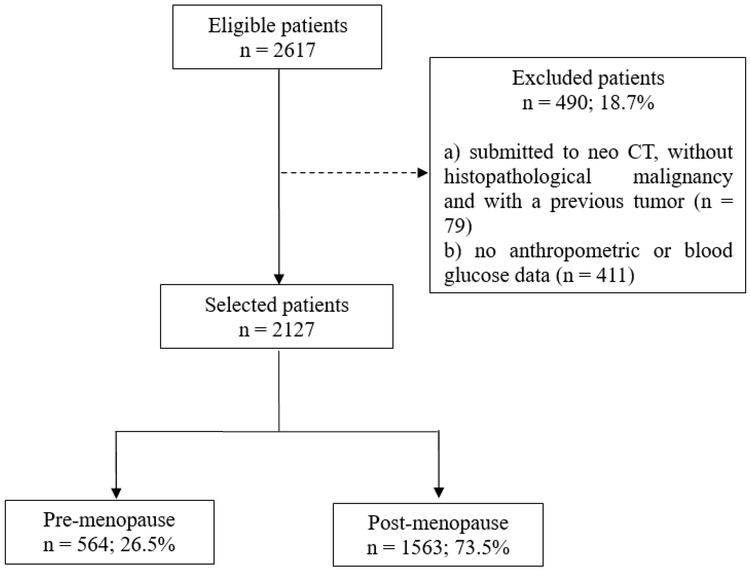
2. Methods
2.1. Data Collection
2.2. Statistical Analysis
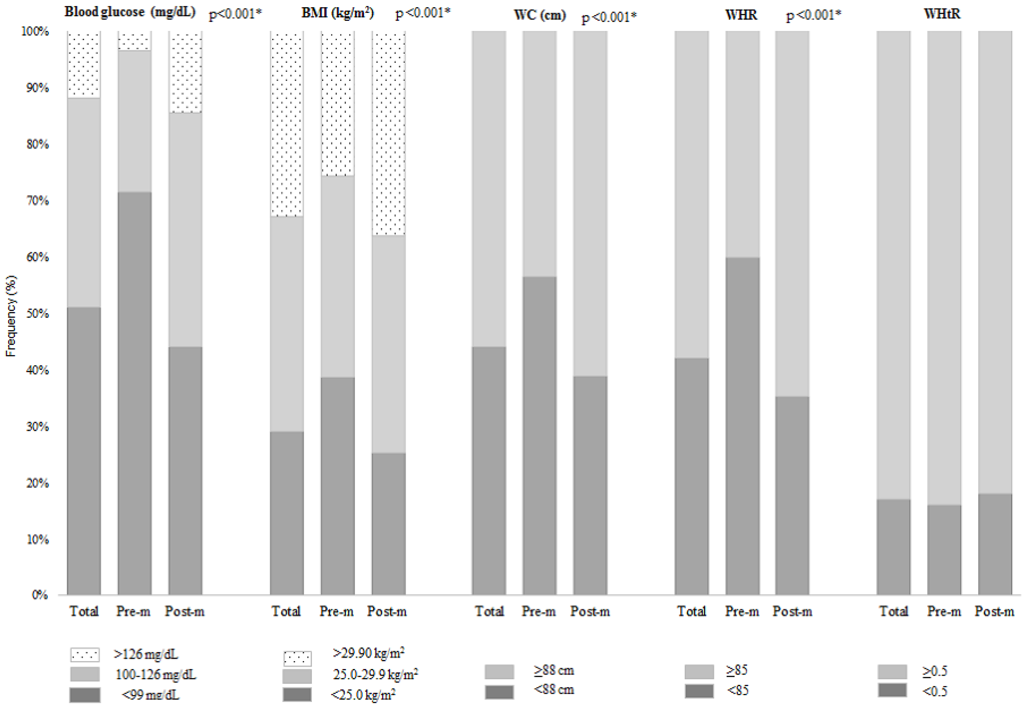
3. Results
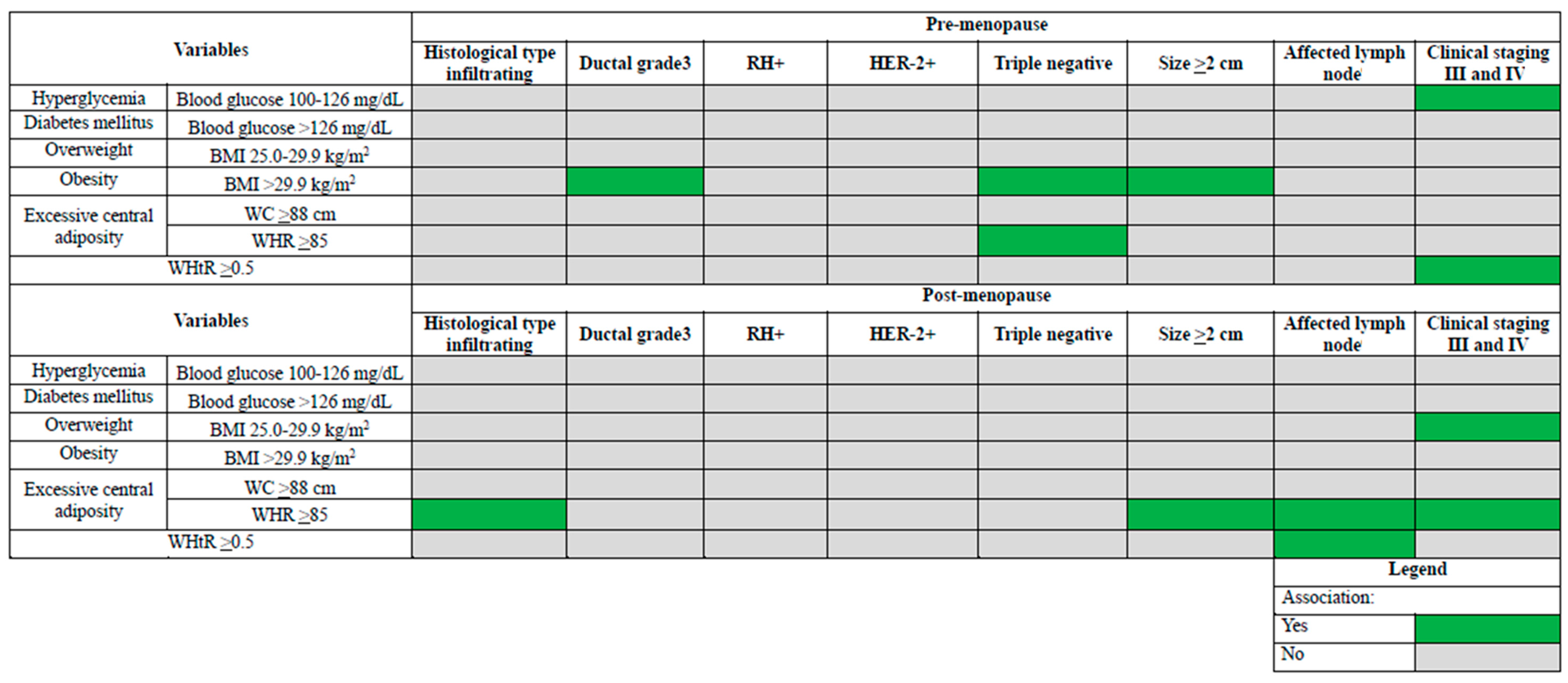
4. Discussion
5. Conclusions
Author Contributions
Funding
Institutional Review Board Statement
Informed Consent Statement
Data Availability Statement
Conflicts of Interest
References
- Sung, H.; Ferlay, J.; Siegel, R.L.; Laversanne, M.; Soerjomataram, I.; Jemal, A.; Bray, F. Global Cancer Statistics 2020: GLOBOCAN Estimates of Incidence and Mortality Worldwide for 36 Cancers in 185 Countries. CA Cancer J. Clin. 2021, 71, 209–249. [Google Scholar] [CrossRef] [PubMed]
- Instituto Nacional de Câncer José Alencar Gomes da Silva (INCA). Estimativa 2020: Incidência de Câncer no Brasil. 2019. Available online: https://www.inca.gov.br/sites/ufu.sti.inca.local/files/media/document/estimativa-2020-incidencia-de-cancer-no-brasil.pdf (accessed on 30 March 2022). (In Portuguese)
- Bougaret, L.; Delort, L.; Billard, H.; Le OKHuede, C.; Boby, C.; De La Foye, A.; Rossary, A.; Mojallal, A.; Damour, O.; Auxenfans, C.; et al. Adipocyte/breast cancer cell crosstalk in obesity interferes with the anti-proliferative efficacy of tamoxifen. PLoS ONE 2018, 13, e0191571. [Google Scholar] [CrossRef] [PubMed]
- Picon-Ruiz, M.; Morata-Tarifa, C.; Valle-Goffin, J.J.; Friedman, E.R.; Slingerland, J.M. Obesity and adverse breast cancer risk and outcome: Mechanistic insights and strategies for intervention. CA Cancer J. Clin. 2017, 67, 378–397. [Google Scholar] [CrossRef] [PubMed]
- World Health Organization (WHO). Obesity: Preventing and Managing the Global Epidemic: Report of a WHO Consultation; Techinical Report for World Health Organization 894; World Health Organization: Geneva, Switzerland, 2000; Available online: https://apps.who.int/iris/handle/10665/42330 (accessed on 2 March 2022).
- Associação Brasileira para o Estudo da Obesidade e da Síndrome Metabólica (ABESO). Mapa da Obesidade. Available online: https://abeso.org.br/obesidade-e-sindrome-metabolica/mapa-da-obesidade/ (accessed on 10 March 2022). (In Portuguese).
- Nagrani, R.; Mhatre, S.; Rajaraman, P.; Soerjomataram, I.; Boffetta, P.; Gupta, S.; Parmar, V.; Badwe, R.; Dikshit, R. Central obesity increases risk of breast cancer irrespective of menopausal and hormonal receptor status in women of South Asian Ethnicity. Eur. J. Cancer 2016, 66, 153–161. [Google Scholar] [CrossRef]
- American Diabetes Association (ADA). Standards of Medical Care in Diabetes—2022 Abridged for Primary Care Providers. Clin. Diabetes 2022, 40, 10–38. [Google Scholar] [CrossRef]
- Browning, L.M.; Hsieh, S.D.; Ashwell, M. A systematic review of waist-to-height ratio as a screening tool for the prediction of cardiovascular disease and diabetes: 0·5 could be a suitable global boundary value. Nutr. Res. Rev. 2010, 23, 247–269. [Google Scholar] [CrossRef]
- Kim, A.; Scharf, K.; Senthil, M.; Solomon, N.; Garberoglio, C.; Lum, S.S. The prevalence of overweight and obesity in a breast clinic population: Consideration for weight loss as a therapeutic intervention. Surg. Obes. Relat. Dis. 2014, 10, 348–353. [Google Scholar] [CrossRef]
- Arnold, M.; Pandeya, N.; Byrnes, G.; Renehan, A.G.; A Stevens, G.; Ezzati, M.; Ferlay, J.; Miranda, J.J.; Romieu, I.; Dikshit, R.; et al. Global burden of cancer attributable to high body-mass index in 2012: A population-based study. Lancet Oncol. 2014, 16, 36–46. [Google Scholar] [CrossRef]
- Kerr, J.; Anderson, C.; Lippman, S.M. Physical activity, sedentary behaviour, diet, and cancer: An update and emerging new evidence. Lancet Oncol. 2017, 18, e457–e471. [Google Scholar] [CrossRef]
- Ewertz, M.; Jensen, M.-B.; Gunnarsdóttir, K.Á.; Højris, I.; Jakobsen, E.H.; Nielsen, D.; Stenbygaard, L.E.; Tange, U.B.; Cold, S. Effect of Obesity on Prognosis After Early-Stage Breast Cancer. J. Clin. Oncol. 2011, 29, 25–31. [Google Scholar] [CrossRef]
- Vaysse, C.; Lømo, J.; Garred, Ø.; Fjeldheim, F.; Lofteroed, T.; Schlichting, E.; McTiernan, A.; Frydenberg, H.; Husøy, A.; Lundgren, S.; et al. Inflammation of mammary adipose tissue occurs in overweight and obese patients exhibiting early-stage breast cancer. NPJ Breast Cancer 2017, 3, 19. [Google Scholar] [CrossRef] [PubMed]
- Renehan, A.G.; Zwahlen, M.; Egger, M. Adiposity and cancer risk: New mechanistic insights from epidemiology. Nat. Cancer 2015, 15, 484–498. [Google Scholar] [CrossRef] [PubMed]
- Healy, L.A.; Ryan, A.M.; Carroll, P.; Ennis, D.; Crowley, V.; Boyle, T.; Kennedy, M.J.; Connolly, E.; Reynolds, J.V. Metabolic Syndrome, Central Obesity and Insulin Resistance are Associated with Adverse Pathological Features in Postmenopausal Breast Cancer. Clin. Oncol. 2010, 22, 281–288. [Google Scholar] [CrossRef] [PubMed]
- Li, H.; Sun, X.; Miller, E.; Wang, Q.; Tao, P.; Liu, L.; Zhao, Y.; Wang, M.; Qi, Y.; Li, J. BMI, reproductive factors, and breast cancer molecular subtypes: A case-control study and meta-analysis. J. Epidemiol. 2017, 27, 143–151. [Google Scholar] [CrossRef]
- Contiero, P.; Berrino, F.; Tagliabue, G.; Mastroianni, A.; Di Mauro, M.G.; Fabiano, S.; Annulli, M.; Muti, P. Fasting blood glucose and long-term prognosis of non-metastatic breast cancer: A cohort study. Breast Cancer Res. Treat. 2013, 138, 951–959. [Google Scholar] [CrossRef]
- Chen, K.; Zhang, J.; Beeraka, N.M.; Tang, C.; Babayeva, Y.V.; Sinelnikov, M.Y.; Zhang, X.; Zhang, J.; Liu, J.; Reshetov, I.V.; et al. Advances in the Prevention and Treatment of Obesity-Driven Effects in Breast Cancers. Front. Oncol. 2022, 12, 820968. [Google Scholar] [CrossRef]
- Houghton, S.C.; Eliassen, H.; Tamimi, R.M.; Willett, W.C.; Rosner, B.A.; Hankinson, S.E. Central Adiposity and Subsequent Risk of Breast Cancer by Menopause Status. JNCI J. Natl. Cancer Inst. 2020, 113, 900–908. [Google Scholar] [CrossRef]
- Chen, H.; Ding, A.; Wang, M. Impact of central obesity on prognostic outcome of triple negative breast cancer in Chinese women. SpringerPlus 2016, 5, 594. [Google Scholar] [CrossRef]
- Masters, G.A.; Krilov, L.; Bailey, H.H.; Brose, M.S.; Burstein, H.; Diller, L.R.; Dizon, D.S.; Fine, H.A.; Kalemkerian, G.P.; Moasser, M.; et al. Clinical Cancer Advances 2015: Annual Report on Progress Against Cancer From the American Society of Clinical Oncology. J. Clin. Oncol. 2015, 33, 786–809. [Google Scholar] [CrossRef]
- Lohmann, A.E.; Soldera, S.V.; Pimentel, I.; Ribnikar, D.; Ennis, M.; Amir, E.; Goodwin, P.J. Association of Obesity With Breast Cancer Outcome in Relation to Cancer Subtypes: A Meta-Analysis. JNCI J. Natl. Cancer Inst. 2021, 113, 1465–1475. [Google Scholar] [CrossRef]
- Cheng, E.; Kirley, J.; Feliciano, E.M.C.; Caan, B.J. Adiposity and cancer survival: A systematic review and meta-analysis. Cancer Causes Control 2022, 33, 1219–1246. [Google Scholar] [CrossRef] [PubMed]
- Caan, B.J.; Feliciano, E.M.C.; Kroenke, C.H. The Importance of Body Composition in Explaining the Overweight Paradox in Cancer—Counterpoint. Cancer Res. 2018, 78, 1906–1912. [Google Scholar] [CrossRef] [PubMed]
- Guasch-Ferré, M.; Salas-Salvadó, J.; Ros, E.; Estruch, R.; Corella, D.; Fitó, M.; Martínez-González, M.A.; PREDIMED Investigators. The PREDIMED trial, Mediterranean diet and health outcomes: How strong is the evidence? Nutr. Metab. Cardiovasc. Dis. 2017, 27, 624–632. [Google Scholar] [CrossRef] [PubMed]
- Freedman, D.S.; Thornton, J.; Pi-Sunyer, F.X.; Heymsfield, S.B.; Wang, J.; Pierson, R.N., Jr.; Blanck, H.M.; Gallagher, D. The body adiposity index (hip circumference ÷ height1.5) is not a more accurate measure of adiposity than is BMI, waist circumference, or hip circumference. Obesity 2012, 20, 2438–2444. [Google Scholar] [CrossRef] [PubMed]
- Lee, C.M.Y.; Huxley, R.R.; Wildman, R.P.; Woodward, M. Indices of abdominal obesity are better discriminators of cardiovascular risk factors than BMI: A meta-analysis. J. Clin. Epidemiol. 2008, 61, 646–653. [Google Scholar] [CrossRef] [PubMed]
- Instituto Nacional de Câncer José Alencar Gomes da Silva (INCA). Detecção Precoce do Câncer. 2021. Available online: https://www.inca.gov.br/publicacoes/livros/deteccao-precoce-do-cancer (accessed on 22 March 2022). (In Portuguese)


| Variables | Total n (%) | Histological Type Infiltrating | Ductal Grade 3 b | RH+ b | HER-2+ b | Triple Negative b | Size ≥ 2 cm b | Affected Lymph Node b | Clinical Staging III and IV b | ||||||||
|---|---|---|---|---|---|---|---|---|---|---|---|---|---|---|---|---|---|
| No 93 (16.5%) | Yes 471 (83.5%) | No 216 (38.4%) | Yes 346 (61.6%) | No 87 (16.6%) | Yes 438 (83.4%) | No 326 (77.8%) | Yes 93 (22.2%) | No 376 (89.7%) | Yes 43 (10.3%) | No 188 (34.6%) | Yes 355 (65.4%) | No 311 (60.6%) | Yes 202 (39.4%) | No 434 (89.1%) | Yes 53 (10.9%) | ||
| Age (years) | |||||||||||||||||
| <60 | 551 (97.7%) | 91 (97.8%) | 460 (97.7%) | 211 (97.7%) | 338 (97.7%) | 86 (98.8%) | 427 (97.5%) | 316 (96.9%) | 92 (98.9%) | 366 (97.3%) | 42 (97.7%) | 184 (97.9%) | 347 (97.7%) | 231 (74.3%) | 137 (67.8%) | 428 (98.6%) | 49 (92.4%) |
| ≥60 | 13 (2.3%) | 2 (2.2%) | 11 (2.3%) | 5 (2.3%) | 8 (92.3%) | 1 (1.2%) | 11 (2.5%) | 10 (3.1%) | 1 (1.1%) | 10 (2.7%) | 1 (2.3%) | 4 (2.1%) | 8 (2.2%) | 80 (25.7%) | 65 (32.2%) | 6 (1.4%) | 4 (4.6%) |
| Blood glucose (mg/dL) | |||||||||||||||||
| ≤99 | 403 (71.4%) | 70 (75.3%) | 333 (70.7%) | 151 (69.9%) | 250 (72.3%) | 64 (73.6%) | 310 (70.8%) | 232 (71.2%) | 68 (73.1%) | 270 (71.8%) | 30 (69.8%) | 138 (73.4%) | 250 (70.4%) | 228 (73.3%) | 139 (68.8%) | 316 (73.5%) | 29 (54.7%) a |
| 100–126 | 141 (25.0%) | 18 (19.3%) | 123 (26.1%) | 58 (27.3%) | 82 (23.7%) | 20 (23.0%) | 113 (25.8%) | 84 (25.8%) | 22 (23.7%) | 94 (25.0%) | 12 (27.9%) | 42 (22.3%) | 94 (26.5%) | 71 (22.8%) | 57 (28.2%) | 100 (23.0%) | 22 (41.5%) |
| >126 | 20 (3.6%) | 5 (5.4%) | 15 (3.2%) | 6 (2.8%) | 14 (4.0%) | 3 (3.4%) | 15 (3.4%) | 10 (3.0%) | 3 (3.2%) | 12 (3.2%) | 1 (2.3%) | 8 (4.3%) | 11 (3.1%) | 12 (3.9%) | 6 (3.0%) | 15 (3.5%) | 2 (3.8%) |
| BMI (kg/m2) | |||||||||||||||||
| ≤24.9 | 218 (38.7%) | 31 (33.3%) | 187 (39.7%) | 94 (43.5%) | 124 (35.8%) a | 33 (38.0%) | 173 (39.5%) | 130 (39.9%) | 30 (32.3%) | 145 (38.6%) | 15 (34.9%) a | 82 (43.6%) | 127 (35.8%) a | 118 (37.9%) | 80 (39.6%) | 177 (40.8%) | 16 (30.2%) a |
| 25.0–29.9 | 201 (35.6%) | 37 (39.8%) | 164 (34.8%) | 78 (36.1%) | 123 (35.6%) | 27 (31.0%) | 160 (36.5%) | 112 (34.3%) | 41 (44.1%) | 142 (37.8%) | 11 (25.6%) | 71 (39.4%) | 119 (33.5%) | 119 (38.3%) | 68 (33.7%) | 154 (35.5%) | 21 (39.6%) |
| >29.9 | 145 (25.7%) | 25 (26.9%) | 120 (25.5%) | 44 (20.4%) | 99 (28.6%) | 27 (31.0% | 105 (24.0%) | 84 (25.8%) | 22 (23.7%) | 89 (23.6%) | 17 (39.5%) | 32 (17.0%) | 109 (30.7%) | 74 (23.8%) | 54 (26.7%) | 103 (23.7%) | 16 (30.2%) |
| WC (cm) | |||||||||||||||||
| <88 | 318 (56.4%) | 52 (55.9%) | 266 (56.5%) | 129 (59.7%) | 189 (54.6%) | 47 (54.0%) | 251 (57.3%) | 189 (58.0%) | 48 (51.6%) | 215 (57.2%) | 22 (51.2%) | 122 (64.9%) | 182 (51.3%) a | 181 (58.2%) | 112 (55.4%) | 255 (58.8%) | 28 (52.8%) |
| ≥88 | 246 (43.6%) | 41 (44.1%) | 205 (43.5%) | 87 (40.3%) | 157 (45.4%) | 40 (46.0%) | 187 (42.7%) | 137 (42.0%) | 45 (48.4%) | 161 (42.8%) | 21 (48.8%) | 66 (35.1%) | 173 (48.7%) | 130 (41.8%) | 90 (40.6%) | 179 (41.2%) | 25 (47.2%) |
| WHR | |||||||||||||||||
| <85 | 338 (59.9%) | 55 (59.1%) | 283 (60.1%) | 129 (59.7%) | 208 (60.1%) | 57 (65.5%) | 259 (59.1%) | 200 (61.3%) | 54 (58.1%) | 224 (59.6%) | 30 (69.8%) | 120 (63.8%) | 203 (57.2%) | 187 (60.1%) | 121 (59.9%) | 268 (61.7%) | 31 (58.5%) |
| ≥85 | 226 (40.1%) | 38 (40.9%) | 188 (39.9%) | 87 (40.3%) | 138 (39.9%) | 30 (34.5%) | 179 (40.9%) | 126 (38.7%) | 39 (41.9%) | 152 (40.4%) | 13 (30.2%) | 68 (36.2%) | 152 (42.8%) | 124 (39.9%) | 81 (40.1%) | 166 (38.3%) | 22 (41.5%) |
| WHtR | |||||||||||||||||
| <0.5 | 88 (15.6%) | 16 (17.2%) | 72 (15.3%) | 32 (14.8%) | 56 (16.2%) | 14 (16.1%) | 66 (15.1%) | 52 (15.9%) | 12 (12.9%) | 54 (14.4%) | 10 (23.3%) | 30 (16.0%) | 53 (14.9%) | 52 (16.7%) | 27 (13.4%) | 74 (17.0%) | 4 (7.6%) a |
| ≥0.5 | 476 (84.4%) | 77 (82.8%) | 399 (84.7%) | 184 (85.2%) | 290 (83.8%) | 73 (83.9%) | 372 (84.9%) | 274 (84.1%) | 81 (87.1%) | 322 (85.6%) | 33 (76.7%) | 158 (54.0%) | 302 (85.1%) | 259 (83.3%) | 175 (86.6%) | 360 (83.0%) | 49 (92.4%) |
| Variables | Total n (%) | Histological Type Infiltrating | Ductal Grade 3 b | RH+ b | HER-2+ b | Triple Negative b | Size ≥ 2 cm b | Affected Lymph Node b | Clinical Staging III and IV b | ||||||||
|---|---|---|---|---|---|---|---|---|---|---|---|---|---|---|---|---|---|
| No 272 (17.4%) | Yes 1291 (82.6%) | No 589 (38.4%) | Yes 945 (61.6%) | No 283 (19.5%) | Yes 1167 (80.5%) | No 915 (81.8%) | Yes 203 (18.2%) | No 983 (87.9%) | Yes 135 (12.1%) | No 573 (37.8%) | Yes 943 (62.2%) | No 844 (60.0%) | Yes 563 (40.0%) | No 1179 (88.6%) | Yes 151 (11.4%) | ||
| Age (years) | |||||||||||||||||
| <60 | 596 (38.1%) | 101 (37.1%) | 495 (38.3%) | 231 (39.2%) | 355 (37.6%) | 104 (36.8%) | 436 (37.4%) | 371 (40.6%) | 85 (41.9%) | 398 (40.5%) | 58 (43.0%) | 231 (40.3%) | 342 (36.3%) | 304 (36.0%) | 219 (38.9%) | 452 (38.3%) | 47 (31.1%) |
| ≥60 | 967(61.9%) | 171 (62.9%) | 796 (61.7%) | 358 (60.8%) | 590 (62.4%) | 179 (63.2%) | 731 (62.4%) | 544 (59.4%) | 118 (58.1%) | 585 (59.5%) | 77 (57.0%) | 342 (59.7%) | 601 (63.7%) | 540 (4.0%) | 344 (61.1%) | 727 (61.7%) | 104 (68.9%) |
| Blood glucose (mg/dL) | |||||||||||||||||
| ≤99 | 689 (44.1%) | 124 (45.6%) | 565 (43.8%) | 244 (41.4%) | 432 (45.7%) | 135 (47.7%) | 502 (43.0%) | 401 (43.8%) | 91 (44.8%) | 425 (43.2%) | 67 (49.6%) | 265 (46.2%) | 392 (41.6%) | 380 (45.0%) | 227 (40.3%) | 512 (43.4%) | 71 (47.0%) |
| 100–126 | 646 (41.3%) | 109 (40.1%) | 537 (41.6%) | 257 (43.6%) | 378 (40.0%) | 107 (37.8%) | 492 (42.2%) | 379 (41.4%) | 80 (39.4%) | 405 (41.2%) | 54 (40.0%) | 232 (40.5%) | 406 (43.0%) | 341 (40.4%) | 248 (44.1%) | 496 (42.1%) | 52 (34.5%) |
| >126 | 228(15.6%) | 39 (14.3%) | 189 (14.6%) | 88 (15.0%) | 135 (14.3%) | 41 (14.5%) | 173 (14.8%) | 135 (14.8%) | 32 (15.8%) | 153 (15.6%) | 14 (10.4%) | 76 (13.3%) | 145 (15.4%) | 123 (14.6%) | 88 (15.6%) | 171 (14.5%) | 28 (18.5%) |
| BMI (kg/m2) | |||||||||||||||||
| ≤24.9 | 394 (25.2%) | 72 (26.5%) | 322 (24.9%) | 142 (24.1%) | 243 (25.7%) a | 73 (25.8%) | 296 (25.4%) | 226 (24.7%) | 53 (26.1%) | 246 (25.0%) | 33 (24.4%) a | 143 (25.0%) | 236 (25.0%) | 211 (25.0%) | 143 (25.4%) | 289 (24.5%) | 48 (31.8%) a |
| 25.0–29.9 | 601 (38.5%) | 107 (39.3%) | 494 (38.3%) | 208 (35.3%) | 378 (40.0%) | 108 (38.2%) | 442 (37.9%) | 346 (37.8%) | 79 (38.9%) | 369 (37.5%) | 56 (41.5%) | 232 (40.5%) | 352 (37.3%) | 342 (40.5%) | 200 (35.5%) | 471 (40.0%) | 26 (26.5%) |
| >29.9 | 568 (36.3%) | 93 (34.2%) | 475 (36.8%) | 239 (40.6%) | 324 (34.3%) | 102 (36.0%) | 429 (36.7%) | 343 (37.5%) | 71 (35.0%) | 368 (37.5%) | 46 (54.1%) | 198 (34.5%) | 355 (37.7%) | 291 (34.5%) | 220 (39.1%) | 419 (35.5%) | 63 (41.7%) |
| WC (cm) | |||||||||||||||||
| <88 | 607 (38.8%) | 111 (40.8%) | 496 (38.4%) | 212 (36.0%) | 382 (40.4%) | 109 (38.5%) | 450 (38.6%) | 348 (38.0%) | 79 (38.9%) | 371 (37.7%) | 56 (41.5%) | 233 (40.7%) | 353 (37.4%) | 335 (39.7%) | 212 (37.7%) | 456 (38.7%) | 65 (43.0%) |
| ≥88 | 956 (61.2%) | 161 (59.2%) | 795 (61.6%) | 377 (64.0%) | 563 (59.6%) | 174 (61.5%) | 717 (61.4%) | 567 (62.0%) | 124 (61.1%) | 612 (62.3%) | 79 (58.5%) | 340 (59.3%) | 590 (62.6%) | 509 (60.3%) | 351 (62.3%) | 723 (61.3%) | 86 (57.0%) |
| WHR | |||||||||||||||||
| <85 | 550 (35.2%) | 112 (41.2%)a | 438 (33.9%) | 199 (33.8%) | 340 (36.0%) | 102 (36.0%) | 397 (34.0%) | 333 (36.4%) | 66 (32.5%) | 344 (35.0%) | 55 (40.7%) | 227 (39.6%) | 304 (32.2%) a | 308 (36.5%) | 177 (31.4%) a | 421 (35.7%) | 36 (23.8%) a |
| ≥85 | 1013 (64.8%) | 160 (58.8%) | 853 (66.1%) | 390 (66.2%) | 605 (64.0%) | 191 (64.0%) | 770 (66.0%) | 582 (63.6%) | 137 (67.5%) | 639 (65.0%) | 80 (59.3%) | 346 (60.4%) | 639 (67.8%) | 536 (63.5%) | 386 (68.6%) | 758 (64.3%) | 115 (76.2%) |
| WHtR | |||||||||||||||||
| <0.5 | 278 (17.8%) | 51 (18.7%) | 227 (17.6%) | 97 (16.5%) | 177 (18.7%) | 59 (20.8%) | 203 (17.4%) | 173 (18.9%) | 34 (16.7%) | 176 (17.9%) | 31 (23.0%) | 97 (16.9%) | 174 (18.5%) | 134 (15.9%) | 115 (20.4%) | 206 (17.5%) | 30 (19.9%) |
| ≥0.5 | 1285 (82.2%) | 221 (81.3%) | 1064 (82.4%) | 492 (83.5%) | 768 (81.3%) | 224 (79.2%) | 964 (82.6%) | 742 (81.1%) | 169 (83.3%) | 807 (82.1%) | 704 (77.0%) | 476 (83.1%) | 769 (81.5%) | 710 (84.1%) | 448 (79.6%) | 973 (82.5%) | 121 (80.1%) |
| Variables | Histological Type Infiltrating OR (95% CI) | Ductal Grade 3 OR (95% CI) | RH + OR (95% CI) | HER-2 + OR (95% CI) | Triple Negative OR (95% CI) | Size ≥ 2 cm OR (95% CI) | Affected Lymph Node OR (95% CI) | Clinical Staging III and IV OR (95% CI) | ||||||||
|---|---|---|---|---|---|---|---|---|---|---|---|---|---|---|---|---|
| Uni | Mult c | Uni | Mult d | Uni | Mult e | Uni | Mult f | Uni | Mult g | Uni | Mult h | Uni | Mult i | Uni | Mult j | |
| Age (years) | ||||||||||||||||
| <60 | 1.00 | - | 1.00 | - | 1.00 | - | 1.00 | - | 1.00 | - | 1.00 | - | 1.00 | - | 1.00 | - |
| ≥60 | 0.91 (0.20–4.22) | - | 0.99 (0.33–3.09) | - | 2.21 (0.28–7.38) | - | 2.91 (0.37–2.04) | - | 0.87 (0.11–6.98) | - | 1.06 (0.31–3.57) | - | 1.10 (0.34–3.52) | - | 5.82 (1.59–21.35) a | - |
| Blood glucose (mg/dL) | ||||||||||||||||
| ≤99 | 1.00 | - | 1.00 | - | 1.00 | - | 1.00 | - | 1.00 | - | 1.00 | - | 1.00 | - | 1.00 | 1.00 |
| 100–126 | 0.72 (0.49–1.06) | - | 0.83 (0.57–1.24) | - | 1.16 (0.68–2.01) | - | 1.12 (0.65–1.92) | - | 1.15 (0.56–2.33) | - | 1.23 (0.81–1.88) | - | 1.32 (0.87–1.98) | - | 2.42 (1.33–4.40) b | 2.37 (1.34–4.56) b |
| >126 | 1.40 (0.53–3.71) | - | 1.41 (0.53–3.74) | - | 1.03 (0.29–3.67) | - | 0.98 (0.26–3.65) | - | 0.75 (0.09–5.97) | - | 0.76 (0.30–1.93) | - | 0.82 (0.31–2.35) | - | 1.47 (0.32–6.73) | 1.21 (0.25–5.94) |
| BMI (kg/m2) | ||||||||||||||||
| ≤24.9 | 1.00 | - | 1.00 | 1.00 | 1.00 | - | 1.00 | - | 1.00 | 1.00 | 1.00 | 1.00 | 1.00 | - | 1.00 | - |
| 25.0–29.9 | 1.03 (0.49–1.09) | - | 1.19 (0.81–1.77) | 1.19 (0.81–1.77) | 1.13 (0.65–1.96) | - | 0.63 (0.37–1.07) | - | 0.75 (0.33–1.69) | 0.87 (0.37–1.98) | 1.04 (0.69–1.55) | 1.04 (0.69–1.55) | 0.84 (0.56–1.27) | - | 1.51 (0.76–2.99) | - |
| >29.9 | 1.27 (0.81–2.23) | - | 1.70 (1.09–2.66) a | 1.70 (1.09–2.66) a | 0.74 (0.42–1.30) | - | 0.88 (0.48–1.62) | - | 1.85 (0.88–3.88) | 1.37 (1.08–3.24) a | 2.20 (1.36–3.56) a | 2.20 (1.36–3.56) a | 1.07 (0.68–1.69) | - | 1.71 (0.82–3.58) | - |
| WC (cm) | ||||||||||||||||
| <88 | 1.00 | - | 1.00 | - | 1.00 | - | 1.00 | - | 1.00 | - | 1.00 | - | 1.00 | - | 1.00 | - |
| ≥88 | 1.02 (0.65–1.99) | - | 1.23 (0.87–1.74) | - | 0.87 (0.55–1.39) | - | 0.77 (0.49–1.23) | - | 1.27 (0.68–2.40) | - | 1.76 (1.22–2.53) a | - | 1.12 (0.78–1.60) | - | 1.27 (0.72–2.25) | - |
| WHR | ||||||||||||||||
| <85 | 1.00 | - | 1.00 | - | 1.00 | - | 1.00 | - | 1.00 | 1.00 | 1.00 | - | 1.00 | - | 1.00 | - |
| ≥85 | 1.04 (0.66–1.63) | - | 1.98 (0.69–1.39) | - | 1.31 (0.81–2.12) | - | 0.87 (0.55–1.39) | - | 1.67 (0.32–1.26) | 1.53 (1.05–1.98) a | 1.32 (0.92–1.90) | - | 1.01 (0.70–1.45) | - | 1.14 (0.64–2.04) | - |
| WHtR | ||||||||||||||||
| <0.5 | 1.00 | - | 1.00 | - | 1.00 | - | 1.00 | - | 1.00 | - | 1.00 | - | 1.00 | - | 1.00 | 1.00 |
| ≥0.5 | 0.87 (0.48–1.57) | - | 0.90 (0.56–1.44) | - | 1.08 (0.57–2.03) | - | 0.78 (0.39–1.53) | - | 0.55 (0.26–1.19) | 1.08 (0.66–1.76) | - | 1.30 (0.79–2.15) | - | 2.52 (0.88–7.19) | 3.50 (1.14–10.77) a | |
| Variables | Histological Type Infiltrating OR (95% CI) | Ductal Grade 3 OR (95% CI) | RH + OR (95% CI) | HER-2 + OR (95% CI) | Triple Negative OR (95% CI) | Size ≥ 2 cm OR (95% CI) | Affected Lymph Node OR (95% CI) | Clinical Staging III and IV OR (95% CI) | ||||||||
|---|---|---|---|---|---|---|---|---|---|---|---|---|---|---|---|---|
| Uni | Mult c | Uni | Mult d | Uni | Mult e | Uni | Mult f | Uni | Mult g | Uni | Mult h | Uni | Mult i | Uni | Mult j | |
| Age (years) | ||||||||||||||||
| <60 | 1.00 | - | 1.00 | - | 1.00 | - | 1.00 | - | 1.00 | - | 1.00 | - | 1.00 | - | 1.00 | - |
| ≥60 | 1.05 (0.80–1.38) | - | 1.07 (0.87–1.32) | - | 0.97 (0.74–1.27) | - | 1.06 (0.77–1.44) | - | 0.90 (0.63–1.30) | - | 1.19 (0.96–1.50) | - | 0.88 (0.71–1.10) | - | 1.37 (0.96–1.98) | - |
| Blood glucose (mg/dL) | ||||||||||||||||
| ≤99 | 1.00 | - | 1.00 | - | 1.00 | - | 1.00 | - | 1.00 | - | 1.00 | - | 1.00 | - | 1.00 | - |
| 100–126 | 1.92 (0.70–2.23) | - | 0.83 (0.66–1.04) | - | 1.24 (0.93–1.64) | - | 1.07 (0.77–1.50) | - | 0.84 (0.58–1.24) | - | 1.18 (0.94–1.48) | - | 1.22 (0.96–1.53) | - | 0.76 (0.52–1.10) | - |
| >126 | 1.94 (0.63–2.40) | - | 0.86 (0.63–1.18) | - | 1.13 (0.77–1.68) | - | 0.96 (0.61–1.50) | - | 1.58 (0.61–1.06) | - | 1.29 (0.94–1.77) | - | 1.20 (0.87–1.65) | - | 1.18 (0.74–1.89) | - |
| BMI (kg/m2) | ||||||||||||||||
| ≤24.9 | 1.00 | - | 1.00 | - | 1.00 | - | 1.00 | - | 1.00 | - | 1.00 | - | 1.00 | - | 1.00 | 1.00 |
| 25.0–29.9 | 1.96 (0.49–3.09) | - | 1.06 (0.81–1.39) | - | 1.01 (0.72–1.41) | - | 1.03 (0.70–1.51) | - | 1.13 (0.71–1.79) | - | 0.92 (0.70–1.20) | - | 0.86 (0.66–1.13) | - | 1.51 (1.32–1.80) a | 1.44 (1.28–1.70) b |
| >29.9 | 1.27 (0.81–2.23) | - | 1.79 (0.96–2.03) | - | 1.04 (0.74–1.45 | - | 1.13 (0.76–1.68) | - | 0.93 (0.57–1.49) | - | 1.09 (0.83–1.42) | - | 1.11 (0.85–1.47) | - | 1.90 (0.90–2.36) | 1.73 (0.47–2.12) |
| WC (cm) | ||||||||||||||||
| <88 | 1.00 | - | 1.00 | - | 1.00 | - | 1.00 | - | 1.00 | - | 1.00 | - | 1.00 | - | 1.00 | - |
| ≥88 | 1.90 (0.69–2.18) | - | 1.83 (1.27–2.02) | - | 0.99 (0.76–1.30) | - | 1.04 (0.76–1.42) | - | 1.85 (0.59–2.23) | - | 1.14 (0.93–1.42) | - | 1.09 (0.87–1.36) | - | 0.83 (0.59–1.17) | - |
| WHR | ||||||||||||||||
| <85 | 1.00 | 1.00 | 1.00 | - | 1.00 | - | 1.00 | - | 1.00 | - | 1.00 | 1.00 | 1.00 | 1.00 | 1.00 | 1.00 |
| ≥85 | 1.73 (1.56–1.95) a | 1.73 (1.56–1.95) a | 1.91 (0.73–2.12) | - | 1.09 (0.83–1.43) | - | 0.84 (0.61–1.16) | - | 1. 87 (0.54–2.13) | - | 1.38 (1.11–1.71) a | 1.38 (1.11–1.71) a | 1.25 (0.99–1.57) | 1.24 (1.02–1.56) a | 1.77 (1.20–2.63) a | 1.76 (1.30–2.65) a |
| WHtR | ||||||||||||||||
| <0.5 | 1.00 | - | 1.00 | - | 1.00 | - | 1.00 | - | 1.00 | - | 1.00 | - | 1.00 | 1.00 | 1.00 | - |
| ≥0.5 | 1.92 (0.66–2.29) | - | 0.85 (0.65–1.12) | - | 1.25 (0.90–1.73) | - | 0.86 (0.58–1.29) | - | 1.73 (0.47–1.13) | - | 0.90 (0.69–1.18) | - | 1.73 (1.06–1.96) a | 1.70 (1.06–1.97) a | 0.83 (0.56–1.31) | - |
Publisher’s Note: MDPI stays neutral with regard to jurisdictional claims in published maps and institutional affiliations. |
© 2022 by the authors. Licensee MDPI, Basel, Switzerland. This article is an open access article distributed under the terms and conditions of the Creative Commons Attribution (CC BY) license (https://creativecommons.org/licenses/by/4.0/).
Share and Cite
Gioseffi, C.; Padilha, P.d.C.; Chaves, G.V.; Oliveira, L.C.d.; Peres, W.A.F. Body Weight, Central Adiposity, and Fasting Hyperglycemia Are Associated with Tumor Characteristics in a Brazilian Cohort of Women with Breast Cancer. Nutrients 2022, 14, 4926. https://doi.org/10.3390/nu14224926
Gioseffi C, Padilha PdC, Chaves GV, Oliveira LCd, Peres WAF. Body Weight, Central Adiposity, and Fasting Hyperglycemia Are Associated with Tumor Characteristics in a Brazilian Cohort of Women with Breast Cancer. Nutrients. 2022; 14(22):4926. https://doi.org/10.3390/nu14224926
Chicago/Turabian StyleGioseffi, Clara, Patricia de Carvalho Padilha, Gabriela Villaça Chaves, Livia Costa de Oliveira, and Wilza Arantes Ferreira Peres. 2022. "Body Weight, Central Adiposity, and Fasting Hyperglycemia Are Associated with Tumor Characteristics in a Brazilian Cohort of Women with Breast Cancer" Nutrients 14, no. 22: 4926. https://doi.org/10.3390/nu14224926
APA StyleGioseffi, C., Padilha, P. d. C., Chaves, G. V., Oliveira, L. C. d., & Peres, W. A. F. (2022). Body Weight, Central Adiposity, and Fasting Hyperglycemia Are Associated with Tumor Characteristics in a Brazilian Cohort of Women with Breast Cancer. Nutrients, 14(22), 4926. https://doi.org/10.3390/nu14224926