Article

Effect of a Bifidobacterium-Containing Acid-Resistant Microcapsule Formulation on Gut Microbiota: A Pilot Study

1
Department of Pediatrics, Kansai Medical University, Osaka 573-1010, Japan
2
Research and Development Department, Morishita Jintan Co., Ltd., Osaka 573-0128, Japan
*
Author to whom correspondence should be addressed.
Nutrients 2022, 14(22), 4829; https://doi.org/10.3390/nu14224829
Submission received: 28 September 2022 / Revised: 8 November 2022 / Accepted: 13 November 2022 / Published: 15 November 2022

Abstract

:
Approximately 10 Bifidobacterium species are known to inhabit the human intestinal tract. Bifidobacteria have been reported to possess a variety of probiotic benefits. However, when bifidobacteria are consumed internally as probiotics, the bacteria are killed by gastric acid. Therefore, we developed acid-resistant microcapsules containing Bifidobacterium breve M-16V and B. longum BB536, which are unaffected by gastric acid, and evaluated whether the microcapsule formulation increased the amount of bifidobacteria in the stool after administration compared with the powder formulation. The results revealed no significant difference in the percentage or number of B. longum between before and after administration of the powder or microcapsule formulation in children. By contrast, the bacterial count of B. breve was significantly increased after microcapsule formulation administration (1.5 × 10⁵ copies/g after administration versus 2.8 × 10⁴ copies/g before administration, p = 0.013). In addition, the increase in the bacterial count of B. breve in stools after administration of microcapsule formulation was approximately 1000-fold higher than that after powder formulation administration (p = 0.018). In conclusion, the results indicate that the microcapsule formulation is efficiently transferred to the large intestine without the adverse effects of gastric acidity in children.

1. Introduction

The human oral cavity, gastrointestinal tract, respiratory tract, skin, and urinary and reproductive organs are inhabited by more than 1000 species of bacteria numbering approximately 38 trillion. The bacterial population in the gastrointestinal tract, which accounts for more than 90% of all inhabiting bacteria, comprises the gut microbiota and lives in symbiosis in the human gut, maintaining a certain balance [1]. The composition and quantity of the gut microbiota change with age: the gut microbiota of 1-year-old children is dominated by bacteria belonging to the Actinobacteria phylum; however, their abundance gradually declines, and by 3 years of age, bacteria belonging to the Firmicutes phylum are dominant, similar to the findings in adults [2]. Previously, evaluation of the gut microbiota was based on the classical method of bacterial culture, but with the advent of next-generation sequencers, it is now possible to perform comprehensive genetic analysis to evaluate bacteria, including those that have been difficult to cultivate in the past [3].
Dysbiosis describes abnormalities in the composition or quantity of the gut microbiota, and it is known that dysbiosis occurring from birth to about 2 years of age is transferred to adulthood [4,5]. It has been reported that dysbiosis occurring in infancy is associated with the subsequent development of chronic diseases such as allergic diseases, inflammatory bowel disease, autoimmune disease, obesity, and diabetes mellitus [6]. The American Academy of Pediatrics has stated that a child’s mental development and risk of diseases in childhood and adulthood, such as obesity, hypertension, and diabetes, are related to nutrition in the first 1000 days of life [7]. Therefore, methods for disease prevention and treatment that maintain a normal gut microbiota or correct dysbiosis are being considered. Probiotic administration is considered one such method. Since the first 1000 days of life coincides with the establishment of the gut microbiota, the use of probiotics from infancy should be considered for children with dysbiosis. Probiotics are live microorganisms that, when consumed in proper amounts, confer beneficial effects on the health of the host [8]. For probiotics to exert their functions, they must be sufficiently safe, be of human origin, be able to withstand gastric juice and bile and reach the intestine, be able to grow in the intestines, and be able to be taken orally and maintain an effective bacterial count in the gastrointestinal tract [8].
Bifidobacterium and Lactobacillus species are used as typical probiotics. Lactobacilli are facultative anaerobic bacteria found mainly in the small intestine and female reproductive organs (vagina, cervix, and uterus). Meanwhile, bifidobacteria are anaerobes that cannot grow in the presence of oxygen, and they are found mainly in the large intestine [9]. More than 50 species of Bifidobacterium have been identified [10]. Human residential bifidobacteria (HRB) are broadly classified into adult and infantile forms, with adult HRB including Bifidobacterium adolescentis, B. pseudocatenulatum, B. angulatum, and B. dentium and infantile HRB including B. breve, B. longum subsp. infantis, and B. bifidum. Among HRBs, only B. longum subsp. longum is found in a wide range of ages ranging from infants to older individuals [11]. Bifidobacteria have been reported to exhibit a variety of beneficial effects, including intestinal regulation, protection against infection, and suppression of allergic reactions [12,13]. Probiotics containing B. infantis reduced the incidence and severity of necrotizing enterocolitis in very-low-birth-weight infants [14,15]. Bifidobacterium treatment improved the atopic dermatitis score [16,17,18] and reduced IgE levels [19] in patients with atopic dermatitis.
Although the effects of bifidobacteria as probiotics are proven, it is reported that approximately 40% of the administered bifidobacteria are killed by stomach acid when consumed as a powder formulation [20]. Therefore, we developed acid-resistant microcapsules containing bifidobacteria to ensure the efficient delivery of the bacteria to the large intestine, where bifidobacteria exert their effects as probiotics. The microcapsule manufacturing technology possessed by the authors makes it possible to prepare nearly spherical microcapsules with diameters ranging from 0.5 to 8 mm, which are relatively smaller than conventional capsules. The microcapsules can be given properties such as acid resistance, heat resistance, and enteric solubility by adding multiple layers of film, and the range of formulation design is remarkably wide. The microcapsules used in this study, which are acid-resistant and enteric-soluble, can be used as a kind of drug delivery system to deliver functional ingredients such as bifidobacteria to the intestines without being altered by the low pH of gastric acid [21]. Using the above-mentioned manufacturing technology, a product containing only B. longum BB536 for adult use is marketed under the trade name “BifinaTM”.
In this study, we developed an acid-resistant microcapsule for children’s use containing B. breve M-16V and B. longum BB536 that is not expected to be affected by gastric acid. The purpose of this study was to evaluate whether the microcapsule formulation more efficiently reaches the intestinal tract after oral administration than the powder formulation.

2. Materials and Methods

2.1. Study Participants

Healthy children were recruited, and after explaining the study to their guardians, 10 children were enrolled after their guardians provided informed consent. The inclusion criteria for participants were age of 1–12 years, no use of antimicrobials in the last 3 months, and no regular visits to hospitals. The participants included eight boys and two girls with a median age of 8.08 years (interquartile range (IQR) = 5.83–9.44) on the study entry date. No study participants were born preterm or low-birth weight, which were considered to be risk factors for gut dysbiosis: the median week of gestation was 40.0 weeks (IQR = 39.3–40.1), and the median birth weight was 2950 g (IQR = 2791–3140).

2.2. Characteristics of the Acid-Resistant Microcapsule

The acid-resistant microcapsule formulation administered to the study participants contained B. breve M-16V and B. longum BB536 at a ratio of 1:9 and a total of 1 billion bacteria per packet. The weight of one packet was 0.2 g. The acid-resistant microcapsule used in this study is a seamless, spherical microcapsule with a structure that prevents external liquids and other substances from entering through the outer membranes, as shown in Figure 1a. The microcapsule was constructed using the “drop-in-liquid” method, which is a manufacturing method that utilizes interfacial tension. The microcapsule has a three-layer structure prepared using a triple nozzle. The outermost layer of the membrane contains a substance (gelatin and pectin) that does not dissolve in the acidic range but does dissolve in the neutral range, and the pH response is designed so that the microcapsule does not dissolve in the stomach but instead dissolves in the intestine. The inner membrane of the middle layer of the capsule is made of vegetable fats and oils to improve the barrier function against the permeation of stomach acid. The innermost layer contains the contents (lyophilized Bifidobacterium powder), and a type of drug delivery system is realized by the aforementioned two-layer outer and middle membranes. Each microcapsule was designed to be 2 mm in diameter. A powdered formulation of B. breve M-16V containing 1 billion bacteria per packet was also produced as a control to confirm the effectiveness of the acid-resistant microcapsules (Figure 1b).

2.3. Evaluation of Gut Microbiota before and after the Oral Administration of Bifidobacteria-Containing Products

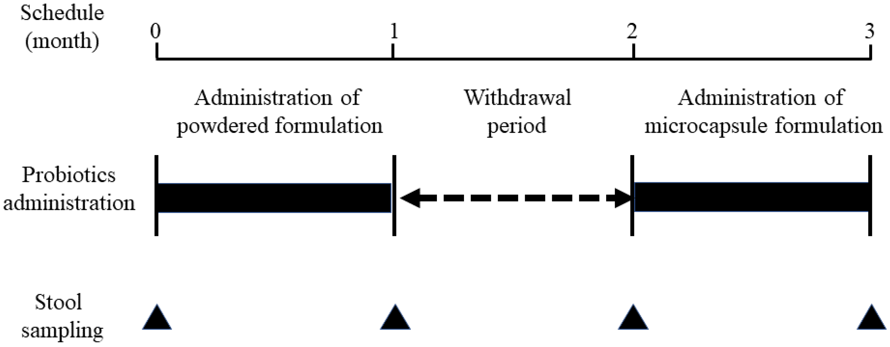
Figure 2 illustrates the study design. Participants were first given one packet of B. breve M-16V powder per day for 1 month. After a 1-month withdrawal period, the microcapsule formulation was administered orally for 1 month (one packet per day). To assess study participants’ adherence to the formulation, a questionnaire regarding the powder or microcapsule formulation was administered to their parents. At the same time, information on the frequency of defecation and changes in stool characteristics was collected. No restrictions were placed on the intake of probiotics other than bifidobacteria. If any probiotics other than bifidobacteria were ingested, we asked the participants to indicate them on the questionnaire form.
To analyze the gut microbiota, spontaneously excreted stool samples of the study participants were collected 4 times before and after 1 month of powder or microcapsule consumption, as shown in Figure 2, and stored at −80 °C until DNA extraction.

2.4. Analysis of Gut Microbiota

For analysis of the gut microbiota, fecal bacterial 16S rRNA sequencing was performed to elucidate the changes in the taxonomic composition and alpha diversity of the study participants for each interventional period. In addition, the relative abundance of B. longum and B. breve. Additionally, quantitative analysis of the bacterial count of B. breve was performed by quantitative real-time polymerase chain reaction (RT-PCR).

2.5. Determination of the 16S rRNA Sequence

DNA extraction was performed using a NucleoSpin DNA Stool Kit (MACHEREY-NAGEL, Düren, Germany) within 1 week after stool collection. Seven hypervariable regions of the 16S rRNA regions of the extracted DNA, excluding v1 and v5, were amplified using a 16S Metagenome Kit (Thermo Fisher Scientific, Waltham, MA, USA). After purification, libraries were constructed using an Ion Plus Fragment Library Kit (Thermo Fisher Scientific) and Ion Xpress Barcode Adapters Kit (Thermo Fisher Scientific). The barcode libraries were quantified using an Agilent Bioanalyzer 2000 (Agilent, Santa Clara, CA, USA). The Ion Chef Instrument and Kit were used to obtain specific concentrations of emulsion PCR and templates. Sequence analysis was performed using an Ion GeneStudio S5 sequencer (Thermo Fisher Scientific) and Ion 318 chip (Thermo Fisher Scientific). Sequence data were analyzed using Metagenomics 16S w1.1 v5.16 workflow (Thermo Fisher Scientific) and Ion Reporter software.
MicroSEQ 16S Reference Library v2013.1, GreenGenes v13.5, Ion Reporter software, and Metagenomics 16S w1.1 v5.16 workflow (Thermo Fisher Scientific) were used to cluster the raw BAM files into operational classification units with 97% identity, and the data were analyzed using the default settings (read length filter, 150; minimum aligned cover age, 90.0; read abundance filter, 10; slash ID reporting rate, 0.2). Ion Reporter’s Quantitative Insights Into Microbial Ecology open-source bioinformatics pipeline was used for alpha and beta diversity analysis and visualization. Only data with a total read count of at least 5000 were used in the analysis.

2.6. Quantitative Measurement of B. breve in Stools Using PCR

RT-PCR targeting the groEL gene of B. breve was used to quantify B. breve in human stool [22]. Quantitative RT-PCR was contracted to PGL LLC (Aichi, Japan). The primer sequences used in RT-PCR were as follows: groEL gene of B. breve forward, GCTCGTCGTTGCCGCCGCCAAGGACGTT; groEL gene of B. breve reverse, ACAGAATGTACGGGATCCTCGAGCACG.
The PCR instrument was an Applied Biosystems 7500 Fast Real-Time PCR System (Thermo Fisher Scientific), and KOD SYBR® qPCR Mix was used to generate reactions. The temperature program for PCR for groEL gene of B. breve was as follows: 98 °C for 2 min, followed by 40 cycles at 98 °C for 10 s, 60 °C for 10 s, and 68 °C for 34 s. The number of B. breve groEL genes in stool samples was calculated by plotting CT values obtained via 10-fold serial dilutions of quantitative PCR standards as a linear function of the base 10 logarithm of the known concentration to create a standard curve.

2.7. Statistical Analysis

Continuous variables were expressed as the median and IQR. The Wilcoxon signed-rank test was used for comparisons between the two corresponding groups. Statistical significance was set at p < 0.05. Statistical analysis was performed using BellCurve for Excel (version 3.21; Social Research and Information Center, Tokyo, Japan).

3. Results

3.1. Characteristics of the Study Participants

Table 1 summarizes the participants’ adherence to the probiotic formulation, the frequency of defecation, and changes in stool characteristics associated with the administration of the probiotic formulation.
Study participants’ adherence to the microcapsule formulation was good, with a median rate of 100% (IQR = 96.6–100). The stool frequency after 1 month of oral administration of the powder or microcapsule formulation was checked. There was no statistically significant difference in the median stool frequency per week between before and after powder formulation administration (5.0 times/week [IQR] 4.5–6.0] vs. 5.75 times/week [IQR = 5.0–6.0], p = 0.35). Similarly, the stool frequency did not differ between before and after oral administration of the microcapsule formulation (5.75 times/week [IQR = 5.0–6.0] vs. 3.25 times/week [IQR = 2.75–5.75], p = 0.18).
Differences in the Bristol Stool Form Scale (BSFS) score [23] between before and after oral administration of the powder or microcapsule formulation were evaluated. The results revealed no statistically significant difference, with a median BSFS score of 4.0 (IQR = 3.0–4.0) after 1 month of powder administration compared with 4.0 (IQR = 3.0–4.0) before oral administration (p = 0.56). Additionally, the median BSFS score after microcapsule formulation administration was 4.0 (IQR = 4.0–4.0) versus 4.0 (IQR = 3.0–4.0) before administration (p = 0.32).

3.2. Results of Gut Microbiota Analysis

3.2.1. Taxonomic Composition

The composition of the gut microbiotas of the study participants for each intervention period is shown in Figure 3. At the order level, the percentage of Bifidobacteriales did not change before or after oral administration of Bifidobacterium-containing powders or microcapsules.

3.2.2. Alpha Diversity

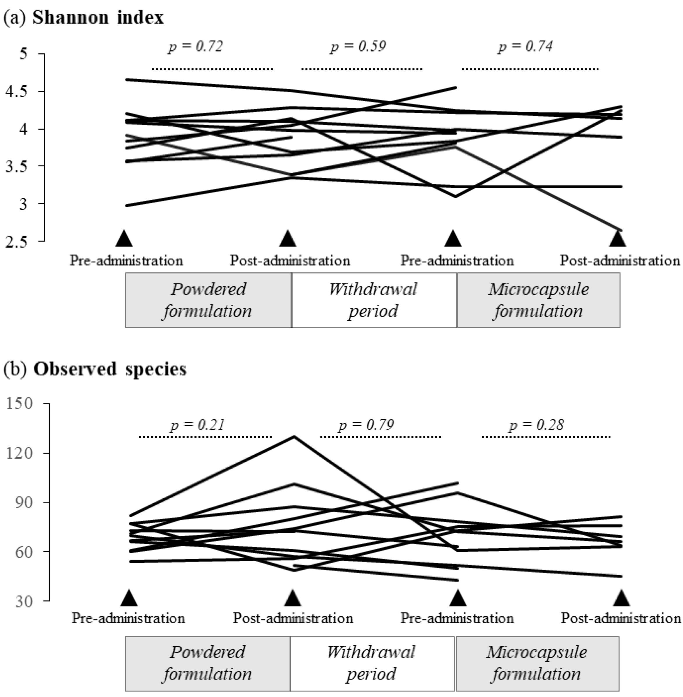
Figure 4a presents the results of the Shannon index and observed species of gut microbiota. The Shannon index was 3.98 (median; IQR = 3.67–4.12) after powder formulation administration, compared with 3.91 (median; IQR = 3.66–4.11) before administration (p = 0.72). There was no significant difference in the Shannon index during the one-month withdrawal period (p = 0.59). Similarly, there was no significant difference in this index before (median 3.83; IQR = 3.49–4.11) and after microcapsule formulation administration (median 4.14; IQR = 3.56–4.21), p = 0.74). Meanwhile, Figure 4b shows the number of observed species of gut microbiota. In total, 73 species (median; IQR = 59–83.5) were observed after powder formulation administration, compared with 70 species (median; IQR = 63–75) before treatment (p = 0.21). There was no significant difference in the change in the observed species during the one-month withdrawal period (p = 0.79). Meanwhile, 73 species (median; IQR = 66.5–76.5) were detected before microcapsule formulation administration versus 66 species (median; IQR = 63.5–72.5) after administration (p = 0.28).

3.2.3. Relative Abundance of B. longum and B. breve in the Gut Microbiota

The relative abundance of B. longum and B. breve was determined by 16S rRNA sequencing before and after administration of the formulations (Figure 5). As shown in Figure 5a, the relative abundance of B. longum was not significantly increased after 1 month of powder administration (median 1.51% [IQR = 1.07–3.25]) compared with the value before administration (median 2.41% [IQR = 1.16–3.93%], p = 0.86). There was no statistically significant difference in the relative amounts before and after the one-month withdrawal period (p = 0.40). Furthermore, the abundance of B. longum did not differ before and after microcapsule formulation administration (median 2.57% [IQR = 0.99–3.29%] versus median 1.85% [IQR = 0.77–2.23, p = 0.40).
Meanwhile, as shown in Figure 5b, the abundance of B. breve was 0.053% (median; IQR = 0.0–0.07) after 1 month of powder formulation administration, compared with 0.011% (median; IQR = 0.0–0.03) before treatment (p = 0.40). There was no statistically significant difference in relative abundance before and after the one-month withdrawal period (p = 0.59). Conversely, the median relative abundance of B. breve tended to increase after microcapsule formulation administration (0.028% [IQR = 0–0.050%] versus 0.093% [IQR = 0–0.167]), although the difference did not reach significance (p = 0.080).

3.2.4. Quantification of B. breve

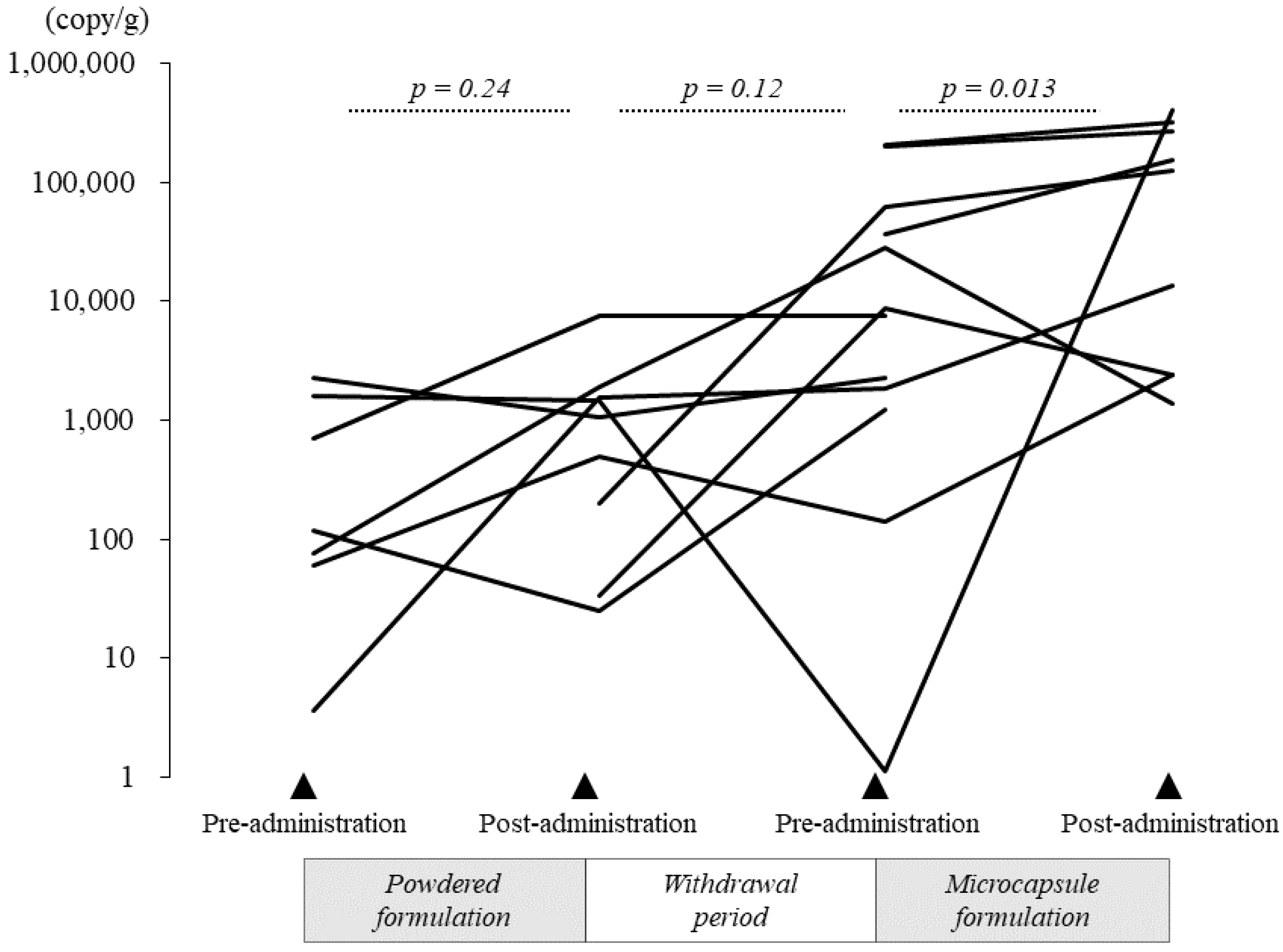
As only B. breve tended to increase in relative abundance after microcapsule formulation administration, as determined by 16S rRNA sequencing (p = 0.080), further quantitative analysis of B. breve using RT-PCR was performed. The results are presented in Figure 6 (bacterial counts were calculated per gram of stool). After administration of the B. breve powder formulation, the median bacterial count slightly increased to 1479.57 copies/g (median; IQR = 769.6–1715.4 copies/g), compared with 116.5 copies/g (median; IQR = 68.1–1163.7 copies/g) before administration, but the difference did not reach statistical significance (p = 0.24). The change in the median bacterial count of B. breve before and after the one-month withdrawal period was not statistically significant (p = 0.12). By contrast, the median bacterial count of B. breve was significantly higher after 1 month of microcapsule formulation administration (28,069 copies/g [IQR = 1866.3–62,721.2] before administration versus 153,576.4 copies/g [IQR = 13,433.4–323,813.1] after administration, p = 0.013).

3.2.5. Comparison of the Colon Delivery Efficiency of the Powder and Microcapsule Formulations of B. breve

The efficiency at which B. breve reached the large intestine was compared between the formulations using the following formula: Number of PCR copies (copies/g) × 1000 g (predicted daily defecation volume)/1 billion (calculated bacterial count of daily oral intake of B. breve by powder formulation) or 100 million (calculated bacterial count of daily oral intake of B. breve by microcapsule formulation).
Figure 7 presents the results of the colon delivery efficiency of B. breve. The microcapsule formulation of B. breve was delivered to the large intestine with significantly better efficiency than the powder formulation (1.54 [IQR = 0.079–2.98] versus 0.0015 [IQR = 0.00077–0.0017], respectively, p = 0.018).

4. Discussion

A previous study confirmed the viability of powdered and capsule probiotic formulations containing bifidobacteria in a validated dynamic in vitro model of the stomach and small intestine, simulating a human adult. The results showed that the survival rate of the capsule formulation was approximately twice that of the powder formulation [24]. Therefore, the administration of a capsule formulation of probiotics is desirable; however, the large size of the capsule formulation makes it difficult for children to swallow. Therefore, we have developed microcapsules containing bifidobacteria of size (2 mm) that can be taken internally, even by toddlers. As shown in Figure 1a, acid-resistant microcapsules are almost spherical in shape with a diameter of 2 mm, differing from the hard or soft capsules that are widely used in general medicine and food products. This structure does not allow external liquids or other substances to penetrate through the membranes. These microcapsules were prepared by the “drop-in-liquid method,” which utilizes interfacial tension to impart properties such as acid resistance and enteric solubility to the membrane. This design enables bifidobacteria, which are vulnerable to stomach acid, to be successfully delivered to the large intestine. After a microcapsule is orally consumed, it passes through the stomach, where the pH is low, and by the time it reaches the intestine, the pH changes to near neutral, which causes the pH-responsive outer skin membrane to dissolve. The microcapsule is then collapsed within the intestinal tract via the physical stimulation of intestinal movement, and living bifidobacteria are delivered [21]. The microcapsules in the present study contained B. breve M-16V and B. longum BB536. B. breve M-16V is a non-motile, non-spore-forming, rod-shaped, anaerobic Gram-positive bacterium discovered in 1963 in the intestines of infants [25]. B. breve M-16V is known to reach the human gastrointestinal tract and has strong adhesion activity [26]. Comprehensive safety evaluations, including functional, genomic, and in vivo analyses, have shown that B. breve M-16V is a non-pathogenic, non-toxigenic, non-hemolytic, non-antibiotic-resistant probiotic bacterium that contains no plasmids and shows no harmful metabolic activity [26,27,28]. The other component, B. longum BB536, was discovered in the intestines of healthy breastfed infants in 1969 [29]. B. longum BB536 has been used as a functional food ingredient in a variety of products, including dairy beverages, yogurt, powdered milk, and nutritional supplements, and has been sold in more than 30 countries for over 40 years [29]. B. longum BB536 is a Gram-positive, anaerobic, catalase-negative bacillus with an irregular morphology and excellent stability during storage. It is known to be highly viable in probiotic foods until consumption, and it does not contain plasmids and exhibits no harmful metabolic activity [26,27]. An in vivo study was also conducted in which high doses of BB536 were orally administered to healthy 4-week-old mice for 7 days, and it was found that BB536 did not migrate to the blood, liver, spleen, kidney, and mesenteric lymph nodes and did not induce damage to the intestinal surface [28].
In this study, we evaluated whether the microcapsule formulation reached the colon more efficiently than the powder formulation by quantitative PCR using specific primers for B. breve. Significantly more B. breve specimens were detected in the stool after microcapsule formulation administration than after powder administration. In addition, the microcapsule formulation reached the intestinal tract more efficiently than the powder formulation, as was calculated from the content of B. breve present in the stool.
The microcapsule formulation contained B. longum in addition to B. breve. Despite the high total bacterial content of B. longum, there was no increase in B. longum after administration. The reasons for these findings were as follows: 1) The results of the present study showed that B. breve and B. longum have absolutely different proportions in the gut microbiota, with B. longum being about 100 times more abundant. Therefore, it is possible that the amount of B. longum in the microcapsule could not be detected in the stool as a significant difference even if the amount of B. longum was 10 times higher than that of B. breve; 2) It has been reported that Gram-positive bacteria must be sufficiently cell-disrupted during the DNA extraction process; otherwise, the extraction sensitivity may be reduced [30]. Both B. longum and B. breve may have low detection sensitivity because they are Gram-positive bacteria, and this may be the reason why no significant change was observed in the relative abundance of B. longum and B. breve by16S-rRNA gene analysis using NGS.
This study had four limitations. First, the efficiency at which viable bacteria reached the intestine was not calculated by culturing bifidobacteria extracted from the stool. However, we believed that we could evaluate the efficiency using quantitative PCR and bacterial 16S rRNA gene analysis. Second, the study participants displayed no changes in stool quality or improvement in constipation following bifidobacterium administration. The study participants did not include children who originally had constipation, and thus, it is necessary to confirm the efficacy of the oral microcapsule formulation in children with constipation in the future. Third, the number of participants in this study was small. Because this was an interventional study in healthy children, the number of participants was small as a result of ethical concerns. However, we were able to demonstrate the efficacy of microcapsules containing bifidobacteria, and we are considering conducting a larger-scale trial in the future. Fourth, the present study was designed based on the report by Saxelin et al. [31] that proposed that the effect of the administered probiotic would disappear from the subject’s colon after a one-month withdrawal period following the end of oral administration. However, as shown in Figure 6, a statistically significant decrease in B. breve was not observed during the withdrawal period, so the “priming effect” cannot be ruled out. Therefore, based on the results of this pilot study, we plan to incorporate a parallel design in our next large-scale intervention study.
In conclusion, we confirmed that the developed acid-resistant microcapsule formulation can efficiently deliver B. breve to the intestine. B. breve is classified as an infantile form of HRB, and acid-resistant microcapsules that can efficiently deliver it to the intestine as a probiotic formulation for children have tremendous advantages. In addition, considering the size of the microcapsule, we believe it will be easy to add the microcapsule to food products such as yogurt in the future.

Author Contributions

Conceptualization, K.K.; Data curation, M.M.; Formal analysis, S.T. and S.A.; Investigation, S.T., S.A., and Y.A.; Methodology, S.T. and S.A.; Project administration, M.M., S.T., and S.A.; Resources, S.T., Y.Y., H.K., M.K., and K.K.; Visualization, M.M. and S.T.; Writing—original draft, M.M.; Writing—review and editing, S.T., S.A., Y.A., and K.K. All authors have read and agreed to the published version of the manuscript.

Funding

This research received no external funding.

Institutional Review Board Statement

The study was conducted in accordance with the guidelines of the Declaration of Helsinki, and the protocol was approved by the Ethics Committee of Kansai Medical University (No. 2018006, 9 July 2018).

Informed Consent Statement

Informed consent was obtained from all subjects involved in the study.

Data Availability Statement

The datasets generated and analyzed during the current study are available from the corresponding authors upon reasonable request.

Acknowledgments

We would like to thank Masaki Hashiyada for his help with bacterial 16S rRNA gene analysis. We also thank Joe Barber Jr. from Edanz (https://jp.edanz.com/ac (accessed on 4 November 2022)) for editing a draft of this manuscript.

Conflicts of Interest

The authors declare no conflict of interest.

References

  1. Sender, R.; Fuchs, S.; Milo, R. Revised Estimates for the Number of Human and Bacteria Cells in the Body. PLoS Biol. 2016, 14, e1002533. [Google Scholar] [CrossRef] [Green Version]
  2. Odamaki, T.; Kato, K.; Sugahara, H.; Hashikura, N.; Takahashi, S.; Xiao, J.Z.; Abe, F.; Osawa, R. Age-related changes in gut microbiota composition from newborn to centenarian: A cross-sectional study. BMC Microbiol. 2016, 16, 90. [Google Scholar] [CrossRef] [PubMed] [Green Version]
  3. Young, R.B.; Marcelino, V.R.; Chonwerawong, M.; Gulliver, E.L.; Forster, S.C. Key Technologies for Progressing Discovery of Microbiome-Based Medicines. Front. Microbiol. 2021, 12, 685935. [Google Scholar] [CrossRef] [PubMed]
  4. Yatsunenko, T.; Rey, F.E.; Manary, M.J.; Trehan, I.; Dominguez-Bello, M.G.; Contreras, M.; Magris, M.; Hidalgo, G.; Baldassano, R.N.; Anokhin, A.P.; et al. Human gut microbiome viewed across age and geography. Nature 2012, 486, 222–227. [Google Scholar] [CrossRef] [Green Version]
  5. Akagawa, S.; Akagawa, Y.; Yamanouchi, S.; Kimata, T.; Tsuji, S.; Kaneko, K. Development of the gut microbiota and dysbiosis in children. Biosci. Microbiota Food Health 2021, 40, 12–18. [Google Scholar] [CrossRef] [PubMed]
  6. Malard, F.; Dore, J.; Gaugler, B.; Mohty, M. Introduction to host microbiome symbiosis in health and disease. Mucosal. Immunol. 2021, 14, 547–554. [Google Scholar] [CrossRef] [PubMed]
  7. Schwarzenberg, S.J.; Georgieff, M.K.; Committee On, N. Advocacy for Improving Nutrition in the First 1000 Days to Support Childhood Development and Adult Health. Pediatrics 2018, 141, e20173716. [Google Scholar] [CrossRef] [Green Version]
  8. Hill, C.; Guarner, F.; Reid, G.; Gibson, G.R.; Merenstein, D.J.; Pot, B.; Morelli, L.; Canani, R.B.; Flint, H.J.; Salminen, S.; et al. Expert consensus document. The International Scientific Association for Probiotics and Prebiotics consensus statement on the scope and appropriate use of the term probiotic. Nat. Rev. Gastroenterol. Hepatol. 2014, 11, 506–514. [Google Scholar] [CrossRef] [Green Version]
  9. Marras, L.; Caputo, M.; Bisicchia, S.; Soato, M.; Bertolino, G.; Vaccaro, S.; Inturri, R. The Role of Bifidobacteria in Predictive and Preventive Medicine: A Focus on Eczema and Hypercholesterolemia. Microorganisms 2021, 9, 836. [Google Scholar] [CrossRef] [PubMed]
  10. Hidalgo-Cantabrana, C.; Delgado, S.; Ruiz, L.; Ruas-Madiedo, P.; Sanchez, B.; Margolles, A. Bifidobacteria and Their Health-Promoting Effects. Microbiol. Spectr. 2017, 5, 73–98. [Google Scholar] [CrossRef]
  11. Wong, C.B.; Sugahara, H.; Odamaki, T.; Xiao, J.Z. Different physiological properties of human-residential and non-human-residential bifidobacteria in human health. Benef. Microbes 2018, 9, 111–122. [Google Scholar] [CrossRef] [PubMed]
  12. Inoue, Y.; Iwabuchi, N.; Xiao, J.Z.; Yaeshima, T.; Iwatsuki, K. Suppressive effects of bifidobacterium breve strain M-16V on T-helper type 2 immune responses in a murine model. Biol. Pharm. Bull. 2009, 32, 760–763. [Google Scholar] [CrossRef] [Green Version]
  13. Fukuda, S.; Toh, H.; Hase, K.; Oshima, K.; Nakanishi, Y.; Yoshimura, K.; Tobe, T.; Clarke, J.M.; Topping, D.L.; Suzuki, T.; et al. Bifidobacteria can protect from enteropathogenic infection through production of acetate. Nature 2011, 469, 543–547. [Google Scholar] [CrossRef] [PubMed]
  14. Lin, H.C.; Su, B.H.; Chen, A.C.; Lin, T.W.; Tsai, C.H.; Yeh, T.F.; Oh, W. Oral probiotics reduce the incidence and severity of necrotizing enterocolitis in very low birth weight infants. Pediatrics 2005, 115, 1–4. [Google Scholar] [CrossRef]
  15. Lin, H.C.; Hsu, C.H.; Chen, H.L.; Chung, M.Y.; Hsu, J.F.; Lien, R.I.; Tsao, L.Y.; Chen, C.H.; Su, B.H. Oral probiotics prevent necrotizing enterocolitis in very low birth weight preterm infants: A multicenter, randomized, controlled trial. Pediatrics 2008, 122, 693–700. [Google Scholar] [CrossRef] [Green Version]
  16. Sistek, D.; Kelly, R.; Wickens, K.; Stanley, T.; Fitzharris, P.; Crane, J. Is the effect of probiotics on atopic dermatitis confined to food sensitized children? Clin. Exp. Allergy 2006, 36, 629–633. [Google Scholar] [CrossRef] [PubMed]
  17. Gerasimov, S.V.; Vasjuta, V.V.; Myhovych, O.O.; Bondarchuk, L.I. Probiotic supplement reduces atopic dermatitis in preschool children: A randomized, double-blind, placebo-controlled, clinical trial. Am. J. Clin. Dermatol. 2010, 11, 351–361. [Google Scholar] [CrossRef]
  18. van der Aa, L.B.; Heymans, H.S.; van Aalderen, W.M.; Sillevis Smitt, J.H.; Knol, J.; Ben Amor, K.; Goossens, D.A.; Sprikkelman, A.B.; Synbad Study, G. Effect of a new synbiotic mixture on atopic dermatitis in infants: A randomized-controlled trial. Clin. Exp. Allergy 2010, 40, 795–804. [Google Scholar] [CrossRef] [PubMed]
  19. Yesilova, Y.; Calka, O.; Akdeniz, N.; Berktas, M. Effect of probiotics on the treatment of children with atopic dermatitis. Ann. Dermatol. 2012, 24, 189–193. [Google Scholar] [CrossRef] [PubMed] [Green Version]
  20. Dianawati, D.; Mishra, V.; Shah, N.P. Viability, Acid and Bile Tolerance of Spray Dried Probiotic Bacteria and Some Commercial Probiotic Supplement Products Kept at Room Temperature. J. Food Sci. 2016, 81, M1472–M1479. [Google Scholar] [CrossRef]
  21. Kohno, M.; Tanaka-Azuma, Y.; Kamaguchi, R.; Tagawa, D. Application of enteric seamless capsules containing Bifidobacterium longum to functional foods. Folia. Pharmacol. Jpn. (Nihon Yakurigaku Zasshi) 2016, 148, 310–314. (In Japanese) [Google Scholar] [CrossRef] [PubMed] [Green Version]
  22. Junick, J.; Blaut, M. Quantification of human fecal bifidobacterium species by use of quantitative real-time PCR analysis targeting the groEL gene. Appl. Environ. Microbiol. 2012, 78, 2613–2622. [Google Scholar] [CrossRef] [PubMed] [Green Version]
  23. Lewis, S.J.; Heaton, K.W. Stool form scale as a useful guide to intestinal transit time. Scand. J. Gastroenterol. 1997, 32, 920–924. [Google Scholar] [CrossRef] [PubMed]
  24. Venema, K.; Verhoeven, J.; Beckman, C.; Keller, D. Survival of a probiotic-containing product using capsule-within-capsule technology in an in vitro model of the stomach and small intestine (TIM-1). Benef. Microbes 2020, 11, 403–409. [Google Scholar] [CrossRef] [PubMed]
  25. Wong, C.B.; Iwabuchi, N.; Xiao, J.Z. Exploring the Science behind Bifidobacterium breve M-16V in Infant Health. Nutrients 2019, 11, 1724. [Google Scholar] [CrossRef] [Green Version]
  26. Toscano, M.; De Vecchi, E.; Gabrieli, A.; Zuccotti, G.V.; Drago, L. Probiotic characteristics and in vitro compatibility of a combination of Bifidobacterium breve M-16 V, Bifidobacterium longum subsp. infantis M-63 and Bifidobacterium longum subsp. longum BB536. Ann. Microbiol. 2014, 65, 1079–1086. [Google Scholar] [CrossRef]
  27. Xiao, J.Z.; Takahashi, S.; Odamaki, T.; Yaeshima, T.; Iwatsuki, K. Antibiotic susceptibility of bifidobacterial strains distributed in the Japanese market. Biosci. Biotechnol. Biochem. 2010, 74, 336–342. [Google Scholar] [CrossRef]
  28. Abe, F.; Muto, M.; Yaeshima, T.; Iwatsuki, K.; Aihara, H.; Ohashi, Y.; Fujisawa, T. Safety evaluation of probiotic bifidobacteria by analysis of mucin degradation activity and translocation ability. Anaerobe 2010, 16, 131–136. [Google Scholar] [CrossRef]
  29. Wong, C.B.; Odamaki, T.; Xiao, J.-z. Beneficial effects of Bifidobacterium longum subsp. longum BB536 on human health: Modulation of gut microbiome as the principal action. J. Funct. Foods 2019, 54, 506–519. [Google Scholar] [CrossRef]
  30. Sjoberg, F.; Nookaew, I.; Yazdanshenas, S.; Gio-Batta, M.; Adlerberth, I.; Wold, A.E. Are all faecal bacteria detected with equal efficiency? A study using next-generation sequencing and quantitative culture of infants’ faecal samples. J. Microbiol. Methods 2020, 177, 106018. [Google Scholar] [CrossRef]
  31. Saxelin, M.; Lassig, A.; Karjalainen, H.; Tynkkynen, S.; Surakka, A.; Vapaatalo, H.; Jarvenpaa, S.; Korpela, R.; Mutanen, M.; Hatakka, K. Persistence of probiotic strains in the gastrointestinal tract when administered as capsules, yoghurt, or cheese. Int. J. Food Microbiol. 2010, 144, 293–300. [Google Scholar] [CrossRef] [PubMed]
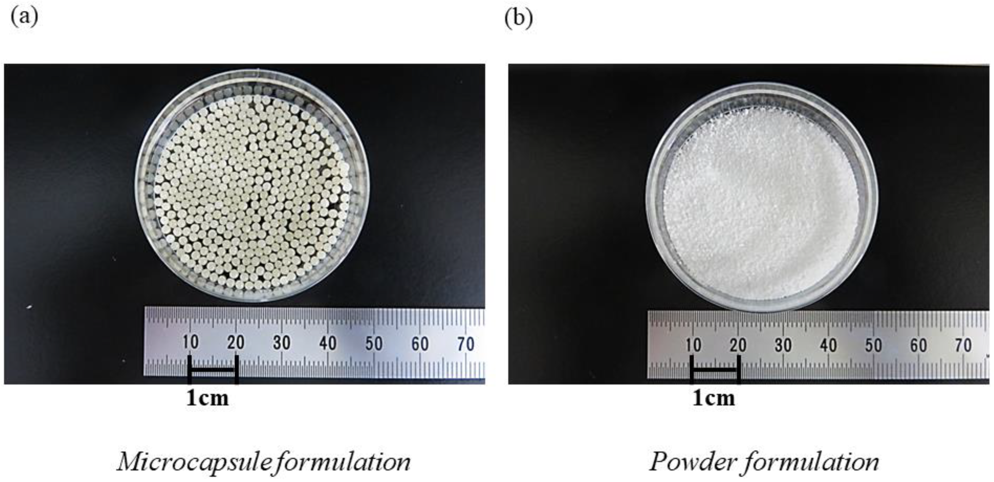
Figure 1. Appearance of formulation of used probiotics. The appearance of the bifidobacteria-containing formulations used in this study are shown: (a) the microcapsule formulation and (b) the powder formulation. Each microcapsule formulation was approximately 2 mm in size.
Nutrients 14 04829 g001
Figure 2. Study design Participants first received one packet of B. breve M-16V powder for one month; after a one-month rest period, a microcapsule formulation was administered orally (one packet per day) for one month. A powder formulation contained 1 billion bacteria per packet, while a microcapsule formulation contained B. breve M-16V and B. longum BB536 at a ratio of 1:9 and a total of 1 billion bacteria per packet.
Nutrients 14 04829 g002
Figure 3. Changes in the composition of the gut microbiota after oral administration of bifidobacterial formulations at the order level. Bars represent the averages of individual data. The percentage of Bifidobacteriales (red bars) did not show any statistically significant change before or after administration of the Bifidobacterium-containing powder and microcapsule formulations.
Nutrients 14 04829 g003
Figure 4. Changes in the Shannon index and observed species of the gut microbiota after administration of the powder or microcapsule formulation containing bifidobacteria. (a) There was no statistically significant difference in the Shannon index representing alpha diversity before or after administration of the powder, the withdrawal period, or administration of the acid-resistant microcapsule formulation containing bifidobacteria. (b) The observed species also displayed no statistically significant difference in abundance after administration.
Nutrients 14 04829 g004
Figure 5. Changes in the relative abundance of B. longum or B. breve in the gut microbiota after the administration of the powder or acid-resistant microcapsule formulation containing bifidobacteria. (a) The relative abundance of B. longum in the gut microbiota, calculated from the results of 16S rRNA gene analysis, was not significantly changed after administration of the powder or acid-resistant microcapsule formulation or before or after the withdrawal period. The relative abundance of B. breve also did not change significantly before or after administration of the powder formulation or before or after the withdrawal period. (b) By contrast, the relative abundance of B. breve tended to increase after administration of the microcapsule formulation, although this difference was not statistically significant compared with the relative abundance before administration.
Nutrients 14 04829 g005
Figure 6. Quantitative changes in B. breve after administration of the powder or microcapsule formulation containing bifidobacteria. The graph presents the quantitative changes in the bacterial count of B. breve before and after administration of the powder formulation, before and after a one-month withdrawal period, and before and after administration of the acid-resistant microcapsule formulation by quantitative PCR. B. breve did not show a statistically significant increase after administration of the powder formulation. However, after administration of the acid-resistant microcapsule formulation, the bacterial count of B. breve was significantly higher than the bacterial count before administration.
Nutrients 14 04829 g006
Figure 7. Differences in the efficiency at which B. breve-containing powder or acid-resistant microcapsule formulations reached the large intestine after administration. The graphs present the efficiency at which B. breve reached the large intestine calculated by quantitative PCR. Compared with the effects of the powder formulation, the acid-resistant microcapsule formulation significantly improved the efficiency at which the bacteria reached the large intestine after administration. Each symbol, such as circle, square, triangle, diamond, bar, and cross, denotes the efficiency data from the individual participants. A.U. (arbitrary unit): For the calculation of the efficiency, the following formula is applied: Number of PCR copies (copies/g) × 1000 g (predicted daily defecation volume)/1 billion (B. breve content in powder formulation) or 100 million (B. breve content in microcapsule formulation).
Nutrients 14 04829 g007
Table 1. Details of the study participants.
Powder FormulationMicrocapsule
Formulation
p-Value
Adherence (%)100 (92.9–100)100 (96.6–100)0.49
Frequency of defecation
per week (pre/post)
5.0 (4.5–6.0)/5.75 (5.0–6.0)5.75 (5.0–6.0)/
3.25 (2.75–5.75)
0.35/0.18
BSFS score (pre/post)4.0 (3.0–4.0)/4.0 (3.0–4.0)4.0 (3.0–4.0)/4.0 (4.0–4.0)0.56/0.32
BSFS, Bristol Stool Form Scale; Statistical comparison between before and after administration.
Publisher’s Note: MDPI stays neutral with regard to jurisdictional claims in published maps and institutional affiliations.