
Vitamin D and Visceral Obesity in Humans: What Should Clinicians Know?


Abstract

1. Introduction
2. Methodology and Literature Search
3. Vitamin D Metabolism
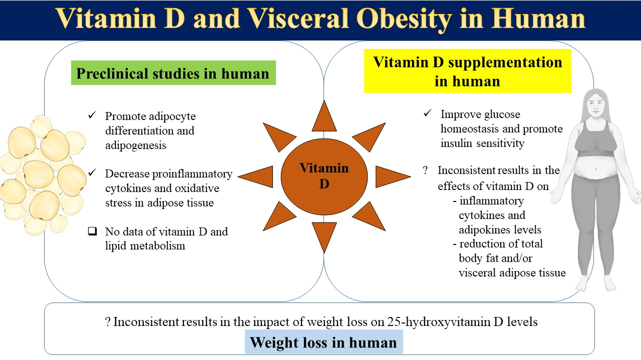
4. Association between Vitamin D and Adipose Tissue
5. Vitamin D Action in Human Adipose Tissue
5.1. Role of Vitamin D in Adipocyte Differentiation and Adipogenesis
5.2. Role of Vitamin D in Energy Homeostasis
5.3. Role of Vitamin D in Inflammation
6. Vitamin D and Obesity
6.1. Effect of Medical and Surgical Weight Loss on Vitamin D Status
6.2. Effect of Vitamin D Supplementation on Weight Reduction and Visceral Fat Loss
7. Discussion
8. Conclusions
Author Contributions
Funding
Institutional Review Board Statement
Informed Consent Statement
Conflicts of Interest
References
- Bouillon, R.; Marcocci, C.; Carmeliet, G.; Bikle, D.; White, J.H.; Dawson-Hughes, B.; Lips, P.; Munns, C.F.; Lazaretti-Castro, M.; Giustina, A.; et al. Skeletal and Extraskeletal Actions of Vitamin D: Current Evidence and Outstanding Questions. Endocr. Rev. 2019, 40, 1109–1151. [Google Scholar] [CrossRef]
- Holick, M.F. Sunlight and vitamin D for bone health and prevention of autoimmune diseases, cancers, and cardiovascular disease. Am. J. Clin. Nutr. 2004, 80, 1678S–1688S. [Google Scholar] [CrossRef] [PubMed]
- Holick, M.F. Vitamin D deficiency. N. Engl. J. Med. 2007, 357, 266–281. [Google Scholar] [CrossRef] [PubMed]
- Despres, J.P.; Lemieux, I. Abdominal obesity and metabolic syndrome. Nature 2006, 444, 881–887. [Google Scholar] [CrossRef] [PubMed]
- Ostman, J.; Arner, P.; Engfeldt, P.; Kager, L. Regional differences in the control of lipolysis in human adipose tissue. Metabolism 1979, 28, 1198–1205. [Google Scholar] [CrossRef]
- Despres, J.P.; Carpentier, A.C.; Tchernof, A.; Neeland, I.J.; Poirier, P. Management of Obesity in Cardiovascular Practice: JACC Focus Seminar. J. Am. Coll. Cardiol. 2021, 78, 513–531. [Google Scholar] [CrossRef]
- Chartrand, D.J.; Murphy-Despres, A.; Almeras, N.; Lemieux, I.; Larose, E.; Despres, J.P. Overweight, Obesity, and CVD Risk: A Focus on Visceral/Ectopic Fat. Curr. Atheroscler. Rep. 2022, 24, 185–195. [Google Scholar] [CrossRef]
- Neeland, I.J.; Ross, R.; Despres, J.P.; Matsuzawa, Y.; Yamashita, S.; Shai, I.; Seidell, J.; Magni, P.; Santos, R.D.; Arsenault, B.; et al. Visceral and ectopic fat, atherosclerosis, and cardiometabolic disease: A position statement. Lancet Diabetes Endocrinol. 2019, 7, 715–725. [Google Scholar] [CrossRef]
- Camacho, P.M.; Petak, S.M.; Binkley, N.; Diab, D.L.; Eldeiry, L.S.; Farooki, A.; Harris, S.T.; Hurley, D.L.; Kelly, J.; Lewiecki, E.M.; et al. American Association of Clinical Endocrinologists/American College of Endocrinology Clinical Practice Guidelines for the Diagnosis and Treatment of Postmenopausal Osteoporosis-2020 Update Executive Summary. Endocr. Pract. 2020, 26, 564–570. [Google Scholar] [CrossRef]
- Cosman, F.; de Beur, S.J.; LeBoff, M.S.; Lewiecki, E.M.; Tanner, B.; Randall, S.; Lindsay, R.; National Osteoporosis, F. Clinician’s Guide to Prevention and Treatment of Osteoporosis. Osteoporos Int. 2014, 25, 2359–2381. [Google Scholar] [CrossRef]
- Eastell, R.; Rosen, C.J.; Black, D.M.; Cheung, A.M.; Murad, M.H.; Shoback, D. Pharmacological Management of Osteoporosis in Postmenopausal Women: An Endocrine Society*.Clinical Practice Guideline. J. Clin. Endocrinol. Metab. 2019, 104, 1595–1622. [Google Scholar] [CrossRef] [PubMed]
- Holick, M.F.; Binkley, N.C.; Bischoff-Ferrari, H.A.; Gordon, C.M.; Hanley, D.A.; Heaney, R.P.; Murad, M.H.; Weaver, C.M.; Endocrine, S. Evaluation, treatment, and prevention of vitamin D deficiency: An Endocrine Society clinical practice guideline. J. Clin. Endocrinol. Metab. 2011, 96, 1911–1930. [Google Scholar] [CrossRef] [PubMed]
- Society, T.R.O. Vitamin D and Bone Health: A Practical Clinical Guideline for Patient Management. December 2018. Available online: theros.org.uk/clinical-publications-and-resources (accessed on 20 March 2022).
- Nimitphong, H.; Park, E.; Lee, M.J. Vitamin D regulation of adipogenesis and adipose tissue functions. Nutr. Res. Pract. 2020, 14, 553–567. [Google Scholar] [CrossRef] [PubMed]
- Silvagno, F.; Pescarmona, G. Spotlight on vitamin D receptor, lipid metabolism and mitochondria: Some preliminary emerging issues. Mol. Cell Endocrinol. 2017, 450, 24–31. [Google Scholar] [CrossRef]
- Bikle, D.D.; Gee, E.; Halloran, B.; Kowalski, M.A.; Ryzen, E.; Haddad, J.G. Assessment of the free fraction of 25-hydroxyvitamin D in serum and its regulation by albumin and the vitamin D-binding protein. J. Clin. Endocrinol. Metab. 1986, 63, 954–959. [Google Scholar] [CrossRef]
- Chun, R.F.; Peercy, B.E.; Orwoll, E.S.; Nielson, C.M.; Adams, J.S.; Hewison, M. Vitamin D and DBP: The free hormone hypothesis revisited. J. Steroid Biochem. Mol. Biol. 2014, 144, 132–137. [Google Scholar] [CrossRef]
- Rosenstreich, S.J.; Rich, C.; Volwiler, W. Deposition in and release of vitamin D3 from body fat: Evidence for a storage site in the rat. J. Clin. Investig. 1971, 50, 679–687. [Google Scholar] [CrossRef]
- Mawer, E.B.; Backhouse, J.; Holman, C.A.; Lumb, G.A.; Stanbury, S.W. The distribution and storage of vitamin D and its metabolites in human tissues. Clin. Sci. 1972, 43, 413–431. [Google Scholar] [CrossRef]
- Heaney, R.P.; Horst, R.L.; Cullen, D.M.; Armas, L.A. Vitamin D3 distribution and status in the body. J. Am. Coll. Nutr. 2009, 28, 252–256. [Google Scholar] [CrossRef]
- Beckman, L.M.; Earthman, C.P.; Thomas, W.; Compher, C.W.; Muniz, J.; Horst, R.L.; Ikramuddin, S.; Kellogg, T.A.; Sibley, S.D. Serum 25(OH) vitamin D concentration changes after Roux-en-Y gastric bypass surgery. Obesity 2013, 21, E599–E606. [Google Scholar] [CrossRef]
- Piccolo, B.D.; Dolnikowski, G.; Seyoum, E.; Thomas, A.P.; Gertz, E.R.; Souza, E.C.; Woodhouse, L.R.; Newman, J.W.; Keim, N.L.; Adams, S.H.; et al. Association between subcutaneous white adipose tissue and serum 25-hydroxyvitamin D in overweight and obese adults. Nutrients 2013, 5, 3352–3366. [Google Scholar] [CrossRef] [PubMed]
- Pramyothin, P.; Biancuzzo, R.M.; Lu, Z.; Hess, D.T.; Apovian, C.M.; Holick, M.F. Vitamin D in adipose tissue and serum 25-hydroxyvitamin D after roux-en-Y gastric bypass. Obesity (Silver Spring) 2011, 19, 2228–2234. [Google Scholar] [CrossRef] [PubMed]
- Nimitphong, H.; Holick, M.F.; Fried, S.K.; Lee, M.J. 25-hydroxyvitamin D(3) and 1,25-dihydroxyvitamin D(3) promote the differentiation of human subcutaneous preadipocytes. PLoS ONE 2012, 7, e52171. [Google Scholar] [CrossRef] [PubMed]
- Wamberg, L.; Cullberg, K.B.; Rejnmark, L.; Richelsen, B.; Pedersen, S.B. Investigations of the anti-inflammatory effects of vitamin D in adipose tissue: Results from an in vitro study and a randomized controlled trial. Horm. Metab. Res. 2013, 45, 456–462. [Google Scholar] [CrossRef]
- Clemente-Postigo, M.; Munoz-Garach, A.; Serrano, M.; Garrido-Sanchez, L.; Bernal-Lopez, M.R.; Fernandez-Garcia, D.; Moreno-Santos, I.; Garriga, N.; Castellano-Castillo, D.; Camargo, A.; et al. Serum 25-hydroxyvitamin D and adipose tissue vitamin D receptor gene expression: Relationship with obesity and type 2 diabetes. J. Clin. Endocrinol. Metab. 2015, 100, E591–E595. [Google Scholar] [CrossRef]
- Mahajan, A.; Stahl, C.H. Dihydroxy-cholecalciferol stimulates adipocytic differentiation of porcine mesenchymal stem cells. J. Nutr. Biochem. 2009, 20, 512–520. [Google Scholar] [CrossRef]
- Manna, P.; Achari, A.E.; Jain, S.K. Vitamin D supplementation inhibits oxidative stress and upregulate SIRT1/AMPK/GLUT4 cascade in high glucose-treated 3T3L1 adipocytes and in adipose tissue of high fat diet-fed diabetic mice. Arch. Biochem. Biophys. 2017, 615, 22–34. [Google Scholar] [CrossRef]
- Marcotorchino, J.; Tourniaire, F.; Landrier, J.F. Vitamin D, adipose tissue, and obesity. Horm. Mol. Biol. Clin. Investig. 2013, 15, 123–128. [Google Scholar] [CrossRef]
- Narvaez, C.J.; Simmons, K.M.; Brunton, J.; Salinero, A.; Chittur, S.V.; Welsh, J.E. Induction of STEAP4 correlates with 1,25-dihydroxyvitamin D3 stimulation of adipogenesis in mesenchymal progenitor cells derived from human adipose tissue. J. Cell. Physiol. 2013, 228, 2024–2036. [Google Scholar] [CrossRef]
- Park, C.Y.; Han, S.N. The Role of Vitamin D in Adipose Tissue Biology: Adipocyte Differentiation, Energy Metabolism, and Inflammation. J. Lipid. Atheroscler. 2021, 10, 130–144. [Google Scholar] [CrossRef]
- Szymczak-Pajor, I.; Miazek, K.; Selmi, A.; Balcerczyk, A.; Sliwinska, A. The Action of Vitamin D in Adipose Tissue: Is There the Link between Vitamin D Deficiency and Adipose Tissue-Related Metabolic Disorders? Int. J. Mol. Sci. 2022, 23, 956. [Google Scholar] [CrossRef] [PubMed]
- Blumberg, J.M.; Tzameli, I.; Astapova, I.; Lam, F.S.; Flier, J.S.; Hollenberg, A.N. Complex role of the vitamin D receptor and its ligand in adipogenesis in 3T3-L1 cells. J. Biol. Chem. 2006, 281, 11205–11213. [Google Scholar] [CrossRef] [PubMed]
- Kong, J.; Li, Y.C. Molecular mechanism of 1,25-dihydroxyvitamin D3 inhibition of adipogenesis in 3T3-L1 cells. Am. J. Physiol. Endocrinol. Metab. 2006, 290, E916–E924. [Google Scholar] [CrossRef] [PubMed]
- Narvaez, C.J.; Matthews, D.; Broun, E.; Chan, M.; Welsh, J. Lean phenotype and resistance to diet-induced obesity in vitamin D receptor knockout mice correlates with induction of uncoupling protein-1 in white adipose tissue. Endocrinology 2009, 150, 651–661. [Google Scholar] [CrossRef] [PubMed]
- Wong, K.E.; Szeto, F.L.; Zhang, W.; Ye, H.; Kong, J.; Zhang, Z.; Sun, X.J.; Li, Y.C. Involvement of the vitamin D receptor in energy metabolism: Regulation of uncoupling proteins. Am. J. Physiol. Endocrinol. Metab. 2009, 296, E820–E828. [Google Scholar] [CrossRef]
- Abulmeaty, M.M.A.; Almajwal, A.M.; Alam, I.; Razak, S.; ElSadek, M.F.; Aljuraiban, G.S.; Hussein, K.S.; Malash, A.M. Relationship of Vitamin D-Deficient Diet and Irisin, and Their Impact on Energy Homeostasis in Rats. Front. Physiol. 2020, 11, 25. [Google Scholar] [CrossRef]
- Yaribeygi, H.; Maleki, M.; Sathyapalan, T.; Iranpanah, H.; Orafai, H.M.; Jamialahmadi, T.; Sahebkar, A. The molecular mechanisms by which vitamin D improve glucose homeostasis: A mechanistic review. Life Sci. 2020, 244, 117305. [Google Scholar] [CrossRef]
- Lemieux, P.; Weisnagel, S.J.; Caron, A.Z.; Julien, A.S.; Morisset, A.S.; Carreau, A.M.; Poirier, J.; Tchernof, A.; Robitaille, J.; Bergeron, J.; et al. Effects of 6-month vitamin D supplementation on insulin sensitivity and secretion: A randomised, placebo-controlled trial. Eur. J. Endocrinol. 2019, 181, 287–299. [Google Scholar] [CrossRef]
- Al-Sofiani, M.E.; Jammah, A.; Racz, M.; Khawaja, R.A.; Hasanato, R.; El-Fawal, H.A.; Mousa, S.A.; Mason, D.L. Effect of Vitamin D Supplementation on Glucose Control and Inflammatory Response in Type II Diabetes: A Double Blind, Randomized Clinical Trial. Int. J. Endocrinol. Metab. 2015, 13, e22604. [Google Scholar] [CrossRef]
- Asemi, Z.; Karamali, M.; Esmaillzadeh, A. Effects of calcium-vitamin D co-supplementation on glycaemic control, inflammation and oxidative stress in gestational diabetes: A randomised placebo-controlled trial. Diabetologia 2014, 57, 1798–1806. [Google Scholar] [CrossRef]
- Sun, X.; Zemel, M.B. Calcium and 1,25-dihydroxyvitamin D3 regulation of adipokine expression. Obesity 2007, 15, 340–348. [Google Scholar] [CrossRef] [PubMed]
- Mousa, A.; Naderpoor, N.; Wilson, K.; Plebanski, M.; de Courten, M.P.J.; Scragg, R.; de Courten, B. Vitamin D supplementation increases adipokine concentrations in overweight or obese adults. Eur. J. Nutr. 2020, 59, 195–204. [Google Scholar] [CrossRef] [PubMed]
- Neve, A.; Corrado, A.; Cantatore, F.P. Immunomodulatory effects of vitamin D in peripheral blood monocyte-derived macrophages from patients with rheumatoid arthritis. Clin. Exp. Med. 2014, 14, 275–283. [Google Scholar] [CrossRef] [PubMed]
- Sadeghi, K.; Wessner, B.; Laggner, U.; Ploder, M.; Tamandl, D.; Friedl, J.; Zugel, U.; Steinmeyer, A.; Pollak, A.; Roth, E.; et al. Vitamin D3 down-regulates monocyte TLR expression and triggers hyporesponsiveness to pathogen-associated molecular patterns. Eur. J. Immunol. 2006, 36, 361–370. [Google Scholar] [CrossRef] [PubMed]
- Villaggio, B.; Soldano, S.; Cutolo, M. 1,25-dihydroxyvitamin D3 downregulates aromatase expression and inflammatory cytokines in human macrophages. Clin. Exp. Rheumatol. 2012, 30, 934–938. [Google Scholar] [CrossRef]
- Zhang, Y.; Leung, D.Y.; Richers, B.N.; Liu, Y.; Remigio, L.K.; Riches, D.W.; Goleva, E. Vitamin D inhibits monocyte/macrophage proinflammatory cytokine production by targeting MAPK phosphatase-1. J. Immunol. 2012, 188, 2127–2135. [Google Scholar] [CrossRef]
- Ding, C.; Wilding, J.P.; Bing, C. 1,25-dihydroxyvitamin D3 protects against macrophage-induced activation of NFkappaB and MAPK signalling and chemokine release in human adipocytes. PLoS ONE 2013, 8, e61707. [Google Scholar] [CrossRef]
- Gao, D.; Trayhurn, P.; Bing, C. 1,25-Dihydroxyvitamin D3 inhibits the cytokine-induced secretion of MCP-1 and reduces monocyte recruitment by human preadipocytes. Int. J. Obes. 2013, 37, 357–365. [Google Scholar] [CrossRef]
- Marcotorchino, J.; Gouranton, E.; Romier, B.; Tourniaire, F.; Astier, J.; Malezet, C.; Amiot, M.J.; Landrier, J.F. Vitamin D reduces the inflammatory response and restores glucose uptake in adipocytes. Mol. Nutr. Food Res. 2012, 56, 1771–1782. [Google Scholar] [CrossRef]
- Nimitphong, H.; Guo, W.; Holick, M.F.; Fried, S.K.; Lee, M.J. Vitamin D Inhibits Adipokine Production and Inflammatory Signaling through the Vitamin D Receptor in Human Adipocytes. Obesity 2021, 29, 562–568. [Google Scholar] [CrossRef]
- Ionica, M.; Aburel, O.M.; Vaduva, A.; Petrus, A.; Ratiu, S.; Olariu, S.; Sturza, A.; Muntean, D.M. Vitamin D alleviates oxidative stress in adipose tissue and mesenteric vessels from obese patients with subclinical inflammation. Can. J. Physiol. Pharmacol. 2020, 98, 85–92. [Google Scholar] [CrossRef] [PubMed]
- De Souza, W.N.; Norde, M.M.; Oki, E.; Rogero, M.M.; Marchioni, D.M.; Fisberg, R.M.; Martini, L.A. Association between 25-hydroxyvitamin D and inflammatory biomarker levels in a cross-sectional population-based study, Sao Paulo, Brazil. Nutr. Res. 2016, 36, 1–8. [Google Scholar] [CrossRef] [PubMed]
- Gangloff, A.; Bergeron, J.; Lemieux, I.; Tremblay, A.; Poirier, P.; Almeras, N.; Despres, J.P. Relationships between circulating 25(OH) vitamin D, leptin levels and visceral adipose tissue volume: Results from a 1-year lifestyle intervention program in men with visceral obesity. Int. J. Obes. 2020, 44, 280–288. [Google Scholar] [CrossRef]
- Beilfuss, J.; Berg, V.; Sneve, M.; Jorde, R.; Kamycheva, E. Effects of a 1-year supplementation with cholecalciferol on interleukin-6, tumor necrosis factor-alpha and insulin resistance in overweight and obese subjects. Cytokine 2012, 60, 870–874. [Google Scholar] [CrossRef] [PubMed]
- Jamka, M.; Wozniewicz, M.; Walkowiak, J.; Bogdanski, P.; Jeszka, J.; Stelmach-Mardas, M. The effect of vitamin D supplementation on selected inflammatory biomarkers in obese and overweight subjects: A systematic review with meta-analysis. Eur. J. Nutr. 2016, 55, 2163–2176. [Google Scholar] [CrossRef] [PubMed]
- Yu, Y.; Tian, L.; Xiao, Y.; Huang, G.; Zhang, M. Effect of Vitamin D Supplementation on Some Inflammatory Biomarkers in Type 2 Diabetes Mellitus Subjects: A Systematic Review and Meta-Analysis of Randomized Controlled Trials. Ann. Nutr. Metab. 2018, 73, 62–73. [Google Scholar] [CrossRef]
- Wortsman, J.; Matsuoka, L.Y.; Chen, T.C.; Lu, Z.; Holick, M.F. Decreased bioavailability of vitamin D in obesity. Am. J. Clin. Nutr. 2000, 72, 690–693. [Google Scholar] [CrossRef]
- Earthman, C.P.; Beckman, L.M.; Masodkar, K.; Sibley, S.D. The link between obesity and low circulating 25-hydroxyvitamin D concentrations: Considerations and implications. Int. J. Obes. 2012, 36, 387–396. [Google Scholar] [CrossRef]
- Saneei, P.; Salehi-Abargouei, A.; Esmaillzadeh, A. Serum 25-hydroxy vitamin D levels in relation to body mass index: A systematic review and meta-analysis. Obes. Rev. 2013, 14, 393–404. [Google Scholar] [CrossRef]
- Vimaleswaran, K.S.; Berry, D.J.; Lu, C.; Tikkanen, E.; Pilz, S.; Hiraki, L.T.; Cooper, J.D.; Dastani, Z.; Li, R.; Houston, D.K.; et al. Causal relationship between obesity and vitamin D status: Bi-directional Mendelian randomization analysis of multiple cohorts. PLoS Med. 2013, 10, e1001383. [Google Scholar] [CrossRef]
- Pereira-Santos, M.; Costa, P.R.; Assis, A.M.; Santos, C.A.; Santos, D.B. Obesity and vitamin D deficiency: A systematic review and meta-analysis. Obes. Rev. 2015, 16, 341–349. [Google Scholar] [CrossRef]
- Rafiq, S.; Jeppesen, P.B. Body Mass Index, Vitamin D, and Type 2 Diabetes: A Systematic Review and Meta-Analysis. Nutrients 2018, 10, 1182. [Google Scholar] [CrossRef] [PubMed]
- Bacopoulou, F.; Kolias, E.; Efthymiou, V.; Antonopoulos, C.N.; Charmandari, E. Vitamin D predictors in polycystic ovary syndrome: A meta-analysis. Eur. J. Clin. Investig. 2017, 47, 746–755. [Google Scholar] [CrossRef] [PubMed]
- Liu, B.; Fan, D.; Yin, F. The Relationship between Vitamin D Status and Visceral Fat Accumulation in Males with Type 2 Diabetes. J. Nutr. Sci. Vitaminol. 2020, 66, 396–401. [Google Scholar] [CrossRef]
- Mansouri, M.; Miri, A.; Varmaghani, M.; Abbasi, R.; Taha, P.; Ramezani, S.; Rahmani, E.; Armaghan, R.; Sadeghi, O. Vitamin D deficiency in relation to general and abdominal obesity among high educated adults. Eat. Weight Disord. 2019, 24, 83–90. [Google Scholar] [CrossRef]
- Zhang, M.; Li, P.; Zhu, Y.; Chang, H.; Wang, X.; Liu, W.; Zhang, Y.; Huang, G. Higher visceral fat area increases the risk of vitamin D insufficiency and deficiency in Chinese adults. Nutr. Metab. 2015, 12, 50. [Google Scholar] [CrossRef][Green Version]
- Hannemann, A.; Thuesen, B.H.; Friedrich, N.; Volzke, H.; Steveling, A.; Ittermann, T.; Hegenscheid, K.; Nauck, M.; Linneberg, A.; Wallaschofski, H. Adiposity measures and vitamin D concentrations in Northeast Germany and Denmark. Nutr. Metab. 2015, 12, 24. [Google Scholar] [CrossRef]
- Hao, Y.; Ma, X.; Shen, Y.; Ni, J.; Luo, Y.; Xiao, Y.; Bao, Y.; Jia, W. Associations of serum 25-hydroxyvitamin D3 levels with visceral adipose tissue in Chinese men with normal glucose tolerance. PLoS ONE 2014, 9, e86773. [Google Scholar] [CrossRef] [PubMed]
- Mallard, S.R.; Howe, A.S.; Houghton, L.A. Vitamin D status and weight loss: A systematic review and meta-analysis of randomized and nonrandomized controlled weight-loss trials. Am. J. Clin. Nutr. 2016, 104, 1151–1159. [Google Scholar] [CrossRef]
- Pannu, P.K.; Zhao, Y.; Soares, M.J. Reductions in body weight and percent fat mass increase the vitamin D status of obese subjects: A systematic review and metaregression analysis. Nutr. Res. 2016, 36, 201–213. [Google Scholar] [CrossRef]
- Stein, E.M.; Strain, G.; Sinha, N.; Ortiz, D.; Pomp, A.; Dakin, G.; McMahon, D.J.; Bockman, R.; Silverberg, S.J. Vitamin D insufficiency prior to bariatric surgery: Risk factors and a pilot treatment study. Clin. Endocrinol. 2009, 71, 176–183. [Google Scholar] [CrossRef] [PubMed]
- Bruno, C.; Fulford, A.D.; Potts, J.R.; McClintock, R.; Jones, R.; Cacucci, B.M.; Gupta, C.E.; Peacock, M.; Considine, R.V. Serum markers of bone turnover are increased at six and 18 months after Roux-en-Y bariatric surgery: Correlation with the reduction in leptin. J. Clin. Endocrinol. Metab. 2010, 95, 159–166. [Google Scholar] [CrossRef] [PubMed]
- Li, Z.; Zhou, X.; Fu, W. Vitamin D supplementation for the prevention of vitamin D deficiency after bariatric surgery: A systematic review and meta-analysis. Eur. J. Clin. Nutr. 2018, 72, 1061–1070. [Google Scholar] [CrossRef]
- Aasheim, E.T.; Bjorkman, S.; Sovik, T.T.; Engstrom, M.; Hanvold, S.E.; Mala, T.; Olbers, T.; Bohmer, T. Vitamin status after bariatric surgery: A randomized study of gastric bypass and duodenal switch. Am. J. Clin. Nutr. 2009, 90, 15–22. [Google Scholar] [CrossRef] [PubMed]
- Johnson, J.M.; Maher, J.W.; DeMaria, E.J.; Downs, R.W.; Wolfe, L.G.; Kellum, J.M. The long-term effects of gastric bypass on vitamin D metabolism. Ann. Surg. 2006, 243, 701–704, discussion 704–705. [Google Scholar] [CrossRef] [PubMed]
- Coates, P.S.; Fernstrom, J.D.; Fernstrom, M.H.; Schauer, P.R.; Greenspan, S.L. Gastric bypass surgery for morbid obesity leads to an increase in bone turnover and a decrease in bone mass. J. Clin. Endocrinol. Metab. 2004, 89, 1061–1065. [Google Scholar] [CrossRef]
- Fleischer, J.; Stein, E.M.; Bessler, M.; Della Badia, M.; Restuccia, N.; Olivero-Rivera, L.; McMahon, D.J.; Silverberg, S.J. The decline in hip bone density after gastric bypass surgery is associated with extent of weight loss. J. Clin. Endocrinol. Metab. 2008, 93, 3735–3740. [Google Scholar] [CrossRef][Green Version]
- Liu, C.; Wu, D.; Zhang, J.F.; Xu, D.; Xu, W.F.; Chen, Y.; Liu, B.Y.; Li, P.; Li, L. Changes in Bone Metabolism in Morbidly Obese Patients after Bariatric Surgery: A Meta-Analysis. Obes. Surg. 2016, 26, 91–97. [Google Scholar] [CrossRef]
- Kalani, A.; Bami, H.; Tiboni, M.; Jaeschke, R.; Adachi, J.D.; Lau, A.N. The effect of bariatric surgery on serum 25-OH vitamin D levels: A systematic review and meta-analysis. Obes. Sci. Pract. 2017, 3, 319–332. [Google Scholar] [CrossRef]
- Tian, Z.; Fan, X.T.; Li, S.Z.; Zhai, T.; Dong, J. Changes in Bone Metabolism after Sleeve Gastrectomy Versus Gastric Bypass: A Meta-Analysis. Obes. Surg. 2020, 30, 77–86. [Google Scholar] [CrossRef]
- Ha, J.; Kwon, Y.; Kwon, J.W.; Kim, D.; Park, S.H.; Hwang, J.; Lee, C.M.; Park, S. Micronutrient status in bariatric surgery patients receiving postoperative supplementation per guidelines: Insights from a systematic review and meta-analysis of longitudinal studies. Obes. Rev. 2021, 22, e13249. [Google Scholar] [CrossRef] [PubMed]
- Cordeiro, M.M.; Biscaia, P.B.; Brunoski, J.; Ribeiro, R.A.; Franco, G.C.N.; Scomparin, D.X. Vitamin D supplementation decreases visceral adiposity and normalizes leptinemia and circulating TNF-alpha levels in western diet-fed obese rats. Life Sci. 2021, 278, 119550. [Google Scholar] [CrossRef] [PubMed]
- Rosenblum, J.L.; Castro, V.M.; Moore, C.E.; Kaplan, L.M. Calcium and vitamin D supplementation is associated with decreased abdominal visceral adipose tissue in overweight and obese adults. Am. J. Clin. Nutr. 2012, 95, 101–108. [Google Scholar] [CrossRef]
- Salehpour, A.; Hosseinpanah, F.; Shidfar, F.; Vafa, M.; Razaghi, M.; Dehghani, S.; Hoshiarrad, A.; Gohari, M. A 12-week double-blind randomized clinical trial of vitamin D(3) supplementation on body fat mass in healthy overweight and obese women. Nutr. J. 2012, 11, 78. [Google Scholar] [CrossRef]
- Zhu, W.; Cai, D.; Wang, Y.; Lin, N.; Hu, Q.; Qi, Y.; Ma, S.; Amarasekara, S. Calcium plus vitamin D3 supplementation facilitated fat loss in overweight and obese college students with very-low calcium consumption: A randomized controlled trial. Nutr. J. 2013, 12, 8. [Google Scholar] [CrossRef]
- Wamberg, L.; Kampmann, U.; Stodkilde-Jorgensen, H.; Rejnmark, L.; Pedersen, S.B.; Richelsen, B. Effects of vitamin D supplementation on body fat accumulation, inflammation, and metabolic risk factors in obese adults with low vitamin D levels results from a randomized trial. Eur. J. Int. Med. 2013, 24, 644–649. [Google Scholar] [CrossRef] [PubMed]
- Jabbour, J.; Rahme, M.; Mahfoud, Z.R.; El-Hajj Fuleihan, G. Effect of high dose vitamin D supplementation on indices of sarcopenia and obesity assessed by DXA among older adults: A randomized controlled trial. Endocrine 2022, 76, 162–171. [Google Scholar] [CrossRef] [PubMed]
- Duan, L.; Han, L.; Liu, Q.; Zhao, Y.; Wang, L.; Wang, Y. Effects of Vitamin D Supplementation on General and Central Obesity: Results from 20 Randomized Controlled Trials Involving Apparently Healthy Populations. Ann. Nutr. Metab. 2020, 76, 153–164. [Google Scholar] [CrossRef]
- Perna, S. Is Vitamin D Supplementation Useful for Weight Loss Programs? A Systematic Review and Meta-Analysis of Randomized Controlled Trials. Medicina 2019, 55, 368. [Google Scholar] [CrossRef]
- Emadzadeh, M.; Rashidmayvan, M.; Sahebi, R.; Sadeghi, R.; Ferns, G.A.; Ghayour-Mobarhan, M. The effect of vitamin D fortified products on anthropometric indices: A systematic review and meta-analysis. Complement. Ther. Clin. Pract. 2020, 41, 101242. [Google Scholar] [CrossRef]
- Xenos, K.; Papasavva, M.; Raptis, A.; Katsarou, M.S.; Drakoulis, N. Vitamin D Supplementation and Genetic Polymorphisms Impact on Weight Loss Diet Outcomes in Caucasians: A Randomized Double-Blind Placebo-Controlled Clinical Study. Front. Med. 2022, 9, 811326. [Google Scholar] [CrossRef] [PubMed]
- Sadat-Ali, M.; AlTabash, K.W.; Al-Turki, H.A.; AlMousa, S.A.; AlSayed, H.N. Time out: Should vitamin D dosing be based on patient’s body mass index (BMI): A prospective controlled study. J. Nutr. Sci. 2021, 10, e106. [Google Scholar] [CrossRef] [PubMed]


| Cell Type | Type of Vitamin D, Dose, Duration | Results |
|---|---|---|
| hASCs derived from women with normal BMI [30] | 1,25(OH)2D3, 10 nM, 7 and 14 days | - Increased the expression of FABP4, FASN, and PPAR-γ mRNA - Increased ACC, FABP4, and FASN protein levels - Promoted lipid accumulation |
| Human subcutaneous preadipocytes derived from obese men and women [24] | 1,25(OH)2D3, 0.1 and 10 nM, 14 days | - Increased the expression of PPAR-γ and LPL mRNA - Increased FABP protein levels - Increased triglyceride accumulation |
| Human subcutaneous preadipocytes derived from obese men and women [24] | 25(OH)D3, 1 and 10 nM, 14 days | - Increased the expression of LPL mRNA - Increased FABP4 protein levels - Increased triglyceride accumulation |
| Participants | Type of Vitamin D, Dose, Duration | Results |
|---|---|---|
| 96 participants with prediabetes or with newly diagnosed type 2 diabetes [39] | Vitamin D3, 5000 IU/day vs. placebo, 6 months | - Increased M-value (a marker of peripheral insulin sensitivity) in the vitamin D group vs. stable in the placebo group (p = 0.009) - Improved β-cell function (disposition index) in the vitamin D group vs. stable in the placebo group (p = 0.039) |
| 20 participants with type 2 diabetes [40] | Vitamin D3, 5000 IU/day vs. placebo, 12 weeks | - Increased HOMA-%B in the vitamin D group (p = 0.03) * vs. non-significant increased HOMA-%B in the placebo group (p = 0.08) * |
| 56 women with gestational diabetes (at 24–28 weeks of gestation) [41] | Vitamin D3 50,000 IU at baseline and day 21 + calcium 1000 mg/day vs. placebo, 3 weeks | - Decreased HOMA-IR in the treatment group vs. stable in the placebo group (p = 0.001) - Increased QUICKI in the treatment group vs. stable in the placebo group (p = 0.003) - A significant increase in GSH in the treatment group when compared with the placebo group (p = 0.03) - A smaller increase in MDA in the treatment when compared with the placebo group (p = 0.03) |
| 54 participants with obesity and vitamin D deficiency (25(OH)D < 20 ng/mL) [43] | Vitamin D3 100,000 IU bolus, then 4000 IU/day vs. placebo, 16 weeks | - Greater increases in adiponectin (p = 0.002) and leptin (p = 0.002) in the vitamin D group when compared with the placebo group a |
| The Association between 25(OH)D Levels and Inflammatory Cytokines | ||
| Study Design, n | Results | |
| A cross-sectional population-based study, 281 [53] | - A negative association between plasma IL-6 and TNF-α levels and serum 25(OH)D concentration in normal-weight participants - A negative association between plasma adiponectin level and serum 25(OH)D concentration in overweight participants | |
| Post hoc analysis from 1-year lifestyle intervention program, 113 men [54] | - An increase in 25(OH)D levels were associated with a decrease in leptin levels after adjustment for changes in adiposity - No association between changes in 25(OH)D levels and changes in adiponectin levels | |
| The Effect of Vitamin D Supplementation on Inflammatory Cytokines | ||
| Study design, n | Type of vitamin D, dose, duration | Results |
| RCT, subcutaneous abdominal adipose tissue from 40 participants with obesity and vitamin D deficiency (25(OH)D < 20 ng/mL) [25] | Vitamin D3 7000 IU/day vs. placebo, 26 weeks | - No differences in the changes in MCP-1, IL-6, IL-8, and adiponectin levels from baseline between the 2 groups - No differences in the expression levels of MCP-1, IL-6, and IL-8 before and after treatment with either placebo or vitamin D |
| RCT, 332 participants with overweight and obesity [55] | Vitamin D3 40,000 IU/week vs. 20,000 IU/week vs. placebo, 1 year | - A non-significant decrease in IL-6 (p = 0.08) and a significant increase in CRP (p < 0.05) in the vitamin D group when compared with the placebo group - No effect of vitamin D supplementation on TNF-α levels |
| A systematic review and meta-analysis of 13 RCTs, 1955 participants with obesity or overweight [56] | Vitamin D3 700–200,000 IU/day or vitamin D2 150,000 IU at 0 and 12 weeks, duration 4–156 weeks (mean 41 weeks) | - No significant reduction in CRP, TNF- α, and IL-6 levels after receiving vitamin D supplementation |
| A systematic review and meta-analysis of 13 RCTs, 875 participants with type 2 diabetes [57] | Vitamin D2 or D3 20–6000 IU/day or 25,000 or 50,000 IU/week, duration 8–52 weeks (median 12 weeks) | - A significant decrease in CRP in the vitamin D group when compared with no vitamin D treatment (p = 0.005) - No effects of vitamin D supplementation on TNF-α and IL-6 |
| Study Design, n | Weight Loss Intervention | Results |
|---|---|---|
| A systematic review and meta-analysis of 15 clinical trials (4 RCTs and 11 non-RCTs), 3471 participants with obesity and overweight [70] | Caloric restriction and/or exercise intervention without weight loss medications ± vitamin D supplementation (median vitamin D intake was 350 IU/day) vs. weight maintenance, first follow-up visit at 6–104 weeks (median 26 weeks) | Weight loss was associated with a small but significant increase in 25(OH)D levels (mean difference 3.76 nmol/L, 95% CI: 2.38, 5.13 nmol/L). |
| A systematic review and meta-analysis of 23 clinical trials (14 RCTs and 9 single-arm studies), 2085 participants with obesity and overweight [71] | Caloric restriction and/or exercise intervention without vitamin D supplementation vs. weight maintenance, study duration 2 weeks to 2 years | Weight loss was not significantly associated with increased 25(OH)D levels (6.0 nmol/L, 95% CI: −12.42, 0.47 in the weighted mean difference of 25(OH)D for weight loss of 10 kg (p = 0.06)). |
| A systematic review and meta-analysis of 7 studies (2 RCTs and 3 observational studies), 4282 cases/15,630 controls participants with obesity [80] | Bariatric surgery (RYGB or DS with or without BPD) compared to non-surgical controls, 1 year postoperative | 25(OH)D levels did not change significantly compared to controls (weight mean difference 6.79%, 95% CI: −9.01, 22.59). |
| A systematic review and meta-analysis of 10 prospective studies, 344 participants with morbid obesity [79] | RYGB, vitamin D, and calcium supplementation after surgery, 6–36 months postoperative | 25(OH)D levels did not increase significantly after RYGB compared to baseline levels despite vitamin D supplementation (mean difference 1.35 ng/mL, 95% CI: −1.12, 3.83). |
| A systematic review and meta-analysis of 12 studies (6 RCTs and 6 single-arm studies), 1285 participants with obesity [74] | Bariatric surgery with vitamin D supplementation compared with different types of bariatric surgery or lifestyle intervention, 1 year postoperative | 25(OH)D levels increased significantly after surgery, and the prevalence of vitamin D deficiency decreased only in RCTs with vitamin D supplementation >800 IU/day (prevalence of vitamin D deficiency was 54% before surgery and 31% after surgery). |
| A systematic review and meta-analysis of 13 studies (2 RCTs, 9 observational studies), 1503 participants with morbid obesity [81] | Bariatric surgery (RYGB or SG), 1–5 years postoperative | 25(OH)D levels were significantly lowered in patients who underwent RYGB compared to SG at 1 year postoperative (mean difference −1.85 ng/mL, 95% CI: −3.32, −0.39). |
| A systematic review and meta-analysis of 5 studies in participants with morbid obesity receiving sufficient vitamin D supplementation according to guidelines [82] | Bariatric surgery (RYGB or SG), 3 months–5 years postoperative | Vitamin D levels significantly increased after RYGB (weighted mean difference 22.71 ng/mL; 95% CI, 15.87, 29.56 at 6–11 month) and SG (weight mean difference 6.03 ng/mL; 95% CI, 4.18, 7.89 at 12–23 months). |
Publisher’s Note: MDPI stays neutral with regard to jurisdictional claims in published maps and institutional affiliations. |
© 2022 by the authors. Licensee MDPI, Basel, Switzerland. This article is an open access article distributed under the terms and conditions of the Creative Commons Attribution (CC BY) license (https://creativecommons.org/licenses/by/4.0/).
Share and Cite
Shantavasinkul, P.C.; Nimitphong, H. Vitamin D and Visceral Obesity in Humans: What Should Clinicians Know? Nutrients 2022, 14, 3075. https://doi.org/10.3390/nu14153075
Shantavasinkul PC, Nimitphong H. Vitamin D and Visceral Obesity in Humans: What Should Clinicians Know? Nutrients. 2022; 14(15):3075. https://doi.org/10.3390/nu14153075
Chicago/Turabian StyleShantavasinkul, Prapimporn Chattranukulchai, and Hataikarn Nimitphong. 2022. "Vitamin D and Visceral Obesity in Humans: What Should Clinicians Know?" Nutrients 14, no. 15: 3075. https://doi.org/10.3390/nu14153075
APA StyleShantavasinkul, P. C., & Nimitphong, H. (2022). Vitamin D and Visceral Obesity in Humans: What Should Clinicians Know? Nutrients, 14(15), 3075. https://doi.org/10.3390/nu14153075