Review

Selenium Supplementation in Pregnant Women with Autoimmune Thyroiditis: A Practical Approach

1
Department of Experimental Medicine, Sapienza University of Rome—Policlinico Umberto I Hospital, 00161 Rome, Italy
2
Neuroendocrinology, Neuromed Institute, IRCCS, 86077 Pozzilli, Italy
*
Author to whom correspondence should be addressed.
Nutrients 2022, 14(11), 2234; https://doi.org/10.3390/nu14112234
Submission received: 19 April 2022 / Revised: 23 May 2022 / Accepted: 23 May 2022 / Published: 27 May 2022
(This article belongs to the Special Issue Nutritional Insight in Thyroid Diseases and Endocrinology)

Abstract

:
Selenium (Se) is an essential trace element with antioxidant and anti-inflammatory properties and a pivotal role in thyroid metabolism. Ensuring a sufficient Se supply is possible via a balanced, wholesome diet; however, Se content in foods may be different throughout geographical areas. Se supplementation is expected to improve inflammatory status in patients with autoimmune thyroiditis, especially in those with high activity, and has been demonstrated as effective in reducing the thyroid peroxidase antibodies titer. Se status seems to affect thyroid function in pregnancy, which prompts the potential role of Se supplementation in such patients. Few clinical trials have investigated the effectiveness of Se supplementation in pregnant women with thyroiditis, and their results suggest the safety and effectiveness of this element in reducing autoantibody levels and preventing postpartum thyroiditis development, although limited. Hence, more robust evidence is needed to confirm these data. The current study aims to summarize published data on the relationship between Se and thyroid status in pregnant women with thyroiditis and the potential use of Se. Moreover, an algorithm for Se supplementation is proposed for pregnant women with thyroiditis to help endocrinologists in daily clinical practice to consider Se status.

1. Introduction

Selenium (Se) is an essential trace element that is involved in the biosynthesis of organic compounds, namely selenomethionine and selenocysteine, being stochastically incorporated due to its nutritional availability [1,2]. Se is incorporated into proteins (named selenoproteins) and a few modified transfer RNAs. Therefore, increasing Se consumption leads to a higher Se content of proteins [3,4,5]. Twenty-five genes from the human genome encode selenoproteins. These selenoproteins have a wide range of functions, from antioxidant and anti-inflammatory activities to active thyroid hormone production [3,4].
The thyroid is the organ with the highest Se concentration of all tissue, reflecting its key role in thyroid metabolism. Thyrocytes express several selenoproteins, including the deiodinase isozymes types 1 and 2 (DIO1 and DIO2), members of the glutathione peroxidase family (GPX1, GPX3, and GPX4), the thioredoxin reductases (TXNRD1, TXNRD2, and TXNRD3), and selenoprotein (F, P, K, N, and S). These proteins are functionally active in the thyroid gland, where they exert oxidoreductase functions and regulate thyroid hormone levels [3,6] (Figure 1). Specifically, DIO1 and DIO2 convert the prohormone thyroxine (tetra-iodothyronine, T4) in the active thyroid hormone 3, 3′,5-tri-iodothyronine (T3) in thyrocytes, whereas this activation is due to DIO2 in target tissues. Deiodinase isozyme type 3 (DIO3) is not expressed in thyrocytes; however, it is expressed in the placenta [7]. Meanwhile, GPX is responsible for protecting thyrocytes from excessive H2O2 accumulation [3,4]. H2O2 is essential for the thyroid peroxidase (TPO) enzyme in the iodide oxidation process. The thyroid produces H2O2 for hormone synthesis and is exposed to free radical damage if H2O2 is not properly reduced to H2O by intracellular defense mechanisms or during the hormone synthesis process. Se deficiency leads to reduced GPX and DIO enzymatic activity and to a decreased H2O2 disposal with thyroid gland damage and the impairment of the thyroid hormone metabolism as a consequence [5]. Recent preclinical studies have provided molecular evidence that the correction of Se deficiency results in thyrocyte apoptosis modulation. Sodium selenite incubation in rat thyroid follicular cell lines can improve cell growth and proliferation and prevent apoptosis [8]. Additionally, Se exerts a protective action against H2O2-induced oxidative stress in human thyrocytes [9].
Oxidative stress has been advocated as a key pathogenetic event in autoimmune disorder development, such as in autoimmune thyroiditis (AIT). A sufficient Se intake is required to modulate immune cells and avoid overshooting immune response [6]. AIT pathogenesis has not yet been fully clarified; however, selenoprotein amount and activity were suggested to interfere with disease risk [10,11,12]. Se supplementation is expected to improve the inflammatory status in patients with AIT, especially in those with high activity [5]. A suggested mechanism for the potential beneficial effect of Se in AIT is linked with the role of selenoproteins in the immunoregulatory processes involving cytokine production and T cell activity [13,14].
Finally, pregnancy is a condition that strengthens demand for micronutrients. Se is transferred from the mother to the fetus during pregnancy, leading to possible Se deficiency [6]. Some selenoprotein activities such as GPXs, TXNRDs, SELENOS, and SELENOP have been implicated to reduce oxidative stress in certain pregnancy pathologies [7]. Collectively, antioxidant selenoprotein activities have been suggested as the biological rationale for Se supplementation as an effective treatment in pregnant patients with AIT.

2. Selenium Intake and Status

The main natural source of Se is food, and a shortlist of Se sources in the diet is depicted in Table 1 (see REF [15,16,17] for a comprehensive list).
Ensuring a sufficient Se supply is possible via a balanced wholesome diet; however, the content of Se in foods may be different throughout geographical areas due to its concentration in the soil, climatic conditions, and methods of preparing food products [3,18,19,20]. There is no reliable way of calculating individual daily Se intake outside of laboratory-based assessments because the Se content of a given food item cannot be precisely predicted due to different variables.
The most commonly used method for Se assessment is determining its levels in plasma or serum, which reflect the Se intake of the past few days [3,21,22]. The selenoproteins, GPX3 and SELENOP (Figure 1) can be used as biologically relevant Se biomarkers in the plasma because they are responsive to minor Se deficiency [16]. Both markers appear saturated at some point—GPX3 earlier than SELENOP—when Se supply is sufficiently high [23]. Se status measurement has been found to cover a wide range and is highly influenced by the ethnicity and the geographical area in which it is determined [3,19,20].
Normal plasma Se concentrations vary according to the geographical area of adult females, from approximately 86.8 μg/L (1.1 μmol/L) in Europe to 126.3 μg/L (1.6 μmol/L) in the United States of America [24]. The Food and Nutrition Board at the Institute of Medicine of the National Academies, United States, has recommended 55 μg of Se/day as a dietary allowance for 19–50-year-old females [25]. In 2015, German, Austrian, and Swiss nutrition societies revised the reference values for Se consumption to 60 μg/day for females [26]. Se levels may fall during pregnancy due to plasma volume expansion [24]. A reference range for Se in pregnant women has not yet been defined. In 2007, Alvarez found a much higher mean serum Se in the first trimester (109 μg/L), which significantly decreased at term (85.3 μg/L) in Spanish pregnant women [27]. In 2019, Grieger and coworkers reported a mean serum Se concentration of 57.6 μg/L at 15 weeks of gestation in 894 primiparous women from Australia without fertility problems [28].
Se is widely available as a supplement, especially in selenomethionine forms [25]. A major limitation in determining standard reference ranges of trace elements during pregnancy is the widespread use of supplement intake by pregnant women. A recent study by Pop and colleagues reports on the first-trimester reference range of plasma Se assessed at 12 weeks of gestation in 2041 pregnant women, including 544 women who were not taking Se-containing supplements. The reference range of the total sample was 59.2–103.4 μg/L, with women not taking Se supplements having a significantly lower mean Se concentration (75.8 μg/L) than those with supplement intake (81.3 μg/L). The study results suggest that a daily dose of 27.5–55 μg Se increases plasma Se concentrations [24].
Some people may show inadequate Se intake. This demographic comprises populations living in countries whose diet primarily consists of vegetables grown in low-Se areas (i.e., Europe, China, Russia, Middle East, and New Zealand) [26,29,30,31,32], vegetarian and vegan populations [33,34], and patients on hemodialysis [35].
Notably, both Se deficiency and excess have been associated with adverse health effects [2,36]. Low Se has been associated with inflammatory conditions, mood and cognitive disorders, cardiovascular diseases, increased viral virulence, and impaired fertility, in addition to thyroid disorders [2,3]. On the other side, selenosis (overexposure to Se) was described in China for the first time in 1983, with hair and nail loss as the most frequent symptoms, and a dosage of 400 μg has been established as the maximum safe daily dietary Se intake [37,38]. Moreover, Se supplements of 200 μg/day, which are generally considered safe, have been associated with toxic effects (alopecia, dermatitis, squamous cell carcinoma, type-2 diabetes mellitus, and high-grade prostate cancer) in North Americans [2]. Ultimately, a U-shaped relationship has been found between Se status and risk of a number of adverse conditions [2,3].

3. Selenium Supplementation and Autoimmune Thyroiditis

AIT is the most common human organ-specific autoimmune disease, accounting for Hashimoto’s thyroiditis in >90% of all cases. Its incidence is approximately 10% in the general population, with a male to female ratio of 1:10 [39]. Thyroglobulin antibodies (TgAb) and thyroid peroxidase antibodies (TPOAb) are the serum markers of AIT. Circulating TPOAb are present in approximately 80–90% of patients with AIT, and their positivity can be used to predict the switch from subclinical hypothyroidism to overt hypothyroidism. TgAb is detected in the serum of 60–80% of patients with AIT and determining their titer has a sensitivity of 30–50% for AIT diagnosis [40].
Se deficiency has been implicated in thyroid gland damage and impaired thyroid hormone metabolism. Moreover, based on the known Se antioxidant and anti-inflammatory properties (Figure 1), its supplementation has been promoted as a potential AIT therapy. Notably, both very low or very high Se intake (47 and 297 μg/day, respectively) alters thyroid hormone concentrations by reducing T3 in the case of low intake, and increasing T3 in the case of high Se intake [41]. Moreover, GPx activity is impaired in subjects with low–normal plasma Se concentrations because the mean concentration necessary for optimal GPx activities is approximately 90 μg/L [42]. Additionally, TPOAb reduction has been reported as dose-dependent and requires Se doses higher than 100 μg/day to maximize GPx activity [43]. Furthermore, achieving a maximal SELENOP concentration requires a plasma Se of ~120 μg/L [44].
Se supplementation has been widely explored in several trials, including patients with AIT, but with mixed results [43,45,46,47,48,49,50,51,52,53,54,55,56,57,58,59,60,61]. However, the majority of the studies found decreased TPOAb after Se treatment.
A recent systematic review and meta-analysis, including 9 randomized controlled trials and a total of 787 patients, evaluated the effectiveness of Se supplementation in AIT treatment [62]. The authors found that Se supplementation of 6- (p = 0.023) or 12-month (p < 0.001) duration significantly reduced the TPOAb titers in patients with AIT; whereas, 12-months of Se supplementation decreased the TgAb titers in these patients (p < 0.001). Reducing the Ab titer in pregnant patients could be particularly important, as explained in the next section. Furthermore, after treatment, mood improvement was found in the Se supplementation group compared with controls (p = 0.045). No serious adverse effects were recorded concerning Se supplementation, except mild gastric discomfort [62].

4. Selenium Status, Autoimmune Thyroiditis, and Fertility

The literature reveals scarce data on the relationship between Se status and fertility in patients with AIT and, sometimes, controversial results. Furthermore, the majority of studies have evaluated Se status or the presence of AIT in females in the early stage of pregnancy, but studies assessing the effect of Se supplementation on fertility outcomes in females with AIT are lacking. Noteworthily, unusual folliculogenesis and embryogenesis may occur in patients with elevated TPOAb levels, causing miscarriage and other fertility problems [63,64]. A lower antral follicular count was associated with low fT3 and TPOAb positivity in women with unexplained infertility or with diminished ovarian reserves [65]. However, these results have not been confirmed in other studies that compare the fertility outcomes between females with positive TPOAb and negative TPOAb [66,67].
The role of Se in male fertility [68] has been much studied; however, its function in female fertility is not yet completely understood. Se has been shown to play a role in granulosa cell proliferation through its antioxidant action [69]. Se improves GPX action, reducing the ROS production that has a pivotal role in placental oxidative stress [70,71], with proposed immunomodulatory and anti-inflammatory effects in pregnant females affected by AIT [72,73]. Hence, a study has been conducted to determine whether using immunomodulatory drugs, including the administration of a low Se dose, could improve the reproductive outcome of infertile patients with AIT. Unfortunately, the results have not yet been published (NCT03289403). However, the administration of Se in the early stages of conception in women with AIT could be hypothesized to improve their fertility, but further data will be necessary to better explain the main mechanisms underlying this process.

5. Autoimmune Thyroiditis in Pregnancy

Thyroid disease is a common clinical condition in pregnancy, and autoimmune thyroid disease represents the most prevalent one, with 2–17% of pregnant women presenting with positive TPOAb or TgAb [72].
Regular thyroid function during pregnancy is essential for both maternal and fetal health. The fetal thyroid gland is not functionally mature until 18–20 weeks of gestation; therefore, the fetus is strictly dependent on maternal T4 during the first trimester of pregnancy [73]. Increased levels of maternal thyroid hormone production are physiologically required during pregnancy due to estrogen-mediated thyroxine-binding globulin increases, leading to a relatively decreased active free T4 levels and the placental DIO3 deactivation of T4 to the inactive reverse T3 [74].
Untreated maternal hypothyroidism during pregnancy leads to an enhanced risk of pregnancy complications, such as an increased risk of preterm birth, low birth weight, and stillbirth [72]. Maternal hypothyroidism may also have adverse effects on the cognitive function of offspring since adequate thyroid hormone levels are crucial for neuronal migration, myelination, and other structural fetal brain modifications [75,76,77]. Maternal hypothyroidism is defined as an elevated thyroid-stimulating hormone (TSH) concentration beyond the upper limit of the pregnancy-specific reference range; when this is unavailable, a TSH upper reference limit of 4.0 mU/L may be used [72]. Females with euthyroidism and positive TPOAb before pregnancy may develop high TSH levels during pregnancy [78]; hence, not only women with preconceptional hypothyroidism, but also euthyroid patients with thyroid autoimmunity should have regular serum TSH concentration measurements during pregnancy [72]. Moreover, a two- to three-fold increase in the risk of pregnancy loss has been found in patients with positive thyroid autoantibodies, although the exact underlying mechanism still remains unclear [79,80,81,82,83]. Furthermore, an increased risk of preterm birth has been found in women with normal thyroid function and the presence of thyroid autoantibodies [82,84].
Well-established evidence and consensuses are available on the need to treat women with overt hypothyroidism during pregnancy [72]. Regarding subclinical hypothyroidism, a condition with mildly elevated TSH and peripheral thyroid hormone levels within the normal reference laboratory range, levothyroxine (LT4) therapy is recommended for (1) TPOAb-positive females with a TSH greater than the pregnancy-specific reference range, and (2) TPOAb-negative females with a TSH greater than 10.0 mU/L. Furthermore, LT4 therapy may be considered for (1) TPOAb-positive females with TSH levels of >2.5 mU/L and (2) TPOAb-negative females with TSH levels greater than the pregnancy-specific reference range and below 10.0 mU/L [72].
Moreover, euthyroid females with positive TPOAb and/or TgAb may develop postpartum thyroiditis (PPT) [85], a painless destructive thyroiditis due to the immunity rebound within the first postpartum year [85,86]. Noteworthily, patients with the highest TPOAb titers during pregnancy have the highest PPT risk [87].
In conclusion, an effective treatment to prevent them is still unavailable, although evidence has suggested that euthyroid females with AIT have a high risk of experiencing adverse obstetric outcomes. This was further confirmed by the recent T4LIFE trial, which showed that LT4 administration did not result in higher live birth rates in patients with AIT with euthyroidism with recurrent pregnancy loss [88], thus leading to research on other treatment strategies.

6. Selenium Status, Thyroid, and Pregnancy

Noteworthily, Se is essential for a healthy pregnancy [7] and low Se status has been associated with adverse pregnancy outcomes, such as miscarriages, neural tube defects, premature birth, low birth weight, preeclampsia, glucose intolerance, gestational diabetes, and even diaphragmatic hernia [89]. Moreover, women with Se deficiency showed an approximately eight-fold higher risk of preterm delivery and a low birth weight infant compared to females with normal Se concentrations [89].
Clinical conditions associated with declined Se status, such as pregnancy, show a considerable overlap to potential triggers for AIT development, and these processes may be causally linked. Therefore, a declined Se below a certain threshold may impair the balance from regular lymphocyte function to self-tolerance disruption, triggering autoreactive processes [6].
Several studies have evaluated the correlation between Se status and thyroid outcomes in pregnancy, with the majority of them suggesting better Se status is linked with better thyroid function [24,90,91,92,93]. Table 2 summarizes the details of studies that evaluate Se status and thyroid outcomes in pregnant women.
A cross-sectional study by Arikan evaluated Se levels in the plasma of 37 hyperthyroid (group 1) and 70 healthy (group 2) pregnant women to investigate the association between maternal plasma Se concentrations and thyroid hormone levels. Se levels were significantly lower in hyperthyroid than in healthy females (p < 0.05) with a positive correlation between Se and FT4 in group 1 and with TSH in group 2 (p < 0.05), supporting the hypothesis that hyperthyroidism is associated with decreased antioxidant response [90].
A recent prospective birth cohort study by Guo et al. evaluated Se status in 1931 pregnant women and found a non-linear association between maternal Se and TSH levels and, adjusting for potential confounders, when maternal serum Se levels were <103.7 μg/L, each unit increase in Se levels (μg/L) was significantly associated with a decrease of 0.014 μIU/mL in TSH levels (p = 0.023). Moreover, maternal TSH levels were significantly inversely associated with infant birth weight (p = 0.010). Therefore, the authors speculated that low Se status during pregnancy may be associated with low thyroid function related to low birth weights [91].
Hofstee et al. investigated the relationship between maternal Se and thyroid hormone status during pregnancy by utilizing data from a retrospective, cross-sectional study and dividing 63 women into three groups as follows: 21 with the lowest serum Se concentrations (51.2 ± 1.2 μg/L), 21 with mean concentrations that represented the entire cohort (78.8 ± 0.4 μg/L), and 21 with optimal serum Se concentrations (106.9 ± 2.3 μg/L). Iodine concentrations between the mean and the low serum Se groups were not different, whereas women in the optimal serum Se group had significantly higher iodine concentrations (p = 0.016). Women with low serum Se concentrations demonstrated reduced FT3 levels (p < 0.05) and increased TPOAb (p < 0.01), and serum Se was positively correlated with FT3 (p < 0.05) and negatively correlated with TPOAb (p < 0.001). Furthermore, the FT4/TSH ratio was increased in the low-Se cohort. Pregnancy disorder incidence, most notably gestational diabetes mellitus, was increased within the low-Se cohort [93].
Pop et al. reported on the first-trimester reference ranges of plasma mineral Se/zinc/copper concentrations concerning FT4, TSH, and TPOAb, assessed at 12 weeks of gestation in 2041 pregnant women, including 544 women not taking mineral supplements. Plasma Se concentrations negatively correlated with logFT4 (p = 0·014) and positively correlated with logTSH (p = 0·001), and women taking mineral supplements were 1.46 times less likely to have elevated TPOAb at 12 weeks of gestation [24].
Conversely, Ambroziak et al. assessed Se status in the form of serum Se and SELENOP concentrations in 74 consecutively recruited pregnant women—45 healthy, and 29 with autoimmune thyroid disease—and concluded that a high proportion of women developed a severe Se deficit during pregnancy, irrespective of autoimmune thyroid disease, in a geographic area of habitually low Se intake (i.e., Poland). Furthermore, an increased iodine excretion between the first and third trimester was observed in the group of women with AIT (p < 0.01), probably due to the co-excretion of iodine derived from the multivitamin tablets and LT4 given to 48% of women in this group [92].
Definitively, Se status seems to affect thyroid function in pregnancy, suggesting a potential role of Se supplementation in this setting of patients. Notably, the 2017 American Thyroid (ATA) association guidelines recommend against Se supplementation in pregnant patients with AIT (a weak recommendation with moderate-quality evidence), although both the presence of AIT and low Se status can lead to adverse obstetric outcomes as reported before, and recent evidence suggests promising results of Se supplementation [72,89,94]. Additionally, to avoid excessive Se intake, which is possibly correlated with several conditions [36], the measurement of Se status could be beneficial before supplementation to achieve an optimum Se status and reduce disease risk (Figure 2).

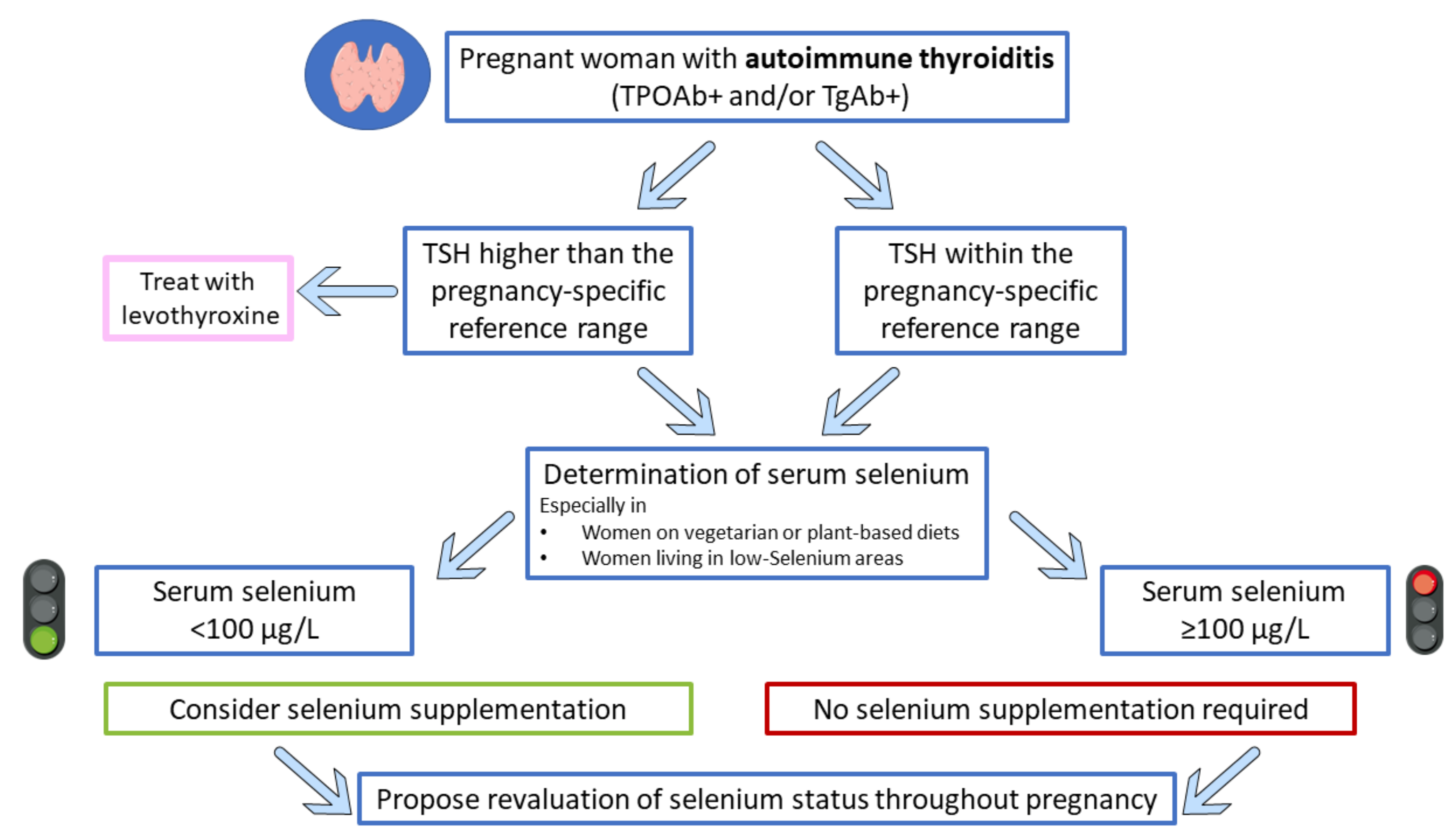
7. Selenium Supplementation in Pregnant Women with Autoimmune Thyroiditis: Has the Time Come?

To date, three placebo-controlled trials have evaluated the impact of Se supplementation on thyroid function in pregnancy. Table 3 summarizes the details of the Se supplementation trials evaluating thyroid outcomes in pregnant women.
Negro et al. examined the influence of Se supplementation both during and after pregnancy in terms of thyroidal autoimmune pattern and function. This prospective, randomized, placebo-controlled study enrolled 77 TPOAb-positive women receiving selenomethionine 200 μg/day (group S1), 74 TPOAb-negative women receiving a placebo (group S0), and 81 TPOAb-negative age-matched women as the control group (group C). All the participants were advised to use iodized salt at the start of the study. At the thyroid ultrasound evaluation, the parenchymal echogenicity was classified as normal (grade 0) or mild thyroiditis (grade 1), moderate thyroiditis (grade 2), or advanced thyroiditis (grade 3). After delivery, patients were considered to have hypothyroidism if their TSH values were above the normal range, whether or not their FT4 values were below or within the normal range. The authors found that PPT (p < 0.01) and permanent hypothyroidism (p < 0.01) were significantly lower in group S1 compared with S0. No adverse effects due to excess Se intake were observed in the Se-treated group [95].
These data have been partially confirmed by a more recent Italian study by Mantovani et al., the SERENA study, a multicenter, randomized, double-blind, placebo-controlled trial that included 45 pregnant women with thyroiditis who were randomly assigned to selenomethionine 83 µg/day or placebo. The authors found a significant autoantibody reduction in the treatment group (p < 0.01) and a rebound in their titer in the placebo group after delivery (p < 0.01). No differences were found in adverse pregnancy outcomes [97].
Conversely, Mao et al. evaluated the effect of a Se nutritional dose (60 μg/day) on thyroid autoimmunity and thyroid function in pregnant women with mild-to-moderate iodine deficiency in the United Kingdom and found that Se supplementation was no more beneficial than placebo in reducing TPOAb levels, and that it determined a decrease in both TSH and FT4 levels in women with thyroiditis. Meanwhile, the authors found that Se supplementation improved thyroid function in women with thyroiditis, for which the decreased TSH levels became almost significantly lower than in females with thyroiditis on the placebo (p = 0.05). Moreover, they found that the iodine-to-creatinine ratio in the Se group was significantly lower than that in the placebo group (p = 0.034), but only in women without thyroiditis [96].
The different findings between the study by Mao et al. and the other two aforementioned studies have many reasonable explanations as follows: (1) the number of TPOAb-positive women was lower in the United Kingdom study compared to the Italian ones (25 vs. 151 and 25 vs. 45), resulting in a lower chance of seeing an effect; (2) baseline TPOAb levels were much lower in the study by Mao compared to the other (110 vs. 627 U/mL and 110 vs. 352 U/mL) and a higher concentration is needed for better treatment responses; (3) the Se dose was lower (60 vs. 200 μg/day and 60 vs. 83 μg/day) in Mao’s study and was probably too low to sustain an effect; (4) none of the study participants received LT4 treatment in Mao’s study in contrast to 20% of the TPOAb-positive women in Negro’s study, and 71% of the women in Mantovani’s study had a potential LT4 additive effect in reducing TPOAb levels in patients with thyroiditis.
The results derived from these trials are not completely consistent; however, they suggest that Se supplementation in pregnant women with positive TPOAb is safe and could effectively reduce autoantibody levels and prevent PPT development, a clinical condition with a prevalence ranging from 33% to 50% [98]. The higher the autoantibody level, the higher the risk of developing PPT and, consequently, hypothyroidism that occurs from 3 to 12 months after delivery [87]. More robust evidence is needed to confirm these data.
Finally, a recent Italian study by Negro et al. evaluated the use of Se in daily clinical practice among Italian endocrinologists by a web-based survey that investigated the use of Se supplementation in different clinical conditions. The survey results showed that approximately 40% of participants had suggested Se use for patients with AIT who were planning a pregnancy or who were already pregnant, with different aims as follows: 39.9% to prevent hypothyroidism development; 24.7% to prevent PPT; 30.7% to reduce TPOAb titers. Moreover, almost 30% of the respondents suggested a Se supplementation of <100 μg/day, 60% recommended 100–200 μg/day, and 10% suggested >200 μg/day [99]. The aforementioned survey results suggest that Se use is widely considered in daily clinical practice, at least in Italy, beyond what is supported by evidence-based medicine and patients’ Se status.
In conclusion, it is important to underline that Se supplementation is not recommended by the 2017 ATA guidelines for the treatment of TPOAb-positive women during pregnancy. The authors explained that any generalized recommendation was unreliable due to conflicting data regarding Se supplementation published before 2017 [72]. Therefore, due to adverse health effects related to both Se deficiency and excess [2,36], special care around Se supplementation during pregnancy is needed, and Se status assessment before Se administration could be an effective and safe option to prevent impaired thyroid function during pregnancy and in the postpartum period [72,89]. In this contest, additional studies are needed to clarify how endocrinologists can appropriately determine Se treatment for AIT management in pregnancy. Figure 2 shows an algorithm proposal indicating a good practical approach for considering Se status in these cases.

8. Conclusions

Se is an essential trace element with a pivotal role in thyroid metabolism. The antioxidant activities of selenoproteins have been suggested as the biological rationale for the use of Se supplementation as an effective treatment in these patients, since oxidative stress has been advocated as a key pathogenetic event in AIT development. Se supplementation has been demonstrated as being effective in reducing the TPOAb titer. Moreover, Se status seems to affect thyroid function during pregnancy, prompting investigation into the potential role of Se supplementation in this setting. To date, few clinical trials have investigated the effectiveness of Se supplementation in pregnant women with thyroiditis and their results suggest its safety and effectiveness in reducing autoantibody levels and preventing the development of postpartum thyroiditis, although these studies are limited. However, Se exhibits U-shaped effects, which should be considered when Se supplementation is planned. Therefore, an individualized approach taking Se status into account may be a good strategy to improve thyroid function in women with AIT both during pregnancy and in the postpartum period.

Funding

This research received no external funding.

Institutional Review Board Statement

Not applicable.

Informed Consent Statement

Not applicable.

Data Availability Statement

Not applicable.

Conflicts of Interest

M.M., V.S., T.F., E.G., C.P., D.G. and A.C. declare no conflict of interest; A.M.I. does consultancy for Novartis, Takeda, Recordati, and Sandoz companies and has received research grants and honorariums from Shire, IPSEN, and Pfizer company.

References

  1. Rayman, M.P. The importance of selenium to human health. Lancet 2000, 356, 233–241. [Google Scholar] [CrossRef] [Green Version]
  2. Rayman, M.P. Selenium intake, status, and health: A complex relationship. Hormones (Athens) 2020, 19, 9–14. [Google Scholar] [CrossRef] [Green Version]
  3. Winther, K.H.; Rayman, M.P.; Bonnema, S.J.; Hegedus, L. Selenium in thyroid disorders—Essential knowledge for clinicians. Nat. Rev. Endocrinol. 2020, 16, 165–176. [Google Scholar] [CrossRef] [PubMed]
  4. Kohrle, J. Selenium in Endocrinology-Selenoprotein-Related Diseases, Population Studies, and Epidemiological Evidence. Endocrinology 2021, 162, bqaa228. [Google Scholar] [CrossRef] [PubMed]
  5. Kohrle, J.; Jakob, F.; Contempre, B.; Dumont, J.E. Selenium, the thyroid, and the endocrine system. Endocr. Rev. 2005, 26, 944–984. [Google Scholar] [CrossRef]
  6. Schomburg, L. Selenium Deficiency Due to Diet, Pregnancy, Severe Illness, or COVID-19-A Preventable Trigger for Autoimmune Disease. Int. J. Mol. Sci. 2021, 22, 8532. [Google Scholar] [CrossRef] [PubMed]
  7. Hogan, C.; Perkins, A.V. Selenoproteins in the Human Placenta: How Essential Is Selenium to a Healthy Start to Life? Nutrients 2022, 14, 628. [Google Scholar] [CrossRef]
  8. Nettore, I.C.; De Nisco, E.; Desiderio, S.; Passaro, C.; Maione, L.; Negri, M.; Albano, L.; Pivonello, R.; Pivonello, C.; Portella, G.; et al. Selenium supplementation modulates apoptotic processes in thyroid follicular cells. BioFactors 2017, 43, 415–423. [Google Scholar] [CrossRef] [Green Version]
  9. Ruggeri, R.M.; D’Ascola, A.; Vicchio, T.M.; Campo, S.; Giani, F.; Giovinazzo, S.; Frasca, F.; Cannavo, S.; Campenni, A.; Trimarchi, F. Selenium exerts protective effects against oxidative stress and cell damage in human thyrocytes and fibroblasts. Endocrine 2020, 68, 151–162. [Google Scholar] [CrossRef]
  10. Santos, L.R.; Neves, C.; Melo, M.; Soares, P. Selenium and Selenoproteins in Immune Mediated Thyroid Disorders. Diagnostics 2018, 8, 70. [Google Scholar] [CrossRef] [Green Version]
  11. Santos, L.R.; Duraes, C.; Mendes, A.; Prazeres, H.; Alvelos, M.I.; Moreira, C.S.; Canedo, P.; Esteves, C.; Neves, C.; Carvalho, D.; et al. A polymorphism in the promoter region of the selenoprotein S gene (SEPS1) contributes to Hashimoto’s thyroiditis susceptibility. J. Clin. Endocrinol. Metab. 2014, 99, E719–E723. [Google Scholar] [CrossRef] [PubMed] [Green Version]
  12. Kucharzewski, M.; Braziewicz, J.; Majewska, U.; Gozdz, S. Concentration of selenium in the whole blood and the thyroid tissue of patients with various thyroid diseases. Biol. Trace Elem. Res. 2002, 88, 25–30. [Google Scholar] [CrossRef]
  13. Balazs, C.; Kaczur, V. Effect of Selenium on HLA-DR Expression of Thyrocytes. Autoimmune Dis. 2012, 2012, 374635. [Google Scholar] [CrossRef] [PubMed] [Green Version]
  14. Hu, Y.; Feng, W.; Chen, H.; Shi, H.; Jiang, L.; Zheng, X.; Liu, X.; Zhang, W.; Ge, Y.; Liu, Y.; et al. Effect of selenium on thyroid autoimmunity and regulatory T cells in patients with Hashimoto’s thyroiditis: A prospective randomized-controlled trial. Clin. Transl. Sci. 2021, 14, 1390–1402. [Google Scholar] [CrossRef] [PubMed]
  15. Kieliszek, M.; Blazejak, S. Current Knowledge on the Importance of Selenium in Food for Living Organisms: A Review. Molecules 2016, 21, 609. [Google Scholar] [CrossRef] [Green Version]
  16. Fairweather-Tait, S.J.; Bao, Y.; Broadley, M.R.; Collings, R.; Ford, D.; Hesketh, J.E.; Hurst, R. Selenium in human health and disease. Antioxid Redox Signal. 2011, 14, 1337–1383. [Google Scholar] [CrossRef]
  17. Navarro-Alarcon, M.; Cabrera-Vique, C. Selenium in food and the human body: A review. Sci. Total Environ. 2008, 400, 115–141. [Google Scholar] [CrossRef]
  18. Johnson, C.C.; Fordyce, F.M.; Rayman, M.P. Symposium on ‘Geographical and geological influences on nutrition’: Factors controlling the distribution of selenium in the environment and their impact on health and nutrition. Proc. Nutr. Soc. 2010, 69, 119–132. [Google Scholar] [CrossRef] [Green Version]
  19. Xia, Y.; Hill, K.E.; Byrne, D.W.; Xu, J.; Burk, R.F. Effectiveness of selenium supplements in a low-selenium area of China. Am. J. Clin. Nutr. 2005, 81, 829–834. [Google Scholar] [CrossRef] [Green Version]
  20. Yang, G.Q.; Xia, Y.M. Studies on human dietary requirements and safe range of dietary intakes of selenium in China and their application in the prevention of related endemic diseases. Biomed. Environ. Sci. 1995, 8, 187–201. [Google Scholar]
  21. Ashton, K.; Hooper, L.; Harvey, L.J.; Hurst, R.; Casgrain, A.; Fairweather-Tait, S.J. Methods of assessment of selenium status in humans: A systematic review. Am. J. Clin. Nutr. 2009, 89, 2025S–2039S. [Google Scholar] [CrossRef] [PubMed] [Green Version]
  22. Di Dato, C.; Gianfrilli, D.; Greco, E.; Astolfi, M.; Canepari, S.; Lenzi, A.; Isidori, A.M.; Giannetta, E. Profiling of selenium absorption and accumulation in healthy subjects after prolonged L-selenomethionine supplementation. J. Endocrinol. Investig. 2017, 40, 1183–1190. [Google Scholar] [CrossRef] [PubMed]
  23. Xia, Y.; Hill, K.E.; Li, P.; Xu, J.; Zhou, D.; Motley, A.K.; Wang, L.; Byrne, D.W.; Burk, R.F. Optimization of selenoprotein P and other plasma selenium biomarkers for the assessment of the selenium nutritional requirement: A placebo-controlled, double-blind study of selenomethionine supplementation in selenium-deficient Chinese subjects. Am. J. Clin. Nutr. 2010, 92, 525–531. [Google Scholar] [CrossRef] [PubMed] [Green Version]
  24. Pop, V.; Krabbe, J.; Maret, W.; Rayman, M. Plasma mineral (selenium, zinc or copper) concentrations in the general pregnant population, adjusted for supplement intake, in relation to thyroid function. Br. J. Nutr. 2021, 125, 71–78. [Google Scholar] [CrossRef] [PubMed]
  25. Institute of Medicine; Food and Nutrition Board; Panel on Dietary Antioxidants and Related Compounds; Subcommittee on Upper Reference Levels of Nutrients; Subcommittee on Interpretation and Uses of Dietary Reference Intakes; Standing Committee on the Scientific Evaluation of Dietary Reference Intakes. Dietary Reference Intakes for Vitamin C, Vitamin E, Selenium, and Carotenoids; National Academies Press: Washington, DC, USA, 2000. [Google Scholar] [CrossRef]
  26. Kipp, A.P.; Strohm, D.; Brigelius-Flohe, R.; Schomburg, L.; Bechthold, A.; Leschik-Bonnet, E.; Heseker, H.; German Nutrition, S. Revised reference values for selenium intake. J. Trace Elem. Med. Biol. 2015, 32, 195–199. [Google Scholar] [CrossRef] [PubMed] [Green Version]
  27. Izquierdo Alvarez, S.; Castanon, S.G.; Ruata, M.L.; Aragues, E.F.; Terraz, P.B.; Irazabal, Y.G.; Gonzalez, E.G.; Rodriguez, B.G. Updating of normal levels of copper, zinc and selenium in serum of pregnant women. J. Trace Elem. Med. Biol. 2007, 21 (Suppl. S1), 49–52. [Google Scholar] [CrossRef] [PubMed]
  28. Grieger, J.A.; Grzeskowiak, L.E.; Wilson, R.L.; Bianco-Miotto, T.; Leemaqz, S.Y.; Jankovic-Karasoulos, T.; Perkins, A.V.; Norman, R.J.; Dekker, G.A.; Roberts, C.T. Maternal Selenium, Copper and Zinc Concentrations in Early Pregnancy, and the Association with Fertility. Nutrients 2019, 11, 1609. [Google Scholar] [CrossRef] [Green Version]
  29. Stoffaneller, R.; Morse, N.L. A review of dietary selenium intake and selenium status in Europe and the Middle East. Nutrients 2015, 7, 1494–1537. [Google Scholar] [CrossRef]
  30. Wang, X.; Li, H.; Yang, L.; Kong, C.; Wang, J.; Li, Y. Selenium Nutritional Status of Rural Residents and Its Correlation with Dietary Intake Patterns in a Typical Low-Selenium Area in China. Nutrients 2020, 12, 3816. [Google Scholar] [CrossRef]
  31. Avery, J.C.; Hoffmann, P.R. Selenium, Selenoproteins, and Immunity. Nutrients 2018, 10, 1203. [Google Scholar] [CrossRef] [Green Version]
  32. Casey, C.E.; Guthrie, B.E.; Friend, G.M.; Robinson, M.F. Selenium in human tissues from New Zealand. Arch. Environ. Health 1982, 37, 133–135. [Google Scholar] [CrossRef] [PubMed]
  33. Fallon, N.; Dillon, S.A. Low Intakes of Iodine and Selenium Amongst Vegan and Vegetarian Women Highlight a Potential Nutritional Vulnerability. Front. Nutr. 2020, 7, 72. [Google Scholar] [CrossRef] [PubMed]
  34. Bakaloudi, D.R.; Halloran, A.; Rippin, H.L.; Oikonomidou, A.C.; Dardavesis, T.I.; Williams, J.; Wickramasinghe, K.; Breda, J.; Chourdakis, M. Intake and adequacy of the vegan diet. A systematic review of the evidence. Clin. Nutr. 2021, 40, 3503–3521. [Google Scholar] [CrossRef] [PubMed]
  35. Tonelli, M.; Wiebe, N.; Hemmelgarn, B.; Klarenbach, S.; Field, C.; Manns, B.; Thadhani, R.; Gill, J.; Alberta Kidney Disease, N. Trace elements in hemodialysis patients: A systematic review and meta-analysis. BMC Med. 2009, 7, 25. [Google Scholar] [CrossRef] [Green Version]
  36. Rayman, M.P. Selenium and human health. Lancet 2012, 379, 1256–1268. [Google Scholar] [CrossRef]
  37. Yang, G.Q.; Wang, S.Z.; Zhou, R.H.; Sun, S.Z. Endemic selenium intoxication of humans in China. Am. J. Clin. Nutr. 1983, 37, 872–881. [Google Scholar] [CrossRef]
  38. Yang, G.; Zhou, R. Further observations on the human maximum safe dietary selenium intake in a seleniferous area of China. J. Trace Elem. Electrolytes Health Dis. 1994, 8, 159–165. [Google Scholar]
  39. Pearce, E.N.; Farwell, A.P.; Braverman, L.E. Thyroiditis. N. Engl. J. Med. 2003, 348, 2646–2655. [Google Scholar] [CrossRef]
  40. Ragusa, F.; Fallahi, P.; Elia, G.; Gonnella, D.; Paparo, S.R.; Giusti, C.; Churilov, L.P.; Ferrari, S.M.; Antonelli, A. Hashimotos’ thyroiditis: Epidemiology, pathogenesis, clinic and therapy. Best Pract. Res. Clin. Endocrinol. Metab. 2019, 33, 101367. [Google Scholar] [CrossRef]
  41. Hawkes, W.C.; Keim, N.L. Dietary selenium intake modulates thyroid hormone and energy metabolism in men. J. Nutr. 2003, 133, 3443–3448. [Google Scholar] [CrossRef]
  42. Duffield, A.J.; Thomson, C.D.; Hill, K.E.; Williams, S. An estimation of selenium requirements for New Zealanders. Am. J. Clin. Nutr. 1999, 70, 896–903. [Google Scholar] [CrossRef] [PubMed] [Green Version]
  43. Duntas, L.H.; Mantzou, E.; Koutras, D.A. Effects of a six month treatment with selenomethionine in patients with autoimmune thyroiditis. Eur. J. Endocrinol. 2003, 148, 389–393. [Google Scholar] [CrossRef] [PubMed] [Green Version]
  44. Hurst, R.; Armah, C.N.; Dainty, J.R.; Hart, D.J.; Teucher, B.; Goldson, A.J.; Broadley, M.R.; Motley, A.K.; Fairweather-Tait, S.J. Establishing optimal selenium status: Results of a randomized, double-blind, placebo-controlled trial. Am. J. Clin. Nutr. 2010, 91, 923–931. [Google Scholar] [CrossRef] [PubMed] [Green Version]
  45. Gartner, R.; Gasnier, B.C.; Dietrich, J.W.; Krebs, B.; Angstwurm, M.W. Selenium supplementation in patients with autoimmune thyroiditis decreases thyroid peroxidase antibodies concentrations. J. Clin. Endocrinol. Metab. 2002, 87, 1687–1691. [Google Scholar] [CrossRef] [PubMed] [Green Version]
  46. Wang, W.; Mao, J.; Zhao, J.; Lu, J.; Yan, L.; Du, J.; Lu, Z.; Wang, H.; Xu, M.; Bai, X.; et al. Decreased Thyroid Peroxidase Antibody Titer in Response to Selenium Supplementation in Autoimmune Thyroiditis and the Influence of a Selenoprotein P Gene Polymorphism: A Prospective, Multicenter Study in China. Thyroid 2018, 28, 1674–1681. [Google Scholar] [CrossRef]
  47. Yu, L.; Zhou, L.; Xu, E.; Bi, Y.; Hu, X.; Pei, X.; Jin, G. Levothyroxine monotherapy versus levothyroxine and selenium combination therapy in chronic lymphocytic thyroiditis. J. Endocrinol. Investig. 2017, 40, 1243–1250. [Google Scholar] [CrossRef]
  48. Esposito, D.; Rotondi, M.; Accardo, G.; Vallone, G.; Conzo, G.; Docimo, G.; Selvaggi, F.; Cappelli, C.; Chiovato, L.; Giugliano, D.; et al. Influence of short-term selenium supplementation on the natural course of Hashimoto’s thyroiditis: Clinical results of a blinded placebo-controlled randomized prospective trial. J. Endocrinol. Investig. 2017, 40, 83–89. [Google Scholar] [CrossRef]
  49. Pirola, I.; Gandossi, E.; Agosti, B.; Delbarba, A.; Cappelli, C. Selenium supplementation could restore euthyroidism in subclinical hypothyroid patients with autoimmune thyroiditis. Endokrynol. Pol. 2016, 67, 567–571. [Google Scholar] [CrossRef] [Green Version]
  50. Pilli, T.; Cantara, S.; Schomburg, L.; Cenci, V.; Cardinale, S.; Heid, E.C.; Kuhn, E.C.; Cevenini, G.; Sestini, F.; Fioravanti, C.; et al. IFNgamma-Inducible Chemokines Decrease upon Selenomethionine Supplementation in Women with Euthyroid Autoimmune Thyroiditis: Comparison between Two Doses of Selenomethionine (80 or 160 mug) versus Placebo. Eur. Thyroid J. 2015, 4, 226–233. [Google Scholar] [CrossRef] [Green Version]
  51. De Farias, C.R.; Cardoso, B.R.; de Oliveira, G.M.; de Mello Guazzelli, I.C.; Catarino, R.M.; Chammas, M.C.; Cozzolino, S.M.; Knobel, M. A randomized-controlled, double-blind study of the impact of selenium supplementation on thyroid autoimmunity and inflammation with focus on the GPx1 genotypes. J. Endocrinol. Investig. 2015, 38, 1065–1074. [Google Scholar] [CrossRef]
  52. Eskes, S.A.; Endert, E.; Fliers, E.; Birnie, E.; Hollenbach, B.; Schomburg, L.; Kohrle, J.; Wiersinga, W.M. Selenite supplementation in euthyroid subjects with thyroid peroxidase antibodies. Clin. Endocrinol. 2014, 80, 444–451. [Google Scholar] [CrossRef] [PubMed]
  53. Anastasilakis, A.D.; Toulis, K.A.; Nisianakis, P.; Goulis, D.G.; Kampas, L.; Valeri, R.M.; Oikonomou, D.; Tzellos, T.G.; Delaroudis, S. Selenomethionine treatment in patients with autoimmune thyroiditis: A prospective, quasi-randomised trial. Int. J. Clin. Pract. 2012, 66, 378–383. [Google Scholar] [CrossRef] [PubMed]
  54. Duntas, L.H. Selenium and the thyroid: A close-knit connection. J. Clin. Endocrinol. Metab. 2010, 95, 5180–5188. [Google Scholar] [CrossRef] [PubMed] [Green Version]
  55. Krysiak, R.; Okopien, B. Haemostatic effects of levothyroxine and selenomethionine in euthyroid patients with Hashimoto’s thyroiditis. Thromb. Haemost. 2012, 108, 973–980. [Google Scholar] [CrossRef] [PubMed]
  56. Krysiak, R.; Okopien, B. The effect of levothyroxine and selenomethionine on lymphocyte and monocyte cytokine release in women with Hashimoto’s thyroiditis. J. Clin. Endocrinol. Metab. 2011, 96, 2206–2215. [Google Scholar] [CrossRef] [Green Version]
  57. Nacamulli, D.; Mian, C.; Petricca, D.; Lazzarotto, F.; Barollo, S.; Pozza, D.; Masiero, S.; Faggian, D.; Plebani, M.; Girelli, M.E.; et al. Influence of physiological dietary selenium supplementation on the natural course of autoimmune thyroiditis. Clin. Endocrinol. 2010, 73, 535–539. [Google Scholar] [CrossRef]
  58. Wichman, J.; Winther, K.H.; Bonnema, S.J.; Hegedus, L. Selenium Supplementation Significantly Reduces Thyroid Autoantibody Levels in Patients with Chronic Autoimmune Thyroiditis: A Systematic Review and Meta-Analysis. Thyroid 2016, 26, 1681–1692. [Google Scholar] [CrossRef]
  59. Karanikas, G.; Schuetz, M.; Kontur, S.; Duan, H.; Kommata, S.; Schoen, R.; Antoni, A.; Kletter, K.; Dudczak, R.; Willheim, M. No immunological benefit of selenium in consecutive patients with autoimmune thyroiditis. Thyroid 2008, 18, 7–12. [Google Scholar] [CrossRef]
  60. Mazokopakis, E.E.; Papadakis, J.A.; Papadomanolaki, M.G.; Batistakis, A.G.; Giannakopoulos, T.G.; Protopapadakis, E.E.; Ganotakis, E.S. Effects of 12 months treatment with L-selenomethionine on serum anti-TPO Levels in Patients with Hashimoto’s thyroiditis. Thyroid 2007, 17, 609–612. [Google Scholar] [CrossRef]
  61. Turker, O.; Kumanlioglu, K.; Karapolat, I.; Dogan, I. Selenium treatment in autoimmune thyroiditis: 9-month follow-up with variable doses. J. Endocrinol. 2006, 190, 151–156. [Google Scholar] [CrossRef] [Green Version]
  62. Fan, Y.; Xu, S.; Zhang, H.; Cao, W.; Wang, K.; Chen, G.; Di, H.; Cao, M.; Liu, C. Selenium supplementation for autoimmune thyroiditis: A systematic review and meta-analysis. Int. J. Endocrinol. 2014, 2014, 904573. [Google Scholar] [CrossRef] [PubMed]
  63. Vissenberg, R.; Manders, V.D.; Mastenbroek, S.; Fliers, E.; Afink, G.B.; Ris-Stalpers, C.; Goddijn, M.; Bisschop, P.H. Pathophysiological aspects of thyroid hormone disorders/thyroid peroxidase autoantibodies and reproduction. Hum. Reprod. Update 2015, 21, 378–387. [Google Scholar] [CrossRef] [PubMed] [Green Version]
  64. Seungdamrong, A.; Steiner, A.Z.; Gracia, C.R.; Legro, R.S.; Diamond, M.P.; Coutifaris, C.; Schlaff, W.D.; Casson, P.; Christman, G.M.; Robinson, R.D.; et al. Preconceptional antithyroid peroxidase antibodies, but not thyroid-stimulating hormone, are associated with decreased live birth rates in infertile women. Fertil. Steril. 2017, 108, 843–850. [Google Scholar] [CrossRef] [PubMed] [Green Version]
  65. Korevaar, T.I.M.; Minguez-Alarcon, L.; Messerlian, C.; de Poortere, R.A.; Williams, P.L.; Broeren, M.A.; Hauser, R.; Souter, I.C. Association of Thyroid Function and Autoimmunity with Ovarian Reserve in Women Seeking Infertility Care. Thyroid 2018, 28, 1349–1358. [Google Scholar] [CrossRef] [PubMed]
  66. Kilic, S.; Tasdemir, N.; Yilmaz, N.; Yuksel, B.; Gul, A.; Batioglu, S. The effect of anti-thyroid antibodies on endometrial volume, embryo grade and IVF outcome. Gynecol. Endocrinol. 2008, 24, 649–655. [Google Scholar] [CrossRef] [PubMed]
  67. Birjandi, B.; Ramezani Tehrani, F.; Amouzegar, A.; Tohidi, M.; Bidhendi Yarandi, R.; Azizi, F. The association between subclinical hypothyroidism and TPOAb positivity with infertility in a population-based study: Tehran thyroid study (TTS). BMC Endocr. Disord. 2021, 21, 108. [Google Scholar] [CrossRef]
  68. Ahsan, U.; Kamran, Z.; Raza, I.; Ahmad, S.; Babar, W.; Riaz, M.H.; Iqbal, Z. Role of selenium in male reproduction—A review. Anim. Reprod. Sci. 2014, 146, 55–62. [Google Scholar] [CrossRef]
  69. Basini, G.; Tamanini, C. Selenium stimulates estradiol production in bovine granulosa cells: Possible involvement of nitric oxide. Domest. Anim. Endocrinol. 2000, 18, 1–17. [Google Scholar] [CrossRef]
  70. Paszkowski, T.; Traub, A.I.; Robinson, S.Y.; McMaster, D. Selenium dependent glutathione peroxidase activity in human follicular fluid. Clin. Chim. Acta 1995, 236, 173–180. [Google Scholar] [CrossRef]
  71. Khera, A.; Dong, L.F.; Holland, O.; Vanderlelie, J.; Pasdar, E.A.; Neuzil, J.; Perkins, A.V. Selenium supplementation induces mitochondrial biogenesis in trophoblasts. Placenta 2015, 36, 863–869. [Google Scholar] [CrossRef]
  72. Alexander, E.K.; Pearce, E.N.; Brent, G.A.; Brown, R.S.; Chen, H.; Dosiou, C.; Grobman, W.A.; Laurberg, P.; Lazarus, J.H.; Mandel, S.J.; et al. 2017 Guidelines of the American Thyroid Association for the Diagnosis and Management of Thyroid Disease During Pregnancy and the Postpartum. Thyroid 2017, 27, 315–389. [Google Scholar] [CrossRef] [PubMed] [Green Version]
  73. Korevaar, T.I.M.; Medici, M.; Visser, T.J.; Peeters, R.P. Thyroid disease in pregnancy: New insights in diagnosis and clinical management. Nat. Rev. Endocrinol. 2017, 13, 610–622. [Google Scholar] [CrossRef] [PubMed]
  74. Di Girolamo, R.; Liberati, M.; Silvi, C.; D’Antonio, F. Levothyroxine Supplementation in Euthyroid Pregnant Women With Positive Autoantibodies: A Systematic Review and Meta-Analysis. Front. Endocrinol. 2022, 13, 759064. [Google Scholar] [CrossRef] [PubMed]
  75. Teng, W.; Shan, Z.; Patil-Sisodia, K.; Cooper, D.S. Hypothyroidism in pregnancy. Lancet Diabetes Endocrinol. 2013, 1, 228–237. [Google Scholar] [CrossRef]
  76. Man, E.B.; Jones, W.S.; Holden, R.H.; Mellits, E.D. Thyroid function in human pregnancy. 8. Retardation of progeny aged 7 years; relationships to maternal age and maternal thyroid function. Am. J. Obstet. Gynecol. 1971, 111, 905–916. [Google Scholar] [CrossRef]
  77. Haddow, J.E.; Palomaki, G.E.; Allan, W.C.; Williams, J.R.; Knight, G.J.; Gagnon, J.; O’Heir, C.E.; Mitchell, M.L.; Hermos, R.J.; Waisbren, S.E.; et al. Maternal thyroid deficiency during pregnancy and subsequent neuropsychological development of the child. N. Engl. J. Med. 1999, 341, 549–555. [Google Scholar] [CrossRef] [PubMed] [Green Version]
  78. Negro, R.; Formoso, G.; Mangieri, T.; Pezzarossa, A.; Dazzi, D.; Hassan, H. Levothyroxine treatment in euthyroid pregnant women with autoimmune thyroid disease: Effects on obstetrical complications. J. Clin. Endocrinol. Metab. 2006, 91, 2587–2591. [Google Scholar] [CrossRef]
  79. Stagnaro-Green, A.; Roman, S.H.; Cobin, R.H.; el-Harazy, E.; Alvarez-Marfany, M.; Davies, T.F. Detection of at-risk pregnancy by means of highly sensitive assays for thyroid autoantibodies. JAMA 1990, 264, 1422–1425. [Google Scholar] [CrossRef]
  80. Prummel, M.F.; Wiersinga, W.M. Thyroid autoimmunity and miscarriage. Eur. J. Endocrinol. 2004, 150, 751–755. [Google Scholar] [CrossRef] [Green Version]
  81. Xie, J.; Jiang, L.; Sadhukhan, A.; Yang, S.; Yao, Q.; Zhou, P.; Rao, J.; Jin, M. Effect of antithyroid antibodies on women with recurrent miscarriage: A meta-analysis. Am. J. Reprod. Immunol. 2020, 83, e13238. [Google Scholar] [CrossRef] [Green Version]
  82. Beneventi, F.; De Maggio, I.; Bellingeri, C.; Cavagnoli, C.; Spada, C.; Boschetti, A.; Magri, F.; Spinillo, A. Thyroid autoimmunity and adverse pregnancy outcomes: A prospective cohort study. Endocrine 2022, 76, 198–207. [Google Scholar] [CrossRef] [PubMed]
  83. Chen, L.; Hu, R. of antithyroid antibodies on women with: A meta-analysis. Clin. Endocrinol. 2011, 74, 513–519. [Google Scholar] [CrossRef]
  84. Thangaratinam, S.; Tan, A.; Knox, E.; Kilby, M.D.; Franklyn, J.; Coomarasamy, A. Association between thyroid autoantibodies and miscarriage and preterm birth: Meta-analysis of evidence. BMJ 2011, 342, d2616. [Google Scholar] [CrossRef] [PubMed] [Green Version]
  85. Peng, C.C.; Pearce, E.N. An update on thyroid disorders in the postpartum period. J. Endocrinol. Investig. 2022, 1–10. [Google Scholar] [CrossRef] [PubMed]
  86. Stagnaro-Green, A.; Roman, S.H.; Cobin, R.H.; el-Harazy, E.; Wallenstein, S.; Davies, T.F. A prospective study of lymphocyte-initiated immunosuppression in normal pregnancy: Evidence of a T-cell etiology for postpartum thyroid dysfunction. J. Clin. Endocrinol. Metab. 1992, 74, 645–653. [Google Scholar] [CrossRef] [PubMed]
  87. Stagnaro-Green, A. Approach to the patient with postpartum thyroiditis. J. Clin. Endocrinol. Metab. 2012, 97, 334–342. [Google Scholar] [CrossRef] [Green Version]
  88. Van Dijk, M.M.; Vissenberg, R.; Fliers, E.; van der Post, J.A.M.; van der Hoorn, M.P.; de Weerd, S.; Kuchenbecker, W.K.; Hoek, A.; Sikkema, J.M.; Verhoeve, H.R.; et al. Levothyroxine in euthyroid thyroid peroxidase antibody positive women with recurrent pregnancy loss (T4LIFE trial): A multicentre, randomised, double-blind, placebo-controlled, phase 3 trial. Lancet Diabetes Endocrinol. 2022, 10, 322–329. [Google Scholar] [CrossRef]
  89. Duntas, L.H. Selenium and at-risk pregnancy: Challenges and controversies. Thyroid Res. 2020, 13, 16. [Google Scholar] [CrossRef]
  90. Arikan, T.A. Plasma Selenium Levels in First Trimester Pregnant Women with Hyperthyroidism and the Relationship with Thyroid Hormone Status. Biol. Trace Elem. Res. 2015, 167, 194–199. [Google Scholar] [CrossRef]
  91. Guo, X.; Zhou, L.; Xu, J.; Liu, Z.; Liu, J.; Yan, C. Prenatal Maternal Low Selenium, High Thyrotropin, and Low Birth Weights. Biol. Trace Elem. Res. 2021, 199, 18–25. [Google Scholar] [CrossRef]
  92. Ambroziak, U.; Hybsier, S.; Shahnazaryan, U.; Krasnodebska-Kiljanska, M.; Rijntjes, E.; Bartoszewicz, Z.; Bednarczuk, T.; Schomburg, L. Severe selenium deficits in pregnant women irrespective of autoimmune thyroid disease in an area with marginal selenium intake. J. Trace Elem. Med. Biol. 2017, 44, 186–191. [Google Scholar] [CrossRef] [PubMed]
  93. Hofstee, P.; James-McAlpine, J.; McKeating, D.R.; Vanderlelie, J.J.; Cuffe, J.S.M.; Perkins, A.V. Low serum selenium in pregnancy is associated with reduced T3 and increased risk of GDM. J. Endocrinol. 2021, 248, 45–57. [Google Scholar] [CrossRef] [PubMed]
  94. Mirone, M.; Giannetta, E.; Isidori, A.M. Selenium and reproductive function. A systematic review. J. Endocrinol. Investig. 2013, 36, 28–36. [Google Scholar]
  95. Negro, R.; Greco, G.; Mangieri, T.; Pezzarossa, A.; Dazzi, D.; Hassan, H. The influence of selenium supplementation on postpartum thyroid status in pregn.nant women with thyroid peroxidase autoantibodies. J. Clin. Endocrinol. Metab. 2007, 92, 1263–1268. [Google Scholar] [CrossRef]
  96. Mao, J.; Pop, V.J.; Bath, S.C.; Vader, H.L.; Redman, C.W.; Rayman, M.P. Effect of low-dose selenium on thyroid autoimmunity and thyroid function in UK pregnant women with mild-to-moderate iodine deficiency. Eur. J. Nutr. 2016, 55, 55–61. [Google Scholar] [CrossRef] [Green Version]
  97. Mantovani, G.; Isidori, A.M.; Moretti, C.; Di Dato, C.; Greco, E.; Ciolli, P.; Bonomi, M.; Petrone, L.; Fumarola, A.; Campagna, G.; et al. Selenium supplementation in the management of thyroid autoimmunity during pregnancy: Results of the “SERENA study”, a randomized, double-blind, placebo-controlled trial. Endocrine 2019, 66, 542–550. [Google Scholar] [CrossRef] [Green Version]
  98. Nicholson, W.K.; Robinson, K.A.; Smallridge, R.C.; Ladenson, P.W.; Powe, N.R. Prevalence of postpartum thyroid dysfunction: A quantitative review. Thyroid 2006, 16, 573–582. [Google Scholar] [CrossRef] [Green Version]
  99. Negro, R.; Attanasio, R.; Grimaldi, F.; Marcocci, C.; Guglielmi, R.; Papini, E. A 2016 Italian Survey about the Clinical Use of Selenium in Thyroid Disease. Eur. Thyroid J. 2016, 5, 164–170. [Google Scholar] [CrossRef] [Green Version]
Figure 1. Thyroid gland selenoproteins. Selenoprotein P (SELENOP) and glutathione peroxidase 3 (GPX3) are actively secreted from the thyrocyte and exert oxidoreductase functions, protecting thyrocytes from excessive H2O2 accumulation. Other selenoproteins, including Selenoprotein K, N, and S (SELENOK, SELENON, and SELENOS, respectively) take part in the quality control pathways within the endoplasmic reticulum. Type 1 (DIO1) and 2 iodothyronine deiodinase (DIO2) regulate thyroid hormone levels, converting T4 into T3. GPX4 and thioredoxin reductase 2 (TXNRD2) are localized in the mitochondria, affording protection against the hydrogen peroxide produced by the mitochondrial respiratory chain. This figure was created using Servier Medical Art templates, which is licensed under the Creative Commons Attribution 3.0 Unported License; https://smart.servier.com (accessed on 17 February 2022).
Nutrients 14 02234 g001
Figure 2. Proposed algorithm for selenium supplementation in pregnant women with thyroiditis. This figure was created using Servier Medical Art templates, which is licensed under the Creative Commons Attribution 3.0 Unported License; https://smart.servier.com (accessed on 17 February 2022).
Nutrients 14 02234 g002
Table 1. Examples of selenium food sources.
FoodSe Content (µg/g Fresh Weight)
Brazil nuts~0.85–7
Tuna (in oil)~0.8
Chicken~0.6
Sardines~0.6
Lamb~0.3-0.4
Shellfish~0.4–1.3
Beef~0.35–0.5
Salmon~0.2–0.4
Ham~0.2
Eggs~0.2
Milk products~0.1–0.5
Table 2. Details of studies evaluating selenium status and thyroid outcomes in pregnant women.
First Author, YearCountryStudy DesignStudy Population No. of Patients Gestational AgeOutcomesMain Results
Arikan et al. 2015 [90]TurkeyCross-sectional studyHealthy pregnant vs. hyperthyroid pregnant107: 70 healthy (group 1); 37 hyperthyroid (group 2)First trimesterTSH, FT3, FT4, serum SeSignificantly higher FT3 and FT4 levels and significantly lower TSH and Se levels in group 2 than in group 1; positive correlation between Se and FT4 in group 1 and with TSH in group 2
Ambroziak et al. 2017 [92]PolandProspective studyHealthy pregnant and pregnant women with AITD74: 45 healthy;
39 AITD
First, second and third trimesterTSH, FT3, FT4, TPOAb, TgAb, thyroid US, serum Se and SELENOPRelatively low serum Se and SELENOP levels in both healthy and AITD; from first to third trimester TPOAb and TgAb declined in AITD, but this was unrelated to Se status
Guo et al. 2021 [91]ChinaProspective cohort studyPregnant193128–36 wksTSH, serum Se, birth weight and lengthSerum Se levels <103.7 μg/L, each unit increase significantly associated with a decrease of 0.014 μIU/mL in TSH; maternal TSH levels inversely associated with infant birth weights
Hofstee et al. 2021 [93]AustraliaRetrospective cross-sectional studyPregnant euthyroid63:
21 with low Se;
21 with mean Se;
21 with optimal Se
26–30 wksTSH, FT3, FT4, TPOAb, serum SeFemales with low Se concentrations showed reduced FT3 and increased TPOAb and incidence of pregnancy disorders; Se levels positively correlated with FT3 and negatively correlated with TPOAb
Pop et al. 2021 [24]The NetherlandsLongitudinal prospective studyPregnant2041:
1479 taking mineral supplements;
544 not taking mineral supplements
12 wksTSH, FT4, TPOAb, serum Se, Zn and CuNegative correlation between Se levels and logFT4; positive correlation between Se levels and logTSH; women taking supplements were 1.46 times less likely to have elevated TPOAb at 12 wks
Se—selenium; wks—weeks; AITD—autoimmune thyroid disease; SELENOP—selenoprotein P; US—ultrasound.
Table 3. Details of selenium supplementation trials evaluating thyroid outcomes in pregnant women.
First Author, Year CountryNo. of Patients (Selenium/Placebo/Control)Selenium SupplementationDuration of Treatment (Months)Patients Requiring LT4 during Pregnancy (%)OutcomesMain Results
Negro et al. 2007 [95]Italy232 (77/74/81)selenomethionine 200 μg/day1833 (14.2)TSH, FT4, TPOAb, thyroid US, PPTLower prevalence of PPT and permanent hypothyroidism
Mao et al. 2016 [96]UK230 (120/110)selenium 60 μg/day6NoneTSH, FT4, TPOAb, TgAbDecrease in TSH and FT4 during pregnancy but no effect on TPOAb
Mantovani et al. 2019 [97]Italy45 (21/24)selenomethionine 83 μg/day1213 * (28.9)TSH, FT3, FT4, TPOAb, TgAb, thyroid US, HRQoLDecrease in TPOAb and TgAb in the Se group but increase in the PLB group at PP
* Ten patients required LT4 dosage adjustment and three patients began LT4 treatment; PPT—postpartum thyroiditis; PLB—placebo; PP—postpartum.
Publisher’s Note: MDPI stays neutral with regard to jurisdictional claims in published maps and institutional affiliations.

