Highbush Blueberry (Vaccinium corymbosum L.) Leaves Extract and Its Modified Arginine Preparation for the Management of Metabolic Syndrome—Chemical Analysis and Bioactivity in Rat Model

 ,
,  ,
,  ,
,
Abstract
:
1. Introduction
2. Materials and Methods
2.1. Chemicals and General Experiments
2.2. Plant Material
2.3. Preparation of Extracts
2.4. Quantification of Major Phytochemicals Using Non-Specific Chemical Methods
2.5. HPLC-DAD-MS Analysis and Quantification of Major Compounds
2.6. The Anti-MS Potential of Extracts Using In Vivo Rat Model
2.7. Statistical Analysis
3. Results
3.1. Quantification of Major Phytochemicals Using Chemical Methods
3.2. Phytochemical Analysis of Investigated Extracts and Quantification of Major Compounds Using HPLC-DAD-MS Approach
3.3. The Pharmacological Activity of the Extracts
4. Discussion
5. Conclusions
Author Contributions
Funding
Institutional Review Board Statement
Informed Consent Statement
Data Availability Statement
Conflicts of Interest
References
- Hu, X.; Li, X.; Adams, H.; Kubena, K.; Guo, S. Etiology of Metabolic Syndrome and Dietary Intervention. Int. J. Mol. Sci. 2019, 20, 128. [Google Scholar] [CrossRef] [Green Version]
- Rochlani, Y.; Pothineni, N.V.; Kovelamudi, S.; Mehta, J.L. Metabolic syndrome: Pathophysiology, management, and modulation by natural compounds. Ther. Adv. Cardiovasc. Dis. 2017, 11, 215–225. [Google Scholar] [CrossRef]
- Ferri, F.F. Metabolic syndrome. In Ferri’s Clinical Advisor 2019; Elsevier: Philadelphia, PA, USA, 2019; Available online: https://www.clinicalkey.com (accessed on 12 February 2019).
- Metabolic Syndrome. National Heart, Lung, and Blood Institute. Available online: https://www.nhlbi.nih.gov/health-topics/metabolic-syndrome (accessed on 10 February 2019).
- Samson, S.L.; Garber, A.J. Metabolic syndrome. Endocrinol. Metab. Clin. N. Am. 2014, 43, 1–23. [Google Scholar] [CrossRef]
- About Metabolic Syndrome. American Heart Association. Available online: https://www.heart.org/en/health-topics/metabolic-syndrome/about-metabolic-syndrome (accessed on 12 February 2019).
- Aguilar-Salinas, C.A.; Viveros-Ruiz, T. Recent Advances in Managing/Understanding the Metabolic Syndrome. F1000Research 2019, 8. F1000 Faculty Rev-370. [Google Scholar] [CrossRef] [PubMed] [Green Version]
- Saklaeyn, M.G. The Global Epidemic of the Metabolic Syndrome. Curr. Hypertens. Rep. 2018, 20, 8. [Google Scholar] [CrossRef] [Green Version]
- Kovalenko, V.N. Compendium 2020—Medicines; MORION: Kiiv, Ukraine, 2020; p. 2700. [Google Scholar]
- Koshovyi, O.M.; Zagayko, A.L.; Kolychev, I.O.; Akhmedov, E.Y.; Komissarenko, A.N. Phytochemical study of the dry extract from bilberry leaves. Azerbaijan Pharm. Pharmacother. J. 2016, 16, 18–23. [Google Scholar]
- Zagayko, A.L.; Kolisnyk, T.Y.; Chumak, O.I.; Ruban, O.A.; Koshovyi, O.M. Evaluation of anti-obesity and lipid-lowering properties of Vaccinium myrtillus leaves powder extract in a hamster model. J. Basic Clin. Physiol. Pharmacol. 2018, 29, 697–703. [Google Scholar] [CrossRef] [PubMed]
- Song, G.Q. Blueberry (Vaccinium corymbosum L.). In Agrobacterium Protocols. Methods in Molecular Biology; 1224; Wang, K., Ed.; Springer: New York, NY, USA, 2015. [Google Scholar] [CrossRef]
- Strik, B.C.; Finn, C.E.; Moore, P.P. Blueberry Cultivars for the Pacific Northwest; Oregon State University Extension Service: Corvallis, OR, USA, 2014; Volume 656, pp. 1–13. [Google Scholar]
- Vaughan, J.G.; Geissler, C.A. Roґsliny Jadalne; Prуszy ґnski i Spуłka: Warszawa, Poland, 2001. [Google Scholar]
- Export-Oriented Blueberry Cultivation. 2020. Available online: https://inventure.com.ua/investments/vyrashivanie-golubiki (accessed on 1 December 2019).
- Stefanescu, B.E.; Calinoiu, L.F.; Ranga, F.; Fetea, F.; Mocan, A.; Vodnar, D.C.; Crișan, G. The Chemical and Biological Profiles of Leaves from Commercial Blueberry Varieties. Plants 2020, 9, 1193. [Google Scholar] [CrossRef]
- Sater, H.M.; Bizzio, L.N.; Tieman, D.M.; Muñoz, P.D. A Review of the Fruit Volatiles Found in Blueberry and Other Vaccinium Species. J. Agric. Food Chem. 2020, 21, 5777–5786. [Google Scholar] [CrossRef]
- Zorzi, M.; Gai, F.; Medana, C.; Aigotti, R.; Morello, S.; Peiretti, P.G. Bioactive Compounds and Antioxidant Capacity of Small Berries. Foods 2020, 9, 623. [Google Scholar] [CrossRef]
- Grodzinsky, A.M. Medical Plants; Kiiv: Bazhana, Ukraine, 1992; p. 74. [Google Scholar]
- Fern, K. Vaccinium uliginosum. Temperate Plants. 2020. Available online: http://temperate.theferns.info/plant/Vaccinium+uliginosum (accessed on 18 August 2020).
- Stremoukhov, A.A. The Prospects of Using Highbush Blueberry Leaves for the Creation of New Dietary Supplements. Commodity Analysis of Products of Limited Pharmacy Assortment: Materials of the III Scientific-Practical Internet-Conference with International Participation (15 April 2016); NUPH: Kharkiv, Ukraine, 2016; p. 166. [Google Scholar]
- Stull, A.J. Blueberries’ Impact on Insulin Resistance and Glucose Intolerance. Antioxidants 2016, 5, 44. [Google Scholar] [CrossRef] [Green Version]
- Rocha, D.M.U.P.; Caldas, A.P.S.; Pereira da Silva, B.; Hermsdorff, H.H.M.; Alfenas, R.C.G. Effects of blueberry and cranberry consumption on type 2 diabetes glycemic control: A systematic review. Crit. Rev. Food Sci. Nutr. 2019, 59, 1816–1828. [Google Scholar] [CrossRef]
- Müller, D.; Schantz, M.; Richling, E. High Performance Liquid Chromatography Analysis of Anthocyanins in Bilberries (Vaccinium myrtillus L.), Blueberries (Vaccinium corymbosum L.), and Corresponding Juices. J. Food Sci. 2012, 77, 340–345. [Google Scholar] [CrossRef]
- Liu, B.; Hu, T.; Yan, W. Authentication of the Bilberry Extracts by an HPLC Fingerprint Method Combining Reference Standard Extracts. Molecules 2020, 25, 2514. [Google Scholar] [CrossRef]
- Braic, R.Ş.; Imre, S.; Eşianu, S.; Laczko-Zold, E.; Dogaru, T.M. Vaccinium corymbosum leaves, a potential source of polyphenolic compounds. Rom. Biotechnol. Lett. 2019, 24, 755–760. [Google Scholar] [CrossRef]
- Kim, S.-M.; Shang, Y.F.; Um, B.-H. Preparative separation of chlorogenic acid by centrifugal partition chromatography from highbush blueberry leaves (Vaccinium corymbosum L.). Phytochem. Anal. 2010, 21, 457–462. [Google Scholar] [CrossRef]
- Wang, L.-J.; Wu, J.; Wang, H.-X.; Li, S.-S.; Zheng, X.-C.; Du, H.; Xu, Y.J.; Wang, L.-S. Composition of phenolic compounds and antioxidant activity in the leaves of blueberry cultivars. J. Funct. Foods 2015, 16, 295–304. [Google Scholar] [CrossRef]
- Pertuzatti, P.B.; Barcia, M.T.; Gomez-Alonso, S.; Godoy, H.T.; Hermosin-Gutierrez, I. Phenolics profiling by HPLC-DAD-ESI-MSn aided by principal component analysis to classify Rabbiteye and Highbush blueberries. Food Chem. 2021, 340, 127958. [Google Scholar] [CrossRef]
- Vrancheva, R.; Ivanov, I.; Dincheva, I.; Badjakov, I.; Pavlov, A. Triterpenoids and Other Non-Polar Compounds in Leaves of Wild and Cultivated Vaccinium Species. Plants 2021, 10, 94. [Google Scholar] [CrossRef]
- Parfenov, V.A. Use of L-lysine aescinate in central nervous system diseases. Neurol. Neuropsychiatry Psychosom. 2011, 3, 99–104. [Google Scholar] [CrossRef] [Green Version]
- Koshovyi, O.; Raal, A.; Kireyev, I.; Tryshchuk, N.; Ilina, T.; Romanenko, Y.; Kovalenko, S.M.; Bunyatyan, N. Phytochemical and Psychotropic Research of Motherwort (Leonurus cardiaca L.) Modified Dry Extracts. Plants 2021, 10, 230. [Google Scholar] [CrossRef]
- Chaika, N.; Koshovyi, O.; Raal, A.; Kireyev, I.; Zupanets, A.; Odyntsova, V. Phytochemical profile and pharmacological activity of the dry extract from Arctostaphylos uva-ursi leaves modified with phenylalanine. Sci. Pharm. Sci. 2020, 6, 74–78. [Google Scholar] [CrossRef]
- Altas, M.; Var, A.; Köse, C.; Ozbilgin, K.; Arı, Z. Endothelial dysfunction in high fructose containing diet fed rats: Increased nitric oxide and decreased endothelin-1 levels in liver tissue. Dicle Univ. Med. School 2010, 37, 193–198. [Google Scholar]
- Zagayko, A.L.; Voronina, L.M.; Strelchenko, K.V. Metabolic Syndrom: Mechanisms of Development and Prospects for Antioxidant Therapy; Publiser of NUPH: Kharkiv, Ukraine, 2007; p. 216. [Google Scholar]
- Liu, J.; Yong, H.; Yao, X.; Hu, H.; Yun, D.; Xiao, L. Recent advances in phenolic–protein conjugates: Synthesis, characterization, biological activities and potential applications. RSC Adv. 2019, 9, 35825–35840. [Google Scholar] [CrossRef] [Green Version]
- Quan, T.H.; Benjakul, S.; Sae-Leaw, T.; Balange, A.K.; Maqsood, S. Protein–polyphenol conjugates: Antioxidant property, functionalities and their applications. Trends Food Sci. Technol. 2019, 91, 507–517. [Google Scholar] [CrossRef]
- Dobrochaeva, D.N.; Kotov, M.I.; Prokudin, Y.N.; Barbarich, A.I. Key to Higher Plants of Ukraine, 2nd ed.; Science Dumka: Kiev, Ukraine, 1999. (In Russian) [Google Scholar]
- Zabolotnyi, O.V.; Zabolotnyi, V.A.; Koshevoi, M.D. Conditionality examination of the new testing algorithms for coal-water slurries moisture measurement. Nauk. Visnyk Natsionalnoho Hirnychoho Univ. 2018, 1, 51–59. [Google Scholar] [CrossRef]
- Ilina, T.; Kashpur, N.; Granica, S.; Bazylko, A.; Shinkovenko, I.; Kovalyova, A.; Goryacha, O.; Koshovyi, O. Phytochemical profiles and in vitro immunomodulatory activity of ethanolic extracts from Galium aparine L. Plants 2019, 8, 541. [Google Scholar] [CrossRef] [Green Version]
- Koshovyi, O.N.; Vovk, G.V.; Akhmedov, E.Y.; Komissarenko, A.N. The study of the chemical composition and pharmacological activity of Salvia officinalis leaves extracts getting by complex processing. Azerbaijan Pharm. Pharmacother. J. 2015, 15, 30–34. [Google Scholar]
- Starchenko, G.; Hrytsyk, A.; Raal, A.; Koshovyi, О. Phytochemical profile and pharmacological activities of water and hydroethanolic dry extracts of Calluna vulgaris (L.) Hull. herb. Plants 2020, 9, 751. [Google Scholar] [CrossRef]
- Shinkovenko, I.L.; Kashpur, N.V.; Ilyina, T.V.; Kovalyova, A.M.; Goryacha, O.V.; Koshovyi, O.M.; Toryanyk, E.L.; Kryvoruchko, O.V. The immunomodulatory activity of the extracts and complexes of biologically active compounds of Galium verum L. herb. Ceska A Slov. Farm. 2018, 67, 25–29. [Google Scholar]
- Krivoruchko, E.; Markin, A.; Samoilova, V.A.; Ilina, T.; Koshovyi, O. Research in the chemical composition of the bark of sorbus aucuparia. Ceska A Slov. Farm. 2018, 67, 113–115. [Google Scholar]
- State Pharmacopoeia of Ukraine. (2004–2018) Amendment 1–4; State Enterprise Ukrainian Scientific Pharmacopoeial Center for Drug Quality: Kharkiv, Ukraine, 2018; Volume 1–3. (In Ukrainian)
- Grace, M.H.; Esposito, D.; Dunlap, K.L.; Lila, M.A. Comparative Analysis of Phenolic Content and Profile, Antioxidant Capacity, and Anti-inflammatory Bioactivity in Wild Alaskan and Commercial Vaccinium Berries. J. Agric. Food Chem. 2014, 62, 4007–4017. [Google Scholar] [CrossRef] [PubMed] [Green Version]
- Rigolon, T.C.B.; Barros, F.A.R.; Vieira, É.N.R.; Stringheta, P.C. Prediction of total phenolics, anthocyanins and antioxidant capacity of blackberry (Rubus sp.), blueberry (Vaccinium sp.) and jaboticaba (Plinia cauliflora (Mart.) Kausel) skin using colorimetric parameters. Food Sci. Technol. 2020, 40, 620–625. [Google Scholar] [CrossRef]
- Clifford, M.N.; Knight, S.; Kuhnert, N. Discriminating between the six isomers of dicaffeoylquinic acid by LC-MS(n). J. Agric. Food Chem. 2005, 53, 3821–3832. [Google Scholar] [CrossRef] [PubMed]
- Ma, C.; Dastmalchi, K.; Whitaker, B.D.; Kennelly, E.J. Two new antioxidant malonated caffeoylquinic acid isomers in fruits of wild eggplant relatives. J. Agric. Food Chem. 2011, 59, 9645–9651. [Google Scholar] [CrossRef] [PubMed]
- Jaiswal, R.; Sovdat, T.; Vivan, F.; Kuhnert, N. Profiling and characterization by LC-MSn of the chlorogenic acids and hydroxycinnamoylshikimate esters in maté (Ilex paraguariensis). J. Agric. Food Chem. 2010, 58, 5471–5484. [Google Scholar] [CrossRef] [PubMed]
- Rezende, F.M.; Ferreira, M.J.P.; Clausen, M.H.; Rossi, M.; Furlan, C.M. Acylated Flavonoid Glycosides are the Main Pigments that Determine the Flower Colour of the Brazilian Native Tree Tibouchina pulchra (Cham.) Cogn. Molecules 2019, 24, 718. [Google Scholar] [CrossRef] [PubMed] [Green Version]
- Kravchenko, G.B. Hypoglycemic and hypolipidemic activity of arginine containing bearberry leaves extract in insulin resistant rats. Med. Clin. Chem. 2020, 22, 5–10. [Google Scholar] [CrossRef]
- Lia, D.; Lia, B.; Ma, Y.; Sun, X.; Lin, Y.; Meng, X. Polyphenols, anthocyanins, and flavonoids contents and the antioxidant capacity of various cultivars of highbush and half-high blueberries. J. Food Compos. Anal. 2017, 62, 84–93. [Google Scholar] [CrossRef]
- Kasprzak-Drozd, K.; Oniszczuk, T.; Stasiak, M.; Oniszczuk, A. Beneficial Effects of Phenolic Compounds on Gut Microbiota and Metabolic Syndrome. Int. J. Mol. Sci. 2021, 22, 3715. [Google Scholar] [CrossRef]
- Castro Barbosaa, T.; Jiang, L.Q.; Zierath, J.R.; Nunes, M.T. L-Arginine enhances glucose and lipid metabolism in rat L6 myotubes via the NO/ c-GMP pathway. Metabolism 2013, 62, 79–89. [Google Scholar] [CrossRef] [PubMed]
- Wu, Z.; Hou, Y.; Hu, S.; Bazer, F.W.; Meininger, C.J.; Wu, G. Catabolism and safety of supplemental L-arginine in animals. Amino Acids 2016, 48, 1541–1552. [Google Scholar] [CrossRef]
- Hayashi, T.; Juliet, P.A.; Matsui-Hirai, H.; Miyazaki, A.; Fukatsu, A.; Funami, J.; Iguchi, A.; Ignarro, L.J. l-Citrulline and l-arginine supplementation retards the progression of high-cholesterol-diet-induced atherosclerosis in rabbits. Proc. Natl. Acad. Sci. USA 2005, 102, 13681–13686. [Google Scholar] [CrossRef] [PubMed] [Green Version]


| No | Compound Name | Retention Time [min] | UV-Vis Maxima [nm] | MS− Ions | MS2− Ions | MS3− Ions | MS+ Ions | MS2+ Ions | MS3+ Ions | Content (μg/mg) | Quantification Standard | Ions Used for Quantifiaction |
|---|---|---|---|---|---|---|---|---|---|---|---|---|
| 1 | 3-O-caffeoylquinic acid (neochlorogenic acid) s | 16.2 | 242, 302, 324 | 353 | 191 b, 179, 135 | - | 355 | 163 b, 135 | - | 29.45 ± 0.57 | chlorogenic acid | 353, 707 |
| 2 | 5-O-caffeoylquinic acid (chlorogenic acid) s | 22.8 | 242, 300, 325 | 353 | 191 b, 179, 135 | - | 355 | 163 | - | 136.47 ± 4.26 | chlorogenic acid | 353, 707 |
| 3 | 4-O-caffeoylquinic acid (cryptochlorogenic acid) | 24.7 | 300,324 | 353 | 191, 179, 173 b | - | 355 | 163 | - | 6.09 ± 0.17 | chlorogenic acid | 353, 707 |
| 4 | O-mallonyl-O-caffeoylquinic acid isomer | 28.9 | 300, 324 | 439 | 233, 395 b, 353 | - | 441 | 423 b, 404, 163 | - | 17.85 ± 0.56 | chlorogenic acid | 439, 879 |
| 5 | O-caffeoylshikimic acid isomer | 30.5 | 300, 326 | 335 | 135, 179 b | - | 337 | 163 | - | 2.53 ± 0.07 | chlorogenic acid | 335, 667 |
| 6 | O-mallonyl-O-caffeoylquinic acid isomer | 32.7 | 305, 326 | 439 | 233, 353, 395 b | - | 441 | 163, 193, 244, 396, 423,645 | - | 13.07 ± 0.77 | chlorogenic acid | 439, 878 |
| 7 | myricetin3-O-galactoside s | 36.3 | 265, 353 | 479 | 179, 316 b, 461 | - | 481 | 319 | 165 | 0.91 ± 0.03 | hyperoside | 479, 959 |
| 8 | myricetin 3-O-glucoside s | 36.9 | 265, 353 | 479 | 179, 205, 297, 271, 316 b, 383, 461 | - | 481 | 319 | - | 1.03 ± 0.02 | hyperoside | 479, 959 |
| 9 | quercetin rhamnohexoside | 40.5 | 264, 353 | 609 | 301 b | - | 611 | 303 b, 345 | - | 0.41 ± 0.01 | hyperoside | 609, 1219 |
| 10 | quercetin 3-O-galactoside (hyperoside) s | 41.1 | 254, 262 sh, 353 | 463 | 301 b, 343, 179, 151, 229, 283, 255 | - | 465 | 303 b, 345 | 165 b, 195, 284 | 4.29 ± 0.10 | hyperoside | 463, 927 |
| 11 | quercetin 3-O-rutinoside (rutin) s | 41.4 | slope | 609 | 301 b, 179, 271, 343, 395, 457 | - | 611 | 465, 303 b | 447, 303 b | 1.97 ± 0.05 | hyperoside | 609, 1219 |
| 12 | quercetin 3-O-glucoside (isoquercitrin) s | 42.1 | 253, 264 sh, 353 | 463 | 301 b, 151, 179, 255, 273, 298, 344 | - | 465 | 303 | - | 2.33 ± 0.04 | hyperoside | 463, 927 |
| 13 | quercetin 3-O-arabinoside (avicularin) s | 44.5 | 265, 354 | 433 | 301 | - | 435 | 303 | 137, 229 b, 257, 285 | 1.17 ± 0.04 | hyperoside | 433, 867 |
| 14 | kaempferol O-rhamnohexoside | 45.8 | 265, 342 | 593 | 199, 257, 285 b, 327, 447, 486, 565, 286 b | - | 595 | 287 b, 449 | 287 | 0.72 ± 0.01 | hyperoside | 593, 1187 |
| 15 | quercetin O-malonylhexoside | 46.1 | 264, 353 | 549 | 505 b, 301 | 301 | 551 | 303 | - | 0.26 ± 0.01 | hyperoside | 549, 1099 |
| 16 | kaempferol 3-O-glucoside (astragalin) s | 46.5 | 265, 343 | 447 | 284 | - | 449 | 287 b, 303 | 137, 191, 229 b | 0.41 ± 0.01 | hyperoside | 447, 895 |
| 17 | 4,5-O-dicaffeoylquinic acid s | 51.0 | 328, 389 | 515 | 173, 179, 203, 255, 299, 353 b, 404 | 135, 173 b | 517 | 163, 296, 499 b | - | 2.80 ± 0.30 | chlorogenic acid | 515, 1031 |
| 18 | mirycetin p-coumaroylhexoside | 52.6 | 259, 274, 281, 319, 321, 352 | 625 | 317, 479 b | 179, 316 b, 461 | 627 | 309, 319 b, 489, 609 | - | 0.36 ± 0.02 | hyperoside | 625, 1251 |
| 19 | caffeic acid derivative | 53.6 | 296, 324 | 207 | - | 209 | - | 1.44 ± 0.04 | chlorogenic acid | 207, 415 | ||
| 20 | undefined phenolic acid | 54.8 | 281, 335 | 451 | 341 | - | 453 | 191, 301, 343 b, 435 | 191 | 0.58 ± 0.02 | chlorogenic acid | 451, 905 |
| 21 | p-coumaroyl-caffeoylquinic acid | 56.5 | 287, 315 | 499 | 173, 337 b | 173 | 501 | 321, 483 b | 147, 303 b | 1.17 ± 0.20 | chlorogenic acid | 499, 999 |
| 22 | quercetin p-coumaroylhexoside | 58.0 | 256, 281, 321, 354 | 609 | 463, 301 b | - | 303 | 165, 221, 267 b, 393, 428, 459 | - | 0.65 ± 0.02 | hyperoside | 609, 301 |
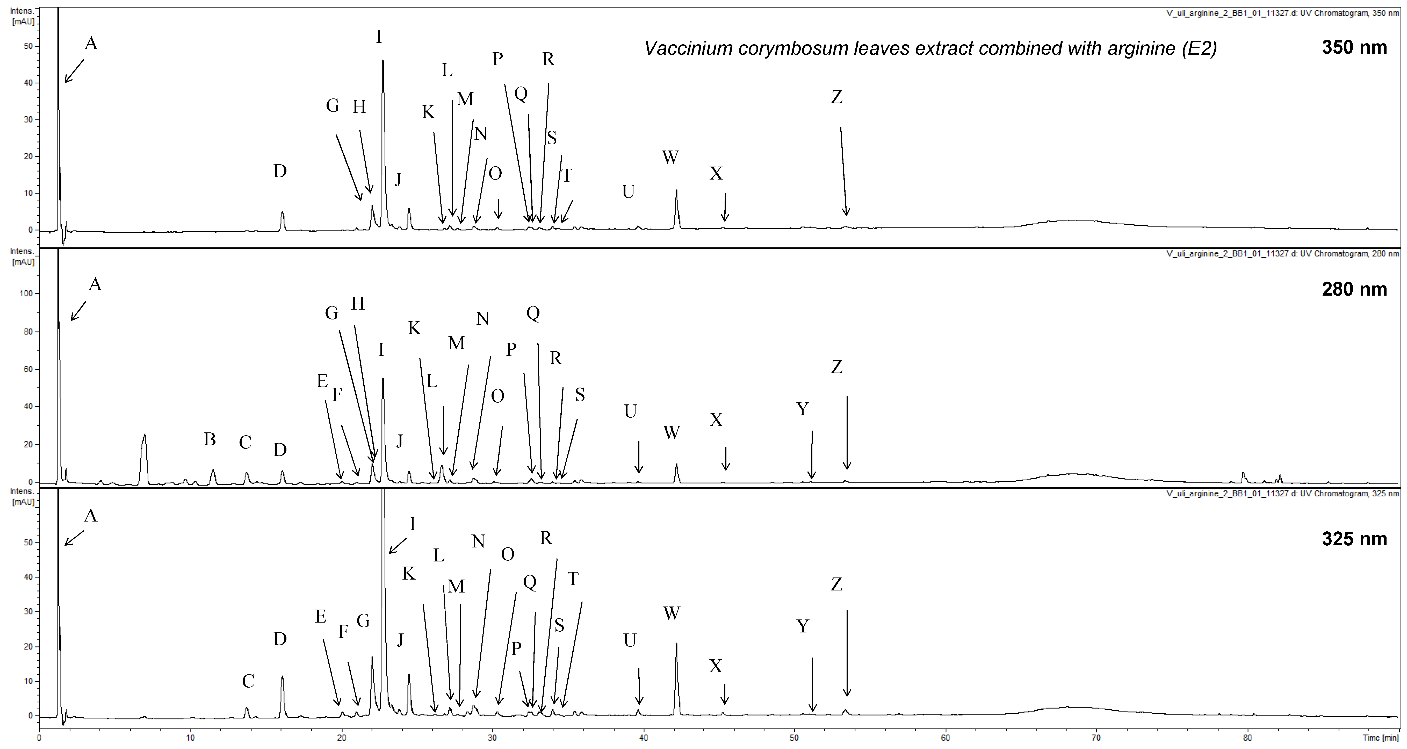
| No | Compound Name | Retention Time [min] | UV-Vis Maxima [nm] | MS− Ions | MS2− Ions | MS+ Ions | MS2+ Ions | Content/μg/mg | Qunatification Standard | Ions Used for Qunatification |
|---|---|---|---|---|---|---|---|---|---|---|
| A | quinic acid s | 1.4 | 227, 275 | 191 | - | 193 | - | 0.23 ± 0.21 | chlorogenic acid | 191 |
| B | phenolic acid derivative | 11.7 | 278 | 299 | 111, 173 b, 255 | 301 | 109, 224 b, 255 | 3.79 ± 0.05 | chlorogenic acid | 299 |
| C | phenolic acid derivative | 14.0 | 279, 311 | 299 | 109, 149, 173 b, 262, 281 | 301 | - | 0.06 ± 0.04 | chlorogenic acid | 299 |
| D | 3-O-caffeoylquinic acid (neochlorogenic acid) s | 16.3 | 300, 324 | 353 | 135, 179, 191 b | 355 | 145, 163 b, 337 | 1.06 ± 0.20 | chlorogenic acid | 353, 707 |
| E | 3-O-p-coumaroylquinic acid | 20.2 | 314 | 337 | 163 b, 173, 290 | 339 | 147 b | 0.14 ± 0.02 | chlorogenic acid | 337 |
| F | phenolic acid derivative | 21.1 | 288, 326 | 533 | 179, 191, 353, 489 b | 535 | 163, 355, 517 b | 0.17 ± 0.08 | chlorogenic acid | 533 |
| G | caffeic acid s | 22.3 | 243, 300 sh, 324 | 179 | - | 709 | 355 b, 447, 499, 517, 691 | 0.27 ± 0.06 | chlorogenic acid | 179 |
| H | caffeoylquinic acid-arginine conjugate I | 22.5 | 280, 325 | 527 | 365, 353 b, 191 | 529 | - | 0.58 ± 0.14 | chlorogenic acid | 527 |
| I | 5-O-caffeoylquinic acid (chlorogenic acid) s | 22.9 | 245, 300 sh, 325 | 353 | 215, 191 b, 173 | 355 | 145, 163 b | 9.57 ± 1.89 | chlorogenic acid | 353, 707 |
| J | 4-O-caffeoylquinic acid (cryptochlorogenic acid) s | 24.7 | 243, 300 sh, 326 | 353 | 173 b, 191 | 355 | 117, 163 b | 1.30 ± 0.26 | chlorogenic acid | 353, 707 |
| K | caffeoylquinic acid-arginine conjugate II | 26.0 | 280, 324 | 527 | 179, 191 b, 353, 489 | 529 | - | 0.43 ± 0.02 | chlorogenic acid | 533 |
| L | caffeoylquinic acid-arginine conjugate III | 26.7 | 283, 325 | 527 | 353 b, 191 | 529 | - | 0.20 ± 0.05 | chlorogenic acid | 527 |
| M | caffeoylquinic acid isomer | 27.3 | 242, 300 sh, 324 | 353 | 215, 191 b, 173 | 353 | - | 0.15 ± 0.07 | chlorogenic acid | 353, 707 |
| N | undefined compound | 28.9 | 312 | 439 | 233, 395 b | 441 | - | 0.12 ± 0.01 | chlorogenic acid | 439 |
| O | caffeoylshikimic acid isomer | 30.5 | 326 | 335 | 135, 179 b | 337 | 114, 209, 322 b | 0.18 ± 0.03 | chlorogenic acid | 335 |
| P | caffeoylshikimic acid isomer | 32.5 | 240, 324 | 335 | 135, 179 b | 337 | - | 0.16 ± 0.02 | chlorogenic acid | 335 |
| Q | dicaffeoylquinic acid ethyl ester isomer | 32.7 | 280, 325 | 543 | 353 b, 335, 191 | 545 | - | 0.08 ± 0.04 | chlorogenic acid | 543 |
| R | dicaffeoylquinic acid ethyl ester isomer | 33.2 | 280, 324 | 543 | 353 b, 289, 191 | 545 | - | 0.26 ± 0.13 | chlorogenic acid | 543 |
| S | caffeoylquinic acid ethyl ester isomer | 34.1 | 243, 300 sh, 326 | 381 | 135, 161 b, 207, 335 | 383 | - | 0.52 ± 0.11 | chlorogenic acid | 381, 763 |
| T | caffeoylshikimic acid isomer | 34.5 | 243, 300 sh, 323 | 335 | 135, 179 b | 337 | 0.11 ± 0.01 | chlorogenic acid | 335 | |
| U | caffeoylquinic acid ethyl ester isomer | 39.7 | 243, 300 sh, 326 | 381 | 161 b, 207, 335 | 383 | 163 b, 221 | 0.56 ± 0.14 | chlorogenic acid | 381, 763 |
| W | caffeoylquinic acid ethyl ester isomer | 42.3 | 243, 300 sh, 326 | 381 | 135, 179 b, 191 | 383 | 145, 163 b, 221, 365 | 6.23 ± 0.97 | chlorogenic acid | 381, 763 |
| X | caffeoylquinic acid ethyl ester isomer | 45.5 | 243, 300 sh, 326 | 381 | 135, 179 b, 191, 335 | 383 | 163b, 221 | 0.07 ± 0.01 | chlorogenic acid | 381, 763 |
| Y | undefined compound | 51.4 | 370 | 489 | 243, 269, 287 b | - | - | 0.15 ± 0.04 | chlorogenic acid | 489 |
| Z | undefined compound | 53.5 | 293, 333 | 207 | - | 227 | - | 0.24 ± 0.06 | chlorogenic acid | 207 |
| Phytochemical Group | Method Used | Content, % | |
|---|---|---|---|
| Extract 1 (E1) | Extract 2 (E2) | ||
| hydroxycinnamic acids derivatives | spectrophotometric as chlorogenic acid equivalents (λ = 327 nm) | 2.92 ± 0.12 # | 1.82 ± 0.02 |
| flavonoids | spectrophotometric as rutin equivalents (λ = 417 nm) | 3.03 ± 0.11 # | 1.96 ± 0.05 |
| total phenolics | spectrophotometric as gallic acid equivalents (λ = 270 nm) | 18.42 ± 0.97 # | 12.09 ± 0.07 |
| Treatment | Glucose, mmol/L | Insulin, pg/mL | Triacylglycerols (TAG), mmol/L | Ch-HDL, mmol/L | Ch-LDL, mmol/L |
|---|---|---|---|---|---|
| normal | 4.4 ± 0.09 | 1199 ± 25 | 0.78 ± 0.03 | 1.31 ± 0.03 | 2.73 ± 0.06 |
| HFD | 14.2 ± 0.19 * | 3005 ± 48 * | 2.26 ± 0.06 * | 0.69 ± 0.03 * | 3.56 ± 0.06 * |
| HFD_E1_150 mg/kg | 11.4 ± 0.17 *# | 2986 ± 37 * | 2.23 ± 0.12 * | 0.77 ± 0.11 * | 3.05 ± 0.09 * |
| HFD_E1_250 mg/kg | 9.1 ± 0.10 *# | 2347 ± 21 *# | 1.85 ± 0.09 *# | 0.79 ± 0.02 * | 3.44 ± 0.14 * |
| HFD_E1_350 mg/kg | 9.0 ± 0.21 *# | 2325 ± 35 *# | 1.93 ± 0.15 *# | 0.75 ± 0.07 * | 3.35 ± 0.07 * |
| HFD_E2_150 mg/kg | 11.6 ± 0.19 *# | 2793 ± 41 *# | 1.96 ± 0.12 * | 0.79 ± 0.08 *# | 3.41 ± 0.12 * |
| HFD_E2_250 mg/kg | 8.5 ± 0.18 *# | 2207 ± 19 *# | 1.56 ± 0.09 *# | 0.93 ± 0.08 *# | 3.31 ± 0.10 *# |
| HFD_E2_350 mg/kg | 8.7 ± 0.15 *# | 2211 ± 23 *# | 1.63 ± 0.13 *# | 0.91 ± 0.08 *# | 3.29 ± 0.09 * |
| HFD_Arg_250 mg/kg | 10.7 ± 0.25 * | 2604 ± 32 * | 1.98 ± 0.06 * | 0.73 ± 0.05 * | 3.58 ± 0.08 * |
| HFD_Q_50 mg/kg | 11.2 ± 0.10 *# | 2734 ± 19 *# | 2.24 ± 0.18 * | 0.80 ± 0.08 * | 3.39 ± 0.11 * |
Publisher’s Note: MDPI stays neutral with regard to jurisdictional claims in published maps and institutional affiliations. |
© 2021 by the authors. Licensee MDPI, Basel, Switzerland. This article is an open access article distributed under the terms and conditions of the Creative Commons Attribution (CC BY) license (https://creativecommons.org/licenses/by/4.0/).
Share and Cite
Koshovyi, O.; Granica, S.; Piwowarski, J.P.; Stremoukhov, O.; Kostenko, Y.; Kravchenko, G.; Krasilnikova, O.; Zagayko, A. Highbush Blueberry (Vaccinium corymbosum L.) Leaves Extract and Its Modified Arginine Preparation for the Management of Metabolic Syndrome—Chemical Analysis and Bioactivity in Rat Model. Nutrients 2021, 13, 2870. https://doi.org/10.3390/nu13082870
Koshovyi O, Granica S, Piwowarski JP, Stremoukhov O, Kostenko Y, Kravchenko G, Krasilnikova O, Zagayko A. Highbush Blueberry (Vaccinium corymbosum L.) Leaves Extract and Its Modified Arginine Preparation for the Management of Metabolic Syndrome—Chemical Analysis and Bioactivity in Rat Model. Nutrients. 2021; 13(8):2870. https://doi.org/10.3390/nu13082870
Chicago/Turabian StyleKoshovyi, Oleh, Sebastian Granica, Jakub P. Piwowarski, Oleksandr Stremoukhov, Yuliia Kostenko, Ganna Kravchenko, Oksana Krasilnikova, and Andriy Zagayko. 2021. "Highbush Blueberry (Vaccinium corymbosum L.) Leaves Extract and Its Modified Arginine Preparation for the Management of Metabolic Syndrome—Chemical Analysis and Bioactivity in Rat Model" Nutrients 13, no. 8: 2870. https://doi.org/10.3390/nu13082870
APA StyleKoshovyi, O., Granica, S., Piwowarski, J. P., Stremoukhov, O., Kostenko, Y., Kravchenko, G., Krasilnikova, O., & Zagayko, A. (2021). Highbush Blueberry (Vaccinium corymbosum L.) Leaves Extract and Its Modified Arginine Preparation for the Management of Metabolic Syndrome—Chemical Analysis and Bioactivity in Rat Model. Nutrients, 13(8), 2870. https://doi.org/10.3390/nu13082870