Article

Phycobiliproteins Ameliorate Gonadal Toxicity in Male Mice Treated with Cyclophosphamide

1
Departamento de Farmacia, Escuela Nacional de Ciencias Biológicas, Instituto Politécnico Nacional, Unidad Zacatenco, Mexico City 07738, Mexico
2
Departamento de Toxicología, Centro de Investigación y de Estudios Avanzados del Instituto Politécnico Nacional (Cinvestav-IPN), Unidad Zacatenco, Mexico City 07360, Mexico
3
Centro de Investigación en Ciencias de la Salud (CICSA), Facultad de Ciencias de la Salud, Universidad Anáhuac México, Mexico City 52786, Mexico
*
Author to whom correspondence should be addressed.
Nutrients 2021, 13(8), 2616; https://doi.org/10.3390/nu13082616
Submission received: 14 May 2021 / Revised: 16 July 2021 / Accepted: 26 July 2021 / Published: 29 July 2021
(This article belongs to the Section Proteins and Amino Acids)

Abstract

:
Cyclophosphamide (CP)—which is used to treat autoimmune diseases and cancer—is related to gonadotoxicity attributed to oxidative stress. As phycobiliproteins (PBPs) are strong antioxidants that are unexplored as protective agents against male gonadotoxicity, our work aimed to investigate the effects of PBP crude extract on testicular damage and sperm parameter alterations caused by CP in mice. Three doses of PBP (50, 100, and 200 mg/kg) were tested in the experimental groups (n = 8 per group), administered concomitantly with 100 mg/kg CP. After 42 days receiving PBP daily and CP weekly, body and relative testicular weights, serum testosterone levels, testicular lipoperoxidation and antioxidant enzyme activity levels, and testicular histology and sperm parameter alterations were assessed. The results showed that PBP crude extract at 200 mg/kg prevented testosterone serum reduction, body weight loss, lipoperoxidation and enzyme activity increments, and sperm parameter alterations and partially ameliorated relative testicular weight reductions and histological damage in CP-treated mice. In conclusion, we showed that PBP crude extract (200 mg/kg) mitigated oxidative damage in the testes and ameliorated alterations in sperm parameters in mice treated with CP (100 mg/kg); therefore, PBP extract could be considered as a potential protective agent against CP toxicity.

1. Introduction

Cyclophosphamide (CP) is a prodrug with alkylating activity that is widely used in chemotherapy, autoimmune disorders, and organ transplantation [1]. Despite its pharmacological benefits, CP has been associated with gonadotoxicity [2,3,4]. CP deteriorates sperm quality and atrophies testicular tissue in both humans and mice [5,6]. It also reduces superoxide dismutase (SOD) and glutathione peroxidase (GPX) activities in rat testes [7] and causes reductions correlated with impaired sperm quality in infertile patients [8,9]. CP metabolism generates active molecules such as phosphoramide mustard and acrolein [10], which are associated with therapeutic effect and toxicity, respectively [11]. Acrolein—a strong electrophile—creates oxidative stress conditions, modifies antioxidant enzymes [3], and reduces fertility in CP-treated patients [12]. As sperm cryopreservation—which is inaccessible to the majority of the population—is recommended before receiving chemotherapy [13,14], supplementation with phytocompounds and natural antioxidants has been proposed because they restore antioxidant balance and enhance male fertility [15]. C-phycocyanin (C-PC), allophycocyanin (APC), and phycoerythrin (PE)—known as phycobiliproteins (PBPs) [16]—are water-soluble fluorescent pigments found in cryptomonads, cyanelles, and cyanobacteria such as Spirulina maxima (SP) [17]. PBP extracts demonstrate antioxidant, antimicrobial, antiviral, antitumor, anti-inflammatory [18], antiteratogenic [19], antiulcerogenic [20], and nephroprotective [21] effects as well as preeclampsia-preventive activity [22]. Considering that PBPs’ activity against drug-induced gonadotoxicity is unexplored, the present work aimed to probe whether PBP extracted from SP could ameliorate testicular damage caused by multiple doses of CP in mice.

2. Materials and Methods

2.1. Reagents and Spirulina

Spirulina maxima powder was donated by Alimentos Esenciales para la Humanidad S.A. de C.V. (Mexico City, Mexico). Reagents and CP (CAS 6055-19-2, purity ≥ 98%) were purchased from Sigma-Aldrich Chemicals Co. (St. Louis, MO, USA) except for RANSOD and RANSEL kits obtained from Randox Laboratories (Crumlin, UK) and a testosterone enzyme-linked immunosorbent assay (ELISA) kit purchased from Cayman Chemical Co. (Ellsworth, MI, USA). All reagents were analytical grade.

2.2. PBP Extraction and Determination

Phycobiliproteins were extracted as described by Maurya et al. [23] by diluting SP (5 g) in phosphate-buffered saline (20 mL, 20 mM, pH 7.2). SP solution (0.25 g/mL) was frozen (−70 °C), thawed (24 ± 2 °C), shaken (5 min, 24 ± 2 °C) in darkness, and centrifuged twice at 14,000 rpm (JA-17 rotor, Beckman Coulte Co., Indianapolis, IN, USA) for 30 min at 4 °C. The supernatant was collected, PBPs were identified, and then the remaining supernatant was lyophilized and stored at −70 °C in darkness conditions. C-PC, APC, and PE optical densities (O.D.) were, respectively, determined at 615, 652, and 562 nm, while PBP concentrations were estimated as described by Bennett and Bogorad [24] and described using the following formulas:
C-PC (mg/mL) = [O.D.615 − 0.474(O.D.652)]/5.34,
APC (mg/mL) = [O.D.652 − 0.208(O.D.615)]/5.09,
PE (mg/mL) = [O.D.562 − 2.41(C-PC (mg/mL)) − 0.849(APC (mg/mL))]/9.62.
C-PC, APC, and PE purity indexes were estimated by dividing each O.D. by the total protein O.D. determined at 280 nm, while extraction yields were calculated from the wet basis by the following formula:
Extraction yield (mg/g) = [(PBPC-PC, APC, or PE (mg/mL)) (v(mL))]/SP (g),
where PBPC-PC, APC, and PE (mg/mL) represent the C-PC, APC, or PE concentration obtained, while v indicates the volume of phosphate-buffered saline.

2.3. Animals

The CD-1 male mice (25–30 g, 6–7 weeks old) from Bioterium of Universidad Autónoma del Estado de Hidalgo (Pachuca de Soto, Mexico) were housed in cages with access to LabDiet Rodent 5001 food (Fort Worth, TX, USA) and water ad libitum at constant temperature (24 ± 2 °C) and relative humidity (50 ± 10%) and with 12 h light/dark cycles. Conditions were maintained after and during the experimental procedures. The investigation protocol was previously approved by the Bioethical Committee of the National School of Biological Sciences of the National Polytechnic Institute (protocol code: ZOO-016-2020; 21 December 2020), and procedures with animals were performed in accordance with Mexican Official Standard NOM-062-ZOO-2001.

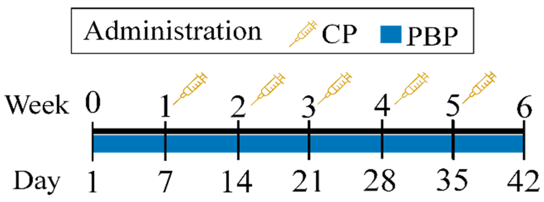
2.4. Experimental Design

Cyclophosphamide was dissolved in physiological solution (0.9% NaCl) and administered intraperitoneally at 100 mg/kg as described by Elangovan et al. [25], and Lu et al. [7], while PBPs were dissolved in phosphate-buffered saline and administered intragastrically at 50, 100, and 200 mg/kg—doubling and halving the 100 mg/kg dose reported by Castro-García et al. [22]. After an acclimatization week, treated male mice received daily i.g. doses of 50, 100, or 200 mg/kg PBP for 42 days concomitantly with weekly i.p. doses of 100 mg/kg CP (Figure 1). Control groups were included; whereby the vehicle control group received i.g. 0.9% NaCl daily for 42 days, the CP group received weekly i.g. doses of 100 mg/kg for 5 weeks, and the PBP group received daily i.p. doses of 200 mg/kg for 42 days; thus, experimental groups were defined as control, CP, PBP, PBP50 + CP, PBP100 + CP, and PBP200 + CP. At the end of treatment (day 42), the procedures described in Section 2.5, Section 2.6, Section 2.7, Section 2.8 and Section 2.9 were performed.

2.5. Serum Testosterone, Body Weight, and Relative Testicular Weight

Blood was collected by retro-orbital bleeding on day 42, and serum samples were separated to quantify testosterone levels using an ELISA kit. Mice were weighed and euthanized by cervical dislocation to immediately dissect the testes and epididymis and to determine the relative testicular weight.

2.6. Lipoperoxidation and SOD and GPX Activity in the Testes

The left testicle was homogenized in physiological solution by sonication for 40 s (output 40%, 20 kHz; Ultrasonic Homogenizer VP-050N, Taitec Corp., Koshigaya City, Saitama, Japan). Testicle homogenates were centrifuged at 10,000 rpm (1619 Rotor Universal 320 Centrifuge, Hettich®, Tuttlingen, DE) for 10 min at 4 °C and the supernatants were collected to evaluate lipoperoxidation and the enzymatic activity levels of SOD and GPX. Before determination, the total protein concentrations in supernatants were calculated by Bradford assay. Lipoperoxidation was determined by malondialdehyde (MDA) identification using thiobarbituric acid as proposed by Buege and Aust [26]. SOD and GPX activity levels were evaluated using RANSOD and RANSEL kits, respectively, following the manufacturers’ instructions.

2.7. Testicular Histology

The right testicle was conserved in buffered formaldehyde (4%, pH 7.4) until histologic observation and histomorphometry. After being dehydrated and embedded in paraffin [27], testicle sections of 5 µm thickness were prepared for hematoxylin and eosin (H&E) staining [28]. Ten stained sections per testicle were analyzed using an Olympus BX61 microscope (Olympus Corp., Shinjuku, Tokyo, Japan).

2.8. Histomorphometry

Morphology changes in seminiferous tubules were determined in five different fields from each testicle section. The total seminiferous tubule area (TSTA) and lumen tubule area (LTA) were measured with Image-Pro Plus® software (Media Cybernetics Inc., Rockville, MD, USA). Next, the seminiferous tubule area (STA) was estimated as the difference between the TSTA and LTA (Figure 2).

2.9. Sperm Parameter Assessment

Epididymis spermatozoa were collected via flushing with M-16 medium (100 mM NaCl, 25 mM NaHCO3, 5.5 mM glucose, 2.6 mM KCl, 1.56 mM Na2HPO4, 0.5 mM sodium pyruvate, 1.8 mM CaCl2, 0.5 mM, MgCl2, 20 mM sodium lactate, 100 IU/mg penicillin, and 100 µg/mL streptomycin at pH 7.2 and 37 °C). The progressive motility, sperm count, and cell viability were evaluated as sperm parameters according to World Health Organization guidelines [29]. Progressive motility was determined on a glass slide, sperm were counted in a hemocytometer, and sperm viability was evaluated by eosin–nigrosin exclusion assay; a phase contrast microscope (Carl Zeiss Microscopy Co., Oberkochen, Germany) was used for all determinations.

2.10. Statistical Analysis

The results were analyzed by one-way ANOVA followed by the Holm–Sidak multiple comparison test. Data were represented as means ± standard error of the mean (SEM), and p < 0.05 was considered statistically significant. The PBP, PBP50 + CP, PBP100 + CP, and PBP200 + CP groups were compared with the control and CP groups. Analysis was performed and graphs were produced in SigmaPlot v12.0 software. All samples were evaluated in triplicate.

3. Results

3.1. PBP Determination

The PBP concentrations, purity index values, and extraction yields are indicated in Table 1. C-PC, APC, and PE, respectively, corresponded to 69.04, 30.34, and 0.62% of the PBPs extracted from SP, with C-PC being 2.3- and 111.5-fold more abundant than APC and PE, respectively. Additionally, the purity index and extraction yield of C-PC were higher than the purity index values and extraction yields of APC and PE.

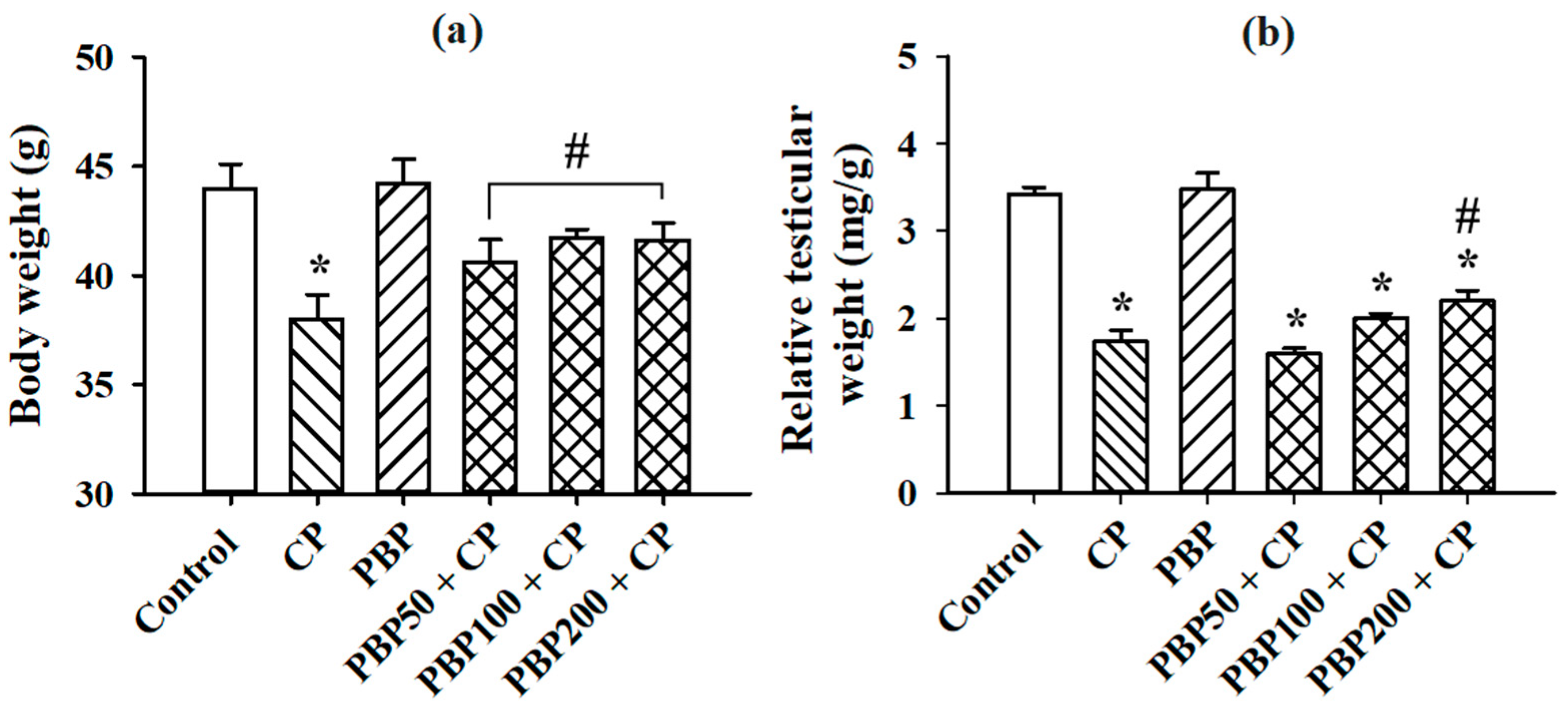
3.2. Serum Testosterone, Body Weight, and Relative Testicular Weight

Figure 3 shows the results obtained from the testosterone serum determination. PBP at 100 and 200 mg/kg prevented the reductions in testosterone levels observed in the CP group. Although the PBP50 + CP group showed testosterone levels statistically lower than control, PBP50 + CP testosterone levels were 7.9% higher than the CP group. The body weight results (Figure 4a) showed that PBP at 50, 100, and 200 mg/kg prevented weight loss caused by CP, while the relative testicular weight data (Figure 4b) indicated that all groups receiving CP decreased their relative testicular weight compared to control; however, the statistical analysis showed that the PBP200 + CP group presented relative testicular weights higher than those of the CP group, suggesting that the PBP 200 mg/kg dose ameliorated testicular weight reduction.

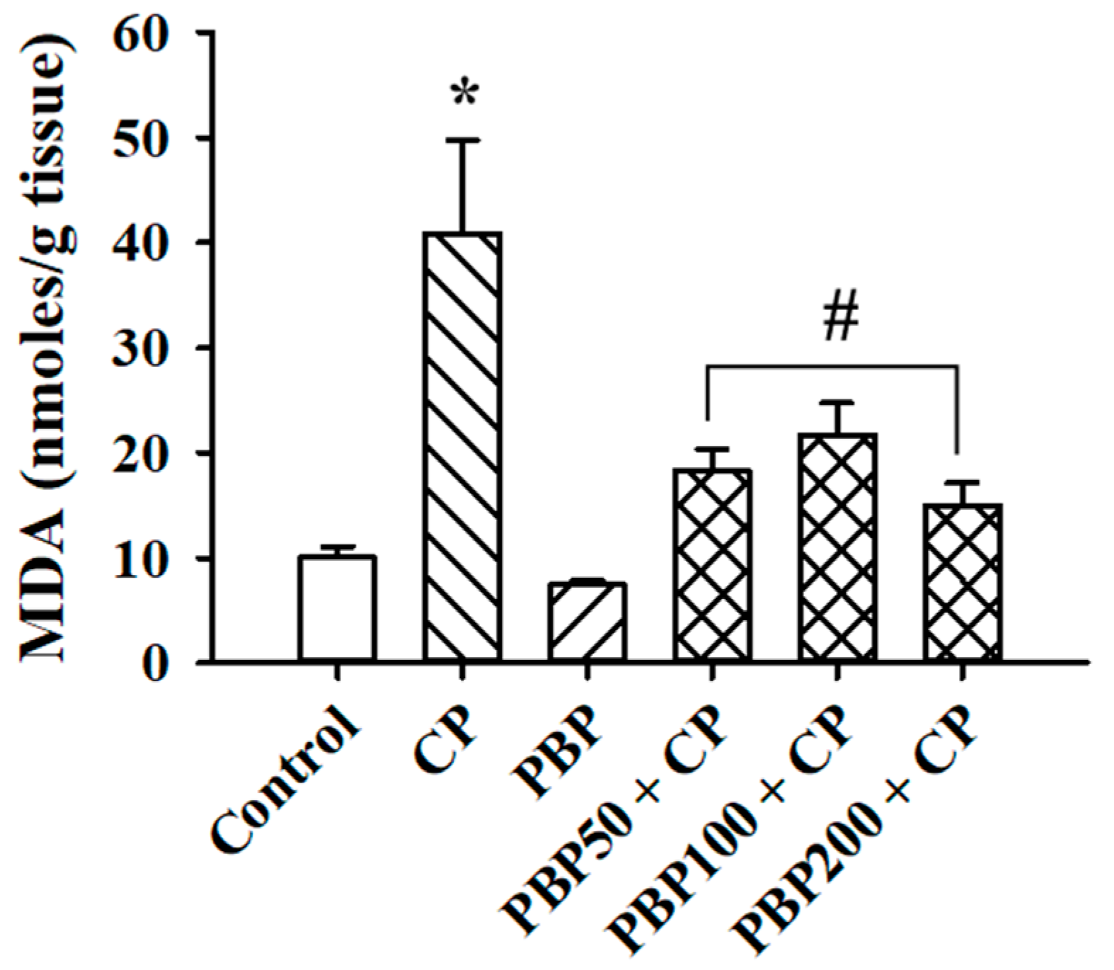
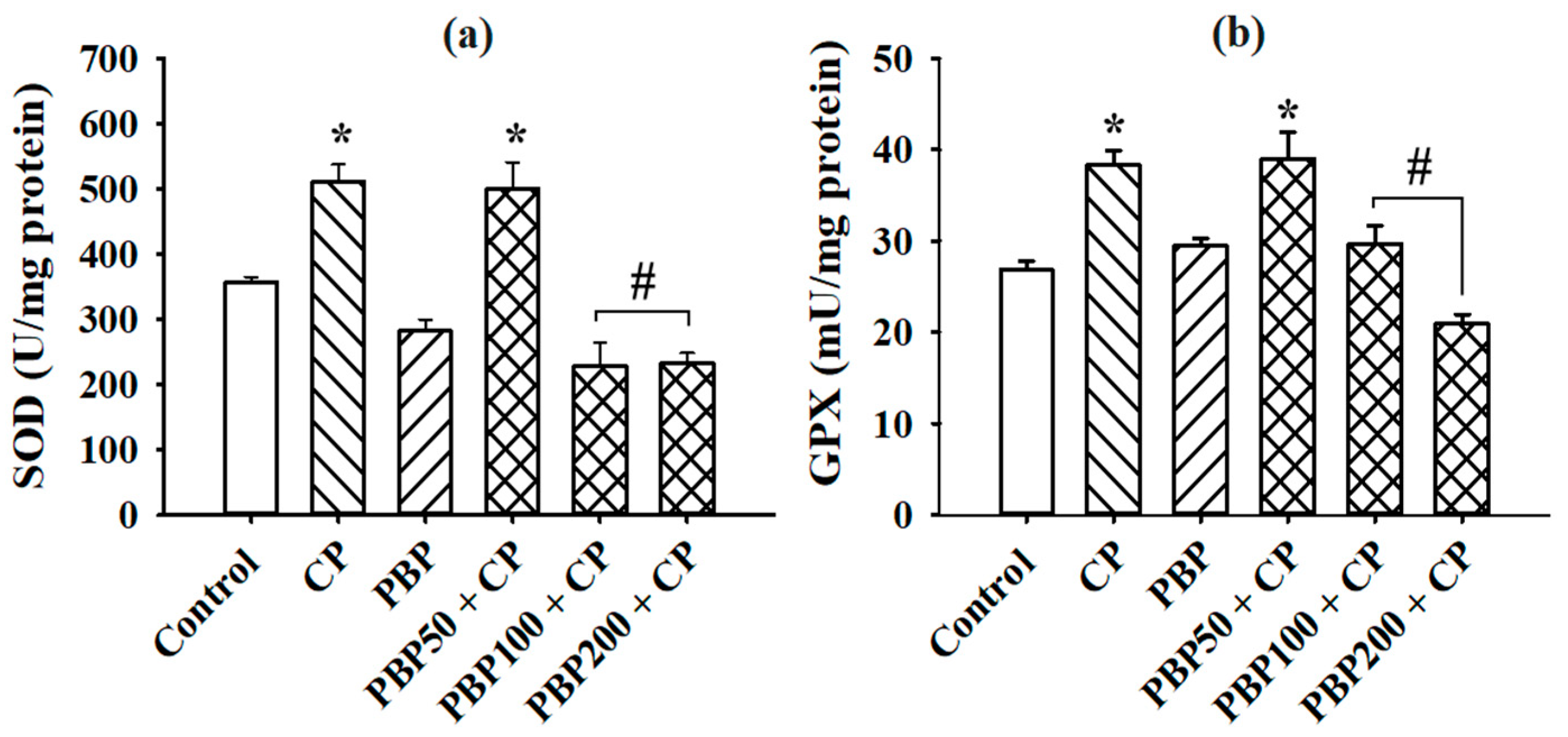
3.3. Lipoperoxidation and SOD and GPX Activity in the Testes

The MDA levels, determined as lipoperoxidation indicators, are shown in Figure 5. MDA in the CP group was fourfold greater than the control, while PBP50 + CP, PBP100 + CP, and PBP200 + CP groups maintained MDA at similar levels to the control group, suggesting a protective effect of PBP (50–200 mg/kg) against testicular lipoperoxidation induced by CP. On the other hand, as presented in Figure 6, the enzymatic activity of SOD and GPX increased in the CP and PBP50 + CP groups compared with the control, while the PBP100 + CP and PBP200 + CP groups showed similar SOD and GPX activity levels to the control group.

3.4. Testicular Histology

Figure 7 shows representative micrographs from histological observations. The control and PBP groups presented intact seminiferous tubules where spermatogonium, spermatocytes, and spermatids were identified. Similarly, the PBP50 + CP, PBP100 + CP, and PBP200 + CP groups preserved germinal cells from the basal membrane to the tubule lumen, while the CP group showed small tubules and large spaces in both the tubule lumen and interstitium, indicating possible depletion of cells, especially mature spermatids and Leydig cells. As interstitial spaces were also observed in PBP50 + CP micrographs, the testicular histology indicated that PBPs at the 50 mg/kg dose partially ameliorated damage caused by CP, while the 100 and 200 mg/kg doses improved testicle histology.

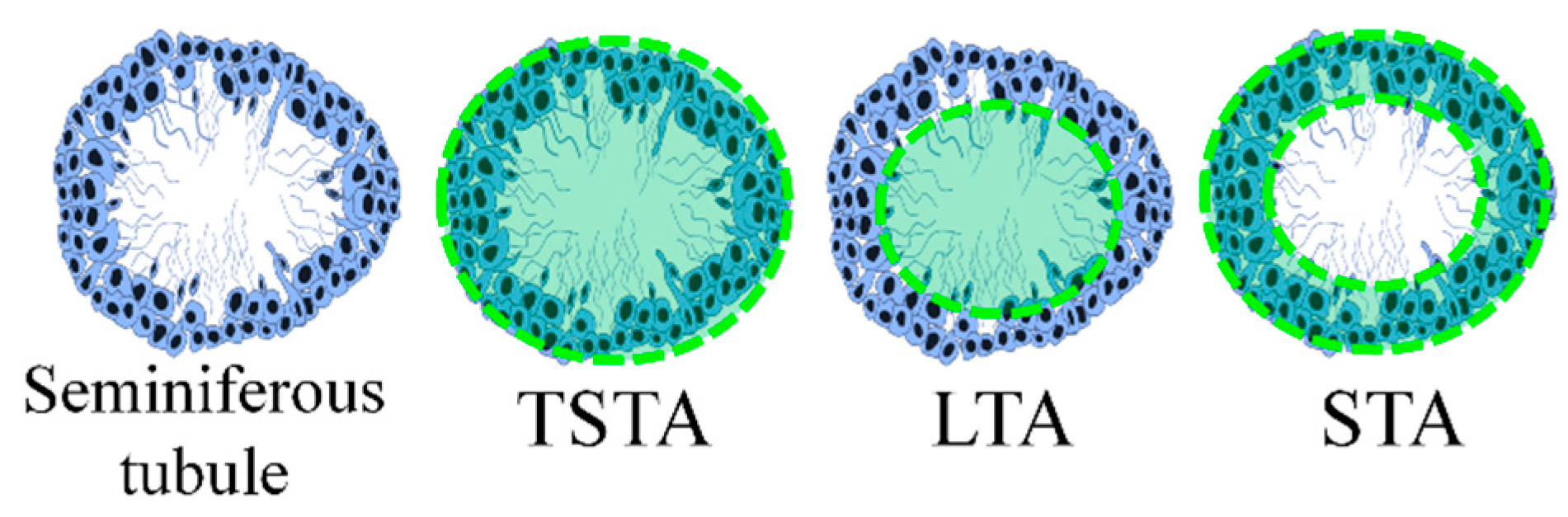
3.5. Histomorphometry

The TSTA, LTA, and STA are shown in Table 2. Although large spaces in the seminal lumen were observed only in the CP group micrograph (Figure 7b), the LTA diminished in all the groups compared with the control, while TSTA was reduced only in the CP group. STA, which represented the seminal tubule cellular structure (Figure 2), was decreased in the CP group compared with the control, confirming tubule reduction in the CP micrograph (Figure 7b). The PBP50 + CP, PBP100 + CP, and PBP200 + CP groups presented STA values lower than the control but greater than the CP group, indicating partial improvement of testicular CP damage. Interestingly, TSTA and STA levels in the PBP group (without CP) were 12.8 and 21.9% higher than the control, respectively, suggesting that PBP consumption (200 mg/kg) contributed to germinal cell increase.

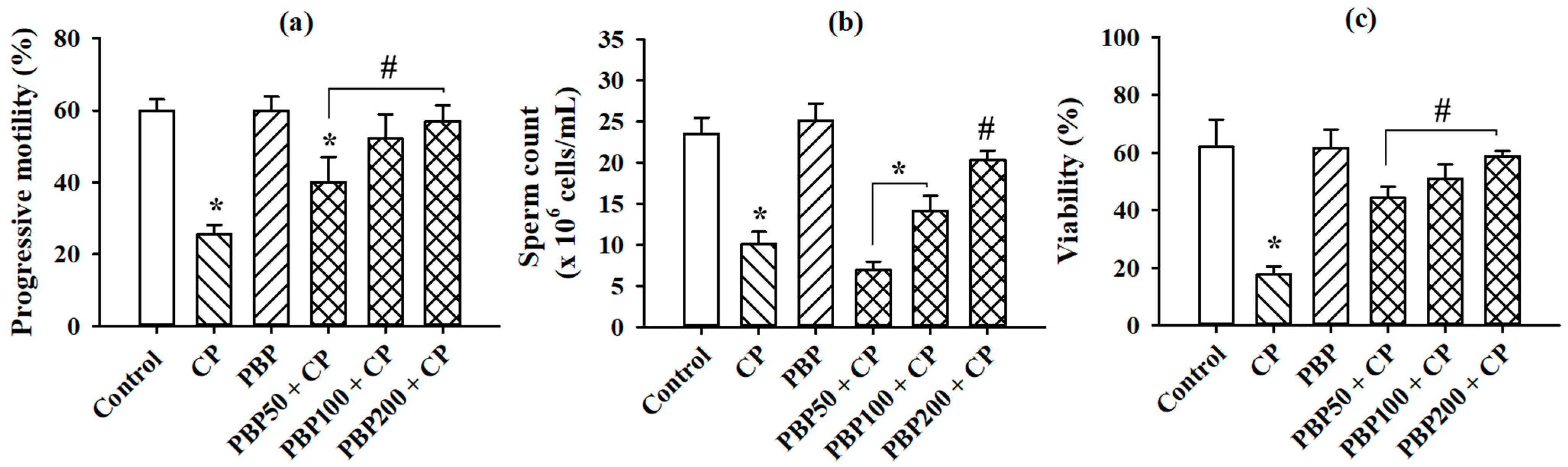
3.6. Sperm Parameters

The progressive motility, sperm count, and sperm viability—evaluated as parameters of sperm quality—are shown in Figure 8. The three evaluated parameters were reduced in the CP group compared with the control. PBP50 + CP, PBP100 + CP, and PBP200 + CP maintained cell viability; PBP100 + CP and PBP200 + CP preserved progressive motility; and PBP200 + CP sustained sperm count at similar levels to the control. The data suggested that PBPs at 200 mg/kg were required to recover sperm parameter alterations caused by CP administration in mice.

4. Discussion

Although CP is used to treat autoimmune diseases and cancer, reproductive organ toxicity has been shown in patients and experimental models [30,31,32]. As CP toxicity is attributed to oxidative stress [10], and PBPs demonstrate strong antioxidant properties [33], we explored the effects of PBP crude extract on the testicular damage caused by multiple CP doses in mice. In PBP crude extract obtained from SP, we identified a higher C-PC percentage (69.04%) than APC (30.34%) and PE (0.62%) (Table 1). C-PC, which is related to high antioxidant capacity [16], was reported in PBP crude extracts from SP by Rodríguez-Sánchez et al. at a low percentage (C-CP, 47%) [21] and by Walter et al. at a reduced concentration (C-PC, 0.237 mg/mL) [34] as compared with our results; however, the C-PC purity index reported by Walter (0.8) correlates with our C-PC purity index result (0.7), with both index values considered to be of food grade (purity index ≥ 0.7) [35]. PBP concentrations (50, 100, and 200 mg/mL) were selected according to the results observed by Castro-García et al. [22] in a rat preeclampsia model, while the duration of CP treatment was determined considering the spermatogenesis cycle [36].
Our results are in agreement with the CP effects previously reported by Elangovan et al. [25], Lu et al. [7], and Iqubal et al. [37] including reductions in serum testosterone, body weight, relative testis weight, and sperm parameter alterations; increases in lipoperoxidation and enzyme activity (SOD and GPX); and testicle damage observed by histology. The PBP crude extract mitigated certain CP effects, depending on the PBP dose administered. In particular, PBP at 200 mg/kg prevented changes in testosterone serum, body weight, lipoperoxidation, enzyme activity, and evaluated sperm parameters while partially ameliorating relative testicular weight loss and histological damage. Body weight loss is attributed to appetite disruption caused by sensory taste cell modifications after CP administration in mice [38]. Protein consumption is recommended to avoid weight loss and malnutrition in patients receiving chemotherapy [39,40]; thus, body weight recuperation observed in CP-treated mice at PBP doses of 50, 100, and 200 mg/kg was probably an effect of the protein content in the PBP crude extract.
The antioxidant effects of PBPs have been previously demonstrated [17,41], which were confirmed in our results, whereby lipoperoxidation and SOD and GPX enzymatic activity increments were prevented with PBP doses of 100 and 200 mg/kg. As SOD and GPX activity, as well as lipoperoxidation [42], are proportional to oxidative stress conditions [43,44,45] and because the antioxidant effects of PBP are related to the capacity of C-PC to trap reactive molecules such as acrolein [46], oxidative stress—generated as a consequence of acrolein interactions—was possibly reduced by the presence of C-PC in the PBP crude extract; however, the PBP crude extract at 200 mg/kg—the highest concentration tested in our study—partially ameliorated the relative testis weight reduction and testicular damage observed by histology. Decrements in relative testicular weight were probably related to STA reductions and decreases in Leydig cells and spermatids in the CP group. Despite our histological results revealing that spermatogonia were preserved after multiple CP doses as previously described by Drumond et al. [47], the interstitial spaces observed in the CP and PBP50 + CP groups suggest Leydig cells loss. Gu et al. [48] related this to the autophagy process of Leydig cells that is induced by CP. As Leydig cells maintain testosterone secretion [49], observed testosterone reductions confirm Leydig cell loss. Although testosterone levels and interstitial spaces improved in PBP 100 and 200 mg/kg groups, suggesting that Leydig cells were recovered, STA values and relative testicular weights were lower than the control, suggesting that cell populations were deteriorated. Interestingly, the PBP (200 mg/kg)-treated group without CP presented higher STA values than the control, probably because the antioxidant activity of PBPs reduced the oxidant molecules required for regular apoptosis processes [50].
On the other hand, sperm parameters such as progressive motility, sperm count, and cell viability were completely recovered at the PBP dose of 200 mg/kg. The effects of the PBP crude extract on sperm parameter alterations in CP-treated mice are attributable to the antioxidant activities of PBPs because damage observed in sperm is directly related to oxidative stress conditions. For example, decreased sperm motility is a result of flagellum defects caused by oxidative damage in the tubulin [51], reduced sperm count is related to spermatogenesis inhibition or apoptosis induction by oxidant molecules, and cell viability is associated with apoptosis triggered by oxidative stress [52].
The NADPH oxidase system (NOX)—embedded in the plasma membrane—catalyzes O2•− production through the oxidation of NADPH. NADPH oxidase isoform 5 (NOX5) is a potential candidate for the ROS-generating system in spermatozoa [53,54]. NOX5 can be directly activated by PKCα independent of an increase in intracellular calcium [55]. Acrolein, which is likely the agent responsible for the toxicity of CP to spermatozoa, is known to activate PKCα [56]; hence, overactivation of NOX5 may be largely responsible for the pro-oxidative impact of CP on spermatozoa. Intracellular free bilirubin generated by heme oxygenase is known to inhibit various isoforms of NADPH oxidase in low nanomolar concentrations [57], although its specific effect on the NOX5 form has not been reported. Phycocyanobilin—a compound present in the chromophores of phycocyanin and allophycocyanin [17]—is converted within cells to phycocyanorubin, which is nearly identical in structure to bilirubin [58]; this may explain why phycocyanobilin has been found to mimic the NADPH oxidase-inhibitory effect of biliverdin or bilirubin in cell cultures. Our results, therefore, suggest that bilirubin and phycocyanobilin can function as inhibitors of NOX5, thereby protecting spermatozoa from acrolein. This possibility could be easily tested in vitro.
As PBP crude extract was studied in the present work, the beneficial effects observed against CP gonadotoxicity in male mice could be increased by other phytochemicals with antioxidant properties such as the polyphenols [59] and polysaccharides phycobiliproteins (PBPs) [60] previously identified in SP. Although our results suggest that PBPs ameliorate CP damage, this study was limited to evaluating healthy mice; hence, the effects of PBPs must be assessed in an animal model of cancer or autoimmune disease to exclude an inactivation of the therapeutic effects of CP.

5. Conclusions

Phycobiliproteins crude extract at 200 mg/kg ameliorated gonadotoxicity caused by CP in male mice. Alterations in testosterone serum, body weight, lipoperoxidation, enzyme activity, and sperm parameters were completely prevented, while relative testicular weight loss and histological damage were attenuated; however, additional CP function tests are recommended.

Author Contributions

Conceptualization, J.B.-B., I.H.-O., M.A.M.-V. and G.C.-C.; data curation, J.B.-B.; formal analysis, J.B.-B.; funding acquisition, G.C.-C.; investigation, J.B.-B., M.A.M.-V. and R.I.M.-G.; methodology, J.B.-B.; project administration, I.H.-O. and G.C.-C.; resources, G.C.-C.; supervision, I.H.-O. and G.C.-C.; visualization, X.A.-P.; writing—original draft, J.B.-B. and X.A.-P.; writing—review and editing, X.A.-P. and G.G.-S. All authors have read and agreed to the published version of the manuscript.

Funding

This research received no external funding.

Institutional Review Board Statement

The study was conducted according to the guidelines of Mexican Official Standard NOM-062-ZOO-2001 and approved by the Bioethical Committee of the National School of Biological Sciences of the National Polytechnic Institute (protocol code: ZOO-016-2020; 21 December 2020).

Acknowledgments

J.B.-B. appreciates the graduate scholarship awarded by the National Council of Science and Technology, Mexico (589497) as well as the scholarship furnished by Postgraduate Studies and Research Science, National Polytechnic Institute, Mexico (20180176). Thanks are given to Fermín Florentino Luna Coronel for helping with the testicular histology and Sihomara García Zepeda for her technical assistance.

Conflicts of Interest

The authors declare no conflict of interest.

References

  1. Jalali, A.S.; Hasanzadeh, S.; Malekinejad, H. Achillea millefolium inflorescence aqueous extract ameliorates cyclophosphamide-induced toxicity in rat testis: Stereological evidences. Chin. J. Nat. Med. 2012, 10, 247–254. [Google Scholar] [CrossRef]
  2. Potnuri, A.G.; Allakonda, L.; Lahkar, M. Crocin attenuates cyclophosphamide induced testicular toxicity by preserving glutathione redox system. Biomed. Pharmacother. 2018, 101, 174–180. [Google Scholar] [CrossRef]
  3. Ghobadi, E.; Moloudizargari, M.; Asghari, M.H.; Abdollahi, M. The mechanisms of cyclophosphamide-induced testicular toxicity and the protective agents. Expert Opin. Drug Metab. Toxicol. 2017, 13, 525–536. [Google Scholar] [CrossRef]
  4. Wetzels, J. Cyclophosphamide-induced gonadal toxicity: A treatment dilemma in patients with lupus nephritis? Neth. J. Med. 2004, 62, 347–352. [Google Scholar]
  5. Drobnis, E.Z.; Nangia, A.K. Immunosuppressants and Male Reproduction. Adv. Exp. Med. Biol. 2017, 1034, 179–210. [Google Scholar] [CrossRef]
  6. Hamzeh, M.; Hosseinimehr, S.J.; Karimpour, A.; Mohammadi, H.R.; Khalatbary, A.R.; Talebpour Amiri, F. Cerium Oxide Nanoparticles Protect Cyclophosphamide-Induced Testicular Toxicity in Mice. Int. J. Prev. Med. 2019, 10, 5. [Google Scholar] [CrossRef]
  7. Lu, W.P.; Mei, X.T.; Wang, Y.; Zheng, Y.P.; Xue, Y.F.; Xu, D.H. Zn(II)-curcumin protects against oxidative stress, deleterious changes in sperm parameters and histological alterations in a male mouse model of cyclophosphamide-induced reproductive damage. Environ. Toxicol. Pharmacol. 2015, 39, 515–524. [Google Scholar] [CrossRef]
  8. Crisol, L.; Matorras, R.; Aspichueta, F.; Expósito, A.; Hernández, M.L.; Ruiz-Larrea, M.B.; Mendoza, R.; Ruiz-Sanz, J.I. Glutathione peroxidase activity in seminal plasma and its relationship to classical sperm parameters and in vitro fertilization-intracytoplasmic sperm injection outcome. Fertil. Steril. 2012, 97, 852–857.e1. [Google Scholar] [CrossRef]
  9. Yan, L.; Liu, J.; Wu, S.; Zhang, S.; Ji, G.; Gu, A. Seminal superoxide dismutase activity and its relationship with semen quality and SOD gene polymorphism. J. Assist. Reprod. Genet. 2014, 31, 549–554. [Google Scholar] [CrossRef] [Green Version]
  10. Liu, F.; Li, X.L.; Lin, T.; He, D.W.; Wei, G.H.; Liu, J.H.; Li, L.S. The cyclophosphamide metabolite, acrolein, induces cytoskeletal changes and oxidative stress in Sertoli cells. Mol. Biol. Rep. 2012, 39, 493–500. [Google Scholar] [CrossRef]
  11. Kim, S.H.; Lee, I.C.; Lim, J.H.; Moon, C.; Bae, C.S.; Kim, S.H.; Shin, D.H.; Park, S.C.; Kim, H.C.; Kim, J.C. Protective effects of pine bark extract on developmental toxicity of cyclophosphamide in rats. Food Chem. Toxicol. 2012, 50, 109–115. [Google Scholar] [CrossRef]
  12. Watcho, P.; Mpeck, I.R.; Deeh Defo, P.B.; Wankeu-Nya, M.; Ngadjui, E.; Bonsou Fozin, G.R.; Kamtchouing, P.; Kamanyi, A. Cyclophosphamide-induced reproductive toxicity: Beneficial effects of Helichrysum odoratissimum (Asteraceae) in male Wistar rats. J. Integr. Med. 2019, 17, 366–373. [Google Scholar] [CrossRef]
  13. Özatik, F.Y.; Özatik, O.; Tekşen, Y.; Yiğitaslan, S.; Ari, N.S. Protective and therapeutic effect of Hydrogen sulfide on hemorrhagic cystitis and testis dysfunction induced with Cyclophosphamide. Turkish J. Med. Sci. 2021. [Google Scholar] [CrossRef] [PubMed]
  14. Mulder, R.L.; Font-Gonzalez, A.; Green, D.M.; Loeffen, E.A.H.; Hudson, M.M.; Loonen, J.; Yu, R.; Ginsberg, J.P.; Mitchell, R.T.; Byrne, J.; et al. Fertility preservation for male patients with childhood, adolescent, and young adult cancer: Recommendations from the PanCareLIFE Consortium and the International Late Effects of Childhood Cancer Guideline Harmonization Group. Lancet Oncol. 2021, 22, e57–e67. [Google Scholar] [CrossRef]
  15. Adewoyin, M.; Ibrahim, M.; Roszaman, R.; Isa, M.L.M.; Alewi, N.A.M.; Rafa, A.A.A.; Anuar, M.N.N. Male Infertility: The Effect of Natural Antioxidants and Phytocompounds on Seminal Oxidative Stress. Diseases 2017, 5, 9. [Google Scholar] [CrossRef] [PubMed]
  16. Yu, P.; Wu, Y.; Wang, G.; Jia, T.; Zhang, Y. Purification and bioactivities of phycocyanin. Crit. Rev. Food Sci. Nutr. 2017, 57, 3840–3849. [Google Scholar] [CrossRef]
  17. Pagels, F.; Guedes, A.C.; Amaro, H.M.; Kijjoa, A.; Vasconcelos, V. Phycobiliproteins from cyanobacteria: Chemistry and biotechnological applications. Biotechnol. Adv. 2019, 37, 422–443. [Google Scholar] [CrossRef] [PubMed]
  18. Righini, H.; Francioso, O.; Di Foggia, M.; Quintana, A.M.; Roberti, R. Preliminary Study on the Activity of Phycobiliproteins against Botrytis cinerea. Mar. Drugs 2020, 18, 600. [Google Scholar] [CrossRef]
  19. Vazquez-Sanchez, J.; Ramon-Gallegos, E.; Mojica-Villegas, A.; Madrigal-Bujaidar, E.; Perez-Pasten-Borja, R.; Chamorro-Cevallos, G. Spirulina maxima and its protein extract protect against hydroxyurea-teratogenic insult in mice. Food Chem. Toxicol. 2009, 47, 2785–2789. [Google Scholar] [CrossRef]
  20. Guzman-Gomez, O.; Garcia-Rodriguez, R.V.; Quevedo-Corona, L.; Perez-Pasten-Borja, R.; Rivero-Ramirez, N.L.; Rios-Castro, E.; Perez-Gutierrez, S.; Perez-Ramos, J.; Chamorro-Cevallos, G.A. Amelioration of Ethanol-Induced Gastric Ulcers in Rats Pretreated with Phycobiliproteins of Arthrospira (Spirulina) Maxima. Nutrients 2018, 10, 763. [Google Scholar] [CrossRef] [Green Version]
  21. Rodríguez-Sánchez, R.; Ortiz-Butrón, R.; Blas-Valdivia, V.; Hernández-García, A.; Cano-Europa, E. Phycobiliproteins or C-phycocyanin of Arthrospira (Spirulina) maxima protect against HgCl2-caused oxidative stress and renal damage. Food Chem. 2012, 135, 2359–2365. [Google Scholar] [CrossRef]
  22. Castro-Garcia, S.Z.; Chamorro-Cevallos, G.; Quevedo-Corona, L.; McCarty, M.F.; Bobadilla-Lugo, R.A. Beneficial effects of phycobiliproteins from Spirulina maxima in a preeclampsia model. Life Sci. 2018, 211, 17–24. [Google Scholar] [CrossRef]
  23. Maurya, S.S.; Maurya, J.N.; Pandey, V.D. Factors regulating phycobiliprotein production in cyanobacteria. Int. J. Curr. Microbiol. Appl. Sci. 2014, 3, 764–771. [Google Scholar]
  24. Bennett, A.; Bogorad, L. Complementary chromatic adaptation in a filamentous blue-green alga. J. Cell Biol. 1973, 58, 419–435. [Google Scholar] [CrossRef] [PubMed]
  25. Elangovan, N.; Chiou, T.J.; Tzeng, W.F.; Chu, S.T. Cyclophosphamide treatment causes impairment of sperm and its fertilizing ability in mice. Toxicology 2006, 222, 60–70. [Google Scholar] [CrossRef] [PubMed]
  26. Buege, J.A.; Aust, S.D. Microsomal lipid peroxidation. In Methods in Enzymology; Fleischer, S., Packer, L., Eds.; Academic Press: Cambridge, MA, USA, 1978; Volume 52, pp. 302–310. ISBN 0076-6879. [Google Scholar]
  27. Layton, C.; Bancroft, J.D.; Suvarna, S.K. 4—Fixation of tissues. In Bancroft’s Theory and Practice of Histological Techniques, 8th ed.; Suvarna, S.K., Layton, C., Bancroft, J.D., Eds.; Elsevier: Amsterdam, The Netherlands, 2019; pp. 40–63. ISBN 978-0-7020-6887-4. [Google Scholar]
  28. Bancroft, J.D.; Layton, C. 10—The hematoxylins and eosin. In Bancroft’s Theory and Practice of Histological Techniques, 8th ed.; Suvarna, S.K., Layton, C., Bancroft, J.D., Eds.; Elsevier: Amsterdam, The Netherlands, 2019; pp. 126–138. ISBN 978-0-7020-6887-4. [Google Scholar]
  29. WHO. WHO Laboratory Manual for the Examination and Processing of Human Semen. Available online: https://apps.who.int/iris/handle/10665/44261 (accessed on 18 September 2017).
  30. Tripathi, D.N.; Jena, G.B. Astaxanthin inhibits cytotoxic and genotoxic effects of cyclophosphamide in mice germ cells. Toxicology 2008, 248, 96–103. [Google Scholar] [CrossRef] [PubMed]
  31. Aguilar-Mahecha, A.; Hales, B.F.; Robaire, B. Chronic Cyclophosphamide Treatment Alters the Expression of Stress Response Genes in Rat Male Germ Cells. Biol. Reprod. 2002, 66, 1024–1032. [Google Scholar] [CrossRef] [Green Version]
  32. Gajjar, R.; Miller, S.D.; Meyers, K.E.; Ginsberg, J.P. Fertility preservation in patients receiving cyclophosphamide therapy for renal disease. Pediatr. Nephrol. 2015, 30, 1099–1106. [Google Scholar] [CrossRef] [Green Version]
  33. Li, W.; Su, H.-N.; Pu, Y.; Chen, J.; Liu, L.-N.; Liu, Q.; Qin, S. Phycobiliproteins: Molecular structure, production, applications, and prospects. Biotechnol. Adv. 2019, 37, 340–353. [Google Scholar] [CrossRef]
  34. Walter, A.; De Carvalho, J.; Thomaz-Soccol, V.; Faria, A.; Ghiggi, V.; Soccol, C. Study of Phycocyanin Production from Spirulina platensis under Different Light Spectra. Braz. Arch. Biol. Technol. 2011, 54, 675–682. [Google Scholar] [CrossRef]
  35. Chen, X.; Wu, M.; Yang, Q.; Wang, S. Preparation, characterization of food grade phycobiliproteins from Porphyra haitanensis and the application in liposome-meat system. Lwt 2017, 77, 468–474. [Google Scholar] [CrossRef]
  36. Marchetti, F.; Aardema, M.; Beevers, C.; van Benthem, J.; Douglas, G.R.; Godschalk, R.; Yauk, C.L.; Young, R.; Williams, A. Simulation of mouse and rat spermatogenesis to inform genotoxicity testing using OECD test guideline 488. Mutat. Res. Toxicol. Environ. Mutagen. 2018, 832–833, 19–28. [Google Scholar] [CrossRef] [PubMed]
  37. Iqubal, A.; Syed, M.A.; Najmi, A.K.; Ali, J.; Haque, S.E. Ameliorative effect of nerolidol on cyclophosphamide-induced gonadal toxicity in Swiss Albino mice: Biochemical-, histological- and immunohistochemical-based evidences. Andrologia 2020, 52, e13535. [Google Scholar] [CrossRef]
  38. Jewkes, B.C.; Gomella, M.G.; Lowry, E.T.; Benner, J.A.; Delay, E.R. Cyclophosphamide-Induced Disruptions to Appetitive Qualities and Detection Thresholds of NaCl: Comparison of Single-Dose and Dose Fractionation Effects. Chem. Senses 2018, 43, 399–410. [Google Scholar] [CrossRef]
  39. Ravasco, P. Nutrition in Cancer Patients. J. Clin. Med. 2019, 8, 1211. [Google Scholar] [CrossRef] [Green Version]
  40. Yalcin, S.; Gumus, M.; Oksuzoglu, B.; Ozdemir, F.; Evrensel, T.; Sarioglu, A.A.; Sahin, B.; Mandel, N.M.; Goker, E. Nutritional Aspect of Cancer Care in Medical Oncology Patients. Clin. Ther. 2019, 41, 2382–2396. [Google Scholar] [CrossRef]
  41. Chandra, R.; Parra, R.; Iqbal, H.M.N. Phycobiliproteins: A Novel Green Tool from Marine Origin Blue-Green Algae and Red Algae. Protein Pept. Lett. 2017, 24, 118–125. [Google Scholar] [CrossRef]
  42. Mladenov, M.; Gokik, M.; Hadzi-Petrushev, N.; Gjorgoski, I.; Jankulovski, N. The relationship between antioxidant enzymes and lipid peroxidation in senescent rat erythrocytes. Physiol. Res. 2015, 64, 891–896. [Google Scholar] [CrossRef]
  43. Aliciguzel, Y.; Ozen, I.; Aslan, M.; Karayalcin, U. Activities of xanthine oxidoreductase and antioxidant enzymes in different tissues of diabetic rats. J. Lab. Clin. Med. 2003, 142, 172–177. [Google Scholar] [CrossRef]
  44. Ceribasi, A.O.; Turk, G.; Sonmez, M.; Sakin, F.; Atessahin, A. Toxic effect of cyclophosphamide on sperm morphology, testicular histology and blood oxidant-antioxidant balance, and protective roles of lycopene and ellagic acid. Basic Clin. Pharmacol. Toxicol. 2010, 107, 730–736. [Google Scholar] [CrossRef] [Green Version]
  45. Pavin, N.; Izaguirry, A.; Soares, M.; Spiazzi, C.; Mendez, A.; Gallas, L.; Brum, D.; Cibin, F. Tribulus terrestris Protects against Male Reproductive Damage Induced by Cyclophosphamide in Mice. Oxid. Med. Cell. Longev. 2018, 2018, 5758191. [Google Scholar] [CrossRef] [Green Version]
  46. Zhu, Q.; Sun, Z.; Jiang, Y.; Chen, F.; Wang, M. Acrolein scavengers: Reactivity, mechanism and impact on health. Mol. Nutr. Food Res. 2011, 55, 1375–1390. [Google Scholar] [CrossRef]
  47. Drumond, A.L.; Weng, C.C.; Wang, G.; Chiarini-Garcia, H.; Eras-Garcia, L.; Meistrich, M.L. Effects of multiple doses of cyclophosphamide on mouse testes: Accessing the germ cells lost, and the functional damage of stem cells. Reprod. Toxicol. 2011, 32, 395–406. [Google Scholar] [CrossRef] [Green Version]
  48. Gu, Y.P.; Yang, X.M.; Duan, Z.H.; Shang, J.H.; Luo, P.; Xiao, W.; Zhang, D.Y.; Liu, H.Z. Squid ink polysaccharide prevents autophagy and oxidative stress affected by cyclophosphamide in Leydig cells of mice: A pilot study. Iran. J. Basic Med. Sci. 2017, 20, 1194–1199. [Google Scholar] [CrossRef]
  49. Smith, L.B.; Walker, W.H. The regulation of spermatogenesis by androgens. Semin. Cell Dev. Biol. 2014, 30, 2–13. [Google Scholar] [CrossRef] [Green Version]
  50. Liu, T.; Wang, L.; Chen, H.; Huang, Y.; Yang, P.; Ahmed, N.; Wang, T.; Liu, Y.; Chen, Q. Molecular and Cellular Mechanisms of Apoptosis during Dissociated Spermatogenesis. Front. Physiol. 2017, 8, 188. [Google Scholar] [CrossRef] [Green Version]
  51. O’Flaherty, C.; Matsushita-Fournier, D. Reactive oxygen species and protein modifications in spermatozoa. Biol. Reprod. 2017, 97, 577–585. [Google Scholar] [CrossRef]
  52. Agarwal, A.; Virk, G.; Ong, C.; du Plessis, S.S. Effect of oxidative stress on male reproduction. World J. Mens Health 2014, 32, 1–17. [Google Scholar] [CrossRef] [Green Version]
  53. Musset, B.; Clark, R.A.; DeCoursey, T.E.; Petheo, G.L.; Geiszt, M.; Chen, Y.; Cornell, J.E.; Eddy, C.A.; Brzyski, R.G.; El Jamali, A. NOX5 in human spermatozoa: Expression, function, and regulation. J. Biol. Chem. 2012, 287, 9376–9388. [Google Scholar] [CrossRef] [Green Version]
  54. Villaverde, A.I.S.B.; Netherton, J.; Baker, M.A. From Past to Present: The Link between Reactive Oxygen Species in Sperm and Male Infertility. Antioxidants 2019, 8, 616. [Google Scholar] [CrossRef] [Green Version]
  55. Chen, F.; Yu, Y.; Haigh, S.; Johnson, J.; Lucas, R.; Stepp, D.W.; Fulton, D.J.R. Regulation of NADPH oxidase 5 by protein kinase C isoforms. PLoS ONE 2014, 9, e88405. [Google Scholar] [CrossRef]
  56. Higashi, T.; Mai, Y.; Mazaki, Y. Protein kinase C-dependent cell damage by unsaturated carbonyl compounds in vascular cells. J. Biosci. Bioeng. 2018, 126, 527–532. [Google Scholar] [CrossRef]
  57. DiNicolantonio, J.J.; McCarty, M.F.; O’Keefe, J.H. Antioxidant bilirubin works in multiple ways to reduce risk for obesity and its health complications. Open Heart 2018, 5, e000914. [Google Scholar] [CrossRef] [Green Version]
  58. Zheng, J.; Inoguchi, T.; Sasaki, S.; Maeda, Y.; McCarty, M.F.; Fujii, M.; Ikeda, N.; Kobayashi, K.; Sonoda, N.; Takayanagi, R. Phycocyanin and phycocyanobilin from Spirulina platensis protect against diabetic nephropathy by inhibiting oxidative stress. Am. J. Physiol. Regul. Integr. Comp. Physiol. 2013, 304, R110–R120. [Google Scholar] [CrossRef] [Green Version]
  59. Al-Dhabi, N.A.; Valan Arasu, M. Quantification of Phytochemicals from Commercial Spirulina Products and Their Antioxidant Activities. Evid. Based Complement. Altern. Med. 2016, 2016, 7631864. [Google Scholar] [CrossRef] [Green Version]
  60. Chaiklahan, R.; Chirasuwan, N.; Triratana, P.; Loha, V.; Tia, S.; Bunnag, B. Polysaccharide extraction from Spirulina sp. and its antioxidant capacity. Int. J. Biol. Macromol. 2013, 58, 73–78. [Google Scholar] [CrossRef] [PubMed]
Figure 1. Cyclophosphamide (CP) and phycobiliprotein (PBP) extract administration. Treated male mice received daily i.g. doses of 50, 100, or 200 mg/kg PBP for 42 days concomitantly with weekly i.p. doses of 100 mg/kg CP.
Nutrients 13 02616 g001
Figure 2. Schematic representation of the seminiferous tubule area. The total seminiferous tubule area (TSTA), lumen tubule area (LTA), and seminiferous tubule area (STA) were measured as the marked green areas indicate.
Nutrients 13 02616 g002
Figure 3. Effects of PBPs on testosterone levels of CP-treated mice. Testosterone levels evaluated in serum after treatment ended are represented as means ± SEM. Results analyzed by one-way ANOVA followed by Holm–Sidak test. * Indicates p < 0.05 vs. control group; # indicates p < 0.05 vs. CP group.
Nutrients 13 02616 g003
Figure 4. Effects of PBPs on body weight and relative testicular weight of CP-treated mice: (a) body weights measured at end of treatment, (b) relative testicular weights. Means ± SEM analyzed by one-way ANOVA followed by Holm–Sidak test. * Indicates p < 0.05 vs. control group; # indicates p < 0.05 vs. CP group.
Nutrients 13 02616 g004
Figure 5. Effects of PBPs on testis lipoperoxidation of CP-treated mice. Malonaldehyde (MDA) was measured as an indicator of lipoperoxidation. Means ± SEM analyzed by one-way ANOVA followed by Holm–Sidak test. * Indicates p < 0.05 vs. control group; # indicates p < 0.05 vs. CP group.
Nutrients 13 02616 g005
Figure 6. Effects of PBPs on SOD and GPX enzyme activities of CP-treated mice: (a) superoxide dismutase (SOD), (b) glutathione peroxidase (GPX). The activity levels were evaluated in mice testicles at the end of treatment. Means ± SEM analyzed by one-way ANOVA followed by Holm–Sidak test. * Indicates p < 0.05 vs. control group; # indicates p < 0.05 vs. CP group.
Nutrients 13 02616 g006
Figure 7. Effects of PBPs on the testicular histology of CP-treated mice: (a) The control group presented spermatogonium (green arrows), spermatocytes (black arrows), spermatids (red arrows), and mature spermatids (purple arrows) in seminiferous tubules and Leydig cells in interstitial space (blue circles); (b) the CP group showed small seminiferous tubules, large interstitial spaces (yellow squares), and several spermatids; (c) PBP, (d) PBP50 + CP, (e) PBP100 + CP, and (f) PBP200 + CP groups presented preserved germinal cells; however, large interstitial spaces were identified in PBP50 + CP (d). Boxes in the upper right corner are maximizations (X2) from specific field areas, as indicated by dashed lines. H&E-stained sections observed at 10×.
Nutrients 13 02616 g007
Figure 8. Effects of PBPs on sperm parameters of CP-treated mice: (a) progressive motility; (b) sperm count; (c) cell viability. Means ± SEM analyzed by one-way ANOVA followed by Holm–Sidak test. * Indicates p < 0.05 vs. control group; # indicates p < 0.05 vs. CP group.
Nutrients 13 02616 g008
Table 1. PBP concentrations, purity index values, and extraction yields.
PBPConcentration
(mg/mL)
Purity IndexExtraction Yield
(mg/g, wb SP)
C-PC2.23 ± 0.020.7 ± 0.028.93 ± 0.44
APC0.98 ± 0.140.3 ± 0.023.91 ± 0.56
PE0.02 ± 0.0010.005 ± 0.0010.08 ± 0.004
Mean ± standard deviation (SD) from triplicates; C-PC: phycocyanin; APC: allophycocyanin; PE: phycoerythrin; PBPs: phycobiliproteins; SP: Spirulina maxima; wb: wet basis.
Table 2. Effects of PBP on seminiferous tubule areas of CP-treated mice. The total seminiferous tubule area (TSTA), lumen tubule area (LTA), and seminiferous tubule area (STA) were determined.
GroupTSTA (µm2)LTA (µm2)STA (µm2)
Control50,082 ± 945.410,771 ± 342.439,311 ± 755.1
CP24,764 ± 511.3 a7763 ± 251.0 a17,001 ± 396.9 a
PBP56,481 ± 1535 ab8575 ± 600.2 a47,906 ± 1249 ab
PBP50 + CP35,576 ± 1247 b7452 ± 600.4 a28,124 ± 996.4 ab
PBP100 + CP38,683 ± 1237 b8209 ± 407.6 a30,474 ± 1085 ab
PBP200 + CP40,320 ± 1898 b9217 ± 659.9 a31,102 ± 1855 ab
Means ± SEM analyzed by one-way ANOVA followed by Holm–Sidak test; a p < 0.05 vs. control group; b p < 0.05 vs. CP group.
Publisher’s Note: MDPI stays neutral with regard to jurisdictional claims in published maps and institutional affiliations.

