Article

The Effect of Interactions between Folic Acid Supplementation and One Carbon Metabolism Gene Variants on Small-for-Gestational-Age Births in the Screening for Pregnancy Endpoints (SCOPE) Cohort Study

by
Rhodi E. Bulloch
1,*,
Clare R. Wall
1,
Lesley M. E. McCowan
2,
Rennae S. Taylor
2,
Claire T. Roberts
3,4 and
John M. D. Thompson
2,5
1
Discipline of Nutrition and Dietetics, The University of Auckland, Auckland 1010, New Zealand
2
Department of Obstetrics and Gynaecology, The University of Auckland, Auckland 1010, New Zealand
3
Robinson Research Institute, Adelaide Medical School, University of Adelaide, Adelaide 5005, Australia
4
Flinders Health and Medical Research Institute, Flinders University, Bedford Park 5042, Australia
5
Department of Paediatrics, Child and Youth Health, The University of Auckland, Auckland 1010, New Zealand
*
Author to whom correspondence should be addressed.
Nutrients 2020, 12(6), 1677; https://doi.org/10.3390/nu12061677
Submission received: 30 April 2020 / Revised: 30 May 2020 / Accepted: 1 June 2020 / Published: 4 June 2020
(This article belongs to the Special Issue Prenatal Development and Nutrition Intake)

Abstract

:
Small-for-gestational-age (SGA) is associated with significant perinatal morbidity and mortality. Our aim was to investigate gene-nutrient interactions between maternal one-carbon single nucleotide polymorphisms (SNPs) and folic acid supplement (FAS) use, and their association with SGA. Nulliparous New Zealand women with singleton pregnancy were recruited as part of the Screening for Pregnancy Endpoints prospective cohort study. Data on FAS use was collected via face-to-face interview at 15 weeks’ gestation; participants were followed prospectively and birth outcome data collected within 72 h of delivery. Participants were genotyped for MTHFR 677, MTHFR 1298, MTHFD1 1958, MTR 2756, MTRR 66 and TCN2 776 SNPs. Genotype data for at least one SNP was available for 1873 (93%) of eligible participants. Analysis showed a significant SNP-FAS interaction for MTHFR 1298 (p = 0.020), MTHFR 677 (p = 0.019) and TCN2 776 (p = 0.017) in relation to SGA: MTHFR 1298 CC variant non-FAS users had an increased likelihood [Odds Ratio (OR) = 2.91 (95% Confidence Interval (CI) = 1.52, 5.60] compared with wild-type (MTHFR 1298 AA) FAS users. MTHFR 677 variant allele carrier (MTHFR 677 CT + MTHFR 677 TT) non-FAS users had an increased likelihood [OR = 1.87 (95% CI = 1.21, 2.88)] compared to wild-type (MTHFR 677 CC) FAS users. TCN2 776 variant (TCN2 776 GG) non-FAS users had an increased likelihood [OR = 2.16 (95% CI = 1.26, 3.71)] compared with wild type homozygote + heterozygote (TCN2 776 CC + TCN2 776 CG) FAS users. No significant interactions were observed for MTHFD1 1958, MTR 2756 or MTRR 66 (p > 0.05). We observed an overall pattern of FAS attenuating differences in the likelihood of SGA seen between genotype groups in FAS non-users. Future research should focus on how intake of other one-carbon nutrients might mediate these gene-nutrient interactions.

1. Introduction

Small for gestational age (SGA), defined as a birthweight below the 10th centile, is associated with significant perinatal morbidity and mortality, and there are currently few preventive strategies [1,2]. Fetal growth and SGA are influenced by a number of factors including maternal socio-demographic characteristics, nutritional intake and status, and genetics [3,4,5].
Folate, a water soluble B-vitamin, is essential for fetal and placental growth and development, through its role in one-carbon metabolism, and DNA replication, synthesis and methylation [5,6,7,8,9,10]. Folate requirements are increased during pregnancy [8,10,11,12], and adequate maternal folate intake pre-conception and during pregnancy is important for healthy development of the placenta and fetus [8,10,13,14]. Previous systematic reviews indicate a positive association between maternal prenatal folic acid supplementation (FAS) use and fetal growth [15,16]. The World Health Organization Antenatal Care Guidelines include the following recommendation: “Daily iron and folic acid supplementation with 30 mg to 60 mg of elemental iron and 400 µg (0.4 mg) of folic acid is recommended for pregnant women to prevent maternal anaemia, puerperal sepsis, low birth weight, and preterm birth”. [14] (p. 23) and that “Folic acid should be commenced as early as possible (ideally before conception) to prevent neural tube defects” [14] (p. 23).
Functional polymorphisms of genes encoding enzymes involved in folate-mediated one-carbon metabolism can cause disturbances in folate status due to a reduction in enzyme activity [6,8,17,18]. Women with these genetic polymorphisms are at risk of low folate status and adverse pregnancy outcomes including poor fetal growth and SGA, although findings are inconsistent [6,17,19,20,21,22,23,24,25].
Genetic polymorphisms interact with environmental factors to modify disease risk [18,26]. Maternal folate intake plays an important role in the phenotypic expression of mutations in the folate metabolic pathway [18,27]. Differences in folate intakes (including FAS, dietary folate and folic acid from fortified foods) between populations may account for the differences in effects of polymorphisms on outcomes [18,22], which are especially prevalent when folate intake is low [18]. Women with polymorphisms in the folate-mediated one-carbon metabolic pathway may have an increased folate requirement [19], and may benefit from an increased dose and duration of FAS in pregnancy [28].
Few studies have examined the influence of folate gene-nutrient interactions on fetal growth outcomes, with inconsistent findings [23,29]. The aim of this study was to use data from the New Zealand participants in the Screening for Pregnancy Endpoints (SCOPE) prospective cohort study to investigate how gene-nutrient interactions between maternal polymorphisms of the folate mediated one-carbon metabolic pathway and maternal FAS are associated with SGA.
Specific objectives were to: (1) describe the distribution of the maternal SNP genotypes in our cohort, specifically: MTHFR 677 (rs1801133), MTHFR 1298 (rs1801131), MTHFD1 1958 (rs2236225), MTR 2756 (rs1805087), MTRR 66 (rs1801394), TCN 776 (rs1801198); (2) examine the association between these maternal SNPs and SGA; and (3) determine whether there is a genotype-FAS use interaction in relation to SGA.

2. Materials and Methods

2.1. Participant Recruitment and Selection

This study is a secondary analysis using data from New Zealand participants of the SCOPE international multi-centre prospective cohort study. SCOPE was conducted in nulliparous women with singleton pregnancies, with the primary aim of producing clinically useful screening tests to detect nulliparous pregnant women at high risk of adverse birth outcomes of preeclampsia, spontaneous preterm birth and/or SGA (www.scopestudy.net). The SCOPE study was registered with the Australia New Zealand Clinical Trials Registry (ID 82254) [30]. Participants from Auckland (New Zealand) were recruited between 2004 and 2009. Details of SCOPE study methodology have been published in detail previously [31]. Women were excluded if they were considered at high risk of preeclampsia, spontaneous preterm birth or SGA birth due to major medical conditions or medical/gynaecological history [31].

2.2. Ethics

Ethical approval was obtained [New Zealand AKX/02/00/364–23 April 2003] and all participants provided written informed consent for inclusion before they participated in the study. The study was conducted in accordance with the Declaration of Helsinki.

2.3. Sociodemographic, Lifestyle and FAS Data Collection

Participants who agreed to take part were interviewed face-to-face and examined by a trained research midwife at a research visit conducted at 15 (±1) weeks’ gestation. A detailed sociodemographic, family and medical history, as well as lifestyle data, were collected via an interview-administered questionnaire at this research visit. Questions on maternal FAS use were included in the questionnaire [31,32]. Information on FAS use covered the pre-conception time-period (defined as one month before conception), the first trimester, and current supplement use (at 15 (±1) weeks’ gestation). Participants were asked to bring their pregnancy supplements to the SCOPE visit, to verify their folic acid content. Maternal FAS use (Yes/No) at 15 (±1) weeks was chosen as the FAS exposure variable in this study, as maternal report of FAS use at 15 (±1) weeks has previously been confirmed by plasma folate levels in the SCOPE Auckland Cohort [32], suggesting that maternal report of FAS with researcher verification was a reliable proxy of FAS intake [32]. FAS users for this analysis were therefore defined as those participants who took FAS at 15 weeks’ gestation vs. those who did not take FAS at 15 weeks’ gestation. Data were entered at the time of the interview by trained research midwives into an internet-accessed, password-protected centralised database with a complete audit trail (MedSciNet AB, Stockholm, Sweden).

2.4. Single-Nucleotide Polymorphisms

Six SNPs were included in this study: MTHFR 677 (rs1801133), MTHFR 1298 (rs1801131), MTHFD1 1958 (rs2236225), MTR 2756 (rs1805087), MTRR 66 (rs1801394), TCN2 776 (rs1801198). Supplementary Table S1 describes the chromosomal location of each SNP, the enzyme encoded by the associated gene, enzyme function and effect of the polymorphism [6,22,33,34,35,36,37,38,39].

2.5. Analysis of Single Nucleotide Polymorphisms

Blood was drawn from participants at the 15 (±1) weeks’ SCOPE research visit by trained research midwives, into purple-top EDTA-vacutainers (6 mL per vacutainer) and centrifuged at 3000 rpm (2400× g) for 10 min at 4 degrees Celsius. Vacutainers and second spin tubes were kept in ice until aliquots and buffy coat were completed. EDTA plasma was pipetted into lavender-capped barcoded cryotubes in 250 uL quantities without disturbing the buffy coat, leaving a small amount of plasma on top. A new sterile ‘transfer’ pipette tip was then used to carefully remove the white cells (buffy coat) just above the red blood cells (interface between the plasma and red blood cells). Buffy coat was transferred into a sterile brown-top barcoded cryotube which was centrifuged at 3000 rpm (2400× g) for 10 min at 4 degrees Celsius. With a new sterile pipette tip any residual supernatant was aspirated from the buffy coat and discarded. A brown cap was fitted to the cryotube, barcode scanned immediately into SCOPE database and cryotube of buffy coat was placed in −80 degrees Celsius freezer for storage. Buffy coat samples stored in a unique barcoded aliquot were drawn from storage, placed into dry ice and couriered to the SCOPE Adelaide research group.
DNA was extracted from maternal buffy coat samples using a QIAamp 96 DNA blood kit (QIAGEN) using the manufacturer’s instructions, by the Australian Genome Research Facility (AGRF, Adelaide) and was then transported to AGRF Brisbane for multiplex genotyping using the Sequenom Mass Array System. For quality control all samples were also genotyped for Amelogenin to ensure the sex of the sample was correct.

2.6. Outcomes and Definitions

Participants were followed prospectively from the 15 (±1) weeks’ gestation research visit until delivery. Pregnancy outcome data and birth size measurements were collected, usually within 72 h of birth. The primary outcome of this study was SGA, defined as a birth weight <10th customised birthweight centile (adjusted for maternal ethnicity, maternal booking weight and height, and infant sex and delivery gestation) [40].

2.7. Statistical Analysis

Analyses were carried out using STATA Version 5 (StataCorp). Maternal characteristics are reported as counts (n) and percentages (%) for categorical variables, and means and standard deviations (SD) for continuous variables. Chi-square tests were used to examine whether genotype distributions were in Hardy-Weinberg equilibrium, using the genhw command in STATA Version 5 (StataCorp). Logistic regression analysis was used to investigate the association between maternal SNPs (genotype status) and SGA, using the genotype model of inheritance. This model compares heterozygous and homozygous variant genotypes separately to the wild-type homozygote genotype (reference category). To investigate the SNP-FAS interaction on SGA, logistic regression analysis was used, which included a genotype-FAS interaction term. FAS use was chosen as the FAS reference category. Homozygous wild-type genotype was chosen as the SNP reference category. Interaction analysis was conducted under the genotype model of inheritance. SNP genotypes were then combined into dominant (homozygous wild-type (reference category) vs. heterozygous + homozygous variant genotypes) or recessive (homozygous wild-type + heterozygous variant genotypes (reference category) vs. homozygous variant genotype) models in order to investigate associations further [41,42]. Combining genotype groups into recessive or dominant models increases the ability to detect SNP-disease associations when the assumed inheritance model is the true one [42]. To check for any potential confounding effects of ethnicity on gene-nutrient interactions, a sensitivity analysis was conducted using the largest maternal ethnic group in the sample (NZ/Other European subjects). Statistical significance was defined at the 5% level; odds ratios (for categorical outcomes) were estimated using logistic regression and reported with 95% confidence intervals.

3. Results

3.1. Description of Sample

The flow of participant selection is shown in Figure 1. There were 2022 participants eligible for inclusion in this analysis, and blood samples for maternal genotyping and results of at least one SNP were available for n = 1873 (92.6%) of eligible participants. Figure 1 gives the number of participants who were genotyped for each of the six SNPs.
Table 1 describes the characteristics of participants in this sample (n = 1873), including FAS use at 15 (±1) weeks’ gestation, and pregnancy outcomes. Full details of FAS use, dose and plasma folate in the SCOPE New Zealand cohort have previously been described [32]. Seventy-three percent of this sample reported taking a FAS supplement at the first research visit (15 (±1) weeks’ gestation). The prevalence of SGA in this sample was 10.1% (n = 189). Mean birth weight was 3414 g (s.d. = 570 g).

3.2. Genotype Distribution and Allele Frequency

The distributions of maternal genotypes for all six SNPs were in Hardy-Weinberg equilibrium (p > 0.05), and are shown in Table 2. The allele frequencies for the variant allele were as follows: 30.8% for MTHFR 677, 29.3% for MTHFR 1298, 42.2% for MTHFD1 1958, 19.2 for MTR 2756, 51.8 for MTRR 66 and 45.6 for TCN2 766. Supplementary Table S2 gives the genotype and allele frequencies according to each ethnic group included in this sample.

3.3. Associations between Maternal Polymorphisms and SGA

No statistically significant associations (p < 0.05) were observed between maternal genotype status and SGA for any of the six SNPs studied (Table 2).

3.4. SNP-FAS Interactions and SGA

The associations between maternal SNP-FAS interaction and SGA are shown in Table 3. Under the genotype model of inheritance, analysis showed a significant interaction between FAS and MTHFR 1298 (p = 0.020), with MTHFR 1298 CC genotype (homozygous variant) non-FAS users having an increased likelihood of SGA [Odds Ratio (OR) = 2.91 (95% Confidence Interval (CI) = 1.52, 5.60] compared to wild-type (AA) FAS users. Under the dominant model of inheritance, analysis showed a significant interaction between FAS and MTHFR 677 (p = 0.019), with carriers of the variant allele (CT + TT) who did not use FAS having an increased likelihood [OR = 1.87 (95% CI = 1.21, 2.88)] of SGA compared to wild-type (CC) FAS users. Under the recessive model of inheritance, a significant interaction was observed for MTHFR 1298 (p = 0.005), with results showing a significantly higher likelihood of SGA in the homozygous variant group (CC) who did not use FAS, in comparison with the wild type homozygotes (AA) + heterozygotes (AC) who used FAS [OR = 3.11 (95% CI = 1.66, 5.85)]. Under the recessive model of inheritance, a significant interaction was also observed between FAS and TCN 2 776 (p = 0.017), with TCN2 776 variant homozygote (GG) non-FAS users having an increased likelihood of SGA [OR = 2.16 (95% CI = (1.26, 3.71] compared with wild type homozygote + heterozygote (CC + CG) FAS users. No statistically significant interactions were observed for MTHFD1 1958, MTR 2756 or MTRR 66 (p > 0.05) (Table 3).
A sensitivity analysis conducted using NZ/Other European participants (the largest ethnic group in the sample) showed that interactions did not differ notably from those of the total sample, and the statistical significance and effect size of the three interactions described above remained consistent.
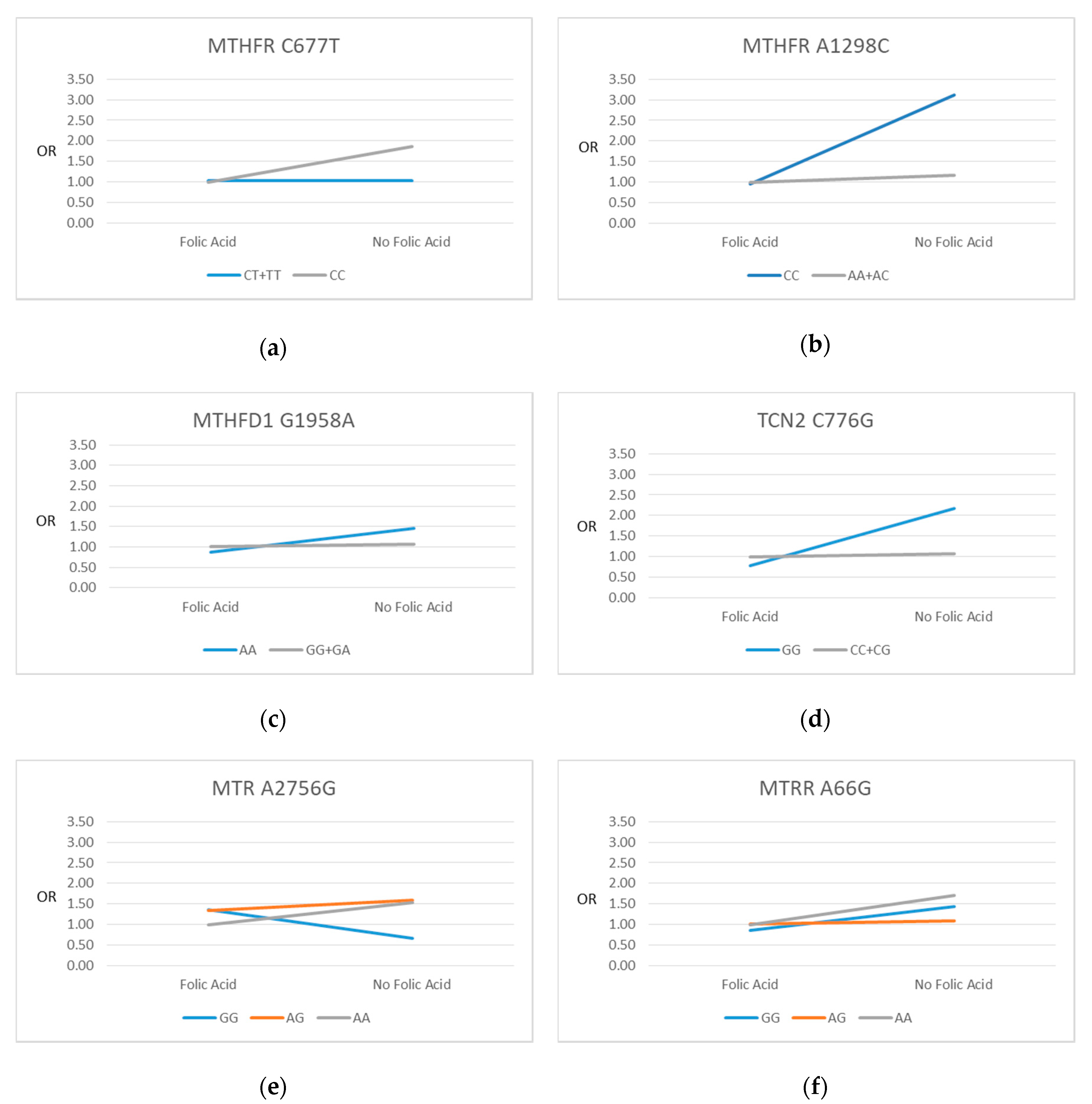
Figure 2 gives a visual representation of the directions of the SNP-FAS interactions on SGA for each of the six SNPs. Although not all interactions were significant, FAS use showed a general pattern of attenuating the differences in the likelihood of SGA seen between genotype groups who did not use FAS.

4. Discussion

In this study of nulliparous pregnant women living in New Zealand, significant gene-nutrient interactions in relation to SGA were observed between three maternal one-carbon metabolism gene polymorphisms—MTHFR 677, MTHFR 1298, and TCN2 776 and maternal FAS use at 15 (±1) weeks’ gestation. Although interactions for the other SNPs were not significant, we observed a general pattern of FAS attenuating the differences in the likelihood of SGA seen between genotypes in FAS non-users. The exception was for the MTR 2756 homozygous variant (GG) genotype, which showed a non-statistically significant reduction in SGA in FAS non-users in comparison with FAS users. However, as the prevalence of the MTR 2756 GG genotype was very low in this sample (3.8%), this may be a chance finding, and this genotype and its interaction with folate requires investigation in future studies.
Few previous studies have examined the effect of folate gene-nutrient interactions on birth size-parameters or SGA. To the best of our knowledge, this is the first study to examine maternal gene-nutrient interactions between FAS use (supplement form) and these six SNPs of the folate-mediated one-carbon metabolic pathway, and their effects on SGA.
One previous study that examined gene-nutrient interactions between maternal total folate intake (dietary folate, folic acid from fortified foods and FAS) and MTHFR 677, MTHFR 1298, MTR 2756 and MTRR 66 polymorphisms and SGA [23] found a statistically significant lower odds of SGA in MTHFR 1298 AA wild-type participants vs. variant homozygote (CC) Caucasian participants in the lowest quartile of total folate intake (OR = 0.3; 95% CI: 0.1–0.9). Although the study focused on total folate intake, the finding of a significant result for MTHFR 1298 aligns with our finding of a significantly higher likelihood of SGA seen in MTHFR 1298 variant CC participants who did not use FAS, in comparison with wild-type AA FAS users. Although Engel and colleagues did not observe statistically significant associations for MTHFR 677, MTR 2756 and MTRR 66 in the lowest quartile of folate, point estimates of the effect sizes were in the same direction as seen in MTHFR 1298 (OR: 0.6, 0.3 and 0.6 respectively) [23]. A second folate gene-nutrient interaction study found no significant interaction between maternal dietary folate and MTHFR 677 or 1298 polymorphisms on anthropometric birth parameters (n = 231) [29]. This is in contrast to our study (n = 1873) which showed a significant interaction effect for both of these polymorphisms. The difference between findings may be due to Torres-Sánchez and colleagues using dietary folate in their gene-nutrient analysis, whereas the present study focused specifically on FAS use.
A number of biological mechanisms are likely to explain our findings of an interaction effect between FAS and maternal genotype on SGA. During pregnancy, folate is important for fetal growth and development through its role in one-carbon metabolism, DNA synthesis, replication and methylation, and requirements are increased, due to rapid placental and fetal growth [43]. Folate plays an essential role in embryonic and fetal development [44]. Adequate maternal folate status is important during pregnancy to ensure optimal maternal folate status and homocysteine levels [15]. Maternal plasma folate and homocysteine levels are influenced by both folate intake and polymorphisms of the folate-mediated homocysteine metabolic pathway [45]. These polymorphisms can cause disturbances in folate metabolism, one-carbon metabolism, DNA synthesis and methylation [6,17,35,46], which can in turn impair fetal growth and development. An increased supply of folic acid might be required for one-carbon metabolism in the presence of these polymorphisms [44]. FAS is likely to attenuate the effect of these SNPs on pregnancy outcomes by supplying additional folic acid and therefore an increased availability of one-carbon groups for the conversion of homocysteine to methionine, helping to maintain normal homocysteine levels [15,20,47]. Previous research shows that folic acid (in supplement form) interacts significantly with the MTHFR 677 polymorphism to influence homocysteine levels [19]. The authors concluded that MTHFR 677 homozygous variant (TT) genotype carriers need increased intakes of total folate compared to those with CC and CT genotypes, to maintain similar plasma homocysteine levels [19]. Similarly, a review by Hiroaka and colleagues (2017) of studies that examined differences in maternal FAS intake and serum folate and homocysteine levels, found that differences in mean levels at baseline seen between all three genotype groups of the MTHFR 677 polymorphism were attenuated in the presence of FAS [45,48,49,50].
A recent review by Zinck and MacFarlane suggests that limitations of previous gene-nutrient interaction studies include small sample sizes, and lack of dietary supplement data [18]. SCOPE contains time-specific data on maternal FAS use in a relatively large sample size. Participants were followed prospectively from 15 (±1) weeks’ gestation until after delivery, allowing capture of FAS data before birth outcomes. When collecting FAS data, participants brought their supplements to the research visit so that the investigator could verify details and record brand names and details directly into the study database. Reported FAS use was previously confirmed by plasma folate analysis in the NZ SCOPE cohort [32].
The current study has a number of limitations that must be considered in the context of our findings. SCOPE includes an opportunistic population of nulliparous participants from Auckland, New Zealand, and was predominantly of NZ/Other European ethnicity, and therefore not representative of New Zealand’s overall ethnic composition. This did not allow separate investigation of SNP-SGA effects in each ethnic group, due to small numbers of other ethnic groups included in the sample. However, we hypothesised that the biological effects of the FAS-SNP interaction would likely be similar across ethnic groups, and sensitivity analysis showed results remained consistent when limited to European participants. Although numbers of non-European ethnic groups in our study were small, our results contribute data on the distribution of the six included SNPs in the New Zealand population. Further research is required to describe these genotype distributions in larger samples of NZ ethnic groups. An important limitation of our study is that SCOPE study data collection did not include a comprehensive food frequency questionnaire or 24-h dietary recall, and we were therefore unable to estimate participant total dietary folate intake. Future interaction studies should examine the influence of FAS genotype interactions across groups with different background dietary folate intakes, to investigate further the FAS-polymorphism interaction association with fetal growth parameters. Folic acid in supplements is a synthetic form and is different to dietary folate. Folic acid in supplement form is more bioavailable than folate occurring naturally in food, hence the two might have different effects in the presence of folate enzyme SNPs.

5. Conclusions

Significant gene-nutrient interactions in relation to SGA were observed for FAS and three gene polymorphisms of the one-carbon metabolic pathway. FAS showed an overall pattern of attenuating differences in the likelihood of SGA seen between genotype groups in FAS non-users. This highlights the importance of ensuring adequate folic acid intake for women with these polymorphisms. Further research is required to investigate how differences in total folate intake (from dietary sources, food fortification and supplements) influence this interaction. Future research should focus on how folate biomarkers, FAS at other time points (such as pre-conception) and intake of other nutrients involved in one carbon metabolism (B12, choline, biotin) might mediate these interactions.

Supplementary Materials

The following are available online at https://www.mdpi.com/2072-6643/12/6/1677/s1, Table S1: Details of Included Polymorphisms, Table S2: Maternal Genotype and Allele Frequency by Ethnicity in New Zealand SCOPE Participants.

Author Contributions

Conceptualization, R.E.B., J.M.D.T., C.R.W., L.M.E.M., R.S.T. and C.T.R.; methodology, R.E.B., J.M.D.T., C.R.W., L.M.E.M., R.S.T. and C.T.R.; software, R.E.B., J.M.D.T. and R.S.T.; validation, R.E.B. and J.M.D.T.; formal analysis, R.E.B., J.M.D.T.; investigation, R.E.B.; R.E.B., J.M.D.T., C.R.W., L.M.E.M., R.S.T. and C.T.R.; data curation, R.E.B., R.S.T., J.M.D.T. and C.T.R.; writing—original draft preparation, R.E.B.; writing—review and editing, J.M.D.T., C.R.W., L.M.E.M., R.S.T. and C.T.R.; visualization, R.E.B.; supervision, J.M.D.T., C.R.W., L.M.E.M.; project administration, R.E.B. and R.S.T.; funding acquisition, L.M.E.M. and C.T.R. All authors have read and agreed to the published version of the manuscript.

Funding

The New Zealand SCOPE study was funded by the New Enterprise Research Fund, Foundation for Research Science and Technology; Health Research Council (04/198); Evelyn Bond Fund, Auckland District Health Board Charitable Trust. Genotyping was funded by the South Australian Premier’s Science and Research Fund South Australian Government and a grant from the National Health and Medical Research Council (NHMRC) of Australia awarded to CTR (GNT519225). CTR is supported by an Australian National Health and Medical Research Council (NHMRC) Investigator Grant (GNT1174971) and a Matthew Flinders Fellowship from Flinders University.

Acknowledgments

The authors would like to thank the SCOPE study participants, research laboratory technicians and research midwives.

Conflicts of Interest

The authors declare no conflict of interest. The funders had no role in the design of the study; in the collection, analyses, or interpretation of data; in the writing of the manuscript, or in the decision to publish the results.

References

  1. Finken, M.J.; van der Steen, M.; Smeets, C.C.; Walenkamp, M.J.; de Bruin, C.; Hokken-Koelega, A.C.; Wit, J.M. Children born small for gestational age: Differential diagnosis, molecular genetic evaluation, and implications. Endocr. Rev. 2018, 39, 851–894. [Google Scholar] [CrossRef] [PubMed] [Green Version]
  2. McCowan, L.M.; Thompson, J.M.; Taylor, R.S.; Baker, P.N.; North, R.A.; Poston, L.; Roberts, C.T.; Simpson, N.A.; Walker, J.J.; Myers, J. Prediction of small for gestational age infants in healthy nulliparous women using clinical and ultrasound risk factors combined with early pregnancy biomarkers. PLoS ONE 2017, 12, e0169311. [Google Scholar] [CrossRef] [PubMed] [Green Version]
  3. McCowan, L.; Horgan, R.P. Risk factors for small for gestational age infants. Best Pract. Res. Clin. Obstet. Gynaecol. 2009, 23, 779–793. [Google Scholar] [CrossRef] [PubMed]
  4. Englund-Ögge, L.; Brantsæter, A.L.; Juodakis, J.; Haugen, M.; Meltzer, H.M.; Jacobsson, B.; Sengpiel, V. Associations between maternal dietary patterns and infant birth weight, small and large for gestational age in the Norwegian Mother and Child Cohort Study. Eur. J. Clin. Nutr. 2018, 73, 1270–1282. [Google Scholar] [CrossRef] [PubMed]
  5. Kalhan, S.C. Maternal and Child Nutrition: The First 1,000 Days. In One-Carbon Metabolism, Fetal Growth and Long-Term Consequences; Karger Publishers: Basel, Switzerland, 2013; Volume 74, pp. 127–138. [Google Scholar]
  6. Nazki, F.H.; Sameer, A.S.; Ganaie, B.A. Folate: Metabolism, genes, polymorphisms and the associated diseases. Gene 2014, 533, 11–20. [Google Scholar] [CrossRef] [PubMed]
  7. Smith, A.D.; Kim, Y.; Refsum, H. Is folic acid good for everyone? Am. J. Clin. Nutr. 2008, 87, 517–533. [Google Scholar] [CrossRef] [Green Version]
  8. Talaulikar, V.S.; Arulkumaran, S. Folic acid in obstetric practice: A review. Obstet. Gynecol. Surv. 2011, 66, 240–247. [Google Scholar] [CrossRef]
  9. Field, M.S.; Kamynina, E.; Agunloye, O.C.; Liebenthal, R.P.; Lamarre, S.G.; Brosnan, M.E.; Brosnan, J.T.; Stover, P.J. Nuclear enrichment of folate cofactors and methylenetetrahydrofolate dehydrogenase 1 (MTHFD1) protect de novo thymidylate biosynthesis during folate deficiency. J. Biol. Chem. 2014, 289, 29642–29650. [Google Scholar] [CrossRef] [Green Version]
  10. Lassi, Z.S.; Salam, R.A.; Haider, B.A.; Bhutta, Z.A. Folic acid supplementation during pregnancy for maternal health and pregnancy outcomes. Cochrane Database Syst. Rev. 2013. [Google Scholar] [CrossRef]
  11. World Health Organization. Guideline: Daily Iron and Folic Acid Supplementation in Pregnant Women; World Health Organizatio: Geneva, Switzerland, 2012. [Google Scholar]
  12. Ministry of Health. Food and Nutrition Guidelines for Healthy Pregnant and Breastfeeding Women—A Background Paper; Ministry of Health: Wellington, New Zealand, 2006.
  13. Williams, P.J.; Bulmer, J.N.; Innes, B.A.; Broughton Pipkin, F. Possible roles for folic acid in the regulation of trophoblast invasion and placental development in normal early human pregnancy. Biol. Reprod. 2011, 84, 1148–1153. [Google Scholar] [CrossRef]
  14. World Health Organization. Recommendations on Antenatal Care for A Positive Pregnancy Experience; World Health Organization: Geneva, Switzerland, 2016. [Google Scholar]
  15. Hodgetts, V.A.; Morris, R.K.; Francis, A.; Gardosi, J.; Ismail, K.M. Effectiveness of folic acid supplementation in pregnancy on reducing the risk of small-for-gestational age neonates: A population study, systematic review and meta-analysis. BJOG 2015, 122, 478–490. [Google Scholar] [CrossRef] [PubMed]
  16. Zhang, Q.; Wang, Y.; Xin, X.; Zhang, Y.; Liu, D.; Peng, Z.; He, Y.; Xu, J.; Ma, X. Effect of folic acid supplementation on preterm delivery and small for gestational age births: A systematic review and meta-analysis. Reprod. Toxicol. 2017, 67, 35–41. [Google Scholar] [CrossRef] [PubMed]
  17. Solanky, N.; Jimenez, A.R.; D’Souza, S.W.; Sibley, C.P.; Glazier, J.D. Expression of folate transporters in human placenta and implications for homocysteine metabolism. Placenta 2010, 31, 134–143. [Google Scholar] [CrossRef] [PubMed]
  18. Zinck, J.W.; de Groh, M.; MacFarlane, A.J. Genetic modifiers of folate, vitamin B-12, and homocysteine status in a cross-sectional study of the Canadian population. Am. J. Clin. Nutr. 2015, 101, 1295–1304. [Google Scholar] [CrossRef] [Green Version]
  19. Ashfield-Watt, P.A.; Pullin, C.H.; Whiting, J.M.; Clark, Z.E.; Moat, S.J.; Newcombe, R.G.; Burr, M.L.; Lewis, M.J.; Powers, H.J.; McDowell, I.F. Methylenetetrahydrofolate reductase 677C→ T genotype modulates homocysteine responses to a folate-rich diet or a low-dose folic acid supplement: A randomized controlled trial. Am. J. Clin. Nutr. 2002, 76, 180–186. [Google Scholar] [CrossRef]
  20. Klai, S.; Fekih-Mrissa, N.; El Housaini, S.; Kaabechi, N.; Nsiri, B.; Rachdi, R.; Gritli, N. Association of MTHFR A1298C polymorphism (but not of MTHFR C677T) with elevated homocysteine levels and placental vasculopathies. Blood Coagul. Fibrinol. 2011, 22, 374–378. [Google Scholar] [CrossRef]
  21. Mtiraoui, N.; Zammiti, W.; Ghazouani, L.; Braham, N.J.; Saidi, S.; Finan, R.R.; Almawi, W.Y.; Mahjoub, T. Methylenetetrahydrofolate reductase C677T and A1298C polymorphism and changes in homocysteine concentrations in women with idiopathic recurrent pregnancy losses. Reproduction 2006, 131, 395–401. [Google Scholar] [CrossRef] [Green Version]
  22. Furness, D.; Dekker, G.A.; McCormack, C.D.; Nowak, R.C.; Thompson, S.D.; Roberts, C.T. 522. The association of folate pathway enzyme polymorphisms and pregnancy outcome. Reprod. Fertil. Dev. 2009, 21, 121. [Google Scholar] [CrossRef]
  23. Engel, S.M.; Olshan, A.F.; Siega-Riz, A.M.; Savitz, D.A.; Chanock, S.J. Polymorphisms in folate metabolizing genes and risk for spontaneous preterm and small-for-gestational age birth. Am. J. Obstet. Gynecol. 2006, 195, 1231.e1–1231.e11. [Google Scholar] [CrossRef]
  24. Wu, H.; Zhu, P.; Geng, X.; Liu, Z.; Cui, L.; Gao, Z.; Jiang, B.; Yang, L. Genetic polymorphism of MTHFR C677T with preterm birth and low birth weight susceptibility: A meta-analysis. Arch. Gynecol. Obstet. 2017, 295, 1105–1118. [Google Scholar] [CrossRef]
  25. Yila, T.A.; Sasaki, S.; Miyashita, C.; Braimoh, T.S.; Kashino, I.; Kobayashi, S.; Okada, E.; Baba, T.; Yoshioka, E.; Minakami, H. Effects of maternal 5, 10-methylenetetrahydrofolate reductase C677T and A1298C Polymorphisms and tobacco smoking on infant birth weight in a Japanese population. J. Epidemiol. 2012, 22, 91–102. [Google Scholar] [CrossRef] [PubMed] [Green Version]
  26. Hobbs, C.A.; Cleves, M.A.; Karim, M.A.; Zhao, W.; MacLeod, S.L. Maternal folate-related gene environment interactions and congenital heart defects. Obstet. Gynecol. 2010, 116, 316. [Google Scholar] [CrossRef] [PubMed]
  27. Isotalo, P.A.; Wells, G.A.; Donnelly, J.G. Neonatal and fetal methylenetetrahydrofolate reductase genetic polymorphisms: An examination of C677T and A1298C mutations. Am. J. Hum. Genet. 2000, 67, 986–990. [Google Scholar] [CrossRef] [PubMed] [Green Version]
  28. Cabo, R.; Hernes, S.; Slettan, A.; Haugen, M.; Ye, S.; Blomhoff, R.; Mansoor, M.A. Effect of genetic polymorphisms involved in folate metabolism on the concentration of serum folate and plasma total homocysteine (p-tHcy) in healthy subjects after short-term folic acid supplementation: A randomized, double blind, crossover study. Genes Nutr. 2015, 10, 1–12. [Google Scholar] [CrossRef] [Green Version]
  29. Torres-Sánchez, L.; López-Carrillo, L.; Blanco-Munoz, J.; Chen, J. Maternal dietary intake of folate, vitamin B 12 and MTHFR 677C> T genotype: Their impact on newborn’s anthropometric parameters. Genes Nutr. 2014, 9, 429. [Google Scholar] [CrossRef] [Green Version]
  30. Australian and New Zealand Clinical Trials Registry. Screening Nulliparous Women to Identify the Combinations of Clinical Risk Factors and/or Biomarkers Required to Predict Preeclampsia, Small for Gestational Age Babies and Spontaneous Preterm Birth. Available online: www.anzctr.org.au/Trial/Registration/TrialReview.aspx?id=82254 (accessed on 24 April 2020).
  31. North, R.A.; McCowan, L.M.; Dekker, G.A.; Poston, L.; Chan, E.H.; Stewart, A.W.; Black, M.A.; Taylor, R.S.; Walker, J.J.; Baker, P.N. Clinical risk prediction for pre-eclampsia in nulliparous women: Development of model in international prospective cohort. BMJ 2011, 342, d1875. [Google Scholar] [CrossRef] [Green Version]
  32. Bulloch, R.E.; McCowan, L.M.; Thompson, J.M.; Houghton, L.A.; Wall, C.R. Plasma folate and its association with folic acid supplementation, socio-demographic and lifestyle factors among New Zealand pregnant women. Br. J. Nutr. 2019, 122, 910–918. [Google Scholar] [CrossRef]
  33. Friso, S.; Choi, S. Gene-nutrient interactions in one-carbon metabolism. Curr. Drug Metab. 2005, 6, 37–46. [Google Scholar] [CrossRef]
  34. Boyles, A.L.; Billups, A.V.; Deak, K.L.; Siegel, D.G.; Mehltretter, L.; Slifer, S.H.; Bassuk, A.G.; Kessler, J.A.; Reed, M.C.; Nijhout, H.F. Neural tube defects and folate pathway genes: Family-based association tests of gene–gene and gene–environment interactions. Environ. Health Perspect. 2006, 114, 1547–1552. [Google Scholar] [CrossRef]
  35. Barbosa, P.R.; Stabler, S.P.; Machado, A.; Braga, R.C.; Hirata, R.; Hirata, M.H.; Sampaio-Neto, L.F.; Allen, R.H.; Guerra-Shinohara, E.M. Association between decreased vitamin levels and MTHFR, MTR and MTRR gene polymorphisms as determinants for elevated total homocysteine concentrations in pregnant women. Eur. J. Clin. Nutr. 2008, 62, 1010–1021. [Google Scholar] [CrossRef]
  36. Chedraui, P.; Salazar-Pousada, D.; Villao, A.; Escobar, G.S.; Ramirez, C.; Hidalgo, L.; Pérez-López, F.R.; Genazzani, A.; Simoncini, T. Polymorphisms of the methylenetetrahydrofolate reductase gene (C677T and A1298C) in nulliparous women complicated with preeclampsia. Gynecol. Endocrinol. 2014, 30, 392–396. [Google Scholar] [CrossRef] [PubMed]
  37. Bufalino, A.; Ribeiro Paranaíba, L.M.; Nascimento de Aquino, S.; Martelli-Júnior, H.; Oliveira Swerts, M.S.; Coletta, R.D. Maternal polymorphisms in folic acid metabolic genes are associated with nonsyndromic cleft lip and/or palate in the Brazilian population. Birth Defects Res. Part A Clin. Mol. Teratol. 2010, 88, 980–986. [Google Scholar] [CrossRef] [PubMed]
  38. Christensen, K.E.; Rohlicek, C.V.; Andelfinger, G.U.; Michaud, J.; Bigras, J.; Richter, A.; MacKenzie, R.E.; Rozen, R. The MTHFD1 p. Arg653Gln variant alters enzyme function and increases risk for congenital heart defects. Hum. Mutat. 2009, 30, 212–220. [Google Scholar] [CrossRef] [PubMed]
  39. Oussalah, A.; Levy, J.; Filhine-Trésarrieu, P.; Namour, F.; Guéant, J. Association of TCN2 rs1801198 c. 776G> C polymorphism with markers of one-carbon metabolism and related diseases: A systematic review and meta-analysis of genetic association studies. Am. J. Clin. Nutr. 2017, 106, 1142–1156. [Google Scholar] [CrossRef] [Green Version]
  40. McCowan, L.; Stewart, A.W.; Francis, A.; Gardosi, J. A customised birthweight centile calculator developed for a New Zealand population. Aust. N. Z. J. Obstet. Gynaecol. 2004, 44, 428–431. [Google Scholar] [CrossRef]
  41. Shaw, G.M.; Iovannisci, D.M.; Yang, W.; Finnell, R.H.; Carmichael, S.L.; Cheng, S.; Lammer, E.J. Risks of human conotruncal heart defects associated with 32 single nucleotide polymorphisms of selected cardiovascular disease-related genes. Am. J. Med. Genet. Part A 2005, 138, 21–26. [Google Scholar] [CrossRef]
  42. Garcia-Closas, M.; Ye, Y.; Rothman, N.; Figueroa, J.D.; Malats, N.; Dinney, C.P.; Chatterjee, N.; Prokunina-Olsson, L.; Wang, Z.; Lin, J. A genome-wide association study of bladder cancer identifies a new susceptibility locus within SLC14A1, a urea transporter gene on chromosome 18q12.3. Hum. Mol. Genet. 2011, 20, 4282–4289. [Google Scholar] [CrossRef] [Green Version]
  43. Fekete, K.; Berti, C.; Cetin, I.; Hermoso, M.; Koletzko, B.V.; Decsi, T. Perinatal folate supply: Relevance in health outcome parameters. Matern. Child Nutr. 2010, 6, 23–38. [Google Scholar] [CrossRef]
  44. Finnell, R.H.; Shaw, G.M.; Lammer, E.J.; Rosenquist, T.H. Gene–nutrient interactions: Importance of folic acid and vitamin B12 during early embryogenesis. Food Nutr. Bull. 2008, 29, S86–S98. [Google Scholar] [CrossRef] [Green Version]
  45. Hiraoka, M.; Kagawa, Y. Genetic polymorphisms and folate status. Congenit. Anom. 2017, 57, 142–149. [Google Scholar] [CrossRef] [Green Version]
  46. Amigou, A.; Rudant, J.; Orsi, L.; Goujon-Bellec, S.; Leverger, G.; Baruchel, A.; Bertrand, Y.; Nelken, B.; Plat, G.; Michel, G. Folic acid supplementation, MTHFR and MTRR polymorphisms, and the risk of childhood leukemia: The ESCALE study (SFCE). Cancer Causes Control 2012, 23, 1265–1277. [Google Scholar] [CrossRef] [PubMed]
  47. Hogeveen, M.; Blom, H.J.; Den Heijer, M. Maternal homocysteine and small-for-gestational-age offspring: Systematic review and meta-analysis. Am. J. Clin. Nutr. 2012, 95, 130–136. [Google Scholar] [CrossRef] [PubMed]
  48. Crider, K.S.; Zhu, J.; Hao, L.; Yang, Q.; Yang, T.P.; Gindler, J.; Maneval, D.R.; Quinlivan, E.P.; Li, Z.; Bailey, L.B. MTHFR 677C→ T genotype is associated with folate and homocysteine concentrations in a large, population-based, double-blind trial of folic acid supplementation. Am. J. Clin. Nutr. 2011, 93, 1365–1372. [Google Scholar] [CrossRef] [PubMed] [Green Version]
  49. Anderson, C.A.; Beresford, S.A.; McLerran, D.; Lampe, J.W.; Deeb, S.; Feng, Z.; Motulsky, A.G. Response of serum and red blood cell folate concentrations to folic acid supplementation depends on methylenetetrahydrofolate reductase C 677 T genotype: Results from a crossover trial. Mol. Nutr. Food Res. 2013, 57, 637–644. [Google Scholar] [CrossRef] [PubMed] [Green Version]
  50. Miyaki, K.; Murata, M.; Kikuchi, H.; Takei, I.; Nakayama, T.; Watanabe, K.; Omae, K. Assessment of tailor-made prevention of atherosclerosis with folic acid supplementation: Randomized, double-blind, placebo-controlled trials in each MTHFR C677T genotype. J. Hum. Genet. 2005, 50, 241–248. [Google Scholar] [CrossRef] [Green Version]
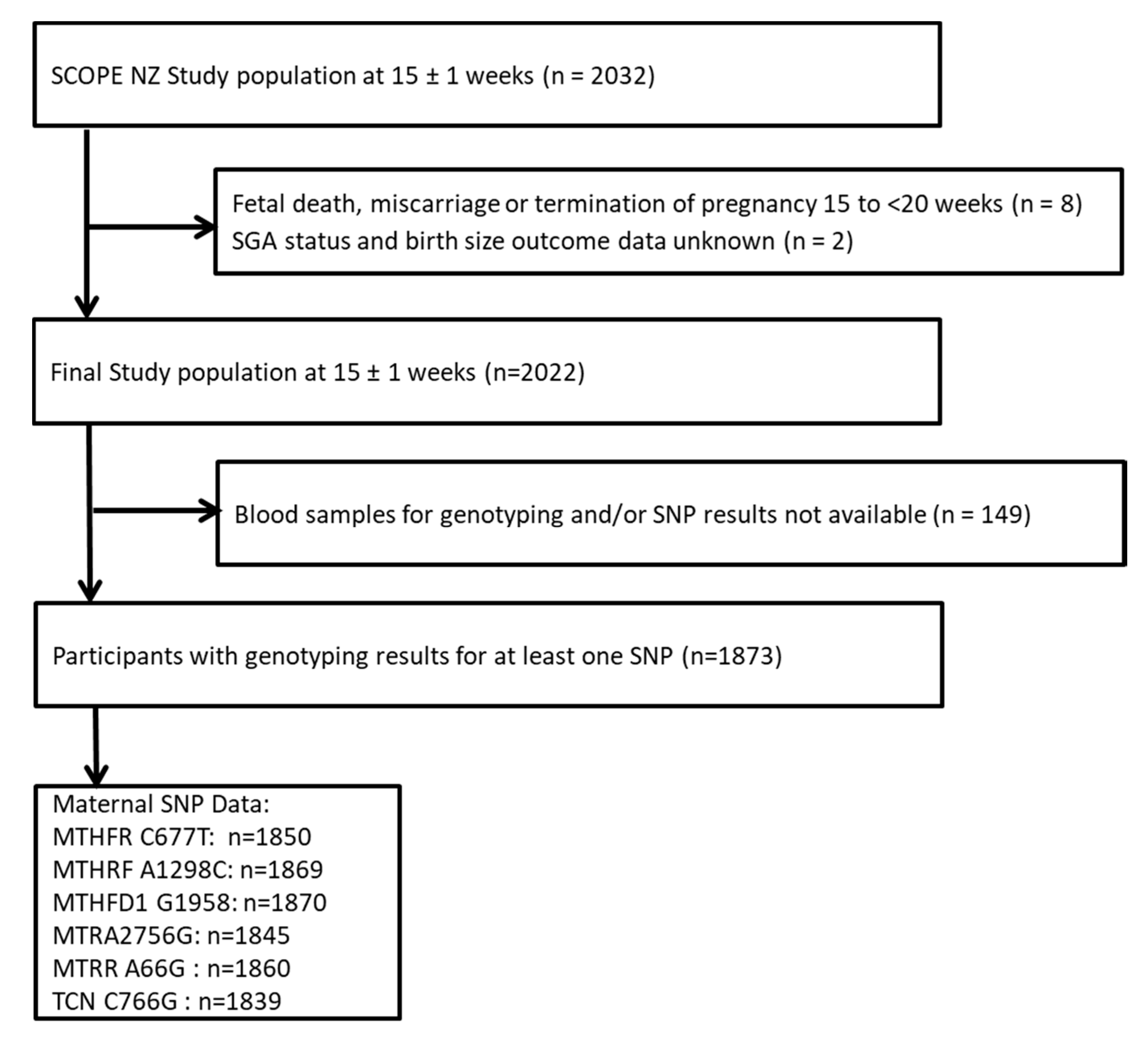
Figure 1. Participant selection flow chart.
Nutrients 12 01677 g001
Figure 2. Plots of regression odds ratios (OR): SNP vs. SGA by maternal folic acid supplement use at 15 weeks’ gestation. (a) MTHFR C677T; (b) MTHFR C1298A; (c) MTHFD1 G1958A; (d) TCN2 C776G; (e) MTR A2756G; (f) MTRR A66G. Folic Acid: reference category (defined as those who took folic acid supplements). Reference genotype category is shown in grey.
Nutrients 12 01677 g002
Table 1. Participant Characteristics.
CharacteristicMean ± SD or n (%)
Total n (%)1873 (100)
Maternal age (years)30.4 ± 4.7
Ethnicity:
NZ/Other European1577 (84.2)
Māori61 (3.3)
Pacific35 (1.9)
Asian97 (5.2)
Indian74 (4.0)
Other Non-European29 (1.6)
Socioeconomic Index *48 ± 15
Education > 12 years 1160 (64.8)
Marital partner (yes)1811 (96.7)
BMI at 15 weeks’ research visit (kg/m2)24.8 (±4.2)
Smoking (at 15 weeks’)73 (3.9)
Folic Acid Supplement Users $1367 (73.0)
Folic Acid Supplement Dose (µg/day)564 (14) ¥
Plasma Folate (nmol/L) §48.6 (1.6) ¥
Fetal Sex:
Male973 (52.0)
Female900 (48.0)
Final Delivery Gestation (weeks)39.6 (±2.1)
Birthweight (grams)3414 (±570)
Customised Birthweight Centile49 (±29)
SGA189 (10.1)
Spontaneous Preterm Birth 77 (4.1)
SD, standard deviation; BMI, body mass index; SGA, small-for-gestational-age. * Socioeconomic Index scale ranges from a score of 10 to 90, with a higher score representing a higher socio-economic status) (35). $ Defined as participants taking folic acid supplements at 15 weeks’ gestation. § Non-fasting plasma folate at 15 (±1) weeks’ gestation (assessed by microbiological assay [32]); geometric mean. ¥ Mean (SE).
Table 2. Maternal Genotype Frequency and Association between SNPs and SGA Risk in New Zealand SCOPE Participants.
SNP:Total n (%)SGA n (%)OR (95% CI)p-Value (p > z)p-Value for Overall Genotype Effect
MTHFR C677T1850 (100)
Genotype: 0.457
MTHFR 677 CC (Ref)887 (48.0)96 (10.8)1.00 (Ref)
MTHFR 677 CT786 (42.5)15 (9.3)0.84 (0.61–1.16)0.299
MTHFR 677 TT177 (9.6)73 (8.5)0.76 (0.43–1.35)0.352
Dominant Model 0.227
MTHFR 677 CC (Ref)887 (48.0)96 (10.8)1.00 (Ref)
MTHFR 677 CT + TT963 (52.1)88 (9.1)0.83 (0.61–1.12)0.227
MTHFR A1298C1869 (100)
Genotype: 0.141
MTHFR 1298 AA (Ref)932 (49.9)93 (10.0)1.00 (Ref)
MTHFR 1298 AC778 (41.6)72 (9.3)0.92 (0.67–1.27)0.614
MTHFR 1298 CC159 (8.5)23 (14.5)1.53 (0.93–2.49)0.092
Recessive Model 0.055
MTHFR 1298 AA + AC (Ref)1710 (91.5)165 (9.6)1.00 (Ref)
MTHFR 1298 CC159 (8.5)23 (14.5)1.58 (0.99–2.53)0.055
MTHFD1 G1958A1870 (100)
Genotype: 0.766
MTHFD1 1958 GG (Ref)621 (33.2)67 (10.8)1.00 (Ref)
MTHFD1 1958 GA920 (49.2)91 (9.9)0.91 (0.65–1.27)0.569
MTHFD1 1958 AA329 (17.6)31 (9.4)0.86 (0.55–1.35)0.510
Recessive Model 0.650
MTHFD1 1958 GG + GA (Ref)1541 (82.4)158 (10.3)1.00 (Ref)
MTHFD1 1958 AA329 (17.6)31 (9.4)0.91 (0.61–1.37)0.650
MTR A2756G1845 (100)
Genotype: 0.438
MTR 2756 AA (Ref)1206 (65.4)116 (9.6)1.00 (Ref)
MTR 2756 AG569 (30.8)66 (11.6)1.23 (0.90–1.70)0.200
MTR 2756 GG70 (3.8)7 (10.0)1.04 (0.47–2.33)0.916
Recessive Model 0.945
MTR 2756 AA + AG (Ref)1775 (96.2)182 (10.3)1.00 (Ref)
MTR 2756 GG 70 (3.8)7 (10.0)0.97 (0.44–2.16)0.945
MTRR A66G1860 (100)
Genotype: 0.714
MTRR 66 AA (Ref)450 (24.2)50 (11.1)1.00 (Ref)
MTRR 66 AG892 (48.0)88 (9.9)0.88 (0.61–1.26)0.478
MTRR 66 GG518 (27.9)50 (9.7)0.85 (0.57–1.29)0.457
Dominant Model 0.418
MTRR 66 AA (Ref)450 (24.2)50 (11.1)1.00 (Ref)
MTRR 66 AG + GG 1410 (75.8)138 (9.8)0.87 (0.62–1.22)0.418
TCN2 C766G1839 (100)
Genotype: 0.420
TCN2 766 CC (Ref)540 (29.4)51 (9.4)1.00 (Ref)0.597
TCN2 766 CG919 (50.0)96 (10.8)1.16 (1.16–0.26)0.435
TCN2 766 GG380 (20.7)41 (10.8)1.12 (0.75–1.790)
Recessive Model 0.682
TCN2 766 CC + CG (Ref)1459 (79.3)147 (10.1)1.00 (Ref)
TCN2 766 GG 380 (20.7)41 (10.8)1.08 (0.75–1.56)0.682
SGA, small-for-gestational-age. OR, odds ratio. Ref, reference category.
Table 3. Effect of Maternal Genotype—FAS Interaction on SGA Risk in New Zealand SCOPE Participants.
SNPAll n (%)FAS Yes n (%) $FAS No n (%) $FAS Yes aOR (95% CI) *FAS No aOR (95% CI) *p-Value *
MTHFR C677T1850 (100)
CC (Ref)887 (48.0)628 (33.9)259 (14.0)1.00 (Ref)1.87 (1.21–2.88)0.072
CT786 (42.5)587 (31.7)199 (10.8)1.06 (0.71–1.56)1.02 (0.58–1.77)
TT177 (9.6)135 (7.3)42 (2.3)0.91 (0.46–1.78)1.08 (0.37–3.12)
CC (Ref)887 (48.0)628 (33.9)259 (14.0)1.00 (Ref)1.87 (1.21–2.88)0.019
CT + TT963 (52.1)722 (39.0)241 (13.0)1.03 (0.71–1.49)1.03 (0.61–1.72)
MTHFR A1298C1869 (100)
AA (Ref)932 (49.9)702 (37.6)230 (12.3)1.00 (Ref)1.07 (0.65–1.75)0.020
AC778 (41.6)560 (30.0)218 (11.7)0.86 (0.58–1.27)1.13 (0.69–1.86)
CC159 (8.5)101 (5.4)58 (3.1)0.90 (0.43–1.86)2.92 (1.52–5.60)
AA + AC (Ref)1710 (91.5)1262 (67.5)448 (24.0)1.00 (Ref)1.17 (0.82–1.67)0.005
CC159 (8.5)101 (5.4)58 (3.1)0.96 (0.47–1.953.11 (1.66–5.85)
MTHFD1 G1958A1870 (100)
GG (Ref)621 (33.2)439 (23.5)182 (9.7)1.00 (Ref)1.40 (0.82–2.38)0.133
GA920 (49.2)680 (36.4)240 (12.8)1.01 (0.67–1.51)1.02 (0.60–1.73)
AA329 (17.6)245 (13.1)84 (4.5)0.69 (0.38–1.23)1.84 (0.96–3.54)
GG + GA (Ref)1541 (82.4)1119 (59.8)422 (22.6)1.00 (Ref)1.18 (0.82–1.69)0.062
AA329 (17.6)245 (13.1)84 (4.5)0.68 (0.40–1.16)1.83 (1.00–3.36)
MTR A2756G1845 (100)
AA (Ref)1206 (65.4)879 (47.6)327 (17.7)1.00 (Ref)1.54 (1.03–2.30)0.253
AG569 (30.8)415 (22.5)154 (8.3)1.34 (0.91–1.97)1.60 (0.95–2.71)
GG70 (3.8)53 (2.9)17 (0.9)1.37 (0.57–3.31)0.67 (0.09–5.12)
MTRR A66G1860 (100)
AA (Ref)450 (24.2)333 (17.9)117 (6.2)1.00 (Ref)1.71 (0.92–3.18)0.245
AG892 (48.0)650 (34.9)242 (13.0)1.01 (0.65–1.58)1.08 (0.62–1.88)
GG518 (27.9)375 (20.2)143 (7.7)0.85 (0.51–1.42)1.44 (0.79–2.64)
TCN2 C776G1839 (100)
CC (Ref)540 (29.4)401 (21.8)139 (7.6)1.00 (Ref)1.23 (0.65–2.32)0.057
CG919 (50.0)660 (35.9)259 (14.1)1.18 (0.77–1.81)1.18 (0.70–2.00)
GG380 (20.7)281 (15.3)99 (5.4)0.86(0.50 –1.50)2.41 (1.31–4.41)
CC + CG (Ref)1459 (79.3)1061 (57.7)398 (21.6)1.00 (Ref)1.07 (0.74–1.57)0.017
GG380 (20.7)281 (15.3)99 (5.4)0.77 (0.48–1.25)2.16 (1.26–3.71)
Ref, reference category. FAS, folic acid supplement use at 15 weeks’ gestation. * p-value for interaction effect. $ Percentage of total number of participants genotyped for each SNP.