Early Detection of Ganoderma boninense in Oil Palm Seedlings Using Support Vector Machines

 , , ,
, , ,
Abstract
:
1. Introduction
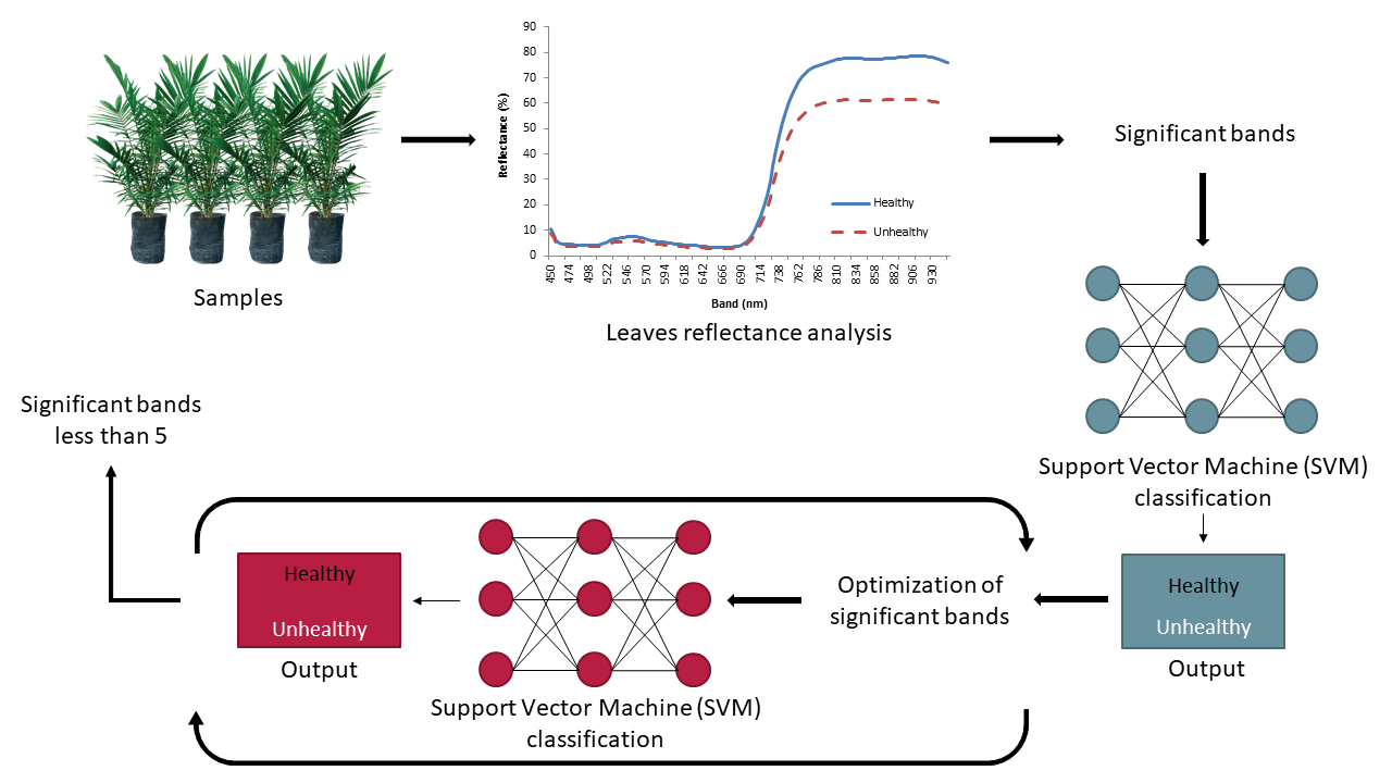
2. Materials and Methods
2.1. Study Area
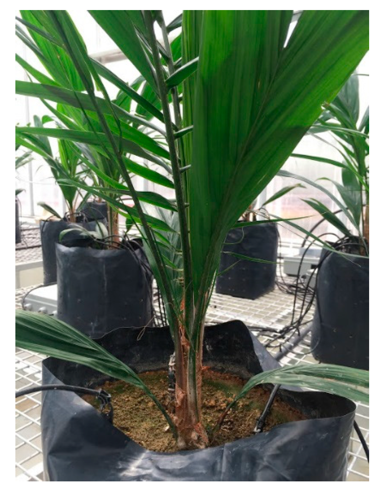
2.2. Artificial Inoculation of Samples
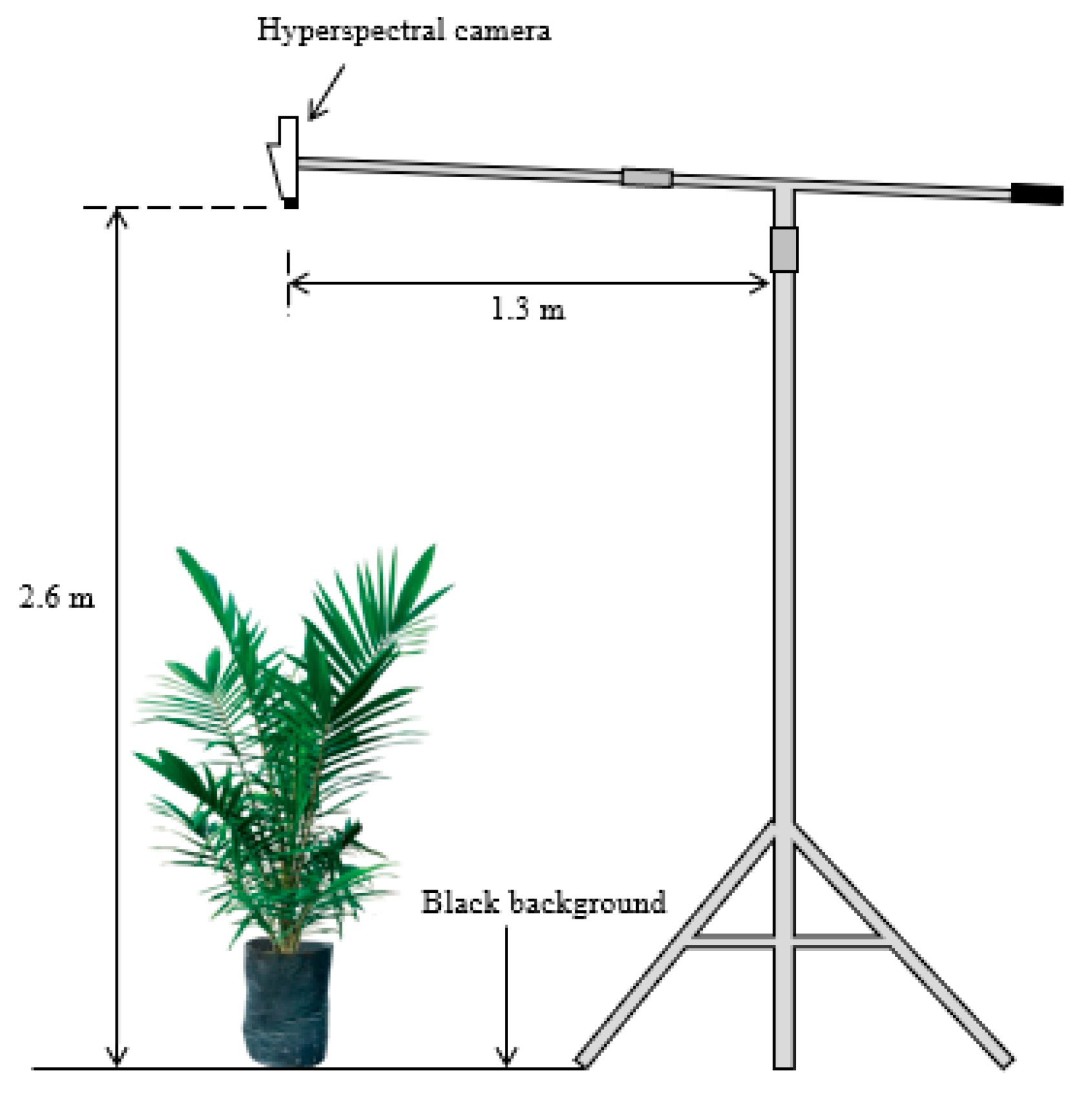
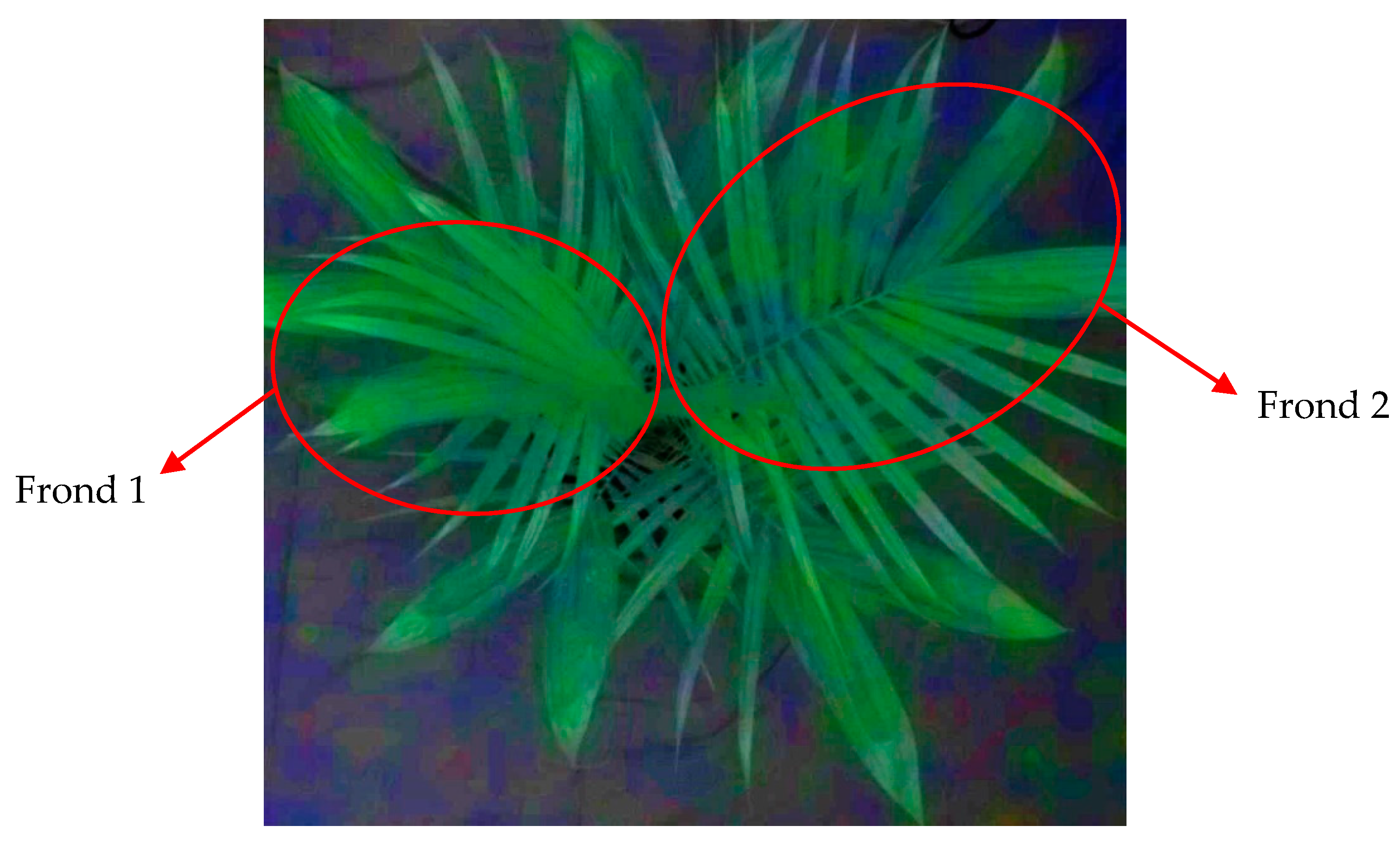
2.3. Data Collection
2.4. Data Pre-Processing
2.5. Data Analysis
3. Results
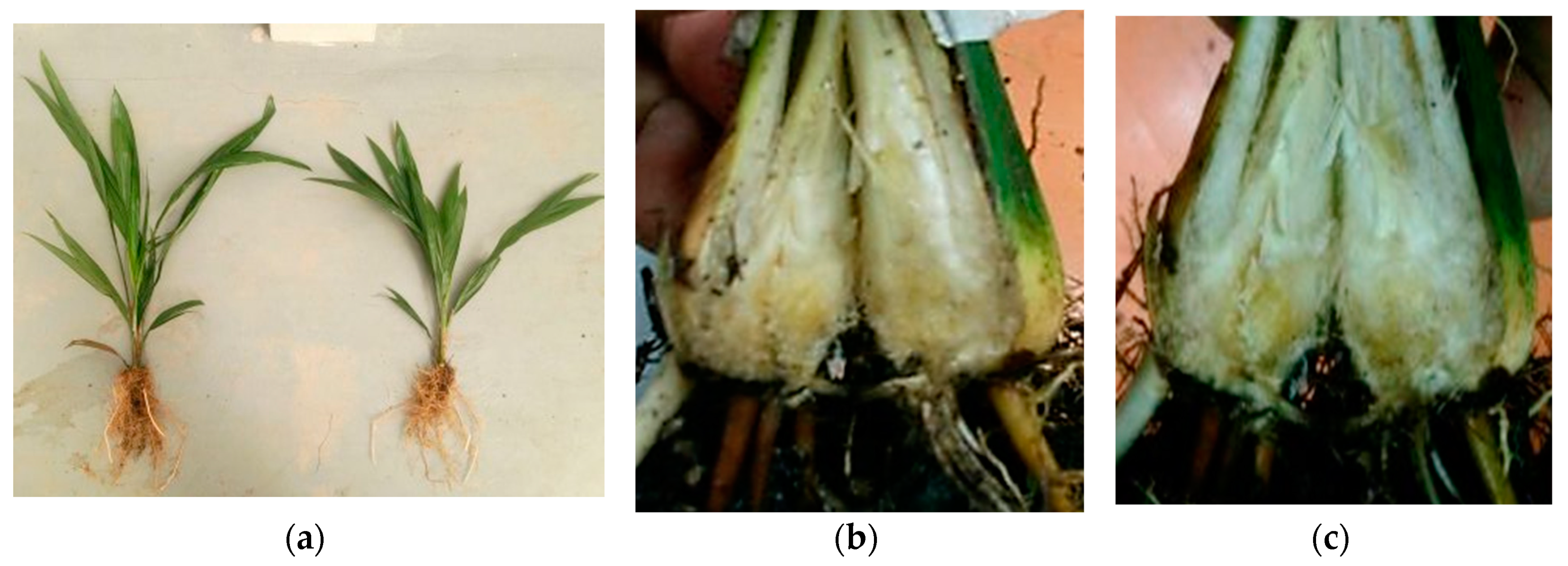
3.1. Status of the Inoculated Sample
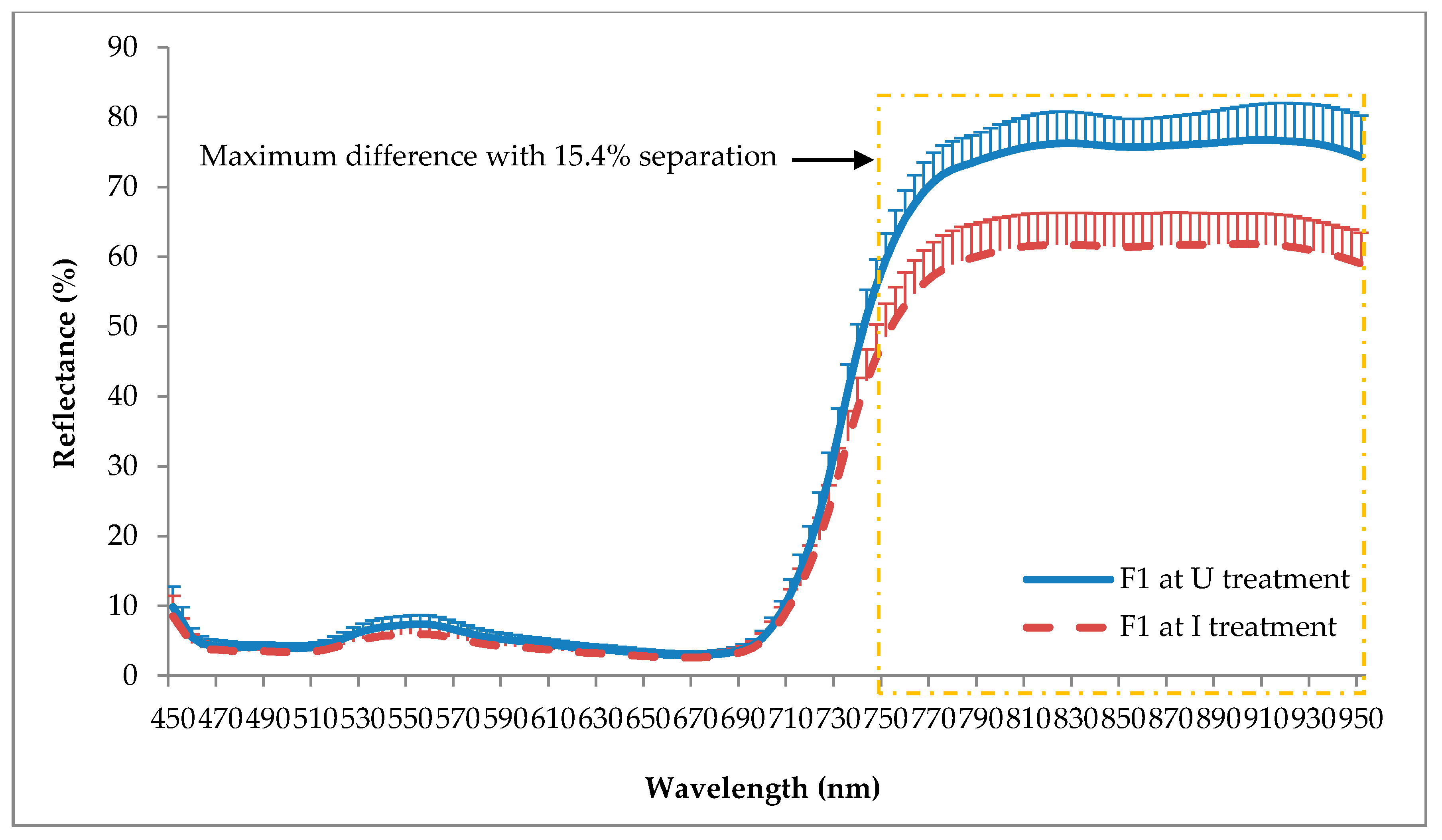
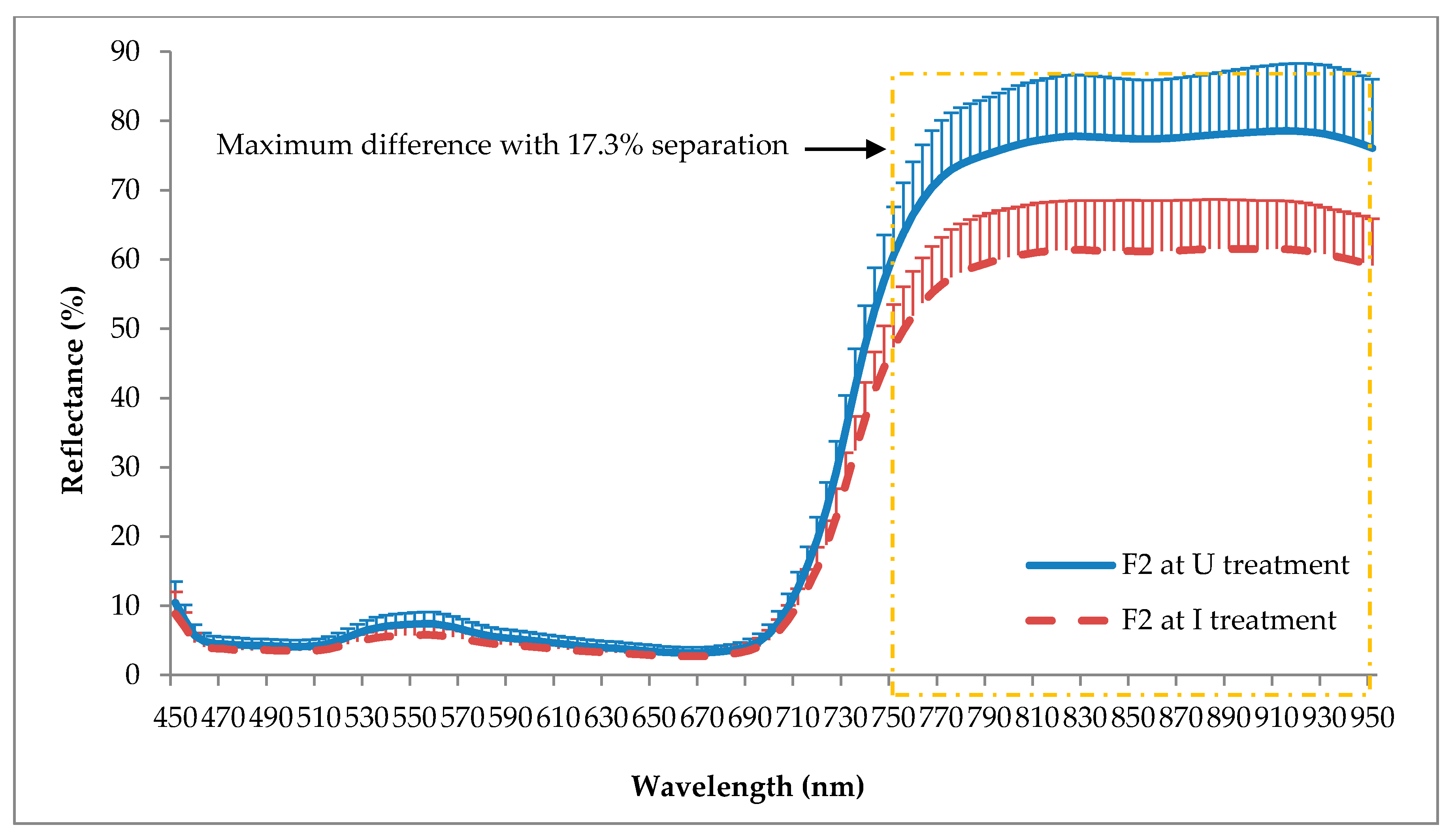
3.2. Reflectance Analysis
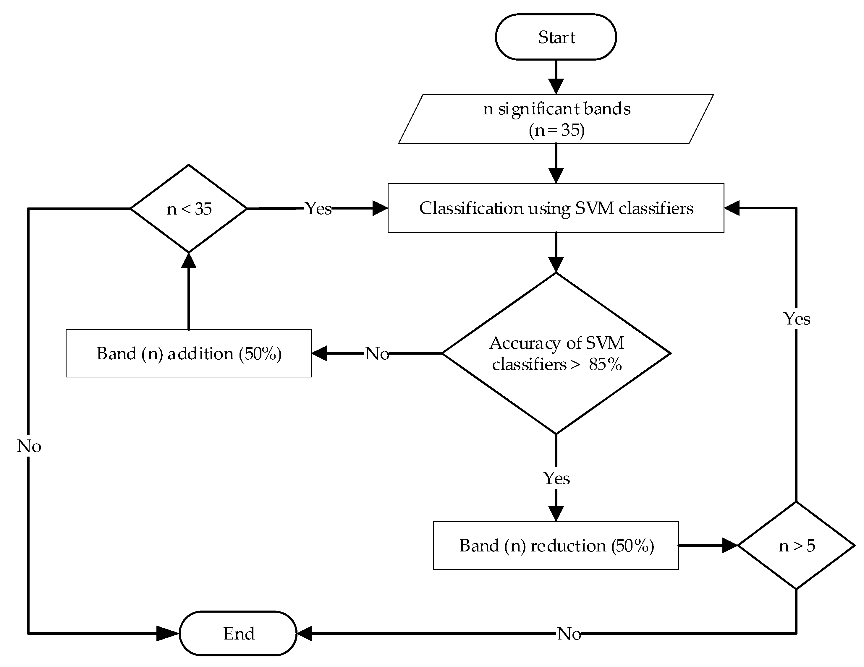
3.3. SVM Classification
3.3.1. Frond 1 (F1)
3.3.2. Frond 2 (F2)
3.3.3. Combination of Frond 1 and Frond 2 (F12)
4. Discussion
5. Conclusions
Author Contributions
Funding
Conflicts of Interest
References
- Sanderson, F.R. An insight into spore dispersal of Ganoderma boninense on oil palm. Mycopathologia 2005, 159, 139–141. [Google Scholar] [CrossRef] [PubMed]
- Flood, J.; Hasan, Y.; Turner, P.D.; O’Grady, E.B. The spread of Ganoderma from infective sources in the field and its implication for management of the disease in oil palm. In Ganoderma Diseases of Perennial Crops; Flood, J., Bridge, P.D., Holderness, M., Eds.; CABI: New York, NY, USA, 2000; pp. 101–112. [Google Scholar]
- Subagio, A.; Foster, H.L. Implications of Ganoderma disease on loss in stand and yield production of oil palm in North Sumatra. In Proceedings of the MAPPS Conference 2003, Kuala Lumpur, Malaysia, 11 August 2003; Malaysian Plant Protection Society: Kuching, Malaysia, 2003. [Google Scholar]
- Roslan, A.; Idris, A.S. Economic impact of Ganoderma incidence on Malaysian oil palm plantation—A case study in Johor. Oil Palm Ind. Econ. J. 2012, 12, 24–30. [Google Scholar]
- Idris, A.S.; Kushairi, D.; Ariffin, D.; Basri, M.W. Technique for inoculation of oil palm germinated seeds with Ganoderma. Mpob Inf. Ser. 2006, 314, 1–4. [Google Scholar]
- Meor, M.S.Y.; Khalid, M.A.; Idris, A.S. Identification of basal stem rot disease in local palm oil by microfocus XRF. J. Nucl. Relat. Technol. 2009, 6, 282–287. [Google Scholar]
- Naher, L.; Siddiquee, S.; Yusuf, U.K.; Mondal, M.M.A. Issues of Ganoderma spp. and basal stem rot disease management in oil palm. Am. J. Agric. Sci. 2015, 2, 103–107. [Google Scholar]
- Sariah, M.; Hussin, M.Z.; Miller, R.N.G.; Holderness, M. Pathogenicity of Ganoderma boninense tested by inoculation of oil palm seedlings. Plant Pathol. 1994, 43, 507–510. [Google Scholar] [CrossRef]
- Haniff, M.H.; Ismail, S.; Idris, A.S. Gas exchange responses of oil palm to Ganoderma boninense infection. Asian J. Plant Sci. 2005, 4, 438–444. [Google Scholar]
- Natarajan, S.; Bhaskaran, R.; Shanmugan, N. Preliminary studies to develop techniques for early detection of Thanjavur wilt in coconut. Indian Coconut J. (India) 1986, 17, 3–6. [Google Scholar]
- Ariffin, D.; Idris, A.S.; Marzuki, A. Spread of Ganoderma boninense and vegetative compatibility studies of a single field palm isolates. In Proceedings of the 1996 PORIM International Palm Oil Congress (Agriculture), Kuala Lumpur, Malaysia, 23–28 September 1996; Palm Oil Research Institute Malaysia: Kuala Lumpur, Malaysia, 1996. [Google Scholar]
- Darmono, T.W. Detection of basal stem rot disease of oil palm using polyclonal antibody. Menara Perkeb. 1999, 67, 32–39. [Google Scholar]
- Madihah, A.Z.; Idris, A.S.; Rafidah, A.R. Polyclonal antibodies of Ganoderma boninense isolated from Malaysian oil palm for detection of basal stem rot disease. Afr. J. Biotechnol. 2014, 13, 3455–3463. [Google Scholar] [CrossRef] [Green Version]
- Idris, A.S.; Rafidah, A.R. Polyclonal Antibody for Detection of Ganoderma; MPOB Information Series No.405; MPOB: Bandar Baru Bangi, Malaysia, 2008. [Google Scholar]
- Kandan, A.; Radjacommare, R.; Ramanathan, A.; Raguchander, T.; Balasubramanian, P.; Samiyappan, R. Molecular biology of Ganoderma pathogenicity and diagnosis in coconut seedlings. Folia Microbiol. 2009, 54, 147–152. [Google Scholar] [CrossRef] [PubMed]
- Utomo, C.; Niepold, F. Development of diagnostic methods for detecting Ganoderma-infected oil palms. J. Phytopathol. 2000, 148, 507–514. [Google Scholar] [CrossRef]
- Idris, A.S.; Yamaoka, M.; Hayakawa, S.; Basri, M.W.; Noorhasimah, I.; Ariffin, D. PCR Technique for Detection of Ganoderma; MPOB Information Series No.188; MPOB: Bandar Baru Bangi, Malaysia, 2003. [Google Scholar]
- Markom, M.A.; Shakaff, A.M.; Adom, A.H.; Ahmad, M.N.; Hidayat, W.; Abdullah, A.H.; Fikri, N.A. Intelligent electronic nose system for basal stem rot disease detection. Comput. Electron. Agric. 2009, 66, 140–146. [Google Scholar] [CrossRef]
- Abdullah, A.H.; Adom, A.H.; Ahmad, M.N.; Saad, M.A.; Tan, E.S.; Fikri, N.A.; Zakaria, A. Electronic nose system for Ganoderma detection. Sens. Lett. 2011, 9, 353–358. [Google Scholar] [CrossRef]
- Abdullah, J.; Hassan, H.; Shari, M.R.; Mohd, S.; Mustapha, M.; Mahmood, A.A.; Hamid, N.H. GammaScorpion: Mobile gamma-ray tomography system for early detection of basal stem rot in oil palm plantations. Opt. Eng. 2013, 52, 036502. [Google Scholar] [CrossRef]
- Khairunniza-Bejo, S.; Vong, C.N. Detection of basal stem rot (BSR) infected oil palm tree using laser scanning data. Agric. Agric. Sci. Procedia 2014, 2, 156–164. [Google Scholar] [CrossRef] [Green Version]
- Azuan, N.H.; Khairunniza-Bejo, S.; Abdullah, A.F.; Kassim, M.S.M.; Ahmad, D. Analysis of changes in oil palm canopy architecture from basal stem rot using terrestrial laser scanner (TLS). Plant Dis. 2019, 103, 3218–3225. [Google Scholar] [CrossRef] [Green Version]
- Husin, N.A.; Khairunniza-Bejo, S.; Abdullah, A.F.; Kassim, M.S.; Ahmad, D.; Azmi, A.N. Application of ground-based LiDAR for analysing oil palm canopy properties on the occurrence of basal stem rot (BSR) Disease. Sci. Rep. 2020, 10, 1–16. [Google Scholar] [CrossRef] [Green Version]
- Bravo, C.; Moshou, D.; West, J.; McCartney, A.; Ramon, H. Early disease detection in wheat fields using spectral reflectance. Biosyst. Eng. 2003, 84, 137–145. [Google Scholar] [CrossRef]
- Yang, Z.; Rao, M.N.; Elliott, N.C.; Kindler, S.D.; Popham, T.W. Differentiating stress induced by greenbugs and Russian wheat aphids in wheat using remote sensing. Comput. Electron. Agric. 2009, 67, 64–70. [Google Scholar] [CrossRef]
- Kumar, A.; Lee, W.S.; Ehsani, R.J.; Albrigo, L.G.; Yang, C.; Mangan, R.L. Citrus greening disease detection using aerial hyperspectral and multispectral imaging techniques. J. Appl. Remote Sens. 2012, 6, 063542. [Google Scholar]
- Zhang, J.; Pu, R.; Huang, W.; Yuan, L.; Luo, J.; Wang, J. Using in-situ hyperspectral data for detecting and discriminating yellow rust disease from nutrient stresses. Field Crops Res. 2012, 134, 165–174. [Google Scholar] [CrossRef]
- Alexander, A.; Dayou, J.; Sipaut, C.S.; ChongKhim, P.; LeePing, C. Some interpretations on FTIR results for the detection of Ganoderma boninense in oil palm tissue. Adv. Environ. Biol. 2014, 8, 30–32. [Google Scholar]
- Susič, N.; Žibrat, U.; Širca, S.; Strajnar, P.; Razinger, J.; Knapič, M.; Stare, B.G. Discrimination between abiotic and biotic drought stress in tomatoes using hyperspectral imaging. Sens. Actuators Chem. 2018, 273, 842–852. [Google Scholar] [CrossRef] [Green Version]
- Ye, D.; Sun, L.; Tan, W.; Che, W.; Yang, M. Detecting and classifying minor bruised potato based on hyperspectral imaging. Chemom. Intell. Lab. Syst. 2018, 177, 129–139. [Google Scholar] [CrossRef]
- Li, B.; Xu, X.; Zhang, L.; Han, J.; Bian, C.; Li, G.; Jin, L. Above-ground biomass estimation and yield prediction in potato by using UAV-based RGB and hyperspectral imaging. ISPRS J. Photogramm. Remote Sens. 2020, 162, 161–172. [Google Scholar] [CrossRef]
- Jiang, H.; Zhang, C.; He, Y.; Chen, X.; Liu, F.; Liu, Y. Wavelength selection for detection of slight bruises on pears based on hyperspectral imaging. Appl. Sci. 2016, 6, 450. [Google Scholar] [CrossRef] [Green Version]
- Rivera, N.V.; Gómez-Sanchis, J.; Chanona-Pérez, J.; Carrasco, J.J.; Millán-Giraldo, M.; Lorente, D.; Blasco, J. Early detection of mechanical damage in mango using NIR hyperspectral images and machine learning. Biosyst. Eng. 2014, 122, 91–98. [Google Scholar] [CrossRef]
- Pantazi, X.E.; Tamouridou, A.A.; Alexandridis, T.K.; Lagopodi, A.L.; Kontouris, G.; Moshou, D. Detection of Silybum marianum infection with Microbotryum silybum using VNIR field spectroscopy. Comput. Electron. Agric. 2017, 137, 130–137. [Google Scholar] [CrossRef]
- Guo, C.; Guo, X. Estimating leaf chlorophyll and nitrogen content of wetland emergent plants using hyperspectral data in the visible domain. Spectrosc. Lett. 2016, 49, 180–187. [Google Scholar] [CrossRef]
- Krezhova, D.; Velichkova, K.; Petrov, N.; Maneva, S. The effect of plant diseases on hyperspectral leaf reflectance and biophysical parameters. In Proceedings of the 5th International Conference on Radiation and Application in Various Fields of Research (RAD-2017), Budva, Montenegro, 11–16 June 2017. [Google Scholar]
- Lowe, A.; Harrison, N.; French, A.P. Hyperspectral image analysis techniques for the detection and classification of the early onset of plant disease and stress. Plant Methods 2017, 13, 80. [Google Scholar] [CrossRef] [PubMed]
- Jin, J.; Wang, Q. Hyperspectral indices based on first derivative spectra closely trace canopy transpiration in a desert plant. Ecol. Inform. 2016, 35, 1–8. [Google Scholar] [CrossRef]
- Peng, Y.; Fan, M.; Song, J.; Cui, T.; Li, R. Assessment of plant species diversity based on hyperspectral indices at a fine scale. Sci. Rep. 2018, 8, 1–11. [Google Scholar] [CrossRef] [PubMed] [Green Version]
- Rumpf, T.; Mahlein, A.K.; Steiner, U.; Oerke, E.C.; Dehne, H.W.; Plümer, L. Early detection and classification of plant diseases with support vector machines based on hyperspectral reflectance. Comput. Electron. Agric. 2010, 74, 91–99. [Google Scholar] [CrossRef]
- Nagasubramanian, K.; Jones, S.; Sarkar, S.; Singh, A.K.; Singh, A.; Ganapathysubramanian, B. Hyperspectral band selection using genetic algorithm and support vector machines for early identification of charcoal rot disease in soybean stems. Plant Methods 2018, 14, 86. [Google Scholar] [CrossRef] [PubMed]
- Helmi, Z.; Mohanad, S.E. Quantitative performance of spectral indices in large scale plant health analysis. Am. J. Agric. Biol. Sci. 2009, 4, 187–191. [Google Scholar]
- Shafri, H.Z.; Hamdan, N. Hyperspectral imagery for mapping disease infection in oil palm plantation using vegetation indices and red-edge techniques. Am. J. Appl. Sci. 2009, 6, 1031. [Google Scholar]
- Shafri, H.Z.M.; Hamdan, N.; Izzuddin Anuar, M. Detection of stressed oil palms from an airborne sensor using optimized spectral indices. Int. J. Remote Sens. 2012, 33, 4293–4311. [Google Scholar] [CrossRef]
- Izzuddin, M.A.; Idris, A.S.; Nisfariza, N.M.; Ezzati, B. Spectral based analysis of airborne hyperspectral remote sensing image for detection of Ganoderma disease in oil palm. In Proceedings of the 2015 International Conference on Biological and Environmental Science (BIOES 2015), Phuket, Thailand, 1–2 October 2015; pp. 13–20. [Google Scholar]
- Izzuddin, M.A.; Nisfariza, M.N.; Ezzati, B.; Idris, A.S.; Steven, M.D.; Boyd, D. Analysis of airborne hyperspectral image using vegetation indices, red edge position and continuum removal for detection of Ganoderma disease in oil palm. J. Oil Palm Res. 2018, 30, 416–428. [Google Scholar]
- Shafri, H.Z.M.; Anuar, M.I. Hyperspectral signal analysis for detecting disease infection in oil palms. In Proceedings of the International Conference on Computer and Electrical Engineering 2008, Phuket, Thailand, 20–22 December 2008; IEEE: New York, NY, USA, 2008. [Google Scholar]
- Shafri, H.Z.; Anuar, M.I.; Seman, I.A.; Noor, N.M. Spectral discrimination of healthy and Ganoderma-infected oil palms from hyperspectral data. Int. J. Remote Sens. 2011, 32, 7111–7129. [Google Scholar] [CrossRef]
- Chung, C.L.; Huang, K.J.; Chen, S.Y.; Lai, M.H.; Chen, Y.C.; Kuo, Y.F. Detecting Bakanae disease in rice seedlings by machine vision. Comput. Electron. Agric. 2016, 121, 404–411. [Google Scholar] [CrossRef]
- Ebrahimi, M.A.; Khoshtaghaza, M.H.; Minaei, S.; Jamshidi, B. Vision-based pest detection based on SVM classification method. Comput. Electron. Agric. 2017, 137, 52–58. [Google Scholar] [CrossRef]
- Ramos, P.J.; Prieto, F.A.; Montoya, E.C.; Oliveros, C.E. Automatic fruit count on coffee branches using computer vision. Comput. Electron. Agric. 2017, 137, 9–22. [Google Scholar] [CrossRef]
- Zhang, M.; Li, C.; Yang, F. Classification of foreign matter embedded inside cotton lint using short wave infrared (SWIR) hyperspectral transmittance imaging. Comput. Electron. Agric. 2017, 139, 75–90. [Google Scholar] [CrossRef]
- Kaur, P.; Pannu, H.S.; Malhi, A.K. Plant disease recognition using fractional-order Zernike moments and SVM classifier. Neural Comput. Appl. 2019, 31, 8749–8768. [Google Scholar] [CrossRef]
- Santoso, H.; Tani, H.; Wang, X. Random Forest classification model of basal stem rot disease caused by Ganoderma boninense in oil palm plantations. Int. J. Remote Sens. 2017, 38, 4683–4699. [Google Scholar] [CrossRef]
- Santoso, H.; Tani, H.; Wang, X.; Prasetyo, A.E.; Sonobe, R. Classifying the severity of basal stem rot disease in oil palm plantations using WorldView-3 imagery and machine learning algorithms. Int. J. Remote Sens. 2019, 40, 7624–7646. [Google Scholar] [CrossRef]
- Khaled, A.Y.; Abd Aziz, S.; Bejo, S.K.; Nawi, N.M.; Jamaludin, D.; Ibrahim, N.U.A. A comparative study on dimensionality reduction of dielectric spectral data for the classification of basal stem rot (BSR) disease in oil palm. Comput. Electron. Agric. 2020, 170, 105288. [Google Scholar] [CrossRef]
- Shafri, H.Z.M.; Anuar, M.I.; Saripan, M.I. Modified vegetation indices for Ganoderma disease detection in oil palm from field spectroradiometer data. J. Appl. Remote Sens. 2009, 3, 033556. [Google Scholar]
- Tawfik, O.; Shafri, H.M.; Mohammed, A.A. Disease detection from field spectrometer data. Iium Eng. J. 2013, 14, 133–143. [Google Scholar] [CrossRef]
- Izzuddin, M.A.; Seman Idris, A.; Nisfariza, M.N.; Nordiana, A.A.; Shafri, H.Z.M.; Ezzati, B. The development of spectral indices for early detection of Ganoderma disease in oil palm seedlings. Int. J. Remote Sens. 2017, 38, 6505–6527. [Google Scholar] [CrossRef]
- Nisfariza, M.N.; Idris, A.S.; Shafri, Z.H.; Steven, M.; Boyd, D. Hyperspectral derivative band ratios of oil palm stress associated with Ganoderma basal stem rot disease. In Proceedings of the Malaysia Remote Sensing Society (MRSS) 2010, Kuala Lumpur, Malaysia, 28–29 April 2010. [Google Scholar]
- Nisfariza, M.N.; Shafri, Z.H.; Idris, A.; Steven, M.; Boyd, D.; Mior, M. Hyperspectral sensing possibilities using continuum removal index in early detection of Ganoderma in oil palm plantation. In Proceedings of the World Engineering Congress 2010, Conference on Geomatics and Geographical Information Science, Kuching, Sarawak, Malaysia, 2–5 August 2010; pp. 233–239. [Google Scholar]
- Ahmadi, P.; Muharam, F.M.; Ahmad, K.; Mansor, S.; Abu Seman, I. Early detection of Ganoderma basal stem rot of oil palms using artificial neural network spectral analysis. Plant Dis. 2017, 101, 1009–1016. [Google Scholar] [CrossRef] [PubMed] [Green Version]
- Lelong, C.C.; Roger, J.M.; Bregand, S.; Dubertret, F.; Lanore, M.; Sitorus, N.A.; Caliman, J.P. Discrimination of fungal disease infestation in oil-palm canopy hyperspectral reflectance data. In Proceedings of the First Workshop on Hyperspectral Image and Signal Processing: Evolution in Remote Sensing, Grenoble, France, 26–28 August 2009; IEEE: New York, NY, USA, 2009. [Google Scholar]
- Lelong, C.C.; Roger, J.M.; Brégand, S.; Dubertret, F.; Lanore, M.; Sitorus, N.A.; Caliman, J.P. Evaluation of oil-palm fungal disease infestation with canopy hyperspectral reflectance data. Sensors 2010, 10, 734–747. [Google Scholar] [CrossRef] [PubMed]
- Liaghat, S.; Mansor, S.; Ehsani, R.; Shafri, H.Z.M.; Meon, S.; Sankaran, S. Mid-infrared spectroscopy for early detection of basal stem rot disease in oil palm. Comput. Electron. Agric. 2014, 101, 48–54. [Google Scholar] [CrossRef]
- Dayou, J.; Alexander, A.; Sipaut, C.S.; Phin, C.K.; Chin, L.P. On the possibility of using FTIR for detection of Ganoderma boninense in infected oil palm tree. J. Adv. Agric. Environ. Eng. 2014, 1, 161–163. [Google Scholar]
- Liaghat, S.; Ehsani, R.; Mansor, S.; Shafri, H.Z.; Meon, S.; Sankaran, S.; Azam, S.H. Early detection of basal stem rot disease (Ganoderma) in oil palms based on hyperspectral reflectance data using pattern recognition algorithms. Int. J. Remote Sens. 2014, 35, 3427–3439. [Google Scholar] [CrossRef]
- Izzuddin, M.A.; Idris, A.S.; Wahid, O.; Nishfariza, M.N.; Shafri, H.Z.M. Field Spectroscopy for Detection of Ganoderma Disease in Oil Palm; MPOB Information Series No.532; MPOB: Bandar Baru Bangi, Malaysia, 2013. [Google Scholar]
- Kamil, N.N.; Omar, S.F. Climate variability and its impact on the palm oil industry. Oil Palm Ind. Econ. J. 2016, 16, 18–30. [Google Scholar]
- Naidu, Y.; Siddiqui, Y.; Rafii, M.Y.; Saud, H.M.; Idris, A.S. Inoculation of oil palm seedlings in Malaysia with white-rot hymenomycetes: Assessment of pathogenicity and vegetative growth. Crop Prot. 2018, 110, 146–154. [Google Scholar] [CrossRef]
- Asaari, M.S.M.; Mishra, P.; Mertens, S.; Dhondt, S.; Inzé, D.; Wuyts, N.; Scheunders, P. Close-range hyperspectral image analysis for the early detection of stress responses in individual plants in a high-throughput phenotyping platform. ISPRS J. Photogramm. Remote Sens. 2018, 138, 121–138. [Google Scholar] [CrossRef]
- Wilcox, R.R. A fundamental problem. In Fundamentals of Modern Statistical Methods: Substantially Improving Power and Accuracy, 2nd ed.; Springer: New York, NY, USA, 2010; pp. 109–126. [Google Scholar]
- Kwak, S.K.; Kim, J.H. Statistical data preparation: Management of missing values and outliers. Korean J. Anesthesiol. 2017, 70, 407. [Google Scholar] [CrossRef]
- Izzati, M.Z.N.A.; Abdullah, F. Disease suppression in Ganoderma-infected oil palm seedlings treated with Trichoderma harzianum. Plant Prot. Sci. 2008, 44, 101–107. [Google Scholar] [CrossRef] [Green Version]
- Kok, S.; Goh, Y.; Tung, H.; Goh, K.; Wong, W.; Goh, Y. In vitro growth of Ganoderma boninense isolates on novel palm extract medium and virulence on oil palm (Elaeis guineensis) seedlings. Malays. J. Microbiol. 2013, 9, 33–42. [Google Scholar]
- Lim, S.; Kim, S.H.; Kim, Y.; Cho, Y.S.; Kim, T.Y.; Jeong, W.K.; Sohn, J.H. Coefficient of Variance as Quality Criterion for Evaluation of Advanced Hepatic Fibrosis Using 2D Shear-Wave Elastography. J. Ultrasound Med. 2018, 37, 355–362. [Google Scholar] [CrossRef] [PubMed] [Green Version]
- Gausman, H.W. Reflectance of leaf components. Remote Sens. Environ. 1977, 6, 1–9. [Google Scholar] [CrossRef]
- Chappelle, E.W.; Kim, M.S.; McMurtrey, J.E., III. Ratio analysis of reflectance spectra (RARS): An algorithm for the remote estimation of the concentrations of chlorophyll a, chlorophyll b, and carotenoids in soybean leaves. Remote Sens. Environ. 1992, 39, 239–247. [Google Scholar] [CrossRef]
- Slaton, M.R.; Raymond Hunt, E., Jr.; Smith, W.K. Estimating near-infrared leaf reflectance from leaf structural characteristics. Am. J. Bot. 2001, 88, 278–284. [Google Scholar] [CrossRef] [PubMed]
- Ausmus, B.S.; Hilty, J.W. Reflectance studies of healthy, maize dwarf mosaic virus-infected, and Helminthosporium maydis-infected corn leaves. Remote Sens. Environ. 1971, 2, 77–81. [Google Scholar] [CrossRef]
- Schmidt, K.S.; Skidmore, A.K. Spectral discrimination of vegetation types in a coastal wetland. Remote Sens. Environ. 2003, 85, 92–108. [Google Scholar] [CrossRef]
- Rapaport, T.; Hochberg, U.; Rachmilevitch, S.; Karnieli, A. The effect of differential growth rates across plants on spectral predictions of physiological parameters. PLoS ONE 2014, 9, e88930. [Google Scholar] [CrossRef]


| Applied Sensor | Spectral Range | Study Scale | Age of Sample | Applied Methods | Specific Bands (nm) | Accuracy (%) | Reference |
|---|---|---|---|---|---|---|---|
| APOGEE spectroradiometer | 300 to 1000 nm | Nursery | n.a. | Mann–Whitney U test and Jeffries–Matusita (JM) distance analysis | 460, 461.5, 462, 462.5, 467.5, 468.5, 469, 480, 480.5, 483, 488, 490.5, 500.5, 501.5, 503.5, 524, 524.5, 525, 525.5, 528.5, 567, 568, 700, 717, 718, 720.5 744, 744.5 | n.a. | [47] |
| n.a. | Mann–Whitney U test and Band ratio and Optimum index factor (OIF) and K-means clustering and Average silhouette width (ASW) plot | 610.5, 738 | n.a. | [57] | |||
| 10 months old | Analysis of variance (ANOVA) and JM distance analysis and Maximum Likelihood classification | 495, 495.5, 496, 651.5, 652, 652.5, 653, 653.5, 654, 654.5, 655, 655.5, 656, 656.5, 657, 657.5, 658, 658.5, 659, 659.5, 660, 660.5, 661, 908 | 82 | [48] | |||
| n.a. | Modified red-edge simple ratio and JM distance analysis | 460, 705 | n.a. | [58] | |||
| 10 months old | ANOVA and Band ratio and OIF and ASW | 495.5, 477.5 | n.a. | [59] | |||
| GER 1500 handheld spectrometer | 350 to 1050 nm | Nursery | n.a. | Band ratio in relation with leaf chlorophyll content | 702, 725 | n.a. | [60] |
| Plantation | 5 and 17 years old | Vegetation indices and Continuum removal | 400 to 550 | n.a. | [61] | ||
| 273 to 1100 nm | 12 years old | Artificial neural network | 550 to 560 | 83.3 100 | [62] | ||
| Unispec spectroradiometer | 310 to 1130 nm | Plantation | n.a. | Partial least squares discriminant analysis (PLS-DA) | n.a. | 92 | [63] |
| n.a. | 94 | [64] | |||||
| FT-IR spectrometer | 255 to 2505 nm | Plantation | 15 years old | Principle component analysis (PCA) and Multivariate pattern recognition classification | n.a. | 92 | [65] |
| 2500 to 15,384 nm | n.a. | Spectral pattern analysis | 2857 to 3125, 6060 to 7194, 8000 to 10,000 | n.a. | [28] | ||
| 7692 to 10,000 | n.a. | [66] | |||||
| ASD field spectroradiometer | 325 to1075 nm | Plantation | 15 years old | PCA and Multivariate pattern recognition classification and ANOVA | n.a. | 97 | [67] |
| Spectroradiometer | 273.13 to 1099.57 nm | Plantation | 2, 5 and 17 years old | Spectral pattern analysis using oil palm spectral analyzer system (OPSAS) software | 662 | 80.8 | [68] |
| Airborne AISA sensor | 400 to 900 nm | Plantation | n.a. | Vegetation indices | 705, 750 | 82.86 | [42] |
| n.a. | Red-edge indices | 715, 734, 791 | 84 | [43] | |||
| n.a. | Vegetation indices and Minimum distance classification and Spectral angle mapper | 616, 734 | 86 | [44] | |||
| 5 years old | Vegetation indices and Continuum removal | 400 to 500 | 44.4 | [45] | |||
| 401 to 997 nm | 17 years old | Vegetation indices and Red-edge position and Continuum removal | 400 to 500 | 44.4 | [46] |
| Frond Number | Total Significant Bands | Significant Bands (nm) |
|---|---|---|
| F1 | 35 | 810 814 818 822 826 830 834 838 842 846 850 854 858 866 870 874 878 882 886 890 894 898 902 906 910 914 918 922 926 930 934 938 942 946 950 |
| F2 | 35 | 814 818 822 826 830 834 838 842 846 850 854 858 862 866 870 874 878 882 886 890 894 898 902 906 910 914 918 922 926 930 934 938 942 946 950 |
| Classification Accuracy (%) | ||||
|---|---|---|---|---|
| Number of Significant Bands | ||||
| Classifier | 35 | 18 | 9 | 5 |
| Linear SVM | 100 | 100 | 99 | 99 |
| Quadratic SVM | 100 | 100 | 97 | 97 |
| Cubic SVM | 100 | 100 | 98 | 98 |
| Fine Gaussian SVM | 100 | 100 | 99 | 99 |
| Medium Gaussian SVM | 100 | 100 | 99 | 99 |
| Coarse Gaussian SVM | 100 | 100 | 97 | 97 |
| Total Significant Bands | Significant Bands (nm) |
|---|---|
| 18 | 826 830 890 894 898 902 906 910 914 918 922 926 930 934 938 942 946 950 |
| 9 | 918 922 926 930 934 938 942 946 950 |
| 5 | 930 934 938 942 946 |
| Classification Accuracy (%) | ||||
|---|---|---|---|---|
| Number of Significant Bands | ||||
| Classifier | 35 | 18 | 9 | 14 |
| Linear SVM | 92 | 91 | 91 | 91 |
| Quadratic SVM | 90 | 89 | 88 | 90 |
| Cubic SVM | 89 | 89 | 47 | 86 |
| Fine Gaussian SVM | 93 | 91 | 91 | 92 |
| Medium Gaussian SVM | 92 | 91 | 90 | 91 |
| Coarse Gaussian SVM | 91 | 90 | 90 | 90 |
| Total Significant Bands | Significant Bands (nm) |
|---|---|
| 18 | 882 886 890 894 898 902 906 910 914 918 922 926 930 934 938 942 946 950 |
| 9 | 914 918 922 926 930 934 938 942 946 |
| 14 | 898 902 906 910 914 918 922 926 930 934 938 942 946 950 |
| Total Significant Bands | Significant Bands (nm) |
|---|---|
| 35 | 814 818 822 826 830 834 838 842 846 850 854 858 862 866 870 874 878 882 886 890 894 898 902 906 910 914 918 922 926 930 934 938 942 946 950 |
| 18 | 826 886 890 894 898 902 906 910 914 918 922 926 930 934 938 942 946 950 |
| 9 | 914 922 926 930 934 938 942 946 950 |
| 14 | 898 902 906 910 914 918 922 926 930 934 938 942 946 950 |
| Classification Accuracy (%) | ||||
|---|---|---|---|---|
| Number of Significant Bands | ||||
| Classifier | 35 | 18 | 9 | 14 |
| Linear SVM | 95 | 95 | 93 | 95 |
| Quadratic SVM | 94 | 93 | 93 | 93 |
| Cubic SVM | 94 | 92 | 78 | 89 |
| Fine Gaussian SVM | 95 | 95 | 94 | 95 |
| Medium Gaussian SVM | 95 | 95 | 94 | 95 |
| Coarse Gaussian SVM | 95 | 95 | 94 | 94 |
Publisher’s Note: MDPI stays neutral with regard to jurisdictional claims in published maps and institutional affiliations. |
© 2020 by the authors. Licensee MDPI, Basel, Switzerland. This article is an open access article distributed under the terms and conditions of the Creative Commons Attribution (CC BY) license (http://creativecommons.org/licenses/by/4.0/).
Share and Cite
Noor Azmi, A.N.; Bejo, S.K.; Jahari, M.; Muharam, F.M.; Yule, I.; Husin, N.A. Early Detection of Ganoderma boninense in Oil Palm Seedlings Using Support Vector Machines. Remote Sens. 2020, 12, 3920. https://doi.org/10.3390/rs12233920
Noor Azmi AN, Bejo SK, Jahari M, Muharam FM, Yule I, Husin NA. Early Detection of Ganoderma boninense in Oil Palm Seedlings Using Support Vector Machines. Remote Sensing. 2020; 12(23):3920. https://doi.org/10.3390/rs12233920
Chicago/Turabian StyleNoor Azmi, Aiman Nabilah, Siti Khairunniza Bejo, Mahirah Jahari, Farrah Melissa Muharam, Ian Yule, and Nur Azuan Husin. 2020. "Early Detection of Ganoderma boninense in Oil Palm Seedlings Using Support Vector Machines" Remote Sensing 12, no. 23: 3920. https://doi.org/10.3390/rs12233920
APA StyleNoor Azmi, A. N., Bejo, S. K., Jahari, M., Muharam, F. M., Yule, I., & Husin, N. A. (2020). Early Detection of Ganoderma boninense in Oil Palm Seedlings Using Support Vector Machines. Remote Sensing, 12(23), 3920. https://doi.org/10.3390/rs12233920